Note: Descriptions are shown in the official language in which they were submitted.
CA 02441878 2003-09-18
WO 02/074055 PCT/US02/08646
System and Method for Increasing the Contrast of an Image Produced by an
Epifluorescence Microscope
CROSS REFERENCE TO RELATED APPLICATIONS
This application claims priority of U.S. Provisional Application No.
60/276,906 filed March 19, 2001, the entire contents of which is incorporated
herein by reference in its entirety.
FIELD OF THE INVENTION
The present invention relates to microscope slides and the like for use in
epifluorescence microscopy of biological specimens.
BACKGROUND OF THE INVENTION
Citation or identification of any reference in this section or any section of
this application shall not be construed as an admission that such reference is
available as prior art to the present invention.
An epifluorescence microscope is similar to a conventional reflecting optical
microscope in that both microscopes illuminate the sample and produce a
magnified image of the sample. An epifluorescence microscope, however, uses
the emitted fluorescent light to form an image whereas a conventional
reflecting
optical microscope uses the scattered illumination light to form an image. The
epifluorescent microscope uses a higher intensity illumination, or excitation,
light
than a conventional microscope. The higher intensity excitation light is
needed to
excite a fluorescent molecule in the sample thereby causing the fluorescent
molecule to emit fluorescent light. The excitation light has a higher energy,
or
shorter wavelength, than the emitted light. The epifluorescence microscope
uses
the emitted light to produce a magnified image of the sample. The advantage of
a
epifluorescence microscope is that the sample may be prepared such that the
fluorescent molecules are preferentially attached to the biological structures
of
interest thereby producing an image of the biological structures of interest.
A common problem in epifluorescence microscopy is the low contrast, or
low signal-to-noise (S/N) ratio, of the fluorescent image. This is due to the
low
CA 02441878 2003-09-18
WO 02/074055 PCT/US02/08646
intensity of the emitted light compared to the high intensity of the
excitation light.
A dichroic mirror is usually used to reduce the scattered excitation light
before the
image is viewed or recorded.
The dichroic mirror is only partially effective in removing the excitation
light
from the emitted light so other measures must be taken to increase the S/N
ratio
of the fluorescent image. In order to assist in the discussion of the other
approaches to increasing the SlN ratio of the fluorescent image, reference to
Fig.
1 is helpful.
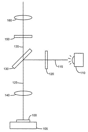
Fig. 1 illustrates the optical path and components of a typical
epifluorescence microscope. A sample 100 is placed on a sample holder 105,
which is normally a microscope slide. The sample is prepared prior to being
placed on the holder 105 with fluorescent tags that bind to the biological
structures
of interest. The fluorescent tags may be a single type of fluorescent tag that
binds
to a particular biological structure or may be a mixture of several
fluorescent tag
types with each tag type binding to a different biological structure. The
sample
100 is illuminated by a light source 110 that produces the excitation light
with
sufficient intensity to cause the tags to emit fluorescent light. The
excitation light
generated by the light source 110 follows a path 115 through an excitation
filter
120 that acts as a band-pass filter allowing only a narrow range of
frequencies to
pass through the excitation filter 120. The excitation filter 120 is chosen to
allow
only the light of a frequency that will cause the tags to fluoresce. The
excitation
light is reflected by a dichroic mirror 130 into the objective lens 140 of the
microscope following path 125. A dichroic mirror separates the excitation
light
from the emission light, in this example, by reflecting the excitation light
while
transmitting the emission light. The excitation light propagates through the
objective lens 140 and illuminates the sample 100 and excites the tags in the
sample to emit fluorescent light, also referred to as emission light. The
emission
light propagates along path 125 in the opposite direction as the excitation
light.
The emission light passes through the objective lens 140 and through the
dichroic
mirror 130 and continues along path 135 through an emission filter 150. The
emission filter 150 is selected to allow only light matching the frequency of
the
emission light to pass through the filter. The emission filter 150 may be a
band-
pass filter, or a long-pass filter that allows the longer wavelength emission
light to
pass through while stopping the shorter wavelength excitation light. After
filtering
CA 02441878 2003-09-18
WO 02/074055 PCT/US02/08646
3
by the emission filter 150, the emission light is formed into an image by an
imaging lens 160.
If the emission filter 150 is perfectly efficient in removing all but the
emission light, the magnified fluorescent image would have a very high
contrast
and S/N ratio. Unfortunately, emission filters are not perfectly efficient so
a small
amount of excitation light is transmitted though the emission filters. Because
the
intensity of the excitation light is very high, the small fraction of
excitation light that
passes through the emission filter is sufficient to severely degrade the
contrast of
the fluorescent image. In addition, the excitation frequency is usually very
close to
the emission frequency of the fluorescent tag molecule. The closeness of the
two
frequencies adds a further requirement on the emission filter that the filter
have a
very steep adsorption edge between the emission frequency and excitation
frequency.
U.S. Patent No. 6,094,274 issued on July 25, 2000 to Yokoi teaches the
use of two interference films as an emission filter. The two interference
films act
to sharpen the adsorption edge between the emission frequency and excitation
frequency. The sharp adsorption edge blocks more of the excitation light while
transmitting more of the emission light to the imaging lens.
Another approach to increasing the S/N ratio of a fluorescent image is
disclosed in Japanese Application Publication No. 9-292572 by Sudo, et al.
published on November 11, 1997 (hereinafter referred to as "Sudo"). Sudo
discloses the use of a mirror behind the sample that reflects the excitation
light
back through the sample. The reflected excitation light approximately doubles
the
excitation light seen by the sample and therefore approximately doubles the
amount of emission light given off by the sample. A portion of the reflected
excitation light will, however, also pass through the dichroic mirror and
emission
filter adding to the "noise" of the higher emission signal. In addition, the
increased
illumination of the sample from the reflected excitation light increases the
bleaching effect on the tagged sample. Bleaching occurs when the fluorescent
tag molecules emit decreasing amounts of fluorescent light as the molecules
are
illuminated by the excitation light. For example, a fluorescent tag molecule
will
emit less than 10% of its emission intensity after only a minute of being
illuminated
by the excitation light. As the intensity of the excitation light increases
the
CA 02441878 2003-09-18
WO 02/074055 PCT/US02/08646
bleaching rate increases thereby decreasing the emission light and reducing
the
contrast of the fluorescent image.
Therefore, there stiff remains a need to provide a microscope system
capable of producing a high contrast fluorescent image while reducing
unnecessary bleaching of the sample.
SUMMARY OF THE INVENTION
One aspect of the present invention is directed to an epifluorescence
microscope for imaging a biological sample having fluorescent tag molecules,
the
tag molecules emitting an emission light at an emission frequency when
illuminated by an excitation light having an excitation frequency, the
microscope
comprising: an excitation light source generating an excitation light; a first
dichroic
mirror reflecting the excitation light; an objective lens disposed to receive
the
excitation light reflected by the dichroic mirror and to illuminate the sample
with
the excitation light; an imaging lens disposed to receive emission light from
the
sample through the objective lens and first dichroic mirror; and a dichroic
sample
reflector disposed behind the sample reflecting the emission light back
through the
sample, objective lens, first dichroic mirror and imaging lens, while
transmitting the
excitation light through the reflector.
Another aspect of the present invention is directed to a sample holder for
supporting a sample for epifluorescence microscopy, the sample emitting an
emission light when illuminated by an excitation light, the sample holder
comprising a base and a dichroic reflector disposed on the base, wherein the
dichroic reflector reflects the emission light emitted by the sample while
transmitting the excitation light illuminating the sample.
Another aspect of the present invention is directed to a microscope slide for
supporting a sample, the slide comprising a top surface and an infra-red
reflecting
film deposited on the top surface, the film directly supporting the sample.
Another aspect of the present invention is directed to a sample holder
holding a sample for an epifluorescence microscope, the sample emitting an
emission light when illuminated by an excitation light, the sample holder
comprising: a base supporting the sample; and a sample reflector disposed on
the base between the sample and base, wherein the reflector reflects the
emission light emitted by the sample while transmitting the excitation light
CA 02441878 2003-09-18
WO 02/074055 PCT/US02/08646
illuminating the sample, wherein the sample reflector is concave having a
focal
point disposed in the sample.
Another aspect of the present invention is directed to a sample holder for
supporting a sample emitting an emission light when illuminated by an
excitation
light, the sample holder comprising: a top surface for directly supporting a
sample,
the top surface having an infra-red reflecting film deposited on the top
surface;
and a bottom surface having a dichroic film deposited on the bottom surface,
the
dichroic film reflecting emission light and transmitting excitation light.
Another aspect of the present invention is directed to a sample holder for
supporting a sample emitting an emission light when illuminated by an
excitation
light, the sample holder comprising: a top surface for directly supporting a
sample;
and a dichroic film deposited on the top surface, the dichroic film
transmitting
excitation light and reflecting emission light.
Another aspect of the present invention is directed to a method for
increasing the contrast of an image produced by an epifluorescence microscope
of a sample emitting an emission light when illuminated by an excitation light
comprising the steps of: illuminating the sample with the excitation light;
collecting
a first portion of the emission light; reflecting a second portion of the
emission
light; collecting the reflected portion of the emission light; and producing
an image
using the collected first portion of the emission light and the collected
reflected
portion of the emission light.
BRIEF DESCRIPTION OF THE DRAWINGS
The present invenfiion may be understood more fully by reference to the
following detailed description of the preferred embodiments of the present
invention, illustrative examples of specific embodiments of the invention and
the
appended figures in which:
Fig. 1 is a view of a conventional epifluorescence microscope.
Fig. 2 is a view of an embodiment of the present invention.
Fig. 3 is a detail view of the sample holder of the embodiment shown in Fig.
2.
Fig. 4 is a view of another embodiment of the present invention.
DETAILED DESCRIPTION OF THE PREFERRED EMBODIMENTS
CA 02441878 2003-09-18
WO 02/074055 PCT/US02/08646
Fig. 2 is a view of an embodiment of the present invention. Excitation light
generated by a light source 210 is filtered by an excitation filter 220. The
excitation filter 220 is preferably a band-pass filter allowing excitation
frequencies
matched to the fluorescent tags in the sample to pass through while absorbing
the
rest. The excitation light is redirected by reflection from a dichroic mirror
230
through an objective lens 240 to illuminate a sample 200 having fluorescent
tag
molecules. The excitation light causes the fluorescent tag molecules to emit
fluorescent light, The fluorescent light emitted by the tag molecules is
collected by
the objective lens 240 and is transmitted through the dichroic mirror 230. The
dichroic mirror 230 is selected to reflect the excitation light emitted by the
light
source 210 toward the sample 200 while transmitting the emission light emitted
by
the sample through the dichroic mirror. The emission light is filtered by an
emission filter 250 to remove extraneous light such as scattered excitation
light.
The emission light is formed into an image by an imaging lens 260. The details
of
mounting and aligning the optical elements described above are known to one of
ordinary skill in the optical microscopy art and are therefore not discussed.
The sample 200 is supported by a sample holder 205. The sample holder
205 includes a sample reflector 207 positioned directly behind the sample 200.
It
is understood that the term "behind" is relative to the direction of the
incident
excitation light. The sample reflector 207, in a preferred embodiment, is a
dichroic
mirror selected to reflect the emission light while transmitting the
excitation light.
Fig. 3 is a detail view of the sample holder 205 and sample reflector 207. A
sample 300 such as a blood or cell smear is placed on a sample support 305
such
as a glass slide. The sample is treated with a fluorescent tag that
preferentially
adsorbs to the biological structures of interest. The sample 300 and sample
support 305 are supported by a sample holder 310. The sample holder 310 has a
base 315 supporting a reflector 320 that, in turn, supports the glass slide
305 and
sample 300.
Excitation light 350 illuminates the sample 300 and interacts with the
sample 300, sample support 305 and reflector 320. For example, the excitation
light 350 may be back-scattered from the sample, shown as ray 352, or may be
back-scattered from the sample support 305, shown as ray 354, or may be back-
scattered from the reflector, shown as ray 356. Some of the back-scattered
light
350 352 356 is collected by the objective lens (not shown) and transmitted
CA 02441878 2003-09-18
WO 02/074055 PCT/US02/08646
through to the imaging lens. The back-scattered light 352 354 356 collected by
the imaging lens contributes to the background noise level of the image and
therefore reduces the SlN ratio of the image.
A small fraction of the excitation light 350 interacts with the fluorescent
tags
302 causing the fluorescent tags 302 to emit fluorescent light 360 362. Some
of
the emission light 360 is collected by the objective lens and imaged by the
imaging lens thereby forming the image of the biological structures of
interest.
Less than one-half of the emission light 360 362 is directly collected by the
objective lens because at least one-half of the emission light is emitted in a
direction away from the objective lens as represented by ray 362.
In a preferred embodiment, the reflector 320 is a dichroic mirror that allows
the short wave-length excitation light 351 to pass through the mirror 320
while
reflecting the longer wave-length emission light 362. Selection of the
reflector 320
to match the excitation and emission frequencies of the specific fluorescent
tag
molecule used to prepare the sample is well known to one of ordinary skill in
the
fluorescent microscopy art.
The novel feature of the reflector 320 is that, unlike the dichroic mirror
commonly used in typical epifluorescent microscopes, the reflector 320
reflects
the emission light instead of the excitation light. In the preferred
embodiment, the
reflector 320 transmits or absorbs most of the excitation light 350 and
therefore
reduces the amount of back-scattered excitation light 356 that may be
collected by
the objective lens. Reducing the amount of back-scattered excitation light 356
also reduces the noise in the image and results in a higher contrast image of
the
sample. In addition, the reflected emission light 362 may be collected by the
objective lens and contribute to the "signal portion" of the image and thereby
create a higher contrast image.
The reflected emission light 362 is reflected from the surface of the
reflector
320. The reflector surfiace is behind, with respect to the direction of the
excitation
light, the tag molecule in the sample and therefore will not be in the same
focal
plane 370 as the sample. The resulting image will have a higher intensity due
to
the reflected emission light but will have a lower resolution due to the
spatial
displacement of the reflector surFace with respect to the plane of the sample.
In
many situations, the higher intensity image is more important than the slight
loss
of resolution. For example, if the emission light is used to detect the
presence of
CA 02441878 2003-09-18
WO 02/074055 PCT/US02/08646
a rare cell in a sample, a brighter image is preferred because a bright image
is
easier to detect. The slight loss in resolution in this example is not as
important
because the detection of the rare cell depends primarily on image brightness,
not
image resolution.
In another embodiment of the present invention, the sample is placed
directly on the reflector 320. Placing the sample directly on the reflector
320 .
eliminates the need for a sample support 305 and reduces the distance between
the plane of the reflector and the plane of the sample 370 thereby reducing
the
focal mismatch between the image formed by the emission light collected
directly
from the sample and the image formed by the reflected emission light 362.
The reflection surface may also be used as a reference plane for
automatically focusing the image using laser tracking such as the Teletrac
LTAF-
8000 series Laser Tracking Autofocus from Axsys Technologies of Rocky Hill,
Connecticut. In typical auto-focusing methods, the image is focused based on
the
reflected light from a surface, usually a cover slide. In typical laser
autofocusing
systems, the frequency of the laser light is usually in the infrared portion
of the
spectrum and has a longer wavelength than the light emitted by the fluorescent
tags. The amount of reflected light is usually less than about 5% of the
incident
light. The small signal strength of the reflected light causes the microscope
to
lose focus if the sample is perturbed. In an embodiment where the reflector
acts
as a high-pass filter allowing the higher frequency excitation light through
the filter
while reflecting the lower frequency emission and autofocusing lighfi back
through
the objective lens. Although the reflector may not reflect all of the infra-
red
focusing light, a sufficient amount of focusing light will be reflected for
the laser
auto-focus system to maintain focus on the top surface of the reflector.
Fig. 4 is a side view of another embodiment of the present invention. An
infra-red reflecting film 410 is deposited on the top surface of a sample
support
420 and a dichroic film 430 reflecting emission light while transmitting
excitation
light is deposited on the bottom surface of the sample support. The sample
support may be a single-use disposable glass slide. The sample 405 is placed
directly on the infra-red reflector 410 and illuminated by both the excitation
light
450 and a focusing beam 460. The focusing beam 460 is preferably an infra-red
beam, characterized by a wavelength between 700 - 800 nm, that is part of a
CA 02441878 2003-09-18
WO 02/074055 PCT/US02/08646
laser auto-focus system such as the one described above. The focusing beam
460 is reflected (indicated by ray 465) by the infra-red reflecting film 410
back to
the laser auto-focus system that automatically focuses the microscope on the
infra-red reflecting film 410. In most situations, the biological structures
of interest
usually settle onto the surface of the infra-red reflecting film 410.
Therefore,
focusing on the reflecting film 410 will likely bring the biological
structures of
interest into focus. The dichroic film 430 on the bottom surface of the sample
support 420 will reflect the emission light (indicated by ray 455) back
through the
sample for collection by the objective lens while transmitting or absorbing
the
excitation light (indicated by ray 451 ).
In a preferred embodiment, the infra-red reflecting film 410 is metal film,
such as for example titanium, between 0.6 - 90 nm. The metal film may be
deposited using any of the known techniques for depositing thin films such as
physical deposition. In a preferred embodiment, magnetron sputtering may be
used to apply the infra-red reflecting film to the glass slide. The sputtering
composition, in a preferred embodiment, is substantially titanium with
impurities
such as carbon, nitrogen, iron, oxygen, and hydrogen cumulatively comprising
less than 1 % of the sputtering composition. Other sputtering compositions
. comprising metals different than titanium may be used to form the metal
film.
The selection of the sputtering composition and film thickness may
determined by one of skill in the art by measuring the intensity of the
reflected
auto-focus beam from the reflecting film. In one embodiment of the present
invention, the thickness and composition of the film is adjusted to reflect
between
4 - 8 % of the incident infra-red auto-focus beam. In a preferred embodiment,
the
thickness and composition of the film is adjusted to reflect between 5.5 - 7 %
of
the incident auto-focus beam.
In other situations, however, a high contrast, high resolution image is
preferred. In another embodiment of the present invention, the reflector is
shaped
into a concave surface having a focal point in the plane (defined by the
excitation
light ray) of the sample. This has the advantage of being able to focus both
the
direct and reflected emission light on the same focal plane.
In another embodiment of the present invention, more than one kind of
fluorescent tag may be used to image different biological structures of the
sample.
CA 02441878 2003-09-18
WO 02/074055 PCT/US02/08646
A mixture of different kinds of fluorescent tag molecules is used to prepare
the
sample. Each kind of fluorescent tag attaches to different biological
structures.
The light emitted by the fluorescent tags may have a different frequency and
the
excitation light required to cause the tags to fluoresce may have a different
5 frequency depending on the kind of fluorescent tag. Each tag may require its
own
set of excitation and emission filters selected for the excitation and
emission light
frequencies of the specific tag. The sample holder reflector is chosen to
transmit
or absorb all the excitation frequencies of the fluorescent tags while
reflecting all
the emission frequencies of the fluorescent tags.
10 The invention described herein is not to be limited in scope by the
preferred
embodiments herein described, since these embodiments are intended as
illustrations of several aspects of the invention. Any equivalent embodiments
are
intended to be within the scope of this invention. Indeed, various
modifications of
the invention in addition to those shown and described herein will become
apparent to those skilled in the art from the foregoing description. For
example,
instead of transmitting the excitation light, the sample reflector may absorb
the
excitation light. Another example includes the use of a laser as the
excitation light
source. Since the laser produces essentially monochromatic light, using a
laser
as the excitation light source eliminates the need for an excitation fitter.
Such
modifications are also intended to fall within the scope of the invention.