Note: Descriptions are shown in the official language in which they were submitted.
CA 02442445 2003-09-26
WO 02/077616 PCT/US02/09542
ATR CRYSTAL DEVICE
This application claims the benefit under 35 U.S.C. ~ 119(e) of prior-filed,
copending U.S. Provisional Patent Application No. 60/279,187, filed on March
27,
2001.
FIELD OF THE INVENTION
[0001] The present invention relates to the field of spectroscopic detectors.
BACKGROUND OF THE INVENTION
[0002] ATR (Attenuated Total Reflectance) spectroscopy is a technique that is
based on molecular vibration and the curvature of light beams when passing
through
different mediums. An ATR spectrum is generated by transmitting radiation,
which can
be IR (from about 0.1 x 10⁻⁵ cm to about 7.5 x 10⁻⁵ cm), VIS (from about 7.0 x
10-5 to
about 4.0 x 10⁻⁵ cm), or UV (from about 4.0 x 10⁻⁵ cm to about 2.2 x 10⁻⁵ cm),
through
an optical crystal in contact with a sample and then determining what portion
of the
incident radiation is attenuated by the sample at a particular wavelength. ATR
spectrometry is used extensively in clinical assays, medical diagnostics, and
laboratory
testing. Since the depth of penetration for the evanescent wave in ATR
spectrometry is
shallow, there is a low incidence of Fresnel Reflection. Thus, reliable
spectral analysis
of murky, semisolid, turbid, and optically dense solutions is possible with
ATR
spectrometry.
[0003] When light travels from one medium to another, a speed change results
that
causes the light to bend. The amount that a beam of light bends on passing
from a first
medium to a second medium can be determined by calculating the refractive
index of
both mediums, defined as the ratio of the speed of light in a vacuum to the
speed of
light in a medium (n=c/v), and applying Snell's Law. Snell's Law: n~ sin e,=n2
sin 92
(where n, is the refractive index of the first medium, n2 is the refractive
index of the
CA 02442445 2003-09-26
WO 02/077616 PCT/US02/09542
second medium, sin 9, is the angle of light to the normal in the first medium,
and sin e2
is the refracted angle of the light to the normal in the second medium),
calculates the
amount of curvature of the beam of light on moving from the first medium to
the
second medium. Pursuant to Snell's Law, when the beam of light impinges an
interface
between the first and second medium at or above a critical angle, defined as
sin~~,t = siri
' nz/n,, there is no refracted ray, i.e., the incident light is totally
internally reflected, and
an evanescent wave is generated.
[0004] In ATR spectrometry, a sample is measured by passing a beam of light
through an optical crystal, which can be mounted on a probe. The beam, which,
for
example, can be UV, IR, or VIS, is directed onto the optical crystal at an
angle of
incidence such that all incident light undergoes total internal reflection.
When the
beam undergoes total internal reflection, an electro-magnetic radiation field,
described
by N.J. Harrick (1965) as an evanescent wave, extends beyond the surface of
the crystal
into the sample next to the crystal. The depth of penetration of the
evanescent wave,
which is generally quite shallow, is a function of the refractive index of the
crystal
material, refractive index of the sample material, angle of incidence of the
beam, and
wavelength of the light. In regions of the spectrum where the sample absorbs
energy,
the evanescent wave is attenuated and the attenuated energy is passed back to
the beam
of light. The beam of light then exits the optical crystal and impinges a
detector. The
detector records the attenuated beam, which can then be transformed to
generate a
spectrum, e.g., an absorption spectra.
[0005] Detectors used in spectroscopy generally fall into two classes,
photographic
detectors, in which radiation impinges upon an unexposed photographic film,
and
electronic detectors, in which the radiation impinges upon a detector and is
converted
into an electrical signal. Electronic detectors provide the advantage of
increased speed
and accuracy, as well as the ability to convert the spectral data into an
electronic format,
which can be displayed, processed, and/or stored. Examples of electronic
detectors
include photomultiplier tubes and photodetectors. Photomultiplier tubes are
quite
sensitive, but are relatively large and expensive. Photodetectors provide the
advantage
2
CA 02442445 2003-09-26
WO 02/077616 PCT/US02/09542
of reduced size and cost. Some examples of photodetectors are pin diode
detectors,
charge coupled device detectors, and charge injection device detectors.
[0006] According to the Beer-Lambert Law, a linear relationship exists between
the
spectrum and the concentration of a sample. In mathematical terms: A=Ebc,
where A is
the absorbance value of a sample at a specific wavelength, b is the pathlength
through
the sample, c is the concentration, and E is the absorbency coefficient of the
material at
the specific wavelength. In order to determine the relationship between the
spectrum
and the concentration, an instrument measures a set of standard samples, which
reflect
the compositions of unknown samples as closely as possible and span the
expected
range of concentrations and compositions of the unknowns. The measurements of
the
standard samples along with measured data from a training set are then used to
create a
set of calibration equations. However, in order to apply the equations to a
set of
unknown samples, finding the constant for the absorptivity coefficient is
necessary. As
the absorptivity coefficient for a given compound at a selected wavelength is
constant, a
least squares regression method, classical least squares regression method, or
inverse
least squares regression method can be used to solve the equation. Once the
calibration
equations have been solved, calculation of quantities or properties of unknown
samples
is possible. However, in order for the quantities and properties to be
predicted
accurately, the unknown samples should be measured under the same conditions.
Spectrometers are ideal measurement devices because unlike other methods,
which give
single point measurements for each calibration and unknown sample, the
spectrum of a
sample contains many data points. Furthermore, every response value in a
spectrum
has some relation to the properties or constituents that make up the sample.
[0007] Dissolution testing is required for all solid oral pharmaceutical
dosage forms
in which absorption of the drug is necessary for the product to exert the
desired
therapeutic effect. One way to calculate the amount of dissolution of a
substance in a
medium is by creating and solving a calibration equation that accurately
predicts the
quantity of the constituents of interest.
[0008] The U.S. Pharmacopoeia (USP) is one well-known standard source of
CA 02442445 2003-09-26
WO 02/077616 PCT/US02/09542
information that provides for dissolution and drug release testing in the
majority of
monographs for such dosage forms. Exceptions are for tablets meeting a
requirement
for completeness of solution or for rapid (10 to 15 minutes) disintegration of
soluble or
radiolabled drugs. The apparatus and procedure conform to the requirements and
specifications given, e.g., USP 23rd edition Chapter 711 (Dissolution) pages
1791-
1793. Dissolution testing serves as a measure of quality control, stability
and
uniformity as well as a means by which to correlate in-vitro with in-vivo drug
release
characteristics. Current USP dissolution methods most commonly employ a
temperature programmable water bath, maintained at about 37 ° C, in
which sample
vessels are submerged. These vessels contain a predetermined volume of a
dissolution
media and a mechanism to agitate the contents of the vessel. This may be
accomplished with a rotating basket attached to a shaft or with a paddle that
is also
attached to a shaft, both of which are generally described in USP 23rd edition
Chapter
711 (Dissolution) pages 1791-1793. The solid dosage form is placed into the
media-
filled vessel at time zero, and specific vessel temperature and mixing speeds
are
maintained as dissolution of the dosage form in the medium is monitored over
time.
[0009] A number of systems are currently used to perform dissolution testing
of
dosage forms. For example, it is known to use a pumping system which removes
dissolution media from the vessel and then provides it to a spectrometer for
analysis.
However, this system has the disadvantage of removing the dissolution media
from the
vessel during dissolution, thereby, changing the dissolution conditions. It is
also known
to use fiber optic flow cell probes disposed within the dissolution media to
monitor
dissolution. However, such probes have apertures which may become clogged,
thus,
affecting the dissolution measurements.
SUMMARY OF THE INVENTION
[0010] In accordance with a first embodiment of the present invention, an ATR
crystal system is provided which includes an ATR crystal, a containment
vessel, a
radiation source, and a detector. One face of the ATR crystal forms a portion
of an
4
CA 02442445 2003-09-26
WO 02/077616 PCT/US02/09542
interior surface of the containment vessel. The radiation source generates a
beam of
radiation, and is optically connected to an input of the ATR crystal. The
detector
optically connected to an output of the ATR crystal, and records the beam of
radiation.
[0011] In accordance with a second embodiment of the present invention, the
ATR
crystal system is incorporated into an apparatus and method for determining a
dissolution profile of a pharmaceutical dosage form containing a releasable
quantity of
a therapeutically active agent. In accordance with this embodiment, the vessel
includes
a dosage form immersed in a dissolution media, and processor is coupled to the
detector. The processor receives information from the ATR crystal as the
dissolution
of the dosage form in the dissolution medium proceeds, analyzes the
information, and
generates a dissolution profile of the dosage form.
[0012] In accordance with a third embodiment of the present invention, a
method of
sample analysis utilizing an ATR crystal in a containment vessel is provided
which
comprises the steps of providing a containment vessel having an ATR crystal,
the ATR
crystal forming a portion of an interior surface of the vessel; generating a
monochromatic beam of light; transmitting the beam to an input of the ATR
crystal;
transmitting the beam from an output of the ATR crystal to a detector; and
recording a
signal from the detector.
[0013] In accordance with a fourth embodiment of the present invention, an
apparatus and method for determining a dissolution profile of a pharmaceutical
dosage
form containing a releasable quantity of a therapeutically active agent is
provided. A
vessel for immersing a pharmaceutical dosage form in a dissolution medium is
provided along with an elongated probe which includes an ATR crystal. The
elongated
probe is disposed in the vessel such that the ATR crystal immersed in the
dissolution
medium. A radiation source for creating a beam of radiation is optically
connected to
an input of the ATR crystal, and a detector is optically connected to an
output of the
ATR crystal to record the beam of radiation. A processor is coupled to the
detector, and
receives information from the detector as the dissolution of the dosage form
in the
dissolution medium proceeds, analyzes the information, and generates a
dissolution
CA 02442445 2003-09-26
WO 02/077616 PCT/US02/09542
profile of the dosage form.
[0014] In accordance with a fifth embodiment of the present invention, an
apparatus
is provided which includes a dissolution vessel and an ATR crystal, wherein
one face of
the ATR crystal forms a portion of an interior surface of the dissolution
vessel.
BRIEF DESCRIPTION OF THE DRAWINGS
[0015] Figures 1 (a,b) illustrates alternative embodiments of the present
invention in
which an ATR crystal is embedded in a vessel, and is coupled between a
radiation
source and a detector via a first connecting device, and a second connecting
device,
respectively.
[0016] Figure 2 illustrates a schematic side view of the present invention
having a
first a first mirror and a second mirror in place of the first connecting
device and the
second connecting device.
[0017] Figures 3(a,b) illustrates another embodiment of the present invention,
which shows the ATR crystal mounted on a probe.
[0018] Figure 4 illustrates a multiple bounce embodiment of the ATR crystal.
[0019] Figure 5 illustrates a single bounce embodiment of the ATR crystal.
(0020] Figure 6 shows a monochromatic filter-type device.
[0021] Figure 7 shows a rotating tilting filter wheel utilizing wedge
interference
filters having a light blocking flag.
[0022] Figure 8 shows a spinning filter system in which the light passes
through an
encoder wheel.
[0023] Figure 9 shows a typical pre-dispersive monochromator-based instrument,
where the light is dispersed prior to striking the sample.
[0024] Figure 10 shows a typical post-dispersive monochromator using the ATR
crystal.
[0025] Figure 11 depicts an Acousto Optic Tunable Filter spectrometer
utilizing an
RF signal to generate acoustic waves in a Te02 crystal.
[0026] Figure 12 depicts a Fourier Transform device.
6
CA 02442445 2003-09-26
WO 02/077616 PCT/US02/09542
[0027] Figure 13 shows another embodiment of the present invention, wherein an
ATR crystal is disposed in a double helix along an inner surface of a vessel.
[0028] Figure 14 shows an embodiment of the present invention which includes
an
ATR crystal embedded in a dissolution vessel.
DETAILED DESCRIPTION OF THE PREFERRED EMBODIMENTS
[0029] Current ATR devices can detect spectral data from samples with a high
incidence of Fresnel Reflection. However, the results are often inaccurate due
to the
general hydrodynamic disruption that occurs when a probe is placed within a
sample
medium (especially if the sample is aqueous).
[0030] In accordance with a first embodiment of the present invention, an
embedded ATR crystal system is provided. The system includes an ATR crystal.
One
face of the ATR crystal forms a portion of the interior surface of a
containment vessel
and is shaped to fit in an aperture of the containment vessel. A radiation
source is
optically connected to an input of the ATR crystal and a detector is optically
connected
to an output of the ATR crystal. The containment vessel also includes a second
aperture for receiving a sample and dissolution media. In use a sample is
placed in the
containment vessel, which includes dissolution media. The radiation source
then
generates a beam of light, which enters the crystal. The beam of light is
redirected in
the crystal so that the light impinges the interface, located between the
crystal and the
dissolution media in the containment vessel, at least once before exiting the
crystal. So
long as the beam impinges the interface between the crystal and the
composition at an
angle of incidence at or above a critical angle, an evanescent wave is
generated. The
composition attenuates the evanescent wave, and the attenuated energy from the
wave
passes back to the beam. On exiting the crystal, the beam intersects a
detector, which
records the beam and this detected beam is then converted into an absorbance
spectra.
[0031] In accordance with a second embodiment of the present invention, an
apparatus for determining a dissolution profile of a pharmaceutical dosage
form
containing a releasable quantity of a therapeutically active agent is
provided, wherein
7
CA 02442445 2003-09-26
WO 02/077616 PCT/US02/09542
the dosage form is immersed in a dissolution medium contained in a vessel. The
apparatus includes a vessel for immersing a pharmaceutical dosage form in a
dissolution medium. The vessel has an ATR crystal, and one face of the ATR
crystal
forms a portion of an interior surface of the vessel and is shaped to fit in
an aperture in
a side of the containment vessel. A radiation source, which creates a beam of
radiation,
is optically connected to an input of the ATR crystal. A detector for
recording the beam
of radiation is optically connected to an output of the ATR crystal. A
processor is
coupled to the detector, and the processor receives information from the ATR
crystal
as the dissolution of the dosage form in the dissolution medium proceeds,
analyzes the
information, and generates a dissolution profile of the dosage form. Most
preferably,
the processor receives, analyzes, and displays the dissolution profile as
dissolution in
the dissolution medium proceeds. Software for providing such functionality is
described, for example, in PCT US00/23800, entitled "In Situ Methods for
Measuring
the Release of a Substance from a Dosage Form," the entire disclosure of which
is
hereby incorporated by reference.
[0032] In using this apparatus, an operator preferably performs a baseline
correction
by taking measurements without a sample present in the vessel to obtain a
baseline
spectra. Thereafter, the operator places a dissolution medium and a sample
material in
the containment vessel. The apparatus is then used to generate spectral data
from the
dissolution media. The baseline spectra is then subtracted from the spectral
data to
provide the spectra of the dissolution medium.
[0033] In the first and second embodiments described above, by placing the
crystal
in the wall of the containment vessel, hydrodynamic disruption is reduced,
resulting in
more accurate measurements of spectral data. Additionally, multiple
measurements can
be taken without having to readjust the ATR crystal. Moreover, since the
location of
the ATR crystal is fixed, similar conditions for measuring standard and
unknown
samples are facilitated. Preferably, the ATR crystal is secured to the wall of
the
containment vessel.
[0034] In accordance with a third embodiment of the present invention, an ATR
8
CA 02442445 2003-09-26
WO 02/077616 PCT/US02/09542
crystal is provided in an elongated probe. The elongated probe is configured
to direct a
beam of radiation to the ATR crystal and is disposed within a dissolution
vessel.
Radiation is optically connected to an input of the ATR crystal; and a
detector is
optically connected to an output of the ATR crystal. In use, the dissolution
vessel
includes dissolution media and a dosage form is submerged in the dissolution
media.
The radiation source generates a beam of light, which is directed to the ATR
crystal
through the probe. The probe may be constructed of chalcogenide fiber or Fused
Silica
Fiber. Preferably, embedded in the probe are two internal connection devices
that are
attached to a first and second connecting devices, which, in turn, are coupled
to the
radiation source and detector, respectively. A glass with a refractive index
lower than
the probe may coat the outer surface of the probe. Thus, the probe directs the
light to
and from the crystal with minimal interference. On entering the crystal, the
beam of
light is redirected so that the light impinges the interface between the
crystal and the
composition in the containment vessel at least once before exiting the
crystal. So long
as the beam impinges the interface between the crystal and the composition at
an angle
of incidence at or above a critical angle, an evanescent wave is generated.
The
composition attenuates the evanescent wave, and the attenuated energy from the
wave
passes back to the beam. On exiting the crystal, the beam intersects a
detector, which
records the beam. The detected beam can then be used to generate a spectrum,
which,
in turn, is processed to provide a dissolution profile of the dosage form in
the
dissolution medium.
(0035] Preferably, the probe is constructed of chalcogenide fiber, e.g., from
glass
composed of arsenic, selenium, and tellurium (AsSeTe glass). Chalcogenide
fiber
performs well in the mid-IR range, transmitting across a substantial part of
the mid-IR
region, namely 900-5000 nm. The probe can also be clad with a glass of lower
refractive index to prevent escape or "leakage" of radiation from the fiber,
which results
in more precise spectral readings. In the LJVNis range, Fused Silica Fiber may
be used
as the ATR crystal.
[0036] The probe can be configured to form a seal between the outer surface of
the
9
CA 02442445 2003-09-26
WO 02/077616 PCT/US02/09542
probe and an opening of the containment vessel (or an opening in a containment
vessel
cover), thus, minimizing loss of the material in the containment vessel due to
spillage.
or evaporation. A securing device, e.g., a screw, can be used to fix the
location of the
probe with respect to the containment vessel.
[0037] Due to the low incidence of Fresnel Reflection associated with ATR
crystals, analysis of murky, turbid, and semisolid substances are possible.
Moreover,
spectra can be obtained from liquid samples having a high molar extinction
coefficient,
such as pastes and other viscous mixtures.
[0038] The radiation source can be, but is not limited to, a QTH lamp, a
deuterium
lamp, a light emitting diode, or a laser. The radiation source can also be a
Xenon
Lamp, a Mercury Xenon Lamp, a Xenon Flash Lamp, a Metal Halide Lamp, a GaAs
Infrared LED, a GaAIA Infrared LED, GaAIA Infrared LED,or a GaAAs Infrared LED
(produced by Hamamatsu Corporation). The differing sources allow spectroscopic
analysis in the ultraviolet and visible regions as well as the infrared
regions, thus,
facilitating the analysis of dyes and other strongly absorbing water soluble
substances.
[0039] In embodiments where the light source is not a monochromatic-light
source,
a filter, for example, a monochromator, a spectrograph, a linear variable
filter, a
bandpass filter, or an interference filter is provided either between the
radiation source
and the ATR crystal, or between the ATR crystal and detector. The filter can
also be a
monochromator-filter type device, rotating tilting filter wheels, spinning
filter wheel,
AOTF(Acousto Optic Tunable Filter), pre-dispersive grating monochromator, or
post-
dispersive grating monochromator. The filter acts to separate a monochromatic
beam
of light from a polychromatic beam of light, thus, allowing spectral analysis.
[0040] One or more connecting devices can transfer the beam of light from a
radiation source to the ATR crystal or probe, and from the ATR crystal or
probe to the
detector. One or more mirrors can also transfer the beam of light from the
radiation
source to the ATR crystal or probe, and from the ATR crystal or probe to the
detector.
The first and second connecting devices can be rigid wave tubes or fiber optic
cables.
Both the rigid wave tubes and fiber optic cables guide the beam of light from
a source
CA 02442445 2003-09-26
WO 02/077616 PCT/US02/09542
to a destination, however; the fiber optic cables have greater flexibility and
are less
prone to damage. Also, fiber optic cables can be embedded in the interior of
the probe.
Mirrors are generally less expensive than wave tubes or fiber optic guides and
can be
used in environments not conducive to wave tubes or fiber optic guides. A UV
spot
light source produced by Hamamatsu Corporation can also be used as the
radiation
source and connected to the crystal.
[0041] The probe can be constructed of or coated in an inert material, e.g.,
Teflon,
fluoroplastic, PTFE, NALGENE, or Teflon fluoropolymer resin, and contain one
or
more internal connecting devices, which are connected to the first and second
connecting devices and the ATR crystal. In order to analyze radioactive or
highly
corrosive substances, the probe may also be constructed and/or coated with
lead or
steel. Instead of using internal connecting devices, the first and second
connecting
devices can be embedded in the probe. Thus, the exterior surface of the probe
protects
the connecting devices from a corrosive composition.
[0042] The containment vessel preferably can be constructed of or coated in an
inert
material, e.g., Teflon, fluoroplastic, PTFE, NALGENE, or Teflon fluoropolymer
resin,
thus, allowing analysis of corrosive materials. Preferably, in order to
prevent loss of the
composition, a cover covers and forms a seal with the opening in the vessel.
In order to
analyze radioactive or highly corrosive substances, the containment vessel may
also be
constructed and/or coated with lead or steel. The containment vessel can also
be
constructed of glass, PYREX, plastic or other materials typically used to hold
non-
corrosive substances.
[0043] The embedded ATR may be composed of ZnSe, Ge, SeAs, Cds, CdTe, CsI,
C, InSb, Si, Sapphire (A1203), Anneled Glass, borosilicate crown glass, BK7
Anneled
Glass, UBK7 Annealed Glass, LaSF N9 Anneled Glass, BaKI Annealed Glass, SFl 1
Annealed Glass, SK11 Annealed Glass, SFS Annealed Glass, Flint Glass, F2
Glass,
Optical Crown Glass, Low-Expansion Borosilicate Glass(LEBG), Pyrex, Synthetic
Fused Silica (amorphous silicon dioxide), Optical Quality Synthetic Fused
Silica, UV
Grade Synthetic Fused Silica, ZERODUR, Agar, AgCI, KRS-5 (a TIBr and T1C1
compound), KRS-6 (a TIBr and TICI compound), ZnS, Zr02, AMTIR, or diamond.
11
CA 02442445 2003-09-26
WO 02/077616 PCT/US02/09542
[0044] The entire ATR crystal or a portion thereof can be coated with a
metallic
coating, dielectric coating, bare aluminum, protected aluminum, enhanced
aluminum,
UV-enhanced aluminum, internal silver, protected silver, bare gold, protected
gold,
MAXBRIte, Extended MAXBRIte, Diode Laser MAXBRIte, UV MAXBRIte, or Laser
Line MAX-R. The coating increases the amount of light reflected, thus,
improving the
accuracy of the data. Furthermore, the coating can be a material that only
reflects a
specific wavelength of light.
[0045] The ATR crystal can have a variety of shapes including, but not limited
to,
trapezoidal, cylindrical (e.g., pen shaped), hemispherical, spherical, and
rectangular.
The differing shapes provide different refraction indexes, which are useful
for
analyzing different samples. Spherical ATR crystals reduce the beam diameter
by a
factor of two, thus, concentrating the beam to a smaller spot size. As a
result, the beam
exerts more pressure on the sample and allows for improved analysis of small
samples.
[0046] The ATR crystal can be configured so that a beam of light enters the
crystal,
reflects off the interface, and exits the crystal. Such a crystal is known as
a single
bounce crystal. A single bounce crystal reduces Fresnel reflection losses due
to the
shorter path length of the beam. Because of the reduction of Fresnel
reflection losses,
the single bounce ATR improves both qualitative and quantitative analysis of
strongly
absorbing samples, e.g., aqueous liquids, organic liquids, pastes, and
powdered solids.
[0047] Multiple bounce ATR crystals can also be used. These provide the
advantage of attenuating the beam multiple times, thus, providing a higher
sensitivity to
smaller sample concentrations or the percentage of components within a sample.
In
certain embodiments, the ATR crystal can be coated in order to restrict
pathlength (e.g.,
to reduce the number of bounces that will impinge the sample).
[0048] Detectors can include, but are not limited to, silicon detectors (PDA,
CCD
detectors, individual photo diodes), photomultiplier tubes, Ga detectors, InSb
detectors,
GaAs detectors, Ge detectors, PbS detectors, PbSi photoconductive photon
detectors,
PbSe photon detectors, InAs photon detectors, InGaAs photon detectors,
photoconductive photon detectors, photovoltaic photon detectors, InSb photon
12
CA 02442445 2003-09-26
WO 02/077616 PCT/US02/09542
detectors, photodiodes, photoconductive cells, CdS photoconductive cells, opto-
semiconductors, or HgCdTe photoconductive detectors. A single detector or an
array
of detectors can be used. Preferably, the detector connects to a processing
unit, which
can convert the interferogram signal to a spectrum.
(0049] FT (Fourier Transform) Spectrometers, FTIR (Fourier Transform Infra-
Red)
Spectrometers, or Double-beam Spectrometers can also be used with the ATR
crystal.
These devices are configured in a conventional manner, except that instead of
the beam
of light impinging the sample, the beam of light impinges the ATR crystal in
contact
with the sample.
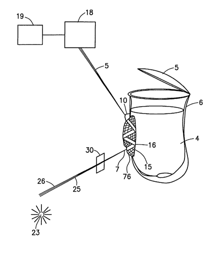
[0050] Fig. 1 (a) illustrates a schematic side view of a preferred embodiment
of the
present invention, which includes an ATR crystal 76, a first connecting device
26, and a
second connecting device 3. The first connecting device 26 transmits a beam 25
of
light, which is generated by a radiation source 23, to the ATR crystal 76 in
the side of a
containment vessel 6. While traveling to the ATR crystal 76, the beam impinges
a filter
30, which changes the beam 25 from polychromatic light to monochromatic light.
The
beam 25 enters the ATR crystal 76 and while traveling through the ATR crystal
76
impinges an interface 15, located between the ATR crystal 76 and a substance
4, at
least once. Each time the beam 25 contacts the interface 15 at or above a
critical angle
44 (See Figs. 4,5), an evanescent wave 16 is generated. The evanescent waves
16
penetrate the substance 4 and are attenuated in the regions of the spectrum
where the
substance 4 absorbs energy. The attenuated energy of each evanescent wave 16
is
passed back to the beam 25. The beam 25 then exits the ATR crystal 76 and is
directed
to a detector 18 by the second connecting device 3. The detector 18, which can
be
connected to a processing device 19, records the attenuated beam 25. The
detected
beam can then be processed by the processing device 19 to generate an
absorbance
spectrum of the dissolution medium. In certain embodiments of the present
invention,
processing device 19 may subtract a baseline spectrum (the data obtained by
using the
present invention without a substance present) from a sample spectrum (the
data
obtained by using the present invention with a substance present) to obtain a
baseline
corrected absorption spectra.
13
CA 02442445 2003-09-26
WO 02/077616 PCT/US02/09542
[0051] As one of ordinary skill in the art will appreciate, the spectrum
generated by
the ATR crystal may be affected and deviate from the general linearity of the
Beer-
Lambert law by such chemical and instrumental factors including, but not
limited to,
deviations in the absorptivity coefficients at high concentrations of the
sample due to
electrostatic interactions between molecules in close proximity within the
sample, the
randomized scattering of the radiation beam due to particulates in the sample,
any
innate fluoresecence or phosphorescence of the sample, any shifts in chemical
equilibria as a function of concentration in the sample, and stray light.
These problems,
however, can be avoided or compensated for using conventional techniques.
[0052] An aperture or opening is formed in the side of the containment vessel.
The
ATR crystal 76 is secured to the side of the containment vessel 6, covering
the aperture,
such that one side of the ATR crystal forms a portion of the interior surface
of the
vessel. The ATR crystal can be secured to the containment vessel in any
suitable
manner, including, for example, with a fastener, gasket, or adhesive (such as
silicone
adhesives). In this way, the ATR crystal is embedded in the containment
vessel.
[0053] A cover 5 can be placed over the containment vessel, so as to prevent
loss,
e.g., through evaporation, of the substance 4.
[0054] The equations N=L cot F/2t and DP w/2n n~ [sin2F-(ns/n~)2 ]'~' (where N
is the
number of reflections in the ATR crystal 76, L is the length of the ATR
crystal 76, F is
the angle of incidence, t is the thickness of the ATR crystal 76, DP is the
depth of
penetration of the evanescent wave 16, n~ is the refractive index of the ATR
crystal 76,
n5 is the refractive index of the substance 4, and w is the wavelength of the
light)
determine the number of reflections of the beam 25 within the ATR crystal 76
and the
depth of penetration of the beam 25 into the sample 4. The effective
pathlength of the
beam 25 may then be determined by P=N x DP (where N is the number of
reflections
and DP is the depth of penetration). The effective pathlength gives a measure
of the
intensity of the resulting spectrum.
[0055] The filter 30 can be a monochromator, a spectrograph, a linear variable
filter, a bandpass filter, or an interference filter. The filter 30 can also
be a
monochromator-filter type device, rotating tilting filter wheel, spinning
filter wheel,
14
CA 02442445 2003-09-26
WO 02/077616 PCT/US02/09542
AOTF(Acousto Optic Tunable Filter), or pre-dispersive grating monochromator.
[0056] Although the filter 30 is located between the radiation source 23 and
the
ATR crystal 76 in Fig. 1(a), the filter 30 can be located between the ATR
crystal 76 and
the detector 18, as shown in Fig. 1 (b), with similar components bearing
identical
reference numbers to Figure 1 (a). However, if the filter 30 is between the
ATR crystal
76 and the detector 18, the filter can not be a pre-dispersive grating
monochromator,
but can be a post-dispersive grating monochromator.
[0057] The radiation source 23 can be a QTH lamp, a deuterium lamp, a set of
light
emitting diodes, or a laser. The radiation source 23 can also be a Xenon Lamp,
a
Mercury Xenon Lamp, a Xenon Flash Lamp, a Metal Halide Lamp, a GaAs Infrared
LED, a GaAIA Infrared LED, GaAIA Infrared LED, or a GaAAs Infrared LED (all
produced by Hamamatsu Corporation). If a laser, LED, or other monochromatic
light
source is used, there is no need for a filter 30, either before, or after, the
ATR crystal.
[0058] The embedded ATR crystal 76 may be composed of ZnSe, Ge, SeAs, Cds,
CdTe, CsI, InSb, Si, Sapphire (A1203), diamond Anneled. Glass, borosilicate
crown
glass, BK7 Anneled Glass, UBK7 Annealed Glass, LaSF N9 Anneled Glass, BaKl
Annealed Glass, SF11 Annealed Glass, SK11 Annealed Glass, SFS Annealed Glass,
Flint Glass, F2 Glass, Optical Crown Glass, Low-Expansion Borosilicate
Glass(LEBG),
Pyrex, Synthetic Fused Silica (amorphous silicon dioxide), Optical Quality
Synthetic
Fused Silica, UV Grade Synthetic Fused Silica, ZERODUR, Agar, AgCI, KRS-5 (a
TIBr and T1CI compound), KRS-6 (a TIBr and TICI compound), ZnS, ZrOz, AMTIR,
or
diamond. Movements for UV/VIS applications, other crystals, such as Fused
Silica can
be used.
[0059] Although toxic, the Zinc Selenide (ZnSe) crystal, which is clear and
has a
polycrystalline lattice-work with a grain size of approximately 70 ,u~m, is
essentially
free of extrinsic impurity absorptions, and thus provides extremely low bulk
losses
from scatter. Moreover, the ZnSe crystal has a wide spectroscopic range of
about
20,000-S00 cm', transmits in the range of about O.S~m to about 15~m, and has a
refractive index of about 2.43.
[0060] As an optical element, the ZnS crystal, which has a refractive index of
about
CA 02442445 2003-09-26
WO 02/077616 PCT/US02/09542
2.25, has a spectroscopic range of about 50,000-770 cm' and functions in wet
or
aqueous solutions, but does not function in acidic solutions.
[0061] Si crystals, which have a higher refractive index than KRS-5 and ZnSe
crystals, are suitable for applications involving wet samples and aqueous
solutions,
even acids and alkalis, however, HF and HN03 attack the crystal. Spectroscopic
range
is about 4000-1500 cm' and in the far IR about 400-30crri'. The refractive
index of the
crystal is about 3.42.
[0062] Germanium optical elements with a refractive index of about 4.01 have
the
highest refractive index of common IR materials. Due to a wide transmission
range
covering 1.8 -- 17 Vim, spectroscopic range of about 5000-550 cm', and opacity
in
visible ranges, Germanium crystals are useful for analyzing hard polymers and
carbon
filled samples. The Germanium optical element is suitable for wet samples and
aqueous solutions, even acids and alkalis. However, the Geranium optical
element is
attacked by hot sulphuric acids and aqua regia, and is subject to thermal
shock.
[0063] KRS-5 is a general purpose crystal for may experiments, however, the
crystal is not well suited for applications involving wet or aqueous solution
and can be
distorted by pressure. Moreover, the KRS-S crystal is extremely toxic. The
crystal has
a refractive index of about 2.38 and a spectroscopic range of the crystal is
about
17,000-250ctri'.
[0064] AMTIR (Amorphous Material which Transmits Infrared Radiation) is a
chalcogenide glass with a refractive index of about 2.5. The AMTIR crystal,
which has
a spectroscopic range of about 4000-725crri', is suitable for acidic solutions
involving
wet samples or aqueous solutions, but the crystal is attacked by bases.
[0065] Diamond crystals, which have a spectroscopic range of about 4000-
400crri'
and a type 2A absorption band in the 2500-2000 cm' region, are suitable for
applications involving aqueous solutions from pH 1 to 14. Furthermore, the
durability
of the diamond allows contact efficiencies approaching 100%. The refractive
index of
the diamond crystal is about 2.35. A bullet shaped focusing crystal, made from
ZnSe or
KRS-5, can be placed in optical contact with the diamond crystal, so as to
provide
interfacing optics for the input and output radiation.
16
CA 02442445 2003-09-26
WO 02/077616 PCT/US02/09542
[0066] The entire ATR crystal 76 or a portion thereof, e.g., the side that
reflects the
beam of light onto the interface 15, can be coated with a metallic coating,
dielectric
coating, bare aluminum, protected aluminum, enhanced aluminum, UV-enhanced
aluminum, internal silver, protected silver, bare gold, protected gold,
MAXBRIte,
Extended MAXBRIte, Diode Laser MAXBRIte, UV MAXBRIte, or Laser Line MAX-
R. The coating increases the amount of light reflected, thus, improving the
accuracy of
the data. Furthermore, the coating can be a material that only reflects (or
allows
transmittance) of specific wavelengths of light. In this manner, the coating
can be
arranged on the ATR crystal 76 so that only specific wavelengths of light
reach the
sample.
[0067] The ATR crystal 76 can be trapezoidal, cylindrical, hemispherical,
spherical,
or rectangular. The differing shapes affect the refraction index and the
number of times
the beam 25 reflects while in the ATR crystal 76.
[0068] Single bounce crystals, i.e., the beam enters the crystal, reflects off
the
interface, and exits the crystal, reduce Fresnel reflection losses due to the
shorter path
length of the beam 25. Fresnel reflection losses result from diffraction which
involves
spherical waves incident upon an obstruction, effectively originating from a
point
Because of the reduction of Fresnel reflection losses, the single bounce
crystal improves
both qualitative and quantitative analysis of strongly absorbing samples,
e.g., aqueous
liquids, organic liquids, pastes, and powdered solids.
[0069] Spherical crystals, as described by Nicolet Instrument Corp., reduce
the
beam 25 diameter by a factor of two, resulting in a concentration of energy to
a smaller
spot size. As such, the beam 25 exerts more pressure on the substance 4 and
allows for
improved analysis of small samples.
[0070] The first and second connecting devices 26,3 can be hollow, rigid tube
wave
guides (the "light-pipe," W. M. Doyle and N.A. Jennings, Spectroscopy 5 (1) 34-
38
(1990)). However, the tubes are fairly inflexible, requiring careful
mechanical design
dictated by the geometry of the reaction vessel being used, and thus do not
lend
themselves to repeated use in environments where the reaction vessel
dimensions
and/or shape may vary. Furthermore, since rigid tube wave guides depend upon
17
CA 02442445 2003-09-26
WO 02/077616 PCT/US02/09542
carefully aligned mirrors to transmit the signal around bends or corners in
the tube, the
rigid tube wave guides are extremely sensitive to vibration, making the wave
guides
unsuitable for use in typical industrial environments.
[0071] The first and second connecting devices 26,3 can also be flexible fiber
optic
cable, which may contain one or more optical fibers that transmit radiation in
the
appropriate part of the electromagnetic spectrum. Fiber optic cables can be
used in a
wide variety of environments because of their durability and malleability. For
example,
cables for use in the visible region of the spectrum can be made using fibers
of silica
glass.
[0072] The first and second connecting devices 26,3 can be constructed of, but
are
not limited to, Optical Glass, Fused Silica Fiber, low OH Fused Silica Fiber,
Fluoride,
or Chalcogenide fiber. High quality optical glass transmits wavelengths from
about
400nm to about 900nm. However, transmission in the UV range is very low and
wavelengths below about 350nm are not transmitted. When the application
requires
UV light, more expensive Fused Silica fibers can be used. At about 1.4
microns, all
fibers except those specifically designed for IR transmission show a
significant drop in
transmission because of absorption in the glass. Low OH Fused Silica Fibers
specifically designed for the NIR do not show the transmission drop at about
1.4
microns and transmit well between about .4 microns and about 2.5 microns.
Fluoride
and Chalcogenide Fibers can cover a range form about 1 micron to about 10
microns.
[0073] The containment vessel 6 can be constructed of an inert material, e.g.,
Teflon, fluoroplastic, PTFE, NALGENE, lead, stainless steel, or Teflon
fluoropolymer
resin. Alternatively, the vessel 6 could be made of glass, PYREX, crown, Fused
Silica
borosilicate glass, or Flint.
[0074] A wide variety of detectors 18 can be used, including, but not limited
to,
silicon detectors (PDA, CCD detectors, individual photo diodes),
photomultiplier tubes,
Ga detectors, InSb detectors, GaAs detectors, Ge detectors, PbS detectors,
PbSi
photoconductive photon detectors, PbSe photon detectors, InAs photon
detectors,
InGaAs photon detectors, photoconductive photon detectors, photovoltaic photon
detectors, InSb photon detectors, photodiodes, photoconductive cells, CdS
18
CA 02442445 2003-09-26
WO 02/077616 PCT/US02/09542
photoconductive cells, opto-semiconductors, or HgCdTe photoconductive
detectors.
The detector 18 can consist of one element (i.e., one detector unit) or of a
plurality of
elements organized into an array.
[0075] Fig. 2 illustrates a schematic side view the present invention having a
first a
first mirror 77 and a second mirror 78 in place of the first connecting device
26 and the
second connecting device 3 (Fig. 1). The first mirror 77 reflects the beam 25
of light,
which is generated by the radiation source 23, to the ATR crystal 76. In route
to the
ATR crystal 76, the beam impinges the filter 30, which changes the beam of
light from
polychromatic light to monochromatic light. The beam 25 enters the ATR crystal
76
and while traveling through the ATR crystal 76 impinges an interface 15,
located
between the ATR crystal 76 and a substance 4, at least once. Each time the
beam 25
contacts the interface 15 at or above the critical angle 44 (See Figs. 3,4),
an evanescent
wave 16 is generated. The evanescent waves 16 penetrate the substance 4 and
are
attenuated in the regions of the spectrum where the substance 4 absorbs
energy. The
attenuated energy of each evanescent wave 16 is passed back to the beam 25.
The
beam 25 then exits the ATR crystal 76 and is directed to the detector 18 by
the second
mirror 78. The detector 18, which is connected to the processing device 19,
records the
attenuated beam 25 and the detected beam is then processed in the manner
described
above to generate a spectrum of the dissolution medium. Alternatively, the
filter 30 can
be placed between the ATR crystal 76 and the detector 18 instead of between
the ATR
crystal 76 and the radiation source 23.
[0076] Fig. 3(a) illustrates an alternate embodiment of the present invention,
which
shows the ATR crystal 76 mounted on a probe 100. Components 18, 19, 30, 77, 78
and
23 (not shown) can be configured in the same manner as described above with
reference to Figures l and 2. The probe 100 is inserted into the containment
vessel 6
through an aperture. If desired, the probe 100 can be shaped so that a seal is
formed
with the containment vessel 6, thereby, preventing loss of the substance 4 by
evaporation. The radiation source (not shown) generates the beam of light. The
beam
of light passes through the filter (not shown) and enters the first connecting
device 26.
The first connecting device 26 then transmits the beam 25 to the probe 100.
The probe
19
CA 02442445 2003-09-26
WO 02/077616 PCT/US02/09542
100, in turn, transmits the beam to the ATR crystal 76. The beam 25 then
enters the
ATR crystal 76 and travels through the ATR crystal 76. While traveling through
the
ATR crystal 76, the beam 25 impinges the interface 15 between the ATR crystal
76 and
the substance 4 at least once and in so doing generates the at least one
evanescent wave.
The evanescent wave penetrates the substance 4 and is attenuated in the
regions of the
spectrum where the substance 4 absorbs energy. The attenuated energy of the
evanescent wave is passed back to the beam 25, which then exits the ATR
crystal 76
and re-enters the probe 100. The probe 100 transmits the beam 25 to the second
connecting device 3. The second connecting device 3 then directs the bean to
the
detector (not shown). The detector, which can be connected to the processing
device
(not shown), records the attenuated light beam and the detected beam is then
processed
to generate a spectrum report of the dissolution medium.
[0077] Fig. 3(b) illustrates a side view of the probe 100. A probe cover 101
is also
shown. The probe cover 101 can selectively cover the outer surface of the ATR
crystal
76, so that absorbance contacts with the substance 4 are decreased.
[0078] In order to prevent interference between the portion of the beam 25
exiting
the ATR crystal 76 and the portion of the beam 25 entering the ATR crystal 76,
a
partition 5000 may be placed in the middle of the probe 100. Also, the first
and second
connecting devices 26,3 can also (or alternatively) be connected to the probe
100 in
such a way that there is a direct connection between the output of the first
connecting
26 and the input of the ATR crystal 76, and a direct connection between the
output of
the ATR crystal 76 and the input of the second connecting device 3.
[0079] The probe 100 can be constructed of chalcogenide fiber, e.g., from
glass
composed of arsenic, selenium, and tellurium (AsSeTe glass). Chalcogenide
fiber
performs well in the mid-IR range, transmitting across a substantial part of
the mid-IR
region, namely 900-5000 cm. The probe 100 can also be clad with a glass of
lower
refractive index to prevent escape or "leakage" of radiation from the fiber.
[0080] Alternatively, the probe 100 can be constructed of an inert material,
e.g.,
Teflon, fluoroplastic, PTFE, NALGENE, or Teflon fluoropolymer resin, and
contain a
plurality of internal connecting devices 105, e.g., fiber optic cables, as
shown in Fig.
CA 02442445 2003-09-26
WO 02/077616 PCT/US02/09542
3(b). To transfer the light from the first connection device 26 to the ATR
crystal 76, a
first set of the internal connecting devices 1 OS is attached to the output of
the first
connecting device 26 and input of the ATR crystal 76. To transfer the light
from the
output of the ATR crystal 76 to the input of the second connecting device 3, a
second
set of the internal connecting devices 105 is attached to the input of the
second
connecting device 3 and the output of the ATR crystal 76. Preferably, the
internal
connection devices 105 are connected to the first and second connecting
devices 26,3
and the ATR crystal 76, so as to transmit the beam of light 25 to and from the
ATR
crystal 76.
[0081] Instead of internal connecting devices 105, the first and second
connecting
devices 26,3 can be embedded inside the probe 100, with the output of the
first
connecting device 26 connected to the input of the ATR crystal 76, and with
the input
of the second connecting device 3 connected to the output of the ATR crystal
76.
[0082] Figures 4 illustrates a multiple bounce embodiment of the ATR crystal
76.
The beam 25 of light enters the ATR crystal 76 at an angle of incidence 42 and
reflects
off the interface 15 of the ATR crystal 76 and the substance 4. The beam then
reflects
off the opposing side 49 of the ATR crystal 76, which can be coated with a
material to
enhance reflectiveness, and returns to the interface 15 a second time. Each
time the
beam reflects off the interface 15, the beam does so at the angle of incidence
42. If the
angle of incidence 42 equals or is greater than the critical angle 44, the
incident light
undergoes total internal reflection and the evanescent wave 16 is generated.
Snell's
Law determines the critical angle 44: sine,, - siri 1 nz/n, (where n~ is the
index of
refraction of the ATR crystal 76 and n2 is the index of refraction for the
substance 4).
In regions of the spectrum where the substance 4 absorbs energy, the
evanescent wave
16 is attenuated, and the attenuated energy passed back to the beam 25. After
impinging the interface 15 the second time, the beam 25 exits the ATR crystal
76. Any
spectral data derived from the interface 1 S can be masked out by subtracting
the
spectral data from the detector 18 from spectral data generated by an empty
vessel.
Alternatively, a material can be placed at the opposing interface 49 that does
not absorb
radiation in the wavelength band of the light transmitted through the crystal.
21
CA 02442445 2003-09-26
WO 02/077616 PCT/US02/09542
[0083] The refractive index of the ATR crystal 76, the refractive index of the
substance 4, and the angle of incidence 42 affect the depth of penetration.
For example,
reducing the angle of incidence 42 or using a crystal with a lower refractive
index
increases the depth of penetration of the evanescent wave 16, however, at
shallow
depths of penetration, the evanescent wave 16 generally provides more reliable
results.
[0084] Figures 5 illustrates a single bounce embodiment of the ATR crystal 76.
The beam 25 enters the ATR crystal 76 at the angle of incidence 42 and
reflects off the
interface 15 at a reflection point 78. If the angle of incidence 42 at which
the beam
contacts the interface 15 equals or is greater than the critical angle 44, the
incident light
undergoes total internal reflection and the evanescent wave 16 is generated.
Snell's
Law determines the critical angle 44: sin~~;~ = siri' n2/n, (where n, is the
index of
refraction of the ATR crystal 76 and n2 is the index of refraction for the
substance 4).
In regions of the spectrum where the substance 4 absorbs energy, the
evanescent wave
16 is attenuated, and the attenuated energy passed back to the beam 25. The
beam 25
then exits the ATR crystal 76.
[0085] Figures 6 shows a monochromatic filter-type device. A beam of light
impinges a rotating circular disk 504, which includes a plurality of narrow
bandpass
optical filters 507. The disk can be rotated so that the beam of light passes
through
each of the narrow bandpass optical filters 507. An encoder 511 controls which
optical
filter 507 is presently under the light source. The optical filters 507 filter
the beam of
light so that only a narrow selected wavelength range passes through. The
monochromatic filter-type device can be used as the filter 30 in any of the
devices
described in Figs. 1-3, upstream or downstream of the ATR crystal 76.
[0086] Figures 7 and 8 illustrate two basic forms of filter-type NIR devices
utilizing
a tilting filter concept.
(0087] Figures 7 shows a rotating tilting filter wheel utilizing wedge
interference
filters having a light blocking flag 604. Light is transmitted through a
filter wheel 600
at varying wavelengths and bandpasses, which are dependent on the incident
angle of
the light passing through the interference filter wedge 602. The rotating
filter wheel
can be used as the filter 30 in any of the devices as described in Figs. 1-3,
upstream or
22
CA 02442445 2003-09-26
WO 02/077616 PCT/US02/09542
downstream of the ATR crystal 76.
[0088] Figures 8 shows a spinning filter wheel in which the light passes
through an
encoder wheel 700, having a plurality of interference filters 701. The
spinning filter
wheel operates using the same basic principle as the tilting filter of Fig. 7,
but the
interference filters 701 of the spinning filter wheel are mounted in the
encoder wheel
700 for greater positioning accuracy (wavelength reproducibility) and greater
reliability.
The spinning filter wheel can be used as the filter 30 in any of the devices
as described
in Figs. 1-3, upstream or downstream of the ATR crystal 76.
[0089] Figures 9 shows a typical pre-dispersive monochromator-based
instrument,
where the light is dispersed prior to striking the sample. The beam of light
passes
through an entrance slit 800 and onto a grating 810. The grating 810 separates
the light
into a plurality of beams of different wavelengths. Via the order sorting 820
(to
eliminate undesired wavelengths) and stds 830 (to provide a wavelength
standard for
calibration) components, a desired band of wavelengths is selected for
transmission.
The device may also be used as the filter 30 with any one of the embodiments
of the
present invention described above in Figures 1 (b), 2, or 3, provided that it
is located
upstream of the ATR crystal 76.
[0090] Figures 10 shows a typical post-dispersive monochromator. This type of
instrument provides the advantage of allowing the transmission of more energy
on the
sample. After the beam of light has exited the ATR crystal 76 (Figs. 1-5),
where the
beam of light was attenuated, the light is reflected back to a grating 910
(the dispersive
element). On striking the grating 910, the light is separated into the various
wavelengths. An order sorting 920 (to eliminate undesired wavelengths) and an
stds
930 ( to provide a wavelength standard for calibration) component provide the
desired
band of wavelengths selected for transmission. As illustrated, this filter may
be used as
the filter 30 in with any one of the embodiments of the present invention
described
above in Figs. 1 (b), 2 or 3, provided that it is located downstream of the
ATR crystal
76.
[0091] Figures 11 depicts an Acousto Optic Tunable Filter spectrometer
utilizing an
RF signal 1229 to generate acoustic waves in a Te02 crystal 1232. The beam of
light
23
CA 02442445 2003-09-26
WO 02/077616 PCT/US02/09542
transmits through the crystal 1232, and the crystal 1232 splits the beam of
light into
three beams: a center beam of unaltered white light 1237 and two beams of
monochromatic and orthogonally polarized light 1240. The wavelength of the
monochromatic light can be incremented across a wavelength band of interest by
varying the RF frequency. One of the two beams of monochromatic light 1240
then
pass to any of the devices described in Figs. 1-3.
[0092] Figures 12 depicts a Fourier Transform device. A light emitting source
1300, e.g., an IR diode, produces a beam 1302. The beam 1302 impinges a beam
sputter 1304, which splits the beam 1302 into a first half 1308 and a second
half 1306
and in so doing sends the first half 1308 and second half 1306 in two
directions at right
angles. The first half 1308 continues to a stationary mirror 1390 and then
back to the
beam splitter 1304. The second half 1306 impinges a moving mirror 1310 and
returns
to the beam splitter 1304. The moving mirror 1310 changes the total path
length of the
second half 1306, so that when the first half 1304 and second half 1306
recombine to
form a recombined beam 1312, the difference in path length creates
constructive and
destructive interference: an interferogram. The recombined beam 1312 passes
into any
of the devices described in Figs. 1-3. A Fourier transform converts the data
obtained
from the detector 18 of the devices of Figs. 1-3 to an intensity vs. time
spectrum, and
since time is the reciprocal of frequency, the spectrum can be converted into
an
intensity vs. frequency spectrum.
[0093] Fig. 13 schematically illustrates a side view of a preferred embodiment
of
the present invention, which includes a spiral ATR crystal 76. The ATR crystal
76,
which can be composed of fused silica, is disposed along the inner surface of
the vessel
6 in a double helix configuration. The double helix configuration spirals
downward
until it reaches the bottom of the vessel 6, and then spirals upward. This
architecture
allows a large number of bounces which are distributed throughout the interior
surface
of the vessel.
[0094] Fig. 14 schematically illustrates a side view of a preferred embodiment
of
the present invention for performing dissolution testing of a dosage form. The
processing device 19, detector 18, first connecting device 26, second
connecting device
3, filter 30, ATR crystal 76, radiation source 26, vessel 6, and substance 4
are
24
CA 02442445 2003-09-26
WO 02/077616 PCT/US02/09542
configured in the same manner described above with regard to Figure 1(b). The
arrangements shown in Figures 1(b) and 2 can similarly be used in conjunction
with
this embodiment. Also shown in Figure 14 is a mixing shaft and paddle 10000
which
conform to the requirements and specifications given, e.g., in USP 23rd
edition Chapter
711 (Dissolution) pages 1791-1793. Referring to Figure 14, processor 19
receives
information from the ATR crystal 76 via detector 18 as the dissolution of the
dosage
form in the dissolution medium 4 proceeds, analyzes the information, and
generates a
dissolution profile of the dosage form. Most preferably, the processor
receives,
analyzes, and displays the dissolution profile as dissolution in the
dissolution medium
proceeds. Software for providing such functionality is described, for example,
in PCT
US00/23800, entitled "In Situ Methods for Measuring the Release of a Substance
from
a Dosage Form," the entire disclosure of which is hereby incorporated by
reference. In
using this apparatus, an operator preferably performs a baseline correction by
taking
measurements without a sample present in the vessel to obtain a baseline
spectra.
Thereafter, the operator places a dissolution medium and a sample material in
the
containment vessel. The apparatus is then used to generate spectral data from
the
dissolution media. The baseline spectra is then subtracted from the spectral
data to
provide the spectra of the dissolution medium.