Note: Descriptions are shown in the official language in which they were submitted.
CA 02442572 2003-09-17
WO 02/076299 PCT/US02/07900
MICRO-INVASIVE TISSUE REMOVAL DEVICE
Field of the Invention
The present invention relates generally to medical
devices and methods and, more particularly, to medical
devices, for example, micro-invasive devices, and methods for
removing material, for example, tissue and/or other material,
from the bodies of humans or animals for beneficial purposes,
such as diagnosis and/or therapeutic treatment.
Background of the Invention
The medical industry is constantly evolving through the
adaptation of improved pharmaceutical, biotechnology, and
medical device products and procedures. Techniques and
technologies are being developed to treat internal areas of
the body through less invasive means.
It is often desirable and frequently necessary to remove
a portion of tissue from humans and other animals,
particularly in the diagnosis and treatment of patients with
cancerous tumors, pre-malignant conditions and other diseases
or disorders. Typically, in the case of cancer, when the
~0 physician establishes by means of procedures such as
palpation, x-ray or ultrasound imaging that suspicious
circumstances exist, a biopsy is performed to determine
whether the cells are cancerous . Biopsy may be done by an open
or percutaneous technique. Open biopsy removes the entire mass
(excisional biopsy) or a part of the mass (incisional biopsy) .
Percutaneous biopsy, on the other hand, is usually done with
a needle-like instrument and may be either a fine needle
aspiration (FNA) or a core biopsy. In FNA biopsy, individual
cells or clusters of cells are obtained for cytologic
examination and may be prepared such as in a Papanicolaou
smear. In core biopsy, as the term suggests, a core or
fragment of tissue is obtained for histologic examination
which may be done via a frozen section or paraffin section.
The type of biopsy utilized depends in large part on
circumstances present with respect to the patient and no
CA 02442572 2003-09-17
WO 02/076299 PCT/US02/07900
2
single procedure is ideal for all cases. However, core biopsy
is extremely useful in a number of conditions and continues
to be used frequently by the medical profession.
To arrive at a definitive tissue diagnosis, intact tissue
is needed from an organ or lesion within the body. In most
instances, only part of the organ or lesion need be sampled.
However, the portions of tissue obtained must be
representative of the organ or lesion as a whole. In the
past, to obtain tissue from organs or lesions within the body,
surgery had to be performed to locate, identify and remove the
tissue. With the advent of medical imaging equipment (x-rays
and fluoroscopy, computed tomography, ultrasound, nuclear
medicine, and magnetic resonance imaging) it has become
possible to identify small abnormalities even deep within the
body. However, definitive tissue characterization still
requires obtaining adequate tissue samples to characterize the
histology of the organ or lesion. For example, mammography
can identify non-palpable (not perceptible by touch) breast
abnormalities earlier than they can be diagnosed by physical
examination.
Percutaneous biopsy techniques are desirable in many
instances, particularly in light of modern imaging techniques
which are able to pinpoint abnormal tissue masses, for example
in areas of the body such as the breast, brain and spinal
column. A well known instrument used quite extensively for
core biopsies of in the past is manufactured by Travenol
Laboratories of Deerfield, I11., and is sold under the mark
"TRU-CUT." This manual biopsy instrument at one time enjoyed
as much as 98% of the market for such devices. As disclosed
in U.S. Pat. No. 3,477,423, the instrument comprises a two-
piece assembly: an outer cutting cannula mounted to one hub
member and an inner stylet with a sampling notch ground into
it mounted to a second hub, with the hubs being slidably
interlocked. The instrument is assembled and placed into the
body with the outer cutting cannula just to the rear of a
lancet point or beveled distal end of the stylet. Upon
CA 02442572 2003-09-17
WO 02/076299 PCT/US02/07900
3
inserting the device up to or in front of the area to be
biopsied, advancement of the assembly is halted. The stylet
is manually advanced distally of the cannula with the cannula
held stationery. Upon advancement of the stylet, the specimen
notch is exposed. Tissue surrounding the stylet prolapses
into the specimen notch and the cutting cannula is then
manually advanced distally over the stylet, slowly shearing
off the tissue entrapped in the stylet's specimen notch. The
instrument is then either (a) withdrawn and the stylet
advanced distally to expose the tissue for preparation for
study or (b) left in place and only the stylet is proximally
removed from within the cannula so a determination of
successful sampling may be made. If the sampling was not
successful, the stylet may be reinserted into the cannula,
which remains positioned within the patient, and an attempt
to reposition the assembly of stylet and cannula and repeat
sampling can be made.
Such a technique using this basic design of a biopsy
instrument is referred to as a manual technique. One drawback
to the manual technique is that it requires a great deal of
manual dexterity and motor coordination, along with the use
of both hands, to advance the stylet while maintaining the
position of the cannula and then to maintain the position of
the stylet while advancing the cannula. Another drawback is
that the cannula is advanced relatively slowly, resulting in
an extremely poor cutting action and allowing the surrounding
tissue an opportunity to collapse, thus making no use of the
stored kinetic energy in the material being severed. Further
disadvantages are encountered when the tissue volume to be
sampled contains areas of higher density than that of
surrounding tissue, such as areas of calcification commonly
associated with certain types of cancerous growths. A
manually inserted sampling device is often incapable of
penetrating the denser area of tissue which merely deflects
the course of the cannula/stylet structure around the dense
area and into the more compliant surrounding tissue.
CA 02442572 2003-09-17
WO 02/076299 PCT/US02/07900
4
A variety of automatic and semiautomatic biopsy
instruments have been developed which are spring loaded gun-
type devices. A biopsy gun currently used is described in
U.S. Pat. No. Re. 34,056, entitled "TISSUE SAMPLING DEVICE,"
issued to Lindgren et al. Additional examples of biopsy gun
devices are disclosed in U.S. Pat. Nos. 4,600,014 and
4,958,625.
Such devices use a design comprising a handle held in a
physician's palm, and a guide tube extending forward of the
handle. A cannula is slidably disposed within the guide tube
and is movable from within the guide tube forward out of the
distal end of the guide tube. A sampling stylet is
telescopically disposed within the cannula and projects from
the rear of the handle. In an automatic mode of operation,
the cannula, when in the retracted position, is spring loaded
by means of a compressed spring. A release lever, which works
against the compressed spring, is activated to release
compression of the spring which then expands and pushes the
cannula outwardly over the stylet. This instrument, as
stated, requires two handed operation. Also, since the stylet
is not removable proximally from within the handle, the entire
instrument must be withdrawn to obtain access to the sample.
A fully automatic instrument manufactured by Radiplast,
Inc, of Sweden is described in U.S. Pat. No. 4,699,154. This
instrument comprises a reusable, spring-loaded box-shaped
housing or handpiece, which activates a disposable cannula and
stylet set. Both the stylet and cannula are activated in
rapid succession. The instrument has the advantage of
reducing the dexterity and motor coordination necessary in the
use of manual devices and also eliminates the slow cutting
action of the manually advanced cannula, replacing it with a
very quick, clean cut. This instrument, however, also has its
drawbacks. First, the reusable handpiece is very large,
heavy, cumbersome, and expensive. They are also typically
spring-powered devices and must be manually cocked with some
sort of plunger bar. Such "cocking" of the gun requires
CA 02442572 2003-09-17
WO 02/076299 PCT/US02/07900
considerable force and the gun must be cocked for each biopsy
cut. When actuated, the springs provided in the gun
accelerate the needles until a mechanical stop position is
reached which can create a loud snapping noise and jerking
5 motion which is a problem both to the physician and the
patient. A further drawback is encountered in automatically
activating both the stylet and the cannula, as opposed to
activating the stylet manually, in that the rapid speed at
which the cannula follows the stylet into the tissue does not
allow much tissue to collapse into the specimen notch,
limiting the size of the sample.
U.S. Pat. No. 5,183,054, entitled "ACTUATED BIOPSY
CUTTING NEEDLE WITH REMOVABLE STYLET," issued to Burkholder
et al., discloses a biopsy device having a tubular cannula
through which a stylet, having a stylet cavity near the distal
end, is placed. The stylet is removable from the cannula and
removed from the biopsy device through the housing so that the
tissue sample obtained by the biopsy device may be manually
retrieved while the cannula remains in place within the
patient, near the area being sampled. Thereafter, the stylet
may be reinserted through the housing and cannula into the
patient's tissue where additional tissue samples may be
obtained. In this way, trauma to the tissue that ordinarily
occurs upon reinsertion of the cannula and stylet is
minimized.
U.S. Pat. No. 5,234,000, entitled "AUTOMATIC BIOPSY
DEVICE HOUSING A PLURALITY OF STYLETS, " issued to Hakky et al .
describes a biopsy device for taking a plurality of samples
of tissue from a living being. The device comprises a housing
having a portion arranged to be held by a person using the
device, a cannula having a proximal portion and a distal
portion and being coupled to the housing. A plurality of
stylets are located in the housing, with each of the stylets
having a proximal end, a distal end, and a tissue receiving
notch located adjacent the distal end. Each stylet is
individually propelled through the cannula into the body so
CA 02442572 2003-09-17
WO 02/076299 PCT/US02/07900
6
that a portion of the tissue prolapses into the notch.
There currently exists a need for a more effective
microsurgical device for obtaining and/or collecting a sample
of tissue from a target area in a patient.
Sununary of the Invention
New apparatus and methods for removing tissue and/or
other material from human or animal bodies have been
discovered. The present invention provides apparatus, for
example, micro-invasive apparatus, to remove tissue or other
material from a target area of a human or animal body to
provide one or more benefits, such as diagnostic benefits,
therapeutic benefits and the like.
The apparatus of the invention are useful for removing
unwanted, diseased, or even healthy bodily materials, tissues
or foreign materials for medical treatment and/or therapeutic
purposes. Advantageously, the present invention is suitable
for use in many surgical settings and is suitable for
performing various material removal procedures using
methodologies, for example, in terms of methods of introducing
the apparatus into the body and removing the apparatus from
the body, which are substantially analogous to conventional
surgical techniques. Necessary or desirable adaptations of
the apparatus of the present invention for specific medical
treatment, e.g., diagnostic, and therapeutic purposes will be
readily appreciated by those of skill in the art.
Accordingly, apparatus for removing material from a
target area of a human or animal body are provided. In one
broad aspect, the apparatus comprise a handpiece and a tissue
removal element connected or coupled thereto. The tissue
removal element includes a cannula, for example, a
substantially rigid or flexible cannula, and a rotational
element at least partially disposed in the cannula. The
rotational element is structured to be operatively coupled to
a source of rotational energy, for example, a motor. The
rotational element is disposed at least partially in the
CA 02442572 2003-09-17
WO 02/076299 PCT/US02/07900
7
cannula. The cannula includes an open distal tip structured
to be placed in a target area of a body, and preferably a
proximal end portion structured to be coupled, for example,
removably coupled, to the handpiece. The tissue removal
element is structured and effective to draw material from the
target area or site, for example, into the open distal tip,
in response to, for example, as a result of, rotation of the
rotational element relative to the cannula.
In one embodiment, the rotational element is at least
partially disposed in the cannula and is structured to at
least assist in drawing material from the target area into the
cannula. For example, the rotational element and the cannula
are structured to cooperatively engage to form or create a
source of suction effective in drawing, preferably sufficient
to draw the material from the target area into the cannula in
response to rotation of the rotational element relative to the
cannula. Advantageously, the cannula, in particular the
interior hollow space formed or defined by the cannula, and
the rotational element are sized and positioned, relative to
each other, to create a source of suction or pumping action
in response to the rotational element rotating relative to the
cannula. Without wishing to limit the invention to any
particular theory of operation, it is believed that this
functioning of the cannula/rotational element combination is
at least somewhat analogous to the functioning of a pump, for
example, a pump based on the principles of the "Archimedes'
screw", causing the material to be drawn or pulled or pumped
into the open distal tip of the cannula and through the
cannula in being removed from the target area of the
human/animal body.
Preferably, the suction/pumping action created or formed
by the cannula/rotational element combination is itself
sufficient and effective so that no other, for example, no
additional or supplemental, source of suction, aspiration, or
pumping action is employed, needed or required to effectively
remove material from the target area in accordance with the
CA 02442572 2003-09-17
WO 02/076299 PCT/US02/07900
8
present invention.
In one embodiment of the invention, the rotational
element includes a shaft and one or more outwardly extending
projections, for example threads, having a substantially
helical configuration. Advantageously, the rotational element
includes a distal portion with such projections. The proximal
portion of the rotational element may or may not include such
projections or threads. In a very useful embodiment, the
proximal portion of substantially free of such projections or
threads.
A portion of the shaft, for example the distal portion
of the rotational element, in a useful embodiment, extends
beyond the open distal tip or inlet of the cannula, for
example, by a distance in a range of about 0.02 inches to
about 1 inch beyond the open distal tip of the cannula. The
rotational element distal portion preferably extends a
distance at least about one half of the spacing between
adjacent projections or threads, beyond the open distal tip
of the cannula. The rotational element distal portion may
extend beyond the open distal tip by a distance equal to more
than about one spacing, for example, about two spacings or
more between adjacent projections or threads beyond the open
distal tip of the cannula. The rotational element may
advantageously include an elongated shaft having a proximal
portion which is substantially smooth to allow sufficient
annular space between the shaft and cannula for removal of
material.
The cannula may be of any suitable size. However, in
order to obtain the reduced invasiveness benefits of the
present invention, it is preferred that the cannula size have
an outer diameter of no greater than about 5 mm or less, and
more preferably no greater than about 2 mm, or less.
It has unexpectedly been found that the present apparatus
including such small size cannulas providing for reduced, or
even micro, invasive procedures (which reduce surgical trauma
and promote healing) and are effective in removing materials
CA 02442572 2003-09-17
WO 02/076299 PCT/US02/07900
9
from a human or animal body to achieve therapeutic benefits,
for example, therapeutic removal of soft tissues, soft tissue
tumors, breast tissue, spinal disc nucleus materials and the
like.
In one embodiment of the invention, the open distal tip
of the cannula is angled or is beveled with respect to a
longitudinal axis of the cannula. Alternatively, the open
distal tip is substantially perpendicular with respect to the
longitudinal axis of the cannula.
The present apparatus advantageously includes a tissue
collection chamber in communication, for example, fluid
communication, with the cannula and structured to collect and
contain material passed through the cannula. The collection
chamber preferably is structured to facilitate quantification
and/or other analysis of the material removed from the human
or animal body. In one particularly useful embodiment, the
collection chamber comprises a substantially transparent
conical section removably engaged to a housing of the
handpiece and preferably circumscribing a portion, for
example, the proximal portion of the shaft of the rotational
element.
The cannula and/or the rotation element, preferably both,
advantageously are structured to be manually deformable, for
example, to enable the physician to alter the normal
configuration, for example, the normal substantially straight
configuration, thereof to create a curved configuration if
desired, for example, to deal with the material removal
application at hand.
In another broad aspect of the present invention, methods
of removing material from a body of a human or an animal are
provided. Such methods comprise placing into a body of a
human or an animal a cannula having an open distal tip and a
rotational element disposed at least partially in the cannula,
and rotating the rotational element relative to the cannula,
thereby at least assisting in drawing a material from the body
into the open distal tip of the cannula. The method
CA 02442572 2003-09-17
WO 02/076299 PCT/US02/07900
preferably further comprises passing the material from the
body through the cannula. Apparatus in accordance with the
present invention described herein, can be advantageously
employed in accordance with the present methods.
5 The cannula used in accordance with the present methods
preferably have outer diameters of about 5 mm or Less, for
example, 2 mm or less.
The placing step of the present methods preferably
includes percutaneously introducing the cannula into the human
l0 or animal body, and positioning the open distal tip of the
cannula in close proximity to the material to be removed. The
cannula and rotational element preferably are sued and
positioned relative to each other so that the rotating step
is effective in drawing the material from the body of a human
or an animal into the open distal tip of the cannula.
Preferably, the material from the body is removed without
applying additional suction or aspiration to the open distal
tip of the cannula.
In one very useful embodiment, the rotating of the
rotational element relative to the cannula is effective to
draw the material into the cannula as a substantially single
continuous piece. Thus, although some shearing and/or cutting
of the material to be removed may occur in accordance with the
present invention, for example, so that the removed material
is compatible with the space within the cannula through which
the material is to be moved proximally, the present apparatus
and methods preferably are not dependent or based on cutting
or chopping the material to be removed into small discrete
segments.
The present methods preferably further comprise
collecting the material removed and/or observing and/or
otherwise testing the material removed.
Any suitable material can be removed from the body of a
human or an animal using the present apparatus and/or methods .
Preferably, such material to be removed can be effectively
removed using the present apparatus and/or methods without
CA 02442572 2003-09-17
WO 02/076299 PCT/US02/07900
11
employing or requiring additional suction or aspiration,
beyond that formed or created by the rotation of the
rotational element relative to the cannula.
Advantageously, the material to be removed is soft and/or
semi-solid and/or a viscous flowable material and/or a
material which is at least somewhat free to move toward a
source of lower pressure or suction. Examples of such
materials include, without limitation, material located in the
nucleus of spinal discs, material located in breasts, eyes,
soft tissue tumors, cysts, bone marrow, sinus tissue, fatty
tissues (e.g. for removing excess fat from an area of the
body), clots, blockages, cancerous and/or suspected cancerous
tissues, other diseased and/or suspected diseased tissues,
other tissues, bodily materials, materials normally foreign
to the body and the like. Moreover, the present apparatus and
methods may be employed in any suitable part or parts of the
body of a human or animal in which the material or materials
to be removed are located.
Incorporated herein by this specific reference are the
entire disclosures of U.S. Patent Application for Micro
invasive Nucleotomy Device and Method, having Serial No.
(attorney docket no. D-3039) , filed on even date herewith, and
commonly assigned and U.S. Patent Application for Micro
invasive Breast Biopsy Device, having Serial No. (attorney
docket no. D-3026), filed on even date herewith and commonly
assigned herewith.
Each and every feature described herein, and each and
every combination of two or more of such features, is included
within the~scope of the present invention provided that the
features included in such a combination are not mutually
inconsistent.
The present invention and the objects and advantages
thereof will be more clearly understood and appreciated with
respect to the following Detailed Description, when considered
in conjunction with the accompanying Drawings.
CA 02442572 2003-09-17
WO 02/076299 PCT/US02/07900
12
Brief Description of the Drawings
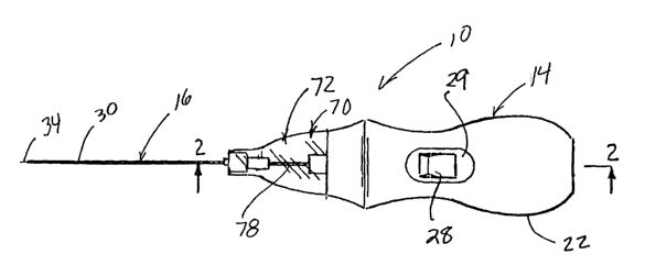
Fig. 1 shows a side view of a micro-invasive tissue
removal apparatus in accordance with the present invention
including a handpiece and a tissue removal element connected
thereto.
Fig. 2 is a partial cross-sectional view of the apparatus
taken along line 2-2 of Fig. 1.
Fig. 3 is a partial cross sectional view of a preferred
distal tip of the tissue removal element of the apparatus in
accordance with the present invention; and
Figs. 4 is side view of the apparatus shown in Fig. 1,
with the tissue removal element having a curve for
facilitating access to tissue.
Figs. 5 and 6 each show a partial cross sectional view
of an alternative distal tip of the tissue removal apparatus
of the present invention.
Detailed Description
Turning now to Figs. 1 and 2, a micro-invasive tissue
removal apparatus in accordance with the present invention is
shown generally at 10. The apparatus 10 generally comprises
a handpiece 14 and a tissue removal mechanism 16 to be
described in detail hereinafter.
The handpiece 14 is preferably sized and contoured to fit
comfortably within a palm of a surgeon, and includes for
example a molded plastic housing 22. As shown in Fig. 2, the
housing 22 of the handpiece 14 encloses a small motor 24 and
a power supply, for example a 9 volt battery 26 for driving
the tissue removal mechanism 16. Suitable electrical
connectors 27 are provided. For convenient, one handed
operation, an ON/OFF switch 28 is preferably provided on a
recessed, lateral portion 29 of the housing 22.
Turning now as well to Fig. 3, the tissue removal
mechanism 16 generally includes a cannula 30 and a rotatable
element 34 disposed therein. As shown most clearly in Fig. 3,
the cannula 30 includes a distal portion 40 defining an inlet
CA 02442572 2003-09-17
WO 02/076299 PCT/US02/07900
13
42 for receiving tissue drawn from a target area within a
patient. The inlet 42 is defined, for example, by flat,
distal edge 44 of the cannula 30. The distal edge 44, in the
embodiment shown in Fig. 3, lies along a plane that is
substantially perpendicular with respect to the longitudinal
axis of the cannula 30. During operation of the apparatus 10,
as will be described in greater detail hereinafter, tissue
and/or other material is drawn, suctioned or pumped, through
the inlet 42 and into a cylindrical bore 46 defined between
the cannula 30 and a shaft 50 of the rotatable element 34.
In a preferred embodiment of the invention, such as shown
in Figs. 1-3, the tissue removal mechanism 16 is structured
to draw tissue into the cannula 30 by a pumping action
produced by rotation of the rotatable element 34, preferably
without the use of supplemental aspiration or other means for
drawing tissue into the threaded distal portion 52 or cannula
30. In other words, the rotational element 34 and the cannula
30 are designed to cooperatively engage to form a source of
suction that is, in itself, sufficient to draw the tissue
material into the cannula 30. Advantageously, the present
invention 10 has been found to be safe and highly effective
for removing soft tissues from a body less invasively, without
being connected to external sources of aspiration or other
external machines and devices. In the preferred embodiment
of the invention, the rotational element 34 includes a distal
portion 52 which extends beyond the open distal tip (defined
by edge 44) of the cannula 30. More preferably, the distal
portion 52 extends a length of about 0.066 inches beyond the
cannula distal edge 44. A blunt, rounded tip 53 of the
rotational element 34 is preferably provided.
As shown, the rotational element 34 includes one or more
outwardly extending projections, for example threads such as
helical threading 56 shown, disposed about at least a portion
of the shaft 50, for urging tissue into the bore 46.
Preferably, outer radial edge 58 of the threading 56, or other
projection, is disposed closely proximate an inner wall 62 of
CA 02442572 2003-09-17
WO 02/076299 PCT/US02/07900
14
the cannula. As shown, the distal end 52 of the rotational
element 34 extends at least one-half thread turn beyond the
cannula inlet 42. This structure allows tissue material to
prolapse between the outer, distal-most threading turns, and
be pulled into the inlet without necessarily being discretely
cut or severed by the threading 56. The present invention is
designed such that upon insertion of the open distal tip of
the cannula 30 into the target region of the body, tissue or
other material will prolapse into and at least partially fill
the open spaces between the projections or threading 56.
Rotation of the rotational element 34, for example at about
12,000 RPM, causes the tissue material to be pulled in a
proximal direction proximally into the bore 46, for example,
as a continuous piece or strand of material.
Although the threading 56 is only shown as a single
thread located on the distal portion 52 of the rotational
element 34, it is to be appreciated that in some embodiments
of the invention, the threading 56 may involve multiple
threads, and/or may be disposed on more proximally located
portions of the rotatable element shaft 50. Furthermore,
although only about 4.5 turns of threading 56 are shown, it
is to be appreciated that in some embodiments of the present
invention, fewer or more than 4.5 turns of threading 56 may
be provided. It is also contemplated by the present invention
that rather than continuous threading 56, the shaft 50 may be
provided with discontinuous threading. It is contemplated
that with appropriate modifications and the like, these and
other structures may be provided which would operate in a
manner similar to the pumping action provided by the structure
shown.
Preferably, the cannula 30 has an outer diameter of less
than about 5 mm, for example, an outer diameter of about 2.0
mm or less. The cannula 30 is made of any suitable, medical
grade material or materials, but is preferably somewhat rigid
yet bendable.
Advantageously, as will be appreciated by those of skill
CA 02442572 2003-09-17
WO 02/076299 PCT/US02/07900
in the art, the apparatus 10 of the present invention is
minimally invasive to the patient. For example, the cannula
30 can be introduced into the target area of the patient by
means of a conventional, rigid stylet (not shown) disposed
5 through the cannula 30 (detached from the handpiece 14). The
cannula/ stylet are introduced percutaneously through the
skin, underlying muscle/fatty tissue and into the target area
of the body such that the inlet 42 is positioned within or
closely adjacent the target area. The stylet is then removed
10 and the cannula 30 is left in place. The rotational element
34, attached to the handpiece 14, is then introduced into the
cannula 30. Preferably, this procedure is facilitated through
the use of fluoroscopy and x-ray imaging techniques as known
in the art, which do not require direct endoscopic or direct
15 viewing of the target tissue.
Advantageously, unlike prior art surgical tissue removal
devices, the action of the tissue removal mechanism 16 urges
tissue into the cannula 30 in. many instances as a
substantially continuous segment rather than in relatively
smaller, distinct portions of the tissue. Generally, the
cannula 30 and rotational element 34 are structured to
cooperatively function in a manner that will form a source of
suction within the cannula 30 when the rotational element 34
is rotated while the cannula inlet 42 is disposed within the
target tissue. It has been found that the level of suction
so created is sufficient to gently and effectively draw soft
tissue, for example gelatinous, viscous, or any suitable
tissue that can be drawn by the action of the present
invention into the cannula without need for any other, for
example, supplemental, source of suction applied to the inlet
42. For example, the suction formed or created is sufficient
to pull or soft tissues into the open tip without causing
damage to other structures.
The tissue removal mechanism 16 can be left to remain in
substantially the same position within the target area during
the tissue removal procedure, or alternatively may be
CA 02442572 2003-09-17
WO 02/076299 PCT/US02/07900
16
advanced, or withdrawn during the procedure, for example in
a direction along the longitudinal axis of the cannula in
order to facilitate tissue removal.
Fig. 4 shows another advantageous feature of the present
invention. The tissue removal mechanism 16 may be structured
to be deformed, for example, manually deformed, into a curve
shape such as shown. The flexibility and deformability of the
tissue removal mechanism 16 allows custom shaping or curving
of the apparatus 10 for further facilitating access to tissue .
Unlike prior art devices designed to remove substantially
liquid substances, the present invention can be used to remove
highly viscous substances.
Fig. 5 shows an alternative cannula distal portion 40a,
which is beveled, includes sharp distal tip 80, and a
relatively wider inlet 42a than inlet 42. Also shown is a
narrower threading 56a (relative to threading 56 of Fig. 3)
on rotational element 34a. It is contemplated that in some
embodiments of the present invention, a beveled cannula may
be provided (such as in Fig. 5) and the rotational element may
be somewhat recessed within the cannula, in that it does not
extend further than a distal-most tip 80 thereof. Thus, it
is contemplated that as long as at least a portion of
threading is exposed to tissue through the angled inlet, the
tissue will be drawn into the inlet 42a and effectively
removed upon rotation of the rotatable element.
Fig. 6 shows a cannula distal portion 40 similar to that
shown in Fig. 3. However the rotational element 34a is
similar to that shown in Fig. 5, having narrow helical
threading 56a, and a flat tip 53a rather than the rounded tip
53 shown in Fig. 3.
As shown in Figs. 1, 2 and 4, the apparatus l0 may
further comprise a collection chamber 70, for example, defined
by a subhousing 72 removably engaged to the housing 22. More
specifically, the collection chamber 72 is in fluid
communication with a proximal portion 76 of the cannula 30.
For example, the collection chamber 70 is adapted to collect,
CA 02442572 2003-09-17
WO 02/076299 PCT/US02/07900
17
temporarily contain, .and allow analysis of tissue, for example
during and/or after the tissue removal procedure.
Generally, the collection chamber 70 is structured to
contain material that is drawn from the surgical site. The
removed material enters the collection chamber 70 as shown by
arrows 74 in Fig. 2. The collection chamber 70 is preferably
adapted to allow observation of the tissue material during the
procedure. For example, the subhousing 72 may be transparent.
In addition, the collection chamber 70 is preferably
structured to allow quantification or measurement of the
tissue, for example, the subhousing 72 may be provided with
suitable indices (not shown) showing milliliters (ml) of
material collected therein. As shown, a proximal portion 78
of the rotatable element 34 is circumscribed by the collection
chamber 70.
It is further contemplated that in many applications of
the present invention, the cannula 30 may alternatively or
additionally be used as a passageway for introducing
medications and other agents into the target region before or
after the tissue removal, if desirable.
It can be appreciated that the present apparatus is less
invasive in comparison to other percutaneous tissue removal
devices in the art. Despite its simplicity, the present
device is designed to be highly efficient in removing soft
tissue, for example, cystic materials, muscle, brain tissue,
and gelatinous tissue material (such as within an
intervertebral disc nucleus). Because there is no external
suction source or supplemental aspiration required to pull
material into the cannula, it can further be appreciated that
the apparatus is smaller, safer and requires less monitoring
than devices that include a separate or external source of
suction or additional idler shafts for removing material.
It is also to be appreciated that the apparatus of the
present invention may be modified to include a connector for
enabling the handpiece to be connected to an external
aspiration source. In this case, means for monitoring the
CA 02442572 2003-09-17
WO 02/076299 PCT/US02/07900
18
vacuum level in the cannula is preferably provided in order
to indicate and prevent buildup of excess vacuum in the event
the cannula becomes clogged for example.
While this invention has been described with respect to
various specific examples and embodiments, it is to be
understood that the invention is not limited thereto and that
it can be variously practiced within the scope of the
following claims.