Note: Descriptions are shown in the official language in which they were submitted.
CA 02442598 2003-09-26
WO 02/080796 PCT/US01/11413
VESSEL SEALER AND DIVIDER WITH
NON-CONDUCTIVE STOP MEMBERS
BACKGROUND
The present disclosure relates to an electrosurgical instrument and
method for performing endoscopic surgical procedures. More particularly, the
present disclosure relates to an endoscopic bipolar electrosurgical forceps
and
method of using same which includes a non-conductive stop member associated
with one or both of the opposing jaw members. The non-conductive stop member
is
designed to control the gap distance between opposing jaw members and enhance
the manipulation and gripping of tissue during the sealing and dividing
process.
Technical Field
Endoscopic forceps utilize mechanical action to constrict, grasp,
dissect and/or clamp tissue. Endoscopic electrosurgical forceps utilize both
mechanical clamping action and electrical energy to effect hemostasis by
heating
the tissue and blood vessels to coagulate, cauterize and/or seal tissue.
Endoscopic instruments are inserted into patient through a
cannula, or port, that has been made with a trocar or similar such device.
Typical
sizes for cannulas range from three millimeters to twelve millimeters. Smaller
cannulas are usually preferred, and this presents a design challenge to
instrument
manufacturers who must find ways to make surgical instruments that fit through
the
cannulas.
1
CA 02442598 2003-09-26
WO 02/080796 PCT/US01/11413
Certain endoscopic surgical procedures require cutting blood vessels
or vascular tissue. However, due to space limitations surgeons can have
difficulty
suturing vessels or performing other traditional methods of controlling
bleeding, e.g.,
clamping and/or tying-off transected blood vessels. Blood vessels, in the
range
below two millimeters in diameter, can often be closed using standard
electrosurgical techniques. However, if a larger vessel is severed, it may be
necessary for the surgeon to convert the endoscopic procedure into an open-
surgical procedure and thereby abandon the benefits of laparoscopy.
Several journal articles have disclosed methods for sealing small blood
vessels using electrosurgery. An article entitled Studies on Coagulation and
the
Development of an Automatic Computerized Bipolar Coagulator, J. Neurosurg.,
Volume 75, July 1991, describes a bipolar coagulator which is used to seal
small
blood vessels. The article states that it is not possible to safely coagulate
arteries
with a diameter larger than 2 to 2.5 mm. A second article is entitled
Automatically
Controlled Bipolar Electrocoagulation - "COA-COMP", Neurosurg. Rev. (1984),
pp.187-190, describes a method for terminating electrosurgical power to the
vessel
so that charring of the vessel walls can be avoided.
As mentioned above, by utilizing an electrosurgical forceps, a surgeon
can either cauterize, coagulate/desiccate and/or simply reduce or slow
bleeding, by
controlling the intensity, frequency and duration of the electrosurgical
energy applied
through the jaw members to the tissue. The electrode of each jaw member is
charged to a different electric potential such that when the jaw members grasp
tissue, electrical energy can be selectively transferred through the tissue.
2
CA 02442598 2003-09-26
WO 02/080796 PCT/US01/11413
In order to effect a proper seal with larger vessels, two predominant
mechanical parameters must be accurately controlled - the pressure applied to
the
vessel and the gap distance between the electrodes - both of which are
affected by
the thickness of the sealed vessel. More particularly, accurate application of
pressure is important to oppose the walls of the vessel; to reduce the tissue
impedance to a low enough value that allows enough electrosurgical energy
through
the tissue; to overcome the forces of expansion during tissue heating; and to
contribute to the end tissue thickness which is an indication of a good seal.
It has
been determined that a typical fused vessel wall is optimum between 0.001 and
0.005 inches. Below this range, the seal may shred or tear and above this
range the
lumens may not be properly or effectively sealed.
Electrosurgical methods may be able to seal larger vessels using an
appropriate electrosurgical power curve, coupled with an instrument capable of
applying a large closure force to the vessel walls. It is thought that the
process of
coagulating small vessels is fundamentally different than electrosurgical
vessel
sealing. For the purposes herein, "coagulation" is defined as a process of
desiccating tissue wherein the tissue cells are ruptured and dried. Vessel
sealing is
defined as the process of liquefying the collagen in the tissue so that it
reforms into a
fused mass. Thus, coagulation of small vessels is sufficient to permanently
close
them. Larger vessels need to be sealed to assure permanent closure.
U.S. Patent No. 2,176,479 to Willis, U.S. Patent Nos. 4,005,714 and
4,031,898 to Hiltebrandt, U.S. Patent Nos. 5,827,274, 5,290,287 and 5,312,433
to
3
CA 02442598 2003-09-26
WO 02/080796 PCT/US01/11413
Boebel et al., U.S. Patent Nos. 4,370,980, 4,552,143, 5,026,370 and 5,116,332
to
Lottick, U.S. Patent No. 5,443,463 to Stern et al., U.S. Patent No. 5,484,436
to
Eggers et al. and U.S. Patent No. 5,951,549 to Richardson et al., all relate
to
electrosurgical instruments for coagulating, cutting and/or sealing vessels or
tissue.
However, some of these designs may not provide uniformly reproducible pressure
to
the blood vessel and may result in an ineffective or non-uniform seal.
For the most part, these instruments rely on clamping pressure alone
to procure proper sealing thickness and are not designed to take into account
gap
tolerances and/or parallelism and flatness requirements which are parameters
which, if properly controlled, can assure a consistent and effective tissue
seal. For
example, it is known that it is difficult to adequately control thickness of
the resulting
sealed tissue by controlling clamping pressure alone for either of two
reasons: 1) if
too much force is applied, there is a possibility that the two poles will
touch and
energy will not be transferred through the tissue resulting in an ineffective
seal; or 2)
if too low a force is applied the tissue may pre-maturely move prior to
activation and
sealing and/or a thicker, less reliable seal may be created.
Typically and particularly with respect to endoscopic electrosurgical
procedures, once a vessel is sealed, the surgeon has to remove the sealing
instrument from the operative site, substitute a new instrument through the
cannula
and accurately sever the vessel along the newly formed tissue seal. As can be
appreciated, this additional step may be both time consuming (particularly
when
sealing a significant number of vessels) and may contribute to imprecise
separation
of the tissue along the sealing line due to the misalignment or misplacement
of the
4
CA 02442598 2003-09-26
WO 02/080796 PCT/US01/11413
severing instrument along the center of the tissue sealing line.
Several attempts have been made to design an instrument which
incorporates a knife or blade member which effectively severs the tissue after
forming a tissue seal. For example, U.S. Patent No. 5,674,220 to Fox et at.
discloses a transparent vessel sealing instrument which includes a
longitudinally
reciprocating knife which severs the tissue once sealed. The instrument
includes a
plurality of openings which enable direct visualization of the tissue during
the sealing
and severing process. This direct visualization allows a user to visually and
manually regulate the closure force and gap distance between jaw members to
reduce and/or limit certain undesirable effects known to occur when sealing
vessels,
thermal spread, charring, etc. As can be appreciated, the overall success of
creating a tissue seal with this instrument is greatly reliant upon the user's
expertise,
vision, dexterity, and experience in judging the appropriate closure force,
gap
distance and length of reciprocation of the knife to uniformly, consistently
and
effectively seal the vessel and separate the tissue at the seal.
U.S. Patent No. 5,702,390 to Austin et al. discloses a vessel sealing
instrument which includes a triangularly-shaped electrode which is rotatable
from a
first position to seal tissue to a second position to cut tissue. Again, the
user must
rely on direct visualization and expertise to control the various effects of
sealing and
cutting tissue.
CA 02442598 2003-09-26
WO 02/080796 PCT/US01/11413
Thus, a need exists to develop an endoscopic electrosurgical
instrument which effectively and consistently seals and separates vascular
tissue
and solves the aforementioned problems. This instrument regulates the gap
distances between opposing jaws members, reduces the chances of short
circuiting
the opposing jaws during activation and assists in manipulating, gripping and
holding
the tissue prior to and during activation and separation of the tissue.
SUMMARY
The present disclosure relates to an endoscopic bipolar electrosurgical
forceps for clamping, sealing and dividing tissue. The forceps includes an
elongated
shaft having opposing jaw members at a distal end thereof. The jaw members are
movable relative to one another from a first position wherein the jaw members
are
disposed in spaced relation relative to one another to a second position
wherein the
jaw members cooperate to grasp tissue therebetween. An electrosurgical energy
source is connected to the jaw members such that the jaw members are capable
of
conducting energy through tissue held therebetween to effect a tissue seal. At
least
one non-conductive and spaced-apart stop member is disposed on an inner-
facing
surface of at least one of the jaw members and is positioned to control the
gap
distance between the opposing jaw members when the tissue is held
therebetween.
A longitudinally reciprocating knife severs the tissue proximate the sealing
site once
an effective seal is formed.
6
CA 02442598 2003-09-26
WO 02/080796 PCT/US01/11413
One embodiment of the presently disclosed forceps includes a drive
rod assembly which connects the jaw members to the source of electrical energy
such that the first jaw member has a first electrical potential and the second
jaw
member has a second electrical potential. Preferably, a handle mechanically
engages the drive rod assembly and imparts movement of the first and second
jaw
members relative to one another.
In one embodiment of the present disclosure, one of the jaw members
includes an electrically conductive surface having a longitudinally-oriented
channel
defined therein which facilitates longitudinal reciprocation of the knife for
severing
tissue. Preferably, the forceps includes a trigger for actuating the knife
which is
independently operable from the drive assembly.
In one embodiment, the forceps includes at least two stop members
arranged as a series of longitudinally-oriented projections which extend along
the
inner-facing surface from the proximal end to the distal end of the jaw
member. In
another embodiment, the stop members include a series of circle-like tabs
which
project from the inner facing surface and extend from the proximal end to the
distal
end of the jaw member. The stop members may be disposed on either opposing jaw
member on opposite sides of the longitudinally-oriented channel and/or in an
alternating, laterally-offset manner relative to one another along the length
of the
surface of either or both jaw members.
7
CA 02442598 2003-09-26
WO 02/080796 PCT/US01/11413
In another embodiment of the present disclosure, a raised lip is
provided to act as a stop member which projects from the inner-facing surface
and
extends about the outer periphery of the jaw member to control the gap
distance
between opposing jaw members. In another embodiment, at least one
longitudinally-oriented ridge extends from the proximal end to the distal end
of one
of the jaw members and controls the gap distance between the jaw members.
Preferably, the stop members are affixed/attached to the jaw member(s) by
stamping, thermal spraying, overmolding and/or by an adhesive. The stop
members
project from about 0.001 inches to about 0.005 inches and, preferably, from
about
0.002 inches to about 0.003 inches from the inner-facing surface of at least
one of
the jaw members. It is envisioned that the stop members may be made from an
insulative material such as parylene, nylon and/or ceramic. Other materials
are also
contemplated, e.g., syndiotactic polystryrenes such as QUESTRA manufactured
by
DOW Chemical, Syndiotactic-polystryrene (SPS), Polybutylene Terephthalate
(PBT),
Polycarbonate (PC), Acrylonitrile Butadiene Styrene (ABS), Polyphthalamide
(PPA),
Polymide, Polyethylene Terephthalate (PET), Polyamide-imide (PAI), Acrylic
(PMMA), Polystyrene (PS and HIPS), Polyether Sulfone (PES), Aliphatic
Polyketone, Acetal (POM) Copolymer, Polyurethane (PU and TPU), Nylon with
Polyphenylene-oxide dispersion and Acrylonitrile Styrene Acrylate.
8
CA 02442598 2003-09-26
WO 02/080796 PCT/US01/11413
Another embodiment of the present disclosure includes an endoscopic
bipolar forceps for sealing and dividing tissue having at least one elongated
shaft
having opposing jaw members at a distal end thereof. The jaw members are
movable relative to one another from a first position wherein the jaw members
are
disposed in spaced relation relative to one another to a second position
wherein the
jaw members cooperate to grasp tissue therebetween. A drive rod assembly
connects the jaw members to a source of electrical energy such that the first
jaw
member has a first electrical potential and the second jaw member has a second
electrical potential. The jaw members, when activated, conduct energy through
the
tissue held between the jaw members to effect a tissue seal. A handle attaches
to
the drive rod assembly and, when actuated, imparts movement of the first and
second jaw members relative to one another via the drive rod assembly. At
least
one non-conductive and spaced-apart stop member is disposed on the inner
facing
surface of one of the jaw members and operates to control the overall gap
distance
between the opposing seal surfaces of the jaw members when tissue is held
therebetween. A trigger mechanically activates a knife for severing the tissue
proximate the tissue sealing site.
The present disclosure also relates to a method for sealing and
dividing tissue and includes the steps of: providing an endoscopic bipolar
forceps
which includes:
an elongated shaft having opposing jaw members at a distal
end thereof which cooperate to grasp tissue therebetween;
9
CA 02442598 2003-09-26
WO 02/080796 PCT/US01/11413
at least one non-conductive and spaced-apart stop member
disposed on an inner facing surface of at least one of the jaw members which
controls the distance between the jaw members when tissue is held
therebetween;
and
a knife.
The method further includes the steps of: connecting the jaw members
to a source of electrical energy; actuating the jaw members to grasp tissue
between
opposing jaw members; conducting energy to the jaw members to through tissue
held therebetween to effect a seal; and actuating the knife to sever tissue
proximate
the seal.
Preferably, at least one of the jaw members of the providing step
includes an electrically conductive surface having a longitudinally-oriented
channel
defined therein which facilitates actuation of the knife in a longitudinally
reciprocating
fashion within the channel for severing the tissue.
BRIEF DESCRIPTION OF THE DRAWINGS
Various embodiments of the subject instrument are described herein
with reference to the drawings wherein:
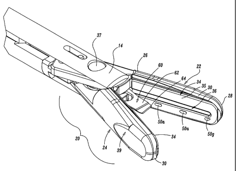
Fig. 1 is a perspective view of an endoscopic forceps showing a handle
and an end effector according to the present disclosure;
CA 02442598 2003-09-26
WO 02/080796 PCT/US01/11413
Fig. 2 is a partial cross-section of the forceps of Fig. 1 showing the
internal working components of the handle and showing the end effector in a
closed
configuration;
Fig. 3 is an enlarged, perspective view of the end effector assembly
shown in open configuration;
Fig. 4 is a greatly enlarged, side view of a proximal end of the end
effector of Fig. 3;
Fig. 5 is a greatly enlarged perspective view of a distal end of the end
effector of Fig. 3 showing a knife and a series of stop members disposed along
an
inner facing surface of a jaw member;
Figs. 6A-6F show various configurations for the stop members on the
inner facing surface of one of the jaw members;
Fig. 7 is an enlarged perspective view of a sealing site of a tubular
vessel;
Fig. 8 is a longitudinal cross-section of the sealing site taken along line
8-8 of Fig. 7; and
Fig. 9 is a longitudinal cross-section of the sealing site of Fig. 7 after
separation of the tubular vessel;
11
CA 02442598 2003-09-26
WO 02/080796 PCT/US01/11413
DETAILED DESCRIPTION
Referring now to Figs. 1-5, one embodiment of an endoscopic bipolar
forceps 10 is shown for use with various surgical procedures and includes a
housing
and handle assembly 80 having an end effector assembly 20 attached thereto.
More particularly, forceps 10 includes a shaft 12 which has a distal end 14
dimensioned to mechanically engage with the end effector assembly 20 and a
proximal end 16 which mechanically engages the housing and handle assembly 80.
In the drawings and in the descriptions which follow, the term "proximal", as
is
traditional, will refer to the end of the forceps 10 which is closer to the
user, while the
term "distal" will refer to the end which is further from the user.
The end effector assembly 20 is attached to the distal end 14 of shaft
12 and includes a pair of opposing jaw members 22 and 24. Preferably, housing
and handle assembly 80 is attached to the proximal end 16 of shaft 12 and
includes
internally-disposed activating mechanisms, e.g., a movable handle 82 and a
drive
assembly 70, which mechanically cooperate to impart movement of the jaw
members 22 and 24 from an open position wherein the jaw members 22 and 24 are
disposed in spaced relation relative to one another, to a clamping or closed
position
wherein the jaw members 22 and 24 cooperate to grasp tissue 150 (Fig. 7)
therebetween.
It is envisioned that the forceps 10 may be designed such that it is fully
or partially disposable depending upon a particular purpose or to achieve a
particular
result. For example, end effector assembly 20 may be selectively and
releasably
12
CA 02442598 2010-05-03
engageable with the distal end 14 of the shaft 12 and/or the proximal end 16
of the
shaft 12 may be selectively and releasably engageable with the housing and
handle
assembly 80. In either of these two instances, the forceps 10 would be
considered
"partially disposable", i.e., a new or different end effector assembly 20 (or
end
effector assembly 20 and shaft 12) selectively replaces the old end effector
assembly 20 as needed .
Figs. I and 2 show the operating elements and the internal-working
components of the housing and handle assembly 80 which for the purposes of the
present disclosure are generally described herein. The specific
functions and operative relationships of these elements and the
various internal working components are described in more detail
in commonly assigned U.S. Patent No. 7,101,372 entitled "VESSEL
SEALER AND DIVIDER".
As best shown in Fig. 2, housing and handle assembly 80 includes
movable handle 82 and a fixed handle 84. The movable handle 82 includes an
aperture 89 defined therethrough which enables a user to grasp and move the
handle 82 relative to the fixed handle 84. Movable handle 82 is selectively
moveable about a pivot 87 from a first position relative to fixed handle 84 to
a
second position in closer proximity to the fixed handle 84 which, as explained
below,
imparts relative movement of the jaw members 22 and 24 relative to one
another.
13
CA 02442598 2010-05-03
More particularly, housing and handle assembly 80 houses a drive
assembly 70 which cooperates with the movable handle 82 to impart movement of
the jaw members 22 and 24 from an open position wherein the jaw members 22 and
24 are disposed in spaced relation relative to one another, to a clamping or
closed
position wherein the jaw members 22 and 24 cooperate to grasp tissue 150 (Fig.
7)
therebetween. The general operating parameters of the drive assembly 70 and
the
internal-working components of the same are explained in a more generalized
fashion below but are explained in specific detail in the above-mentioned
commonly
assigned, 'VESSEL SEALER AND DIVIDER" patent. For the
purposes of the present disclosure, the housing and handle assembly 80 can
generally be characterized as a four-bar mechanical linkage composed of the
following elements: movable handle 82, a link 73, a cam-like link 76 and a
base link
embodied by fixed pivot points 75 and 76. Movement of the handle 82 activates
the
four-bar linkage which, in turn, actuates the drive assembly 70 for imparting
movement of the opposing jaw members 22 and 24 relative to one another to
grasp
tissue 150 therebetween. It is envisioned that employing a four-bar mechanical
linkage will enable the user to gain a significant mechanical advantage when
compressing the jaw members 22 and 24 against the tissue 150 as explained in
further detail below with respect the generally disclosed operating parameters
of the
drive assembly 70.
14
CA 02442598 2010-05-03
Preferably, fixed handle 84 includes a channel 85 defined therein
which is dimensioned to receive a flange 83 which extends proximally from
movable
handle 82. Preferably, flange 83 includes a fixed end 90 which is affixed to
movable
handle 82 and a free end 92 which is dimensioned for facile reception within
channel
85 of handle 84. It is envisioned that flange 83 may be dimensioned to allow a
user
to selectively, progressively and incrementally move jaw members 22 and 24
relative
to one another from the open to closed positions. For example, it is also
contemplated that flange 83 may include a ratchet-like interface which
Iockingly
engages the movable handle 82 and, therefore, jaw members 22 and 24 at
selective, incremental positions relative to one another depending upon a
particular
purpose. Other mechanisms may also be employed to control and/or limit the
movement of handle 82 relative to handle -84 (and jaw members 22 and 24) such
as, e.g., hydraulic, semi-hydraulic and/or gearing systems.
As can be appreciated by the present disclosure and as explained in
more detail with respect to the above-mentioned commonly assigned
"VESSEL SEALER AND DIVIDER" patent, channel 85 of fixed handle 84
includes an entrance pathway 91 and an exit pathway 95 for reciprocation of
flange
83. As best shown in Fig. 2, as handle 82 moves in a generally pivoting
fashion
towards fixed handle 84 about pivot 87, link 73 rotates about a guide pin 74
disposed within handle 82. As a result, link 73 rotates proximally about a
pivot 76.
As can be appreciated, the pivoting path of handle 82 relative to fixed handle
84
biases cam-like link 76 to rotate about pivot 75 in a generally proximal
direction.
CA 02442598 2003-09-26
WO 02/080796 PCT/US01/11413
Movement of the cam-like link 76 imparts movement to the drive assembly 70 as
explained below.
As best shown in Fig. 2, upon initial movement of handle 82 towards
fixed handle 84, the free end 92 of flange 83 moves generally proximally and
upwardly along entrance pathway 91 until end 92 passes or mechanically engages
a
rail member 97 disposed along pathway 91. It is envisioned that rail 97
permits
movement of flange 83 proximally until the point where end 92 clears rail 97.
Once
end 92 clears rail 97, distal movement of the handle 82 and flange 83, i.e.,
release,
is redirected by rail 97 into the exit pathway 95.
More particularly, upon initial release, i.e., a reduction in the closing
pressure of handle 82 against handle 84, the handle 82 returns slightly
distally
towards pathway 91 but is directed towards exit pathway 95. At this point, the
release or return pressure between the handles 82 and 84 which is attributable
and
directly proportional to the release pressure associated with the compression
of the
drive assembly 70 (explained below) causes the end 92 of flange 83 to settle
or lock
within a catch basin 93. Handle 82 is now secured in position within handle 84
which, in turn, locks the jaw members 22 and 24 in a closed position against
the
tissue. The instrument is now positioned for selective application of
electrosurgical
energy to form the tissue seal 152. Again, the various operating elements and
their relevant functions are explained in more detail with respect to the
above-
16
CA 02442598 2010-05-03
mentioned commonly assigned "VESSEL SEALER AND DIVIDER" patent.
As best shown in Fig. 2, re-initiation or re-grasping of the handle 82
again moves flange 83 generally proximally along the newly re-directed exit
path 95
until end 92 clears a lip 94 disposed along exit pathway 95. Once lip 94 is
sufficiently cleared, handle 82 and flange. 83 are fully and freely releasable
from
handle 84 along exit pathway 95 upon the reduction of grasping pressure which,
in
turn, returns the jaw members 22 and 24 to the open, pre-activated position.
As mentioned above, the housing and handle assembly 80 houses a
drive assembly 70 which cooperates with the movable handle 82 to impart
relative
movement of the jaw members 22 and 24 to grasp the tissue 150. The operation
of
the drive rod assembly 70 and the various working components of the drive
assembly 70 are explained in detail in the above-mentioned commonly assigned
"VESSEL SEALER AND DIVIDER" patent.
Generally and for the purposes of the present disclosure, the drive
assembly 70 includes a compression spring 72, a drive rod 40 and a compression
sleeve 98 (Fig. 2). As best shown in the enlarged view of Fig. 4, the drive
rod 40 is
telescopically and internally reciprocable within a knife sleeve 48. Movement
of the
drive rod 40 relative to the knife sleeve 48 imparts movement to the jaw
members 22
and 24. A tab member 46 is disposed at a free end 42 of the drive rod 40 which
17
CA 02442598 2010-05-03
defines a notch 43 between the tab 46 and end 42. The tab 46 and the notch 43
mechanically cooperate with the compression spring 72 to impart movement of
the
shaft 40 relative to the knife sleeve 48 which, in turn, opens and closes the
jaw
members 22 and 24 about the tissue 150.
As explained above, movement of the handle assembly 80 via the four-
bar linkage, ultimately causes cam-like link 76 to rotate generally clockwise
about
pivot 75 (i.e. proximally) which, in turn, compresses spring 72 proximally
against a
flange 77 disposed within the upper portion of the fixed handle 84. Movement
of
the spring 72, in turn, moves the drive rod 40 relative to the knife sleeve 48
which
moves the opposing jaw members 22 and 24 relative to one another. As can be
appreciated, the significant mechanical advantage associated with the four-bar
linkage permits facile, consistent and uniform compression of the spring 72
which, in
turn, permits facile, consistent and uniform compression of the jaw members 22
and
24 about the tissue 150. Other details and advantages of the four-bar
mechanical
linkage are more fully discussed with respect to the above-mentioned commonly
assigned "VESSEL SEALER AND DIVIDER" patent,
Once the tissue 150 is grasped between opposing jaw members 22
and 24, electrosurgical energy can be supplied to the jaw members 22 and 24
through an electrosurgical interface 110 disposed within the handle 84 . Again
these
features are explained in more detail with respect to the above-mentioned
commonly
assigned "VESSEL SEALER AND DIVIDER" patent.
18
CA 02442598 2010-05-03
Forceps 10 also includes a trigger 86 which reciprocates the knife
sleeve 48 which, in turn, reciprocates a knife 60 disposed within the end
effector
assembly 20 as explained below (Fig. 5). Once the a tissue seal 152 is formed
(Fig. 7), the user can activate the trigger 86 to separate the tissue 150 as
shown in
Fig. 9 along the tissue seal 152. As can be appreciated, the reciprocating
knife 60
allows the user to quickly separate the tissue 150 immediately after sealing
without
substituting a cutting instrument through the cannula or trocar port (not
shown). It is
envisioned that the knife 60 also facilitates a more accurate separation of
the vessel
150 along an ideal cutting plane "B-B" associated with the newly formed tissue
seal
152 (See Figs. 7-9). Knife 60 preferably includes a sharpened edge 62 for
severing
the tissue 150 held between the jaw members 22 and 24 at the tissue sealing
site
152 (Fig. 7). It is envisioned that knife 60 may also be coupled to the
electrosurgical
energy source to facilitate separation of the tissue 150 along the tissue seal
152.
Preferably and as explained in more detail with respect to the above-
mentioned commonly assigned "VESSEL SEALER AND DIVIDER"
patent; handle assembly 80 may also include a lockout mechanism (not shown)
which restricts activation of trigger 86 until the jaw members 22 and 24 are
closed
and/or. substantially closed about tissue 150. For example and as best seen in
Fig.
2, exit pathway 95 may be dimensioned such that the trigger 86 is only
activatable
when flange 83 is disposed in a predetermined or predefined position which
provides
sufficient clearance for the activation of the trigger 86, e.g., seated within
catch basin
19
CA 02442598 2010-05-03
93. It is envisioned that configuring the handle assembly 80 in this fashion
may
reduce the chances of premature activation of the trigger 86 prior to
electrosurgical
activation and sealing.
A rotating assembly 88 may also be incorporated with forceps 10.
Preferably, rotating assembly 88 is mechanically associated with the shaft 12
and
the drive assembly 70. As seen best in Fig. 4, the shaft 12 includes an
aperture 44
located therein which mechanically interfaces a corresponding detent (not
shown)
affixed to rotating assembly 88 such that rotational movement of the rotating
assembly 88 imparts similar rotational movement to the shaft 12 which, in
turn,
rotates the end effector assembly 20 about a longitudinal axis "A". These
features
along with the unique electrical configuration for the transference of
electrosurgical
energy through the handle assembly 80, the rotating assembly 88 and the drive
assembly 70 are described in more detail in the above-mentioned commonly
assigned "VESSEL SEALER AND DIVIDER" patent.
As best seen with respect to Figs. 3, 5 and 6A-6F, end effector
assembly 20 attaches to the distal end 14 of shaft 12. The end effector
assembly 20
includes the first jaw member 22, the second jaw member 24 and the
reciprocating
knife 60 disposed therebetween. The jaw members 22 and 24 are preferably
pivotable about a pivot 37 from the open to closed positions upon relative
reciprocation, i.e., longitudinal movement, of the drive rod 42 as mentioned
above.
Again, the mechanical and cooperative relationships with respect to the
various
CA 02442598 2010-05-03
moving elements of the end effector assembly 20 are further described with
respect
to the above-mentioned commonly assigned "VESSEL SEALER AND
DIVIDER" patent.
Each of the jaw members includes an electrically conductive sealing
surface 35 dispose on inner-facing surface 34 thereof and an insulator 30
disposed
on an outer-facing surface 39 thereof. It is envisioned that the electrically
conductive surfaces 35 cooperate to seal tissue 150 held therebetween upon the
application of electrosurgical energy. The insulators 30 together with the
outer, non-
conductive surfaces 39 of the jaw members 22 and 24 are preferably dimensioned
to
limit and/or reduce many of the known undesirable effects related to tissue
sealing,
e.g., flashover, thermal spread and stray current dissipation.
It is envisioned that the electrically conductive sealing surfaces 35 may
also include a pinch trim which facilitates secure engagement of the
electrically
conductive surface 35 to the insulator 30 and also simplifies the overall
manufacturing process. It is envisioned that the electrically conductive
sealing
surface 35 may also include an outer peripheral edge which has a radius and
the
insulator 30 meets the electrically conductive sealing surface 35 along an
adjoining
edge which is generally tangential to the radius and/or meets along the
radius.
Preferably, at the interface, the electrically conductive surface 35 is raised
relative to
the insulator 30. These and other envisioned embodiments are discussed in
commonly assigned PCT Publication No. WO 2002/080786
21
CA 02442598 2010-05-03
published October 17, 2002 and entitled "ELECTROSURGICAL
INSTRUMENT WHICH REDUCES COLLATERAL DAMAGE TO
ADJACENT TISSUE" by Johnson et al and commonly assigned
U.S. Patent No. 7,135,020 entitled "ELECTROSURGICAL
INSTRUMENT WHICH IS DESIGNED TO REDUCE THE
INCIDENCE OF FLASHOVER" by Johnson et al.
Preferably, a least one of the electrically conductive surfaces 35 of the
jaw members, e.g., 22, includes a longitudinally-oriented channel 36 defined
therein
which extends from a proximal end 26 to a distal end 28 of the jaw member 22.
It is
envisioned that the channel 36 facilitates longitudinal reciprocation of the
knife 60
along a preferred cutting plane "B-B" to effectively and accurately separate
the
tissue 150 along the formed tissue seal 152 (See Figs. 7-9). Preferably and as
explained in detail in the above-mentioned commonly assigned, co-pending
"VESSEL SEALER AND DIVIDER" application, the jaw members 22 and 24 of the
end effector assembly 22 are electrically isolated from one another such that
electrosurgical energy can be effectively transferred through the tissue 150
to form
seal 152.
As mentioned above, upon movement of the handle 82, the jaw
members 22 and 24 close together and grasp tissue 150. At this point flange 83
becomes seated within catch 93 which, together with the mechanical advantage
22
CA 02442598 2003-09-26
WO 02/080796 PCT/US01/11413
associated with the four-bar mechanism and the spring 70, maintains a
proportional
axial force on the drive rod 40 which, in turn, maintains a compressive force
between
opposing jaw members 22 and 24 against the tissue 150. It is envisioned that
the
end effector assembly 20 may be dimensioned to off-load excessive clamping
forces
to prevent mechanical failure of certain internal operating elements of the
end
effector.
By controlling the intensity, frequency and duration of the
electrosurgical energy applied to the tissue 150, the user can either
cauterize,
coagulate/desiccate seal and/or simply reduce or slow bleeding. As mentioned
above, two mechanical factors play an important role in determining the
resulting
thickness of the sealed tissue and effectiveness of the seal, i.e., the
pressure
applied between opposing jaw members 22 and 24 and the gap distance between
the opposing sealing surfaces 35 of the jaw members 22 and 24 during the
sealing
process. However, thickness of the resulting tissue seal 152 cannot be
adequately
controlled by force alone. In other words, too much force and the two jaw
members
22 and 24 would touch and possibly short resulting in little energy traveling
through
the tissue 150 thus resulting in a bad tissue seal 152 . Too little force and
the seal
152 would be too thick.
Applying the correct force is also important for other reasons: to
oppose the walls of the vessel; to reduce the tissue impedance to a low enough
value that allows enough current through the tissue 150; and to overcome the
forces
23
CA 02442598 2003-09-26
WO 02/080796 PCT/US01/11413
of expansion during tissue heating in addition to contributing towards
creating the
required end tissue thickness which is an indication of a good seal.
Preferably, the electrically conductive sealing surfaces 35 of the jaw
members 22 and 24 are relatively flat to avoid current concentrations at sharp
edges
and to avoid arcing between high points. In addition and due to the reaction
force of
the tissue 150 when engaged, jaw members 22 and 24 are preferably manufactured
to resist bending. For example and as best seen in Fig. 6A, the jaw members 22
and 24 are preferably tapered along width "W" which is advantageous for two
reasons: 1) the taper will apply constant pressure for a constant tissue
thickness at
parallel; 2) the thicker proximal portion of the jaw members 22 and 24 will
resist
bending due to the reaction force of the tissue 150.
As best seen in Figs. 5-6F, in order to achieve a desired spacing
between the electrically conductive surfaces 35 of the respective jaw members
22
and 24, (i.e., gap distance) and apply a desired force to seal the tissue 150,
at least
one jaw member 22 and/or 24 includes at least one stop member, e.g., 50a,
which
limits the movement of the two opposing jaw members 22 and 24 relative to one
another. Preferably, the stop member, e.g., 50a, extends from the sealing
surface
or tissue contacting surface 35 a predetermined distance according to the
specific
material properties (e.g., compressive strength, thermal expansion, etc.) to
yield a
consistent and accurate gap distance during sealing. Preferably, the gap
distance
between opposing sealing surfaces 35 during sealing ranges from about 0.001
24
CA 02442598 2003-09-26
WO 02/080796 PCT/US01/11413
inches to about 0.005 inches and, more preferably, between about 0.002 and
about
0.003 inches.
Preferably, stop members 50a-50g are made from an insulative
material, e.g., parylene, nylon and/or ceramic and are dimensioned to limit
opposing
movement of the jaw members 22 and 24 to within the above mentioned gap range.
It is envisioned that the stop members 50a-50g may be disposed one or both of
the
jaw members 22 and 24 depending upon a particular purpose or to achieve a
particular result.
Figs. 6A-6F show various contemplated configurations of the non-
conductive stop members 50a-50g disposed on, along or protruding through the
jaw
member 24. It is envisioned that one or more stop members, e.g., 50a and 50g,
can
be positioned on either or both jaw members 22 and 24 depending upon a
particular
purpose or to achieve a desired result. As can be appreciated by the present
disclosure, the various configurations of the stop members 50a-50g are
designed to
both limit the movement of the tissue 150 prior to and during activation and
prevent
short circuiting of the jaw members 22 and 24 as the tissue 150 is being
compressed.
Figs. 6A and 6B show one possible configuration of the stop members
50a-50g for controlling the gap distance between opposing seal surfaces 35.
More
particularly, a pair of longitudinally-oriented tab-like stop members 50a are
disposed
CA 02442598 2003-09-26
WO 02/080796 PCT/US01/11413
proximate the center of sealing surface 35 on one side of the knife channel 36
of jaw
member 24. A second stop member, e.g., 50b, is disposed at the proximal end 26
of jaw member 24 and a third stop member 50g is disposed at the distal tip 28
of jaw
member 24. Preferably, the stop members 50a-50g may be configured in any
known geometric or polynomial configuration, e.g., triangular, rectilinear,
circular,
ovoid, scalloped, etc., depending upon a particular purpose. Moreover, it is
contemplated that any combination of different stop members 50a-50g may be
assembled along the sealing surfaces 35 to achieve a desired gap distance. It
is
also envisioned that the stop members may be designed as a raised lip (not
shown)
which projects from the outer periphery of the jaw member 24.
Fig. 6C shows a first series of circle-like stop members 50c extending
from the proximal end 26 to the distal end 28 of jaw member 24 in an
alternating,
laterally-offset manner relative to one another on one side of the knife
channel 36
and a second series of circle-like stop members 50c extending from the
proximal
end 26 to the distal end 28 of jaw member 24 in an alternating, laterally-
offset
manner relative to one another on the other side of the knife channel 36. It
is
envisioned that circle-like stop members 50c are substantially equal in size,
however, one or more of the stop members 50c may be dimensioned larger or
smaller than the other stop members 50c depending upon a particular purpose or
to
achieve a desired result.
26
CA 02442598 2003-09-26
WO 02/080796 PCT/US01/11413
Fig. 6D shows yet another configuration wherein the stop member is
configured as a longitudinally-oriented ridge 50e extending from a proximal
end 26
to a distal end 28 of jaw member 82 along one side of knife channel 36. As
mentioned above, a second longitudinally-oriented ridge 50e may be disposed on
opposing jaw member 22 on the opposite side of knife channel 36 for sealing
purposes. Fig. 6E shows a series of elongated tab-like members 50f which are
disposed at an angle relative to knife channel 36. Fig. 6F shows yet another
configuration wherein different stop members, e.g., 50a, 50c and 50g are
disposed
atop sealing surface 35 on both sides of the knife channel 36.
Preferably, the non-conductive stop members 50a-50g are molded
onto the jaw members 22 and 24 (e.g., overmolding, injection molding, etc.),
stamped onto the jaw members 22 and 24 or deposited (e.g., deposition) onto
the
jaw members 22 and 24. The stop members 50a-50g may also be slideably
attached to the jaw members and/or attached to the electrically conductive
surfaces
35 in a snap-fit manner. Other techniques involves thermally spraying a
ceramic
material onto the surface of the jaw member 22 and 24 to form the stop members
50a-50g. Several thermal spraying techniques are contemplated which involve
depositing a broad range of heat resistant and insulative materials on the
electrically
conductive surfaces 35 to create stop members 50a-50g, e.g., High velocity Oxy-
fuel
deposition, plasma deposition, etc.
27
CA 02442598 2003-09-26
WO 02/080796 PCT/US01/11413
It is envisioned that the stop members 50a-50g protrude about 0.001 to
about 0.005 inches from the inner-facing surfaces 35 of the jaw members 22 and
24
which, as can be appreciated by the present disclosure, both reduces the
possibility
of short circuiting between electrically conductive surfaces and enhances the
gripping characteristics of the jaw members 22 and 24 during sealing and
dividing.
Preferably, the stop members 50a-50g protrude about 0.002 inches to about
0.003
inches from the electrically conductive surface 35 which has been determined
yield
an ideal gap distance for producing effective, uniform and consistent tissue
seals.
Alternatively, the stop members 50a-50g can be molded onto the
inner-facing surface 35 of one or both jaw members 22 and 24 or, in some
cases, it
may be preferable to adhere the stop member 50a-50g to the inner facing
surfaces
35 of one or both of the jaw members 22 and 24 by any known method of
adhesion.
Stamping is defined herein to encompass virtually any press operation known in
the
trade, including but not limited to: blanking, shearing, hot or cold forming,
drawing,
bending, and coining.
Figs. 6A-6F show some of the possible configurations of the stop
members 50a-50f, however, these configurations are shown by way of example and
should not be construed as limiting. Other stop member configurations are also
contemplated which may be may be equally effective in reducing the possibility
of
short circuiting between electrically conductive surfaces 35 and enhancing
tissue
grip during sealing and dividing.
28
CA 02442598 2003-09-26
WO 02/080796 PCT/US01/11413
Further, although it is preferable that the stop members 50a-50g
protrude about 0.001 inches to about 0.005 and preferably about 0.002 inches
to
about 0.003 inches from the inner-facing surfaces 35 of the jaw member 22 and
24,
in some cases it may be preferable to have the stop members 50a-50g protrude
more or less depending upon a particular purpose. For example, it is
contemplated
that the type of material used for the stop members 50a-50g and that
material's
ability to absorb the large compressive closure forces between jaw members 22
and
24 will vary and, therefore, the overall dimensions of the stop members 50a-
50g may
vary as well to produce the desired gap distance.
In other words, the compressive strength of the material along with the
desired or ultimate gap distance required for effective sealing are parameters
which
are carefully considered when forming the stop members 50a-50g and one
material
may have to be dimensioned differently from another material to achieve the
same
gap distance or desired result. For example, the compressive strength of nylon
is
different from ceramic and, therefore, the nylon material may have to be
dimensioned differently, e.g., thicker, to counteract the closing force of the
opposing
jaw members 22 and 24 and to achieve the same desired gap distance when
utilizing a ceramic stop member.
29
CA 02442598 2003-09-26
WO 02/080796 PCT/US01/11413
The present disclosure also relates to a method of sealing and dividing
tissue and includes the steps of: providing an endoscopic bipolar forceps 10
which
includes:
an elongated shaft 12 having opposing jaw members 22 and 24
at a distal end 14 thereof which cooperate to grasp tissue 150 therebetween;
at least one non-conductive and spaced-apart stop member
50a-50g disposed on an inner facing surface 35 of at least one of the jaw
members,
e.g., 24, which controls the distance between the jaw members 22 and 24 when
tissue 150 is held therebetween; and
a knife 60.
The method further includes the steps of: connecting the jaw members
22 and 24 to a source 110 of electrical energy; actuating the jaw members 22
and
24 to grasp tissue 150 between opposing jaw members 22 and 24; conducting
energy to the jaw members 22 and 24 to through tissue 150 held therebetween to
effect a seal 152 (Figs. 7-9); and actuating the knife 60 to sever tissue
proximate the
seal 152.
Preferably, at least one of the jaw members, e.g., 24, of the providing
step includes an electrically conductive surface 35 having a longitudinally-
oriented
channel 36 defined therein which facilitates actuation of the knife 60 in a
longitudinally reciprocating fashion within the channel 36 for severing the
tissue 150
proximate the tissue site.
CA 02442598 2003-09-26
WO 02/080796 PCT/US01/11413
From the foregoing and with reference to the various figure drawings,
those skilled in the art will appreciate that certain modifications can also
be made to
the present disclosure without departing from the scope of the present
disclosure.
For example, it may be preferable to add other features to the forceps 10,
e.g., an
articulating assembly to axially displace the end effector assembly 20
relative to the
elongated shaft 12.
Moreover, it is contemplated that the presently disclosed forceps may
include a disposable end effector assembly which is selectively engageable
with at
least one portion of the electrosurgical instrument, e.g., shaft 12 and/or
handle
assembly 80.
While several embodiments of the disclosure have been shown in the
drawings, it is not intended that the disclosure be limited thereto, as it is
intended
that the disclosure be as broad in scope as the art will allow and that the
specification be read likewise. Therefore, the above description should not be
construed as limiting, but merely as exemplications of a preferred
embodiments.
Those skilled in the art will envision other modifications within the scope
and spirit of
the claims appended hereto.
31