Note: Descriptions are shown in the official language in which they were submitted.
CA 02442675 2003-09-30
WO 02/082989 PCT/US02/06831
METHOD FOR OPTICAL MEASUREMENTS OF TISSUE TO DETERMINE
DISEASE STATE OR CONCENTRATION OF AN ANALYTE
BACKGROUND OF THE INVENTION
1. Field of the Invention
This invention relates to an apparatus and method for the non-invasive
diagnosis
to of a disease state or the non-invasive determination of concentrations of
analytes in vivo.
2. Discussion of the Art
Diabetes mellitus is a chronic systemic disease in which the body either fails
to
is produce or fails to respond to the hormone insulin, which regulates the
metabolism of
glucose. It is estimated that there are 16 million diabetics in the United
States and 100
million diabetics worldwide. The growth rate in the number of diabetics is
estimated at
11.5% annually. The number of diabetics is estimated to be as high as 154
million
worldwide by the year 2000 (H. King, R. E. ~Aubert, and W. H. Herman, "Global
burden of
2o diabetes, 1995 - 2025 prevalence, numerical estimates and projections"
Diabetes Care
1998;21:1414), and to exceed 200 million worldwide by the year 2010. A large
number
of diabetics remain undiagnosed. A method for screening for diabetes would be
beneficial for early diagnosis and for starting treatment and management well
before the
onset of complications.
2s Diabetes is frequently associated with microangiopathy. Microangiopathy
results
from the effect of diabetes on microcirculation, which involves the small
blood vessels
such as capillaries, venules, arterioles, and shunts. Microangiopathy can lead
to micro-
vessel complications such as neuropathy (nerve damage), retinopathy (eye
damage),
and nephropathy (kidney failure). The expression "diabetic angiopathy" deals
with effect
30 of diabetes on the arterial as well as the other elements of the vascular
system such as
venules, veins, and lymph subsystem. The relationship between diabetes and
impaired
circulation has been known in the medical art for the past two decades. Laser
Doppler
flowmetry has been used to diagnose peripheral vascular disease and vascular
complications in diabetic patients. Impaired circulation is manifested by a
decrease in
1
CA 02442675 2003-09-30
WO 02/082989 PCT/US02/06831
cutaneous blood flow and a decrease in response to temperature changes, i.e.,
cooling
or warming of the skin.
There is growing evidence that microcirculatory defects can be detected well
before detection of fasting hyperglycemia, i.e. high blood glucose level for a
fasting
s subject (N. Wiernsperger, Diabetologia 2000; 43; 1439-1449). Laser Doppler
flowmetry
and capillary microscopy studies have indicated microcirculation disturbances
due to
diabetes and have shown differences in cutaneous blood flow between diabetics
and
non-diabetics (S. B. Wilson, "Detection of microvascular impairment in type 1
diabetes by
laser Doppler flowmetry, Clinical Physiology, 1992; 12; 195). In diabetic
subjects,
io heating of a body part, or contralateral cooling of a body part, resulted
in impaired blood
flow, as measured by laser Doppler flowmetry (M. Rendell et al, "Microvascular
blood
flow, volume and velocity measurements by laser Doppler techniques in IDDM"
Diabetes; 1989: 819-824). However, these studies of capillary blood flow and
laser
Doppler flowmetry were reported for advanced stages of diabetes (M Rendell et
al,
is "Diabetic cutaneous microangiopathy" American Journal of Medicine 1992; 93:
611 ).
Additionally, X-ray crystallographic studies showed differences in structure
of tissues of
diabetic subjects, due to cross-linking of collagen fibers resulting from
glycation (V. J.
James et al., "Use of X-ray Diffraction in Study of Human Diabetic and Aging
Collagen",
Diabetes, Vol. 40 (1991) 391-394).
Diabetes and certain other diseases cause structural changes to the skin that
can
affect the optical properties thereof, the response of these optical
properties to changes
in concentration of glucose or other analytes, and the response of these
optical
properties to cutaneous temperature changes. R. G. Sibbald et al., "Skin and
Diabetes",
Endocrinology and Metabolism Clinics of North America, Vol. 25, No. 2 (1996)
463-472,
2s summarize a set of structural effects of the skin that are associated with
diabetes.
Included among these effects is thickened skin, which may relate
pathophysiologically to
accelerated collagen aging, with elastic fiber fraying and increased
crosslinking, resulting
from glycosylation of collagen fibers. Another effect of diabetes is "yellow
skin", which
also results from glycosylation of dermal collagen. Change in dermal collagen
structure
3o in diabetic patients has been also reported by V. M. Monnier et al., "Skin
Collagen
Glycation, Glycoxidation, and Crosslinking Are Lower in Subjects With Long-
Term
Intensive Versus Conventional Therapy of Type 1 Diabetes", Diabetes, Vol. 48
(1999)
870-880. Further, V. J. James et al., "Use of X-ray Diffraction in Study of
Human
Diabetic and Aging Collagen", Diabetes, Vol. 40 (1991 ) 391-394, shows that
collagen
3s skin fiber undergoes a structural change as a result of diabetes. The net
effect of these
2
CA 02442675 2003-09-30
WO 02/082989 PCT/US02/06831
findings is that there are structural differences, i.e., size, level of
crosslinking, and
distribution of collagen fibers, in the skin of diabetic subjects as compared
with the skin
of non-diabetic subjects. These differences result in a difference in the
scattering
characteristics of the skin of diabetic subjects.
In order to understand the effect of the structural differences between the
skin of
diabetics and that of non-diabetics on the measured optical signals, it is
useful to
examine the scattering of light in human tissue.
The scattering of light by human tissue can be approximated by an equation
that
expresses the reduced scattering coefficient p'S for a tissue or a turbid
medium as:
to
~,'S = 3.28~a2P(27tar1,7,eaium~~) o.s~ (m_~ X2.09
where "a" represents the average cell diameter, p represents the number
concentration
of CeIIS, "nmedium" represents the refractive index of interstitial fluid, ~,
represents the
is wavelength, and m represents the ratio of the refractive index of the cells
to that of the
interstitial fluid (m = n~ells~rlmedium). The scattering coefficient changes
as cell size "a" or
refractive index "nmedium' change. Temperature can affect the scattering
coefficient by a
change in cell diameter "a", a change in the number concentration of cells p,
or a change
in the refractive index mismatch "m". Because the diabetic status is
independent of
2o glucose concentration, i.e., a diabetic patient can have high or low blood
glucose level, it
is possible to assume that the diabetic status is independent of "m". However,
differences in crosslinking of collagen for diabetics may lead to a different
range for the
dimensional parameter "a" between the diabetic and the non-diabetic groups.
Differences in the variable "a" will lead to a difference in the scattering
characteristics of
2s the skin of diabetic subjects, because the scattering characteristics
affect the term "a" in
Equation (1 ). Thus, the response of the scattering coefficient to changes in
glucose
concentration, or other concentrations of analytes, and the response of the
scattering
coefficient to cutaneous temperature changes are expected to be different for
diabetic
subjects as compared to those responses of the same parameters determined for
non-
3o diabetic subjects.
Scattering properties of tissue can vary with temperature as a result of one
or
more of the following changes:
(a) an increase in temperature can decrease the refractive index of
interstitial
fluid and increase the scattering coefficient of tissue;
3
CA 02442675 2003-09-30
WO 02/082989 PCT/US02/06831
(b) an increase in temperature can change the refractive index of cell
membranes;
(c) an increase in temperature can increase cell size, and hence, can increase
the scattering coefficient.
In the case of (a) or (b), an increase in the refractive index mismatch "m" in
Equation (1 ),
which increases as the temperature increases, can also increase the scattering
coefficient.
Methods of diagnosing diabetes typically require a large number of laboratory
tests, such as, for example, successive blood glucose level measurements while
the
to patient is in a fasting state, determination of serum glycated hemoglobin
HbA1 c, and oral
glucose tolerance (or meal tolerance) tests. These tests are usually performed
after
clinical symptoms of diabetes are observed. These symptoms include thirst,
fatigue, and
frequent urination (Report of the Expert Committee on the Diagnosis and
Classification
of Diabetes Mellitus, Diabetes Care 1997; 20:117-135). The use of a glycated
is hemoglobin test has been equivocal in diagnosing diabetes, even though it
is time-
consuming and requires drawing of blood. See C. L. Rohlfing et al., "Use of
HbA1 c in
screening for undiagnosed diabetes in US population", Diabetes Care 2000; 23:
187-
191.
A non-invasive test for screening diabetics will save a great number of
laboratory
2o tests and will allow screening larger populations, even if clinical
symptoms of diabetes
are not evident. A non-invasive test will also allow early diagnosis and
subsequent
control of diabetes, which in turn will delay the onset of complications from
diabetes. If
uncontrolled, diabetes can result in a variety of adverse clinical
manifestations, including
retinopathy, atherosclerosis, microangiopathy, nephropathy, and neuropathy. In
its '
2s advanced stages, diabetes can cause blindness, coma, and ultimately death.
Non-invasive determination of glucose has been the subject of several patents.
U. S. Patent Nos. 5, 082,787; 5,068,536; 5,077,476; 5,086,229; 5,204,532;
5,237,178;
5,362,966 describe transmission measurements through the finger. U. S. Patent
Nos.
5,321,265; and, US 5,434,412 describe I<romoscopic methods for the
determination of
3o glucose. U. S. Patent Nos. 5,492,118 and 5,551,422 describe measurements
based on
light scattering. United States Patent Nos. 4,655,225; 4,882,492; 5,460,177;
4,975,581
describe methods for the detection of glucose with light of long wavelength (
> 1100 nm)
where glucose does, presumably, have stronger absorption bands. United States
Patent
Nos. 5,009,230; 4,975,581; 5,379,764; 4,655,225; 5,893,364; 5,497,769;
5,209,231; and
4
CA 02442675 2003-09-30
WO 02/082989 PCT/US02/06831
5,348,003 describe a variety of optical methods for the non-invasive
determination of
blood glucose level in the human body.
U. S. Patent No. 5,362,966 describes measurement of finger temperature away
from the optical measurement area. WO 95/04924 describes a near infrared non-
s invasive measurement instrument, where light is introduced and measured at
an
extremity, such as a finger tip, while the temperature of the same extremity
is measured
at another location remote from the location of the optical measurement area.
The
temperature value measured is used in the calculation algorithm together with
the optical
data to determine the concentration of an analyte. The temperature at the
measurement
to site is not controlled or varied according to a preset program. U. S.
Patent No.
5,551,422 describes a glucose sensor that is brought to a specified
temperature,
preferably somewhat above the body normal temperature, with a thermostatically
controlled heating system. U. S. Patent No. 5,666,956 describes a method for
the
determination of glucose from the infrared emission of the tympanic membrane.
U. S.
is Patent No. 5,978,691 describes a method of measuring changes in molecular
behavior,
induced by a change in thermal energy, to facilitatrr the measurement of
physiological
parameters in blood.
U. S. Patent No. 5,844,239 describes a fiber-optics-based optical device for
determination of the optical properties at a shallow depth in a tissue. The
sensor
2o comprises several unit fiber bundles. Each unit fiber bundle has a light
introduction fiber and several light collection fibers arranged in concentric
rings.
Signals from each group of fibers at the same distance are detected to enhance
the signal to noise ratio. Further, signals from the plurality of unit bundles
are
added up, or averaged, to further improve the signal to noise ratio. The
2s temperature is not controlled at the positions where the unit bundles
contact the
skin. The temperature is not varied according to a preset program.
U. S. Application Serial No. 09/080,470, filed May 18, 1998, assigned to the
assignee of this application, describes a sensor employing a temperature
control for non-
invasive determination of blood glucose level. U. S. Application Serial No.
09/098,049,
3o filed November 23, 1998, assigned to the assignee of this application,
describes
methods for determining optical properties of tissue having a plurality of
layers non-
invasively. Both applications disclose the use of a temperature controllable
optical
element that contacts the skin.
Cutaneous microcirculation occurs at depths of 1 to 2 mm below the epidermal
3s surface of the skin (I. M. Braverman, "The Cutaneous Microcirculation:
Ultrastructure and
CA 02442675 2003-09-30
WO 02/082989 PCT/US02/06831
Microanatomical Organization", Microcirculation (1997) Vol. 4, No. 3, 329-
340). Thus,
measurement of optical properties of skin close to the surface thereof can
provide useful
information on the effect of blood circulation on the concentration of
metabolites in
tissues that are close to the surface of the skin. Also, studies of blood
circulation close
s to the surface of the skin by means of laser Doppler flowmetry (referred to
as LDF
herein) have shown that laser Doppler flowmetry is a good tool for diagnosing
peripheral
circulatory disease. Laser Doppler flowmetry (LDF) measurements are restricted
to the
top-most layer of the skin (~ 200 microns) because the beam loses its
coherence due to
scattering. Temperature dependence of laser Doppler flowmetry studies does not
io incorporate structural changes in the skin due to diabetes. Thus, a
deficiency in the LDF
prior art is the lack of inclusion of temperature dependence of scattering in
the
classification and diagnosis of diabetes complications.
Although a variety of detection techniques have been disclosed in the art,
there is
still no commercially available device that provides reliable non-invasive
measurements
is of blood glucose level. As a result, current approaches to non-invasive
metabolite
testing, such as glucose monitoring, have not achieved wide acceptance.
SUMMARY OF THE INVENTION
This invention provides a method for collecting optical data at two
morphologically
similar, substantially non-overlapping, and preferably adjacent, areas on the
surface of a
human tissue, while the temperature in each area is being maintained or
modulated
according to a temperature program. The optical data obtained are inserted
into a
2s mathematical relationship, e.g., an algorithm, that can be used to predict
a disease state
(such as the diabetes mellitus disease state) or the concentration of an
analyte for
indicating a physical condition (such as blood glucose level).
This invention can be used to differentiate between disease status, such as,
for
example, diabetic and non-diabetic. The discovery underlying the method of
this
3o invention is that certain optical properties of human tissue change in
response to
changes in temperature of the tissue. The method involves the generation of a
calibration (or training) set that utilizes the relationship between optical
signals
emanating from the skin under different thermal stimuli and disease status,
e.g., diabetic
status, established clinically. This calibration set can be used to predict
the disease
3s state of other subjects. Because thermal stimuli affect microcirculatory
action within the
6
CA 02442675 2003-09-30
WO 02/082989 PCT/US02/06831
capillary loops, the method depends upon measuring the optical properties of
the tissue
at different areas on the surface of the tissue, to a depth of up to two
millimeters, as a
function of thermal stimuli. Structural changes, as well as circulatory
changes, due to a
disease state are determined at two morphologically similar, but substantially
non-
s overlapping areas on the surface of human tissue, e.g., the skin of a
forearm, with each
area being subjected to different temperature modulation programs. In addition
to
determination of a disease state, this invention can also be used to determine
the
concentration of an analyte in a human tissue. This invention also provides an
apparatus for the determination of a disease state, such as diabetes, or
concentration of
to an analyte in a human tissue, such as blood glucose level, by the method of
this
invention.
In one aspect, this invention provides a method for determining a disease
state of
a subject. The method comprises the steps of:
(a) measuring at least one optical property at a first area on a human tissue
to
is obtain a first set of data, the first area being subjected to a first
temperature
program;
(b) measuring at least one optical property at a second area on the human
tissue to obtain a second set of data, the second area being subjected to a
second temperature program, the second temperature program being
2o different from the first temperature program, the second area being
morphologically similar to but not substantially overlapping with the first
area;
(c) inserting the first set of data and the second set of data into a
mathematical relationship to calculate a mathematical output; and
2s (d) comparing the mathematical output to a category selector to determine
the
disease state of the human.
The mathematical relationship is typically established by correlating the
parameter with
the disease state, which is determined by invasive methods. As used herein,
the
3o expression "disease state" means the status of a subject having an abnormal
cardiovascular condition, a neoplasmic condition, or other disease that
affects the
tissues. A representative example of a disease state is diabetes. The thus-
established
mathematical relationship can be used to determine the disease state of a
subject.
In another aspect, this invention provides a method for determining the
ss concentration of an analyte in a tissue of a subject. The method comprises
the steps of:
7
CA 02442675 2003-09-30
WO 02/082989 PCT/US02/06831
(a) measuring at least one optical property at a first area on the tissue to
obtain a
first set of data, the first area being subjected to a first temperature
program;
(b) measuring at least one optical property at a second area on the tissue
to obtain a second set of data, the second area being subjected to a
s second temperature program, the second temperature program being
different from the first temperature program, the second area being
morphologically similar to but not substantially overlapping with the first
area; and
(c) inserting the first set of data and the second set of data into a
mathematical
to relationship to calculate the concentration of the analyte.
The mathematical relationship is typically established by correlating the
parameter with
the concentration of the analyte, which is determined by invasive methods. The
thus-
established mathematical relationship can be used to determine the
concentration of the
is analyte in the tissue of a subject.
In another aspect, this invention provides an apparatus for carrying out the
method of this invention. The apparatus comprises:
(a) means for illuminating at least two areas of tissue with light;
(b) means for collecting light re-emitted from the at least two areas of
tissue;
20 (c) means for measuring the intensity of the re-emitted light collected at
the two
areas of tissue; and
(d) means for controlling the temperature of the at least two areas of the
tissue
simultaneously by means of temperature programs.
2s One embodiment of this invention involves a method for non-invasive
diagnosis of
a disease state, such as, for example, diabetes, or the concentration of an
analyte, such
as, for example, blood glucose level.
The measurements at the first area and at the second area can be made
simultaneously. Alternatively, the measurements at the first area and at the
second area
3o can be made sequentially.
BRIEF DESCRIPTION OF THE DRAWINGS
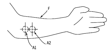
3s FIG. 1 Illustrates measurement areas on a human forearm.
8
CA 02442675 2003-09-30
WO 02/082989 PCT/US02/06831
FIG. 2 is a schematic diagram illustrating a device suitable for use in this
invention. The device employs one optical head and is suitable for sequential
measurements. The portion of the device that contacts a body part is shown as
a cross-
section. .
s FIG. 3 is a schematic diagram illustrating a device suitable for use in this
invention. The device employs two optical heads and is suitable for
simultaneous
measurements. The portion of the device that contacts a body part is shown as
a cross-
section.
FIG. 4 is a schematic diagram illustrating a device suitable for use in this
to invention. The device employs one optical head and is suitable for
sequential
measurements. The portion of the device that contacts a body part is shown as
a cross-
section. The device of FIG. 4 is a variation of the device shown in FIG. 2.
is DETAILED DESCRIPTION OF THE INVENTION
As used herein, the expression °'optical properties" refers to the
absorption,
scattering, emission, reflectance, and depolarization properties of biological
tissues. The
expression "optical parameter" refers to a parameter that describes and
defines an
20 optical property of a medium and its components. Examples of optical
parameters
include, but are not limited to, absorption coefficient, scattering
coefficient, and extinction
coefficient of analytes. The expression "scattering media" refers to media
that both
scatter light and absorb light. The expression "absorption coefficient" (i.e.
~.a) refers to
the probability of light absorption per unit path length, which is equal to
2.303 sC in cm-~,
2s where, E is molar extinction coefficient and C is the molar concentration.
The expression
"reduced scattering coefficient" (i.e. ~,S') refers to the probability of
equivalently isotropic
(uniform in all directions) scattering per unit path length, which is equal to
ap in cm-~,
where, 6 is scattering cross section and p is the number density of scattering
centers.
The expression °'light penetration depth" (i.e. 8) refers to the rate
of decay of intensity of
30 light in scattering media with respect to the path traveled by the light in
the same
direction as the incident light. Light penetration depth represents the depth
at which light
intensity in the tissue is attenuated to 1/e of its original value and is
related to the
absorption and scattering coefficients as 8 = 1/x(3 ~, a(p.a + ~,'S)).
9
CA 02442675 2003-09-30
WO 02/082989 PCT/US02/06831
The expression "diffuse reflectance" (reflectance therein unless specified
otherwise) refers to measurement of light that is re-emitted from a sample at
all angles
different from the direction of the incident light, and over an area wider
than the area
where the incident light is introduced into the sample. The expressions
"spatially
resolved scattering" or "spatially resolved diffuse reflectance" and
"localized reflection"
refer to a measurement of light that is re-emitted from a sample and collected
at several
light collection sites at specific distances from a light introduction site.
Alternatively,
these expressions can refer to the light collected at a given light collection
site on the
sample boundary as a result of introducing light at discrete light
introduction sites located
io on the same boundary at a set of defined distances from the light
collection site. In both
instances, pa and p,'S are calculated from the intensity distribution of the
re-emitted light
with respect to distances, i.e., the re-emitted light intensity at a
multiplicity of sampling
distances. The expressions "re-emitted light" and "reflected light" are used
synonymously herein, as are the expressions "reflectance" and the "intensity
of re-
15 emitted light", unless otherwise indicated.
The expression "light introduction site" means a location on the surface of a
sample, e.g., a body part, tissue, or the like, at which light is injected or
introduced into
the sample, by means of, for example, an optical fiber. The source of the
light can be
located at the light introduction site or can be located remote from the light
introduction
2o site. If the source of light is located remote from the light introduction
site, the light must
be transmitted to the light introduction site by light transmitting means,
such as, for
example, optical fibers. The expression "light collection site" means a
location on the
surface of a sample, e.g., a body part, tissue, or the like, at which light
that is re-emitted
from the sample is collected for measurement. The detector, which determines
the
2s intensity of the re-emitted light, can be located at the light collection
site or can be
located remote from the light collection site. If the detector is located
remote from the
light collection site, the light must be transmitted to the detector by light
transmitting
means, such as, for example, optical fibers. The distance between a light
introduction
site and a light collection site, as measured along the surface of a sample,
is defined as
3o the "sampling distance". For a given sample, the sampling distance
determines the
mean distance from the surface of the sample into the interior of the sample
at which the
scattering and absorption events contribute to the measured re-emitted light.
Such
mean distance is hereinafter referred to as the "sampling depth", which is a
function of
the sampling distance. In this invention an "area on the surface of the
tissue" may have
CA 02442675 2003-09-30
WO 02/082989 PCT/US02/06831
multiple light introduction sites, multiple light collection sites, at least
one sampling
distance and at least one sampling depth and its optical properties are
affected an
independently-run temperature program. The expression "temperature program"
refers
to a sequence of temperature levels as a function of time. Examples of
temperature
programs include, but are not limited to, (1 ) maintaining a constant
temperature over a
period of time; (2) decreasing temperature over a period of time; (3)
increasing
temperature over a period of time; and (4) combinations of (1 ), (2), and (3).
The
expression "not substantially overlapping" means that the areas subjected to
temperature programs can overlap slightly so long as the temperature programs
to which
io the areas are subjected are distinguishable. However, it is preferred that
the areas that
are subjected to the temperature programs not overlap.
The expression "blood flow" means the velocity of red blood cells in blood
vessels.
Blood flow is usually measured by laser Doppler flowmetry. The term
"vasodilatation"
refers to the increase in diameter of a blood or lymph vessel by the action of
a nerve. A
is chemical agent such as insulin or increasing tissue temperature can induce
vasodilatation. The term "microcirculation" refers to the movement of blood in
capillaries,
arterioles, and venules as a result of constriction and relaxation of vessel
walls. The
term "artery" means a blood vessel that conducts blood from the heart to
tissues and
organs. Arteries are lined up with smooth flat cells (endothelium) and are
surrounded by
2o thick muscular elastic walls containing fibrous tissue. Arteries branch
repeatedly until
their diameter is less than 300 microns; these small-branched arteries are
called
"arterioles." Walls of arterioles are formed from smooth muscle. The function
of
arterioles is to control blood supply to the capillaries. The term "capillary"
refers to a
minute hair-like tube (5-20 microns in diameter) having a wall consisting of a
single layer
2s of flattened cells (endothelium). Capillary walls are permeable to water,
oxygen,
glucose, amino acids, carbon dioxide and inorganic ions. The capillaries form
a network
in all tissues. They are supplied by oxygenated blood by the arterioles and
pass
deoxygenated blood to the venules.
A "vein" is a blood vessel that conducts blood from the tissues and organs
back to
3o the heart; the vein is lined with smooth flat cells (endothelium) and is
surrounded by
muscular and fibrous tissue. Walls of veins are thin and the diameter of veins
is large
compared with the diameter of arteries. The vein contains valves that allow
unidirectional flow of blood to the heart. A "venule" is a small vein that
collects blood
from capillaries and joins other venules to form a vein. A venule has more
connective
3s tissue than a capillary, but has similar small molecules permeability as a
capillary.
11
CA 02442675 2003-09-30
WO 02/082989 PCT/US02/06831
Arterioles and venules are connected through the capillary loop or through
shunts. The
term "shunt" refers to a passage or a connection (anastomosis) between two
blood
vessels. An arteriovenous shunt is a passage of blood from an artery (or
arteriole) to a
vein (or venule) that does not go through the capillary loop. The term
"plexus" refers to a
s braid of blood vessels. In the skin, the "upper plexus" or the "superficial
plexus" refers to
the braid of arterioles and venules found at the top layer of the dermis. The
"lower
plexus" or deep plexus" refers to the braid of arterioles and venules that
found at the
lower layer of the dermis. Each of the braids is referred to as a "vascular
plexus" and
both are interconnected. Arterioles, venules, capillary loops, the upper
plexus and the
lo lower plexus comprise the microvasulature system and are responsible for
controlling
skin temperature and the flow of blood and nutrients to the skin and disposal
of
metabolic products from the skin.
FIG. 1 shows a human forearm "F", on which are marked of two morphologically
similar, substantially non-overlapping areas "A1" and "A2" on the surface of
the tissue.
is The two areas are selected to have similar morphology (such as the presence
of hair,
bone, appearance of veins). While not required, it is preferred that the areas
be
adjacent. Each area is subjected to a temperature program to induce optical
changes,
which result from changes in absorption and scattering properties of the
tissue. The
temperature programs are not identical. Changes in absorption properties of
the tissue
2o can be induced by changes in microcirculation, while changes in scattering
properties of
the tissue can be induced by changes in the refractive index mismatch between
the
scattering centers in the tissue and the fluid medium surrounding these
centers. This
mismatch is caused by changes in temperature.
FIG. 2 illustrates an embodiment of a device suitable for use in this
invention.
2s This device can be used to subject two morphologically similar, non-
overlapping areas
on the surface of human skin to different temperature programs while optical
measurements are made at these two morphologically similar, non-overlapping
area on
the surface of human skin. The device is similar to that described in WO
99/59464,
which is incorporated herein by reference. As shown schematically in FIG. 2,
the device
30 10 comprises a light source module 12, a human interlace module 14, and a
signal
detection module 16. The human interface module 14 has a single optical head.
As will
be described later, different embodiments may have a plurality of optical
heads. These
three modules are interconnected through a branched optical fiber bundle 18.
The light
source module 12 comprises four light emitting diodes (LED's) 20, 22, 24, and
26, by
ss means of which the output of light can be modulated. The LED's are mounted
in a
12
CA 02442675 2003-09-30
WO 02/082989 PCT/US02/06831
circular holder 28 and the light from the LED's is collected and then
transferred onto an
end 30 of an illuminating element 32 by means of a lens assembly 34, e.g., a
28 mm
focal length RKE precision eyepiece (Edmund Scientific part No 30787). Each
LED is
modulated at a different frequency. A portion of the light is diverted by a
beam splitter 36
and focused onto a silicon photodiode 38 (Model S-2386-44K 6C, Hamamatsu,
Hamamatsu City, Japan) and a pre-amplifier 40 to generate a reference signal,
which is
used to correct for fluctuations in intensity of the source of light. The
remainder of the
light beam continues onto the end 30 of the illuminating element 32 housed at
the source
tip 42 of a fiber bundle 44.
io An end 45 of the illuminating element 32 and the ends of the light
collecting
elements 46, 48, 50, and 52 are mounted in a commori tip 54, which is situated
at the
center of a temperature-controlled disc 56 (2-cm diameter). The common tip 54
and the
temperature-controlled disc 56 are parts of the human interface module 14. All
of the
elements 32, 46, 48, 50, and 52 are fibers made of low OH silica, and each has
a
is diameter of 400 wm (Fiberguide Industries, Stirling, NJ). The light re-
emitted from the
skin is collected by the light collecting elements 46, 48, 50, and 52 and
transmitted to the
signal detection module 16. A detector 60, e.g., a~quadrant silicon photodiode
detector
(Advanced Photonics, P/N SD225-2321-040), located in the signal detection
module 16
measures the intensity of light transmitted from the four light collecting
elements 46, 48,
20 50, and 52. The distal end of each light collecting element is located in a
detection tip
62.
The body interface module 14 of the device 10 can be mounted on a cradle (not
shown) that is, in turn, mounted on an arm of a standard clinical reclining
chair (not
shown). The subject sits in the chair so that the forearm of the subject rests
on the
2s cradle. The optical head of the device, which is located in the body
interface module 14,
is pressed against the dorsal side of the subject's forearm at a constant
force of, for
example, 160 grams (approximately 45 grams per cm2). Other means of placing
the
forearm of the subject in contact with the optical head of the device can also
be used. A
thermoelectric cooling/heating element 64 (Model SP1507-01AC, Marlow
Industries,
3o Dallas, TX) and a controller/power supply unit 66 (Marlow Industries,
SE5000-02)
controls the temperature of the disc 56, which is placed in contact with the
skin of the
forearm. A thermocouple (or thermistor) 68 has the function of sensing the
temperature
in the aluminum disc 56 and providing a feedback to the temperature controller
66. A
personal computer employing LabViewTM (version 5.1, National Instruments,
Austin, TX)
13
CA 02442675 2003-09-30
WO 02/082989 PCT/US02/06831
software program sets the temperature of the disc 56 by means of the
controller 66. The
personal computer and its accompanying software also manage the acquisition of
data.
Light from the illuminating element 32 enters the skin through a body
interface
module 58 attached on the arm of the reclining clinical chair. The signals
from four of the
light collecting elements 46, 48, 50, and 52 are transmitted to the detector
60 (Advanced
Photonics, P/N SD225-2321-040), one signal to each quadrant of the detector
60. The
signal from each quadrant of the detector 60 is amplified separately by an
amplifier 70
and measured by means of a multimeter (Model No. 3458A, Hewlett-Packard, Palo
Alto,
CA). The optical signals are collected and integrated every 30 seconds,
because of the
limitations of the data transfer rate between the multimeter and the personal
computer.
A calibration algorithm is used to correct for fluctuation in the intensity
and
spectral output of the LED's, spectral response of the detector, relative
light throughput
of the illuminating element and each light collecting element, and dark
current of the
detection system (i.e., the current of the detection system when the light
source is turned
is off). Accordingly, the magnitude of the reflectance signal thus obtained
differs from its
true value only by a common multiplicative factor that is unique for each set
of elements,
detector, and type of lamp.
The device in FIG. 2 has a single optical head; the device is capable of
providing
a defined temperature program while optical measurements are being made. In
order to
2o carry out the method of this invention, the optical head of the device is
brought in contact
with the tissue (e.g., human skin) at a first area thereof. A first defined
temperature
program is run while optical measurements are being made. The arm is moved to
allow
the device to contact the second area of the tissue, whereat a second defined
temperature program is run while optical measurements are being made. In other
2s words, the two areas are contacted sequentially, and the two optical
measurements are
made sequentially.
FIG. 3 illustrates a device having two optical heads, each optical head
capable of
providing a defined temperature program while optical measurements are being
made.
The two optical heads of the device are mounted on a common bracket (not
shown).
3o The two optical heads~are applied to the skin of the forearm of the subject
in the same
way as is the device having the single optical head described previously. A
constant
force spring is used to maintain the optical heads in contact with the
forearm. The spring
force applied is typically about 200 grams, although the precise amount of
force is not
critical to the operation of the invention.
14
CA 02442675 2003-09-30
WO 02/082989 PCT/US02/06831
Referring now to FIG. 3, a device 100 can be brought into contact with a body
part
102 at two test areas 104, 106 on the body part 102. The device comprises two
optical
heads, which in turn comprise aluminum discs 108, 110, the temperatures of
which can
be controlled, and thermoelectric cooling/heating elements 112, 114. The
temperature of
each disc 108, 110 at each head is controlled by thermoelectric
cooling/heating elements
112, 114. The thermoelectric coolinglheating elements 112, 114 (Model SP1507-
01AC,
Marlow Industries, Dallas, TX) and controller/power supply units 116, 118
(Marlow
Industries, SE5000-02) control the temperature of the discs 108, 110, which
are placed
in contact with the skin, through power inputs from temperature controllers
116, 118. A
to thermocouple (or thermistor) 120, 122 has the function of sensing the
temperature in
each aluminum disc 108, 110 and providing a feedback to the temperature
controller
associated with the particular disc.
The optical heads also include illuminating fibers 124, 126. Light emitted
from the
skin is collected by fiber groups 128, 130 and fed into detector 132, which
also contains
- is electronics to amplify the signal collected from the first area, and
detector 134, which
also contains electronics to amplify the signal collected from the second area
. Light
source electronics 140 provide power to operate the light sources of the
optical head that
is in contact with the first area. In the same manner, light source
electronics 142 provide
power to operate the light sources of the optical head that is in contact with
the second
2o area. A microprocessor/computer 150 controls the temperature controllers
116, 118,
light source electronics 140, 142, and signal amplification electronics 132,
134 through
cables/connectors 152, 154, 156, 158, 160, and 162. When this device is
brought in
contact with the tissue, the two optical heads contact the tissue at the two
areas
simultaneously. The two temperature programs and the measurements accompanying
2s them can be run simultaneously.
Thus, the method of this invention can be characterized as a method in which.
optical signals are collected at two morphologically similar, non-overlapping,
preferably
adjacent, areas as the temperature in each area is being maintained or
modulated
according to a temperature program. The measured values derived from the
optical
3o signals obtained are inserted into an algorithm that can be used for
predicting a disease
state (such as the diabetes mellitus disease state) or into an algorithm that
can be used
for determining the concentration of a substance in the body, such as blood
glucose
level.
CA 02442675 2003-09-30
WO 02/082989 PCT/US02/06831
When the optical measuring device is brought in contact with the skin or
another
tissue, several phenomena are observed with respect to the optical signal. In
the case of
skin, we have discovered that when light is introduced into the skin by an
optical
measuring device in contact with the skin and the intensity of light
transmitted or
reflected from the skin is measured, the optical signal measured follows a
definite course
over time. First, a sharp change in signal is observed over a time period of
from about 1
to about 30 seconds. This change is manifested as a decrease in reflectance as
a
function of time. This decrease is the largest component of the signal change.
The
magnitude of the decrease varies with the geometry of the optical measuring
device, the
to pressure imparted on the skin, and the nature of the skin of the
individual. The change
in optical signal as a function of time can be attributed to conformance of
the skin,
especially the stratum corneum, to the shape of the measuring device.
Second; a decrease in the intensity of reflected light or scattered light
appears as
a function of time. This decrease takes place over a longer time period,
extending over
is minutes, and exhibits a more gradual slope than does the initial decrease.
An increase
in glucose concentration in the skin leads to a decrease in the reflected
signal in a
scattering measurement. An increase in glucose concentration also leads to a
decrease
in the scattering coefficient of skin. The change in the scattering
coefficient is estimated
to be 1 x10'4 per mM of glucose. The change in optical signal due to the
initial interaction
20 of the device or the mechanical compression of tissue is in the same
direction as the
effect of increasing glucose concentration, but is at least ten times greater
than the
increase that can be attributed to a change in glucose concentration.
Third, the difference between the temperature of the measuring device and the
temperature of the skin causes a drift in the optical signal over time. If the
measuring
2s device is at a temperature higher than that of the skin, heat will flow
from the device to
the skin, thereby leading to an increase in temperature of the skin and,
hence, an
increase in scattering coefficient and the intensity of reflected light, as
described in U. S.
Serial No. 09/419,461, filed October 15, 1999. On the other hand, if the
measuring
device is at a temperature lower than that of the skin, heat will flow from
the skin to the
3o device. This flow of heat will lead to a temporary decrease in the
temperature of the
skin, a decrease in the scattering coefficient, and a concomitant decrease in
intensity of
reflected light.
Finally, changes in the surface and subsurface structure of the tissue as a
function of time will also affect the optical signal. Thus, opening and
closing of arterio
16
CA 02442675 2003-09-30
WO 02/082989 PCT/US02/06831
venous shunts, movement of interstitial fluid, etc., will lead to a slow
change in the
measured optical signal.
The method of this invention provides optical measurements along with
controlled
temperature changes of the cutaneous vascular bed, thereby allowing tracking
of
microvascular defects and cutaneous structural differences. This tracking is
achieved
without inconvenience to the subject, such as may be encountered in
contralateral
cooling of the arm or heating to 44 °C, as in capillary video
microscopy. The method of
this invention can also eliminate the need for physical movement, such as
sitting and
standing, during the measurement.
to The two-area measurement accompanied by temperature programming corrects
for the spontaneous change in optical signal (drift) resulting from the
interaction between
the probe and skin, during the measurement.
The optical measurements required in this invention can be measurements
of diffuse reflectance or spatially resolved diffuse reflectance. Transmission
is measurements can be used with the method of this invention, when the
optical
device is applied to a thin body part where temperature can be controlled and
varied over the volume interrogated by the light beam. The ear lobe or the
webs
between the fingers are potential sites for transmission measurements.
One aspect of this invention is the determination of the disease state , e.g.,
2o diabetic status, of a subject. A mathematical function can be derived from
optical signals
at two areas of the tissue (e.g., skin), wherein the temperature is controlled
at each area
by means of temperature programs. A temperature program can specify a constant
temperature value, a set of decreasing temperature values, a set of increasing
temperature values, or a set of temperature values that increase and decrease
over the
2s given period of time. One such function is expressed by Equation (2) as:
,f (RA1T1~ RA1T2~ RA2T3~ RA2T4) _ [Ln (RA1T1~RA1T2)] - [Ln (RA2T3RA2T4)~
where
3o RA1T1 represents the measured light intensity at temperature T1 at the
first area of
skin,
RA1T2 represents the measured light intensity at temperature T2 at the same
first
area of the skin,
RA2T3 represents the measured light intensity at temperature T3 at a second
area
ss of the skin, and
17
CA 02442675 2003-09-30
WO 02/082989 PCT/US02/06831
RA2T4 represents the measured light intensity at temperature T4 at the same
second area of the skin.
T1 and T2 represent the limits of the first temperature program. T3 and T4
represent the
limits of the second temperature program. The temperature program in the first
area
should differ from the temperature program in the second area. Otherwise, the
measurements are merely repetitive.
The derived functions) f( RA1T1, RA1T2~ RA2T3, RA2T4)e determined at a
plurality of
sampling distances and at a plurality of wavelengths, and the known disease
states, e.g.,
io diabetic or non-diabetic, of each of a set of subjects (i.e., the
calibration set) can be
processed to generate a discriminant function D. Discriminant functions are
used in the .
art of pattern recognition. See for example Duda and Hart, Pattern
Classification and
Scene Analysis, John Wiley & Sons (1973), pages 17 to 20, and pages 130 to
138,
which pages are incorporated herein by reference. The discriminant function is
a
is decision rule for categorizing objects, in this case placing diabetic
subjects and non-
diabetic subjects in their respective categories. A subject is classified as
diabetic if D >
0, and non-diabetic if D < 0. As used herein, the decision rule for
categorizing objects
has been referred to as a "category selector."
The true disease state must, of course, be known in order to utilize optical
measurements for the calibration set. D is typically a quadratic expression
comprising a
plurality of the functions of the type f ( I~A1T1, RA1T2, RA2T3~ RA2T4)~
An example of a discriminant function is a quadratic expression of the form
D = ~Z~; ~; (~~.f )(~;.f; ) + ~lai~t.f + a° (3)
where
as ~l = 1 0~ 0; and ~ 8~ = K (4a)
t
8~ = 1 0~ 0; and ~ 8~ = K (4b)
ar, a;, and ao are constants determined from the calibration set, and
subscripts i or j are indices to specific combinations of wavelength and
sampling
distance. The number 6C limits the total number of individual wavelength-
sampling
3o distance combinations used in the discriminant function. Those skilled in
the art
can derive and use discriminant functions of other forms, or use neural
networks
18
CA 02442675 2003-09-30
WO 02/082989 PCT/US02/06831
to achieve similar classifications. Those skilled in the art will recognize
that the
two-category classification mentioned above is merely a special case of a
multi-
category classification and that discriminant functions for multi-category
situations
can be generated.
The diabetic status of a subject is represented by S; where
S; _ +1 for a diabetic subject
S; _ - 1 for a non-diabetic subject
The value of the function D can be calculated for each subject i and expressed
as
D;. If D; and S; have the same sign, the subject is categorized as concordant
(i.e., the
to subject is classified properly). If D; and S; have different signs, the
subject is categorized
as discordant (i.e., the subject is classified improperly). The number of
subjects in each
category (concordant or discordant) is determined.
The coefficients of the quadratic function D of the calibration (training) set
are
used to calculate the value of the function D; for the prediction set. If the
calculated value
is of D; for a given subject in the prediction set is positive (D; > 0), the
subject is classified
as diabetic. If the calculated value of D; for a given subject in the
prediction set is
negative (D; < 0), the subject is classified as non-diabetic. A 2x2 prediction
matrix of the
type shown in Table 1 is then established for each experimental condition
examined
(e.g., initial temperature, final temperature, and cooling rate) at each area.
The number
20 of true diabetic subjects identified as diabetic is designated "a", the
number of true non-
diabetic subjects identified as non-diabetic is designated "d", the number of
diabetic
subjects identified as non-diabetic (false negative) is designated "c", and
the number of
non-diabetic designated as diabetic (false positive classification) is
designated "b". The
number of concordant subjects (a and d) are placed along a diagonal that
represents the
2s concordant diabetics and non-diabetics. The numbers of discordant subjects
(b and c)
are placed on the other diagonal, as indicated in Table 1. The above-described
classification method can be used for screening for diabetes in human
subjects.
Table 1
True Status
~
Optical Test Result Diabetic Non-diabetic
Diabetic a b
Non-diabetic c d
19
CA 02442675 2003-09-30
WO 02/082989 PCT/US02/06831
The quality of the differentiation between diabetic and non-diabetic subjects
is
judged by sensitivity, specificity, positive predictive value (PPV), and
negative predicted
value (NPV). The term "sensitivity" refers to the ability of a test to
identify individuals
s who are truly positive. The measure of sensitivity is ratio of the number of
subjects
identified as positive by the test method divided by the total number of truly
positive
samples as determined by the reference method (i.e., the true status). The
term
"specificity" refers to the ability of a test to identify individuals who are
truly negative.
The measure of specificity is the ratio of the number of subjects identified
as negative by
to the test method divided by the total number of truly negative subjects as
determined by
the reference method . The expression "positive predictive value" or "PPV" is
the
probability of being truly positive given a positive test result. It is the
ratio of the number
of truly positive subjects identified as positive by the test method divided
by the total
number of positive subjects as determined by the test method (in this
invention, an
is optical method). The expression "negative predictive value" or "NPV" is the
probability of
being truly negative given a negative test result. It is the ratio of the
number of truly
negative subjects identified by the test method divided by the total number of
negative
subjects as determined by the test method. The expression "p value" is a
parameter that
refers to the ability to separate two partially overlapping populations using
the x2 test.
2o The smaller the value of p, the better is the separation between the two
populations.
The performance parameters of the method, namely, sensitivity, specificity,
positive predictive value (PPV), and negative predictive value (NPV) are
calculated from
the population number a, b, c, and d, where
2s Sensitivity = a/(a+c),
Specificity = d/(d+b),
Positive predictive value (PPV) = al(a+b), and
Negative predictive value (NPV) = d/(c+d). '
3o Another suitable application for the method of this invention is a method
for the
non-invasive determination of the concentration of an analyte, e.g., glucose,
in human
tissue. The method involves contacting the skin with an optical head at two
areas, the
areas being morphologically similar but non-overlapping, the areas being
subject to
different temperature programs.
CA 02442675 2003-09-30
WO 02/082989 PCT/US02/06831
A linear regression relationship utilizing, for example, differences of
optical
measurements at the two areas can be derived for each subject to generate a
calibration
relationship that relates the concentration of glucose in tissue and the
optical signals
observed at the first and second areas while these areas are subject to
different
temperature programs. The concentration of an analyte, e.g., glucose, can be
determined from an equation such as the following:
LG-' - UO + ~i~ j Vii; Lln(R(/~.i, Y'j, t1, A1 )) - ~~R(/~'i, Y'j, t1, Az))
~7 1.~ (5)
+~i~j~J~~~R~~i~~jatZ~Al)) ln(R~~i~~jat2~A2))~
to where [G] represents the concentration of analyte (e.g., glucose); R(~,;,
ri, t~, A~)
represents the quantity of reflected light at 7~;, ri , t~ ,A~; R(~,;, ri ,
t~, A2) represents the
quantity of reflected light at ~,;, ri , t~ , A2; R(7~;, ri , t2, A~)
represents the quantity of reflected
light at ~,;, ri, t2, A~; R(~,;, ri ,t2, A2) represents the quantity of
reflected light at ~,;, ri t2, A2; ~,;
represents the wavelength of light; ri represents the sampling distance; t~
represents a
is first point in time from the time of contact of the optical device with the
tissue, e.g., skin;
t2 represents a second point in time from the time of contact of the optical
device with the
tissue, e.g., skin; A~ refers to the first area; and A2 refers to the second
area. The
quantities bo, cj, d;~ are constant coefficients that are determined by means
of calibration.
Because the temperature program and the time elapsing between applying the
20 optical head to the tissue determines the subcutaneous temperature,
Equation (5) is
actually an expression of the value of concentration of the analyte as a
function of
temperature at each measurement area as shown by equation (6):
[~~-bo +~i~joijLln(R(~,i,~j,T,,A~))-ln(R(~,i,~j,T~,A2))~
+ ~i~ j dij[ln(R(~,i, ~j,T3, A~)) - ln(R(~,i, ~j,T4, A2))~
where [G] represents the concentration of analyte (e.g., glucose); R(7~;, ri,
T~, A~)
represents the quantity of reflected fight at ~,;, ri , T~ ,A~; R(7~;, ri ,
T2, A2) represents the
quantity of reflected light at ~,;, ri , T2 , A2; R(~,;, ri , Ts, A~ )
represents the quantity of
reflected light at ~,;, ri, T3, A~; R(7~;, ri ,T4, A2) represents the quantity
of reflected light at ~,;,
3o ri T4, A2; ~~ represents the wavelength of light; ri represents the
sampling distance; T~
represents the temperature at the first area at time t~, T2 represents the
temperature in
21
CA 02442675 2003-09-30
WO 02/082989 PCT/US02/06831
the first area at time t2, T3 represents the temperature in the second area at
time t~, and
T4 represents the temperature in the second area at time t2. Other examples of
temperature programs can be developed by those skilled in the art and
equations similar
to equations (5) and (6) can be developed to express the concentration of an
analyte
s without deviating from the scope and spirit of this invention. The
temperature program
applied to the first area must be different from the temperature program
applied to the
second area. The quantities bo, c;~, and d;~ are constant coefficients that
are determined
by means of calibration.
The method of this invention can be used over a temperature range of from
about
l0 0 °C to about 45 °C. A preferred temperature range is from
about 10 °C to about 42 °C,
and a more preferred temperature range is from about 20 °C to about 40
°C. Generally,
the temperature range should be sufficient to provide a detectable change in
light
penetration depth in tissue without any temperature related injury to the
tissue or any
significant discomfort to the subject.
is One of the embodiments of this invention includes both the temperature
dependence of cutaneous circulation and the temperature dependence of
structural
parameters of the skin in the diagnosis of diabetes and diabetes
complications. The
measurement of optical properties of human skin across a boundary of the skin
is
adversely affected by the non-homogeneity of the different layers of the skin.
Hormones,
2o drugs, and metabolites in the blood in capillaries contribute to changes in
the optical
signals measured. The interaction of the optical measuring device and the skin
may
have different optical effects depending on the state of perfusion of the
skin. Thus,
highly perfused skin, with high blood content in the capillaries, will be
affected by the
interaction of the device with the skin and the temperature equilibration
between the
2s device and the skin in a different manner than will be a lightly perfused
skin, with low
blood content in the capillaries. These effects were not mentioned in the
prior art. The
various embodiments of this invention address these effects and compensate for
their
contribution to the optical signals. Such compensation will lead to better
assessment of
the diabetic status and determination of concentration of analytes, such as
glucose, in
3o human tissue, such as, for example, human skin.
Non-invasive optical measurements disclosed in the prior art involve
illumination
and collection of light at a single area on the surface of a body part.
Several attempts at
spatial averaging of signal over more than one measurement area are used for
LDF and
applied for the determination of bilirubin. In the case of spatial averaging,
the
3s measurement is repeated at several areas on the surface of the body part,
the optical
22
CA 02442675 2003-09-30
WO 02/082989 PCT/US02/06831
parameter is determined at each area, and an average value of the optical
parameter is
calculated. Alternatively, the signal can be measured at several areas on the
surface of
the body part, averaged, and the concentration of the analyte calculated from
the mean
of the individually measured signals. The use of a plurality of areas in
effect averages
s out tissue heterogeneity and analyte distribution in tissue. However, in
measurements in
a plurality of areas, either the temperature is not controlled or the
temperature is not
varied according to a temperature program. An example of a device in the art
that uses
spatial averaging of the signal is the bilirubin measurement device having the
trademark
BiliCheck~ and manufactured by SpectRx Inc, (Norcross, Georgia). Another
to commercially available device that uses spatial averaging is the Peri Flux
4041
measurement device, manufactured by Perimid AB (JarfaIla,Sweden). None of the
devices in the prior art discloses the use of measurements of optical
parameters under
conditions of defined temperature programs at a plurality of detection areas.
The prior art is silent as to the effect of the interaction between the
optical
is measuring device and the skin. The prior art is also silent as to the
effect of the
interaction between the test fixture or the body part holder and the body part
on the
measured optical signal, and hence on the value of concentration determined
for the
analyte.
This invention provides methods and devices for non-invasively measuring at
ao least one clinical diagnostic parameter of a biological sample, such as,
for example, a
body part. The parameter can be selected, for example, from those such as the
presence of a disease condition, progression of a disease state.
This invention addresses the deficiency in the prior art by providing a method
to
compensate for or alleviate the following effects:
2s (a) effect of the interaction between the optical measuring device and the
tissue;
(b) effect of the interaction between the body part and the body part holder
on the
measured optical signal, i.e. mechanical compression of the tissue;
(c) effect of thermal equilibration between the body part and the measuring
device on light propagation in skin and the measured optical signal; and
30 (d) effect of surface and subsurface structural differences of the tissue
on the
measured optical signal.
U. S. Patent Nos. 5,057,695; 5,551,422; 5,676,143; 5,492,118; 5,419,321;
5,632,273; 5,513,642; and 5,935,062 are silent as to measuring optical signals
at more
3s than one area on the skin, wherein temperature is varied independently at
each area.
23
CA 02442675 2003-09-30
WO 02/082989 PCT/US02/06831
Other methods known in the art operate on large body masses, such as the
skull, thigh,
or large arm muscles. The cutaneous volumes sampled in the methods in the
prior art
are too large to allow effective temperature control or effective temperature
modulation
over these large masses.
The method of this invention offers an advantage over laser Doppler flow
measurements alone, because laser Doppler flow measurements deal with
cutaneous
blood flow in the upper 200 micrometers of the skin. Laser Doppler flow
measurements
do not account for any structural parameters or spectral effects, because
laser Doppler
flow measurements only measure the Doppler shift at a single wavelength.
to Disease states that can be diagnosed using the method of this invention
include,
but are not limited to, diabetic status, peripheral vascular disease, a dermal
disease
state, or a neoplasmic disease state.
The method of this invention can be used on the surface of any tissue wherein
light can be introduced into a tissue boundary and intensity of re-emitted
light can be
is measured across the same or another tissue boundary, while at the same time
temperature can be modulated to affect a change in light penetration depth in
tissue.
Accordingly, a temperature-controlled endoscopic probe can be used to diagnose
lesions
ulcers on the surface of the esophagus or the surface of the cervix.
EXAMPLES
The following non-limiting examples further illustrate the present invention.
2s EXAMPLE 1
A device of the type shown in FIG. 2 was constructed. The device was capable
of
performing optical measurements at a single area on the surface of the skin at
a given
time. Thus, the device was required to be applied twice to the surface of the
skin at two
3o morphologically similar, adjacent, substantially non-overlapping areas on
the skin.
The wavelengths of the LED's and the frequency at which each one was
modulated are shown in Table 2.
. 24
CA 02442675 2003-09-30
WO 02/082989 PCT/US02/06831
Table 2
LED Number Wavelength Modulation ' Half band
(nm) frequency (Hz) width
(nm)
1 660 1024 15
2 590 819 15
3 935 585 25
4 890 455 25
The distance from the center of each light collecting element 46, 48, 50, and
52 to
s the center of the illuminating element 32 defined the sampling distances r~,
r3, r4, and r6
of this device, which are set forth in Table 3.
Table 3
Element r~ r3 r4 r6
Sampling distance (mm)0.44 ( 0.92 I 1.21 I 1.84
I
io
EXAMPLE 2
The device described in Example 1 was used in this example. Programmed
is temperature changes and the optical measurements were carried out at each
of the two
areas on the surface of the forearm as illustrated in FIG 1. The body
interface module
14 of the device 10 was mounted on a cradle (not shown) that was, in turn,
mounted on
the left arm of a standard clinical reclining chair (not shown). The subject
sat in the chair
so that the forearm of the subject rested on the cradle. The head of the
optical device,
2o which is located in the body interface module 14, was pressed against the
dorsal side of
the subject's forearm and the data were collected from the time the head of
the optical
device contacted the skin. Data were collected at the two area sequentially
while each
area was subjected to a different temperature program. Data from the two areas
were
used to create a calibration relationship. At one area of the skin, the
temperature was
2s maintained at 34 °C for 30 seconds. The temperature was then lowered
to 22 °C at the
rate of 4 °C per minute. Optical readings were collected every five
seconds for four
CA 02442675 2003-09-30
WO 02/082989 PCT/US02/06831
minutes. A second area on the surface of the forearm was maintained at 34
°C for 4 %z
minutes. Optical readings were collected every five seconds for four minutes.
The optical data was divided into two classes - the class of diabetics and the
class
of non-diabetics. A 2x2 table that related the subject's disease status to the
optical
signal was constructed. The subject's disease status was determined by a
reference
method wherein the subject had previously been diagnosed as either diabetic or
non-
diabetic. Four diabetics and four non-diabetics were tested six times to
generate 48 data
points that were used to generate a calibration relationship. This calibration
relationship
was used to predict the diabetic status of subjects who were part of a new
group, the
In prediction group. This group included six diabetics and six non-diabetics;
each member
of the prediction group was tested two times. Prediction of the diabetic
status in 120
seconds is shown in Table 4.
Table 4
True Status
Optical Test Result Diabetic Non-diabetic
Diabetic 11 3
Non-diabetic 1 9
The quality of the differentiation between diabetic and non-diabetic subjects
as
judged by sensitivity, specificity, positive predictive value (PPV), and
negative predicted
value (NPV) at different combinations of temperatures is shown in Table 5.
Table 5
Time Temperature p SensitivitySpecificityPPV NPV
(sec) (~C)
Area Area
1 2
120 34 28 0.0038 92 75 79 90
150 34 26 0.0011 92 83 85 91
180 34 24 0.0007 100 75 80 100
210 34 22 0.0011 83 92 91 85
26
CA 02442675 2003-09-30
WO 02/082989 PCT/US02/06831
The temperature programs exemplified in Table 5 could be used to predict the
diabetics with a sensitivity of 85% to 100% and specificity between 75% and
92%
depending on the temperature differential between the two areas. The PPV was
higher
than 80% and the NPV was higher than 85%.
EXAMPLE 3
The procedure described in Example 2 was repeated in this example. In this
to example, one area on the surface of the forearm was maintained at a
temperature of 38
°C for 30 seconds. The temperature of this area was then lowered to a
temperature of
22 °C at the rate of 5.33 °C per minute. Optical readings were
collected every five
seconds for four minutes. A second area on the surface of the forearm was
maintained
at a temperature of 22 °C for 30 seconds. The temperature of this area
was then raised
is to 38 °C at the rate of 5.33 °C per minute. Optical readings
were collected every five
seconds for four minutes.
The optical data was divided into two classes - the class of diabetics and the
class
of non-diabetics. A 2x2 table that related the subject's disease status to the
optical
signal was constructed. The subject's disease status was determined by a
reference
2o method wherein the subject had previously been diagnosed as either diabetic
or non-
diabetic. Four diabetics and four non-diabetics were tested six times to
generate 48 data
points that were used to generate a calibration relationship. This calibration
relationship
was used to predict the diabetic status of subjects who were part of a new
group, the
prediction group. This group included six diabetics and six non-diabetics;
each member
2s of the prediction group was tested two times. Prediction of the diabetic
status in 120
seconds is shown in Table 6.
Table 6
True Status
Optical Test Result Diabetic* Non-diabetic*
Diabetic 11 1
Non-diabetic 0 9
30 *Total number did not equal 12 because subjects displaying outlying signals
were rejected.
27
CA 02442675 2003-09-30
WO 02/082989 PCT/US02/06831
Again, the quality of the differentiation between diabetic and non-diabetic
subjects
as judged by sensitivity, specificity, positive predictive value (PPV), and
negative
predicted value (NPV) was estimated at different combinations of temperatures
at the
two areas is shown in Table 7.
Table 7
Time Temperature p SensitivitySpecificityPPV NPV
(sec) (C)
Area Area
1 2
120 30 30 0.0001 92 90 92 100
150 27.3 32.7 0.0016 73 100 100 77
180 24.7 35.3 0.0004 90 90 90 90
210 22 38 0.0004 82 92 100 83
The temperature programs in Table 7 could be used to predict the diabetics
with a
to sensitivity of 73% to 92% and specificity between 90% and 100%, depending
on the
temperature differential between the two areas. The PPV was higher than 90%
and the
NPV was higher than 77%.
EXAMPLE 4
The procedure described in Example 2 was repeated in this example. In this
example, one area on the surFace of the forearm was maintained at a
temperature of 30
°C for 30 seconds. The temperature of this area was then lowered to 22
°C at the rate of
2.67 °C per minute. Optical readings were collected every five seconds
for up to 4
2o minutes. A second area on the surface of the forearm was maintained at a
temperature
of 30 °C for 30 seconds. The temperature of this area was then raised
to 38 °C at the
rate of 2.67 °C per minute. Optical readings were collected every five
seconds for up to 4
minutes.
The optical data was divided into two classes - the class of diabetics and the
class
2s of non-diabetics. A 2x2 table that related the subject's disease status to
the optical
signal was constructed. The subject's disease status was determined by a
reference
28
CA 02442675 2003-09-30
WO 02/082989 PCT/US02/06831
method wherein the subject had previously been diagnosed as either diabetic or
non-
diabetic. Four diabetics and four non-diabetics were tested six times to
generate 48 data
points that were used to generate a calibration relationship. This calibration
relationship
was used to predict the diabetic status of subjects who were part of a new
group, the
s prediction group. This group included six diabetics and six non-diabetics;
each member
of the prediction group was tested two times. Prediction of the diabetic
status in 120
seconds is shown in Table 8.
Table 8
io
True Status
Optical Test Result Diabetic Non-diabetic
Diabetic 10 0
Non-diabetic 2 12
The quality of the differentiation between diabetic and non-diabetic subjects
as
judged by sensitivity, specificity, positive predictive value (PPV), and
negative predicted
value (NPV) was estimated at different combinations of temperatures at the two
areas.
is The results are shown in Table 9.
Table 9
Time Temperature p SensitivitySpecificityPPV NPV
(sec) (C)
Area Area
1 2
120 26 34 0.0002 83 100 100 86
150 24.7 35.3 0.0011 92 83 85 91
180 23.3 36.7 0.0007 75 100 100 80
210 22 38 0.0002 100 83 86 100
The temperature programs in Table 9 could be used to predict the diabetics
with a
20 ~ sensitivity of 75% to 100%, and specificity between 83% and 100%
depending on the
temperature differential between the two areas. The PPV was higher than 85%
and the
NPV was higher than 80%.
29
CA 02442675 2003-09-30
WO 02/082989 PCT/US02/06831
EXAMPLE 5
A device similar to that described in Example 1 was used. This device is
illustrated in FIG. 4. The device shown in FIG. 4 is similar to the device
shown in FIG. 2,
s except for the following modifications. The lens assembly 34 (see FIG. 2)
was replaced
by a four-in-one fiber bundle 200 manufactured by Fiberguide Industries. The
fiber
bundle 200 contained four fibers designated by the reference numerals 202,
204, 206,
208. Each of the four fibers 202, 204, 206, 208 had a diameter of 400
micrometers and
were made of the same material as the illuminating element 32 and the light
collecting
to elements 46, 48, 50, and 52 described previously. Light from each of the
LED's 20, 22,
24, and 26 was fed into each of four fibers 202, 204, 206, 208, respectively,
the ends of
which fibers were bundled in a ferrule (not shown), and focused at the beam
splitter 36
leading to the illuminating element 32 by means of a group of lenses 210.
Instead of a
quadrant detector 60 (see FIG. 2), four individual photodiodes 212, 214, 216,
and 218
is were used to detect the light from the four fibers 46, 48, 50, and 52,
respectively. The
electronics and optics were packaged in an aluminum brief case, and the fiber
bundle 44
was routed to the body interface module 14. In the device shown in FIG. 4, the
reference numerals that matched those of FIG. 2 denote components that are
identical
or substantially identical to those components of FIG. 2 that employ the same
reference
2o numerals. The body interface module 14 was placed on a table (not shown).
The subject
sat in a chair in front of the body interface module 14 and the arm of the
subject was
placed on a cradle (not shown) that had been placed on the table. The subject
sat in the
chair so that the forearm of the subject rested on the cradle. The head of the
optical
device, which is located in the body interface module 14, was pressed against
the dorsal
2s side of the subject's forearm and the data were collected from the time the
head of the
optical device contacted the skin. The temperature program was the same as
used in
Example 4 (from 30 °C to 22 °C at the first area, and from 30
°C to 38 °C at the second
area). Data were collected at the two areas sequentially for three minutes.
The test was run at a local hospital; a protocol approved by the hospital's
3o Institutional Review Board was used. Subjects were admitted to the hospital
the night
before the test and were confined for two days. On each day, 10 non-invasive
tests and
finger stick measurements, taken at the same time as the non-invasive
measurements, were conducted. Subjects were fed a standard diet and used
diabetes
medications as prescribed by their physicians. Data from the two areas were
used to
CA 02442675 2003-09-30
WO 02/082989 PCT/US02/06831
create a calibration relationship. A leave-one-out cross validation method was
used to
estimate the standard error of prediction and the correlation coefficient.
A linear regression relationship utilizing differentials of optical
measurements can
be derived for each subject by means of Equations (5) and (6). In this
example, the
values of the measurements used were taken at 30 seconds and 180 seconds after
the
optical head initially contacted an area of the skin.
A summary of the data is shown in Table 10. In this table, the standard error
of
prediction, which is the root means square calculated prediction error using
leave-one-
out cross validation method, is "SEP". The leave-one-out cross validation
method is
to described in Wu et al., "Noninvasive Determination of Hemoglobin and
Hematocrit Using
a Temperature-Controlled Localized Reflectance Tissue Photometer", Analytical
Biochemistry, 2000;287:284-293, incorporated herein by reference. The cross
validation
prediction correlation coefficient is "r", and the standard deviation of the
20 invasively-
determined glucose values over the two days is "SDP". The figure of merit is
defined as
is the ratio of the correlation coefficient "r" to the value of (SEP/SDP). The
higher the figure
of merit, the better the correlation.
The value of (SEP/SDP) is a measure of the spread of the predicted blood
glucose level relative to one standard deviation of the blood glucose level in
the
calibration set. A successful cross-validation requires that the ratio
(SEP/SDP) be less
2o than 1Ø In this example, the cut-off value for a successful cross-
validation prediction of
blood glucose level was set at a SEP/SDP ratio of 0.9. The values of SEP, r,
and
SEP/SDP are tabulated in Table 10. The underlined SEP/SDP values represent the
successful predictions of blood glucose level.
31
CA 02442675 2003-09-30
WO 02/082989 PCT/US02/06831
Table 10
Subject SEP CorrelationSDP SEP/SDP Figure
(mg/dL) coefficient(mg/dL) of
r merit
1 78.0 0.63 99.2 0.79 0.80
2 57.3 0.53 66.6 0.86 0.62
3 44.5 0.66 59.6 0.75 0.88
4 52.6 0.25 53.6 0.98 0.25
39.7 0.62 50.6 0.78 0.79
6 50.6 0.65 66.1 0.77 0.85
7 41.7 0.74 63.0 0.66 1.12
8 31.0 0.71 44.3 0.70 1.01
9 38.5 0.80 64.4 0.60 1.33
51.7 0.38 54.3 0.95 0.40
11 38.8 0.49 44.0 0.88 0.55
12 40.5 0.83 74.2 0.55 1.52
13 63.7 0.52 ~ 71.5 0.89 0.59
14 109 0.40 117 0.93 0.43
42.0 0.50 48.6 0.86 0.58
16 62.9 0.51 72.5 0.87 0.59
17 76.5 0.62 96.2 0.80 0.78
18 46.1 0.45 50.9 0.91 0.50
19 47.0 0.52 53.6 0.88 0.59
45.7 0.45 50.3 0.91 0.49
The value of "r" can be included in the determination of the success rate by
calculating the figure of merit, wherein the figure of merit is equal to r
/(SEP/SDP). A
successful prediction was arbitrarily fixed at a figure of merit cut-off value
of 0.70. This
figure of merit cut-off value is equivalent to the limit when (SEP/SDP) equals
0.90 and r
equals 0.63. The underlined figure of merit values represent a more rigorous
prediction
of blood glucose level in the twenty diabetic subjects studied. Nine of twenty
subjects
to showed successful prediction of blood glucose levels when this more
rigorous figure of
merit cut-off value was used. Thus, the sequential method, wherein a single
optical head
32
CA 02442675 2003-09-30
WO 02/082989 PCT/US02/06831
and the temperature programs described in this example were used, led to
successful
predictions of blood glucose levels.
EXAMPLE 6
s
The device described in FIG. 3 was used in this example. The programmed
temperature changes and the optical measurements were carried out at each of
the two
areas on the surface of the forearm as illustrated in FIG. 1. The body
interface module
14 was placed on a table (not shown). The subject sat in front of the body
interface
io module and the arm of the subject was placed on a cradle (not shown) that
had been
placed on the table. The two heads of the optical device were retracted by the
subject's
other hand. The two heads of optical device were then allowed to contact the
skin and
the data were collected from the time the heads contacted the skin. Data were
collected
at the two areas simultaneously; each area was subjected to a separate
temperature
is program. Data from the two areas were used to derive a calibration
relationship. The
temperature program at the first area (the area closest to the elbow) was
started at 30 °C
and held at that temperature for 30 seconds; the temperature was then lowered
to 22 °C
at a rate of 2.67 °C per minute. The temperature program at the second
area (the area
farthest from the elbow) was started at 30 °C and held at that
temperature for 30
2o seconds; the temperature was then raised to 38 °C at a rate of 2.67
°C per minute. Data
(reflectance at four wavelengths and four distances) were collected for 210
seconds.
The natural logarithm of each reflectance value at the two areas was
determined. The
values of Ln R(~,;; r~, t) were fitted to the capillary blood glucose level
values, as
determined by a finger stick procedure, to generate a calibration relationship
for each
2s subject. The values used for the line fitting were measured at 30 seconds
and 210
seconds after the two optical heads contacted the two areas of the skin. A
four term
linear model was calculated for each subject. A summary of the data is shown
in Table
11. Nineteen of the twenty subjects tested in this example were different from
the
twenty subjects whose data are shown in Table 10. Subject No. 40 in Table 11
is the
3o same as Subject No. 1 in Table 10. In Table 11, the standard error of
prediction, which
is the root means square calculated prediction error using leave-one-out cross
validation
method, is "SEP". The leave-one-out cross validation method is described in Wu
et al.,
"Noninvasive Determination of Hemoglobin and Hematocrit Using a Temperature-
Controlled Localized Reflectance Tissue Photometer", Analytical Biochemistry,
33
CA 02442675 2003-09-30
WO 02/082989 PCT/US02/06831
2000;287:284-293, incorporated herein by reference. The cross validation
prediction
correlation coefficient is "r", and the standard deviation of the 20
invasively-determined
glucose values over the two days is "SDP". As defined in Example 5, the figure
of merit
is equal to r/(SEPISDP).
Table 11
SubjectSEP CorrelationSDP SEP/SDP Figure
(mg/dL) coefficient(mg/dL) of
r merit
21 54.4 0.60 67.9 0.80 0.75
22 76.50 0.55 92.1 0.83 0.67
23 60.4 0.66 80.6 0.75 0.89
24 ~ 52.0 0.75 78.3 0.66 1.13
25 21.6 0.72 31.8 0.68 1.06
26 76.50 0.76 118.7 0.64 1.17
27 59.1 0.58 72.1 0.82 0.71.
28 36.6 0.65 47.3 0.77 0.84
29 60.1 0.66 78.9 0.76 0.86
30 70.3 0.68 96.4 0.73 0.93
31 64.8 0.53 75.3 0.86 0.61
32 40.3 0.55 47.8 0.84 0.65
33 43.3 0.38 46.4 0.93 0.41
34 26.7 0.80 44.5 0.60 1.33
35 29.9 0.58 35.9 0.83 0.70
36 21.9 0.47 24.2 0.91 0.52
37 42.3 0.69 57.9 0.73 0.94
38 62.0 0.63 78.6 0.79 0.80
39 52.4 0.72 76.4 0.69 1.06
~0 56.7 0.48 61.7 ~ 0.92 0.52
It is possible to use the same criteria as for the determination of the
success rate
in the non-invasive measurement as was used in Example 5. The use of a
(SEP/SDP)
to ratio of 0.9 yielded seventeen successful predictions of blood glucose
levels out of
twenty subjects. The successful predictions are designated by underlining. The
use of
the more rigorous figure of merit cut-off value, i.e., the figure of merit cut-
off value being
34
CA 02442675 2003-09-30
WO 02/082989 PCT/US02/06831
0.7, achieved thirteen successful predictions of blood glucose levels out of
twenty
subjects.
Various modifications and alterations of this invention will become apparent
to
those skilled in the art without departing from the scope and spirit of this
invention, and it
should be understood that this invention is not to be unduly limited to the
illustrative
embodiments set forth herein.