Note: Descriptions are shown in the official language in which they were submitted.
CA 02442685 2003-10-02
WO 02/080888 PCT/CA02/00460
ESTROGEN RECEPTOR-RELATED RECEPTOR ALPHA (ERRa) AND
CARTILAGE FORMATION
Field of the Invention
The present invention relates to methods and pharmaceutical
preparations for modulation of cartilage formation.
Background-of the Invention
In the description which follows, references are made to certain
literature citations which are listed at the end of the specification and all
of
which are incorporated herein by reference.
Nuclear receptors are transcription factors involved in various
physiological regulatory processes. The superfamily to which nuclear
receptors belong comprises both ligand-dependent molecules such as the
steroid hormone-, thyroid hormone-, retinoic acid- and vitamin D-receptors,
and an increasing number of so-called orphan receptors for which no ligand
has yet been determined [Gronemeyer, 1995; Enmark, 1996]. Indeed, it is
not yet known whether the orphan receptors have ligands that await
identification or whether they act in a constitutive manner. The orphan
receptors display the same structural organization as do the classic ligand-
dependent receptors: the A/B domain located in the N-terminal part of the
protein harbors a ligand-independent transactivation function (AF-1 ); the C
domain, which is the most conserved part of the molecule, is responsible for
the specific DNA-binding activity; the E domain contains the ligand binding
hydrophobic pocket and contributes to receptor dimerization and to the
ligand-dependent transactivation function (AF-2).
Two orphan receptors, estrogen receptor-related receptor a (ERRa)
and ERR~3 ([Giguere, 1988]; NR3B1 and NR3B2, respectively, according to
CA 02442685 2003-10-02
WO 02/080888 PCT/CA02/00460
2
the Nuclear Receptors Nomenclature Committee, 1999) are closely related to
the estrogen receptors ERa and ER[3 [Green, 1986; Kuiper, 1996]; NR3A1
and NR3A2 respectively). ERRa and ERRS were identified by low-stringency
screening of cDNA libraries with a probe encompassing the DNA-binding
domain of the human estrogen receptor (ER). Recently, a third estrogen
receptor-related receptor, ERRS or ERRS was identified by yeast two-hybrid
screening with the glucocorticoid receptor interacting protein 1 (GRIP1 ) as
bait [Hong, 1999].' The DNA binding domain region of ERRs and ERs is
highly conserved, however the others parts of the protein share very little
homology [Giguere, 1988; Hong, 1999]. Therefore, sequence alignment of
ERRa and the ERs reveals a high similarity (68%) in the 66 amino acids of
the DNA-binding domain and a moderate similarity (36%) in the ligand-
binding E domain, which may explain the fact that ERRa does not bind
estrogen. Although ligands for the ERRs have not been clearly identified, the
pesticides chlordane and toxaphene have been reported to be antagonists of
ERRa [Yang, 1999]. Yang et al. also showed that ERRa modulates the
activating effect of estrogens lactoferrin promoter and suggested that ERRa
may interact with ERs through protein-protein interaction [Yang, 1996; Zhang,
2000].
ERRa has also been described as a modulator of the human
aromatase gene in breast, and hypothesized to be critical for normal breast
development and to play an important role in the pathogenesis and
maintenance of breast cancer via its ability to interact with ERs [Yang,
1998].
Aromatase cytochrome p450 catalyzes the conversion of androgens (C19
steroids) to estrone, the immediate precursor of estradiol. Aromatase
cytochrome p450 is the product of the CYP19 gene which exhibits tissue
specific expression through the use of different promoters [Simpson, 1997;
Simpson, 2000]. The CYP19 gene has been linked to rheumatoid arthritis
susceptibility [John, 1999]. In aged orchidectomized rats, administration of
the aromatase inhibitor vorozole increased bone resorption and increased
CA 02442685 2003-10-02
WO 02/080888 PCT/CA02/00460
3
bone loss suggesting that aromatase activity (i.e., the ability to convert
androgens to estrogens) is required through life to maintain proper bone
homeostasis [Vanderschueren, 1996; Vanderschueren, 2000]. Skeletal
defects associated with deficiency of aromatase in humans are noted at
puberty and are associated with continued longitudinal growth (i.e. failure to
close growth plate) amongst other problems. This is consistent with the
observation that aromatase is present in articular chondrocytes [Sasano,
1997] suggesting a dependence on aromatase activity for proper cartilage
development and homeostasis.
Due to their homology to the ERs, it is possible that the ERRs may
intervene in the signals induced by estrogen in cartilage. ERRS expression,
however, is restricted to early development and to a few adult tissues
[Giguere, 1988; Pettersson, 1996]. In contrast, ERRa has a broader
spectrum of expression, including fat, muscle, brain, testis and skin
[Bonnelye, 1997]. Strikingly, ERRa is also highly expressed in the
ossification zones of the mouse embryo (in long bones, vertebrae, ribs and
skull), and is more widely distributed in osteoblast-like cells than is ERa
[Bonnelye, 1997]. Moreover it has been shown that ERRa positively
regulates the osteopontin gene [Vanacker, 1998], an extracellular matrix
molecule secreted by osteoblasts and thought to play a role in bone
remodeling [Denhardt, 1998].
It has been shown that upregulation of ERRa increased osteoblast
differentiation from progenitor cells and proliferation of progenitor cells in
mammals, while down regulation of ERRa caused inhibition of bone
formation, with reduction of osteoblast numbers and differentiation.
(International Patent Application No. PCT/CA00/01015).
ERRa was shown to be expressed also in osteocytes in both calvaria
and long bones, indicating a role in skeletal maintenance.
It is clear from human and animal studies that destruction of cartilage
occurs in rheumatoid arthritis and other inflammatory arthrides. Available
CA 02442685 2003-10-02
WO 02/080888 PCT/CA02/00460
4
treatments are generally based on administration of anti-inflammatory agents
to reduce symptoms and no therapies are available which act at the level of
cartilage, to promote restoration of the damaged tissue.
No involvement of ERRa in cartilage formation and maintenance has
been previously described.
SummarX,of the Invention
The present inventors have shown that ERRa is highly expressed
during chondrogenesis and plays a physiological role in cartilage formation at
both proliferation and differentiation stages. ERRa has been shown to.have
an important function in the formation and turnover of cartilage, including
articular surfaces.
Stimulating ERRa expression or activity promotes cartilage formation
and antagonising ERRa expression or activity inhibits cartilage formation.
These findings enable therapeutic intervention to promote cartilage
formation where this is desirable, for example in conditions involving
cartilage
loss or destruction, by increasing ERRa cartilage promoting activity.
Interventions to inhibit cartilage formation, for example in
chondrosarcomas or chondrodysplasias, are also enabled, by reducing ERRa
cartilage promoting activity.
One embodiment of the invention is use of an agent selected from the
group consisting of:
(a) an estrogen receptor-related receptor alpha (ERRa) agonist;
(b) a substantially purified ERRa protein; and
(c) a nucleotide sequence encoding ERRa protein or an effective
portion thereof; and
(d) an agent which enhances expression of a gene encoding an
ERRa protein
for the preparation of a medicament for promoting cartilage formation in a
mammal.
CA 02442685 2003-10-02
WO 02/080888 PCT/CA02/00460
A further embodiment is a method for promoting cartilage formation in
a tissue or cell i_n vitro comprising contacting the tissue or cell with an
agent
selected from the group consisting of:
(a) an ERRa agonist;
5 (b) a substantially purified ERRa protein;
(c) a nucleotide sequence encoding ERRa protein or an effective
portion thereof; and
(d) an agent which enhances expression of a gene encoding an
ERRa protein.
A further embodiment is use of an agent selected from the group
consisting of:
(a) an ERRa antagonist;
(b) a purified antibody which binds specifically to ERRa protein;
(c) an antisense nucleotide sequence complementary to and
capable of hybridizing to a nucleotide sequence encoding ERRa
protein; and
(d) an agent which reduces expression of the gene encoding ERRa
protein
for the preparation of a medicament for inhibiting cartilage formation in a
mammal.
A further embodiment is a method of promoting cartilage formation in a
mammal comprising administering to the mammal an effective amount of an
agent selected from the group consisting of:
(a) an estrogen receptor related receptor alpha (ERRa) agonist;
(b) a substantially purified ERRa protein
(c) a nucleotide sequence encoding ERRa protein; and
(d) an agent which enhances expression of a gene encoding an
ERRa protein.
CA 02442685 2003-10-02
WO 02/080888 PCT/CA02/00460
6
A further embodiment is a method of inhibiting cartilage formation in a
mammal comprising administering to the mammal an effective amount of an
agent selected from the group consisting of:
(a) an ERRa antagonist;
(b) a purified antibody which binds specifically to an ERRa protein;
(c) an antisense nucleotide sequence complementary to and
capable of hybridizing to a nucleotide sequence encoding an
ERRa protein; and
(d) an agent which reduces expression of a gene encoding an
ERRa protein.
A further embodiment is a method for screening a candidate
compound for its ability to modulate ERRa cartilage promoting activity
comprising:
(a) providing an assay system for measuring cartilage formation;
and
(b) measuring the cartilage promoting activity of ERRa in the
presence or absence of the candidate compound,
wherein a change in ERRa cartilage promoting activity in the presence of the
compound relative to ERRa cartilage promoting activity in the absence of the
compound indicates an ability to modulate ERRa cartilage promoting activity.
Compounds which effect modulation of the cartilage promoting
activity of ERRa may be useful to promote cartilage formation, if their effect
is
positive, or to inhibit cartilage formation, if their effect is negative.
In accordance with another embodiment of the present invention, a
pharmaceutical composition comprises a chondrogenesis promoting amount
of an agent selected from the group consisting of:
(a) an ERRa agonist;
(b) a substantially purified ERRa protein;
(c) a nucleotide sequence encoding ERRa protein or an effective
portion thereof; and
CA 02442685 2003-10-02
WO 02/080888 PCT/CA02/00460
7
(d) an agent which enhances expression of a gene encoding an
ERRa protein; and
a pharmaceutically acceptable carrier.
In accordance with another embodiment of the present invention, a
pharmaceutical composition comprises a cartilage formation inhibiting amount
of an agent selected from the group consisting of:
(a) an ERRa antagonist;
(b) a purified antibody which binds specifically to ERRa protein;
(c) an antisense nucleotide sequence complementary to and
capable of hybridizing to a nucleotide sequence encoding ERRa
protein; and
(d) an agent which reduces expression of the gene encoding ERRa
protein
and a pharmaceutically acceptable carrier.
Summar~of the Drawings
Certain embodiments of the invention are described, reference being
made to the accompanying drawings, wherein:
Figure 1, Panel A is a Northern blot showing expression, in C5.18
cells, of ERRa, link protein, L32 and aggrecan over a proliferation-
differentiation time course in presence (+Dex) or absence (-Dex) of
dexamethasone (Dex) during proliferation (day 5), early cartilage nodule
formation (days 9, 11 ) and late (day 17) cartilage nodule formation.
Figure 1, Panel B shows ERRa mRNA expression normalized against
that of the ribosomal protein L32; the Y-axis is the ratio of the ERRa signal
to
that of L32. For comparison, mRNA levels for two chondroblast markers,
aggrecan and link protein, are also shown (Panel A) and normalized against
L32 (Panel B).
Figure 2, Panel A, shows ERRa expression in C5.18 cells cultured with
dexamethasone, determined as mRNA level by semi-quantitative RT-PCR
CA 02442685 2003-10-02
WO 02/080888 PCT/CA02/00460
g
and normalised against expression of the ribosomal probe L32 (Y axis-
Markers/L32), at days 3, 6, 11, 15 and 21 of culture. Panels B, C and D show
the expression of three chondroblast markers, aggrecan, type II collagen and
link protein, respectively, also normalised against L32.
Figure 3 shows proliferation of C5.18 cells treated with antisense. (AS)
or sense (S) oligonucleotides at 1 ~M, 2~M or 5pM or with no (Ct)
oligonucleotide during the proliferation stage (days 1-4). Data are expressed
as mean cell number +/- SD of triplicate wells.
Figure 4 shows cartilage nodule formation in cultures of C5.18 cells
treated with antisense (AS) or (S) oligonucleotides at 0.5~M, 1 pM and 2~M or
no (Ct) oligonucleotide during the differentiation time period (days 6-11 ).
Figure 5 shows cartilage formation (expressed as nuri~ber of cartilage
nodules/dish) in C5.18 cells transfected with pcDNA3-ERRa (ERRa vector) or
pcDNA3 empty plasmid (empty vector).
Figure 6, Panel A, shows ERRa expression, determined as mRNA
levels by semi-quantitative RT-PCR and normalised against expression of the
ribosomal probe L32, in two joints from each of three control rats, C1, C2 and
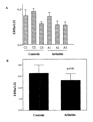
C3, and three rats with arthritis (A1, A2 and A3). Panel B shows ERRa
expression, similarly determined, in data pooled from the six control joints
(controls) and the six arthritic joints (arthritis).
Figure 7 shows ERRa expression, determined and expressed as for
Figure 6, in a femoral bone from each of three control mice (1, 2, 3) and
three
arthritic mice (4, 5, 6) and in pooled joints from three control mice (7) and
three arthritic mice (8).
Figure 8 shows ERRa expression, determined and expressed as for
Figure 6, in C5.18 cell cultures grown - Panel A: in the presence (+) or
absence (-) of fetal bovine serum (FBS) and Panel B: in the presence of
estrogen (10-9M E2) or 0.01% ethanol vehicle (VEH). * p<0.01; ** p<0.005; ns
p<0.06.
CA 02442685 2003-10-02
WO 02/080888 PCT/CA02/00460
9
Detailed Description of the Invention
The present inventors have found a new role for the orphan receptor,
estrogen receptor-related receptor a (ERRa), in the modulation of cartilage
growth and differentiation in mammals.
Cartilage formation involves the proliferation of chondroprogenitor cells
and their differentiation first into chondroblasts and then into mature
chondrocytes which synthesise and deposit cartilage.
Studies of a fetal rat chondrogenic cell line, which is an accepted
model of mammalian chondroprogenitor proliferation and differentiation into
chondrocytes, with formation of cartilage nodules, (Grigoriadis 1996;
McDougall 1996) showed that ERRa was expressed throughout the process
of cartilage formation, from early chondroprogenitor cells in the
perichondrium
to mature cartilage-synthesising chondrocytes.
Stimulation of ERRa expression and increased ERRa activity gave both
increased chondroprogenitor cell and chondrocyte proliferation and increased
differentiation of chondroprogenitors into mature chondrocytes.
Inhibition of ERRa expression and reduced ERRa activity gave decreased
proliferation of chondroprogenitors and chondrocytes and decreased
differentiation and cartilage nodule formation.
Similar results were found it yiyo, in both fetal and adult rat cartilage,
where ERRa expression was high in both progenitor cells and cartilage-
synthesising cells in the cartilage of tibia and metatarsal bones. The
presence of the ERRa receptor in both articular and growth plate
chondrocytes suggests a role for ERRa both in cartilage formation and its
maintenance and integrity throughout the lifetime of the mammal. This role is
further supported by the inventors' findings that ERRa expression was
decreased in the eroding articular cartilage of rats and mice suffering from
induced arthritis, in several accepted models of human inflammatory arthritis.
The invention provides methods and pharmaceutical compositions for
promoting cartilage formation in a mammal by increasing ERRa activity. As
CA 02442685 2003-10-02
WO 02/080888 PCT/CA02/00460
used herein, "ERRa activity" means ERRa chondrogenic or cartilage
promoting activity, ie. stimulation of cartilage production, which may occur
by
stimulation of proliferation of chondroprogenitor cells and/or chondrocytes
and/or promotion of differentiation of chondroprogenitor cells and/or
stimulation of chondrocytes to increase cartilage formation.
ERRa activity may be increased in a mammal by increasing the
amount of ERRa protein present or by increasing the chondrogenic effect of
existing ERRa protein. Increased ERRa activity may be achieved, for
example, by up regulating expression of the ERRa gene, by gene therapy to
10 provide a nucleotide sequence encoding ERRa protein, by administering an
agent which enhances ERRa expression, by administering ERRa protein or
by administering an ERRa agonist. An ERRa agonist is a compound which
increases the chondrogenic activity of ERRa protein.
Agents which increase ERRa activity may be used for preparation of
' medicaments for promoting cartilage formation.
One compound which has been shown by the inventors to increase
ERRa expression is estrogen. Estrogen analogues, including selective
estrogen receptor modifiers (SERMS), may be screened by the methods
described herein to select those active as ERRa agonists or ERRa
expression up-regulators.
The cartilage formation promoting methods and compositions of the
invention can be employed to treat conditions associated with cartilage loss,
cartilage degeneration or cartilage injury. Such conditions include the
various
disorders described collectively as arthritis.
Arthritis is a term used to designate generally diseases of the joint .
Arthritis includes many different conditions but is characterized generally by
the presence of joint inflammation. Inflammation is involved in many forms of
arthritis and results, among other things, in the destruction of joint
cartilage.
The list of diseases that are included in the term arthritis includes, but
is not limited to, ankylosing spondylitis, childhood arthritis, chronic back
injury,
CA 02442685 2003-10-02
WO 02/080888 PCT/CA02/00460
11
gout, infectious arthritis, osteoarthritis, osteoporosis, pagets's disease,
polymyalgia rheumatics, pseudogout, psoriatic arthritis, reactive arthritis,
reiter's syndrome, repetitive stress injury, and rheumatoid arthritis.
Cartilage destruction or injury can also result from joint surgery, joint
injury and obesity.
A number of symptomatic treatments for arthritis exist, including
analgesics and non-steroidal anti-inflammatory agents. Other treatments for
inflammatory arthritis include disease modifying agents (DMARDS) such as
gold salts, methotrexate, sulfasalazine, hydroxychloroquine, chloroquine and
azathioprine. Steroids and corticosteroids are anti-inflammatory agents that
are used to treat the inflammation underlying cartilage destruction.
No current arthritis therapy acts at the level of cartilage. Although many
of the treatments for arthritis may be able to reduce the effects of the
inflammation which causes cartilage destruction, these treatments do not
prbmote cartilage regrowth in the affected tissue.
The present invention provides methods and pharmaceutical
compositions for treating arthritis by increasing ERRa activity. ERRa activity
may be increased as described above.
An ERRa agonist or an agent which enhances ERRa expression, such
as estrogen, may be administered systemically to the subject in need of
treatment, or may be administered locally, for example by intra-articular
injection.
If ERRa activity is to be increased by gene therapy, a preferred
method is by administration of a suitable vector, such as an adenovirus or an
adeno-associated virus carrying the ERRa gene, by intra-articular injection.
Such intra-articular gene administration has been described by Goater et al.,
(2000) and van Lent et al. (2002).
A further preferred method is the e~ VIVO transfection of mesenchymal
stem cells or chondroprogenitor cells with the ERRa gene, followed by intra-
CA 02442685 2003-10-02
WO 02/080888 PCT/CA02/00460
12
articular injection of the treated cells. Such techniques have been described
by Nixon et al., (2000) and Gelse et al., (2001 ).
ERRa protein was found widely distributed in vitro in C5.18 cell
cultures from early proliferation stages through cartilage nodule formation.
ERa and ER(3 were also detected in C5.18 cells at all times analysed,
although ER~3 was present at somewhat lower levels and in a more patchy
appearance. These results indicate that ERRa and ERa and/or ER~i are co-
expressed in chondrogenic cells, and that these receptors may act alone or
together to regulate the expression of target genes in cartilage.
The role played by expression of the estrogen receptors in
chondrocytes has been unclear. The data indicate that ERRa and one or both
of the ERs are co-expressed chondrogenic cells. Protein analysis provided
the result that ERRa and ERa are co-distributed in large cohorts of
chondrogenic cells, suggesting that these receptors may regulate the
expression of the same target genes in cartilage. This may occur via their
known ability to participate in protein-protein interactions and their
recently
described capacity to bind to the same DNA target (SFRE and ERE)
sequence on the osteopontin promoter. ERRa and ER~3 co-expression also
occurs in some chondrogenic cells, but interactions between these two
receptors has not yet been described, although they have recently been
described to recognize the same ERE response element. These data
suggest that ERRa, ERa and ER~i are co-expressed in chondrogenic cells,
and may display at least some functions in common, either singly or through
their interactions, with regulatory capacities to act on target genes.
Consistent with its expression in proliferating chondrogenic C5.18, it
was found that antisense oligonucleotide-induced downregulation of ERRa
inhibited proliferation of C5.18 cell populations as illustrated in Figure 3.
This
decrease in proliferation was an unexpected result, given the previous
observation that ERRa expression appeared to correlate with exit from
proliferation and the onset of the differentiation process in at least certain
CA 02442685 2003-10-02
WO 02/080888 PCT/CA02/00460
13
other cell types, including the nervous system, the epidermis and muscles in
the developing mouse [Bonnelye, 1997). This surprising result suggests that
ERRa may play cell-type specific functions. Cell-type specific treatments can
thus be developed for particular cartilage/joint diseases.
Figure 4 illustrates a critical role for ERRa in cartilage formation, with
down-regulation of cartilage nodule formation concomitant with down-
regulation of ERRa expression in vitro. This result is independent of its
effects on proliferation, since cartilage nodule formation was decreased when
the antisense treatment commenced after proliferation had largely ceased.
These results indicate an unexpected use for ERRa in the regulation of
cartilage formation.
Another group of diseases involves unwanted or inappropriate
cartilage formation. Such diseases include chondrosarcomas and
chondrodysplasias. The present invention provides methods and
pharmaceutical compositions for inhibiting cartilage formation by reducing
ERRa activity and thereby treating such disorders. ERRa activity may be
reduced by reducing the amount of ERRa protein being produced or by
inhibiting the activity of ERRa protein. This may be achieved, for example, by
administering ~an antisense sequence as described herein, or an agent which
reduces ERRa expression, an antibody which binds specifically to ERRa
protein or an ERRa antagonist. An ERRa antagonist is a compound which
decreases the chondrogenic activity of ERRa protein.
An antisense sequence such as an antisense oligo or an antisense
adenovirus can be administered by gene therapy as described above,
preferably by local injection. Antibodies or antagonists can be administered
locally, or systemically if target specific.
A number of ERRa antagonists have been described. For example,
organochlorine compounds such as chlordane and toxaphene have been
shown to antagonise ERRa activity (Yang et al., (1999)).
CA 02442685 2003-10-02
WO 02/080888 PCT/CA02/00460
14
Diethylstilbestrol has also been described as an ERRa antagonist
(Tremblay et al., (2001 a)).
These compounds may be employed or may be used as a starting
point for the development of analogues which can be screened as described
herein for ERRa antagonist properties.
In a further embodiment, the invention provides a method for
assessing the ERRa level or activity of a tissue, which can be used as a
screening method for possible susceptibility to cartilage degeneration or as a
method for monitoring treatment efficacy during treatment of a cartilage
degenerative disorder. For example, subjects such as athletes or the
overweight, who are at increased risk of osteo arthritis, could be screened
for
below normal cartilage ERRa, which would suggest susceptibility to
development of osteo arthritis. Subjects being treated for rheumatoid
arthritis
could have their cartilage ERRa level monitored at intervals to assess
whether normal ERRa levels were being restored or maintained. ERRa
levels can be measured in samples of biopsied joint cartilage tissue, for
example by RT-PCR of mRNA as described herein and in Bonnelye et al.,
(2001 ) or, less quantitatively, by immunolabelling techniques such as those
described in Bonnelye et al., (2001 ).
The invention also provides a method for screening a candidate
compound for its ability to modulate ERRa chondrogenic activity in a suitable
system, by examining ERRa chondrogenic activity in the presence or
absence of the candidate compound. A change in ERRa chondrogenic
activity in the presence of the compound relative to ERRa chondrogenic
activity in the absence of the compound indicates that the compound
modulates ERRa chondrogenic activity. If ERRa chondrogenic activity is
increased relative to the control in the presence of the compound, the
compound is potentially useful as a stimulator of chondrogenesis. By means
of the assays described herein, one of skill in the art can readily determine
whether such a compound caused increased ERRa expression or acted as
CA 02442685 2003-10-02
WO 02/080888 PCT/CA02/00460
an ERRa agonist, to increase activity of ERRa protein. Conversely, if ERRa
chondrogenic activity is decreased in the presence of the compound, relative
to the control, the compound is potentially useful as an inhibitor of
chondrogenesis. It can be determined by means of the assays described
5 herein whether such a compound caused decreased ERRa expression or
acted as an ERRa antagonist, to decrease activity of ERRa protein.
Any assay system which enables one to measure the chondrogenic
activity or cartilage promoting activity of ERRa may be employed as the basis
of the screening method. Suitable assay systems include, for example,
10 measurement of chondroprogenitor proliferation, cartilage nodule formation
or
increase of chondroblast markers stimulated by increased ERRa expression
in a chondrogenic cell line such as C5.18, as described herein.
Candidate compounds may be subjected to an initial screening for their
effect on activation of the ERRa promoter, before proceeding to the more
15 involved testing of their biological effect in the screening method
described
above. While ERRs do not respond to natural estrogens, these receptors
recognise the estrogen response element and have been shown to activate
and repress gene expression in the absence of endogenously added ligand.
One of skill in the art can refer to Shi et al. (1997), Yang et al. (1999) and
Tremblay et al. (2001 ) for suitable methods.
In accordance with a further embodiment of the invention, the ERRa
signalling pathway may be modulated by modulating the binding of the ERRa
to an ERRa binding partner. Such a binding partner may include for example
the estrogen receptor. ERRa can be used to upregulate the transcription and
thus expression of genes which work together with ERRa to affect cartilage
development.
The invention further provides methods for screening candidate
compounds to identify those able to modulate signaling by ERRa through a
pathway involving ERRa.
CA 02442685 2003-10-02
WO 02/080888 PCT/CA02/00460
16
For example, the invention provides screening methods for compounds
able to bind to ERRa which are therefore candidates for modifying the
chondrogenic activity of ERRa. Various suitable screening methods are
known to those in the art, including immobilization of ERRa on a substrate
and exposure of the bound ERRa to candidate compounds, followed by
elution of compounds which have bound to the ERRa.
Co-immunoprecipitation of protein binding partners with an ERRa-
specific antibody will allow the identification of cartilage-specific binding
partners which contribute to ERRa chondrogenic activity.
The invention also provides a method of modulating a ERRa signaling
pathway by increasing or decreasing the availability of ERRa or by
modulating the function of the ERRa.
The invention further provides methods for preventing or treating
diseases characterised by an abnormality in an ERRa signaling pathway
which involves ERRa, by modulating signaling in the pathway.
According to another aspect of the present invention is a method for
suppressing in a mammal, the proliferation of a chondrocytic cell capable of
being stimulated to proliferate by ERRa, the method comprising administering
to the mammal an effective amount of a ERRa antagonist or an antibody
which binds specifically to ERRa.
The invention also enables transgenic non-human animal models,
which may be used for study of the effects on chondrogenesis of over and
under expression of the ERRa gene, for the screening of candidate
compounds as potential agonists or antagonists of this receptor and for the
evaluation of potential therapeutic interventions.
The transgenic animals of the invention may also provide models of
disease conditions associated with abnormalities of ERRa expression.
Animal species suitable for use in the animal models of the invention include
mice, rats, rabbits, dogs, cats, goats, sheep, pigs and non-human primates.
CA 02442685 2003-10-02
WO 02/080888 PCT/CA02/00460
17
Animal models may be produced which over-express ERRa by
inserting a nucleic acid sequence encoding ERRa into a germ line cell or a
stem cell under control of suitable promoters, using conventional techniques
such as oocyte microinjection or transfection or microinjection into stem
cells.
A cartilage specific promoter such as the Type II collagen promoter may be
used, for example. Animal models can also be produced by homologous
recombination to create artificially mutant sequences (knock-in targeting of
the ERRa gene) or loss of function mutations (knock-out targeting of the
ERRa gene). For example, knock-out targeting of the ERRa gene). For
example, knock-out animal models can be made using the tet-receptor
system described U.S. Patent No. 5,654,168 or the Cre-Lox system
described, for example, in U.S.P. Nos. 4,959,717 and 5,801,030.
In accordance with one embodiment of the invention, transgenic
animals are generated by the introduction of a ERRa transgene into a
fertilized animal oocyte, with subsequent growth of the embryo to birth as a
live animal. The ERRa transgene is a transcription unit which directs the
expression of ERRa gene in eukaryotic cells. To create the transgene, ERRa
gene is ligated with an eukaryotic expression module. The basic eukaryotic
expression module contains a promoter element to mediate transcription of
ERRa sequences and signals required for efficient for termination and
polyadenylation of the transcript. Additional elements of the module may
include enhancers which stimulate transcription of ERRa sequences. The
most frequently utilized termination and polyadenylation signals are those
derived from SV40. The choice of promoter and enhancer elements to be
incorporated into the ERRa transgene is determined by the cell types in which
ERRa gene is to be expressed. To achieve expression in a broad range of
cells, promoter and enhancer elements derived from viruses may be utilized,
such as the herpes simplex virus thymidine kinase promoter and polyoma
enhancer. To achieve exclusive expression in a particular cell type, specific
promoter and enhancer elements could be used, such as the promoter of the
CA 02442685 2003-10-02
WO 02/080888 PCT/CA02/00460
18
mb-1 gene and the intronic enhancer of the immunoglobulin heavy chain
gene. In a preferred embodiment, a cartilage specific promoter such as the
promoter of Type II collagen may be used to target expression in
chondrocytes (Bridgewater 1998; Lefebvre 1996).
The ERRa transgene is inserted into a plasmid vector, such as
pBR322 for amplification. The entire ERRa transgene is then released from
the plasmid by enzyme digestion, purified and injected into an oocyte. The
oocyte is subsequently implanted into a pseudopregnant female animal.
Southern blot analysis or other approaches are used to determined the
genotype of the founder animals and animals generated in the subsequent
backcross and intercross.
Such deficient mice will provide a model for study of the role of ERRa
in chondrocyte differentiation and proliferation and general skeletal
development. Such animals will also provide tools for screening candidate
compounds for their interaction with ERRa or the signalling pathway activated
by ERRa.
The invention also provides pharmaceutical compositions for promoting
cartilage formation, comprising as active ingredient a substantially purified
ERRa protein, an ERRa agonist or an isolated nucleotide sequence encoding
ERRa protein. Such compositions are useful, for example, in treating
disorders associated with cartilage degeneration.
ERRa protein may be produced by conventional recombinant
techniques permitting expression of ERRa by a suitable host cell. A DNA
encoding ERRa may be prepared as described, for example, in Giguere et al.
(1998).
Techniques for production of proteins by recombinant expression are
well known to those in the art and are described, for example, in Sambrook et
al. (1989) or latest edition thereof. Suitable host cells include ~. c~oli or
other
bacterial cells, yeast, fungi, insect cells or mammalian cells.
CA 02442685 2003-10-02
WO 02/080888 PCT/CA02/00460
19
The invention provides for compositions for promoting cartilage
formation comprising as active ingredient an ERRa agonist obtained by using
a screening method as described herein.
A nucleotide sequence encoding ERRa protein may be administered to
a subject experiencing cartilage loss due to an absent or defective ERRa
gene either jr vivo or ex vivo. Expression may be targeted to a selected cell
or tissue by use of an appropriate promoter.
The invention also provides pharmaceutical compositions for reducing
or inhibiting cartilage formation, comprising as active ingredient an antibody
which binds specifically to ERRa, an ERRa antagonist or a negative regulator
such as an antisense nucleic acid or a dominant negative mutant version of
the ERRa gene.
The invention provides for compositions for reducing cartilage
formation comprising as active ingredient an ERRa antagonist obtained by
using a screening method as described herein.
Antibodies which bind specifically to ERRa protein may be made by
conventional techniques.
The term "antibodies" includes polyclonal antibodies, monoclonal
antibodies, single chain antibodies and fragments such as Fab fragments.
In order to prepare polyclonal antibodies, fusion proteins containing
defined portions or all of an ERRa protein can be synthesized in bacteria by
expression of the corresponding DNA sequences, as described above.
Fusion proteins are commonly used as a source of antigen for producing
antibodies. Alternatively, the protein may be isolated and purified from the
recombinant expression culture and used as source of antigen. Either the
entire protein or fragments thereof can be used as a source of antigen to
produce antibodies.
The purified protein is mixed with Freund's adjuvant and injected into
rabbits or other appropriate laboratory animals. Following booster injections
at weekly intervals, the animals are then bled and the serum isolated. The
CA 02442685 2003-10-02
WO 02/080888 PCT/CA02/00460
serum may be used directly or purified by various methods including affinity
chromatography to give polyclonal antibodies.
Monoclonal anti-ERRa antibodies may be produced by. methods well
known in the art. Briefly, the purified protein or fragment thereof is
injected in
5 Freund's adjuvant into mice over a suitable period of time, spleen cells are
harvested and these are fused with a permanently growing myeloma partner
and the resultant hybridomas are screened to identify cells producing the
desired antibody. Suitable methods for antibody. preparation may be found in
standard texts such as Barreback, E.D. (1995).
10 The pharmaceutical compositions of the invention may comprise, in
addition to the active ingredient, one or more pharmaceutically acceptable
carriers.
Administration of an effective amount of a pharmaceutical composition
of the present invention means an amount effective, at dosages and for
15 periods of time necessary to achieve the desired result. This may also vary
according to factors such as the disease state, age, sex, and weight of the
subject, and the ability of the composition to elicit a desired response in
the
subject. Dosage regima may be adjusted to provide the optimum therapeutic
response. For example, several divided doses may be administered daily or
20 the dose may be proportionally reduced as indicated by the exigencies of
the
therapeutic situation.
By pharmaceutically acceptable carrier as used herein is meant one or
more compatible solid or liquid delivery systems. Some examples of
pharmaceutically acceptable carriers are sugars, starches, cellulose and its
derivatives, powdered tragacanth, malt, gelatin, collagen, talc, stearic
acids,
magnesium stearate, calcium sulfate, vegetable oils, polyols, agar, alginic
acids, pyrogen-free water, isotonic saline, phosphate buffer, and other
suitable non-toxic substances used in pharmaceutical formulations. Other
excipients such as wetting agents and lubricants, tableting agents,
stabilizers,
anti-oxidants and preservatives are also contemplated.
CA 02442685 2003-10-02
WO 02/080888 PCT/CA02/00460
21
The compositions described herein can be prepared by known
methods for the preparation of pharmaceutically acceptable compositions
which can be administered to subjects, such that an effective quantity of the
active substance is combined in a mixture with a pharmaceutically acceptable
carrier. Suitable carriers and formulations adapted for particular modes of
administration are described, for example, in Remington's Pharmaceutical
Sciences (Remington's Pharmaceutical Sciences, Mack Publishing Company,
Easton, Pa., USA 1985). On this basis the compositions include, albeit not
exclusively, solutions of the substance in association with one or more
pharmaceutically acceptable vehicles or diluents, and contained in buffered
solutions with a suitable pH and iso-osmotic with the physiological fluids.
The pharmaceutical compositions of the invention may be
administered therapeutically by various routes such as by injection or by
oral,
nasal, intra-articular, intra-vertebral, buccal, rectal, vaginal, transdermal
or
ocular administration in a variety of formulations, as is known to those
skilled
in the art.
The present invention enables also a screening method for compounds
of therapeutic utility as antagonists of the chondrogenic activity of ERRa.
Such antagonist compounds are useful, for example, to reduce or prevent
differentiation and maturation of chondrocytes. ERRa antagonists may also
be used in the treatment of cartilage related disorders involving
inappropriate
cartilage growth. Those skilled in the art will be able to devise a number of
possible screening methods for screening candidate compounds for ERRa
antagonism.
A screening method may also be based on binding to the ERRa
receptor. Such competitive binding assays are well known to those skilled in
the art. Once binding has been established for a particular compound, a
biological activity assay is employed to determine agonist or antagonist
potential.
CA 02442685 2003-10-02
WO 02/080888 PCT/CA02/00460
22
Exa a
The examples are described for the purposes of illustration and are not
intended to limit the scope of the invention.
Methods of biochemistry, molecular biology, histology and immunology
referred to but not explicitly described in this disclosure and examples are
reported in the scientific literature and are well known to those skilled in
the
art.
Fxa ple 1 Ex~oression of ERRa in chondrocvte lineage cells throughout
develo~lnent
In order to assess ERRa expression, RCJ.1 C5.18 (C5.18) cells were
grown as described by Grigoriadis, 1996. This cell line is a fetal rat cell
line
which undergoes differentiation into cartilage-producing chondrocytes; it is
widely used as a model system for the study of chondrogenesis and the
regulation of chondrocyte activity. Cells were maintained in a-MEM
containing 15% heat-inactivated FBS (Flow Laboratories, McLean, VA),
antibiotics comprising 100 ~g/ml penicillin G (Sigma Chemical Co., St. Louis,
MO), 50 ~g/ml gentamycin (Sigma), and 0.3 ~g/ml fungizone (Flow
Laboratories) and 10$M dexamethasone (Merck, Sharp, and Dohme,
Canada, Ltd., Kirkland, PQ). Dexamethasone (Dex) stimulates
chondrogenesis and cartilage formation in these cultures. For differentiation
studies, cells were grown in the same medium, with or without
dexamethasone, and with the addition of 50 ~g/ml ascorbic acid and 10 mM
sodium ~-glycerophosphate. Medium was changed every 2 days. All dishes
were incubated at 37oC in a humidified atmosphere in a 95% air/5% C02
incubator.
For Northern blot analysis, total RNA was extracted with guanidine
from C5.18 cells after culture periods corresponding to different stages of
proliferation, differentiation and cartilage nodule formation. Northern blots
were prepared and hybridized with a 750bp fragment corresponding to the rat
CA 02442685 2003-10-02
WO 02/080888 PCT/CA02/00460
23
3' UTR of ERRa (provided by JM Vanacker, Lyon, France) according to
standard procedures (Chirgwin et al, 1979). Mouse cDNAs for link protein and
aggrecan were kindly provided by S. Bernier, London, On.
ERRa mRNA expression levels were assessed over a proliferation-
differentiation time course by Northern blotting of C5.18 cells grown in the
presence (+Dex) or absence (-Dex) of dexamethasone), a stimulator of
differentiation in this model. Under both growth conditions, ERRa mRNA was
expressed constitutively at all times assessed, including proliferation (day
5),
and early (day 9, 11 ) and late (day 17) cartilage nodule formation, as shown
in Panel A of Figure 1. For comparison, levels of mRNA levels for two
cartilage markers, aggrecan and link protein are shown in Panel B of Figure
1.
Semi-quantitative RT-PCR was carried out as described in Bonnelye et
al. (2001 ), over the proliferation/differentiation time course of C5.18
cultures
treated with Dex. The results are shown in Figure 2. Days 3 and 6 were
within the proliferation phase and days 11, 15 and 21 were within the
differentiation stage. ERRa expression was normalised against that of the
ribosomal probe L32. For comparison, mRNA levels for three chondroblast
markers, type II collagen, aggrecan and link protein, were also assessed,
normalised against L32.
In order to assess ERRa protein expression, C5.18 cell cultures, grown
as described above, were immunolabelled essentially as described previously
[Turksen, 1991; Turksen, 1992]. Cultures were rinsed with PBS, fixed with
3.7% formaldehyde in PBS and permeabilized with methanol at -20°C.
Frozen
sections were fixed 10 min in cold acetone. Paraffin sections were
deparaffinized in xylene, then rehydrated in 100%, 95% and 70% ethanol and
water. After rinsing, cells in dishes or frozen or paraffin sections were
incubated for 1 h at room temperature with 10% normal serum in PBS for
ERRa and ERa and in 3% BSA in PBS (denaturated) for ERa. After rinsing,
cells or sections were incubated for 1.5 hours with appropriate dilutions of
CA 02442685 2003-10-02
WO 02/080888 PCT/CA02/00460
24
primary antibodies (1/50, anti-ERRa; anti-ERa or anti- ER~i MC-20 or Y-19,
respectively; Santa Cruz Biotechnology, Inc).
While ERRa protein was expressed throughout the
proliferation/differentiation sequence, protein levels were highest in
maturing
and mature chondrocytes associated with cartilage nodules it1 iv tro. At all
stages (proliferation and differentiation/maturation), the majority of ERRa
appeared to be localized to the nucleus. For comparison, ERa and ERa
levels were also assessed; all three receptors were co-expressed in at least
some chondrocytes. However, based on staining intensity, ERRa levels were
highest (detected in all chondrocytes), followed by ERa (detectable in most
chondrocytes but at lower levels than ERRa) and finally by ER(3 (detectable at
very low levels in only a subset of chondrocytes). As with ERRa, ERa
appeared to be primarily localized to the nucleus at all stages, whereas ER(3
appeared to assume a nuclear localization mainly when cells were in
proliferative stages (data not shown).
Example 2 - In vivo expression of ERRa
To determine the in vivo expression of ERRa protein, immuno-
cytochemistry was performed on sections of 21 day fetal rat tibiae and
metatarsals and on sections of adult rat tibiae and femurs. The sections
were rinsed in PBS and incubated for 1 h at room temperature with
secondary antibody CY-3-conjugated anti-rabbit (Jackson Immunoresearch
Lab, West Grove, PA, USA; 1/300 final dilution) for ERRa. After rinsing,
samples were mounted in Moviol (Hoechst Ltd, Montreal, PQ, Canada) and
observed by epifluorescence microscopy on a Zeiss Photomicroscope III
(Zeiss, Oberkochen, Germany).
ERRa protein was already highly expressed in the chondrocytes of the
growth plates of term-pregnant rat fetuses and continued to be expressed in
the cartilage of adult animals. In fetal growth plate cartilage, intense label
for
ERRa was seen in perichondrial precursors and proliferating chondrocytes,
CA 02442685 2003-10-02
WO 02/080888 PCT/CA02/00460
while staining in hypertrophic chondrocytes was low or absent. In adult
animals, growth plate chondrocytes, including hypertrophic zone, were
intensely labeled, as were articular chondrocytes. Articular zone
chondrocytes, based on labeling intensity, expressed much higher levels of
5 ERRa than cells in surrounding tissues (data not shown).
Example 3- Anfisense and sense olig~onucleofide treatment
Antisense oligonucleotides form DNA:RNA duplexes with specific
mRNA species, thereby blocking binding of the mRNA to the 40S ribosomal
10 subunit and preventing translation [Jen, 2000]. To examine the involvement
of
ERRa in chondrocyte differentiation and cartilage formation, C5.18 cells were
treated either during the proliferation phase or during the differentiation
and
cartilage nodule formation phase. Preliminary experiments were done to
determine effective oligonucleotide concentrations that were not toxic (not
15 shown) and the efficacy of the antisense was confirmed by
immunocytochemistry and Western blots.
C5.18 cells were plated in 24 wells plates at 104 cells/well and treated
with antisense or sense oligonucleotides. Antisense oligonucleotide inhibition
of ERRa expression was accomplished with a 20-base phosphorothioate-
20 modified oligonucleotide, localized to the A/B domain. The ERRa antisense
oligonucleotide sequence was: 5'-TCACCGGGGGTTCAGTCTCA-3'. Control
dishes were treated with the complementary sense oligonucleotide or no
oligonucleotide. Preliminary experiments were done to determine effective
oligonucleotide concentrations that were not toxic. 0.1 ~M to 5~M
25 oligonucleotides were added directly to cells either during the
proliferation
phase (days 1 to 4) and 0.5~,M to 2~M oligonucleotides were added during the
differentiation phase (day 5 to day 13) in standard medium as above
supplemented with 50 ~g/ml ascorbic acid, 10 mM sodium [3-glycerophosphate,
and10$ M dexamethadone. Medium was changed every 2 days and fresh
oligonucleotides were added.
CA 02442685 2003-10-02
WO 02/080888 PCT/CA02/00460
26
For cell growth analysis, the cell layers were rinsed in PBS, released
with trypsin and collagenase, and the harvested cells were counted
electronically. The results are shown in Figure 3. Results are plotted as the
average of three counts for each of three wells for control and each
concentration of antisense or sense primers used. When C5.18 cells were
treated between days 1-4, the proliferation stage, a significant and specific
dose-dependent decrease in cell number in dishes treated with antisense
compared to sense or untreated controls is seen. These results indicate a
role for ERRa in the proliferation or very early differentiation phases of
C5.18
1o cells.
To determine whether ERRa also plays a role in chondrocyte
differentiation independently of an effect on proliferation, C5.18 cells were
treated with the antisense oligonucleotide beginning at day 5 (after cells had
reached confluence and proliferation was decreased) to day 15. For
quantification of cartilage formation, dishes or wells were fixed and with
Alcian
blue and cartilage nodules were counted on a grid [Grigoriadis, 1996].
Results, as shown in Figure 4, are plotted as the mean number of nodules ~
SD of three wells for controls and each concentration of antisense or sense
primers. A striking dose-dependent decrease in cartilage nodule formation
was seen.
E 1e
C5.18 cells at ~50% confluency were transfected with either a pcDNA3
empty plasmid (Empty vector) or pcDNA3-ERRa (ERRa vector) (0.5Ng DNA
per transfection). Five 35-mm dishes per treatment group were fixed, stained
with Alcian blue and the cartilage nodules counted. Cultures transfected with
the ERRa gene showed a significant increase in the number of cartilage
nodules formed (mean +/- SD; Welch test or Mann Whitney test, p<0.05).
Results shown in Figure 5 are representative of two independent
experiments.
CA 02442685 2003-10-02
WO 02/080888 PCT/CA02/00460
27
xamp.Je 5 - Rat model of inflammator)r arthritis
Arthritis was initiated in genetically susceptible female Lewis rats
(Charles River Breeding Laboratories, Wilmington, MA) by intraperitoneal
injection of group A SCW (Streptococcal Cell Wall) peptidoglycan-
polysaccharide complexes (Lee Laboratories Inc., Grayson, GA) as described
[Wahl, 1994]. The severity of arthritis (articular index, AI) was determined
by
blinded scoring of each ankle and wrist joint based on the degree of swelling,
erythema, and distortion on a scale of 0-4 and summing the scores for all four
limbs. As a control, PBS was injected instead of SWC. Hemoglobin as an NO
scavenger, was used as a suppressor of arthritis. In one group of rats, it was
administered daily from day 0 to day 24 of SCW treatment and in a second
group from day 10 to day 24 only.
ERRa expression in chondrocytes was examined in sections of
articular cartilage, compact bone and bone marrow from control rats and from
SCW induced arthritic rats by immunocytochemistry as described above.
In the Streptococcal cell wall (SCW)-induced rat arthritis model, ERRa
expression was decreased in chondrocytes in the eroding articular cartilage
as a function of the severity of the disease.
Semi-quantitative RT-PCR as described in example 1 was used to
determine mRNA levels in samples from a further rat model and a mouse
model of collagen-induced arthritis (Trentham et al. (1997)), to assess ERRa
expression.
RNA was isolated with Trizol reagent (Gibco BRL), according to the
manufacturer's protocol, from joints from three control rats and from three
rats
injected with collagen; joints comprised proximal femur and distal tibiae.
Semi-quantitative RT-PCR with ERRa-specific primers was done and
ERRamRNA expression level was normalized against that of the ribosomal
housekeeping gene L32.
CA 02442685 2003-10-02
WO 02/080888 PCT/CA02/00460
28
In the rat model, ERRa mRNA levels were reduced in arthritic joints
compared to normal joints, as seen in Figure 6 (your Figure 9).
Samples obtained from the mouse model consisted of separated bone
and joint samples from 3 arthritic mice and 3 controls. Joints (proximal femur
and distal tibiae) were carefully dissected away from bone and ERRa
expression was separately determined for cartilage tissue and for bone (left
and right tibiae and femurs, marrow removed). The six normal cartilage
samples were pooled for analysis, as were the six arthritic cartilage samples.
RNA was isolated from the tissues, semi-quantitative RT-PCR
with ERRa-specific primers was done and ERRa mRNA expression level was
normalized against that of the ribosomal housekeeping gene L32. ERRa
mRNA levels were significantly reduced in pooled arthritic joint cartilage
samples compared with those from control samples (Student t-test; p<0.05)
as seen in Figure 7 (your Figure 10). ERRa mRNA levels were higher in
control joint cartilage than in control bone (Student t-test; p<0.005). In
contrast, no significant difference in ERRa expression level was seen
between control and arthritic bone samples.
Exa 1e
C5.18 cells were grown in medium with (+) and without (-) fetal bovine
serum (FBS). ERRa mRNA levels were determined by semi-quantitative RT-
PCR and normalised against ribosomal probe L32. As shown in Figure 8,
Panel A, ERRa mRNA was increased by the presence of FBS at days 1 and
2.
Similar C5.18 cultures were grown without FBS but in the presence of
estrogen (10-9M E2) or vehicle (0.01 % ethanol) (VEH) and mRNA levels were
determined as above. As shown in Figure 8, Panel B, estrogen significantly
increased ERRa mRNA levels at day 2. Estrogen plus FBS did not increase
the ERRa mRNA level over that seen with FBS alone, suggesting that the
latter caused maximal stimulation of ERRa expression.
CA 02442685 2003-10-02
WO 02/080888 PCT/CA02/00460
29
The present invention is not limited to the features of the embodiments
described herein, but includes all variations and modifications within the
scope of the claims.
CA 02442685 2003-10-02
WO 02/080888 PCT/CA02/00460
References
Bonnelye, E., Vanacker, J. M., Dittmar, T., Begue, A., Desbiens, X.,
Denhardt, D. T., Aubin, J. E., Laudet, V., and Fournier, B. (1997a). The
5 ERR-1 orphan receptor is a transcriptional activator expressed during
bone development. Mol Endocrinol 11 (7), 905-16.
Bonnelye, E., Vanacker, J. M., Spruyt, N., Alric, S., Fournier, B., Desbiens,
X.,
and Laudet, V. (1997b). Expression of the estrogen-related receptor 1
(ERR-1 ) orphan receptor during mouse development. Mech Dev 65(1-
10 2), 71-85.
Bonnelye et al., (2001 ), J. Cell Biol., v. 153, pp. 971-983.
Bridgewater, L.C., Lefebvre, V., and de Crombrugghe, B. (1998) Chodrocyte-
specific enhancer elements in the Co111 a2 gene resemble the Col2a1
tissue-specific enhancer. J Biol Chem 273(24), 14998-15006.
15 Denhardt, D. T., and Noda, M. (1998). Osteopontin expression and function:
role in bone remodeling. 25th Anniversary Issue. New directions and
dimensions in cellular biochemistry. J. Cell. 8iochem. Suppl. 30/31, 92-
102.
Enmark, E., and Gustafsson, J. A. (1996). Orphan nuclear receptors--the first
20 eight years. Mol Endocrinol 10(11 ), 1293-307.
Gelse et al., (2001 ), Arthritis Rheum., v. 44, pp. 1943-53.
Giguere, V., Yang, N., Segui, P., and Evans, R. M. (1988). Identification of a
new class of steroid hormone receptors. Nature 331 (6151 ), 91-4.
Goater et al., (2000), J. Rheumatol., v. 27, pp. 983-989.
25 Green, S., Walter, P., Kumar, V., Krust, A., Bornert, J. M., Argos, P., and
Chambon, P. (1986). Human oestrogen receptor cDNA: sequence,
expression and homology to v-erb-A. Nature 320(6058), 134-9.
Grigoriadis, A. E., Heersche, J. N., and Aubin, J. E. (1996). Analysis of
chondroprogenitor frequency and cartilage differentiation in a novel
CA 02442685 2003-10-02
WO 02/080888 PCT/CA02/00460
31
family of clonal chondrogenic rat cell lines. Differentiation 60(5), 299-
307.
Gronemeyer, H., and Laudet, V. (1995). Transcription factors 3: nuclear
receptors. Protein Profile 2(11 ), 1173-308.
Hong, H., Yang, L., and Stallcup, M. R. (1999). Hormone-independent
transcriptional activation and coactivator binding by novel orphan
nuclear receptor ERR3. J Biol Chem 274(32), 22618-26.
Jen, K. Y., and Gewirtz, A. M. (2000). Suppression of gene expression by
targeted disruption of messenger RNA: available options and current
strategies. Stem Cells 18(5), 307-319.
John, S., Myerscough, A., Eyre, S., Roby, P., Hajeer, A., Silman, A. J.,
Oilier,
W. E., and Worthington, J. (1999). Linkage of a marker in intron D of
the estrogen synthase locus to rheumatoid arthritis. Arthritis Rheum
42(8), 1617-1620.
Kuiper, G. G., Enmark, E., Pelto-Huikko, M., Nilsson, S., and Gustafsson, J.
A. (1996). Cloning of a novel receptor expressed in rat prostate and
ovary. Proc Natl Acad Sci U S A 93(12), 5925-30.
Lefebvre, V., Zhou, G., Mukhopadhyay, K., Smith, C. N., Zhang, Z.,
Eberspaecher, H., Zhou, X., Sinha, S., Maity, S. N., and de
Crombrugghe, B. (1996). An 18-base-pair sequence in the mouse
proalpha1 (II) collagen gene is sufficient for expression in cartilage and
binds nuclear proteins that are selectively expressed in chondrocytes.
Mol Cell Biol 16(8), 4512-4523.
McDougall, S:, Fu, Y. H., Lowe, G. N., Williams, A., Polendo, R., Benya, P.
D., lida-Klein, A., Fang, M. A., and Hahn, T. J. (1996). Surface
adhesion-mediated regulation of chondrocyte-specific gene expression
in the nontransformed RCJ 3.1 C5.18 rat chondrocyte cell line. J Bone
Miner Res 11 (8), 1130-1138.
Nixon et al., (2000), Clin. Orthop., 379 Suppl, S 201-13.
CA 02442685 2003-10-02
WO 02/080888 PCT/CA02/00460
32
Pettersson, K., Svensson, K., Mattsson, R., Carlsson, B., Ohlsson, R., and
Berkenstam, A. (1996). Expression of a novel member of estrogen
response element-binding nuclear receptors is restricted to the early
stages of chorion formation during mouse embryogenesis. Mech Dev
54(2), 211-23.
Sasano, H., Uzuki, M., Sawai, T., Nagura, H., Matsunaga, G., Kashimoto, O.,
and Harada, N. (1997). Aromatase in human bone tissue. J Bone
Miner Res 12(9), 1416-1423.
Shi et al., (1997), Genomics, v. 44, pp. 52-60
Simpson, E., Rubin, G., Clyne, C., Robertson, K., O'Donnell, L., Jones, M.,
and Davis, S. (2000). The Role of Local Estrogen Biosynthesis in
Males and Females. Trends Endocrinol Metab 11 (5), 184-188.
Simpson, E. R., Zhao, Y., Agarwal, V. R., Michael, M. D., Bulun, S. E.,
Hinshelwood, M. M., Graham-Lorence, S., Sun, T., Fisher, C. R., Qin,
K., and Mendelson, C. R. (1997). Aromatase expression in health and
disease. Recenf Prog Horm Res 52, 185-213; discussion 213-214.
Tremblay et al., (2001 a), Genes Dev., v.15, pp. 833-838.
Tremblay et al., (2001 b), Endocrinol., v. 142, pp. 4572-5.
Trentham et al., (1977), J. Exp. Med., v. 146, pp. 857-868.
Turksen, K., and Aubin, J. E. (1991 ). Positive and negative immunoselection
for enrichment of two classes of osteoprogenitor cells. J. Cell Biol.
114(2), 373-384.
Turksen, K., Bhargava, U., Moe, H. K., and Aubin, J. E. (1992). Isolation of
monoclonal antibodies recognizing rat bone-associated molecules in
vitro and in vivo. J. Hisfochem. Cytochem. 40(9), 1339-1352.
Vanacker, J. M., Delmarre, C., Guo, X., and Laudet, V. (1998). Activation of
the osteopontin promoter by the orphan nuclear receptor estrogen
receptor related alpha. Cell Growth Differ 9(12), 1007-1014.
Vanderschueren, D., Boonen, S., Ederveen, A. G., de Coster, R., Van Herck,
E., Moermans, K., Vandenput, L., Verstuyf, A., and Bouillon, R. (2000).
CA 02442685 2003-10-02
WO 02/080888 PCT/CA02/00460
33
Skeletal effects of estrogen deficiency as induced by an aromatase
inhibitor in an aged male rat model [In Process Citation]. Bone 27(5),
611-617.
Vanderschueren, D., Van Herck, E., De Coster, R., and Bouillon, R. (1996).
Aromatization of androgens is important for skeletal maintenance of
aged male rats. Calcif Tissue Inf 59(3), 179-183.
van Lent et al., (2002), Osteoarthritis Cartilage, v. 10, pp. 234-243.
Wahl, S. M., Allen, J. B., Hines, K. L., Imamichi, T., Wahl, A. M., Furcht, L.
T.,
and McCarthy, J. B. (1994). Synthetic fibronectin peptides suppress
arthritis in rats by interrupting leukocyte adhesion and recruitment. J
Clin Invesf 94(2), 655-662.
Yang, C., and Chen, S. (1999). Two organochlorine pesticides, toxaphene
and chlordane, are antagonists for estrogen-related receptor alpha-1
orphan receptor. Cancer Res 59(18), 4519-24.
Yang, C., Zhou, D., and Chen, S. (1998). Modulation of aromatase
expression in the breast tissue by ERR alpha-1 orphan receptor.
Cancer Res 58(24), 5695-700.
Yang, N., Shigeta, H., Shi, H., and Teng, C. T. (1996). Estrogen-related
receptor, hERR1, modulates estrogen receptor-mediated response of
human lactoferrin gene promoter. J Biol Chem 271 (10), 5795-804.
Zhang, Z., and Teng, C. T. (2000). Estrogen receptor-related receptor alpha 1
interacts with coactivator and constitutively activates the estrogen response
elements of the human lactoferrin gene. J Biol Chem 275(27), 20837-46.