Note: Descriptions are shown in the official language in which they were submitted.
CA 02442971 2007-12-20
29748-2
DESCRIPTION
CHEMOTHERAPEUTIC INDUCTION OF EGR-l PROMOTER ACTIVITY
BACKGROUND OF THE INVENTION
1. Field of the Invention
The present invention relates generally to the fields of molecular biology and
cancer
therapy. More particularly, it concerns use of the DNA damaging chemicals to
induce
expression of the Egr-1 promoter. This permits tissue specific expression of
therapeutic genes
which, in combination with the DNA damaging chemicals, provide therapy to
patients suffering
from cancer.
2. Description of Related Art
Certain cancer treatment methods, including radiotherapy and chemotherapy,
involve
damaging the DNA of the cancer cell. The cellular response to normal DNA
damage includes
activation of DNA repair, cell cycle arrest and lethality (Hall, 1988). For
example, the induction of
DNA double-strand breaks results in lethal chromosomal aberrations that
include deletions,
dicentrics, rings, and anaphase bridges (Hall, 1994).
Another approach to treating cancers is gene therapy. This involves the
transfer of a
foreign gene into a cancer cell, often a tumor suppressor or inducer of
apoptosis, under
conditions suitable for expression of the gene. Once expressed, the gene
product confers a
beneficial effect on the tumor cell by either slowing its growth, inhibiting
its metastatic potential,
or killing it outright.
Combining one or more of these methods is a powerful tool as heterogeneity in
many
tumors makes mono-therapies far less effective than combinations. However,
radio-, 'chemo-
and gene therapy all have the potential for toxic effects. Thus, being able to
reduce toxicity, for
example, by reducing the amount of radiation/drug/vector administered, is
highly advantageous.
1
CA 02442971 2003-10-03
WO 02/080849 PCT/US02/10733
For example, tumor necrosis factor-alpha (TNF-a), which has antitumor
properties, has
been studied as a systemic gene-therapy treatment for cancer in phase 1
studies, but toxicity has
limited the therapeutic index of this cytokine (Spriggs et al., 1988; Demetri
et al., 1989) Also,
combinations of systemic TNF-a and chemotherapy have been investigated in a
few clinical
trials with limited success (Nakamoto et al., 2000).
On the other hand, chemotherapeutic agents such as cisplatin and other
platinum
analogues are currently employed in the treatment of several cancers including
head and neck,
esophageal, , lung, testis, ovarian, and bladder cancers. Additionally,
cisplatin is used
concurrently with irradiation (IR) as a radiosensitizer. In spite of the
relative efficacy of
cisplatin, tumor-resistance has limited the role of cisplatin in curative
cancer chemotherapy
(Johnson and Stevenson, 2001). Tumor-derived mechanisms of cisplatin-
resistance include an
increase in DNA repair of cisplatin adducts in tumor cells, an increase in
glutathione, which
inhibits free-radical formation and subsequent DNA damage, and a relative
decrease in uptake of
cisplatin by resistant cells (Kartalou and Essigmann, 2001). The combination
of cisplatin with
other chemotherapeutic agents, especially 5-FU and VP-16, has increased the
therapeutic index
of both agents in some human tumors (Kucuk et al., 2000), but other strategies
are needed .to
increase the efficacy of cisplatin.
Thus, there is a need in the art to improve both gene-therapeutic as well as
chemotherapeutic treatment regimens. Therapies that combine the benefits of
different treatment
regimens, at the same time reducing the associated side-effects, are desired.
SUMMARY OF THE INVENTION
The present invention overcomes the deficiencies in the art and provides
methods that
enhance the therapeutic utility of gene-therapy as well chemotherapy. A
transcriptional targeting
strategy has been developed wherein inducible expression vectors that encode
for therapeutic
genes are induced by chemotherapeutic agents. The chemotherapeutic agents
specifically target
inducible promoters of the expression vector to provide targeted therapy. The
therapeutic
methods provided are especially effective in treating tumors.
Therefore, in accordance with the present invention, there are provided
methods for
expressing a protein of interest comprising (a) providing an expression
construct comprising a
nucleic acid segment encoding the protein of interest, the nucleic acid
segment being positioned
under the control of an Egr-1 promoter; (b) transferring the expression
construct into a cell; (c)
-2-
CA 02442971 2003-12-22
28778-148
contacting the cell with at least one free radical-inducing DNA damaging
compound, whereby
the DNA damaging compound induces expression of the protein of interest from
the Egr-1
promoter.
The free radical-inducing DNA damaging compound may be a platinum compound
such
as cisplatin, a nitrogen mustard, cytoxan, cyclophosphamide, mitomycin c,.
adriamycin,
iphosphamide, bleomycin, doxorubicin, procarbazine, actinomycin, chlorambucil,
carboplatinum, busulfan, bcnu, ccnu, hexamethylmelamineoxaliplatin,
epirubicin, daunorubicin,
camptothecin, or mitoxantrone. Step (c) may comprise contacting the cell with
at least a second
free-radical inducing DNA damaging compound. The method may further comprise
contacting
the cell with a cancer chemotherapeutic compound or ionizing radiation. The
cell may be a
cancer cell, for example, a lung cancer cell, prostate cancer cell, ovarian
cancer cell, testicular
cancer cell, brain cancer cell, skin cancer cell, colon cancer cell, gastric
cancer cell, esophageal
cancer cell, tracheal cancer cell, head & neck cancer cell, pancreatic cancer
cell, liver cancer cell,
breast cancer cell, ovarian cancer cell, lymphoid cancer cell, leukemia cell,
cervical cancer cell,
or vulvar cancer cell.
The expression vector may further comprise an origin of replication, a
selectable marker,
or a polyadenylation signal operable linked to the nucleic segment. The
expression vector may
be plasmid or a viral vector, for example, an adenoviral vector, an adeno-
associated viral vector,
a retroviral vector, a' lentiviral vector, a vaccinia viral vector, or a
herpesviral vector. The viral
vector may lack one or more viral genes, thus rendering the viral vector non-
replicative. The cell
may be located in an organism, for example, a human.
The protein of interest may be a tumor suppressor, an inducer of apoptosis, an
enzyme, a
toxin, a cytosine, or any other protein with antitumor activity. Examples of
tumor suppressors
are Rb, p16, p53, PTEN, MDA7 or BRCA1 or BRCA2. Examples of inducers of
apoptosis are
Bax, Bad, Bik, AdE1B, Bim, Bcl-X,, Bak, TRAIL, Harakiri or Bid. Examples of
enzymes are
thymidine kinase, cytosine deaminase, hypoxanthine guanine phosphoribosyl
transferase.
Examples of toxin are pseudomonas exotoxin, diptheria toxin, cholera toxin,
pertussis toxin A
subunit, enterotoxin A, or ricin A chain. Other molecules with antitumor
activity include
interleukins (IL) and cytokines exemplified by, IL-1, IL-2, IL-3, IL-4, IL-5,
IL-6, IL-7, IL-8, IL-
9, IL-10, IL-11, IL-12, IL-13, IL-14, IL-15, 0-interferon, a-interferon, y-
interferon, angiostatin,
thrombospondin, endostatin, METH-1, METH-2, GM-CSF, G-CSF, M-CSF and tumor
necrosis
factos (TNF) such as TNF-a and TNF-P. The skilled artisan will recognize that
the invention is
-3-
CA 02442971 2003-10-03
WO 02/080849 PCT/US02/10733
not limited by any particular protein of interest, such as those disclosed
above, as long as the
protein has an antitumor effect.
In another embodiment, the invention provides methods for treating cancer in a
subject
comprising (a) providing an expression construct comprising a nucleic acid
segment encoding a
cancer therapeutic protein, the nucleic acid segment being positioned under
the control of an
Egr-1 promoter; and (b) administering the expression construct to the subject
in combination
with a free radical-inducing DNA damaging compound, whereby the DNA damaging
compound
induces expression of the cancer therapeutic protein from the Egr-1 promoter,
thereby treating
the cancer in the subject. The expression construct may be delivered local or
regional to a tumor
located in the subject, delivered systemically, or delivered via intratumoral
injection or by direct
injection into tumor vasculature.
The DNA damaging compound may be administered prior to administering the
expression vector, after administering the expression vector, or at the same
time as the
expression vector. The expression vector and or DNA damaging agent may be
administered at
least twice. The cancer therapeutic protein may be a tumor suppressor, an
inducer of apoptosis,
an enzyme, a toxin, a cytokine, or any protein with anti-tumor activity.
In yet another embodiment, there are provided methods for inhibiting tumor
cell growth
in a subject comprising (a) providing an expression construct comprising a
nucleic acid segment
encoding a cancer therapeutic protein, the nucleic acid segment being
positioned under the
control of an Egr-1 promoter; and (b) administering the expression construct
to .the subject in
combination with a free radical-inducing DNA damaging compound, whereby the
DNA
.damaging compound induces expression of the cancer therapeutic protein from
the Egr-1
promoter, thereby inhibiting tumor cell growth in the subject. In one such
embodiment, the
cancer therapeutic protein is TNF-a. In another such embodiment, the free
radical-inducing
DNA damaging compound is cisplatin.
In still yet another embodiment, there are provided methods for killing a
tumor cell in a
subject comprising (a) providing an expression construct comprising a nucleic
acid segment
encoding a cancer therapeutic protein, the nucleic acid segment being
positioned under the
control of an Egr-1 promoter; and (b) administering the expression construct
to the subject in
combination with a free radical-inducing DNA damaging compound, whereby the
DNA
damaging compound induces expression of the cancer therapeutic protein from
the Egr-1
promoter, thereby killing the tumor cell in the subject.
-4-
CA 02442971 2010-09-23
31802-1
In still a further embodiment, there are provided methods
for inhibiting tumor cell metastasis in a subject comprising
(a) providing an expression construct comprising a nucleic acid
segment encoding a cancer therapeutic protein, the nucleic acid
segment being positioned under the control of an Egr-1
promoter; and (b) administering the expression construct to the
subject in combination with a free radical-inducing DNA
damaging compound, whereby the DNA damaging compound induces
expression of the cancer therapeutic protein from the Egr-1
io promoter, thereby inhibiting tumor cell metastasis in the
subject.
In even a further embodiment, there are provided methods
for reducing tumor burden in a subject comprising (a) providing
an expression construct comprising a nucleic acid segment
is encoding a cancer therapeutic protein, the nucleic acid segment
being positioned under the control of an Egr-1 promoter; and
(b) administering the expression construct to the subject in
combination with a free radical-inducing DNA damaging compound,
whereby the DNA damaging compound induces expression of the
20 cancer therapeutic protein from the Egr-1 promoter, thereby
reducing tumor burden in the subject.
In an additional embodiment, there are provided
methods for rendering an inoperable tumor operable comprising
(a) providing an expression construct comprising a nucleic acid
25 segment encoding a cancer therapeutic protein, the nucleic acid
segment being positioned under the control of an Egr-1
promoter; and (b) administering the expression construct to the
subject in combination with a free radical-inducing DNA
damaging compound, whereby the DNA damaging compound induces
3o expression of the cancer therapeutic protein from the Egr-1
promoter, thereby reducing the size or shape of the tumor and
rendering susceptible to resection.
- 5 -
CA 02442971 2010-09-23
31802-1
In another embodiment, there is provided a method for
expressing a protein of interest comprising: (a) providing an
expression construct comprising a nucleic acid segment encoding
said protein of interest, said nucleic acid segment being
s positioned under the control of an Egr-1 promoter; (b)
transferring said expression construct into a cell; and (c)
contacting said cell with a DNA damaging chemotherapeutic agent
that induces said Egr-1 promoter, whereby said chemotherapeutic
agent induces expression of said protein of interest from said
1o Egr-1 promoter.
In yet another embodiment, there is provided a use,
for inhibiting tumor cell growth in a subject, of an expression
construct in combination with a DNA damaging chemotherapeutic
agent that induces an Egr-1 promoter, wherein the expression
15 construct comprises a nucleic acid segment encoding a cancer
therapeutic protein, said nucleic acid segment being positioned
under the control of the Egr-1 promoter; and said
chemotherapeutic agent induces expression of said cancer
therapeutic protein from said Egr-1 promoter.
20 In yet another embodiment, there is provided a use,
for treating cancer in a subject, of an expression construct in
combination with a DNA damaging chemotherapeutic agent, wherein
the expression construct comprises a nucleic acid segment
encoding a cancer therapeutic protein, said nucleic acid
25 segment being positioned under the control of an early growth
response factor 1 (Egr-1) promoter; and said chemotherapeutic
agent induces expression of said cancer therapeutic protein
from said Egr-1 promoter.
In yet another embodiment, there is provided a use,
30 for killing a tumor cell in a subject, of an expression
construct in combination with a DNA damaging chemotherapeutic
- 5a -
CA 02442971 2010-09-23
31802-1
agent, wherein the expression construct comprises a nucleic
acid segment encoding a cancer therapeutic protein, said
nucleic acid segment being positioned under the control of an
Egr-1 promoter; and said chemotherapeutic agent induces
expression of said cancer therapeutic protein from said Egr-1
promoter.
In yet another embodiment, there is provided a use,
for inhibiting tumor cell metastasis in a subject, of an
expression construct in combination with a DNA damaging
1o chemotherapeutic agent, wherein the expression construct
comprises a nucleic acid segment encoding a cancer therapeutic
protein, said nucleic acid segment being positioned under the
control of an early response growth factor 1 (Egr-l) promoter;
and said chemotherapeutic agent induces expression of said
cancer therapeutic protein from said Egr-1 promoter.
In yet another embodiment, there is provided a use,
for reducing tumor burden in a subject, of an expression
construct in combination with a DNA damaging chemotherapeutic
compound, wherein the expression construct comprises a nucleic
acid segment encoding a cancer therapeutic protein, said
nucleic acid segment being positioned under the control of an
early response growth factor 1 (Egr-1) promoter; and said DNA
damaging compound induces expression of said cancer therapeutic
protein from said Egr-1 promoter.
In yet another embodiment, there is provided a use,
in the manufacture of a medicament for rendering a tumor
operable, of an expression construct in combination with a DNA
damaging chemotherapeutic agent, wherein the expression
construct comprises a nucleic acid segment encoding a cancer
therapeutic protein, said nucleic acid segment being positioned
under the control of an early response growth factor 1 (Egr-1)
- 5b -
CA 02442971 2010-09-23
31802-1
promoter; and wherein said chemotherapeutic agent induces
expression of said cancer therapeutic protein from said Egr-1
promoter.
In yet another embodiment, there is provided an in
vitro or ex vivo method for expressing a protein of interest
comprising: (a) providing a cell comprising an expression
construct comprising a nucleic acid segment encoding said
protein of interest, said nucleic acid segment being positioned
under the control of an early response growth factor 1 (Egr-1)
to promoter; and (b) contacting said cell with a DNA damaging
chemotherapeutic agent which induces expression of said protein
of interest from said Egr-1 promoter.
In yet another embodiment, there is provided an
expression vector in combination with at least one DNA damaging
chemotherapeutic agent for use in therapy, wherein the
expression vector comprises a nucleic acid segment encoding a
protein of interest, the nucleic acid segment being positioned
under the control of an early growth response factor 1 (Egr-1)
promoter, wherein said chemotherapeutic agent induces
expression of said protein of interest from said Egr-1 promoter
in a cell.
As used herein the specification, "a" or "an" may
mean one or more. As used herein in the claim(s), when used in
conjunction with the word "comprising", the words "a" or "an"
may mean one or more than one. As used herein "another" may
mean at least a second or more.
Other objects, features and advantages of the present
invention will become apparent from the following detailed
description. It should be understood, however, that the
3o detailed description and the specific examples, while
indicating preferred embodiments of the invention, are given by
- 5c -
CA 02442971 2010-09-23
31802-1
way of illustration only, since various changes and
modifications within the spirit and scope of the invention will
become apparent to those skilled in the art from this detailed
description.
- 5d -
CA 02442971 2003-10-03
WO 02/080849 PCT/US02/10733
BRIEF DESCRIPTION OF THE DRAWINGS
The following drawings form part of the present specification and are included
to further
demonstrate certain aspects of the present invention. The invention may be
better understood by
reference to one or more of these drawings in combination with the detailed
description of
specific embodiments presented herein.
FIG. 1. Chemoinduction in Seg-1. Fractional tumor volume is measured as a
function of time and treatment (Seg-l = esophageal carcinoma cell line; UTC =
untreated
control; Ad.Egr.TNF = adenovirus encoded tumor necrosis factor under control
of the Egr-1
promoter; plat = cisplatinum at 4 mg/kg; A.TNF/plat = Ad.Egr.TNF + plat).
FIG. 2. TNF expression with chemoinduction. TNF production in picograms
per ml as a function of time (5 days or 10 days) and treatment (utc =
untreated control; Ad.TNF
= adenovirus encoded tumor necrosis factor under control of the Egr-1
promoter; Platnm =
cisplatinum; TNF/Pltm = Ad.TNF + Platnm).
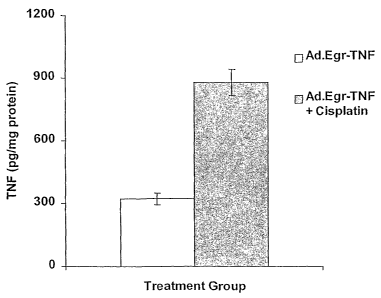
FIG. 3. TNF induction with 4 mg/kg platinum. TNF production in picograms
per mg protein as a function of time (5 days or 10 days) and treatment (Ad.TNF
= adenovirus
encoded tumor necrosis factor under control of the Egr-1 promoter; TNF/Pltmn =
Ad.TNF +
cisplatinum).
FIG. 4. Dose response of platinum in Ad.Egr.TNF-treated Seg-1. Fractional
tumor volume is measured as a function of time and treatment (Seg-1 =
esophageal carcinoma
cell line; pbs = phosphate buffered saline; Ad.TNF = adenovirus encoded tumor
necrosis factor
under control of the Egr-1 promoter; Platl = cisplatinum at 1 mg/kg; Plat3 =
cisplatinum at 3
mg/kg; Plat6 = cisplatinum at 6 mg/kg; Platl/TNF = cisplatinum at 1 mg/kg +
Ad.TNF;
Plat3/TNF = cisplatinum at 3 mg/kg + Ad.TNF; Plat6/TNF = cisplatinum at 6
mg/kg + Ad.TNF).
FIGS. 5A & 5B. In vitro measurement of TNF-a protein. TNF-a production by
Ad.Egr.TNF.I ID-infected cells exposed to IR (5 Gy) or cisplatin (5 M) was
measured using
ELISA. Significant levels of TNF-a protein were detected at 24, 48 and 72 hrs
following
exposure to Ad.Egr.TNF.11D + IR (P < 0.001) and Ad.Egr.TNF.11D + cisplatin (P<
0.001)
compared with vector alone in Seg-l cultures (FIG. 5A) and PROb cultures (FIG.
5B). Data are
reported as mean f SEM.
FIGS. 6A & 6B. In vitro reporter assays. Luciferase reporter constructs were
used
to evaluate induction of the Egr-l promoter by IR or cisplatin. Minimal
luciferase activity was
detectable following transfection with either the pGL3 (negative control) or
the pGL3 660
-6-
CA 02442971 2003-10-03
WO 02/080849 PCT/US02/10733
plasmid (minimal Egr-1 promoter) constructs. FIG. 6A. In Seg-1 cells, a 2.4-
fold increase
(P-0.005) in relative luciferase activity was observed following exposure to
IR (20 Gy) and a
2.0-fold increase (P=0.005) following exposure to cisplatin (50 gM). FIG. 6B.
In PROb cells, a
4.2-fold increase (P=0.004) in relative luciferase activity was observed
following exposure to IR
(20 Gy) and a 3.6-fold increase (P=0.01) following exposure to cisplatin (50
M). Data are
reported as mean SEM.
FIGS. 7A & 7B. In vivo measurement of TNF-a protein. TNF-a production by
Ad.Egr.TNF.11D-injected xenografts was measured by ELISA. A significant
increase in
intratumoral TNF-a protein concentration was observed following combined
treatment with
Ad.Egr.TNF.11D + cisplatin compared with treatment with Ad.Egr.TNF.11D vector
alone in
Seg-1 (FIG. 7A) (3.5-fold increase; P<0.05) and PROb (FIG. 7B) xenografts (2.7-
fold;
P<0.001). Data are reported as mean SEM.
FIGS. 8A & 8B. In vivo regrowth studies. The effect of combined treatment with
Ad.Egr.TNF.11D and cisplatin was evaluated by measuring the volume of
xenografts injected
with Ad.Null.3511.11D or Ad.Egr.TNF.11D with or without cisplatin. FIG. 8A. In
Seg-1
xenografts combined treatment with Ad.Egr.TNF.11D + cisplatin produced
significant tumor
regression compared with tumors treated with the Ad.Null + cisplatin at days
on days 4
(P=0.045), 6 (P<0.005), 8 (P<0.002), 10 (P<0.001), 12 (P<0.004), and 14
(P<0.021). FIG. 8B.
In PROb xenografts significant tumor regression was observed in the tumors
receiving combined
treatment with Ad.Egr.TNF. 11 D + cisplatin compared with tumors treated
Ad.Null + cisplatin at
days on days 4 (P=0.045), 6 (P<0.001), 8 (P=0.048), 10 (P<0.001), 12
(P<0.001), and 14
(P=0.002). Data are reported as mean SEM.
DESCRIPTION OF ILLUSTRATIVE EMBODIMENTS
,,25
The present invention stems in part from the inventors' observation that the
Egr-1
promoter, long known to contain radiation-responsive elements, also may be
induced by DNA
damaging chemicals. This surprising observation provides for a previously
unattempted
combination therapy for hyperproliferative diseases such as cancer - using an
expression
construct containing the Egr-1 promoter encoding an antitumor gene such a
tumor necrosis factor
(TNF) in conjunction with a DNA damaging chemical.
The combined therapeutic effect of the DNA damaging agent and induced
expression of
the therapeutic gene in cancer cells provides a superior result to use of
either agent alone and
also allows for using reduced dose-- of each agent. Being able to reduce any
systemic toxicity,
-7-
CA 02442971 2003-10-03
WO 02/080849 PCT/US02/10733
by reducing the amount of radiation and/or drug and/or vector administered, is
highly
advantageous. The following disclosure provides a detailed description of the
foregoing
embodiments, as well as variations thereof.
A transcriptional targeting strategy is provided whereby chemotherapeutic
agents in
conjunction with inducible expression vectors that encode for genes with
antitumor effects may
be used to effectively treat tumors, where the vectors are induced by the
chemotherapeutic agent.
Thus, expression constructs comprising the inducible Egr-1 promoter and
encoding for any
antitumor gene in conjunction with a chemotherapeutic agent that can induce
and activate the
Egr-1 promoter, via DNA damage or production of ROI's, are provided.
With a selective tumor-targeting vector, a genetic construct that expresses an
antitumor
gene that is inducible by a chemotherapeutic enhances the effects of the
chemotherapeutic. as.
well as the antitumor agent. As both the chemotherapeutic agent and the
antitumor gene will
generally have different mechanisms of tumor cell killing therefore, cells
resistant to one agent
may be sensitive to the other. It is also contemplated that such combinations
may enhance the
local effects of combination chemo-radiation therapy or other adjunct cancer
therapies.
A. Egr-1 Promoter
The Egr-1 promoter is defined herein as those 5' regulatory sequences
necessary to
control the DNA damaging agent-induced transcription of downstream sequences
operably
connected thereto. The Egr-1 promoter has complex structure which has
previously been
analyzed in the context of radiation- and H202-induced gene expression. It
contains multiple
ETS binding sites (ETS are transcriptional regulatory proteins), three of
which exist as parts of
two serum response elements (SRE's), SREI and SREII. The SRE's, also known as
CArG
motifs, are cis-elements that regulate the expression of many growth factor
responsive genes.
There are a total of six SRE's, each comprising the consensus CC(A+T-rich)6GG
sequence.
The present inventors have previously demonstrated that a chimeric genetic
construct
consisting of the 5' Egr-1 CArG elements ligated to the TNF-a cDNA express
high, levels of
intratumoral TNF-a following IR exposure of cells transduced with this
construct. Tumors
transduced with the chimeric Egr-TNF construct and treated with IR exhibited
increased
regression/cures compared with tumors treated with either agent alone, likely
due to the
intratumoral induction of TNF-a production by IR, and the cytotoxic
interaction of TNF-a and
IR on the tumor cells and the tumor vasculature (Weichselbaum et al., 2001;
Staba et al., 1998).
In the present invention, the inventors used cisplatin, a commonly used
chemotherapeutic agent
that alters intracellular radical oxygen formation and damages DNA, to induce
the TNF-a gene
-8-
CA 02442971 2003-10-03
WO 02/080849 PCT/US02/10733
under control of the DNA damage / ROI inducible CArG elements of the Egr-l
promoter. The
invention 'therefore provides the use of agents that cause DNA damage and/or
produce ROI to
induce Egr-1 and therefore to drive the expression of genes under the control
of Egr-1 in
expression vectors.
B. DNA Damaging Chemicals
The term "DNA damaging chemical" refers to the any drug that induces, either
directly
or indirectly, damage, to a DNA molecule. Of particular interest in the
present invention are
those drugs that generate free radicals. The following categories of chemicals
are believed to
effect DNA damage through one or more pathways.
1. Alkylating Agents
Alkylating agents are drugs that directly interact with genomic DNA to prevent
the
cancer cell from proliferating. This category of chemotherapeutic drugs
represents agents that
affect all phases of the cell cycle, that is, they are not phase-specific.
Alkylating agents can be
implemented to treat, for example, chronic leukemia, non-Hodgkin's lymphoma,
Hodgkin's
disease, multiple myeloma, and particular cancers of the breast, lung, and
ovary. An alkylating
agent, may include, but is not limited to, a nitrogen mustard, an
ethylenimene, a
methylmelamine, an alkyl sulfonate, a nitrosourea or a triazines.
They include but are not limited to: busulfan, chlorambucil, cisplatin,
cyclophosphamide
(cytoxan), dacarbazine, ifosfamide, mechlorethamine (mustargen), and
melphalan. In specific
aspects, troglitazaone can be used to treat cancer in combination with any one
or more of these
alkylating agents, some of which are discussed below.
i. Nitrogen Mustards
A nitrogen mustard may be, but is not limited to, mechlorethamine (HN2), which
is used
for Hodgkin's disease and non-Hodgkin's lymphomas; cyclophosphamide and/or
ifosfamide,
which are used in treating such cancers as acute or chronic lymphocytic
leukemias, Hodgkin's
disease, non-Hodgkin's lymphomas, multiple myeloma, neuroblastoma, breast,
ovary, lung,
Wilm's tumor, cervix testis and soft tissue sarcomas; melphalan (L-
sarcolysin), which has been
used to treat such cancers as multiple myeloma, breast and ovary; and
chlorambucil, which has
been used to treat diseases such as, for example, chronic lymphatic
(lymphocytic) leukemia,
malignant lymphomas including lymphosarcoma, giant follicular lymphoma,
Hodgkin's disease
and non-Hodgkin's lymphomas.
-9-
CA 02442971 2003-10-03
WO 02/080849 PCT/US02/10733
a. Chlorambucil
Chlorambucil (also known as leukeran) is a bifunctional alkylating agent of
the nitrogen
mustard type that has been found active against selected human neoplastic
diseases.
Chlorambucil is known chemically as 4-[bis(2-chlorethyl)amino] benzenebutanoic
acid.
Chlorambucil is available in tablet form for oral administration. It is
rapidly and
completely absorbed from the gastrointestinal tract. For example, after a
single oral doses of
about 0.6 mg/kg to about 1.2 mg/kg, peak plasma chlorambucil levels are
reached within one
hour and the terminal half-life of the parent drug is, estimated at about 1.5
hours... About
0.1 mg/kg/day to about 0.2 mg/kg/day or about 3 6 mg/m2/day to about 6
mg/m2/day or
alternatively about 0.4 mg/kg may be used for antineoplastic treatment.
Chlorambucil is not
curative by itself but may produce clinically useful palliation.
b. Cyclophosphamide
Cyclophosphamide is 2H-1,3,2-Oxazaphosphorin-2-amine, N,N-bis(2-
chloroethyl)tetrahydro-, 2-oxide, monohydrate; termed Cytoxan available from
Mead Johnson;
and Neosar available from Adria. Cyclophosphamide is prepared by condensing 3-
amino-l-
propanol with N,N-bis(2-chlorethyl) phosphoramidic dichloride [(C1CH2CH2)2N--
POC12] in
dioxane solution under the catalytic influence of triethylamine. The
condensation is double,
involving both the hydroxyl and the amino groups, thus effecting the
cyclization.
Unlike other 13-chloroethylamino alkylators, it does not cyclize readily to
the active
ethyleneimonium form until activated by hepatic enzymes. Thus, the substance
is stable in the
gastrointestinal tract, tolerated well and effective by the oral and parental
routes and does not
cause local vesication, necrosis, phlebitis or even pain.
Suitable oral doses for adults include, for example, about 1 mg/kg/day to
about
5 mg/kg/day (usually in combination), depending upon gastrointestinal
tolerance; or about
1 mg/kg/day to about 2 mg/kg/day; intravenous doses include, for example,
initially about
40 mg/kg to about 50 mg/kg in divided doses over a period of about 2 days to
about 5 days or
about 10 mg/kg to about 15 mg/kg about every 7 days to about 10 days or about
3 mg/kg to
about 5 mg/kg twice a week or about 1.5 mg/kg/day to about 3 mg/kg/day. In
some aspects, a
dose of about 250 mg/kg/day may be administered as an antineoplastic. Because
of
gastrointestinal adverse effects, the intravenous route is preferred for
loading. During
maintenance, a leukocyte count of about 3000/mm3 to 4000/mm3 usually is
desired. The drug
also sometimes is administered intramuscularly, by infiltration or into body
cavities. It is
-10-
CA 02442971 2003-10-03
WO 02/080849 PCT/US02/10733
available in dosage forms for injection of about 100 mg, about 200 mg and
about 500 mg, and
tablets of about 25 mg and about 50 mg.
c. Melphalan
Melphalan, also known as alkeran, L-phenylalanine mustard, phenylalanine
mustard, L-
PAM, or L-sarcolysin, is a phenylalanine derivative of nitrogen mustard.
Melphalan is a
bifunctional alkylating agent which is active -against selective human
neoplastic diseases. It is
known chemically as 4-[bis(2-chloroethyl)amino]-L-phenylalanine.
Melphalan is the active L-isomer of the compound and was first synthesized in
1953 by
Bergel and Stock; the D-isomer, known as medphalan, is less active against
certain animal
tumors, and the dose needed to produce effects on chromosomes is larger than
that required with
the L-isomer. The racemic (DL-) form is known as merphalan or sarcolysin.
Melphalan is
insoluble in water and has a pKal of about 2.1. Melphalan is available in
tablet form for oral
administration and has been used to treat multiple myeloma. Available evidence
suggests that
about one third to one half of the patients with multiple myeloma show a
favorable response to
oral administration of the drug.
Melphalan has been used in the treatment of epithelial ovarian carcinoma. One
commonly employed regimen for the treatment of ovarian carcinoma has been to
administer
melphalan at a dose of about 0.2 mg/kg daily for five days as a single course.
Courses are
repeated about every four to five weeks depending upon hematologic tolerance
(Smith and
Rutledge, 1975; Young et al., 1978). Alternatively, in certain embodiments,
the dose of
melphalan used could be as low as about 0.05 mg/kg/day or as high as about 3
mg/kg/day or
greater.
ii. Ethylenimenes and Methymelamines
An ethylenimene and/or a methylmelamine include, but are not limited to,
hexamethylmelamine, used to treat ovary cancer; and thiotepa, which has been
used to treat
bladder, breast and ovary cancer.
W. Alkyl Sulfonates
An alkyl sulfonate includes but is not limited to such drugs as busulfan,
which has been
used to treat chronic granulocytic leukemia. Busulfan (also known as myleran)
is a bifunctional
alkylating agent.. Busulfan is known chemically as 1,4-butanediol
dimethanesulfonate. Busulfan
is available in tablet form for oral administration, wherein for example, each
scored tablet
-11-
CA 02442971 2003-10-03
WO 02/080849 PCT/US02/10733
contains about 2 mg busulfan and the inactive ingredients magnesium stearate
and sodium
chloride.
Busulfan is indicated for the palliative treatment of chronic myelogenous
(myeloid,
myelocytic, granulocytic) leukemia. Although not curative, busulfan reduces
the total
granulocyte mass, relieves symptoms of the disease, and improves the clinical
state of the
patient. Approximately 90% of adults with previously untreated chronic
myelogenous leukemia
will obtain hematologic remission with regression or stabilization of
organomegaly following the
use of busulfan. Busulfan, has been shown to be superior to splenic
irradiation with respect to
survival times and maintenance of hemoglobin levels, and to be equivalent to
irradiation at
controlling splenomegaly.
iv. Nitrosoureas
Nitrosureas, like alkylating agents, inhibit DNA repair proteins. They are
used to treat
non-Hodgkin's lymphomas, multiple myeloma, malignant melanoma, in addition to
brain tumors.
A nitrosourea include but is not limited to a carmustine (BCNU), a lomustine
(CCNU), a
semustine (methyl-CCNU) or a streptozocin. Semustine has been used in such
cancers as a
primary brain tumor, a stomach or a colon cancer. Stroptozocin has been used
to treat diseases
such as a malignant pancreatic insulinoma or a malignant carcinoid.
Streptozocin has been used
to treat such cancers as a malignant melanoma, Hodgkin's disease and soft
tissue sarcomas.
a. Carmustine
Carmustine (sterile carmustine) is one of the nitrosoureas used in the
treatment of certain
neoplastic diseases. It is 1,3 bis (2-chloroethyl)-1-nitrosourea. It is
lyophilized pale yellow
flakes or congealed mass with a molecular weight of 214.06. It is highly
soluble in alcohol and
lipids, and poorly soluble in water. Carmustine is administered by intravenous
infusion after
reconstitution as recommended
Although it is generally agreed that carmustine alkylates DNA and RNA, it is
not cross
resistant with other alkylators. As with other nitrosoureas, it may also
inhibit several key
enzymatic processes by carbamoylation of amino acids in proteins.
Carmustine is indicated as palliative therapy as a single agent or in
established
combination therapy with other approved chemotherapeutic agents in brain
tumors such as
glioblastoma, brainstem glioma, medullobladyoma, astrocytoma, ependymoma, and
metastatic
brain tumors. Also it has been used in combination with prednisone to treat
multiple myeloma.
Carnustine has been used in treating such cancers as a multiple myeloma or a
malignant
-12-
CA 02442971 2003-10-03
WO 02/080849 PCT/US02/10733
melanoma. Carmustine has proved useful, in the treatment of Hodgkin's Disease
and in non-
Hodgkin's lymphomas, as secondary therapy in combination with other approved
drugs in
patients who relapse while being treated with primary therapy, or who fail to
respond to primary
therapy.
Sterile carmustine is commonly available in 100 mg single dose vials of
lyophilized
material. The recommended dose of carmustine as a single agent in previously
untreated patients
is about 150,mg/m2 to about 200 mg/m2 intravenously every 6 weeks. This may be
given as a
single dose or divided into daily injections such as about 75 mg/m2 to about
100 mg/m2 on
2 successive days. When carmustine is used in combination with other
myelosuppressive drugs
or in patients in whom bone marrow reserve is depleted, the doses should be
adjusted
accordingly. Doses subsequent to the initial dose should be adjusted according
to the
hematologic response of the patient to the preceding dose. It is of course
understood that other
doses may be used in the present invention, for example about 10 mg/m2, about
20 mg/m2, about
30 mg/m2, about 40 mg/m2, about 50 mg/m2, about 60 mg/m2, about 70 mg/m2,
about 80 mg/m2,
about 90 mg/m2 to about 100 mg/m2.
b. Lomustine
Lomustine is one of the nitrosoureas used in the treatment of certain
neoplastic diseases.
It is 1-(2-chloro-ethyl)-3-cyclohexyl-1 nitrosourea. It is a yellow powder
with the empirical
formula of C9H16C1N302 and a molecular weight of 233.71. Lomustine is soluble
in 10%
ethanol (about 0.05 mg/mL) and in absolute alcohol (about 70 mg/mL). Lomustine
is relatively
insoluble in water (less than about 0.05 mg/mL). It is relatively unionized at
a physiological pH.
Inactive ingredients in lomustine capsules are: magnesium stearate and
mannitol.
Although it is generally agreed that lomustine alkylates DNA and RNA, it is
not cross
resistant with other alkylators. As with other nitrosoureas, it may also
inhibit several key
enzymatic processes by carbamoylation of amino acids in proteins.
Lomustine may be given orally. Following oral administration of radioactive
lomustine
at doses ranging from about 30 mg/m2 to 100 mg/m2, about half of the
radioactivity given was
excreted in the form of degradation products within 24 hours. The serum half-
life of the
metabolites ranges from about 16 hours to about 2 days. Tissue levels are
comparable to plasma
levels at 15 minutes after intravenous administration.
Lomustine has been shown to be useful as a single agent in addition to other
treatment
modalities, or in established combination therapy with other approved
chemotherapeutic agents
in both primary and metastatic brain tumors, in patients who have already
received appropriate
-13-
CA 02442971 2003-10-03
WO 02/080849 PCT/US02/10733
surgical and/or radiotherapeutic procedures. Lomustine has been used to treat
such cancers as
small-cell lung cancer. It has also proved effective in secondary therapy
against Hodgkin's
Disease in combination with other approved drugs in patients who relapse while
being treated
with primary therapy, or who fail to respond to primary therapy.
The recommended dose of lomustine in adults and children as a single agent in
previously untreated patients is about 130 mg/m2 as a single oral dose every 6
weeks. In
individuals with compromised bone marrow function, the dose should be reduced
to about
100 mg/m2 every 6 weeks. When lomustine is used in combination with other
myelosuppressive
drugs, the doses should be adjusted accordingly. It is understood that other
doses may be used
for example, about 20 mg/m2, about 30mg/m2, about 40 mg/m2, about 50 mg/r2,
about
60 mg/m2, about 70 mg/in2, about 80 Mg/1112 , about 90 mg/m2, about 100 mg/m2
to about
120 mg/m2.
c. Triazine
A triazine include but is not limited to such drugs as a dacabazine (DTIC;
dimethyltriazenoimidaz olecarboxamide), used in the treatment of such cancers
as a malignant
melanoma, Hodgkin's disease and a soft-tissue sarcoma.
II. Antimetabolites
Antimetabolites disrupt DNA and RNA synthesis. Unlike alkylating agents, they
specifically influence the cell cycle during S phase. They have used to combat
chronic
leukemias in addition to tumors of breast, ovary and the gastrointestinal
tract. Antimetabolites
can be differentiated into various categories, such as folic acid analogs,
pyrimidine analogs and
purine analogs and related inhibitory compounds. Antimetabolites include but
are not-limited to,
5-fluorouracil (5-FU), cytarabine (Ara-C), fludarabine, gemcitabine, and
methotrexate.
i. Folic Acid Analogs
Folic acid analogs include but are not limited to compounds such as
methotrexate
(amethopterin), which has been used in the treatment of cancers such as acute
lymphocytic
leukemia, choriocarcinoma, mycosis fungoides, breast, head and neck, lung and
osteogenic
sarcoma.
-14-
CA 02442971 2003-10-03
WO 02/080849 PCT/US02/10733
ii. Pyrimidine Analogs
Pyrimidine analogs include such compounds as cytarabine (cytosine
arabinoside), 5-
fluorouracil (fluouracil; 5-FU) and floxuridine (fluorode-oxyuridine; FudR).
Cytarabine has
been used in the treatment of cancers such as acute granulocytic leukemia and
acute lymphocytic
leukemias. Floxuridine and 5-fluorouracil have been used in the treatment of
cancers such as
breast, colon, stomach, pancreas, ovary, head and neck, urinary bladder and
topical premalignant
skin lesions.
5-Fluorouracil (5-FU) has the chemical name of 5-fluoro-2,4(1H,3H)-
pyrimidinedione.
Its mechanism of action is thought to be by blocking the methylation reaction
of deoxyuridylic
acid to thymidylic acid. Thus, 5-FU interferes with the synthesis of
deoxyribonucleic acid
(DNA) and to a lesser extent inhibits the formation of ribonucleic acid (RNA).
Since DNA and
RNA are essential for cell division and proliferation, it is thought that the
effect of 5-FU is to
create a thymidine deficiency leading to cell death. Thus, the effect of 5-FU
is found in cells that
rapidly divide, a characteristic of metastatic cancers.
iii. Purine Analogs and Related Inhibitors
Purine analogs and related compounds include, but are not limited to,
mercaptopurine (6-
mercaptopurine; 6-MP), thioguanine (6-thioguanine; TG) and pentostatin (2-
deoxycoformycin).
Mercaptopurine has been used in acute lymphocytic, acute granulocytic and
chronic granulocytic
leukemias. Thrioguanine has been used in the treatment of such cancers as
acute granulocytic
leukemia, acute lymphocytic leukemia and chronic lymphocytic leukemia.
Pentostatin has been
used in such cancers as hairy cell leukemias, mycosis fungoides and chronic
lymphocytic
leukemia.
III. Natural Products
Natural products generally refer to compounds originally isolated from a
natural source,
and identified has having a pharmacological activity. Such compounds, analogs
and derivatives
thereof may be, isolated from a natural source, chemically synthesized or
recombinantly
produced by any technique known to those of skill in the art. Natural products
include such
categories as mitotic inhibitors, antitumor antibiotics, enzymes and
biological response
modifiers.
-15-
CA 02442971 2003-10-03
WO 02/080849 PCT/US02/10733
L Mitotic Inhibitors
Mitotic inhibitors include plant alkaloids and other natural agents that can
inhibit either
protein synthesis required for cell division or mitosis. They operate during a
specific phase
during the cell cycle. Mitotic inhibitors include, for example, docetaxel,
etoposide (VP16),
teniposide, paclitaxel, taxol, vinblastine, vincristine, and vinorelbine.
a. Epipodophyllotoxins
Epipodophyllotoxins include such compounds as teniposide and VP16. VP16 is
also
known as etoposide and is used primarily for treatment of testicular tumors,
in combination with
bleomycin and cisplatin, and in combination with cisplatin for small-cell
carcinoma of the lung.
Teniposide and VP16 are also active against cancers such as testis, other lung
cancer, Hodgkin's
disease, non-Hodgkin's lymphomas, acute granulocytic leukemia, acute
nonlymphocytic
leukemia, carcinoma of the breast, and Kaposi's sarcoma associated with
acquired
immunodeficiency syndrome (AIDS).
VP16 is available as a solution (e.g., 20 mg/ml) for intravenous
administration and as
50 mg, liquid-filled capsules for oral use. For small-cell carcinoma of the
lung, the intravenous
dose (in combination therapy) is can be as much as about 100 mg/m2 or as
little as about 2 mg/
m2, routinely about 35 mg/m2, daily for about 4 days, to about 50 mg/m2, daily
for about 5 days
have also been used. When given orally, the dose should be doubled. Hence the
doses for small
cell lung carcinoma may be as high as about 200 mg/m2 to about 250 mg/m2. The
intravenous
dose for testicular cancer (in combination therapy) is about 50 mg/m2 to about
100 mg/m2 daily
for about 5 days, or about 100 mg/m2 on alternate days, for three doses.
Cycles of therapy are
usually repeated about every 3 to 4 weeks. The drug should be administered
slowly (e.g., about
minutes to about 60 minutes) as an infusion in order to avoid hypotension and
bronchospasm,
25 which are probably due to the solvents used in the formulation.
b. Taxoids
Taxoids are a class of related compounds isolated from the bark of the ash
tree, Taxzts
brevifolia. Taxoids include but are not limited to compounds such as docetaxel
and paclitaxel.'
30 Paclitaxel binds to tubulin (at a site distinct from that used by the vinca
alkaloids) and
promotes the assembly of microtubules. Paclitaxel is being evaluated
clinically; it has activity
against malignant melanoma and carcinoma of the ovary. In certain aspects,
maximal doses are
about 30 mg/m2 per day for about 5 days or about 210 mg/m2 to about 250 mg/m2
given once
about every 3 weeks.
-16-
CA 02442971 2003-10-03
WO 02/080849 PCT/US02/10733
c. Vinca Alkaloids
Vinca alkaloids are a type of plant alkaloid identified to have pharmaceutical
activity.
They include such compounds as vinblastine (VLB) and vincristine.
1. Vinblastine
Vinblastine is an example of a plant alkaloid that can be used for the
treatment of cancer
and precancer.; When cells are incubated with vinblastine, dissolution of the
microtubules
occurs.
Unpredictable absorption has been reported after oral administration of
vinblastine or:
vincristine. At the usual clinical doses the peak concentration of each drug
in plasma is
approximately' 0.4 mM. Vinblastine and vincristine bind to plasma proteins.
They are
extensively concentrated in platelets and to a lesser extent in leukocytes and
erythrocytes.
After intravenous injection, vinblastine has a multiphasic pattern of
clearance from the
plasma; after distribution, drug disappears from plasma with half-lives of
approximately 1 and
hours. Vinblastine is metabolized in the liver to biologically activate
derivative
desacetylvinblastine. Approximately 15% of an administered dose is detected
intact in the urine,
and about 10% is recovered in the feces after biliary excretion. Doses should
be reduced in
patients with hepatic dysfunction. At least a 50% reduction in dosage is
indicated if the
20 concentration, of bilirubin in plasma is greater than 3 mg/dl (about 50
mM).
Vinblastine sulfate is available in preparations for injection. When the dnig
is given
intravenously;, special precautions must be taken against subcutaneous
extravasation, since this
may cause painful irritation and ulceration. The drug should not be injected
into an extremity
with impaired circulation. After a single dose of 0.3 mg/kg of body weight,
myelosuppression
reaches its maximum in about 7 days to about 10 days. If a moderate level of
leukopenia
(approximately 3000 cells/mm3) is not attained, the weekly dose may be
increased gradually by
increments of about 0.05 mg/kg of body weight. In regimens designed to cure
testicular cancer,
vinblastine is used in doses of about 0.3 mg/kg about every 3 weeks
irrespective of blood cell,
counts or toxicity.
An important clinical use of vinblastine is with bleomycin and cisplatin in
the curative
therapy of metastatic testicular tumors. Beneficial responses have been
reported in various
lymphomas, particularly Hodgkin's disease, where significant improvement may
be noted in 50
to 90% of cases. The effectiveness of vinblastine in a high proportion of
lymphomas is not
diminished when the disease is refractory to alkylating agents. It is also
active in Kaposi's
-17-
CA 02442971 2003-10-03
WO 02/080849 PCT/US02/10733
sarcoma, testis cancer, neuroblastoma, and Letterer-Siwe disease
(histiocytosis X), as well as in
carcinoma of the breast and choriocarcinoma in women.
Doses of about 0.1 mg/kg to about 0.3 mg/kg can be administered or about 1.5
mg/m2 to
about 2 mg/in2 can also be administered. Alternatively, about 0.1 mg/m2, about
0.12 mg/m2,
about 0.14 mg/m2, about 0.15 mg/m2, about 0.2 mg/m2, about 0.25 mg/m2, about
0.5 mg/m2,
about 1.0 mg/m2, about 1.2 mg/m2, about 1.4 mg/m2, about 1.5 mg/m2, about 2.0
mg/m2, about
2.5 mg/m2, about 5.0 mg/m2, about 6 mg/m2, about 8 mg/m2, about 9 mg/m2, about
10 mg/m2, to
about 20 mg/m2, can be given.
2. Vincristine
Vincristine blocks mitosis and produces metaphase arrest. It seems likely that
most of
the biological activities of this drug can be explained by its ability to bind
specifically to tubulin
and to block the ability of protein to polymerize into microtubules. Through
disruption of the
microtubules of the mitotic apparatus, cell division is arrested in metaphase.
The inability to
segregate chromosomes correctly during mitosis presumably leads to cell death.
The relatively low toxicity of vincristine for normal marrow cells and
epithelial cells
make this agent unusual among anti-neoplastic drugs, and it is often included
in combination
with other myelosuppressive agents. Unpredictable absorption has been reported
after oral
administration of vnblastine or vincristine. At the usual clinical doses the
peak concentration of
each drug in plasma is about 0.4 mM.
Vinblastine and vincristine bind to plasma proteins. They are extensively
concentrated in
platelets and to a lesser extent in leukocytes and erythrocytes. Vincristine
has a multiphasic
pattern of clearance from the plasma; the terminal half-life is about ' 24
hours. The. drug is
metabolized in the liver, but no biologically active derivatives have been
identified. Doses
should be reduced in patients with hepatic dysfunction. At least a 50%
reduction in dosage is
indicated if the concentration of bilirubin in plasma is greater than about 3
mg/dl (about 50 mM).
Vincristine sulfate is available as a solution (e.g., 1 mg/ml) for intravenous
injection.
Vincristine used together with corticosteroids is presently the treatment of
choice to induce
remissions in childhood leukemia; the optimal dosages for these drugs appear
to be vincristine,
intravenously, about 2 mg/m2 of body-surface area, weekly; and prednisone,
orally, about
mg/m2, daily. Adult. patients with Hodgkin's disease or non-Hodgkin's
lymphomas usually
receive vincristine as a : part of a complex protocol. When used in the MOPP
regimen, the
recommended dose of vincristine is about 1.4 mg/m2. High doses of vincristine
seem to be
tolerated better by children with leukemia than by adults, who may experience
sever
-18-
CA 02442971 2003-10-03
WO 02/080849 PCT/US02/10733
neurological toxicity. Administration of the drug more frequently than every 7
days or at higher
doses seems to increase the toxic manifestations without proportional
improvement in the
response rate. Precautions should also be used to avoid extravasation during
intravenous
administration of vincristine. Vincristine (and vinblastine) can be infused
into the arterial blood
supply of tumors in doses several times larger than those that can be
administered intravenously
with comparable toxicity.
Vincristine has been effective in Hodgkin's disease and other lymphomas.
Although it
appears to be somewhat less beneficial than vinblastine when used alone in
Hodgkin's disease,
when used with mechlorethamine, prednisone, and procarbazine (the so-called
MOPP regimen),
it is the preferred treatment for the advanced stages (III and IV) of this
disease. In non-
Hodgkin's lymphomas, vincristine is an important agent, particularly when used
with
cyclophosphamide, bleomycin, doxorubicin, and prednisone. Vincristine is more
useful than
vinblastine in lymphocytic leukemia. Beneficial response have been reported in
patients with a
variety of other neoplasms, particularly Wilms' tumor, neuroblastoma, brain
tumors,
rhabdomyosarcoma, small cell lung, and carcinomas of the breast, bladder, and
the male and
female reproductive systems.
Doses of vincristine include about 0.01 mg/kg to about 0.03 mg/kg or about 0.4
mg/m2 to
'about 1.4 mg/m2 can be administered or about 1.5 mg/m2 to about 2 mg/m2 can
also be
administered. Alternatively, in certain embodiments, about 0.02 mg/m2, about
0.05 mg/m2,
about 0.06 mg/m2, about 0.07 mg/m2, about 0.08 mg/m2, about 0.1 mg/m2, about
0.12 mg/m2,
about 0.14 mg/ma, about 0.15 mg/m2, about 0.2 mg/m2, about 0.25 mg/m2 can be
given as a
constant intravenous infusion.
d. Antitumor Antibiotics
Antitumor antibiotics have both antimicrobial and cytotoxic activity. These
drugs also
interfere with DNA by chemically inhibiting enzymes and mitosis or altering
cellular
membranes. These agents are not phase specific so they work in all phases of
the cell cycle.
Thus, they are widely used for a variety of cancers. Examples of antitumor
antibiotics include,
but are not limited to, bleomycin, dactinomycin, daunorubicin, doxorubicin
(Adriamycin),
plicamycin (mithramycin) and idarubicin. Widely used in clinical setting for
the treatment of
neoplasms these compounds generally are administered through intravenous bolus
injections or
orally.
-19-
CA 02442971 2003-10-03
WO 02/080849 PCT/US02/10733
1. Doxorubicin
Doxorubicin hydrochloride, 5,12-Naphthacenedione, (8s-cis)-l0-[(3-amino-2,3,6-
trideoxy-a-L-lyxo-hexopyranosyl)oxy]-7,8,9,10-tetrahydro-6,8,11-trihydroxy-8-
(hydroxyacetyl)-
1-methoxy-hydrochloride (hydroxydaunorubicin hydrochloride, Adriamycin) is
used in a wide
antineoplastic spectrum. It binds to DNA and inhibits nucleic acid synthesis,
inhibits mitosis and
promotes chromosomal aberrations.
Administered alone, it is the drug of first choice for the treatment of
thyroid adenoma and
primary hepatocellular carcinoma. It is a component of 31 first-choice
combinations for the
treatment of diseases including ovarian, endometrial and breast tumors,
bronchogenic oat-cell
carcinoma, - non-small cell lung carcinoma, stomach, genitourinary, thyroid,
gastric
adenocarcinoma, retinoblastoma, neuroblastoma, mycosis fungoides, pancreatic
carcinoma,
prostatic carcinoma, bladder carcinoma, myeloma, diffuse histiocytic lymphoma,
Wilms' tumor,
Hodgkin's disease, adrenal tumors, osteogenic sarcoma, soft tissue sarcoma,
Ewing's sarcoma,
rhabdomyosarcoma and acute lymphocytic leukemia. It is an alternative drug for
the treatment
of'other diseases such as islet cell, cervical, testicular and adrenocortical
cancers. It is also an
immunosuppressant.
Doxorubicin is absorbed poorly and is preferably administered intravenously.
The.
-pharmacokinetics are multicompartmental. Distribution phases have half-lives
of 12 minutes
and 3.3 hours. The elimination half-life is about 30 hours, with about 40% to
about 50%
secreted into the bile. Most of the remainder is metabolized in the liver,
partly to an active
metabolite (doxorubicinol), but a few percent is excreted into the urine. In
the presence of liver
impairment, the dose should be reduced.
In certain embodiments, appropriate intravenous doses are, adult, about 60
mg/m2 to
about 75 mg/m2 at about 21-day intervals or about 25 mg/m2 to about 30 mg/m2
on each of 2 or 3
successive days repeated at about 3 week to about 4 week intervals or about 20
mg/m2 once a
week. The lowest dose should be used in elderly patients, when there is prior
bone-marrow
depression caused by prior chemotherapy or neoplastic marrow invasion, or when
the drug is
combined with other myelopoietic suppressant drugs. The dose should be reduced
by about 50%
if the serum bilirubin lies between about 1.2 mg/dL and about 3 mg/dL and by
about 75% if
above about 3 mg/dL. The lifetime total dose should not exceed about 550 mg/m2
in patients
with normal heart function and about 400 mg/m2 in persons having received
mediastinal
irradiation. In certain embodiments, and alternative dose regiment may
comprise about
30 mg/m2 on each of 3 consecutive days, repeated about every 4 week. Exemplary
doses may be
about 10 mg/m2, about 20 mg/m2, about 30 mg/m2, about 50 mg/m2, about 100
mg/m2, about
-20-
CA 02442971 2003-10-03
WO 02/080849 PCT/US02/10733
150 mg/m2, about 175 mg/m2, about 200 mg/m2, about 225 mg/m2, about 250 mg/m2,
about
275 mg/m2, about 300 mg/m2, about 350 mg/m2, about 400 mg/m2, about 425 mg/m2,
about
450 mg/m2 about 475 mg/m2, to about 500 mg/m2.
2. Daunorubicin
Daunorubicin hydrochloride, 5,12-Naphthacenedione, (8S-cis)-8-acetyl-10-[(3-
amino-
2,3,6-trideoxy-a-L-lyxo-hexanopyranosyl)oxy]-7,8,9,10-tetrahydro-6,8,11-
trihydroxy-10-
methoxy-, hydrochloride; also termed cerubidine and available from Wyeth.
Daunorubicin
(daunomycin; rubidomycin) intercalates into DNA, blocks DAN-directed RNA
polymerase and
inhibits DNA synthesis. It can prevent cell division in -doses that do not
interfere with nucleic
acid synthesis.
In combination with other drugs it is often included in the first-choice
chemotherapy of
diseases such as, for example, acute granulocytic leukemia, acute myelocytic
leukemia in adults
(for induction of remission), acute lymphocytic leukemia and the acute phase
of chronic
, myelocytic leukemia. Oral absorption is poor, and it preferably given by
other methods (e.g.,
intravenously). The half-life of distribution is 45 minutes and of
elimination, about 19 hours.
The half-life of its active metabolite, daunonibicinol, is about 27 hours.
Daunorubicin is
metabolized mostly in the liver and also secreted into the bile (about 40%).
Dosage must be
reduced in liver or renal insufficiencies.
Generally, suitable intravenous doses are (base equivalent): adult, younger
than 60 years,
about 45 mg/m2/day (about 30 mg/m2 for patients older than 60 year.) for about
1 day, about
2 days or about 3 days about every 3 weeks or 4 weeks or about 0.8 mg/kg/day
for about 3 days,
about 4 days, about 5 days to about 6 days about every 3 weeks or about 4
weeks; no more than
about 550 mg/m2 should be given in a lifetime, except only about 450 mg/m2 if
there has been
chest irradiation; children, about 25 mg/m2 once a week unless the age is less
than 2 years. or the
body surface less than about 0.5 in, in which case the weight-based adult
schedule is used. It is
available in injectable dosage forms (base equivalent) of about 20 mg (as the
base equivalent to
about 21.4 mg of the hydrochloride). Exemplary doses may be about 10 mg/m2,
about
20 mg/m2, about 30 mg/m2, about 50 mg/m2, about 100 mg/m2, about 150 mg/m2,
about
175 mg/m2, about 200 mg/m2, about 225 mg/m2, about 250 mg/m2, about 275 mg/m2,
about
300 mg/m2, about 350 mg/m2, about 400 mg/m2, about 425 mg/m2, about 450 mg/m2,
about
475 mg/m2, to about 500 mg/m2.
-21-
CA 02442971 2003-10-03
WO 02/080849 PCT/US02/10733
3. Mitomycin
Mitomycin (also known as mutamycin and/or mitomycin-C) is an antibiotic
isolated from
the broth of Str=eptornyces caespitosus which has been shown to have antitumor
activity. The
compound is heat stable, has a high melting point, and is freely soluble in
organic solvents.
Mitomycin selectively inhibits the synthesis of deoxyribonucleic acid (DNA).
The
guanine and cytosine content correlates with the degree of mitomycin-induced
cross-linking. At
high' concentrations of the drug, cellular RNA and protein synthesis are also
suppressed.
Mitomycin has been used in tumors such as stomach, cervix, colon, breast,
pancreas, bladder and,
head and neck.
. In humans, mitomycin is rapidly cleared from the serum after intravenous
administration.
Time required to reduce the serum concentration by about 50% after a 30 mg.
bolus injection is
17 minutes. After injection of 30 mg, 20 mg, or 10 mg I.V., the maximal serum
concentrations
were 2.4 mg/mL, 1.7 mg/mL, and 0.52 mg/mL, respectively. Clearance is effected
primarily by
metabolism in the liver, but metabolism occurs in other tissues as well. The
rate of clearance is
inversely proportional to the maximal serum concentration because, it is
thought, of saturation of
the degradative pathways. Approximately 10% of a dose of mitomycin is excreted
unchanged in
the urine. Since metabolic pathways are saturated at relatively low doses, the
percent of a dose
excreted in urine increases with increasing dose. In children, excretion of
intravenously
administered mitomycin is similar.
4. Actinomycin D
Actinomycin D (Dactinomycin) [50-76-0]; C62H86N12016 (1255.43) is an
antineoplastic
drag that inhibits DNA-dependent RNA polymerase. It is often a component of
first-choice
combinations for treatment of diseases such as, for example, choriocarcinoma,
embryonal
rhabdomyosarcoma, testicular tumor, Kaposi's sarcoma and Wilms' tumor. Tumors
that fail to
respond to systemic treatment sometimes respond to local perfusion.
Dactinomycin potentiates
radiotherapy. It is a secondary (efferent) immunosuppressive.
In certain specific aspects, actinomycin D is used in combination with agents
such as, for
example, primary surgery, radiotherapy, and other drugs, particularly
vincristine and
cyclophosphamide. Antineoplastic activity has also been noted in Ewing's
tumor, Kaposi's
sarcoma, and soft-tissue sarcomas. Dactinomycin can be effective in women with
advanced
cases of choriocarcinoma. It also produces consistent responses in combination
with
chlorambucil and methotrexate in patients with metastatic testicular
carcinomas. A response
may sometimes be observed in patients with Hodgkin's disease and non-Hodgkin's
lymphomas.
-22-
CA 02442971 2003-10-03
WO 02/080849 PCT/US02/10733
Dactinomycin has also been used to inhibit immunological responses,
particularly the rejection
of renal transplants.
Half of the dose is excreted intact into the bile and 10% into the urine; the
half-life is
about 36 hours. The drug does not pass the blood-brain barrier. Actinomycin D
is supplied as a
lyophilized powder (0/5 mg in each vial). The usual daily dose is about 10
mg/kg to about
mg/kg; this is given intravenously for about 5 days; if no manifestations of
toxicity are
encountered, additional courses may be given at intervals of about 3 weeks to
about 4 weeks.
Daily injections of about 100 mg to about 400 mg have been given to children
for about 10 days
to about 14 days; in other regimens, about 3 mg/kg to about 6 mg/kg, for a
total of about
10 125 mg/kg, and weekly maintenance doses of about 7.5 mg/kg have been used.
Although it is
safer to administer the drug into the tubing of an intravenous infusion,
direct intravenous
injections have been given, with the precaution of discarding the needle used
to withdraw the
drug from the vial in order to avoid subcutaneous reaction. Exemplary doses
may be about
100 mg/m2, about 150 mg/m2, about 175 mg/m2, about 200 mg/m2, about 225 mg/m2,
about
15 250 mg/m2, about 275 mg/m2, about 300 mg/m2, about 350 mg/m2, about 400
mghn2, about
425 mg/m2, about 450 mg/m2, about 475 mg/m2, to about 500 mg/m2.
5. Bleomycin
Bleomycin is a mixture of cytotoxic glycopeptide antibiotics isolated from a
strain of
Streptonayces verticillus. Although the exact mechanism of action of bleomycin
is unknown,
available evidence would seem to indicate that the main mode of action is the
inhibition of DNA
synthesis with some evidence of lesser inhibition of RNA and protein
synthesis.
In mice, high concentrations of bleomycin are found in the skin, lungs,
kidneys,
peritoneum, and lymphatics. Tumor cells of the skin and lungs have been found
to have high
concentrations of bleomycin in contrast to the low concentrations found in
hematopoietic tissue.
The low concentrations of bleomycin found in bone marrow may be related to
high levels of
bleomycin degradative enzymes found in that tissue.
In patients with a creatinine clearance of greater than about 35 mL per
minute, the serum
or plasma terminal elimination half-life of bleomycin is approximately 115
minutes. In patients
with a creatinine clearance of less than about 35 mL per minute, the plasma or
serum terminal
elimination half-life increases exponentially as the creatinine clearance
decreases. In humans,
about 60% to about 70% of an administered dose is recovered in the urine as
active bleomycin.
In specific embodiments, bleomycin may be given by the intramuscular,
intravenous, or
subcutaneous routes. It is freely soluble in water. Because of the possibility
of an anaphylactoid
-23-
CA 02442971 2003-10-03
WO 02/080849 PCT/US02/10733
reaction, lymphoma patients should be treated with two units or less for the
first two doses. If no
acute reaction occurs, then the regular dosage schedule may be followed.
In preferred aspects, bleomycin should be considered a palliative treatment.
It has been
shown to be useful in the management of the following neoplasms either as a
single agent or in
proven combinations with other approved chemotherapeutic agents in squamous
cell carcinoma
such as head and neck (including mouth, tongue, tonsil, nasopharynx,
oropharynx, sinus, palate,
lip, buccal mucosa, gingiva, epiglottis, larynx), esophagus, lung and
genitourinary tract,
Hodgkin's disease, non-Hodgkin's lymphoma, skin, penis, cervix, and vulva. It
has also been
used in the treatment of lymphomas and testicular carcinoma.
Improvement of Hodgkin's Disease and testicular tumors is prompt and noted
within 2
weeks. If no improvement is seen by this time, improvement is unlikely.
Squamous cell cancers
respond more slowly, sometimes requiring as long as 3 weeks before any
improvement is noted.
IV. Miscellaneous Agents
Some chemotherapy agents do not qualify into the previous categories based on
their
activities. They include, but are not limited to, platinum coordination
complexes,
anthracenedione, substituted urea, methyl hydrazine derivative,
adrenalcortical suppressant,
amsacrine, L-asparaginase, and tretinoin. It is contemplated that they are
included within the
compositions and methods of the present invention for use in combination
therapies.
i. Platinum Coordination Complexes
Platinum coordination complexes include such compounds as carboplatin and
cisplatin
(cis-DDP). Cisplatin has been widely used to treat cancers such as, for
example, metastatic
testicular or ovarian carcinoma, advanced bladder cancer, head or neck cancer,
cervical cancer,
lung cancer or other tumors. Cisplatin is not absorbed orally and must
therefore be delivered via
other routes, such as for example, intravenous, subcutaneous, intratumoral or
intraperitoneal
injection. Cisplatin can be used alone or, in combination with other agents,
with efficacious
doses used in clinical applications of about 15 mg/m2 to about 20 mg/m2 for 5
days every three
weeks for a total of three courses being contemplated in certain embodiments.
Doses may be,
for example, about 0.50 mg/m2, about 1.0 mg/m2, about 1.50 mg/m2, about 1.75
mg/m2, about
2.0 mg/m2, about 3.0 mg/m2, about 4.0 mg/m2, about 5.0 mg/m2, to about 10
mg/m2.
The present inventors have found that cisplatin, which stimulates ROI
production,
induces the CArG elements of the Egr-l promoter. For example, cisplatin
induced the
production of TNF-a in human and rodent cancer cells infected with an
adenoviral vector
-24-
CA 02442971 2007-12-20
29748-2
encoding the CArG elements of the Egr-1 promoter ligated upstream to a cDNA
encoding TNF-
a. Thus, the present invention provides a new approach that combines the use
of
chemotherapeutic agents that can produce ROI or DNA damage, such as cisplatin,
with the
temporal and spatial control of gene therapy using antitumor genes.
:ii. Other Agents
Anthracenediones, such as mitoxantrone, have been used for treating acute
granulocytic
leukemia and breast cancer. A substituted urea such as hydroxyurea has been
used in treating
chronic granulocytic leukemia, polycythemia vera, essental thrombocytosis and
malignant
melanoma. A methyl hydrazine derivative such as procarbazine (N-
methylhydrazine, MIH) has
been used in the treatment of Hodgkin's disease. An adrenocortical suppressant
such as mitotane
has been used to treat adrenal cortex cancer, while aminoghitethimide has been
used to treat
Hodgkin's disease.
V. Doses.
Doses for DNA damaging agents are well known to those of skill in the art (see
for example, the "Physicians Desk Reference", Goodman & Gilman's "The
Pharmacological Basis
of Therapeutics", "Remington's Pharmaceutical Sciences", and "The Merck Index,
Eleventh
Edition"), and may be combined with the invention in light of the
disclosures herein. Some variation in dosage will necessarily occur
depending on the condition of the subject being treated. The person
responsible for
administration will, in any event, determine the appropriate dose for the
individual subject.
Examples of specific chemotherapeutic agents and dose regimes are also
described herein. Of
course, all of these dosages and agents described herein are exemplary rather
than limiting, and
other doses or agents may be used by a skilled artisan for a specific patient
or application. Any
dosage in-between these points, or range derivable therein is also expected to
be of use in the
invention.
C. Therapeutic Genes
I. Tumor Suppressors
p53 currently is recognized as a tumor suppressor gene. High levels of mutant
p53 have
been found in many cells transformed by chemical carcinogenesis, ultraviolet
radiation, and
several viruses. The p53 gene is a frequent target of mutational inactivation
in a wide variety of
human tumors and is already documented to be the most frequently-mutated gene
in common
-25-
CA 02442971 2003-10-03
WO 02/080849 PCT/US02/10733
human cancers. It is mutated in over 50% of human NSCLC (Holistein et al.,
1991) and in a
wide spectrum of other tumors.
The,p53 gene encodes a 393-amino acid phosphoprotein that can form complexes
with
host proteins such as SV40large-T antigen and adenoviral E1B. The protein is
found in normal
tissues and cells, but at concentrations which are minute by comparison with
transformed cells or
tumor tissue. Interestingly, wild-type p53 appears to be important in
regulating cell growth and
division. Overexpression of wild-type p53 has been shown in some cases to be
anti-proliferative
in human tumor cell lines. Thus, p53 can act as a negative regulator of cell
growth (Weinberg,
1991) and may directly suppress uncontrolled cell growth or indirectly
activate genes that
suppress this growth. Thus, absence or inactivation of wild-type p53 may
contribute to,
transformation. However, some studies indicate that the presence of mutant p53
may be
necessary for full expression of the transforming potential of the gene.
Wild-type p53 is recognized as an important growth regulator in many cell
types.
Missense mutations are common for the p53 gene and are essential for the
transforming ability of
the oncogene. A single genetic change prompted by point mutations can create
carcinogenic
p53, in as much as mutations in p53 are known to abrogate the tumor suppressor
capability of
wild-type p53. Unlike other oncogenes, however, p53 point mutations are known
to occur in at
least 30 distinct codons, often creating dominant alleles that produce shifts
in cell phenotype
without a reduction to homozygosity. Additionally, many of these dominant
negative alleles
appear to be tolerated in the organism and passed on in the germ line. Various
mutant. alleles
appear to range from minimally dysfunctional to strongly penetrant, dominant
negative alleles
(Weinberg, 1991).
Casey and colleagues have reported.that transfection of DNA encoding wild-type
p53
into two human breast cancer cell lines restores growth suppression control in
such cells (Casey
et al., 1991). A similar effect also has been demonstrated on transfection of
wild-type, but not
mutant, p53 into human lung cancer cell lines (Takahasi et al., 1992). p53
appears dominant
over the mutant gene and will select against proliferation when transfected
into cells with the
mutant gene. Normal expression of the transfected p53 does not affect the
growth of normal or
non-malignant cells with endogenous p53. Thus, such constructs might be taken
up by normal
cells without adverse effects. It is thus proposed that the treatment of p53-
associated cancers
with wild-type p53 will reduce the number of malignant cells or their growth
rate.
The major transitions of the eukaryotic cell cycle are triggered by cyclin-
dependent
kinases, or CDK's. One CDK, cyclin-dependent kinase 4 (CDK4), regulates
progression
through the G1. The activity of this enzyme may be to phosphorylate Rb at late
G1. The activity
-26-
CA 02442971 2003-10-03
WO 02/080849 PCT/US02/10733
of CDK4 is controlled by an activating subunit, D-type cyclin, and by an
inhibitory subunit
p16 K4. The p161NK4 has been biochemically characterized as a protein that
specifically binds to
and inhibits CDK4, and thus may regulate Rb phosphorylation (Serrano et cal.,
1993; Serrano et-
al., 1995). Since the pl61NK4 protein is a CDK4 inhibitor (Serrano, 1993),
deletion of this gene
may increase the .activity of CDK4, resulting in hyperphosphorylation of the
Rb protein. p16:
also is known to regulate the function of CDK6.
p161NK4 belongs to a newly described class of CDK-inhibitory proteins that
also includes
INK4B ~ p 21WAF1 , and p27KIP1. The p161NK4 gene maps to 9p21,, a chromosome
region
p
frequently deleted in many tumor types. Homozygous deletions and mutations of
the p16 K4
10 gene are frequent in human tumor cell lines. This evidence suggests that
the p16INK4 gene is a
tumor suppressor gene. This interpretation has been challenged, however, by
the observation
that the frequency of the p 161NK4 gene alterations is much lower in primary
uncultured tumors
than in cultured dell lines (Caldas et al., 1994; Cheng et at., 1994;
Hussussian et al:, 1994; Kamb
et at., 1994; Kamb et at., 1994; Mori et at., 1994; Okamoto et at., 1994;
Nobori et al., 1995;
15 Orlow et at., 1994; Arap et at., 1995). However, it was later shown that
while the p16 gene was
intact in many primary tumors, there were other mechanisms that prevented p16
protein
expression in a large percentage of some tumor types. p16 promoter
hypermethylation is one of
these mechanisms (Merlo et al., 1995; Herman, 1995; Gonzalez-Zulueta, 1995).
Restoration of
wild-type p16 INK4 function by transfection with a plasmid expression vector
reduced colony
formation by some human cancer cell lines (Okamoto, 1994; Arap, 1995).
Delivery of p16 with
adenovirus vectors inhibits proliferation of some human cancer lines and
reduces the growth of.,
human tumor xenografts.
C-CAM is expressed in virtually all epithelial cells (Odin and Obrink, 1987).
C-CAM,
with an apparent molecular weight of 105 kD, was originally isolated from the
plasma membrane
of the rat hepatocyte by its reaction with specific antibodies that neutralize
cell aggregation
(Obrink, 1991). Recent studies indicate that, structurally, C-CAM belongs to
the
immunoglobulin (Ig) superfamily and its sequence is highly homologous to
carcinoembryonic.
antigen (CEA) (Lin and Guidotti, 1989). Using a baculovirus expression system,
Cheung et al.
(1993) demonstrated that the first Ig domain of C-CAM is critical for cell
adhesive activity.
Cell adhesion molecules, or CAM's are known to be involved in a complex
network of
molecular interactions that regulate organ development and cell
differentiation (Edelman, 1985).
Recent data indicate that aberrant expression' of CAM's maybe involved in the
tumorigenesis of
several neoplasms; for example, decreased expression of E-cadherin, which is
predominantly
expressed in epithelial cells, is associated with the progression of several
kinds of neoplasms
-27-
CA 02442971 2003-10-03
WO 02/080849 PCT/US02/10733
(Edelman and Crossin, 1991; Frixen et al., 1991; Bussemakers et al., 1992;
Matsura et al., 1992;
Umbas et al., 1992). Also, Giancotti and Ruoslahti (1990) demonstrated that
increasing
expression of a5Ri integrin by gene transfer can reduce tumorigenicity of
Chinese hamster ovary
cells in vivo. C-CAM now has been shown to suppress tumor growth in vitro and
in vivo.
Other tumor suppressors that may be employed according to the present
invention
include p21, p15, BRCA1, BRCA2, IRF-1, PTEN, RB, APC, DCC, NF-1, NF-2, WT-1,
MEN-I,
MEN-II, zacl, p73, VHL, FCC, MCC, DBCCRI, DCP4 and p57.
II. Inducers of Apoptosis
Inducers of apoptosis, such as Bax, Bak, Bcl-Xs, Bad, Bim, Bik, Bid, Harakiri,
Ad E1B,
Bad, ICE-CED3 proteases, TRAIL, SARP-2.and apoptin, similarly could find use
according to
the present invention. In addition, the delivery and regulated expression of
cytotoxic genes have
been described in the U.S. Patent Application entitled, "Induction of Apoptic
or Cytotoxic Gene
Expression by Adenoviral Mediated Gene . Codelivery," filed March 11, 1999
(specifically
incorporated herein by reference):
III. Enzymes
Various enzyme genes are of interest according to the present invention. Such
enzymes
include cytosine deaminase, adenosine deaminase, hypoxanthine-guanine
phosphoribosyltransferase, and human thymidine kinase.
IV. Cytokines, Hormones and Growth Factors
Another class of genes that is contemplated to be inserted into the vectors of
the present
invention include interleukins and cytokines: Interleukin 1, IL-2, IL-3, IL-4,
IL-5, IL-6, IL-7,
IL-8, IL-9, IL-10, IL-11, IL-12, IL-13, IL-14, IL-15, (3-interferon, a-
interferon, y-interferon,
angiostatin, thrombospondin, endostatin, METH-1, METH-2, GM-CSF, G-CSF, M-CSF
and
tumor necrosis factor (TNF).
TNF-a is a cytokine secreted by macrophages and other hematopoetic cells that
has
antitumor activity in animal studies (Old, 1985; Fiers, 1991). TNF-a is
cytotoxic for many
malignant cells and also plays an important role in the defense against viral,
bacterial and
parasitic infections and in autoimmune responses (Fiers, 1991). A direct toxic
effect on tumor
cells, as well as cytotoxic and thrombotic effects on the tumor vasculature,
mediate the antitumor
effects of TNF-a (Watanabe et al., 1988; Tartaglia et al., 1993; Robaye et
al., 1991; Havell et
al., 1988; Obrador et al., 2001; Slungaard et al., 1990; Mauceri et al.,
2002). The combination
-28-
CA 02442971 2003-10-03
WO 02/080849 PCT/US02/10733
of TNF-a with chemotherapeutic agents, such as cisplatin and adriamycin, that
damage DNA has
demonstrated synergistic effects in experimental models (Duan et al., 2001;
Bonavida et al.,
1990). Recently, isolated limb perfusion with melphalan, a bi-functional
alkylating agent, and
TNF-a has been reported to be a successful therapeutic strategy for limb
sarcomas and
melanomas (Thom et al., 1995) However, systemic toxicities have limited the
use of TNF-a in
human cancer therapy (Spriggs et al., 1988).
The present invention provides the .chemo-induction of TNF-a under the control
of the
inducible Egr-1 promoter, which can be induced by ROI's, damaged DNA and IR,
by a.
chemotherapeutic agent. Studies in mice models of cancer and human cancer
cells show that the,
chemo-induction of TNF-a in itself did not cause any toxicity.
V. Toxins
Various toxins are also contemplated to be useful as part of the expression
vectors of the
present invention, these toxins include bacterial toxins such as ricin A-chain
(Burbage, 199'7),
diphtheria toxin A (Massuda et al., 1997; Lidor, 1997), pertussis toxin A
subunit, E. coli
enterotoxin toxin A subunit, cholera toxin A subunit and pseudomonas toxin c-
terminal.
Recently, it was demonstrated that transfection of a plasmid containing the
fusion protein
regulatable diphtheria toxin A chain gene was cytotoxic for cancer cells.
Thus, gene transfer of
regulated toxin genes might also be applied to the treatment of cancers
(Massuda et al., 1997).
VI. Antisense Constructs
Antisense methodology takes advantage of the fact that nucleic acids tend to
pair with
"complementary" sequences. By complementary, it is meant that polynucleotides
are those
which are capable of base-pairing according to the standard Watson-Crick
complementarity
rules. That is, the larger purines will base pair with the smaller pyrimidines
to form
combinations of guanine paired with, cytosine (G:C) and adenine paired with
either thymine
(A:T) in the case of DNA, or adenine paired with uracil (A:U) in the case of
RNA. Inclusion of
less common bases such as inosine, 5-methylcytosine, 6-methyladenine,
hypoxanthine and others
in hybridizing sequences does not interfere with pairing.
Targeting double-stranded (ds) DNA with polynucleotides leads to triple-helix
formation;
targeting RNA will lead to double-helix formation. Antisense polynucleotides,
when introduced
into a target cell, specifically bind to their target polynucleotide and
interfere with transcription,
RNA processing, transport, translation and/or stability. Antisense RNA
constructs, or DNA
encoding such antisense RNA's, may be employed to inhibit gene transcription
or translation or
-29-
CA 02442971 2003-10-03
WO 02/080849 PCT/US02/10733
both within a host cell, either in vitro or in vivo, such as within a host
animal, including a human
subject.
Antisense constructs may be designed to -bind to the promoter and other
control regions,
exons, introns or even exon-intron boundaries of a gene. It is contemplated
that the most
effective antisense ; constructs will include regions complementary to =
intron/exon splice
junctions. Thus, it is proposed that a preferred embodiment includes an
antisense construct with
complementarity to regions within 50-200 bases of an intron-exon splice
junction. It has been.
observed that some exon sequences can be included in the construct without
seriously affecting
the target selectivity thereof. The amount of exonic material included will
vary depending on the
particular exon. and intron sequences used. One can readily test whether too
much exon DNA is-
included simply by testing the constructs in vitro to determine whether normal
cellular function
is affected or whether the expression of related genes having complementary
sequences is
affected.
As stated above, "complementary" or "antisense" means polynucleotide sequences
that
are substantially complementary over their entire length and have very few
base mismatches.
For example, sequences'of fifteen bases in length may be termed complementary
when. they have
complementary nucleotides at thirteen or fourteen positions. Naturally,
sequences which are
completely complementary will be sequences which are entirely complementary
throughout their
entire length and have no base mismatches. Other sequences with lower degrees
of homology,
also are contemplated. For example, an antisense construct which has limited
regions of high
homology, but also contains a non-homologous region (e.g., ribozyme; see
below) could be
designed. These molecules, though having less than 50% homology, would bind to
target
sequences under appropriate conditions.
It may be advantageous to combine portions of genomic DNA with cDNA or
synthetic
sequences to generate specific constructs. For example, where an intron is
desired in the
ultimate construct, a genomic clone will need to be used. The cDNA or a
synthesized
polynucleotide may provide more convenient restriction sites for the remaining
portion of the
construct and, therefore, would be used for the rest of the sequence.
Particular oncogenes that are targets for antisense constructs are ras, myc,
neu, raf, erb,
src, fins, jun, trk, ret, hst, gsp, bcl-2 and abl. Also contemplated to be
useful will be anti-
apoptotic genes and angiogenesis promoters.
-30-
CA 02442971 2007-12-20
29748-2
VII. Ribozymes
Although proteins traditionally have been used for catalysis of nucleic acids,
another
class of macromolecules has emerged as useful in this endeavor. Ribozymes are
RNA-protein
complexes that cleave nucleic acids in a site-specific fashion. Ribozymes have
specific catalytic
domains that possess endonuclease activity (Kim and Cook, 1987; Gerlach et
al., 1987; Forster
and Symons, 1987). For example, a large number of ribozymes accelerate
phosphoester transfer
reactions with a high degree of specificity, often cleaving only one of
several phosphoesters in
an oligonucleotide substrate (Michel and Westhof, 1990; Reinhold-Hurek and
Shub, 1992). This
specificity has been attributed to the requirement that the substrate bind via
specific base-pairing
interactions to the internal guide sequence ("IGS") of the ribozyme prior to
chemical reaction.
Ribozyme catalysis has primarily been observed as part of sequence-specific
cleavage/ligation reactions involving nucleic acids (Joyce, 1989; Cook et al.,
1981). For
example, U.S. Patent 5,354,855 reports that certain ribozymes can act as
endonucleases with a
sequence specificity greater than that of known ribonucleases and approaching
that of the DNA
restriction enzymes. Thus, sequence-specific ribozyme-mediated inhibition of
gene expression
may be particularly suited to therapeutic applications (Scanlon et al., 1991;
Sarver et al., 1990).
Recently, it was reported that ribozymes elicited genetic changes in some
cells lines to which
they were applied; the altered genes included the oncogenes H-ras, c-fos and
genes of HIV.
Most of this work involved the modification of a target mRNA, based on a
specific mutant codon
that is cleaved by a specific ribozyme. Targets for this embodiment will
include angiogenic
genes such as VEGFs and angiopoeiteins as well as the oncogenes (e.g., ras,
myc, neu, raf, erb,
sic, fins, jun, trk, ret, hst, gsp, bcl-2, EGFR, grb2 and abi.
VIII. Single Chain Antibodies
In yet another embodiment, one gene may comprise a single-chain antibody.
Methods
for the production of single-chain antibodies are well known to those of skill
in the art. The
skilled artisan is referred to U.S. Patent 5,359,046, for such methods. A
single chain
antibody is created by fusing together the variable domains of the
heavy and light chains using a short peptide linker, thereby reconstituting an
antigen binding site
on a single molecule.
Single-chain antibody variable fragments (scFvs) in which the C-terminus of
one variable
domain is tethered to the N-terminus of the other via a 15 to 25 amino acid
peptide or linker,
have been developed without significantly disrupting antigen binding or
specificity of the
-31-
CA 02442971 2003-10-03
WO 02/080849 PCT/US02/10733
binding (Bedzyk et al., 1990; Chaudhary et al., 1990): These Fvs lack the
constant regions (Fc)
present in the heavy and light chains of the native antibody.
Antibodies to a wide variety of molecules are contemplated, such as oncogenes,
growth
factors, hormones, enzymes, transcription factors or receptors. Also
contemplated are secreted
antibodies, targeted to serum, against angiogenic factors (VEGF/VSP; (3FGF;
aFGF) and
endothelial antigens necessary for angiogenesis (i.e., V3 integrin).
Specifically contemplated are
growth factors such as transforming growth factor and platelet derived growth
factor.
IX. Cell Cycle Regulators
Cell cycle regulators provide possible advantages, when combined with other
genes.
Such cell cycle regulators include p27, p21, p57, p18, p73, p19, p15, E2F-1,
E2F-2, E2F-3,
p107, p130 and E2F-4. Other cell cycle regulators include anti-angiogenic
proteins, such as
soluble Fltl (dominant negative soluble VEGF receptor), soluble Wnt receptors,
soluble
Tie2/Tek receptor, soluble hemopexin domain of matrix metalloprotease 2 and
soluble receptors
of other angiogenic cytokines (e.g. VEGFRl/KDR, VEGFR3/Flt4, both VEGF
receptors).
X. Chemokines
Genes that code for chemokines also may be used in the present invention.
Chemokines
generally act as chemoattractants to recruit immune effector cells to the site
of chemokine
expression. It may be advantageous to express a particular chemokine gene in
combination with,
for example, a cytokine gene, to enhance the recruitment of other immune
system components to
the site of treatment. Such chemokines include RANTES, MCAF, MIPl-a, MIPl-(3
and IP-10.
The skilled artisan will recognize that certain cytokines are also known to
have cheinoattractant
effects and could also be classified under the term chemokines.
D. Expression Constructs
1. Vectors
The term "vector" is used to refer to a carrier nucleic acid molecule into
which a nucleic
acid sequence can be inserted for introduction into a cell where it can be
replicated. A nucleic
acid sequence can be "exogenous," which means that it is foreign to the cell
into which the
vector is being introduced or that the sequence is homologous to a sequence in
the cell but in a
position within the host cell nucleic acid in which the sequence is ordinarily
not found. Vectors
include plasmids, cosmids, viruses (bacteriophage, animal viruses, and plant
viruses), and
artificial chromosomes (e.g., YACs). One of skill in the art would be well
equipped to construct
-32-
CA 02442971 2007-12-20
29748-2
a vector through standard recombinant techniques (see, for example, Goodbourn
and Maniatis et
al., 1988 and Ausubel et al., 19-9,1).
The term "expression vector" refers to any type of genetic construct
comprising a nucleic
acid coding for a RNA capable of being transcribed. In some cases, RNA
molecules are then
translated into a protein, polypeptide, or peptide. In other cases, these
sequences are not
translated, for example, in the production of antisense molecules or
ribozymes. Expression
vectors can contain a variety of "control sequences," which refer to nucleic
acid sequences
necessary for the transcription and possibly translation of an operably linked
coding sequence in
a particular host cell. According to the present invention, the vectors will
contain sufficient
portions of the Egr-1 promoter to confer chemical inducibility. In addition to
control sequences
that govern transcription and translation, vectors and expression vectors may
contain nucleic acid
sequences that serve other functions as well and are described infra.
i. Initiation Signals and Internal Ribosome Binding Sites
A specific initiation signal also may be required for efficient translation of
coding
sequences. These signals include the ATG initiation codon or adjacent
sequences. Exogenous
translational control signals, including the ATG initiation codon, may need to
be provided. One
of ordinary skill in the art would readily be capable of determining this and
providing the
necessary signals. It is well known that the initiation codon must be "in-
frame" with the reading
frame of the desired coding sequence to ensure translation of the entire
insert. The exogenous
translational control signals and initiation codons can be either natural or
synthetic.. The
efficiency of expression may be enhanced by the inclusion of appropriate
transcription enhancer
elements.
In certain embodiments of the invention, the use of internal ribosome entry
sites (IRES)
elements are used to create multigene, or polycistronic, messages. IRES
elements are able to
bypass the ribosome scanning model of 5' methylated Cap dependent translation
and begin
translation at internal sites (Pelletier and Sonenberg, 1988). IRES elements
from two members
of the picornavirus family (polio and encephalomyocarditis) have been
described (Pelletier and
Sonenberg, 1988), as well an IRES from a mammalian message (Macejak and
Sarnow, 1991).
IRES elements can be linked to heterologous open reading frames. Multiple open
reading
frames can be transcribed together, each separated by an IRES, creating
polycistronic messages.
By virtue of the IRES element, each open reading frame is accessible to
ribosomes for efficient
translation. Multiple genes can be efficiently expressed using a single
promoter/enhancer to
-33-
CA 02442971 2007-12-20
29748-2
transcribe a single message (see U.S. Patent Nos, 5,925,565 and 5,935,819).
ii. Multiple Cloning Sites
Vectors can include a multiple cloning site (MCS), which is a nucleic acid
region that
contains multiple restriction enzyme sites, any of which can be used in
conjunction with standard
recombinant technology to digest the vector (see, for example, Carbonelli et
col., 1999, Levenson
et at., 1998, and Cocea, 1997). "Restriction enzyme digestion"
refers to catalytic cleavage of a nucleic acid molecule with an enzyme that
functions only at
specific locations in a nucleic acid molecule. Many of these restriction
enzymes are
commercially available. Use of such enzymes is widely understood by those of
skill in the art.
Frequently, a vector is linearized or fragmented using a restriction enzyme
that cuts within the
MCS to enable exogenous sequences to be ligated to the vector. "Ligation"
refers to the process
of forming phosphodiester bonds between two nucleic acid fragments, which may
or may not be
contiguous with each other. Techniques involving restriction enzymes and
ligation reactions are
well known to those of skill in the art of recombinant technology.
Hi. Splicing Sites
Most transcribed eukaryotic RNA molecules will undergo RNA splicing to remove
introns from the primary transcripts. Vectors containing genomic eukaryotic
sequences may
require donor and/or acceptor splicing sites to ensure proper processing of
the transcript for
protein expression (see, for example, Chandler et al., 19,
7).
iv. Termination Signals
The vectors or constructs of the .present invention will generally comprise at
least one
termination signal. A "termination signal" or "terminator" is comprised of the
DNA sequences
involved in specific termination of an RNA transcript by an RNA polymerase.
Thus, in certain
embodiments a termination signal that ends the production of an RNA transcript
is contemplated.
A terminator may be necessary in vivo to achieve desirable message levels.
In eukaryotic systems, the terminator region may also comprise specific DNA
sequences
that permit site-specific cleavage of the new transcript so as to expose a
polyadenylation site.
This signals a specialized endogenous polymerase to add a stretch of about 200
A residues
(polyA) to the 3' end of the transcript. RNA molecules modified with this
polyA tail appear to
more stable and are translated more efficiently. Thus, in other embodiments
involving
-34-
CA 02442971 2003-10-03
WO 02/080849 PCT/US02/10733
eukaryotes, it is preferred that that terminator comprises a signal for the
cleavage of the RNA,
and it is more preferred that the terminator signal promotes polyadenylation
of the message. The
terminator and/or polyadenylation site elements can serve to enhance message
levels and to
minimize read through from the cassette into other sequences.
Terminators. contemplated for use in the invention include any known
terminator of
transcription described herein or known to one of ordinary skill in the art,
including but not
limited to, for example, the termination sequences of genes, such as for
example the bovine
growth, hormone terminator or viral termination sequences, such as for example
the SV40
terminator. In certain embodiments, the termination signal may be a lack of
transcribable or
translatable sequence, such as due to a sequence truncation.
v. Polyadenylation Signals
In expression, particularly eukaryotic expression, one will typically include
a
polyadenylation signal to effect proper polyadenylation of the transcript. The
nature of the
polyadenylation signal is not believed to be crucial to the successful
practice of the invention,
and any such sequence may be employed. Preferred embodiments include the SV40
polyadenylation signal or the bovine growth hormone polyadenylation signal,
convenient and
known to function well in various target cells. Polyadenylation may increase
the stability of the
transcript or may facilitate cytoplasmic transport.
vi. Origins of Replication
In order to propagate a vector in a host cell, it may contain one or more
origins of
replication sites (often termed "ori"), which is a specific nucleic acid
sequence at which
replication is initiated. Alternatively an autonomously replicating sequence
(ARS) can be
employed if the host cell is yeast.
vii. Selectable and Screenable Markers
In certain embodiments of the invention, cells containing a nucleic acid
construct of the
present invention may be identified in vitro or in vivo by including a marker
in the expression
vector. Such markers would confer an identifiable change to the cell
permitting easy
identification of cells containing the expression vector. Generally, a
selectable marker is one
that confers a property that allows for selection. A positive selectable
marker is one in which the
presence of the marker allows for its selection, while a negative selectable
marker is one in
-35-
CA 02442971 2003-10-03
WO 02/080849 PCT/US02/10733
which its presence prevents its selection. An example of a positive selectable
marker is a drug
resistance marker.
Usually the inclusion of a drug selection marker aids in the cloning and
identification of
transformants, for example, genes that confer resistance to neomycin,
puromycin, hygromycin,
DHFR, GPT, zeocin and histidinol are useful selectable markers. In addition to
markers
conferring a phenotype that allows for the discrimination of transformants
based on the
implementation of conditions, other types of markers including screenable
markers such as GFP,
whose basis is colorimetric analysis, are also contemplated. Alternatively,
screenable enzymes
such as herpes simplex virus thymidine kinase (tk) or chloramphenicol
acetyltransferase (CAT)
may be utilized. One of skill in the art would also know how to employ
immunologic markers,
possibly in conjunction with FACS analysis. The marker used is not believed to
be important, so
long as it is capable of being expressed simultaneously with the nucleic acid
encoding a gene
product. Further examples of selectable and screenable markers are well known
to one of skill in
the art.
viii. Plasmid Vectors
In certain embodiments, a plasmid vector is contemplated for use to transform
a host cell.
In general, plasmid vectors containing replicon and control sequences which
are derived from
species compatible with the host cell are used in connection with these hosts.
The vector
ordinarily carries a replication site, as well as marking sequences which are
capable of providing
phenotypic selection in transformed cells. In a non-limiting example, E. coli
is often
transformed using derivatives of pBR322, a plasmid derived from an E. coli
species. pBR322
contains genes for ampicillin and tetracycline resistance and thus provides
easy means. for
identifying transformed cells. The pBR plasmid, or other microbial plasmid or
phage must also
contain, or be modified to contain, for example, promoters which can be used
by the microbial
organism for expression of its own proteins.
In addition, phage vectors containing replicon and control sequences that are
compatible
with the host microorganism can be used as transforming vectors in connection
with these hosts.
For example, the phage lambda GEMTM-11 may be utilized in making a recombinant
phage
vector which can be used to transform host cells, such as, for example, E.
coli LE392.
Further useful plasmid vectors include pIN vectors (Inouye et al., 1985); and
pGEX
vectors, for use in generating glutathione S-transferase (GST) soluble fusion
proteins for later
purification and separation or cleavage. Other suitable fusion proteins are
those with
(3-galactosidase, ubiquitin, and the like.
-36-
CA 02442971 2003-10-03
WO 02/080849 PCT/US02/10733
Bacterial host cells, for example, E. coli, comprising the expression vector,
are grown in
any of a number of suitable media, for example, LB. The expression of the
recombinant protein
in certain vectors may be induced, as would be understood by those of skill in
the art, by
contacting a host cell with an agent specific for certain promoters, e.g., by
adding IPTG to the
media or by switching incubation to a higher temperature. After culturing the
bacteria for a
further period, generally of between 2 and 24 h, the cells are collected by
centrifugation and
washed to remove residual media.
ix. . Viral Vectors
The ability of certain viruses to infect cells or enter cells via receptor-
mediated
endocytosis, and to integrate into host cell genome and express viral genes
stably and efficiently
have made them attractive candidates for the transfer of foreign nucleic acids
into cells (e.g.,
mammalian cells). Non-limiting examples of virus vectors that may be used to
deliver a nucleic
acid of the present invention are described below.
a. Adenoviral Vectors
A particular method for delivery of the nucleic acid involves the use of an
adenovirus
expression vector. Although adenovirus vectors are known to have a low
capacity for integration
into genomic DNA, this feature is counterbalanced by the high efficiency of
gene transfer
afforded by these vectors. "Adenovirus expression vector" is meant to include
those constructs
containing adenovirus sequences sufficient to (a) support packaging of the
construct and (b) to
ultimately express a tissue or cell-specific construct that has been cloned
therein. Knowledge of
the genetic organization or adenovirus, a 36 kb, linear, double-stranded DNA
virus, allows
substitution of large pieces of adenoviral DNA with foreign sequences up to 7
kb (Grunhaus and
Horwitz, 1992).
b. AAV Vectors
The nucleic acid may be introduced into the cell using adenovirus assisted
transfection.
Increased transfection efficiencies have been reported in cell systems using
adenovirus coupled
systems (Kelleher and Vos, 1994; Cotten et al., 1992; Curiel, 1994). Adeno-
associated virus
(AAV) is an attractive vector system for use according to the present
invention as it has a high
frequency of integration and it can infect nondividing cells, thus making it
useful for delivery of
genes into mammalian cells, for example, in tissue culture (Muzyczka, 1992) or
in vivo. AAV
has a broad host range for infectivity (Tratschin et al., 1984; Laughlin et
al., 1986; Lebkowski et
-37-
CA 02442971 2007-12-20
29748-2
al., 1988; McLaughlin et al., 1988). Details concerning the generation and use
of rAAV vectors
are described in U.S. Patents 5,139,941 and 4,797,365.
c. Retroviral Vectors
Retroviruses have promise as gene delivery vectors due to their ability to
integrate their
genes into the host genome, transferring a large amount of foreign genetic
material, infecting a
broad spectrum of species and cell types and of being packaged in special cell-
lines (Miller,
1992).
In order to construct a retroviral vector, a nucleic acid is inserted into the
viral genome in
the place of certain viral sequences to produce a virus that is replication-
defective. In order to
produce virions, a packaging cell line containing the gag, pol, and env genes
but without the
LTR and packaging components is constructed (Mann et al., 1983). When a
recombinant
plasmid containing a cDNA, together with the retroviral LTR and packaging
sequences is
introduced into a special cell line (e.g., by calcium phosphate precipitation
for example), the
packaging sequence allows the RNA transcript of the recombinant plasmid to be
packaged into
viral particles, which are then secreted into the culture media (Nicolas and
Rubenstein, 1988;
Temin, 1986; Mann et al., 1983).. The media containing the recombinant
retroviruses is then
collected, optionally concentrated, and used for gene transfer. Retroviral
vectors are able to
infect a broad variety of cell types. However, integration and stable
expression require the
division of host cells (Paskind et al., 1975).
Lentiviruses are complex retroviruses, which, in addition to the common
retroviral genes
gag, pol, and env, contain other genes with regulatory or structural function.
Lentiviral vectors
are well known in the art (see, for example, Naldini et al., 1996; Zufferey et
al., 1997; Blomer et
al., 1997; U.S. Patents 6,013,516 and 5,994,136). Some examples of lentivirus
include the
Human Immunodeficiency Viruses: HIV-1, HIV-2 and the Simian Immunodeficiency
Virus:
SIV. Lentiviral vectors have been generated by multiply attenuating the HIV
virulence genes,
for example, the genes env, vif, vpr, vpu and nef are deleted making the
vector biologically safe.
Recombinant lentiviral vectors are capable of infecting non-dividing cells and
can be
used for both in vivo and ex vivo gene transfer and expression of nucleic acid
sequences. For
example, recombinant lentivirus capable of infecting a non-dividing cell
wherein a suitable host
cell is transfected with two or more vectors carrying the packaging functions,
namely gag, pol
and env, as well as rev and tat is described in U. S. Patent No. 5,994,136.
One
may target the recombinant virus by linkage of the envelope protein with an
antibody or a particular ligand for targeting to a receptor of a particular
cell-type. By inserting a
-38-
CA 02442971 2007-12-20
29748-2
sequence (including a regulatory region) of interest into the viral vector,
along with another gene
which encodes the ligand for a receptor on a specific target cell, for
example, the vector is now
target-specific.
d. Other Viral Vectors
Other viral vectors may be employed as vaccine constructs in the present
invention.
Vectors derived from viruses such as vaccinia virus (Ridgeway, 1988; Baichwal
and Sugden,
1986; Coupar et al., 1988), sindbis virus, cytomegalovirus and herpes simplex
virus may be
employed. They offer several attractive features for various mammalian cells
(Friedmann, 1.989;
Ridgeway, 1988; Baichwal and Sugden, 1986; Coupar et al., 1988; Horwich et
al., 1990).
e. Delivery Using Modified Viruses
A nucleic acid to be delivered may be housed within an infective virus that
has been
engineered to express a specific binding ligand. The virus particle will thus
bind specifically to
the cognate receptors of the target cell and deliver the contents to the cell.
A novel approach
designed to allow specific targeting of retrovirus vectors was developed based
on the chemical
modification of a retrovirus by the chemical addition of lactose residues to
the viral envelope.
This modification can permit the specific infection of hepatocytes via
sialoglycoprotein
receptors.
Another approach to targeting of recombinant retroviruses was designed in
which
biotinylated antibodies against a retroviral envelope protein and against a
specific cell receptor
were used. The antibodies were coupled via the biotin components by using
streptavidin
(Roux et al.. 1989). Using antibodies against major histocompatibility complex
class 1 and class
II antigens, they' demonstrated the infection of a variety of human cells that
bore those surface
antigens with an ecotropic virus in vitro (Roux et al., 1989).
II. Vector Delivery and Cell Transformation
Suitable methods for nucleic acid delivery for transformation of an organelle,
a cell, a
tissue or an organism for use with the current invention are believed to
include virtually any
method by which a nucleic acid (e.g., DNA) can be introduced into an
organelle, a cell, a tissue
or an organism, as described herein or as would be known to one of ordinary
skill in the art.
Such methods include, but are not limited to, direct delivery of DNA such as
by ex vivo
transfection (Wilson et al., 1989, Nabel et al, 1989), by injection (U.S.
Patents 5,994,624,
5,981,274, 5,945,100, 5,780,448, 5,736,524, 5,702,932, 5,656,610, 5,589,466
and 5,580,859),
-39-
CA 02442971 2007-12-20
29748-2
including microinjection (Harlan and Weintraub, 1985; U.S. Patent No.
5,789,215); by
electmporation (U.S. Patent No. 5,384,253; Tur-Kaspa et al., 1986; Potter el
al., 1984); by
calcium phosphate precipitation (Graham and Van Der Eb, 1973; Chen and
Okayama, 1987;
Rippe et al., 1990); by using DEAE-dextran followed by polyethylene glycol
(Gopal, 1985); by
direct sonic loading (Fechheimer et al., 1987); by liposome mediated
transfection (Nicolau and
Sene, 1982; Fraley et al., 1979; Nicolau et al., 1987; Wong et al., 1980;
Kaneda et al., 1989;
Kato et al., 1991) and receptor-mediated transfection (Wu and Wu, 1987; Wu and
Wu, 1988); by
microprojectile bombardment (PCT Application Nos_ WO 94/09699 and 95/06128;
U.S. Patents
5,610,042; 5,322,783, 5,563,055, 5,550,318, 5,538,877 and 5,538,880);
by agitation with silicon carbide fibers (Kaeppler et al., 1990;
U.S. Patents 5,302,523 and 5,464,765); by Agrobacterium-mediated
transformation (U.S. Patents 5,591,616 and 5,563,055); by PEG-mediated
transformation of protoplasts (Omirulleh et al., 1993; U.S. Patents
4,684,611 and 4,952,500); by desiccation/inhibition-mediated DNA
uptake (Potrykus et al., 1985), and any combination of such methods. Through
the application
of techniques such as these, organelle(s), cell(s), tissue(s) or organism(s)
may be stably or
transiently transformed.
i. Ex Vivo Transformation
Methods for tranfecting vascular cells and tissues removed from an organism in
an ex
vivo setting are known to those of skill in the art. For example, tannin
endothelial cells have
been genetically altered by retrovial gene tranfer in vitro and transplanted
into a canine (Wilson
et al., 1989). In another example, yucatan minipig endothelial cells were
tranfected by retrovirus
in vitro and transplated into an artery using a double-ballonw catheter (Nabel
et al., 1989). Thus,
it is contemplated that cells or tissues may be removed and tranfected ex vivo
using the nucleic
acids of the present invention. In particular aspects, the transplanted cells
or tissues may be
placed into an organism. In preferred facets, a nucleic acid is expressed in
the transplated cells
or tissues.
ii. Injection
In certain embodiments, a nucleic acid may be delivered to an organelle, a
cell, a tissue or
an organism via one or more injections (i.e., a needle injection), such as,
for example,
subcutaneously, intradermally, intramuscularly, intervenously,
intraperitoneally, etc. Methods
-40-
CA 02442971 2007-12-20
29748-2
of injection of vaccines are well known to those of ordinary skill in the art
(e.g,, injection of a
composition comprising a saline solution). Further embodiments of the present
invention
include the introduction of a nucleic acid by direct microinjection. Direct
microinjection has
been used to introduce nucleic acid constructs into Xenopus oocytes (Harland
and Weintraub,
1985),
iii. Electroporation
In certain embodiments of the present invention, a nucleic acid is introduced
into an
organelle, a cell,- a. tissue or an organism via electroporation.
Electroporation involves the
exposure of a=suspension of cells and DNA to a high-voltage electric
discharge. In some variants
of this method, certain cell wall-degrading enzymes, such as pectin-degrading
enzymes, are
employed. to render the target recipient cells more susceptible to
transformation by
electroporation than untreated cells (U.S. Patent No. 5,384,25)=
Alternatively, recipient cells can be made more susceptible to transformation
by
mechanical wounding.
Transfection of eukaryotic cells using electroporation. has been quite
successful. Mouse
pre-B lymphocytes have been transfected with human kappa-immunoglobulin genes
(Potter et al., 1984), and rat hepatocytes have been transfected with the
chloramphenicol
acetyltransferase gene (Tur-Kaspa et al., 1986) in this manner.
iv. Calcium Phosphate
In other embodiments of the present invention, a nucleic acid is introduced to
the cells
using calcium phosphate precipitation. Human KB cells have been transfected
with adenovirus 5
DNA (Graham and Van Der Eb, 1973) using this technique. Also in this manner,
mouse L(A9),
mouse C127, CHO, CV-1, BHK, NIH3T3 and HeLa cells were transfected with a
neomycin
marker gene (Chen and Okayama, 1987), and rat hepatocytes were transfected
with a variety of
marker genes (Rippe et al., 1990).
v. DEAE-Dextran
In another embodiment, a nucleic acid is delivered into a cell using DEAE-
dextran
followed by polyethylene glycol. In this manner, reporter plasmids were
introduced into mouse
myeloma and erythroleukemia cells (Gopal, 1985).
-41-
CA 02442971 2003-10-03
WO 02/080849 PCT/US02/10733
vi. Sonication Loading
Additional embodiments of the present invention include the introduction of a
nucleic
acid by direct sonic loading. LTK- fibroblasts have been transfected with the
thymidine kinase
gene by sonication loading (Fechheimer et al., 1987).
vii. Liposome-Mediated Transfection
In a further embodiment of the invention, a nucleic acid may be entrapped in a
lipid
complex such as, for example, a liposome. Liposomes are vesicular structures
characterized by a
phospholipid bilayer membrane and an inner aqueous medium. Multilamellar
liposomes have
multiple lipid' layers separated by aqueous medium. 'They form spontaneously
when
phospholipids are suspended in an excess of aqueous solution. The lipid
components undergo
self-rearrangement before the formation of closed structures and entrap water
and dissolved
solutes between the lipid bilayers (Ghosh and Bachhawat, 1991). Also
contemplated is an
nucleic acid complexed with Lipofectamine (Gibco BRL) or Superfect (Qiagen).
Liposome-mediated nucleic acid delivery and expression of foreign DNA in vitro
has
been very successful (Nicolau and Sene, 1982; Fraley et al., 1979; Nicolau et
al., 1987). The
feasibility of liposome-mediated delivery and expression of foreign DNA in
cultured chick
embryo, HeLa and hepatoma cells has also been demonstrated (Wong et al.,
1980).
In certain embodiments of the invention, a liposome may be complexed with a
hemagglutinating virus (HVJ). This has been shown to facilitate fusion with
the cell membrane
and promote cell entry of liposome-encapsulated DNA (Kaneda et al., 1989). In
other
embodiments, 'a liposome may be complexed or employed in conjunction with
nuclear
non-histone chromosomal proteins (HMG-1) (Kato et al., 1991). In yet further
embodiments, a
liposome may be complexed or employed in conjunction with both HVJ and HMG-1.
In other
embodiments, a delivery vehicle may comprise a ligand and a liposome.
viii. Receptor Mediated Transfection
Still further, a nucleic acid may be delivered to a target cell via receptor-
mediated
delivery vehicles. These take advantage of the selective uptake of
macromolecules by
receptor-mediated endocytosis that will be occurring in a target cell. In view
of the cell
type-specific distribution of various receptors, this delivery method adds
another degree of
specificity to the present invention.
Certain receptor-mediated gene targeting vehicles comprise a cell receptor-
specific ligand
and a, nucleic acid-binding agent. Others comprise a cell receptor-specific
ligand to which the
-42-
CA 02442971 2007-12-20
29748-2
nucleic acid to be delivered has been operatively attached. Several ligands
have been used for
receptor-mediated gene transfer (Wu and Wu, 1987; Wagner et al., 1990; Perales
et al., 1994;
Myers, EPO 0273085), which establishes the operability of the technique.
Specific delivery in
an cell tvne has been described (Wu and W,Vu.
In certain aspects of the present invention, a ligand will be chosen to
C''IT' B? nu io 2 receptor sped lcilly expressed on the target cell
population.
In other embodiments, a nucleic acid delivery vehicle component of a cell-
specific
nucleic acid targeting vehicle may comprise a specific binding ligand in
combination with a
liposome. The nucleic acid(s) to be delivered are housed,within the liposome
and the specific
binding ligand is. functionally incorporated into the liposome membrane. The
liposome will thus
specifically bind to the receptor(s) of a target cell and deliver the contents
to a cell. Such
systems have been shown to be functional using systems in which, for example,
epidermal
growth factor (EGF) is used in the receptor-mediated delivery of a nucleic
acid to cells that
exhibit upregulation of the EGF receptor.
In still further embodiments, the nucleic acid delivery vehicle component of a
targeted
delivery vehicle may be a liposome itself, which will preferably comprise one
or more lipids or
glycoproteins that direct cell-specific binding. For example, lactosyl-
ceramide, a
galactose-terminal asialganglioside, have been incorporated into liposomes and
observed an
increase in the uptake of the insulin gene by hepatocytes (Nicolau et al.,
1987). It is
contemplated that the tissue-specific transforming constructs of the present
invention can be
specifically delivered into a target cell in a similar manner.
E. Combined Administration of Therapeutic Genes and DNA Damaging Agents
1. Adminstration
Tumors that can be treated with the present invention include, but are not
limited to,
tumors of the brain (glioblastomas, medulloblastoma, astrocytoma,
oligodendroglioma,
ependymomas), lung, liver, spleen, kidney, lymph node, small intestine,
pancreas, blood cells,
colon, stomach, breast, endometrium, prostate, testicle, ovary, skin, head and
neck, esophagus,
bone marrow, blood or other tissue. The tumor may be distinguished as
metastatic and
non-metastatic. Various embodiments include tumor cells of the skin, muscle,
facia, brain,
prostate, breast, endometrium, lung, head & neck, pancreas, small intestine,
blood cells, liver,
testes, ovaries, colon, skin, stomach, esophagus, spleen, lymph node, bone
marrow or kidney.
.Other embodiments include fluid samples such as peripheral blood, lymph
fluid, ascites, serous
fluid, pleural effusion, sputum, cerebrospinal fluid, lacrimal fluid, stool or
urine.
-43-
CA 02442971 2003-10-03
WO 02/080849 PCT/US02/10733
In accordance with the present invention, delivery of an Egr-l-driven
expression vector
and a DNA damaging agent is provided. This combination is capable of affecting
a
hyperproliferative disease (e.g., cancer) in a subject, for example, by
killing one or more target
cells, inducing apoptosis in one or more target cells, reducing the growth
rate of one or more
target cells, reducing the incidence or number of metastases, reducing a
tumor's size, inhibiting a
tumor's growth, reducing the blood supply to a tumor or one, or more target
cells, promoting, an
immune response against one or more target cells or a tumor, preventing or
inhibiting the
progression of a cancer, or increasing the lifespan of a subject with a
cancer.
More generally, the agents are provided in a combined amount with an effective
dose to
kill or inhibit proliferation of a cancer cell. This process may involve
contacting the cell(s) with
the agents at the same time or within a period of time wherein separate
administration of the
agents to a cell, ,tissue or organism produces a desired therapeutic benefit.
This may be achieved
by contacting the cell, tissue or organism with a single composition or
pharmacological
formulation that includes both agents, or by contacting the cell with two or
more distinct
compositions or formulations.
The terms contacted" and "exposed," when applied to a cell, tissue or
organism, are
used herein to describe the process by which a therapeutic construct and DNA.
damaging agent
are delivered to a target cell, tissue or organism or are placed in direct
juxtaposition with the
target cell, tissue or organism. To achieve cell killing or stasis, the agents
are delivered to one or
more cells in a combined amount effective to kill the cells or prevent them
from dividing.
The expression construct may precede, be concurrent with and/or follow the DNA-
damaging agent by intervals ranging from minutes to weeks. In embodiments
where the agents
are applied separately to a cell, tissue or organism, one would generally
ensure that a significant
period of time did not expire between the time of each delivery, such that the
DNA damaging
agent would still be able to induce expression from the the Egr-1 promoter in
the cell, tissue or
organism. For example, in such instances, it is contemplated that one may
contact the cell, tissue
or organism with the agents substantially simultaneously (i.e., within less
than about a minute).
In other aspects, the agents may be administered about 1 minute, about 5
minutes, about 10
minutes, about 20 minutes about 30 minutes, about 45 minutes, about 60
minutes, about 2 hours,
about 3 hours, about 4 hours, about 5 hours, about 6 hours, about 7 hours
about 8 hours, about 9
hours, about 10 hours, about 11 hours, about 12 hours, about 13 hours, about
14 hours, about 15
hours, about 16 hours, about 17 hours, about 18 hours, about 19 hours, about
20 hours, about 21
hours, about 22 hours, about 22 hours, about 23 hours, about 24 hours, about
25 hours, about 26
hours, about 27 hours, about 28 hours, about 29 hours, about 30 hours, about
31 hours, about 32
-44-
CA 02442971 2003-10-03
WO 02/080849 PCT/US02/10733
hours, about 33 hours, about 34 hours, about 35 hours, about 36 hours, about
37 hours, about 38
hours, about 39 hours, about 40 hours, about 41 hours, about 42 hours, about
43 hours, about 44
hours, about 45 hours, about 46 hours, about 47 hours, about 48 hours, about 1
day, about 2 days,
about 3 days, about 4 days, about 5 days, about 6 days; about 7 days, about 8
days, about 9 days,
5, about 10 days, about 11 days, about 12 days, about 13 days, about 14 days,
about 15 days, about
16 days, about 17 days, about 18 days, about 19 days, about 20 days, about 21
days, about 1,
about 2, about 3, about 4, about 5, about 6, about 7 ,or about 8 weeks or more
apart, and' any
range derivable therein.
Various combination may be employed. Non-limiting examples of such
combinations
are shown below, wherein an Egr- 1 vector is "A" and a DNA damaging. agent is
"B":
A/B/A B/A/B B/B/A A/A/B A/B/B . B/A/A A/B/B/B B/A/B/B
B/BB/A BB/AB A/A/B/B AB/A/B A/B/B/A B/B/A/A
B/A/B/A B/A/A/B A/A/A/B B/A/A/A A/B/A/A A/A/B/A
Other combinations are also contemplated.
Administration of the agents of the present invention may follow general
protocols for
the administration of chemo- or gene therapeutics, taking into account the
toxicity, if any. It is
.expected that the treatment cycles would be repeated as necessary. In
particular embodiments, it
is contemplated that various additional agents may be applied in any
combination with the
present invention.
"Effective amount" is defined as an amount. of the agent that will decrease,
reduce, inhibit
or otherwise abrogate the growth of a cancer cell, induce apoptosis, inhibit
metastasis, kill cells
or induce cytotoxicity in cells.
The agents may, in general, be administered intravenously, intraarterially,
intratumorally,
parenterally or intraperitoneally. In particular, it is envisioned that local,
regional and systemic
delivery of the Egr-1 vector and DNA damaging agents to patients with cancers
all will be
suitable methods. A local administration also is useful, and includes direct
injection of tumor
mass, circumferential injection, and injections or bathing of a resected tumor
bed. Regional
delivery may include administration into the tumor vasculature or regional
blood supply.
Alternatively, systemic delivery of either or both DNA damaging agents and Egr-
1 vector is
appropriate in certain circumstances, for example, where extensive metastasis
has occurred.
-45-
CA 02442971 2003-10-03
WO 02/080849 PCT/US02/10733
H. Formulations
The pharmaceutical forms of the agents are generally prepared for use as
injectable
solutions or dispersions. In all cases, the form should be sterile and must be
fluid to the extent
that easy syringability exists. It also should be stable under the conditions
of manufacture and
storage, and be preserved against the contaminating action of microorganisms,
such as bacteria
and fungi. The carrier can be a solvent or dispersion medium containing, for
example, water,
ethanol, polyol (for example, glycerol, propylene glycol, and liquid
polyethylene glycol, and the
like), suitable mixtures thereof, and vegetable oils. The proper fluidity can
be maintained, for
example, by the use of a coating, such as lecithin, by the maintenance of the
required particle
size in the case of dispersion and by the use of surfactants. The prevention
of the action of
microorganisms can be brought about by various antibacterial and antifungal
agents, for
example, parabens, chlorobutanol, phenol, sorbic acid, thimerosal, and the
like. In many cases, it
will be preferable to include isotonic agents, for example, sugars or sodium
chloride. Prolonged
absorption of the injectable compositions can be brought about by the use in
the compositions of
agents delaying absorption, for example, aluminum monostearate and gelatin.
Sterile injectable solutions are prepared by incorporating the active
compounds in the
required amount in the appropriate solvent with various of the other
ingredients enumerated
above, as required, followed by filtered sterilization. Generally, dispersions
are prepared by
incorporating the various sterilized active ingredients into a sterile vehicle
which contains the
basic dispersion medium and the required other ingredients from those
enumerated above. In the
case of sterile powders for the preparation of sterile injectable solutions,
the preferred methods of
preparation are vacuum-drying and freeze-drying techniques which yield a
powder of the active
ingredient plus any additional desired ingredient from a previously sterile-
filtered solution
thereof.
. As used herein, "pharmaceutically acceptable carrier" includes any and all
solvents,
dispersion media, coatings, antibacterial and antifungal agents, isotonic and
absorption delaying
agents and the like. The use of such media and agents for pharmaceutical
active substances is
well known in the art. Except insofar as any conventional media or agent is
incompatible with
the active ingredient, its use in the therapeutic compositions is
contemplated. Supplementary
active ingredients can also be incorporated into the compositions.
-46-
CA 02442971 2003-10-03
WO 02/080849 PCT/US02/10733
F. Adjunct Therapies
1. Radiotherapeutic Agents
Radiotherapeutic agents and factors include radiation and waves that induce
DNA damage
for example, 7-irradiation, X-rays, UV-irradiation, microwaves, electronic
emissions, radioisotopes,
and the like. Therapy may be achieved by irradiating the localized tumor site
with the above
described forms of radiations. It is most likely that all of these factors
effect a broad range of
damage DNA, on the precursors of DNA, the replication and repair of DNA, and
the assembly and
maintenance of chromosomes.
Dosage ranges for X-rays range from daily doses of 50 to 200 roentgens for
prolonged
periods of time (3 to 4 weeks), to single doses of 2000 to 6000 roentgens.
Dosage ranges for
radioisotopes vary widely, and depend on the half-life of the isotope, the
strength and type of
radiation emitted, and the uptake by the neoplastic cells.
II. Surgery
Surgical treatment for removal of the cancerous growth is generally a standard
procedure for
the treatment 'of tumors and cancers. This attempts to remove the entire
cancerous growth or to
reduce it in order to make another therapy more effective, e.g., combined with
chemotherapy and/or
radiotherapy to ensure the destruction of any remaining neoplastic or
malignant cells. Thus, surgery
may be used in combination with the present invention.
Approximately 60% of persons with cancer will undergo surgery of some type,
which
includes, for example, preventative, diagnostic or staging, curative and
palliative surgery.
Surgery, and in particular a curative surgery, may be used in conjunction with
other therapies;
such as the present invention and one or more other agents.
Curative surgery includes resection in which all or part of cancerous tissue
is physically
25' removed, excised and/or destroyed. It is further contemplated that surgery
may remove, excise
or destroy superficial cancers, precancers, or incidental amounts of normal
tissue. Treatment by
surgery includes for example, tumor resection, laser surgery, cryosurgery,
electrosurgery, and
miscopically controlled surgery (Mohs' surgery). Tumor resection refers to
physical removal of
at least part of a tumor. Upon excision of part of all of cancerous cells,
tissue, or tumor, a cavity
may be formed in the body.
Further treatment of the tumor or area of surgery may be accomplished
treatment of the
patient or surgical field with an additional anti-cancer therapy. Such
treatment may be repeated,
for example, about every 1, about every 2, about every 3, about every 4, about
every 5, about
every 6, or about every 7 days, or about every 1, about every 2, about every
3, about every 4, or
-47-
CA 02442971 2007-12-20
29748-2
about every 5 weeks or about every 1, about every 2, about every 3, about
every 4, about every
5, about every 6, about every 7, about every 8, about every 9, about every 10,
about every 11, or
about every 12 months. These treatments may be of varying dosages as well.
III. Immune Therapy
An immunotherapeutic agent generally relies on the use of immune effector
cells and
molecules to target and destroy cancer cells. The immune effector may be, for
example, an
antibody specific for some marker on the surface of a tumor cell. The antibody
alone may serve
as an effector of therapy or it may recruit other cells to actually effect
cell killing. The antibody
also may be conjugated to a drug or toxin (e.g., a chemotherapeutic, a
radionuclide, a ricin A
chain, a cholera toxin, a pertussis toxin, etc.) and serve merely as a
targeting agent. Such
antibody conjugates are called immunotoxins, and are well known in the art
(see U.S. Patent
5,686,072, U.S. Patent 5,578,706, U.S. Patent 4,792,447, U.S. Patent
5,045,451, U.S. Patent
4,664,911, and U.S. Patent 5,767,072). Alternatively, the
effector, may be a lymphocyte carrying a surface molecule that interacts,
either directly or
indirectly, with a tumor cell target. Various effector cells include cytotoxic
T cells and NK cells.
In one aspect of immunotherapy, the tumor cell must bear some marker that is
amenable
to targeting, i.e., is not present on the majority of other cells. Many tumor
markers exist and any
of these may be suitable for targeting in the context of the present
invention. Common tumor
markers ,include carcinoembryonic antigen, prostate specific antigen, urinary
tumor associated
antigen, fetal antigen, tyrosinase (p97), gp68, TAG-72, HIVIFG, Sialyl Lewis
Antigen, MucA,
MucB, PLAP, estrogen receptor, laminin receptor, erb B and p155.
i. Immune Stimulators
In a specific aspect of immunotherapy is to use an immune stimulating molecule
as an
.agent, or more preferably in conjunction with another agent, such as for
example, a cytokines
such as for example IL-2, IL-4, IL-12, GM-CSF, tumor necrosis factor;
interferons alpha, beta,
and gamma; F42K and other cytokine analogs; a chemokine such as for example
MIP-1, MIP-
10, MCP-1, RANTES, IL-8; or a growth factor such as for example FLT3 ligand.
One particular cytokine contemplated for use in the present invention is tumor
necrosis
factor. Tumor necrosis factor (TNF; Cachectin) is a glycoprotein that kills
some kinds of cancer
cells, activates cytokine production, activates macrophages and endothelial
cells, promotes the
production of collagen and collagenases, is an inflammatory mediator and also
a mediator of
septic shock, and promotes catabolism, fever and sleep. Some infectious agents
cause tumor
-48-
CA 02442971 2003-10-03
WO 02/080849 PCT/US02/10733
regression through the stimulation of TNF production. TNF can be quite toxic
when used alone
in effective doses, so that the optimal regimens probably will use it in lower
doses in
combination with other drugs. Its immunosuppressive actions are potentiated by
gamma-
interferon, so that the combination potentially is dangerous. A hybrid of TNF
and interferon-
a also has been found to possess anti-cancer activity.
Another cytokine specifically contemplate is interferon alpha. Interferon
alpha has been
used in treatment of hairy cell leukemia, Kaposi's sarcoma, melanoma,
carcinoid, renal cell
cancer, ovary cancer, bladder cancer, non-Hodgkin's lymphomas, mycosis
fungoides, multiple
myeloma, and chronic granulocytic leukemia.
ii. Passive Immunotherapy
A number of different approaches for passive immunotherapy of cancer exist.
They may
be broadly categorized into the following: injection of antibodies alone;
injection of antibodies
coupled to toxins or chemotherapeutic agents; injection of antibodies coupled
to radioactive
isotopes; injection of anti-idiotype antibodies; and finally, purging of tumor
cells in bone
marrow.
Preferably, human monoclonal antibodies are employed in passive immunotherapy,
as
they produce few or no side effects in the patient. However, their application
is somewhat
limited by their scarcity and have so far only been administered
intralesionally. For example,
20, human monoclonal antibodies to ganglioside antigens have been administered
intralesionally to
patients suffering from cutaneous recurrent melanoma (Irie & Morton, 1986).
Regression was
observed in six out of ten patients, following, daily or weekly, intralesional
injections. In
another study, moderate success was achieved from intralesional injections of
two human
monoclonal antibodies (Irie et al., 1989).
It may be favorable to administer more than one monoclonal antibody directed
against
two different antigens or even antibodies with multiple antigen specificity.
Treatment protocols
also may include administration of lymphokines or other immune enhancers
(Bajorin et al.
1988).
iii. Active Immunotherapy
In' active immunotherapy, an antigenic peptide, polypeptide or protein, or an
autologous
or allogenic tumor cell composition or "vaccine" is administered, generally
with a distinct
bacterial adjuvant (Ravindranath & Morton, 1991; Morton & Ravindranath, 1996;
Morton et al.,
1992; Mitchell et al., 1990; Mitchell et al., 1993). In melanoma
immunotherapy, those patients
-49-
CA 02442971 2003-10-03
WO 02/080849 PCT/US02/10733
who elicit high IgM response often survive better than those who elicit no or
low IgM antibodies
(Morton et at., 1992). IgM antibodies are often transient antibodies and the
exception to.the rule
appears to be anti-ganglioside or anticarbohydrate antibodies.
iv. Adoptive Immunotherapy
In adoptive immunotherapy, the patient's circulating lymphocytes, or tumor
infiltrated
lymphocytes, are isolated in vitro, activated by lymphokines such as IL-2 or
transduced with
genes for tumor necrosis, and readministered (Rosenberg et al., 1988; 1989).
To achieve this,
one would administer to an animal, or human patient, an immunologically
effective amount of
activated lymphocytes in combination with an adjuvant-incorporated anigenic
peptide
composition as described herein. The activated lymphocytes will most
preferably be the patient's
own cells that were earlier isolated from a blood or tumor sample and
activated (or "expanded")
in vitro. This form of immunotherapy has produced several cases of regression
of melanoma and
renal carcinoma, but the percentage of responders were few compared to those
who did not
respond.
G. Screening and Monitoring Effectiveness of Therapy
It is contemplated that in the context of the present invention one may remove
cells,
either tumor, normal or both tumor and normal cells, from an individual in
order to either
monitor the progress of treatment or as a part of the treatment. It is
expected that one may
monitor the effectiveness of treatment by removing such cells and treating
such cells with DAPI
staining to determine the level of chromatin condensation, measuring the level
of apoptosis,
measuring the level of neutral sphingomyelinase production or other methods
such as the
following.
One particular method for determining induction of apoptosis is terminal
deoxynucleotidyl transferase mediated dUTP-biotin nick end labeling (TUNEL)
assays, which
measure the integrity of DNA (Gorczyca, 1993). This assay measures the
fragmentation of DNA
by monitoring the incorporation of labeled UTP into broken DNA strands by the
enzyme
terminal transferase. The incorporation can be monitored by electroscopy or by
cell sorting
methodologies (e.g., FACS).
H. Ex vivo Delivery
In the present invention, it is contemplated that ex vivo gene therapy -
isolation of cells
from an animal or patient, treatment of the cells in vitro, and then the
return of the modified cells
-50-
CA 02442971 2003-10-03
WO 02/080849 PCT/US02/10733
back into an animal or individual - may be employed. This approach permits
higher doses of
'therapy, and the addition of other factors that are may not be possible in an
in vivo setting. In
particular, autologous bone marrow cell (BMC) transplantation is used as a
salvage procedure in
which blood or bone marrow is taken and stored prior to an intensification of
radiation or
chemotherapy. Treatment of such cells to prevent reintroduction of cancer
cells is highly
beneficial.
In preparing human mononuclear cells (MNC), an aliquot of marrow is layered
into a
receptacle such as a centrifuge tube. Initially, MNC may be obtained from a
source of bone
marrow, e.g., tibiae, femora, spine, ribs, hips, sternum, as well as the
humeri, radi, ulna, tibiae,
and fibulae. Additionally, these cells also can be obtained from cord blood,
peripheral blood, or
cytokine-mobilized peripheral blood. Other sources of human hematopoietic stem
cells include
embryonic yolk sac, fetal liver, fetal and adult spleen, and blood. The marrow
layer is
centrifuged to produce a pellet of red cells at the bottom of the tube, a
clear layer of media, an
interface layer which contains the MNC and a plasma medium layer on top. The
interface layer
may then be removed using, for example, suction. Centrifugation of this layer
at 1000g
ultimately yields a MNC pellet. This pellet may then be resuspended in a
suitable buffer for cell
sorting by FACS. The isolated MNC are cloned in vitro to expand the of
immunologically active
cells. The expanded, therapeutically active cells are then provided to the
patient to obtain a
therapeutic effect.
1. Clinical Trials
This example is concerned with the development of human treatment protocols by
.methods comprising: a) providing an expression construct comprising a nucleic
acid segment
encoding a cancer therapeutic protein, the nucleic acid segment being
positioned tinder the
control of an Egr-l promoter; and b) administering the expression construct to
a human subject
in combination with a DNA damaging compound that can induce free radicals. The
methods
may further comprise administering to the patient other cancer therapeutic
compounds, ionizing
radiation and/or any other adjunct cancer therapy. These methods will be of
use in the clinical
treatment of various cancers/tumors and diseases in which transformed or
cancerous cells play a
role. Such treatment will be particularly useful tools in anti-tumor therapy,
for example, in
treating patients with lung cancer, prostate cancer, ovarian cancer,
testicular cancer, brain cancer,
skin cancer, colon cancer, gastric cancer, esophageal cancer, tracheal cancer,
head & neck
cancer, pancreatic cancer, liver cancer, breast cancer, ovarian cancer,
lymphoid cancer, leukemia,
cervical cancer, or vulvar cancer.
-51-
CA 02442971 2003-12-22
28778-148
The free radical-inducing DNA damaging compound may be a platinum compound
such
as cisplatin, a nitrogen mustard, cytoxan, cyclophosphamide, mitomycin c,
adriamycin,
iphosphamide, bleomycin, doxorubicin, procarbazine, actinomycin, chlorambucil,
carboplatinum, busulfan, bcnu, ccnu, hexamethylmelamineoxaliplatin,
epirubicin, daunorubicin,
camptothecin, or mitoxantrone. Any protein with anti-cancer properties may be
used and
examples of such compounds are described elsewhere in the specification.
The various elements of conducting a clinical trial, including patient
treatment and
monitoring, are known to those of skill in the art in light of the present
disclosure. The following
information is being presented as a general guideline for use in establishing
clinical trials using
the methods of the invention.
Candidates for the phase 1 clinical trial will be patients on which all
conventional
therapies have failed. The therapeutic formulations of the invention will be
administered on a
tentative weekly basis. Effectiveness of the therapy and disease course can be
assessed by
monitoring parameters such as tumor size, presence of tumor markers, and/or
bone marrow
infiltration of cancer cells on a periodic basis. Tests that will be used to
monitor the progress of
the patients and the effectiveness of the treatments include: physical exam, X-
ray, blood work
and other clinical laboratory methodologies. In addition, peripheral blood and
bone marrow
samples will be drawn to assess the expression of the anticancer protein
expressed by the vector.
The doses given in the phase I study will be escalated as is done in standard
phase 1 clinical
phase trials, i.e., doses will be escalated until maximal tolerable ranges are
reached.
Clinical responses may be defined by acceptable measure. For example, a
complete
response may be defined by complete disappearance of evidence of cancer cells
for at least 2
months. Whereas a partial response may be defined by a 50% reduction of cancer
cells for at
least 2 months.
The typical course of treatment will vary depending upon the individual
patient and
disease being treated in ways known to those of skill in the art. A typical
treatment course may
comprise about six doses delivered over a 7 to 21 day period. Upon election by
the clinician the
regimen may be continued with six doses every three weeks or on a less
frequent (monthly,
bimonthly, quarterly etc.) basis. For example, a patient with lung cancer
might be treated in
eight week cycles, although longer duration may be used if no adverse effects
are observed with
the patient, and shorter terms of treatment may result if the patient does not
tolerate the treatment
as hoped. Each cycle will consist of between 20 and 35 individual doses spaced
equally,
although this too may be varied depending on the clinical situation. Of
course, these are. only
-52-
CA 02442971 2003-10-03
WO 02/080849 PCT/US02/10733
exemplary times for treatment, and the skilled practitioner will readily
recognize that many other
time-courses are possible.
Patients may, but need not, have received previous or concurrent surgical,
chemo-, radio-
or gene therapeutic treatments. Optimally the patient will exhibit adequate
bone marrow
'function (defined as peripheral absolute granulocyte count of > 2,000/mm3 and
platelet count of
100, 000/mm3, adequate liver function (bilirubin 1.5mg/dl) and adequate renal
function
(creatinine 1.5mg/dl).
The therapeutic compositions of the present' invention will typically be
administered
parenterally in dosage unit formulations containing' ' standard, well known
non-toxic
physiologically acceptable carriers, adjuvants, and vehicles as desired. The
term parenteral as
used herein includes intravenous, introtumoral, subcutaneous, intramuscular,
intra, or infusion
techniques. These compositions will be provided in an amount effective to kill
or inhibit the
proliferation of the cell.
Regional delivery of the compositions are an efficient method for delivering a
therapeutically effective dose to counteract the clinical disease.
Alternatively, systemic delivery
may be appropriate. The therapeutic' compositions of the present invention may
be administered
to the patient directly at the site of the tumor. The volume of the
composition should usually be
sufficient to ensure ' that the entire surface of the tumor is contacted by
the therapeutic
composition. In one embodiment, administration simply entails injection of the
therapeutic
composition into the tumor. In another embodiment, a catheter is inserted into
the site of the
tumor and the cavity may be continuously perfused for a desired period of
time.
Of course, the above-described treatment regimes may be altered in accordance
with the
knowledge gained from pre-clinical trials. Those of skill in the art will be
able to take the
information disclosed in this specification and optimize treatment regimes.
J. Examples
The following examples are included to demonstrate preferred embodiments of
the
invention. It should be appreciated by those of skill in the art that the
techniques disclosed in the
examples which follow represent techniques discovered by the inventor to
function well in the
practice of the invention, and thus can be considered to constitute preferred
modes for its
practice. However, those of skill in the art should, in light of the present
disclosure, appreciate
that many changes 'can be made in the specific embodiments which are disclosed
and still obtain
a like or similar result without departing from the spirit and scope of the
invention.
-53-
CA 02442971 2003-10-03
WO 02/080849 PCT/US02/10733
EXAMPLE I
Methods
Cells and cell culture. Cell lines Seg-1, a human esophageal adenocarcinoma
(Dr.
David Beer, University of Michigan, Ann Arbor, MI) and DHD/K12/TRb (PROb), a
rat colon
adenocarcinoma established in syngeneic BD-IX rats by 1,2-dimethylhydrazine
induction (Dr.
Francois Martin, University of Dijon, France) were maintained in Dulbecco's
Modified Eagle
Medium (DMEM),,(GibcoBRL, Grand Island, NY) supplemented with Fetal Bovine
Serum
(FBS,,10% v/v) (Intergen, Purchase, NY), penicillin (100 IU/ml), and
streptomycin (100 g/ml)
(GibcoBRL), at 37 C and 7.5% CO2.
Animals. Athymic nude mice, (Frederick Cancer Research Institute, Frederick,
MD)
received ,food and water ad libitum. Experiments were in accordance with the
guidelines of the
University of Chicago.
Viral vectors. The viral vectors Ad.Egr.TNF.11D and Ad.Null.3511.11D (GenVec,
Gaithersburg, MD), were stored at -80 C, and diluted to the appropriate
concentration in
formulation buffer.
In vitro measurement of TNF-a protein. Seg-1 and PROb cells were plated at 105
cells/well in 12-well plates (Becton Dickinson, Bedford, MA), grown overnight,
and infected
with either Ad.Null.3511.11D or Ad.Egr.TNF.11D at 100 multiplicities of
infection (MOI) in
serum-free medium for 2-3 hours. IR treated cells in complete medium were
exposed to 5 Gy
using a Pantak PCM 1000 x-ray generator. Cells in the cisplatin group were
exposed to 5 M
cisplatin in complete medium. Cells and supernatants were harvested by
scraping at 24, 48, and
72 hours, and production of human TNF-a was quantified by ELISA (R&D Systems,
Minneapolis, MN) following three cycles of freeze-thaw lysis. Assays were
performed in
triplicate. Duplicate treatment plates were used to adjust for the
cytotoxicity of IR and cisplatin.
Cells were harvested using versene (0.02% EDTA in HESS) and trypsin-EDTA
(0.25% trypsin,
1 mM. EDTA.4Na) (GibcoBRL) and cells were. counted using the hemocytometer
with trypan
blue (0.4%) exclusion (GibcoBRL). Protein assays were performed to normalize
for protein
concentration (Bio-Rad, Hercules, CA).
In vitro luciferase reporter assay. The Egr-1 constructs pE425 (596 base pairs
containing all CArG elements, no AP-1 sites) and pE660 (the minimal Egr-1
promoter, 115 base
pairs no CArG elements) (Datta et al., 1993) were evaluated following sequence
confirmation
and insertion of the PCR product into the pGL3 basic firefly luciferase
reporter plasmid
construct (Promega, Madison, WI) by enzyme restriction and ligation. JM109
competent cells
-54-
CA 02442971 2003-10-03
WO 02/080849 PCT/US02/10733
(Stratagene, La Jolla, CA) were transformed with these plasmids, endotoxin-
free maxipreps
(Qiagen, Valencia, CA) were prepared, and product confirmation was performed
by PCR,,
sequencing, enzyme restriction, and gel electrophoresis. Seg-1 and PROb cells
were plated at
105 cells/well in 12-well plates and transfected with the firefly luciferase
reporter plasmid
constructs, pGL3 basic (promoterless, negative,control),.pGL3 660 (minimal Egr-
1 promoter), or
pGL3 425 (Egr-1 promoter containing all CArG elements) using the TransFast
transfection
reagent (Promega). All groups were co-transfected with the Renilla luciferase
reporter plasmid.
construct pRL-TK (HSV thymidine kinase promoter) to normalize for transfection
efficiency. 48
hr later, cells were exposed to IR (2OGy) or cisplatin (5 M). Cells were
harvested 6 hr later,
and luciferase activity was measured using the Dual-Luciferase reporter assay
system (Promega).
Iii vivo measurement of TNF-a protein. Seg-1 or PROb cells (5 x 10⁶/0.1 ml)
were
injected in the right hind limb of nude mice. Tumor bearing mice were
randomized to one. of 4
groups: intratumoral (IT) Ad.Null.3511.11D (2 x 10⁸ p.u./10 l) with
intraperitoneal (IP) with
normal saline (NS) or cisplatin (8 mg/kg) and IT Ad.Egr.TNF. 11 D (2 x 10⁸
p.u./10 l) with IP .
NS or cisplatin. IP NS or cisplatin treatments were administered after IT
vector. Two
consecutive IT and IP injections were given. Animals were euthanized, and
xenografts were
harvested 48 hours following the second IP injection. Xenografts were snap
frozen in liquid
nitrogen, and homogenized in RIPA buffer (NaCl 150 mM, Tris 10 mM, pH 7.5,
EDTA 5 mM,
pH 7.5, PMSF 100 mM, Leupeptin 1 g/ml, Aprotinin 2 g/ml) using a Brinkman
Polytron
Homogenizer (Kinematica AG, Lucerne, Switzerland). Following three freeze-thaw
lysis cycles,
the homogenate was centrifuged at 10,000 rpm (Sorvall RC5C SS34 rotor) for 10
minutes, 4 C.
TNF-a levels in the supernatants were measured using ELISA and protein assays
were
performed (Bio-Rad, Hercules, CA).
In vivo regrowth studies. Seg-1 or PROb cells (5 x 10⁶/0.1 ml) were injected
in the
right hind limb of nude mice. Tumor bearing mice were assigned to one of 4
groups:
intratumoral (IT) Ad.Null.3511.11D (2 x 10⁸ p.u./10 l) with intraperitoneal
(IP) normal saline
(NS) or cisplatin (3 mg/kg) and IT Ad.Egr.TNF.11D (2 x 10⁸ p.u./10 l) with IP
NS or cisplatin.-
IP NS or cisplatin injections were given following the IT vector injection,
and 4 consecutive,
daily IT and IP injections were given. Xenografts were measured every 2 days
using calipers
and'tumor volume was calculated (length x width x thickness)/2. Fractional
tumor volumes
(V/Vo, Vo = day 0 volume) were calculated and plotted.
Statistical analysis. Statistical significance was determined using two-tail
student's t-
test.
-55-
CA 02442971 2003-10-03
WO 02/080849 PCT/US02/10733
EXAMPLE 2
In vitro Induction of TNF-a in Human and Rat Tumor Cells Following Infection
with Ad.Egr.TNF.11D and Exposure to Cisplatin
Because Egr-1 is induced through the CArG elements of its promoter by ROIs
and/or
DNA damage, TNF-a production by tumor cells infected with an adenoviral vector
in which
CArG elements are upstream to a TNF-a cDNA (Ad.Egr.TNF. 11 D) was analyzed
after exposure
to cisplatin, a DNA damaging agent that alters cellular redox status (Davis et
al., 2001). TNF-a
production was tested in human esophageal Seg-1 cells and rat colorectal PROb
cells following
exposure to 5 M cisplatin. TNF-a concentrations were determined using an
ELISA that is
specific for human TNF-a. No TNF-a protein was detectable in Seg-1 cell
pellets or
supernatants from cultures infected with the null vector (Ad.Null.3511.11D),
and treated with IR
or cisplatin. In contrast, significant levels of TNF-a protein were detected
in cultures of Seg-1
cells infected with the Ad.Egr.TNF. 11 D vector and exposed to IR (5 Gy) at
24, 48 and 72 hrs
(768.8 32.6, 593.0 + 27.6, 746.0 18.5, respectively) compared cells
infected with vector
alone (269.3 1.9, 167.8 8.4, 260.6 + 14.9; P < 0.001). Combined treatment
with
Ad.Egr.TNF.11D + IR resulted in a 2.9, 3.5 and 2.9-fold increase in TNF
production. A similar
induction of TNF-a protein was detected in Seg-l cells infected with the
Ad.Egr.TNF.11D
vector and exposed to 5 M cisplatin compared with vector alone at 24 hrs
(885.3 28.7), 48 hrs
(892.6 21.3) and 72 hrs (901.7 21.7; P < 0.001, FIG. 5A). Combined
treatment with
Ad.Egr.TNF.11D + cisplatin thus resulted in a 3.3, 5.3 and 3.5-fold increase
in TNF production.
Comparable experiments were conducted with PROb cell cultures. Again no TNF-a
protein was detectable in PROb cell pellets or supernatants from cultures
infected with the null
vector (Ad.Null.3511.11D) and treated with IR or cisplatin. Significant levels
of TNF-a protein
were detected in cultures of PROb cells infected with the Ad.Egr.TNF.11D
vector and exposed
to IR (5 Gy) at 24, 48 and 72 hrs (55.1 4.6, 440.5 7.0, 812.7 8.9,
respectively) compared
cells infected with vector alone (17.9 1.7, 169.7 5.2, 522.5 1 11.3; P <
0.001). Combined
treatment with Ad.Egr.TNF.11D + IR resulted in a 3.1, 2.6 and '1.6-fold
increase in TNF
production.' A similar induction of TNF-a protein was detected in Seg-1 cells
infected with the
Ad.Egr:TNF.11D vector and exposed to 5 M cisplatin compared with vector alone
at 24, 48 and
72 hrs (52.4 0.6, 318.6 30.6, 812.2 11.0; P < 0.001, FIG. 5B). Combined
treatment with
Ad.Egr.TNF.11D + cisplatin resulted in a 2.9, 1.9 and 1.6-fold increase in TNF
production.
-56-
CA 02442971 2003-10-03
WO 02/080849 PCT/US02/10733
These findings from the Seg-1 and PROb cell lines demonstrate that IR and
cisplatin induce
TNF-a expression by activating the Egr-1 promoter.
With a selective tumor-targeting vector, a cisplatin inducible genetic
construct enhances
the effects of cisplatin, in this case by TNF-a. Cisplatin and TNF-a have
different mechanisms
of cell killing and therefore, cells resistant to cisplatin may be sensitive
to TNF-a and vice versa.
Also, necrosis is induced by high intratumoral concentrations of TNF-a by
damage to the tumor
microvasculature, which may be useful in treatment of TNF-a and cisplatin
resistant tumors.
The 'cisplatin%Ad.Egr.TNF .11D strategy thus is an effective therapy for
localized tumors not
effectively treated with radiotherapy or surgery. Also, Ad.Egr.TNF.l 1D may
enhance the local
effects of combination chemo-radiation therapy.
EXAMPLE 3
CArG Elements of the Egr-1 Promoter Mediate Induction of TNF-a by Cisplatin
To study whether the CArG elements of the Egr-1 promoter are inducible by
cisplatin,
Egr-1 promoter activity was assessed by measuring activation of the luciferase
reporter gene in
Seg-1 and PROb cells'co-transfected with the firefly luciferase reporter
plasmid constructs pGL3
basic (negative control), pGL3 660 (consisting only of the minimal Egr-1
promoter, no CArG
elements), or pGL3 425 (consisting of all the CArG elements, no AP-1 sites),
and the Renilla
luciferase reporter plasmid construct pRL-TK. Minimal luciferase activity (LA)
was detectable
in Seg-1 cells transfected with the pGL3 basic plasmid construct (LA. = 0.01 -
0.02) or with the
pGL3 660 plasmid construct (LA = 0.10-0.18). However, Seg-1 cells transfected
with the pGL3
425 plasmid'construct exhibited a 2.4-fold increase (P=0.005) in relative
luciferase activity (LA
= 15.07) following exposure to IR (20 Gy) compared to untreated control (LA =
6.37) and a 2.0-
fold increase (P=0.005) in luciferase activity (LA = 2.89) following exposure
to cisplatin (50
M) compared with untreated control (FIG. 6A).
Similar results were obtained with the PROb cell line. Minimal luciferase
activity was
detectable in PROb cells transfected with the pGL3 basic plasmid construct (LA
= 0.21 - 0.30)
or with the pGL3 660 plasmid construct (LA = 0.76 - 1.84). PROb cells
transfected with the
pGL3 425 plasmid construct exhibited a 4.2-fold increase (P=0.004) in
luciferase activity (LA =
57.75) following exposure to IR (20 Gy) compared to untreated control (LA =
13.69) and a 3.6-
fold increase (P=0.01) in luciferase activity (LA = 49.40) following exposure
to cisplatin (50
M) compared with untreated control (FIG. 6B). These data demonstrate that CArG
elements of
-57-
CA 02442971 2003-10-03
WO 02/080849 PCT/US02/10733
the Egr-1 promoter are inducible by cisplatin and mediate the transcriptional
activation of the
chimeric Egr-1.TNF-a gene.
EXAMPLE 4
Induction of TNF-a in Human and Rat Tumor Xenografts Following Treatment
With Ad.Egr.TNF.11D and Cisplatin
TNF-a induction by cisplatin was analyzed following infection of human and
rodent
tumors with the Ad.Egr.TNF.11D vector. Xenografts of Seg-1 or PROb cells
growing the hind.
limbs of athymic nude mice were injected intratumorally (IT) with
Ad.Null.3511.11D or
Ad.Egr.TNF.l1D. Tumor bearing mice were injected IP with either normal saline
(NS) or
cisplatin (3 mg/kg). TNF-a concentration in tumor homogenates was quantified
using ELISA.
No TNF-a protein was detected in Seg-1 tumor homogenates following injection
of the
Ad.Null.3511.11D vector and systemic treatment with either NS or cisplatin. A
significant
increase (3.5-fold) in intratumoral TNF-a protein was observed following
combined treatment
with Ad.Egr.TNF.11'D + cisplatin (1294.0 438.5 pg/mg) compared with
treatment with vector
alone (366.5 52.6 pg/mg; P<0.05, FIG. 7A).
No TNF-a protein was detected in PROb tumor homogenates following injection;
of
Ad.Null.3511.1ID vector and systemic treatment with either NS or cisplatin.
However, a
significant increase (2.7-fold) in intratumoral TNF-a protein was observed
following combined
treatment with Ad.Egr.TNF.11D + cisplatin (878.6 61.9 pg/mg) compared to
treatment with
vector alone (321.4 27.7 pg/mg; P<0.001, FIG. 7B). These findings
demonstrate in vivo
induction of TNF-a protein by cisplatin and verify that the TNF-a protein is a
product of the
Ad.Egr.TNF. I 1D vector rather than the tumor tissue.
EXAMPLE 5
Cisplatin Inducible Ad.Egr.TNF.11D Enhances Treatment of Human and Rat
Xenografts
Potential antitumor effects of chemo-inducible Ad.Egr.TNF.11D and cisplatin
were
examined in Seg-1 and PROb xenografts. In the Seg-1 studies, mean tumor volume
on day 0
(initiation of treatment) was 381.3 10.8 mm3 (n = 48, 12 mice per treatment
group).
Xenografts were injected IT with either Ad.Null.3511.11D or Ad.Egr.TNF.11D.
Mice were
injected IP with either NS or cisplatin. Control tumors (Ad.Null.3511.11D +
NS) doubled in size
-58-
CA 02442971 2003-10-03
WO 02/080849 PCT/US02/10733
by day 4 and exhibited a 4.7 fold increase in mean tumor volume by day 14. A
similar growth
pattern was observed in tumors treated with the Ad.Egr.TNF.11D vector + NS
(2.0-fold increase
at day 4 and 3.8-fold increase in mean volume at day 14). Significant tumor
regression was
observed in the tumors receiving combined treatment with Ad.Egr.TNF.11D +
cisplatin
compared with tumors treated with the null vector + cisplatin on days 4
(P=0.045), 6 (P<0.005),
8 (P<0.002), 10 (P<0.001), 12 (P<0.004), and 14 (P<0.021), (FIG. 8A).
In the PROb studies, mean tumor volume on day 0 was 244.2 6.2 mm3 (n = 40,
10 mice
per treatment group). Control tumors (Ad.Null.351 1.1 1D + NS) grew steadily
doubling in size
by day 4, exhibiting a 4.4 fold increase in mean tumor volume by day 14. A
similar growth
pattern was observed for tumors treated with the Ad.Egr.TNF.l1D vector + NS
(1.6-fold
increase at day 4 and 3.6-fold increase in mean volume at day 14). Significant
tumor regression
was, observed in the tumors receiving combined treatment with Ad.Egr.TNF. I ID
+ cisplatin
compared with tumors treated with the null vector + cisplatin on days 4
(P=0.045), 6 (P<0.001),
8 (P=0.048), 10 (P<0.001), 12 (P<0.001), and 14 (P=0.002), (FIG. 8B). Taken
together, these
data support an antitumor interaction between cisplatin and Ad.Egr.TNF.11D in
xenografts of
human and rodent origin. These findings are consistent with, and supported by,
TNF-a induction
by cisplatin observed in the in vitro and in vivo experiments. Although
toxicity was observed
following treatment with cisplatin, no additional toxicity was observed
following combined
treatment with cisplatin and Ad.Egr.TNF.I 1D.
Thus, cisplatin, a commonly employed chemotherapeutic agent which stimulates
ROI
production, induces the production of TNF-a in human and rodent cancer cells
infected with an
adenoviral vector encoding the CArG elements of the Egr-1 promoter ligated
upstream to a
cDNA encoding, TNF-a. Significant antitumor effects of both TNF-a and
cisplatin were
observed in both experimental tumor systems. Thus, the present invention
provides a new
approach that combines the use of chemotherapeutic agents, such as cisplatin,
with the temporal
and spatial control of gene therapy using antitumor genes.
For most common human neoplasms, grossly visible tumors are not effectively
treated
with most standard chemotherapeutic agents. The transcriptional targeting
strategy such as the
Egrl-TNF-a and cisplatin is useful when it is possible to infuse or directly
inject gross tumors,
even in the presence of micrometastases, since the vector/cisplatin
combination is effective
against gross tumor and cisplatin against micrometastatic disease. The direct
injection of tumors
should be improved with the recent advances in radiographic imaging analysis
of tumor, e.g.
PET scans, combined with CT image reconstruction. Additionally, recent
developments in the
-59-
CA 02442971 2003-10-03
WO 02/080849 PCT/US02/10733
targeting of viral vectors to tumors may provide additional specificity to
chemoinducible gene
therapy of metastatic cancer.
All of the, compositions and methods disclosed and claimed herein can be made
and
executed without undue experimentation in light of the present disclosure.
While the
compositions and' methods of this invention have been described in terms of
preferred
embodiments, it will be apparent to those of skill in the art that variations
may be applied to the
compositions and methods and in the steps or in the sequence of steps of the
method described
herein without departing from the concept, spirit and scope of the invention.
More specifically,
it will be apparent that certain agents which are both chemically and
physiologically related may
be substituted for, the agents described herein while the same or similar
results would be
achieved. All such similar substitutes and modifications apparent to those
skilled in the art are
deemed to be within the spirit, scope and concept of the invention as defined
by the appended
claims.
-60-
CA 02442971 2007-12-20
29748-2
J. References
The following references are listed, to the extent that they provide
exemplary procedural or other details supplementary to those set forth herein.
U. S. Patent 4,664,911
U. S. Patent 4,684,611
U. S. Patent 4,792,447
U.S. Patent 4,797,368
U. S. Patent 4,952,500
U. S. Patent 5,045,451
U. S. Patent 5,139,941
U. S. Patent 5,302,523
U. S. Patent 5,322,783
U. S. Patent 5,354,855
U. S. Patent 5,359,046
U. S. Patent 5,384,253
U. S. Patent 5,464,765
U. S. Patent 5,538,877
U. S. Patent 5,538,880
U. S. Patent 5,550,318
U. S. Patent 5,563,055
U. S. Patent 5,578,706
U. S. Patent 5,580,859
U. S. Patent 5,589,466
U. S. Patent 5,591,616
U. S. Patent 5,610,042
U. S. Patent 5,656,610
U. S. Patent 5,686,072
U. S. Patent 5,702,932
U. S. Patent 5,736,524
U. S. Patent 5,767,072
U. S. Patent 5,780,448
U. S. Patent 5,925,565
U. S. Patent 5,935,819
-61-
CA 02442971 2003-10-03
WO 02/080849 PCT/US02/10733
U. S. Patent 5,945,100,
U. S. Patent 5,981,274
U. S. Patent 5,994,136
U. S. Patent 5,994,624
U. S. Patent 6,013,516
Arap et al., Cancer Res., 55:1351-1354, 1995.
Ausubel et al., In: Current Protocols in Molecular Biology, John, Wiley and
Sons, Inc., 1994
Baichwal and Sugden, In: Gene Transfer, Kucherlapati R, ed., New York, Plenum
Press, pp. 117-
148, 1986.
Bajorin et al., Proc. Annu. Meet. Am. Soc. Clin. Oncol., 7:A967, 1988.
Bedzyk et al., J Biol. Chen., 265:18615, 1990
Blomer et al., J Virol. 71(9): 6641-6649, 1997
Bonavida et al., Gynecol Oncol, 3 8:333-9, 1990.
Burbage et al., LeukRes, 21(7):681-690, 1997.
Bussemakers et al., Cancer Res., 52:2916-2922, 1992.
Caldas et al., Nat. Genet., 8:27-32, 1994.
Carbonelli et al. FEMSMicrobiol Lett. 177(1):75-82, 1999.
Casey et al., Oncogene, 6:1791-1797, 1991.
Chandler et al., Proc Natl Acad Sci USA, 94(8):3596-3601, 1997.
Chaudhary et al. Proc. Natl. Acad. Sci., 87:9491,1990
Chen and Okayama, Mol. Cell Biol., 7:2745-2752, 1987.
Cheng et at., Cancer Res., 54:5547-5551, 1994.
Cheung et al., Arch Biochem Biophys, 305(2):563-569, 1993.
Cocea, Biotechniques, 23:814-816, 1997.
Cotten et al., Proc Natl Acad Sci USA, 89(13):6094-6098, 1992.
Coupar et al., Gene, 68:1-10, 1988.
Curiel, In: Viruses in Human Gene Therapy, J.-M.H. Vos (Ed.), Carolina
Academic Press,
Durham, NC, pp 179-212, 1994.
Datta et al., Proc Natl Acad Sci USA, 90:2419-22, 1993.
Davis et al., JPharmacol Exp Ther, 296:1-6, 2001.
Demetri et al., J Clin Oncol, 7:1545-53, 1989.
Duan et al., JNeurooncol, 52:23-36, 2001.
Edelman and Crossin, Annu. Rev. Biochem., 60:155-190, 1991.
-62-
CA 02442971 2003-10-03
WO 02/080849 PCT/US02/10733
Edelman, Annu. Rev. Biochem., 54:135-169, 1985.
Fechheimer et al., Proc. Natl. Acad. Sci. USA, 84:8463-8467, 1987.
Fiers, FEBSLetters, 285(2):199-212, 1991.
Forster and Symons, Cell, 49:211-220, 1987.
Fraley et al., Proc. Natl. Acad. Sci. USA, 76:3348-3352, 1979.
Friedmann, Science, 244:1275-1281, 1989.
Frixen et al., J. Cell Biol., 113:173-185, 1991.
Gerlach et al., Nature (London), 328:802-805, 1987
Ghosh and Bachhawat, In: Liver diseases, targeted diagnosis and therapy using
specific receptors
and ligands, (Wu G, Wu C ed.), New York: Marcel Dekker, pp. 87-104, 1991.
Giancotti and Ruoslahti, Cell, 60:849-859, 1990.
Gonzalez-Zulueta et al., Cancer Research, 55(20):4531-4535, 1995.
Goodbourn and Maniatis, Proc. Natl. Acad. Sci. USA, 85:1447, 1988.
Gopal, Mot. Cell Biol., 5:1188-1190, 1985.
Gorczyca et al., Cancer Res., 53:1945-1951, 1993.
Graham and Van Der Eb, Virology, 52:456-467, 1973.
Grunhaus and Horwitz, Seminar in Virology, 3:237-252, 1992.
Hall, Radiobiology for the Radiologist, Harper and Row, 1988.
Hall, Radiobiology for the Radiologist, Harper and Row, 1994.
Harland and Weintraub, J. Cell Biol., 101:1094-1099,1985.
Havel] et al., JExp Med, 167:1067-85, 1988.
Herman et al., Cancer Research, 55(20):4525-4530, 1995.
Hollstein et al., Science, 253:49-53, 1991.
Horwich et al. J. Virol., 64:642-650, 1990.
Hussussian et al., Nature Genetics, 15-21, 1994.
Inouye et al., Nucl. Acids Res., 13:3101-3109, 1985.
Irie and Morton, Proc. Nat'lAcad. Sci. USA 83:8694-8698, 1986
Johnson and Stevenson, In: Cancer.Principles and Practice of Oncology (ed.
DeVita, V.T.,
Hellman and Rosenberg, ) 376-88, 2001.
Joyce, Nature, 338:217-244, 1989.
Kamb et al., Nature Genetics, 8:22-26, 1994.
Kamb et al., Science, 2674:436-440, 1994.
Kaneda et al., Science, 243:375-378,1989.
Kartalou and Essigmann, Mutat Res, 478:23-43, 2001.
-63-
CA 02442971 2003-10-03
WO 02/080849 PCT/US02/10733
Kato et al, J. Biol. Chem., 266:3361-3364, 1991.
Kelleher and Vos, Biotechniques, 17(6):1110-1117, 1994.
Kim and Cook, Proc. Natl. Acad. Sci. USA, 84:8788-8792, 1987.
Kucuk et al., Am J Clin Oncol, 23:371-5, 2000.
Levenson et al., Human Gene Therapy, 9:1233-1236, 1998.
Lebkowski et al., Mol Cell Biol, 8(10):3988-3996, 1988.
Lidor et al., Am JObstet Gynecol;177(3):579-585, 1997.
Lin and Guidotti, J. Biol. Chem., 264:14408-14414, 1989.
Laughlin et al., J. Virol., 60(2):515-524, 1986.
McLaughlin et al., J. Virol., 62(6):1963-1973, 1988.
Macejak and Sarnow, Nature, 353:90-94, 1991.
Mann et al., Cell, 33:153-159, 1983.
Massuda et al., Proc Natl Acad Sci USA, 94(26):14701-14706, 1997.
Matsura et al., Brit. J. Cancer, 66:1122-1130, 1992.
Mauceri et al., Int J Cancer, 97:410-5, 2002.
Merlo et al., Nat Med., (7):633-4, 1995.
Michel and Westhof, J. Mol. Biol., 216:585-610, 1990.
Miksicek et al., Cell, 46:203, 1986.
Mizukami et al., Virology, 217:124-130, 1996.
Mori et al., Cancer Res., 54:3396-3397, 1994.
Morton and Ravindranath, In Tumor Immunology, Dalgleish (ed.), London:
Cambridge University
Press, 1-55, 1996.
Morton et al., Ann. Surg., 216:463-482, 1992.
Muzyczka, Curr. Top. Microbiol. Immunol., 158:97-129, 1992.
Myers, EPO 0273085.
Nabel et al., Science, 244:1342-1344, 1989.
Nakamoto et al., Anticancer Res, 20:4087-96, 2000.
Naldini et al., Science, 272(5259):195, 1996
Nicolas and Rubenstein, In: Vectors: A survey of molecular cloning vectors and
their uses,
Rodriguez and Denhardt (eds.), Stoneham: Butterworth, pp. 493-513, 1988.
Nicolau and Sene, Biochim. Biophys. Acta, 721:185-190, 1982.
Nicolau et al., Methods Enzymol., 149:157-176, 1987.
Nobri et al., Nature, 368:753-756, 1995.
Obrador et al., Curr Pharm Biotechnol, 2:119-30, 2001.
-64-
CA 02442971 2003-10-03
WO 02/080849 PCT/US02/10733
Obrink, BioEssays, 13:227-233, 1991.
Odin and Obrink, Exp. Cell Res., 171:1-15, 1987.
Okamoto et at., Proc. Nat'l Acad. Sci. USA, 91:11045-11049, 1994.
Old, Science, 230:630-2, 1985.
Omirulleh et al., Plant Mol. Biol., 21:415-28, 1993.
Orlow et al., Cancer Res., 54:2848-2851, 1994.
Paskind et al., Virology, 67:242-248, 1975.
PCT 94/09699
PCT 95/06128
Pelletier and Sonenberg, Nature, 334:320-325, 1988.
Perales et al., Proc. Natl. Acad. Sci. 91:4086-4090, 1994.
Potrykus et al., Mol. Gen. Genet., 199:183-188, 1985.
Potter et al., Proc. Nat'l Acad. Sci. USA, 81:7161-7165, 1984.
Ravindranath and Morton, Intern. Rev. Immunol., 7: 303-329, 1991.
Reinhold-Hurek and Shub, Nature, 357:173-176, 1992.
Remington's Pharmaceutical Sciences, 15th ed., pp. 1035-1038 and 1570-1580.
Ridgeway, In: Vectors: A survey of molecular cloning vectors and their uses,
Rodriguez RL,
Denhardt DT, ed., Stoneham:Butterworth, pp. 467-492, 1988.
Rippe et at., Mol. Cell Biol., 10:689-695, 1990.
Robaye et al., Am JPathol, 138:447-53, 1991.
Rosenberg, P., Autotransfusion (editorial)" Duodecim., 106 (14) p1027-9, 1990.
Roux et at., Proc. Natl Acad. Sci. USA, 86:9079-9083, 1989.
Sarver et al., Science, 247:1222-1225, 1990.
Scanlon et al., Proc Natl Acad Sci USA, 88:10591-10595, 1991.
Serrano et al., Nature, 366:704-707, 1993.
Serrano et al., Science, 267:249-252, 1995.
Slungaard et al., JExp AMIed, 171:2025-41, 1990.
Smith and Rutledge, Natl Cancer Inst Monogr, 42:169-172, 1975.
Spriggs et al., JNatl Cancer Inst, 80:1039-44, 1988.
Staba et al., Gene Therapy, 5:293-300, 1998.
Takahashi et at., CancerRes., 52:734-736, 1992.
Tartaglia et al., Cell, 74:845-53, 1993.
Temin, In: Gene Transfer, Kucherlapati (ed.), New York: Plenum Press, pp. 149-
188, 1986.
Thom et al., J Clin Oncol, 13:264-73, 1995.
-65-
CA 02442971 2003-10-03
WO 02/080849 PCT/US02/10733
Tratschin et al., Mol. Cell. Biol., 4:2072-2081, 1984.
Tur-Kaspa et al., Mol. Cell Biol., 6:716-718, 1986.
Umbas et al., Cancer Res., 52:5104-5109, 1992.
Wagner et al., Proc. Natl. Acad. Sci. 87(9):3410-3414, 1990.
Watanabe et al., Cancer Res, 48:2179-83, 1988.
Weichselbaum et al., Acta Oncol 40, 735-8, 2001.
Weichselbaum et al., Int JRadiat Oncol Biol Phys, 30:229-34, 1994.
Weinberg, Science, 254:1138-1145, 1991.
Wilson et al., Science, 244:1344-1346, 1989.
Wong et al., Gene, 10:87-94, 1980.
Wu and Wu, Adv. Drug Deliveiy Rev., 12:159-167, 1993.
Wu and Wu, Biochem., 27:887-892, 1988.
Wu and Wu, J. Biol. Chem., 262:4429-4432, 1987.
Young et al., NEngl JMed. 7:299(23):1261-1266, 1978.
Zufferey et al., Biotechnol, 15(9):871-875, 1997
-66-