Note: Descriptions are shown in the official language in which they were submitted.
CA 02443001 2003-10-03
WO 02/080823 PCT/US02/10699
Interbody Spinal Fusion Device
Relationship to Other Patent Application
This application claims the benefit ofUnited States Provisional Patent
Application
Number 60/381,579, filed April 4, 2001.
Background of the Invention
FIELD OF THE INVENTION
This invention relates generally to a spinal implant system, and more
particularly,
to an interbody spinal fusion device for promoting the fusion of two adjacent
vertebrae.
DESCRIPTION OF THE RELATED ART
There is a need in the field of neurosurgery to have spinal implant systems
that
allow surgeons to increase the probability of successful vertebral fusion
procedures.
Intervertebral discs that become degenerated due to various factors such as
trauma,
aging, or disease may be partially or fully removed as a method of pain
control. In a
process that is referred to as "interbody fusion," bone graft material is
placed into the
intervertebral space where the disc was removed to enable adjacent vertebrae
to grow
together and become one solid piece of bone.
Fusion may require weeks, sometimes months, to achieve a desirable result. If
relative movement takes place between the adjacent vertebrae while fusion is
underway
there is a risk of unsatisfactory results, and a minimum, the rate of fusion
will be
2 o retarded. Relative movement may also cause back pain after surgery. In
addition, it is
necessary to support the spine during fusion in order to maintain the height
of the spine
(i.e, intervertebral spacing) and, preferably also to maintain the normal
lordosis of the
spore.
Therefore, once the intervertebral disc is removed, an implant device is
typically
2 5 inserted between neighboring vertebrae to maintain normal disc spacing and
restore
spinal stability while facilitating intervertebral fusion.
Known implant devices for facilitating fizsion include a threaded spinal
implant
comprising a hollow externally-threaded cylinder into which bone chips or
slurry is
placed. The cylinder is inserted into the intervertebral space and has holes
extending
3 o radially therethrough so that bone material grows though the holes to fuse
with the bone
material of the vertebrae. Illustrative devices of this type are described in
U. S. Patent
CA 02443001 2003-10-03
WO 02/080823 PCT/US02/10699
2
Nos. 5,947,971; 5,522,899; 5,489,308; 5,015,247; and 4,961,740. Commercial
devices
of this type include the Sulzer Spine-Tech, Inc. BAKTM surgical implant.
One problem with the implant devices of the type mentioned above is that they
tend not to maintain the normal lordosis of the spine. In a healthy state, the
cervical and
lumbar areas of the human spine are lordotic such that they curve convexly
forward.
Normal lordosis results, at least in significant measure, from the normal
wedge-shaped
nature of the spaces between adjacent pairs of the cervical and lumbar
vertebrae, and the
normal wedge-shaped nature of the intervertebral discs that fill these spaces.
Loss of
lordosis and proper intervertebral spacing may result in an increased risk of
degeneration
1o to other intervertebral discs located adjacent to the fusion level due to
the alteration of
the overall mechanics of the spine. There is, therefore, a need for an
interbody spinal
fusion device that maintains normal disc spacing and lordosis.
As a result of the need to maintain proper intervertebral spacing, in this
known
arrangement, it is necessary to have multiple implant devices, in varying
sizes, available
for any given operation. For example, the Sulzer Spine-Tech, Inc. BAKTM system
includes four different sizes of threaded implants as well as multiple
varieties of other
implements. This, of course, greatly increases the cost of the implant.
A further problem with the implant mentioned above is that the cylindrical
geometry of the engaging element tends to provide a small area of contact
between the
2 0 engaging element and the vertebrae. The small engaging surface tends to
contribute to
subsidence or deformation of the cortical layer of the vertebrae adjacent to
the engaging
element. Moreover, the small engaging surface provides less contact between
the bone
graft material encased in the device and the adjacent vertebrae. Exposure of
the bone
graft material to the surface of the vertebrae is important because the more
exposure, the
2 5 greater the possibility of having fusion occur. There is, therefore, a
need for an interbody
spinal fusion device that permits an increased area of exposure of bone graft
material to
the adjacent vertebrae.
In addition to the foregoing, placement of the known devices is difficult and
can
shift as a result of procedures during the operation and after the operation.
Further,
3 o despite the tendency of the threaded exterior to grip the endplates of
adjacent vertebrae,
CA 02443001 2003-10-03
WO 02/080823 PCT/US02/10699
3
the generally cylindrical character of these implant devices can permit
relative movement
of the vertebrae to take place.
Another type of known implant device is a cage element that has two engaging
plates that fit into the intervertebral region. The height between the two
plates is then
adjusted by some mechanism so that the top plate rests firmly against the
upper vertebrae
and the lower engaging plate rests firmly against the lower vertebrae. Bone
graft material
is then inserted in to the cage element. The top and bottom engaging plates
have holes
that allow the bone graft material to come in contact with the vertebral
surface. The cage
element differs from the externally-threaded cylinder implant discussed above
because the
cage element has more surface area that is in contact with the adjacent
vertebrae.
However, the holes in the top and bottom plates are the only exposure that the
bone graft
material has to the vertebral surface. In this regard, the cage element is not
significantly
better that the externally-threaded cylinder implant.
Further, the cage element rests against the softer cancellous bone in the
center of
the vertebral bodies and/or is provided with protrusions or teeth to
facilitate engagement
with the cancellous bone tissue. The cage element is, therefore, not attached
to the
harder, outer ring of cortical bone. Moreover, engagement of the device to the
cancellous bone surface causes damage that can result in subsidence.
There is, therefore, a need in the art for an interbody spinal fixsion device
that
2 0 decreases the risk of subsidence and provides a larger area of contact
between bone graft
material and adjacent vertebrae. There is also a need in the art for an
interbody spinal
fusion device that maintains normal lordosis.
Summary of the Invention
The foregoing and other objects are achieved by the present invention which
provides an interbody spinal fusion device that facilitates bone fusion
between adjacent
vertebrae of a human spine. In accordance with the invention, the interbody
spinal fission
device is provided with a pair of bone-engaging plate members that are adapted
to
engage respective ones of the vertebrae. The bone-engaging plate members are,
once
installed, accommodated between the adjacent vertebrae, and as will be
described herein,
CA 02443001 2003-10-03
WO 02/080823 PCT/US02/10699
4
facilitate maintenance of the respective vertebrae in a predetermined spaced
apart
relationship. The interbody fusion device having an anterior end distal from a
posterior
end. Each of the bone-engaging plate members is provided with an associated
support
plate that is configured to communicate intimately with an endplate of an
associated one
of the adjacent vertebrae. The support plates each have an outer surface that
contacts
the associated vertebra and an inner surface that is directed toward the other
of the
adjacent vertebrae. In addition, the support plates have apertures
therethrough and at
least one longitudinal support plate portion extending substantially through
the support
plate from the anterior end thereof to the posterior end thereof. There is
additionally
1 o provided a generally curved template having a first template surface
extending outward
substantially orthogonal with respect to the horizontal plane of the support
plate in the
direction of the outer surface of the support plate. The first template
surface faces in the
posterior direction and is arranged for communicating with a substantially
lateral anterior
cortical surface portion of the associated vertebra. In addition, the template
has an
anterior surface. A support stmt is interposed between the respective inner
surfaces of
the bone-engaging plate members, the support strut being arranged to extend
substantially parallel to the longitudinal support plate portion. The support
strut is
adapted to maintain the bone-engaging plate members apart in a predetermined
spatial
relationship wherein a distance between the respective inner surfaces
proximate the
2 0 anterior end is greater that the distance proximate the posterior end. In
this manner, a
substantially natural lordosis of the human spine is maintained.
In one embodiment of the invention, the support plate is configured to be
generally flat. However, in other embodiments the support plate is adapted to
follow the
generally contour of the endplate of the vertebra with which it communicates.
In the
2 5 specific illustrative embodiment of the invention, the support plate is
provided with three
longitudinal members that define two large apertures through the support
plate. The
longitudinal members terminate in a generally curved section at the posterior
end, and
terminate with the template at the anterior end.
In a further embodiment of the invention, the anterior surface of at least one
3 o template is provided with at least three apertures for accommodating
fasteners. Such
CA 02443001 2003-10-03
WO 02/080823 PCT/US02/10699
apertures preferably are configured to determine the angle of penetration of
the fasteners
into each vertebra, and may, in certain embodiments, be orthopedic bone
screws.
In a particularly advantageous embodiment, the support plate includes at least
two tabs that are integrally formed, and coplanar, with the anterior surface
of the
5 template. These tabs extend in the direction of the inner surface of the
support plate.
Two adjacent tabs for guide the support strut, which may have a wedge shape,
between
the bone-engaging plate members during placement. More specifically, the two
adjacent
tabs define an opening into the region between the adjacent vertebrae that has
a width
approximately equal to the width of the support strut.
At least one of the support plates includes a protuberance on the inner
surface of
the longitudinal member proximate the posterior end to preclude over-insertion
of the
support strut in the posterior direction.
Bone graft material is packed into channels that are formed on either side of
the
support strut and that extend between the support plates. The bone graft
material
communicates with the vertebrae through the openings in the support plates.
In a still further embodiment, there is additionally provided an end cap
proximate
the anterior end. The end cap may, in certain embodiment, be coupled to the
anterior
surfaces of the curved templates of the bone-engaging plate members.
Brief Description of the Drawing
2 o Comprehension of the invention is facilitated by reading the following
detailed
description, in conjunction with the annexed drawing, in which:
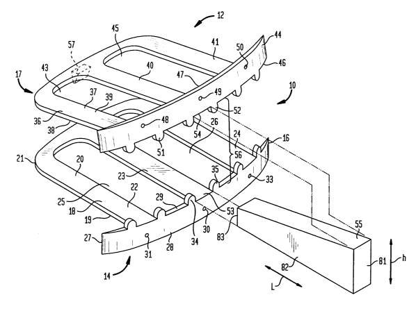
Fig. 1 is a perspective view of an interbody spinal fusion device in
accordance
with the invention;
Fig. 2 is a plan view, from the anterior side, of a portion of the spinal
column with
2 5 the interbody spinal fusion device of Fig. 1 mounted between two adjacent
vertebrae; and
Fig. 3 is a cross-sectional side view of the mounted interbody spinal fusion
device
of Fig. 2, taken along line X-X .
CA 02443001 2003-10-03
WO 02/080823 PCT/US02/10699
6
Detailed Description
Fig. 1 is a perspective view of the interbody spinal fusion device 10.
Interbody
spinal fusion device 0 suitable for implantation in the intervertebral space
between two
adjacent vertebral bodies (not shown), has a pair of bone-engaging plate
members,
specifically top plate member 12 and bottom plate member 14. In use, top plate
member
12 and bottom plate member 14 are arranged above and below each other,
respectively,
in spaced apart relationship. The spaced apart relationship is created and
maintained in
a manner that will be described more completely hereinbelow.
Bottom plate member 14 has a bottom support plate 18 that is adapted to rest
on
the endplate of the lower vertebra (not shown). Bottom support plate 18 has an
outer
surface 19 that contacts the lower vertebra and an inner surface 20. As shown
in this
figure, bottom support plate 18 is generally flat, but may be adapted to
follow the
contour of the vertebra on which it rests. Since the support plates merely
contacts the
endplates of the vertebrae, and can be bent to the contour of the vertebrae,
rather than
engage by means of protuberances or engaging teeth, there is less damage to
the bone
which prevents subsidence. In this embodiment, bottom support plate 18 has
three
longitudinal members 22, 23, and 24 that terminate in a generally curved
section 21 at
posterior end 17. Longitudinal members 22, 23, and 24 define two large
openings 25 and
26.
2 0 At anterior end 16, longitudinal members 22, 23, and 24 terminate with a
generally curved section that is bent substantially orthogonal to the
horizontal plane of
bottom support plate 18 in the direction of communication of the template and
the
vertebral body. The generally curved section is herein referred to as bottom
template 27.
Bottom template 27 has an anterior front face surface 28 at anterior side 16
of the device
2 5 and an opposing posterior surface 29 (designated, but not specifically
shown in this
figure). Posterior surface 29 is adapted to contact and rest flush against the
curved
anterior cortical surface of the lower vertebra (not shown).
In this embodiment, bottom template 27 is provided with three pre-drilled
holes
31, 32 and 33 which may, in some embodiments, be internally threaded.
Fasteners, such
CA 02443001 2003-10-03
WO 02/080823 PCT/US02/10699
7
as threaded orthopedic bone screws (not shown) are inserted through the pre-
drilled
holes and into the hard cortical bone of the anterior surface of the vertebra.
In preferred
embodiments, the pre-drilled holes may be configured to adjust the angle of
placement
of the bone screws so that the bone screws can be set to work against each
other in order
to stabilize the device.
While a total of six bone screws are used in the specific embodiment described
herein, it is to be understood that the bone-engaging plate members can be
attached to
vertebrae using a greater or lesser number of fasteners depending on different
variables,
including, but certainly not limited to, size or bone density of the
vertebrae, spatial
1 o positioning of the vertebrae, and the level of attachment required by the
physician.
Orthopedic bone screws of the type suggested for use in the practice of the
invention are well-known and available from a variety of suppliers known to
those of
ordinary skill in the art. However, it is to be understood, that other known
or new and
improved forms of orthopedic screws and other types of improved orthopedic
fasteners
and fastening systems are within the contemplated scope of the invention.
Top plate member 12 is generally equivalent in structure to bottom plate
member
14, and in some embodiments, may be identical in structure to bottom plate
member.
However, in use, the top plate member 12 is flipped so that top template 44
will be bent
substantially orthogonal to the horizontal plane of top support plate 36 in
the direction
2 o of communication of the template and the vertebral body.
Refernng to Fig. l, top plate member 12 has a top support plate 36 that is
adapted to rest on the endplate of the upper vertebra (not shown). Top support
plate
36 has an outer surface 37 that contacts the upper vertebra and an inner
surface 38
(designated, but not specifically shown in this figure). Top support plate 36
has three
longitudinal members 39, 40, and 41 that terminate in a generally curved
section 42 at
posterior end 17. Longitudinal members 39, 40 and 41 define two large openings
43 an
45. At anterior end 16, longitudinal members 39, 40 and 41 terminate with top
template
44. Top template 44 has an anterior front face surface 46 at anterior side 16
of the
device and an opposing posterior surface 47 (designated, but not specifically
shown).
3 o Posterior surface 47 is adapted to contact and rest flush against the
curved anterior
CA 02443001 2003-10-03
WO 02/080823 PCT/US02/10699
8
cortical surface of the upper vertebra (not shown). Top template 44 is also
provided with
three pre-drilled holes 48, 49, 50.
Bottom template 27 has tabs, illustratively adjacent tabs 34 and 35, that are
integrally formed, and coplanar with, the anterior front face of bottom
template 27, but
extend in a direction opposite to the direction that the template is bent. For
bottom
template 27, the tabs extend upward from its inner surface 20. Tabs 34 and 35
form an
initial guide, or slot 53, that precludes transverse dislocation of support
strut 55 when
inserted into the interbody spinal fusion device. In this specific embodiment,
the tabs are
spaced apart to define an opening having a width approximately equal to the
width of
central longitudinal member 23. Top template 44 also has tabs, illustratively
tabs 51 and
52, that define a slot 54. However, in the case of top template 44, the tabs
extend in a
direction downward from its inner surface 38. When top plate member 12 and
bottom
plate member 14 are mounted to adjacent vertebrae, as will be described
hereinbelow,
tabs 34 and 35 in combination with tabs 51 and 52, are aligned to form
generally an
aperture 56 into which wedge-shaped support strut 55 is inserted.
In addition to the foregoing, in some embodiments additional tabs (shown, but
not specifically designated, in Fig. 1) may be provided. In these embodiments,
the tabs
can operate to define additional slots/apertures for the insertion ofmore than
one support
strut. The tabs, which extend in an opposing direction to the main body of the
template,
2 o and in front of the channels into which one graft material will be placed,
can also operate
to stabilize and anchor the device.
The interbody spinal fusion device 10 has a height that is defined by the
vertical
distance between the outer surface 39 of top support plate 36 and the outer
surface 19
of bottom support plate 18. The height is adjustable by selection and
insertion of a strut
2 5 of the appropriate size into aperture 56, and preferably, varies along the
interbody spinal
fusion device 10 between anterior end 16 and posterior end 17 so as to
maintain the
natural lordosis of the spine.
Refernng to exemplary strut 55, shown in Fig. 1 prior to insertion, support
strut
55 comprises a solid wedge-shaped object of a predetermined maximum height at
3 o anterior end 81 and minimum height at posterior end 83. The angle of the
wedge-shaped
CA 02443001 2003-10-03
WO 02/080823 PCT/US02/10699
9
strut is determined by the height of posterior end 83 relative to the height
of anterior end
81. In a kit embodiment of the invention, a selection of support struts of
varying height
and/or angle would be provided along with the top and bottom plate members in
a
surgical kit so that the practitioner can select the appropriate strut for the
individual
patient. The angle of support strut 55 is chosen to maintain the lordosis of
the vertebral
column. The height of support strut 55 is chosen to approximate the height of
the disc
material that previously occupied the intervertebral spacing. It is
anticipated that as few
as two or three support struts will be all that is required to practice the
invention. Of
course, this number is illustrative and is in no way intended to be limiting.
This is a
1 o significant reduction in the amount of parts required for a surgical kit
for an interbody
fusion operation.
In use, support strut 55 is inserted in aperture 56 between longitudinal
members
23 and 40, spanning the intervertebral region and resting firmly against the
upper and
lower vertebrae. In some embodiments, top plate 36 has a protrusion 57 to
engage
support strut 55 to prevent over-insertion. Of course, either one or both the
bottom plate
18 or top plate 36 can be provided with a protrusion for this purpose.
Fig. 2 is a plan view of a portion of the spinal column with interbody fission
device 10 mounted between two vertebrae. Elements of structure that are
identical to
those in Fig. 1 are similarly designated in Fig. 2. Interbody spinal fusion
device 10 is
2 o placed in intervertebral region 60 between a first vertebra 61 located
below intervertebral
region 60 and a second vertebra 62 located above intervertebral region 60.
Bottom plate
member 14 is attached to vertebra 61 by bone screws 63, 64, and 65 that are
inserted
through pre-drilled holes (see Fig. 1) in bottom template 27. Top plate member
12 is
attached to vertebra 62 by bones screws 63', 64', and 65 'through top template
44.
Support strut 55 is shown inserted in aperture 56.
Channels 66 and 67 are formed on either side of support strut 55 for packing
bone graft material 72 to facilitate fusion of vertebra 61 with vertebra 62.
Referring to
Fig. 1, openings 25 and 26 in bottom support plate 18 (not shown in this
figure) and
openings 43 and 45 in top support plate 36 (not shown in this figure) underlie
or overlie,
CA 02443001 2003-10-03
WO 02/080823 PCT/US02/10699
respectively, channels 66 and 67 so that there is a large area of contact of
bone graft
material with the vertebrae.
In some embodiments, an end cap (not shown in this figure) is placed over the
top
and bottom templates to lock the support strut in place. Advantageously, the
end cap
5 will assist in retaining bone graft material in the channels. Preferably,
the outermost
portions of the lateral and posterior annulus 71 remain intact and serve to
confine the
bone graft material in lateral and posterior directions. Of course, a
retaining plate (not
shown) for the posterior side of spinal fusion device 10 can be devised, by
persons of skill
in the art, for retaining bone graft material, if required.
to The components ofthe interbody spinal fusion device ofthe present invention
are
constructed of biocompatible materials, and presently titanium or titanium
alloys are
preferred. However, it is to be understood that other materials presently
known, and to
be developed, that have the appropriate strength and biocompatibility , such
as ceramics,
metals, and carbon composites, are specifically contemplated for use in
connection with
the invention.
In practice, the interbody spinal fusion device of the present invention is
installed
in accordance with techniques known to those of ordinary skill in the art.
Illustratively,
the technique utilizes an anterior approach to the spine and is particularly
suited to fusion
of lumbar or thoracic vertebrae. The annulus of the affected disc is sharply
incised
2 o anteriorly to allow a complete discectomy to be performed. Preferably, the
entire disc is
removed except for the outermost portions of the lateral and posterior
annulus. The
endplates of the vertebrae are carefully scraped clean of all disc material.
The top plate member 12 and the bottom plate member 14 are respectively placed
into the intervertebral space, as shown in Fig. 3, which is a cross-sectional
side view of
2 5 spinal fixsion device 10, taken along line X-X in Fig. 2, as installed
between neighboring
vertebrae 61 and 62. Elements of structure that are identical to those in Fig.
1 or Fig. 2
are similarly designated in Fig. 3.
Referring to Fig. 3, by way of illustration, top plate member 12 is mounted
into
intervertebral region 60 by placing template 44 on the anterior surface 73 of
the hard
3 0 cortical endplate of the upper vertebra 62. This results in the insertion
of support plate
CA 02443001 2003-10-03
WO 02/080823 PCT/US02/10699
11
36 into intervertebral region so that its outer surface 37 contacts the
cleaned, softer
center of cancellous bone 75 where the disc has been removed (designated, but
not
specifically shown). Template 44 is shown attached to the hard cortical bone
by surgical
screw 64'. Bottom plate member 14 is mounted into intervertebral region 60 and
attached to vertebra 61 in a similar manner by fastening bottom template 27 on
the
anterior surface 74 of the hard cortical endplate of the lower vertebra 61.
The height and width of the disc space are measured, and a support strut 55
having the correct height and/or angle to restore and maintain the appropriate
intervertebral spacing and normal spinal lordosis is selected by the surgeon.
Support
strut 55 is then inserted into aperture 56 created by center slots 54 and 53
(see Fig. l) and
will rest between central longitudinal members 23 and 40. Protuberance 57 on
central
longitudinal member 40 will act as a stop to prevent over-insertion of support
strut 55.
Once the desired height and angle is achieved, bone graft material (not shown
in
this figure) is packed into hollow channels (shown as channels 66 and 67 in
Fig. 2) on
either side of support strut 55. The remaining outermost portions of the
lateral and
posterior annulus (not shown in this figure) may serve to retain the packed
bone graft
material in place. In this particular embodiment, removable end plate 70,
which may be
fastened to template members 44 and 27 by force-fit or fasteners, locks spinal
fusion
device 10 in place and retains bone graft material in the anterior direction.
2 0 Known techniques, such as x-ray imaging or fluoroscopy, can be used to
confirm
correct placement of the device and selection of size/angle of the support
strut.
However, in the practice of the invention, the radius of curvature of the
anterior vertebral
body would dictate the placement of the template. The depth of the support
plate, along
with the curvature as it relates to the anterior vertebral body, creates a
device that is self
2 5 directing as to location. There is no chance of over-penetration of the
device inasmuch
as depth is limited by the anterior aspect of the vertebral approach. This
prevents errors
in placement of the type that routinely occur with the known cylindrical
threaded fission
devices, such as placement which is too far lateral and, theoretically, can go
beyond the
cortical margin into the area of the foramen. It also removes the risk of over-
drilling that
3 0 can happen when a cylindrical threaded fission device is started too far
laterally on the
CA 02443001 2003-10-03
WO 02/080823 PCT/US02/10699
12
vertebral body. Moreover, it prevents the all-too-frequent complications
resulting when
the known device does not obtain equal purchase in each endplate.
The interbody spinal fusion device of the present invention is, mechanically,
a
more stable construct than known prior art devices since there is a greater
amount of
surface area engaged against the anterior aspect of the vertebral body as well
as
impacting the vertebral body endplate. This allows for more aggressive removal
of
cartilaginous endplate and bone from the affected area. Moreover, the
interbody spinal
fusion device of the present invention provides wide channels into which bone
graft
material may be packed. In addition, there large openings in the support plate
provides
1 o for a large area of contact between the bone graft material and the
prepared endplates of
the vertebrae.
Although the invention has been described in terms of specific embodiments and
applications, persons skilled in the art can, in light of this teaching,
generate additional
embodiments without exceeding the scope or departing from the spirit of the
invention
described herein. Accordingly, it is to be understood that the drawing and
description
in this disclosure are proffered to facilitate comprehension of the invention,
and should
not be construed to limit the scope thereof.