Note: Descriptions are shown in the official language in which they were submitted.
CA 02443491 2003-09-30
'
Method of Straddling an Intraosseous Nerve
BACKGROUND OF THE INVENTION
In an effort to reduce back pain through, early intervention techniques, some
investigators have focused upon nerves contained within the vertebral bodies
which are
adjacent the problematic disc.
For example, PCT Patent Publication No. WO 01/0157655 ("Heggeness")
discloses ablating nerves contained within the vertebral body by first boring
into the
vertebral body with a nerve ablation device, placing the tip of the device in
close
proximity to the nerve, and then ablating the nerves with the tip. Heggeness
discloses
using laser devices, electricity transmitting devices, fluid transmitting
devices and
thermal devices, and devices for carrying either chemotherapeutic or
radioactive
substances as candidate nerve ablation devices.
In describing techniques using electricity transmitting devices, Heggeness
discloses "raising the temperature of tip 24 such that the intraosseous nerve
is ablated by
the heat generated by electrical current passing through tip." See Heggeness
at 8,28.
Heggeness further discloses multiple methods of accessing the intraosseous
nerve
(ION). However, each of these methods essentially disclose either i) boring a
straight
channel into the vertebra such that placement of an electrode tip near the end
of that
channel will bring the electrode tip sufficiently close to the ION to effect
its ablation, or
ii) accessing the basivertebral nerve (BVN) via the vertebral foramen. None of
these
techniques recognize how to effectively carry out nerve ablation when the
precise
locations of the ION is unknown, or when the electrode tip can not be
maneuvered
relatively close to the ION.
=
. = =
CA 02443491 2003-09-30
EPO Patent Published Patent Application No, EP 1 059067 Al ("Cosnum")
discloses ablative treatment of metastatic bone tumors, including those within
the spine.
Pain relief is reportedly achieved by penetrating the bone wall with a
suitable probe, and
applying heat through the probe to ablate either the bone tumor or the tissue
near the bone
tumor. Cosman teaches the use of both monopolar and bipolar probes in this
application.
Cosman also teaches that the treatment may also be used to ablate the nerves
and nerve
ramifications in and/or around the bone to desensitize them against further
tumor
encroachment. See Cosman at col. 11, lines 7-11.
However, monopolar approaches require the use of a. grounding pad beneath the
patient and allows energy to flow from the probe and to dissipate in the
surrounding
tissue. Because the path. by which the energy flows from a monopolar probe to
its
corresponding pad is uncontrolled, the energy may undesirably flow through
sensitive
tissue, such as the spinal cord. Since this method may cause undesired local
muscle or
nerve stimulation, it may be difficult or dangerous to operate in sensitive
areas of the
human body.
Cosman discloses devices whose electrodes can deviate from the axis of the
access channel. In particular, Cosman discloses steerable tips, spring-like
electrodes that
take a straight shape within the catheter and then curve upon exiting the
catheter. Cosman
discloses that the curved portion of the electrode may be a rigid and rugged
permanent
curve, or it may be a flexible configuration so that it can be steered, pushed
or guided by
the clinician to be positioned at various location. See Cosman at col. 8,
lines 40-50).
Cosman discloses that electrodes may comprise tubing made of elastic or super-
elastic
metal such as a spring steel or nitonol tubing so that the electrode can be
inserted into
straight segments of the cannula and still describes a curved path when the
curved portion
emerges from the opening. See Cosman at col. 10,1ines 11-16. Cosman also
discloses an
electrode having a flexible but steerable tip which can define an arc, as set
by the
physician. See Cosman at col. 14, line 3.
In sum, Heggeness and Cosman disclose methods of treating that assume the tip
of the electrode can be directed substantially to the target tissue.
A few investigators have examined the effectiveness of heating bone with
= monopolar RF electrodes. DuPuy, A,TR: 175, November 2000,1263-1266 noted
2
CA 02443491 2003-09-30
decreased heat transmission at a 10 mm distance from the electrode through
cancellous
bone in ex vivo studies. DuPuy notes that local heat sinks from the rich
epidural venous
plexus and cerebrospinal fluid pulsations may account for the decreased heat
transmission in cancellous bone. Tillotson, 1nmdgative Radiology, 24:11, Nov.
1989,
888-892, studied the percutaneous ablation of the trigeminal ganglion using RF
energy,
and found that bone marrow necrosis was limited to a sphere of about 1 cm in
diameter,
regardless of the probe size and duration of heating. Tillotson further
reports that
Lindskog showed that the transmission of heat within bone is sharply limited
by blood
flow, and that lethal temperatures cannot be sustained over great distances.
In sum, these investigators appear to report that the well-vaseularized nature
of
. _
bone appears:to limit the heating effect of RF electrodes to a distance
of' less than.about
0.5 cm from the tip.
U.S. Patent No. 6,312,426 ("Goldberg") discloses a system of RF plate-like
electrodes for effecting large, uniform, and extended ablation of the tissue
proximate the
plate-like electrodes. In some embodiments, the plate-like electrodes are
placed on .the
surface of the body tissue, where the ablation is desired, and are configured
to lie
approximately parallel or opposing one another, such that they make a lesion
by
coagulating most of the body tissue volume between them. Goldberg appears to
be
primarily directed to the treatment of rumors. Goldberg states that one
advantage of the
system is that the surgeon need not determine the precise position of the
tumor. See
Goldberg at col. 3, line 59-60. Goldberg does not appear to specifically
discuss the
treatment of nerves.
U.S. Patent No. 6,139,545 ("Utley") discloses a facial nerve ablation system
including at least two spaced apart bi-polar probe electrodes spanning between
them a
percutaneous tissue region containing a facial nerve branch. Utley teaches
that the size
and spacing of the electrodes are purposely set to penetrate the skin to a
depth sufficient
= to span a targeted nerve or nerve within a defined region. See col. 5,
lines 44-47. Utley.
further teaches that the system makes possible the non-invasive selection of
discrete
motor nerve branches, which are small and interspersed in muscle, making them
difficult
to see and detect, for the purpose of specifically targeting them = for
ablation. See col. 2,
3
CA 02443491 2003-09-30
=
lines 20-24. Utley does not disclose the use of such a system for the
treatment of IONS,
nor rigid probes, or deployable electrodes. The probes of Utley
SUMMARY OF THE INVENTION
In attempting to place an electrode in close proximity to the BVN, the present
inventors have found the approaches disclosed in the teachings of the art to
be somewhat
problematic. In particular, although the location of the BVN is somewhat well
known, the
BVN is radiolucent and so its precise location can not be easily identified by
an X-ray.
Since the BVN is also extremely thin, knowingly placing the electrode in close
proximity
to the BVN may be problematic. Moreover, since conventional RF electrodes
appear to
= heat only a fairly limited volume of bone, misplacement of the
electrode tip vis-ii-vis the, .
BVN may result in heating a volume of bone that does not contain the BVN.
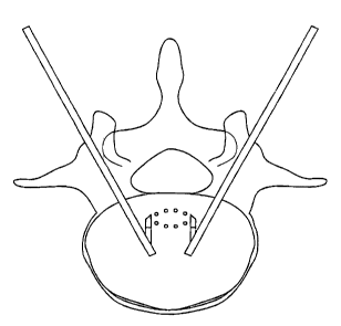
For example, and now referring to FIGS. 1 and 2, there is provided a
representation of a treatment scheme involving the placement of a conventional
bipolar
electrode device in close proximity to the ION. In these FIGS., the ION is
represented by
the solid line identified as ION, while the vertically-disposed dotted lines
identify the
edges of the zone within which the practitioner believes the ION likely
resides (i.e., the
ION residence zone, or "IRZ"). As shown in FIGS. 1 and 2, dale ION is
substantially in
the center of the ION residence zone, then placement of the bipolar electrode
either on
the left hand boundary of the ION residence zone (as in FIG. 1) or
substantially in the
middle of the ION residence zone (as in FIG.2) satisfactorily locates the
electrodes in a
region that allows the current flowing from the electrodes to flow across the
ION. Since
=the current flowing across the ION may resistively and conductive heat the
local bone
tissue and the ION will be heated to therapeutically beneficial temperatures,
these
scenarios may provide beneficial treatment of the ION.
However, and now referring to FIG. 3, if the ION is substantially at the right
edge
= of the ION residence zone, then placement of the bipolar electrodes on
the left hand side
of the ION residence zone fails to locate the electrodes in a region that
allows the current
= flowing from the electrodes to flow across the 10N. Accordingly, current
flowing across
the electrodes can not resistively heat the ION. Moreover, since bone is a
heat sink that
4
CA 02443491 2012-09-27
effectively limits the heat transport to about 0.5 cm, the heat produced by
the electrodes
may be effectively dissipated before it can reach the ION by conduction.
Similarly, and now referring to FIG. 4, if the ION is substantially at the
left edge
of the ION residence zone, then placement of the bipolar electrodes in the
middle of the
ION residence zone fails to locate the electrodes in a region that allows the
current
flowing from the electrodes to flow across the ION. Again current flowing
across the
electrodes cannot resistively heat the ION, and the heat sink quality of bone
may
effectively dissipate the heat produced by the electrodes before it can reach
the ION by
conduction.
Moreover, even if the precise location of the BVN were known, it has been
found
to be difficult to access the posterior portion of the BVN from a
transpedicular approach
with a substantially straight probe.
Therefore, the present inventors set out to produce a system that allows the
practitioner to heat the BVN without having to know the precise location of
the BVN,
and without having to precisely place the electrode tip next to the portion of
the BVN to
be treated.
Illustrative embodiments relate to the production of a large but well-
controlled
heating zone within bone tissue to therapeutically treat an ION within the
heating zone.
Now referring to FIGS. 5-6, there is provided a representation of an
embodiment of the
present invention in which electrodes El and E2 respectively disposed probes
(not shown)
therapeutically treat the ION. FIG. 5 provides a schematic representation of
the electric
field EF produced in the bone tissue by activation of the electrodes. In this
case, the
electric field is relatively thin. FIG. 6 provides a schematic representation
of the total
heating zone THZ produced by the electric field of FIG. 5 including both an
inner
resistive heating zone IR (represented by open circle) and an outer conductive
heating
zone OC (represented by closed circles). In this case, the inner resistive
zone is produced
by the joule heating of bone tissue disposed within the electric field EF,
while the outer
conductive zone is heated by conduction of heat from the resistive heating
zone.
Still referring to FIG. 6, the present inventors have found that positioning
the
active and return electrodes of an energy-transmitting device in a manner that
allows the
electrodes to straddle the ION residence zone IRZ provides a large but well-
controlled
CA 02443491 2013-11-15
total heating zone (IR + OC) within bone tissue to therapeutically treat the
ION within the
heating zone. Since the total heating zone is large and the electrodes
straddle the IRZ,
there is a high level of confidence that a portion of the ION will be present
within the total
heating zone. Since the total heating zone is well controlled, there is no
danger (as with
monopolar systems) that current flowing from the active electrode will
undesirably affect
collateral tissue structures.
Now referring to FIG. 7, if the ION is in fact substantially in the center of
the ION
residence zone, then placement of the bipolar electrodes in a manner that
straddles the ION
residence zone allows the production a total heating zone between the
electrodes that
includes a portion of the ION therein.
Moreover, illustrative embodiments allow the practitioner to therapeutically
treat the
ION even when the ION is in fact located at the edges of the ION residence
zone IRZ. Now
referring to FIGS. 8 and 9, if the ION is located substantially at the right
edge (as in FIG. 8)
or the left edge (as in FIG. 9) of the ION residence zone IRZ, then placement
of the bipolar
electrodes in a manner that straddles the ION residence zone still allows the
production a
total heating zone between the electrodes that includes a portion of the
actual ION therein.
Therefore, the straddling of the ION residence zone by an illustrative
embodiment
satisfactorily locates the electrodes so that the total heating zone produced
by the electrode
activation includes the ION irrespective of the actual location of the ION
within the ION
residence zone IRZ, thereby guaranteeing that the electrodes will always heat
the ION to
therapeutically beneficial temperatures.
Therefore, illustrative embodiments may provide devices and systems configured
to
carry out a method of therapeutically treating a bone having an intraosseous
nerve ION
defining first and second sides of the bone. For example, an illustrative
method may include
the steps of:
a) inserting an energy device baying an active and a return electrode into
the bone,
b) placing the active electrode on the first side of the bone and the
return electrode on
the second side of the bone to define a total heating zone therebetween, and
applying a
sufficiently high frequency voltage between the active and return electrodes
to generate a
current therebetween to resistively heat the total heating zone sufficient to
denervate the ION.
6
CA 02443491 2013-11-15
In addition, an illustrative embodiment may provide a very controlled total
heating
zone which exists substantially only between the paired electrodes. The
ability of such an
embodiment to both therapeutically heat the BVN with substantial certainty and
to minimize
the volume of bone tissue affected by the heating appears to be novel in light
of the
conventional bone-related technology.
Accordingly, such an embodiment is further advantageous because it allows the
clinician to create a sufficiently large heating zone for therapeutically
treating the ION
without requiring direct access to the ION.
Thus, such a preferred embodiment is advantageous because:
1) it does not require knowing the precise location of the ION,
2) it does not require directly accessing the ION, and
3) its controlled heating profile allows the clinician to avoid heating
adjacent structures
such as the healthy adjacent cancellous bone tissue, the spinal cord or
opposing vertebral endplates.
Accordingly, illustrative embodiments may also provide devices and systems
configured to carry out a method of therapeutically treating a vertebral body
having a BVN
defining first and second sides of the vertebral body. For example, an
illustrative method
may include the steps of:
a) determining a BVN residence zone within which the BVN likely resides,
the
BVN residence zone having a first side and a second side,
b) inserting an energy device having an active and a return electrode into
the vertebral
body,
c) placing the active electrode on the first side of the residence zone and
the return
electrode on the second side of the residence zone to define a total heating
zone
therebetween, and
d) applying a sufficiently high frequency voltage between the active and
return
electrodes to generate a current therebetween to resistively heat the total
heating
zone to a temperature sufficient to denervate the BVN.
In an illustrative embodiment, a device for denervating an intraosseous nerve
(ION)
in a bone includes a fixed probe including a shaft having a longitudinal axis,
a distal end
portion, a proximal end portion and a longitudinal bore running from the
proximal end
7
CA 02443491 2013-11-15
portion to the distal end portion. The device further includes a pivotable
probe including a
shaft having a longitudinal axis, a proximal end portion, and a distal end
portion. The distal
end portion is pivotably attached to the fixed probe. The shaft of the
pivotable probe
includes first and second electrodes for electrical connection with a power
supply, and the
fixed probe includes a recess forming a lateral opening in the shaft of the
fixed probe to
house the pivotable probe.
In another illustrative embodiment, a device for denervating an intraosseous
nerve
(ION) in a bone includes a fixed probe including a shaft having a longitudinal
axis, a distal
end portion, a proximal end portion and a longitudinal bore running from the
proximal end
portion. The device further includes a pivotable probe including a shaft
having a longitudinal
axis, a proximal end portion and a distal end portion. The distal end portion
is pivotally
attached to the fixed probe. The fixed probe includes a first electrode for
electric connection
with a power supply and the shaft of the pivotable probe includes a second
electrode for
electric connection with a power supply. The fixed probe includes a recess
forming a lateral
opening in the shaft of the fixed probe to house the pivotable probe.
Other aspects and features of illustrative embodiments will become apparent to
those
ordinarily skilled in the art upon review of the following description of such
embodiments in
conjunction with the accompanying figures.
DESCRIPTION OF THE FIGURES
FIGS. 1 and 2 depict the treatment of the BVN with a conventional bipolar
electrode.
FIGS. 3 and 4 depict the difficulty of treating a BVN with a conventional
bipolar
electrode.
FIGS. 5-6 respectively depict top views of an electric field and a total
heating zone
produced within bone tissue by an embodiment of the present invention.
FIGS. 7-9 depict the treatment of the BVN with a bipolar electrode apparatus
of an
illustrative embodiment.
FIGS. 10a and 10b disclose anterior and upper cross-sectional views of a
straddled
ION that extends in a plane above the electrodes but within the total heating
zone.
FIG. 11 is a cross-sectional anterior view of an illustrative embodiment in
which the
total heating zone has dumb-bell type resistive heating zones.
8
CA 02443491 2013-11-15
FIG. 12 depicts a top view of the treatment of the BVN with a bipolar
electrode
apparatus of an illustrative embodiment wherein the distal ends of the probes
are located
substantially at the midline of the vertebral body.
FIG. 13 discloses cross-sections of components of a preferred dual probe
apparatus
according to an illustrative embodiment.
FIG. 14 discloses an embodiment of the present invention in which a portion of
the
probe shaft acts as an electrode.
FIGS. 15-18 discloses four embodiments of the present invention in which at
least a
portion of the electrode faces thereof are disposed in a substantially
parallel relation.
FIG. 19 discloses a cross-sectional view of an apparatus of an illustrative
embodiment
in which the cannula has a bore having a distal bend and a lateral opening.
FIGS. 20a and 20b disclose cross-sectional views of an apparatus of an
illustrative
embodiment in which the cannula has a proximal bend.
FIGS. 21a and 21 b disclose cross-sectional views of an apparatus of an
illustrative
embodiment in which the probe has a pivoted portion containing an electrode.
FIG. 22 discloses a probe of an illustrative embodiment having reverse conical
electrodes.
FIG. 23 discloses a probe of an illustrative embodiment having a plurality of
active
electrodes and a corresponding plurality of return electrodes.
FIG. 24 discloses a bipolar probe of an illustrative embodiment in which the
return
electrode has a relatively large surface area.
FIG. 25 presents a cross-sectional view of an articulated probe of an
illustrative
embodiment having both active and return electrodes.
FIG. 26 discloses the treatment of a posterior portion of the BVN with a
bipolar
electrode apparatus of an illustrative embodiment.
FIGS. 27 a-d disclose respective top, anterior, lateral and perspective views
of the
placement of a bipolar electrode apparatus of an illustrative embodiment
within a vertebral
body.
FIGS. 28a and 28b show the location of thermocouples T0-T14 within the
vertebral
body.
FIG. 29a-c present the temperatures recorded by thermocouples T0-T1 4.
9
CA 02443491 2013-11-15
FIG. 30a-b present the peak temperatures recorded by thermocouples TO-T14
within
the vertebral body.
FIGS. 31 a-e present top views of a preferred use of the articulated probe of
FIG. 25.
FIG. 32 presents a dual articulated needle embodiment of an illustrative
embodiment.
DETAILED DESCRIPTION
For the purposes of the present invention, the "resistive heating zone" is the
zone of
bone tissue that is resistively heated due to an energy loss incurred by
current travelling
directly through the bone tissue. Resistive heating, "joule" heating and "near-
field" heating
may be used interchangeably herein. The "conductive heating zone" is the zone
of bone
tissue that is heated due to the conduction of heat from an adjacent resistive
heating zone.
The total heating zone THZ in a bone tissue includes both the resistive
heating zone and the
conductive heating zone. The border between the conductive and resistive
heating zones is
defined by the locations where the strength of the electric field is 10% of
the maximum
strength of the electric field between the electrodes. For the purposes of the
present
invention, the heating zones encompass the volume of bone tissue heated to at
least 42 C by
the present invention. For the purposes of the present invention, the "first
and second sides"
of a vertebral body are the lateral-lateral sides intersected by the BVN.
The therapeutic treatment of the ION may be carried out in accordance with
illustrative embodiments by resistive heating, conductive beating, or by
hybrid heating.
In some embodiments, the therapeutic heating of the ION is provided by both
resistive and conductive heating. In some embodiments thereof, as in FIG. 6,
the electrodes
are placed such that the ION passes through resistive heating zone IR, so that
length L1 of the
ION is therapeutically heated by bone tissue in the resistive heating zone
CA 02443491 2003-09-30
;
IR and lengths 1.4 and L3 of the ION are therapeutically heated by the bone
tissue in the
conductive heating zone OC.
In embodiments wherein the therapeutic heating of the ION is provided
substantially by both resistive and conductive heating, it is preferred that
the length L1 of
the ION treated by resistive heating comprise at least 25% of the total
therapeutically
treated length of ION, more preferably at least 50%. In many embodiments, the
peak
temperature in the resistive heating zone IR is between 40 C and 60 C
greater than the
peak temperature in the conductive heating zone OC. Preferably, the peak
temperature in
the resistive heating zone IR is no more than 15 C greater than the peak
temperature in
the conductive heating zone OC, more preferably no more than 10 C, more
preferably no
more than degrees.
Now referring to FIGS. 10a and 10b, in some embodiments, the therapeutic
heating of the ION is provided essentially by the conductive heating zone OC.
This may
occur when the ION is in fact located substantially far from the middle of the
ION
residence zone IRZ. In such an instance, the electrodes are placed such that
the ION
passes only through the conductive heating zone, so that length L2 of the ION
is
therapeutically heated by bone tissue in the conductive heating zone OC.
In preferred embodiments thereof, it is desired that the separation distance
SD
between the ION and the resistive heating zone IR be no more than 1 ern. This
is desired
because the closer the ION is to the resistive heating zone, tbe higher the
temperature
experienced by the ION length L2. More preferably, the separation distance is
no more
than 0.5 cm, more preferably no more than 0.2 cm.
In some embodiments, as in FIG. 10, the electric field is sufficiently strong
to be
located substantially continuously between the two electrodes. This typically
occurs
when the electrodes are very close together (i.e., no more than 5 mrn apart).
In others,
however, as in FIG. 11, the electric field is relatively weak and so resides
substantially
only in the vicinity of the two electrodes. In such cases, and now referring
to FIG. 11,
inward energy flow from the resistive heating zones IR conductively heats the
intermediate area of the conductive heating zone OCT. Preferably, the peak
temperature
in the resistive heating zone IR is no more than 15 C greater than the peak
temperature
11
1lliìli
5
CA 02443491 2003-09-30
in the intermediate conductive heating zone OCA, more preferably no more than
10 C,
more preferably no more than 5 C.
In preferred embodiments, the present invention is carried out via a dual
probe
system. In particular, the present invention preferably comprises an energy
delivery
device comprising a first probe having an active electrode and a second probe
having a
return electrode. Now referring to FIG. 12, this dual probe embodiment allows
the
surgeon to approach the BVN from separate sides of the vertebral body to
easily straddle
the IRZ with the electrodes. With such a device, the surgeon can place the
first probe 601
having an active electrode 603 on a first side of the vertebral body and the
second probe
611 having a retum electrode 613 on a second side of the vertebral body, and
then align
-== the paired electrodes so that their activation produces a total heating
zone that straddles
the IRZ and therefore the BVN therein.
Since aligning the electrodes of such an apparatus to straddle the ION merely
requires advancing the probes into the vertebral body, no complicated
navigation is =
required. The present inventors have appreciated that, even if the location of
the BVN
were precisely known, conventional methods of accessing the BVN require either
i) the
BVN to be naturally located within the vertebral body so as to intersect the
axis = of the
pedicle (Heggeness), or require a complicated probe configuration or
navigation (such as
those described by Cosrnan). Because the dual probe approach simply requires
substantially linear advance of a pair of substantially straight probes, it is
much simpler
and/or much more robust than the conventional methods of accessing nerves in
bone.
Indeed, with this embodiment of the present invention, the clinician may now
desirably
access the vertebral body through the pedicles with substantially straight
probes and .have
a high confidence that their activation can therapeutically treat the BVN.
=
Therefore, in accordance with the present invention, there is provided a
method of
therapeutically treating a vertebral body having a BVN, comprising the steps
of'.
a) providing an energy device having an active electrode having a first face
and a return
electrode having a second face into the vertebral body, and
b) placing the active electrode in the vertebral body to face a first
direction,
12
..=..
CA 02443491 2003-09-30
c) placing the return electrode in the vertebral body to face a second
direction, the first
and second faces defining an angle 24 of no mare than 60 degrees , and
applying a sufficiently high frequency voltage difference between the active
and return
electrodes to generate a current therebetween to produce a total heating zone
to
therapeutically heat the BVN.
Therefore, in accordance with the present invention, there is provided a
method of
therapeutically treating a vertebral body having a BVN, comprising the steps
of
a) providing an energy device having an active electrode and a return
electrode,
- b) placing the active and return electrodes in the -vertebral body to
define an electrode _.
axis, the axis forming an angle (3 of between 50 and 90 degrees with the BVN,
and
c) applying a sufficiently high frequency voltage difference between the
active and return
electrodes to generate a current therebetween to produce a total heating zone
to
therapeutically heat the BVN.
Now referring to FIG. 13, there is provided a preferred dual probe apparatus
according to the present invention comprising first 101 and second 151
cannulae, first
201 and second 251 stylets, first 301 and second 351 probes, and a power
supply 401 in
electrical connection with the probes. For simplicity, only a single cannula,
stylet and
probe will be further described. However, the skilled artisan will appreciate
that preferred
embodiments use two sets of such devices.
Now referring to FIG. 13, cannula 101 comprises a shaft 103 having a
longitudinal bore 10.5 therethrough defining an inner diameter Dc. Distal
opening 109 of
the cannula provides a working portal for the probe. It is further sized to
allow the distal
end of the probe to advance past the distal end 107 of the cannula. The length
Lc of the
cannula is sized to reach from the patient's skin to a location within the
cancellous bone
region of the target bone. Preferably, the cannula is made of a material
selected from the
group consisting of metal and polymer, and is preferably polymer. In many
embodiments,
the cannula is made of an insulating material in order to prevent stray
current from the
probe from contacting non-targeted tissue.
13 =
l
CA 02443491 2003-09-30
In some embodiments, the cannula is shaped so as to guide the probe towards
the
midline of the vertebral body. This inward guidance will help move the
electrodes closer
to the B'VN. In some embodiments, at least a portion of the cannula bore is
curved. In
some embodiments, at least half of the length of the cannula bore is curved.
In other
embodiments, substantially only the distal end portion of the cannula bore is
curved.
Stylet 201 comprises a shaft 203 having a longitudinal axis A and a proximal
205
and distal end 207. Disposed at the distal end of the shall is a tip 209
adapted for boring
or drilling through cortical bone. The outer diameter Do of the stylet shaft
is preferably
adapted to be received within the inner diameter Dc of the cannula.
= For the purposes of the present invention, the combination:of the
.cannula and the
stylet is referred to as a "cannulated needle". In some embodiments, access to
the
vertebral body is gained by first placing the stylet in the cannula to produce
a cannulated
needle, piercing the skin with the cannulated needle, and advancing the
cannulated needle
so that the stylet tip reaches a target tissue region within the cancellous
portion of the
vertebral body, and then withdrawing the stylet. At this point, the cannula is
= conveniently located at the target tissue region to receive a probe of
the present invention.
Probe 301 comprises a shaft 303 having a longitudinal axis B, a distal end
portion
305 and a proximal end portion 307. Disposed near the distal end portion of
the probe is
first electrode 309 having a first face 331 and a connection face 333. The
probe is
designed so that the connection face of the first electrode is placed in
electrical
connection with a first lead 403 of the power supply. In this particular
embodiment, the
shaft has a longitudinal bore 311 extending from the proximal end portion up
to at least
the first electrode. Disposed within the bore is a wire 321 electrically
connected at its first
end 323 to the first electrode and having a second end 325 adapted to be
electrically
connected to a first lead of a power supply.
Therefore, in accordance with the present invention, there is provided an
intraosseous
nerve denervation system, comprising:
a) a cannula having a longitudinal bore,
14
CA 02443491 2003-09-30
b) a stylet having an outer diameter adapted to be received within the
longitudinal bore
and a distal tip adapted to penetrate cortical bone, and
c) a first probe comprising:
i) an outer diameter adapted to be received within the longitudinal bore,
and
ii) a first electrode, and
iii) a lead in electrical connection with the first electrode.
ln some embodiments, the outer surface of the probe is provided with depth
markings so that the clinician can understand the extent to which it has
penetrated the
vertebral body.
In some-embodiments in which a cannulated stylet is first inserted, the stylet
is ,
removed and the cannula remains in place with its distal opening residing in
the target
tissue while the probe is inserted into the cannula. In this embodiment, the
cannula
. provides a secure portal for the probe, thereby insuring that the probe can
enter the bone
safely. This embodiment is especially preferred when the probe is made of a
flexible
material, or is shaped with an irregular cross-section that could undesirably
catch on the
bone during probe advancement into the bone.
In the FIG. 13 probe disclosed above, probe 301 has a blunt tip. In other
embodiments, however, the probe carrying an electrode can be configured to
possess a
sharp distal tip having sufficient sharpness to penetrate cortical bone. With
such a tip, the
clinician can eliminate steps in the procedure that are related to either the
stylet or the
cannulated stylet, and thereby save time.
Now referring to FIG. 14, in some embodiments, the electrode may include a
portion of the probe shaft. For example, in the case of probe 1401, the probe
comprises:
a) an inner electrically conductive shaft 1403 in electrical connection with a
power supply 1409 , and
b) an outer insulating jacket 1405 wrapped around a portion of the shaft.
In this configuration, the placement of the jacket provides a distal
uninsulated shaft
portion 1407 that could be used as an electrode. Preferably, the distal
uninsulated portion
of the shaft has a length of between 3 mm and 8 rrun, and is more preferably
about 5 mm.
In preferred embodiments thereof, the insulation is selected from the group
consisting of
CA 02443491 2003-09-30
polyimide tape, PTFE tape, and heat shrink tubing. Preferred thickness of the
insulation
range from about 0.00025 to 0.0005 inches.
In other embodiments using insulating jackets, the jacket has either a
longitudinally extending slit or slot that exposes a longitudinal surface area
of the
underlying shaft, thereby producing either an essentially linear or an
essentially planar
electrode. In such embodiments, the distal end of the shaft may preferably be
insulated.
In other embodiments using insulating jackets, the insulated portion may
comprises a
proximal jacket and a distal jacket positioned to provide a space therebetween
that
exposes a surface area of the underlying shaft to produce the electrode. In
some
embodiments, the proximal and distal jacket substantially encircle the shaft
to provide an
grmular electa-odetherebetween.
In some embodiments in which a cannulated stylet is used, both the stylet and
the
cannula are removed, and the probe is inserted into the hole created by the
cannulated
stylet. In this embodiment, the hole provides a large portal for the probe.
This
embodiment conserves the annulus of bone removed by the cannula, and so is
preferred
when the probe has a relatively large diameter (e.g., more than 8 min in
diarneter).
In some embodiments in which a cannulated stylet is used, the cannula
comprises
at least one electrode In this embodiment, the cannula acts as the probe as
well. With this
embodiment, the clinician can eliminate steps in the procedure that are
related to
introducing a body into the cannula. In some embodiments, the outer surface of
the
cannula is provided with depth markings so that the clinician can understand
the extent to
which the cannula has penetrated the vertebral body.
In some embodiments in which a cannulated stylet is first inserted, the stylet
comprises at least one electrode. In this embodiment, the stylet acts as the
probe as well.
With this embodiment, the clinician can eliminate steps in the procedure that
are related
to removing the stylet and introducing a body into the cannula. In some
embodiments, the
outer surface of the stylet is provided with depth markings so that the
clinician can
understand the extent to which it has penetrated the vertebral body.
In conducting initial animal experiments with a dual probe embodiment, the
present inventors used a bipedicle approach as shown in FIG. 12, so that each
probe
approached the ION at angle 5 of 45 to about 55 degrees. Since both the probes
and the
16
CA 02443491 2003-09-30
electrodes disposed thereon were essentially cylindrical, the inner faces
605,615 of the
electrodes produced an angle 26. Subsequent testing of the configuration of
FIG. 12
revealed somewhat higher temperatures at the distal portion of the electrodes
and
somewhat lower temperatures near the proximal portions of the electrodes.
Without
wishing to be tied to a theory, it is believed that the shorter path between
the distal
regions produced a lower resistance region (as compared to more proximal inter-
electrode regions) and so caused current to preferentially follow the path of
the least
resistance between the distal portions. Accordingly, the present inventors
sought to
improve upon the relatively uneven temperature profile produced by the
electrode design
of FIG. 12.
In accordance with the present inve.ntion,. the= present inventors modified
its
electrode design to reduce' the angle 28 produces by the inner faces, so that
the distance
between the proximal end of the electrodes is more equal to the distance
between the
proximal end of the electrodes (i.e., the faces are more parallel). When the
electrodes are
provided in such a condition, their orientation reduces the significance of
any path of
least resistance, and so current flows more evenly across the face of each
electrode,
thereby providing even heating and greater control over the system.
Therefore, in accordance with the present invention, there is provided an
intraosseous
nerve denervatioti device, comprising:
a) a first probe having an active electrode and a first lead,
b) a second probe having a return electrode and a second lead,
c) means for creating first and second bores within a bone for accommodating
the
first and second probes,
d) a power supply capable of generating a voltage difference between the
active and
return electrodes, the supply having third and fourth leads,
wherein the first and third leads are in electrical connection, and .the
second and
fourth leads are in electrical connection.
17
CA 02443491 2003-09-30
Preferably, the electrodes are disposed so that the angle 28 produced by the
inner
faces is less than 60 degrees, more preferably no more than 30 degrees. Still
more
preferably, the angle is less than 1 degree. Most preferably, the inner faces
are
substantially parallel.
Now referring to FIG. 15, in some embodiments, substantially parallel
electrodes
are provided by using conical electrodes 501 that taper distally. In this FIG.
15, each
cone electrode 501 has a distal end 503 having a diameter DD and a proximal
end 505
having a diameter Dr, wherein the distal end diameter DE, is larger than the
proximal end
diameter Dp. Preferably, the angle 7 of the cone taper is substantially equal
to the angle 8.
In this condition, the inner faces of the conical electrodes will be
essentially parallel to
each other.
Therefore, in accordance with the present invention, there is provided
intraosseous
nerve denervation system comprising:
a) a first probe having a first electrode and a first lead in electrical
connection with the
first electrode,
wherein the first electrode has a proximal end having a proximal diameter and
a distal
end having a distal diameter,sm. ci the proximal end diameter is less than the
distal end
diameter,
and
b) a second probe having a first electrode and a first lead in electrical
connection with
the first electrode,
wherein the first electrode has a proximal end having a proximal diameter and
a distal
end having a distal diameter, and the proximal end diameter is less than the
distal end
diameter, and
wherein the first and second electrode are disposed so that the electrodes are
parallel.
In FIG. 10, the conical shapes are frustoconical (i.e., they are portions of a
cone).
Frustoconical electrodes are desirable in situations where tissue charring
needs to be
avoided, as the relatively large diameter of the distal end of the electrode
can not provide
18
.=
1
CA 02443491 2003-09-30
an avenue for high current density (relative to the proximal end of the
electrode).
Frustoconical electrodes are also desirable in situations where the probes are
disposed at
a relatively high angle 8, wherein the use of sharp tipped electrodes would
substantially
shorten the distance between the distal tips of the electrodes and thereby
create an
undesirable path of significantly less resistance.
In some embodiments, the frustoconical electrode is shaped so that the
diameter
of its distal end DD is between about 10% and 25% of the diameter of its
proximal end
D. In some embodiments, the frustoconical nature of the electrode is provided
by
physically severing the sharp distal end of the electrode. In others, the
frustoconical
nature of the electrode is provided by insulating the sharp distal end of an
electrode.
As noted above, when the .probes are placed such that their corresponding
electrodes are parallel to each other, the electric field produced by
electrode activation is
substantially uniform between the distal and proximal portions of the
electrodes.
However, as the probes are oriented at an angle from parallel, the electric
field becomes
strongest where the electrodes are closer together. In order to compensate for
this non-
uniform electric field, in some embodiments of the present invention, the
distal ends of
the electrodes are tapered. In this tapered state, the regions of the
electrodes that are
closer together (e.g., the tip) also have a smaller surface area (thereby
reducing the
electric field in that region), while the regions of the electrodes that are
farther apart (e.g.,
the trunk) have a larger surface area (thereby increasing the electric field
in that region).
Typically, the effect is largely determined by the cone size, electrode
spacing and tissue
type therebetween.
In some preferred embodiments of the tapered electrode, and now referring to
FIG. 16, the distal end of the electrode terminates in a sharp tip, so that
the electrode has
a more completely conical shape. Preferably, the conical electrode is shaped
so that the
diameter of its distal end is no more than 20% of the diameter of its proximal
end, more
preferably no more than 10%, more preferably no more than 1%. In addition to
=compensating for non-uniformity in the electric field, the sharp tip may also
be adapted to
penetrate the cortical shell of the vertebral body.
Now referring to FIG. 17, in some embodiments, current flows through an
electrode having only a portion of the conical or frusto-conical. shape. When
electrodes of
= 19
õ,,
CA 02443491 2003-09-30
this embodiment, termed "sectored cones" face each other, their use is
advantageous
because they insure that current will flow the least distance, and so provide
efficiency.
The sectored cones of this embodiment can be produced by first manufacturing
planar
electrodes 511 and placing the planar electrode upon a conveniently angled
probe surface
513. Alternatively, this embodiment can be produced by fn-st manufacturing the
conical
electrode configuration of FIG. 15, and then masking a portion of the conical
electrode
with an insulating material. Unlike the embodiment of FIG. 15, this sectored
cone
embodiment requires careful alignment of the electrode faces and may require
in vivo
rotation of the electrodes to achieve the desired aligrunent.
= Now referring to FIG. 18, in other embodiments, substantially parallel
electrodes
r . can be provided by using elbowed probes 531. =The elbowed
probes have a distal end 533
= and a proximal end 535 meeting at an elbow 537. In some embodiments, the
elbow may
be produced during the manufacturing process (thereby requiring a smaller
diameter
probe in order to fit through the cannula). In other embodiments, the elbow is
produced in
vivo, such as through use of a pull-wire, a pivot or a memory metal disposed
within ,the
probe.
Now referring to FIG. 19, in some embodiments, first 551 and second 552
=
cannulae are each provided with a curved bore 553, 554 forming distal lateral
openings
563,564 in their respective distal end portions 555, 556. When flexible probes
557, 558
containing an electrode 559,560 are passed through the curved bore, the distal
end
561,562 of the probe likewise conforms to the curved bore, thereby forming an
intra-
probe angle e determined by the proximal Ap and distal AD axes of the probe.
Preferably,
this intra-probe angle is between 90 and 135 degrees. Preferably, the intra-
probe angle is
selected so that the distal axes AD of the probes exiting the cannulae form an
angle of no
more than 30 degrees, preferably no more than 10 degrees, more preferably form
a
substantially parallel relation.
Therefore, in accordance with the present invention, there is provided an
intraosseous
nerve denervation system, comprising:
a) a cannula having a longitudinal bore defining a first axis,
Li 11n
CA 02443491 2003-09-30
J
b) a stylet having an outer diameter adapted to be received within the
longitudinal bore.
and a distal tip adapted to penetrate cortical bone, and
c) a first probe comprising:
d) an outer diameter adapted to be received within the longitudinal bore, and
i) a first electrode, and
ii)a lead in electrical connection with the first electrode.
Now referring to FIGS. 20a and 20b, in some embodiments, first 701 and second
751 cannulae are each provided with a curved bore 703, 753 in their respective
distal
portions 705, 755, wherein each bore has a proximal lateral opening 707,757.
The
apparatus further comprises first and second probes 711, 761, each
containing..an-....,
electrode 713,763. In some embodiments, the probe may sit in a distal region
of the bore
(as in FIG. 20a) during advance of the cannula. Once the target tissue region
is reached,
then probes are moved proximally (by, for example, a pull wire ¨ not shown)
and exit the
proximal lateral openings so that the inner faces 715, 765 of the elect/odes
face other.
Therefore, in accordance with the present invention, there is provided an
intraosseous
nerve denervation system, comprising:
a) a cannula having a longitudinal bore defining a first axis,
b) a stylet having an outer diameter adapted to be received within the
longitudinal bore
and a distal tip adapted to penetrate cortical bone, and
, c) a first probe comprising:
i) an outer diameter adapted to be received within the longitudinal bore,
and
ii) a first electrode,.and
iii) a lead in electrical connection with the first electrode.
= Now referring to FIG. 21a and 21b, in some embodiments, at least one
probe 801
comprises i) a distal portion 803 having an electrode 805 and ii) a proximal
portion 807,
the distal portion being pivotally attached to the proximal portion by pivot
809. In some
embodiments, two probes having such pivotally attached electrodes are
introduced
=
21
CA 02443491 2003-09-30
= ,
through the can.nulae in a first linear mode (shown in FIG. 21a) to produce an
angle e
between the electrodes. Next, the respective pivots are actuated (by for
example, a pull
wire ¨ not shown) to produce the angled configuration shown in FIG. 21b which
reduces
the angle 0 between the electrodes. Preferably, the pivoting brings the
electrodes into a
substantially parallel relation.
Therefore, in accordance with the present invention, there is provided
intraosseous
nerve denervation system comprising:
a) a first probe having:
i)
a distal portion having a first electrode, . _ .
ii) a proximal portion comprising a first lead in electrical connection
with the
first electrode, and
iii) a pivot pivotally connecting the proximal and distal portions of the
probe.
In some embodiments, relatively even heating is provided by providing current
density gradients. Now referring to FIG. 22, in some embodiments, first 821
and second
831 probes have first 823 and second 833 electrodes having a reverse conical
shape. In
particular, each electrode has a relatively thick distal portion 827, 837 and
a relatively
thin proximal portion 825, 835. When this probe is activated, it is 'believed
that the
current density of this electrode will vary axially, with a relatively high
current density
present at the proximal portion of each electrode (due to the smaller surface
area) and a =
relatively low current density present at the distal portion of the electrode
(due to the
larger surface area). This current density gradient should provide a more even
heating
zone when the electrodes themselves are oriented at a significant angle, as
the preference
for tip heating (caused by the angled orientation of the electrodes) is
substantially
balanced by the higher current density at the proximal portions of the
electrodes.
Therefore, in accordance with the present invention, there is provided an
intraosseous
nerve denervation system comprising:
22
CA 02443491 2003-09-30
a) a first probe having a first electrode and a first lead in electrical
connection with the
first electrode,
wherein the first electrode has a proximal end having a proximal diameter and
a distal
end having a distal diameter, and
wherein the proximal end diameter is less than the distal end diameter.
Current density gradients can also be produced by providing a plurality of
electrodes on each probe. Now referring to FIG. 23, in some embodiments, first
and
= second electrodes each have a plurality of electrodes. In particular,
first Probe 851 has
first 853, second 854 and third 855 active electrodes, while second probe 861
bas first
863, second 864 and third 865 return electrodes. The voltage across the probes
can be
selected so that there is increasing voltage (and therefore current) across
the more widely
spaced electrodes (i.e., V855-865 < Vs54.864 <V853..863). In some embodiments,
the probes of
FIG. 23 are driven by multiple voltage sources (i.e., a first voltage source
for providing =
voltage between first active electrode 853 and first return electrode 863,
etc.).
Therefore, in accordance with the present invention, there is provided a
method of
therapeutically treating a vertebral body having a B'VN, comprising the steps
of:
a) providing a first energy device having distal and proximal active
electrodes,
b) providing a second energy device having distal and proximal return
electrodes,
c) placing the first and second energy devices in the vertebral body to define
a first
distance between the distal active electrode and the distal return electrode,
and a
second distance between the proximal active electrode and the proximal return
electrode, wherein the first distance is less than the second distance,
d) applying a first high frequency voltage between the distal active and
distal return
electrodes, and
applying a second high frequency voltage between the proximal active and
proximal return electrodes, wherein the first high frequency voltage is less
than the
second high frequency voltage.
23
CA 02443491 2003-09-30
Because multiple voltage sources may add complexity to the device, in other
embodiments, the differences in voltage may be provided by a single voltage
source by
using a poorly conductive electrode. In particular, in some embodiments
thereof, the
probe comprises an electrically conductive probe shaft and a plurality of
spaced apart
insulating jackets wherein the spacing produces the electrodes of FIG. 23. In
this
jacketed embodiment, the probe shaft can be made of a material that is a
relatively poor
electrical conductor (such as tantalum) so that, when a single driving force
is applied
between the jacketed probes, the voltage is highest at the proximal electrode
853, but loss
due to the poor conductance produces a substantially lower voltage at distal
electrode
855. This jacketed embodiment eliminates the need for multiple voltage
sources.
= _
In another dual probe approach, in some embodiments, and now referring to FIG.
24, there is provided an apparatus having first probe 871 having an active
electrode 873,
and a second 881 probe having a return electrode 883, wherein the ratio of the
surface
area of the active electrode to the surface area of the return electrode is
very high, i.e., at
least 2:1 (more preferably at least 5:1). In =this condition, the current
density will be very
high at the active electrode and very low at the return electrode, so that the
total heating
zone THZ will occur essentially only around the active electrode. Since this
device heats
essentially only at the active electrode, this device substantially mimics the
heating
profile of a monopolar electrode, but provides the desirable safety feature of
locally
directing the current to the return electrode.
=
Therefore, in accordance with the present invention, there is provided an
intraosseous nerve denervation system comprising:
a) a first probe having: =
i) an active electrode having a first surface area, and
ii) a first lead in electrical connection with the first electrode,
b) a second probe having:
i) a return electrode having a first surface area, and
24
CA 02443491 2003-09-30
ii) a second lead in electrical connection with the second electrode,
wherein the first surface area is at least two times greater than the second
surface area,
and,
means for creating first and second bores within a bone for accommodating the
first and
second probes.
Although the dual probe approach has many benefits, in other embodiments of
the
present invention, an articulated probe having both active and return
electrodes may be
= used in accordance with the present invention.
= Now referring to FIG. 25, there is provided a preferred articulated
device
= .= according to the present invention. In preferred embodiments,
this device 900 comprises
a fixed probe 901 and a pivotable probe 951.
Fixed probe 901 comprises a shaft 903 having a longitudinal axis and a distal
end
= portion 905 comprising sharpened distal tip 906 and a proximal end
portion 907.
Disposed near the distal end portion of the probe is first electrode 909. The
fixed probe is
designed so that the first electrode is Placed in electrical connection with a
first lead of a
power supply. ln this particular embodiment, the shaft has a longitudinal bore
911
running from the proximal end portion up to at least the first electrode.
Disposed within
the bore is a first wire (not shown) electrically connected at its first end
to the first
electrode and having a second end adapted to be electrically connected to a
first lead of a
power supply (not shown). The fixed probe also comprises a recess 927 forming
a lateral
opening in the shaft and designed to house the pivotable probe when in its
undeployed
mode.
Pivotable probe 951 comprises a shaft 953 having a longitudinal axis, a distal
end
portion 955, and a proximal end portion 957 pivotally attached to the fixed
probe by pivot
961. The pivot allows the pivoting probe to pivot about the fixed probe.
Disposed near
the distal end portion of the pivotable probe is second electrode 963. The
probe is
designed so that the second electrode is placed in electrical connection with
a second lead
of the power supply.
CA 02443491 2003-09-30
The pivotable probe has an undeployed mode and a deployed mode. In the un-
deployed mode, the pivotable probe is seated within the recess of the fixed
probe so that
the axis of its shaft is essentially in line with the axis of the fixed probe
shaft. In this
state, the pivotable probe essentially hides within the fixed probe. In the
deployed mode,
the pivotable probe extends at a significant angle from the fixed probe so
that the axis of
its shaft forms an angle of at least 10 degrees with the axis of the fixed
probe shaft.
In some embodiments, a pusher rod is used to deploy the pivotable probe.
Pusher
rod 975 comprises a proximal handle (not shown) for gripping and a distal end
portion
977 having a shape for accessing the bore of the fixed probe. Distal end
portion has a tip
981 having a shape which, when advanced distally, can push the distal end
portion of the
pivotable probe laterally out of the recess.
Therefore, in accordance with the present invention, there is provided a
device for
denervating an ION in a bone, comprising;
a) a fDted probe having a first electrode thereon in electrical connection
with the
powder supply, and
b) a pivotable probe comprising a second electrode having a proximal portion
pivotally engaged to the fixed probe.
In some embodiments, the pivotable device has both an active and a return
electrode, and the device is introduced through a single pedicle. The location
of these
electrodes may vary depending upon the use of the pivotable device. For
example, when
the active electrode is located on the pivotable probe, the return electrode
may be
positioned in a location selected from the group consisting of:
a) a location on the fixed probe distal of the pivot (as in FIG, 25);
b) a location on the fixed probe proximal of the pivot;
c) a location on the pivotable probe located nearer the pivot; and
d) a location on the pusher rod.
In other embodiments, the locations of the active and return electrodes are
reversed from those described above.
26
CA 02443491 2003-09-30
In general, it is desirable to operate the present invention in a manner that
produces a peak temperature in the target tissue of between about 80 C and 95
C. When
the peak temperature is below 80 C, the off-peak temperatures may quickly
fall below
about 45 C. When the peak temperature is above about 95 C, the bone tissue
exposed to
that peak temperature may experience necrosis and produce charring. This
charring
reduces the electrical conductivity of the charred tissue, thereby making it
more difficult
to pass RF current through the target tissue beyond the char and to
resistively heat the
target tissue beyond the char. In some embodiments, the peak temperature is
preferably
between 86 C and 94 C.
It is desirable to heat the volume of target tissue to a minimum temperature
of at
= least 42 C. When the tissue experiences a temperature above.42 C,
nerves within the
= target tissue may be desirably damaged. However, it is believed that
denervation is a
function of the total quantum of energy delivered to the target tissue, i.e.,
both exposure
temperature and exposure time determine the total dose of energy delivered.
Accordingly,
if the temperature of the target tissue reaches only about 42 C, then it is
believed that
the exposure time of the volume of target tissue to that temperature should be
at least
about 30 minutes and preferably at least 60 minutes in order to deliver the
dose of energy
believed necessary to denervate the nerves within the target tissue.
Preferably, it is desirable to heat the volume of target tissue to a minimum
temperature of at least 50 C. If the temperature of the target tissue reaches
about 50 C,
then it is believed that the exposure time of the volume of target tissue to
that temperature
need only be in the range of about 2 minutes to 10 minutes to achieve
denervation.
More preferably, it is desirable to heat the volume of target tissue to a
minimum
temperature of at least 60 C. If the temperature of the target tissue reaches
about 60 C,
then it is believed that the exposure time of the volume of target tissue to
that temperature
need only be in the range of about 0.01 minutes to 1.5 minutes to achieve
denervation,
preferably 0.1 minutes to 0.25 minutes.
Typically, the period of time that an ION is exposed to therapeutic
temperatures is
in general related to the length of time in which the electrodes are
activated. However,
since it has been observed that the total heating zone remains relatively hot
even after
27
I 0 no
CA 02443491 2003-09-30
power has been turned off (and the electric field eliminated), the exposure
time can
include a period of time in which current is not running through the
electrodes.
In general, the farther apart the electrodes, the greater the likelihood that
the ION
will be contained within the total heating zone. Therefore, in some
embodiments, the
electrodes are placed at least 5 rnrn apart, more preferably at least 10 nun
apart.
However, if the electrodes are spaced too far apart, the electric field takes
on an an
undesirably extreme dumbbell shape. Therefore, in many preferred embodiments,
the
electrodes are placed apart a distance of between 5 min and 25 mm, more
preferably
between 5 nun and 15 mm, more preferably between 10 mm and 15 nun.
In some embodiments, it is desirable to heat the target tissue so that at
least about
= 1 cc of bone tissue experiences the minimum temperature. This volume
corresponds to a
sphere having a radius of about 0.6 cm. Alternatively stated, it is desirable
to heat the
target tissue so the minimum temperature is achieved by every portion of the
bone within
0.6 cm of the point experiencing the peak temperature.
More preferably, it is desirable to heat the target tissue so that at least
about 3 cc
of bone experiences the minimum temperature. This volume corresponds to a
sphere
having a radius of about I cm.
In one preferred embodiment, the present invention provides a steady-state
heated
zone having a peak temperature of between 80 C and 95' C (and preferably
between 86
C and 94 C), and heats at least 1 cc of bone (and preferably at least 3 cc of
bone) to a
temperature of at least 50 C (and preferably 60 C).
Therefore, in accordance with the present invention, there is provided a
method of
therapeutically treating a vertebral body having a BVN, comprising the steps
of:
a) providing an energy device having an active and a return electrode,
a) inserting the active electrode into the vertebral body,
b) inserting the return electrode into the vertebral body, and
c) applyin,g a sufficiently high frequency voltage difference between the
active and
return electrodes to generate a current therebetween to produce a total
heating zone
having a diameter of at least 0.5 cm and a steady state temperature of at
least 50 C.
= 28
CA 02443491 2003-09-30
As noted above, a peak temperature below about 100 C is desirable in order to
prevent charring of the adjacent tissue, steam formation and tissue popping.
In some
embodiments, this is accomplished by providing the power supply with a
feedback means
that allows the peak temperature within the heating zone to be maintained at a
desired
target temperature, such as 90 C. In some embodiments, between about. 24
watts and 30
watts of power is first supplied to the device in order to rapidly heat the
relatively cool
bone, with maximum amperage being obtained within about 10-15 seconds. As the
bone
is further heated to the target temperature, the feedback means gradually
reduces the
power input to the device to between about 6-10 watts.
= If the active electrode has no =active cooling means, it may become be
subject. to
conductive heating by the heated tissue, and the resultant increased
temperature in the
electrode may adversely affect performance by charring the adjacent bone
tissue.
Accordingly, in some embodiments, a cool tip active electrode may be employed.
The
cooled electrode helps maintain the temperature of the electrode at a desired
temperature.
Cooled tip active electrodes are known in the art_ Alternatively, the power
supply may be
designed to provided a pulsed energy input. It has been found that pulsing the
current
= favorably allows heat to dissipate from the electrode tip, and so the
active electrode stays
relatively cooler.
The following section relates to the general structure of preferred energy
devices
in accordance with the present invention:
Tbe apparatus according to the present invention comprises an electrosurgical
probe having a shaft with a proximal end, a distal end, and at least one
active electrode at
= or near the distal end. A connector is provided at or near the proximal
end of the shaft for
= electrically coupling the active electrode to a high frequency voltage
source. in some
embodiments, a return electrode coupled to the voltage source is spaced a
sufficient
distance from the active electrode to substantially avoid or minimize current
shorting
therebetween. The return electrode may be provided integral with the shaft of
the probe
or it may be separate from the shaft
In preferred embodiments, the electrosurgical probe or catheter will comprise
a
shaft or a handpiece= having a proximal end and a distal end which supports
one or more
29
CA 02443491 2003-09-30
electrode terminal(s). The shaft or handpiece may assume a wide variety of
configurations, with the primary purpose being to mechanically support the
active
electrode and permit the treating physician to manipulate the electrode from a
proximal
end of the shaft. The shaft may be rigid or flexible, with flexible shafts
optionally being
combined with a generally rigid external tube for mechanical support. Flexible
shafts
may be combined with pull wires, shape memory actuators, and other known
mechanisms
for effecting selective deflection of the distal end of the shaft to
facilitate positioning of
the electrode array. The shaft will usually include a plurality of wires or
other conductive
elements running axially therethrough to permit connection= of the electrode
array to a
connector at the proximal end of the shaft.
- Preferably,' the shaft may be- a rigid needle that is -introduced
through a .- .
percutaneous penetration in the patient. However, for endoscopic procedures
within the
spine, the shaft will have a suitable diameter and length to allow the surgeon
to reach the
target site (e.g., a disc) by delivering the shaft through the thoracic
cavity, the abdomen
or the like. Thus, the shaft will usually have a length in the range of about
5.0 to 30.0 cm,
and a diameter in the range of about 0.2 mm to about 10 mm, In any of these
embodiments, the shaft may also be introduced through rigid or flexible
endoscopes.
The probe will include one or more active electrode(s) for applying electrical
energy to tissues within the spine. The probe may include one or more return
electrode(s),
OT the return electrode may be positioned on the patient's back, as a
dispersive pad. In
either embodiment, sufficient electrical energy is applied through the probe
to the active
electrode(s) to either necrose the blood supply or nerves within the vertebral
body.
The electrosurgical instrument may also be a catheter that is delivered
percutaneously and/or endoluminally into the patient by insertion through a
conventional
or specialized guide catheter, or the invention may include a catheter having
an active
electrode or electrode array integral with its distal end. The catheter shaft
may be rigid or
flexible, with flexible shafts optionally being combined with a generally
rigid external
tube for mechanical support. Flexible shafts may be combined with pull wires,
shape
memory actuators, and other known mechanisms for effecting selective
deflection of the
distal end of the shaft to facilitate positioning of the electrode or
electrode array. The
catheter shaft will usually include a plurality of wires or other conductive
elements
30 =
CA 02443491 2003-09-30
=
running axially therethrough to permit connection of the electrode or
electrode array and
the return electrode to a connector at the proximal end of the catheter shaft.
The catheter
shaft may include a guide wire for guiding the catheter to the target site, or
the catheter
may comprise a steerable guide catheter. The catheter may also include a
substantially
rigid distal end portion to increase the torque control of the distal end
portion as the
catheter is advanced further into the patient's body. Specific deployment
means will be
described in detail in connection with the figures hereinafter.
In some embodiments, the electrically conductive wires may run freely inside
the =
catheter bore in an unconstrained made, or within multiple lumens within the
catheter
bore.
The tip region of the instrument may comprise many independent electrode
= terminals designed to deliver electrical energy in the vicinity of the
tip. The selective
application of electrical energy is achieved by connecting each individual
electrode
terminal and the return electrode to a power source having independently
controlled or
current limited channels. The return electrode(s) may comprise a single
tubular member
of conductive material proximal to the electrode array. Alternatively, the
instrument may
comprise an array of return electrodes at the distal tip of the instrument
(together with the
active electrodes) to maintain the electric current at the tip. The
application of 'high
frequency voltage between the return electrode(s) and the electrode array
results in the
generation of high electric field intensities at the distal tips of the
electrode terminals with
conduction of high frequency current from each individual electrode terminal
to the
return electrode. The current flow from each individual electrode terminal to
the return
electrode(s) is controlled by either active or passive means, or a combination
thereof, to
deliver electrical energy to the surrounding conductive fluid while minimizing
energy
delivery to surrounding (non-target) tissue.
Temperature probes associated with the apparatus may preferably be disposed on
or within the electrode carrier; between the electrodes =(preferred in bipolar
embodiments); or within the electrodes (preferred for monopolar embodiments).
In some
embodiments wherein =the electrodes are placed on either side of the ION, a
temperature
probe is disposed between the electrodes or in the electrodes. In alternate
embodiments,
the deployable portion of the temperature probe comprises a memory metal.
31
CA 02443491 2003-09-30
The electrode terminal(s) are preferably supported within or by an inorganic
insulating support positioned near the distal end of the instrument shaft. The
return
electrode may be located on the instrument shaft, on another instrument or on
the external
surface of the patient (i.e., a dispersive pad). The close proximity of the
dual needle
design to the intraosseus nerve makes a bipolar design more preferable because
this
minimizes the current flow through non-target tissue and surrounding nerves.
Accordingly, the return electrode is preferably either integrated with the
instrument body,
or another instrument located in close proximity thereto. The proximal end of
the
instrument(s) will include the appropriate electrical connections for coupling
the return
electrode(s) and the electrode terrninal(s) to a high frequency power supply,
such as an
electrosurgical generator,- õ . _
In some embodiments, the active electrode(s) have an active portion or surface
with surface geometries shaped to promote the electric field intensity and
associated
current density along the leading edges of the electrodes. Suitable surface
geometries
may be obtained by creating electrode shapes that include preferential sharp
edges, or by
creating asperities or other surface roughness on the active surface(s) of the
electrodes.
Electrode shapes according to the present invention can include the use of
formed wire
(e.g., by drawing round wire through a shaping die) to form electrodes with a
variety of
cross-sectional shapes, such as siquare, rectangular, L or V shaped, or the
like. Electrode
edges may also be created by removing a portion of the elongate metal
electrode to
reshape the cross-section. For example, material can be ground along the
length of a
round or hollow wire electrode to form D or C shaped wires, respectively, with
edges
facing in the cutting direction. Alternatively, material can be removed at
closely spaced
intervals along the electrode length to form transverse grooves, slots,
threads or the like
along the electrodes. In other embodiments, the probe can be sectored so that
a given
circumference comprises an electrode region and an inactive region. In some
embodiments, the inactive region is masked.
The return electrode is typically spaced proximally from the active
electrode(s) a
suitable. In most of the embodiments described herein, the distal edge of the
exposed
surface of the return electrode is spaced about 5 to 25 mm from the proximal
edge of the
exposed surface of the active electrode(s), in dual needle insertions. Of
course, this
32
CA 02443491 2003-09-30
=
distance may vary with different voltage ranges, the electrode geometry and
depend on
the proximity of tissue structures to active and return electrodes. The return
electrode will
typically have an exposed length in the range of about 1 to 20 ram.
The application of a high frequency voltage between the return electrode(s)
and
the electrode terminal(s) for appropriate time intervals effects modifying the
target tissue.
The present invention may use a single active electrode terminal or an array
of
electrode terminals spaced around the distal surface of a catheter or probe.
In the latter
ernbodiment, the electrode array usually includes a plurality of independently
current-
limited and/or power-controlled electrode terminals to apply electrical energy
selectively
to the target tissue while limiting the unwanted application of electrical
energy to the
surrounding tissue and environment resulting from power dissipation into
surrounding =
electrically conductive fluids, such as blood, normal saline, and the like.
The electrode
terminals may be independently current-limited by isolating the terminals from
each
other and connecting each terminal to a separate power source that is isolated
from the
other electrode terminals. Alternatively, the electrode terminals may be
connected to each
other at either the proximal or distal ends of the catheter to form a single
wire that
couples to a power source.
In one configuration, each individual electrode terminal in the electrode
array is
electrically insulated from all other electrode terminals in the array within
said instrument
and is connected to a power source which is isolated from each of the other
electrode
terminals in the array or to circuitry which limits or interrupts current flow
to the
electrode terminal when low resistivity material (e.g,, blood) causes a lower
impedance
path between the return electrode and the individual electrode terminal. The
isolated
power sources for each individual electrode terminal may be separate power
supply
circuits having internal impedance characteristics which limit power to the
associated
electrode terminal when a low impedance return path is encountered. By way of
example,
the isolated power source may be a user selectable constant current source. In
this
embodiment, lower impedance paths Will automatically result in lower resistive
heating
levels since the heating is proportional to the square of the operating
current times the
impedance. Alternatively, a single power source may be connected to each of
the
electrode terminals through independently actuatable switches, .or by
independent current
33
CA 02443491 2003-09-30
limiting elements, such as inductors, capacitors, resistors and/or
combinations thereof
The current limiting elements may be provided in the instrument, connectors,
cable,
controller or along the conductive path from the controller to the distal tip
of the
instrument. Alternatively, the resistance and/or capacitance may occur on the
surface of
the active electrode terminal(s) due to oxide layers which form selected
electrode
terminals (e.g., titanium or a resistive coating on the surface of metal, such
as platinum).
In a preferred aspect of the invention, the active electrode comprises an
electrode
array having a plurality of electrically isolated electrode terminals disposed
over a contact
surface, which may be a planar or non-planar surface and which may be located
at the
distal tip or over a lateral surface of the shaft, or over both the tip and
lateral surface(s).
= '
The electrode array will include at least two and preferably
more electrode terminals, and .
may further comprise a temperature sensor. In a preferred aspect, each
electrode terminal
will be connected to the proximal connector by an electrically isolated
conductor
disposed within the shaft. The conductors permit independent electrical
coupling of the
electrode terminals to a high frequency power supply and control system with
optional
temperature monitor for operation of the probe. The control system preferably
= incorporate active and/or passive current limiting structures, which are
designed to limit
current flow when the associated electrode terminal is in contact with a low
resistance
return path back to the return electrode.
The use of such electrode arrays in electrosurgical procedures is particularly
advantageous as it has been found to limit the depth of tissue necrosis
without
substantially reducing power delivery. The voltage applied to each electrode
terminal
causes electrical energy to be imparted to any body structure which is
contacted by, or
comes into close proximity with, the electrode terminal, where a current flow
through all
low electrical impedance paths is preferably but not necessarily limited.
Since some of
the needles are hollow, a conductive fluid could be added through the needle
and into the
bone structure for the purposes of lowering the electrical impedance and fill
the spaces in .
the cancellous bone to make them better conductors. to the needle.
It should be clearly understood that the invention is not limited to
electrically
isolated electrode terminals, or even to a plurality of electrode terminals.
For example,
the array of active electrode terminals may be connected to a single lead that
extends
34
_
CA 02443491 2003-09-30
through the catheter shaft to a power source of high frequency current.
Alternatively, the
instrument may incorporate a single electrode that extends directly through
the catheter
shaft or is connected to a single lead that extends to the power source. The
active
electrode(s) may have ball shapes, twizzle shapes, spring shapes, twisted
metal shapes,
cone shapes, annular or solid tube shapes or the like. Alternatively, the
electrode(s) may
comprise a plurality of filaments, rigid or flexible brush electrode(s), side-
effect brush
electrode(s) on a lateral surface of the shaft, coiled electrode(s) or the
like.
The voltage difference applied between the return electrode(s) and the
electrode
terminal(s) will be at high or radio frequency, typically between about 50 kHz
and 20
MHz, usually being between about 100 kHz and 2.5 MHz, preferably being between
about 400 kHz and 1000 kHz, often less than, 600 kHz, and often between about
5:00 kHz .
and 600 kHz. The RMS (root mean square) voltage applied will usually be in the
range
from about 5 volts to 1000 volts, preferably being in the range from about 10
volts to 200
volts, often between about 20 to 100 volts depending on the electrode terminal
size, the
operating frequency and the operation mode of the particular procedure. Lower
peak-to-
peak voltages will be used for tissue coagulation, thermal heating of tissue,
or collagen .
contraction and will typically be in the range from 50 to 1500, preferably.
100 to 1000 and
more preferably 120 to 400 volts peak-to-peak. As discussed above, the voltage
is usually
delivered continuously with a sufficiently high frequency (e.g., on the order
of 50 kHz to
20 MHz) (as compared with e.g., lasers claiming small depths of necrosis,
which are
generally pulsed about 10 to 20 Hz). In addition, the sine wave duty cycle
(i.e.,
cumulative time in any one-second interval that energy is applied) is
preferably on the
order of about 100% for the present invention, as compared with pulsed lasers
which
typically have a duty cycle of about 0.0001%.
The preferred power source of the present invention delivers a high frequency
current selectable to generate average power levels ranging from several
milliwatts to
tens of watts per electrode, depending on the volume of target tissue being
heated, and/or
the niaximum allowed temperature selected for the instrument tip. The power
source
allows the user to select the power level according to the specific
requirements of a
particular procedure.
CA 02443491 2003-09-30
The power source may be current limited or otherwise controlled so that
undesired heating of the target tissue or surrounding (non-target) tissue does
not occur. In
a presently preferred embodiment of the present invention, current limiting
inductors are
placed in series with each independent electrode terminal, where the
inductance of the
inductor is in the range of 10 uH to 50,000 uH, depending on the electrical
properties of
the target tissue, the desired tissue heating rate and the operating
frequency.
Alternatively, capacitor-inductor (LC) circuit structures may be employed, as
described
previously in U.S. Pat. No. 5,697,909. Additionally, current limiting
resistors may be
selected. Preferably, microprocessors are employed to monitor the measured
current and
control the output to limit the current.
The area of the tissue treatment surface can vary widely, and the tissue
treatment
surface can assume a variety of geometries, with particular areas and
geometries being
selected for specific applications. The geometries can be planar, concave,
convex,
hemispherical, conical, linear "in-line" array or virtually any other regular
or irregular
shape. Most commonly, the active electrode(s) or electrode terminal(s) will be
formed at
=
the distal tip of the electrosurgical instrument shaft, frequently being
planar, disk-shaped,
or hemispherical surfaces for use in reshaping procedures or being linear
arrays for use in
cutting. Alternatively or additionally, the active electrode(s) may be formed
on lateral
surfaces of the electrosurgical instrument shaft (e.g., in the manner of a
spatula),
facilitating access to certain body structures in endoseopic procedures. =
The devices of the present invention may be suitably used for insertion into
any
hard tissue in the human body. In some embodiments, the' hard tissue is bone.
In other
embodiments, the hard tissue is cartilage. In preferred embodiments when bone
is
selected as the tissue of choice, the bone is a vertebral body. Preferably,
the present
invention is adapted to puncture the hard cortical shell of the bone and
penetrate at least a
portion of the underlying cancellous bone. In some embodiments, the probe
advances into
the bone to a distance of at least 1/3 of the cross-section of the bone
defined by the
advance of the probe. In some embodiments, the present invention is practiced
in
vertebral bodies substantially free of tumors. In others, the present
invention is practiced
in vertebral bodies having tumors.
36
CA 02443491 2003-09-30
Therefore, in accordance with the present invention, there is provided a
method
of therapeutically treating a healthy vertebral body having a BVN, comprising
the steps
of:
a) providing an energy device having an active and a return electrode,
b) inserting the active electrode into the healthy vertebral body,
c) inserting the return electrode into the healthy vertebral body,
d) placing the active electrode on a first side of the healthy vertebral body
and the return
electrode on a second side of the healthy vertebral body, and
=
applying á sufficiently high frequency .voltage difference between the
active-and _
return electrodes to generate a current therebetween to produce a total
heating zone to
therapeutically heat the BVN.
In some embodiments using two separate probes, the device of the present
invention enters the hard tissue (preferably bone, More preferably the
vertebral body)
through two access points. In preferred embodiments, the pair of separate
probes is
adapted to denervate the BVN and enter through separate pedicles
transpedicularly. In
other embodiments, the pair of separate probes each enters the vertebral body
extrapedicularly. In other embodiments, a first of the pair of separate probes
enters the
vertebral body extrapedicularly and the second enters the vertebral body
transpedicularly.
In embodiments using a single articulated device, the device enters via a
single pedicle.
Now referring to FIG. 26, in some embodiments, the target region of the BVN is
located within the cancellous portion of the bone (i.e., to the interior of
the outer cortical
bone region), and proximal to the junction J of the BVN having a plurality of
branches.
Treatment in this region is advantageous because only a single portion of the
BVN need
be effectively treated to denervate the entire system. In contrast, treatment
of the BVN in
locations more downstream than the junction require the denervation of each
branch.
Therefore, in accordance with the present invention, there is provided a
method of
therapeutically treating a vertebral body having an outer cortical bone region
and an inner
cancellous bone region, and a BVN having a trunk extending from the outer
cortical bone
37
CA 02443491 2003-09-30
region into the inner cancellous region and a branches extending from the
trunk to define
a BVN junction, comprising the steps of:
a) inserting an energy device into the vertebral body, and
b)exclusively depositing energy within the inner cancellous bone region of the
vertebral
body between, but exclusive of the BVN junction and the outer cortical bone
region, to
denervate the BVN.
Typically, treatment in accordance with this embodiment can be effectuated by
placing the electrodes in the region of the vertebral body located between 60%
(point A)
and 90% (Point 131 ofthe distance between the anterior and posterior ends of
the vertebral, . - =
body, as shown in FIG. 26.
= EXAMPLE 31
This prophetic example describes a preferred dual probe embodiment of the
present invention.
First, after induction of an appropriate amount of a local anesthesia, the
human
patient is placed in a prone position on the table. The C-arm of an X-ray
apparatus is
positioned so that the X-rays are perpendicular to the axis of the spine. This
positioning
= provides a lateral view of the vertebral body, thereby allowing the
surgeon to view the =
access of tbe apparatus into the vertebral body.
Next, a cannulated stylet comprising an inner stylet and an outer cannula are
inserted into the skin above each of the respective pedicles so that the
distal tip of each
= stylet is in close proximity to the respective pedicle.
Next, the probe is advanced interiorly into the body so that the stylet tips
bores
through the skin, into and through the pedicle, and then into the vertebral
body. The stylet
is advanced until the tips reach the anterior-posterior midline of the
vertebral body.
Next, the stylet is withdrawn and probe is inserted into the cannula and
advanced
until the first and second electrodes thereof each reach the midline of the
vertebral body.
The location of the two probes is shown from various perspectives in FIG. 27 a-
d. =
38
CA 02443491 2003-09-30
=
Next, the power supply is activated to provide a voltage between the first and
second electrodes. The amount of voltage across the electrodes is sufficient
to produce
an electric current between the first and second electrodes. This current
provides resistive
heating of the tissue disposed between the electrodes in an amount sufficient
to raise the
temperature of the local portion of the BVN to at least 45 C, thereby
denervating the
BVN.
EXAMPLE II
This example describes the efficacy of beating a large zone of a vertebral
body
= with a .bipolar energy device.
=
A pair of probes were inserted into a vertebral body of a porcine cadaver so
that
the tips of the electrodes were located substantially at the midline and
separated by about
4 min. Each electrode had a cylindrical shape, a length of about 20 mm, and a
diameter
of about 1.65 mrn2 (16 gauge) to produce a surface area of about 100 rnm2.
Next, and now referring to FIGS. 28a and 28b, thermocouples 0-14 were placed
within the vertebral body at the 15 locations. Thermocouples 0-4 were placed
halfway
between the electrode tips and were separated by a distance of 2 mm.
Thermocouples 5-9
were placed about at the midpoint between the probe tips, and were vertically
separated
by a distance of 2 min Thermocouples 10-14 were placed along the distal
portion of the
probe and were separated by a distance of 5 mrn.
Next,= about 57 volts of energy was applied across the electrodes, and the
temperature rise in the tissue was recorded at the thermocouple sites. These
temperatures
are provided in FIGS. 29 a-c. In general, the temperature at each site rose
somewhat
steadily from about 22 C to its peak temperature in about 200-300 seconds,
whereupon
feedback controls maintained the peak temperatures. = =
FIGS. 30a and 30b provide the peak temperatures recorded by each thermocouple.
Analysis of the results in FIG. 17a and 17b reveals that peak temperatures of
between
about 80 C and 95 C were able to be sustained over substantial distances. In
particular,
a temperature of 79.4 degrees was reached about 10 mm along the electrode
(T11);
temperatures of between 76.7 and 80.3 C were reached at a depth of about 4
min within
39
CA 02443491 2003-09-30
the tissue (T5 and T9); and a temperature of 76.8 C was reached about 10 ram
along the
electrode (T3).
The positive results provided by this example has great significance to the
problem of therapeutically heating lONs, and the BVN in particular. In
particular, the
results of thermocouples T5-9 indicates that if an ION were located along the
z-axis
within 2 mm of the presumed center of the IRZ, then the ION could be
sufficiently
treated to at least 80 C. Similarly, the results of thermocouples TO-4
indicates that as
much as a 16 mrn length of ION could be sufficiently treated to at least 80
C. Lastly, the
results of thermocouples T 10-14 indicate that the ION could be off-center
laterally in
the IRZ by as much as 2 mm and at least about 10 mm of its length could be
sufficiently
_ treated to at least 80 C. =
EXAMPLE III
This embodiment describes a preferred articulated probe embodiment of the
present invention.
The initial steps described above in Example I are carried out so that the
articulated probe is poised on the patient's skin and held in place by a
ratchet type gun.
See FIG. 31a.
Next, the distal end of the articulated probe is inserted into the skin above
a
pedicle so that the distal end of the fixed probe is in close proximity to the
pedicle.
Now referring to FIG.3 1 b, the probe is advanced interiorly into the body so
that
the distal tip bores through the skin, into and through the pedicle, and then
into the
vertebral body. The distal tip is advanced until it reaches about 30% beyond
the anterior-
posterior midline of the vertebral body.
Now referring to FIG.31c, the distal end of the pusher rod is inserted into
the bore
of the fixed probe and advanced until the angled portion of the pusher rod
contacts the
angled portion of the pivotable probe, thereby nudging the pivotable probe out
of the
recess. The pivotable probe is now in a partially deployed mode.
Now referring to FIG.3 ld, the apparatus is slightly withdrawn from the body.
As
this occurs, the bone disposed between the pivotable and fixed probes prevents
the
pivotable probe from withdrawing along with the fixed probe, but rather forces
open the
pivoting means, thereby bringing the axis of the pivotable probe to a position
CA 02443491 2003-09-30
substantially normal to the axis of the fixed probe. The pivotable probe is
now in
extended mode.
Next, the power supply is activated to provide a voltage between the first and
second electrodes. The amount of voltage across the electrodes is sufficient
to produce
an electric current between the first and second electrodes. This current
provides resistive
heating of the tissue disposed between the electrodes in an amount sufficient
to raise the
temperature of the local portion of the BVN to at least 45 C, thereby
denervating the
BVN.
Next, the fixed probe is pushed forward to bring the pivotable probe back into
the
recess.
Now referring to FIGS.3.1e, the probe is removed ,from the body.
EXAMPLE IV
Now referring to FIG. 32 , there is provided a dual articulated needle
embodiment
of the present invention, wherein the articulated needles are each advanced
down the
pedicles of the vertebral body, and each of the pivotable probes are deployed
at an angle
of less than 90 degrees, so that the electrodes thereon align themselves in an
essentially
parallel relationship. Because the electric field produced by this embodiment
is relatviely
even between the elect-odes, =the resulting total heating zone is also
desirably
= homogeneous. Because the electrodes deploy in = the central posterior
portion of the
= vertebral body, the BVN is desirably denervated near its trunk.
=
41