Note: Descriptions are shown in the official language in which they were submitted.
DEMANDES OU BREVETS VOLUMINEUX
LA PRESENTE PARTIE DE CETTE DEMANDE OU CE BREVETS
COMPREND PLUS D'UN TOME.
CECI EST LE TOME 1 _______________________ DE 2
NOTE: Pour les tomes additionels, veillez contacter le Bureau Canadien des
Brevets.
JUMBO APPLICATIONS / PATENTS
THIS SECTION OF THE APPLICATION / PATENT CONTAINS MORE
THAN ONE VOLUME.
THIS IS VOLUME 1 OF 2
NOTE: For additional volumes please contact the Canadian Patent Office.
õ
CA 02443903 2003-10-10
WO 02/083854 PCT/US02/11521
ANTIBODIES TO VLA-1
FIELD OF THE INVENTION
This invention relates to antibodies to VLA-1 integrin and the use of
these antibodies in treating inflammatory diseases and other immunological
disorders.
This invention also relates to the crystal structure of the complex
between one such antibody and the al-I domain of VLA-1, and to the use of this
structural information for computational drug design.
BACKGROUND OF THE INVENTION
Integrins are a superfamily of cell surface receptors that mediate cell-
cell and cell-matrix adhesion. These proteins are known to provide anchorage
as well
as signals for cellular growth, migration and differentiation during
development and
tissue repair. They have been implicated in immune and inflammatory processes.
Integrins are heterodimeric proteins composed of two noncovalently
linked polypeptide chains, cc and J. The amino terminus of each chain forms a
globular head that contributes to interchain linking and to ligand binding.
The
globular heads are connected to the transmembrane segments by stalks. The
cytoplasmic tails are usually less than 50 amino acid residues long. Integrin
subfamilies were originally defined on the basis of which 13 subunit was used
to form
the heterodimers. The pl-containing integrins are also called VLA molecules,
referring to "very late activation" antigens. VLA-1 to VLA-6 refer to 131
subfamily
members containing al to a6 (i.e., CD49a to CD49f), respectively. For general
CA 02443903 2003-10-10
WO 02/083854 PCT/US02/11521
- 2 -
review, see Cellular and Molecular Immunology, eds. Abul K. Abbas et al., W.B.
Saunders Company, Philadelphia, PA, 2000.
Collagen (both types I and IV) and laminin are known ligands of c. 1I31
integrin (i.e., VLA-1). VLA-1 has been implicated in cell adhesion and
migration on
collagen (Keely et al., 1995, J. Cell Sci. 108:595-607; and Gotwals et al.,
1996, J.
Clin. Invest. 97:2469-2477); in promoting contraction and reorganization of
collagen
matrices, a critical component of wound healing (Gotwals et al., supra; and
Chiro,
1991, Cell 67:403-410); and in regulating the expression of genes involved in
extracellular matrix remodeling (Riikonen et al., 1995, J. Biol. Chem. 270:1-
5; and
Langholz et al., 1995,J. Cell Biol. 131:1903-1915). Thus, improper regulation
of
VLA-1 may result in certain pathological conditions such as fibrosis.
Moreover, it has been suggested that VLA-1 may play a role in T cell
/monocyte-driven diseases. Anti-VLA-1 antibodies block T-cell dependent
cytokine
expression (Miyake et al., 1993, J. Exp. Med. 177:863-868). Expression of VLA-
1 is
increased in persistently activated, 2 to 4 week old cultured T cells (Hemler
et al.,
1985, ELM J. Immunol. 15:502-508). VLA-1 is also expressed on a high
percentage of
T cells isolated from the synovium of patients with rheumatoid arthritis
(Hemler et al.,
1986, J. Clin. Invest. 78:692-702).
Several crystal structures of integrin a subunits have been determined,
including the structures of the a2-I domain of a2131 (PDB accession code laox;
Emsley et al., 1997, J. Biol. Chem. 272:28512-28517); the al-I domain of rat a
1 [31
(PDB accession number lck4; Nolte et al., 1999, FEBS Lett. 452:379-385; WO
00/20459); the al subunit of human alpl (PDB accession code lqc5; Rich et al.,
1999, J. Biol. Chem. 274:24906-24913); the aL-I and aM-I domains; and vWF-A3
(Lee et al., 1995, Cell 80:631-635; Lee et al., 1995, Structure 3:1333-1340;
Qu et al.,
1995, Proc. Natl. Acad. Sci. USA 92:10277-10281; Qu et al., 1996, Structure
4:931-
942). The a2[31 structure revealed a helix (i.e., the C-helix) that created a
trench or
groove on one face of the protein at the metal-ion binding site (Emsley et
al., supra).
The crystal structure of the a2-I domain complexed to a short collagen-based
triple
helical peptide revealed that the collagen-based peptide was bound to that
trench,
where the a2-I amino acids that made intermolecular or metal contacts included
CA 02443903 2014-03-03
- 3 -
Asp151, Asn154, Tyr157, G1n215, Asp219, Leu220, Thr221, Asp254, G1u256,
His258, Tyr285, Leu286, Asn289, Leu291, Asn295, and Lys298 (PDB accession code
ldzi; Emsley et al., 2000, Cell 101:47-56; WO 01/73444). The amino acid
numbering
immediately above is based on PDB accession code ldzi and herein referred to
as
''crystal numbering." The crystal structures of the rat and human al-I domains
revealed a similar trench.
SUMMARY OF THE INVENTION
The present invention provides anti-VLA-1 antibodies and methods of
using these antibodies to treat a variety of inflammatory and immunological
disorders.
Specifically, the invention embraces an antibody that specifically binds
to VLA-1 (e.g., human VLA-1). This antibody contains light chain
complementarity
determining regions ("CDR"s) defined by amino acid residues 24 to 33, 49 to
55, and
88 to 96 of SEQ ID NO:1, and/or heavy chain complementarity determining
regions
defined by amino acid residues 31 to 35, 50 to 65, and 98 to 107 of SEQ ID
NO:2.
These CDRs may contain mutations (e.g., deletions, insertions and/or
substitutions) in
the non-antigen-contacting portions, as determined from the crystal structure
disclosed
herein, without affecting the VLA-1-binding activity of the antibody.
Exemplary
mutations are S24N and G92S in the light chain CDRs, G55S in the heavy chain
CDR2, and D101 A in the heavy chain CDR3. In one embodiment, the antibody of
this invention contains a light chain variable domain sequence of SEQ ID NO:1
and/or a heavy chain variable domain sequence of SEQ ID NO:2.
In a related embodiment, the antibody of this invention contains the
same heavy and light chain polypeptide sequences as an antibody produced by
hybridoma mAQC2, deposited on April 18, 2001 at the American Type Culture
Collection ("ATCC"), 10801 University Boulevard, Manassas, VA 20110-2209 and
having ATCC accession number PTA3273. (All ATCC deposits recited herein were
made under the Budapest Treaty). This antibody can be produced by, for
example,
hybridoma mAQC2, or cells containing nucleic acid sequences isolated from that
hybridoma that encode the heavy and light chains of the mAQC2 monoclonal
antibody.
CA 02443903 2003-10-10
WO 02/083854 PCT/US02/11521
- 4 -
In another embodiment, the antibody is a humanized antibody
comprising at least one (e.g., 2, 3, 4, or 5) of the following residues in its
light chain:
Q1, L4, P46, W47 and Y71; or at least one (e.g., 2, 3, 4, 5, 6 or 7) of the
following
residues in its heavy chain: D1, V12, S28, F29, A49, T93, R94 (Kabat numbering
convention). For instance, the antibody comprises Ql, L4 and Y71 in the light
chain;
and/or (i) F29, A49, T93 and R94, or (ii) A49 and T93, in the heavy chain.
The humanized antibody of this invention may contain a light chain
variable domain sequence defined by amino acid residues 1 to 106 of SEQ TD
NO:3,
and/or a heavy chain variable domain sequence defined by amino acid residues 1
to
118 of SEQ ID NO:4. The humanized antibody may comprise the same heavy and/or
light chain polypeptide sequences as an antibody produced by cell line hAQC2
(ATCC accession number PTA3275; deposited on April 18, 2001).
In another embodiment, the humanized antibody of this invention may
contain a mutation (e.g., deletion, substitution or addition) at one or more
(e.g., 2, 3, 4,
5, 6, 7 or 8) of certain positions in the heavy chain such that an effector
function of the
antibody (e.g., the ability of the antibody to bind to a Fc receptor or a
complement
factor) is altered without affecting the antibody's ability to bind to VLA-1
(U.S.
Patent 5,648,260). These heavy chain positions include, without limitation,
residues
234, 235, 236, 237, 297, 318, 320 and 322 (EU numbering system). The humanized
antibody can, for instance, contain the mutations L234A (i.e., replacing
leucine at
position 234 of an unmodified antibody with alanine) and L235A (EU numbering
system) in its heavy chain. In one related embodiment, the antibody comprises
the
same heavy chain polypeptide sequence as an antibody produced by cell line
hsAQC2
(ATCC accession number PTA3356; deposited on May 4, 2001).
In yet another embodiment, the humanized antibody of this invention
may contain a mutation (e.g., deletion or substitution) at an amino acid
residue that is
a site for glycosylation, such that the glycosylation site is eliminated. Such
an
antibody may be clinically beneficial for having reduced effector functions or
other
undesired functions while retaining its VLA-1 binding affinity. Mutations of
glycosylation sites can also be beneficial for process development (e.g.,
protein
expression and purification). For instance, the heavy chain of the antibody
may
CA 02443903 2003-10-10
WO 02/083854 PCT/US02/11521
- 5 -
contain the mutation N297Q (EU numbering system) such that the heavy chain can
no
longer be glycosylated at this site. In one related embodiment, the humanized
antibody may comprise the same heavy chain polypeptide sequence as an antibody
produced by cell line haAQC2 (ATCC accession number PTA3274; deposited on
April 18, 2001).
In still other embodiments, the heavy and/or light chains of the
antibody of this invention contain mutations that increase affinity for
binding to VLA-
1 and thereby increase potency for treating VLA-1-mediated disorders.
Embraced in this invention are also a composition containing an
antibody of the invention and a pharmaceutically acceptable carrier; an
isolated
nucleic acid containing a coding sequence for SEQ ID NO:1; an isolated nucleic
acid
containing a coding sequence for SEQ ID NO:2; an isolated nucleic acid
containing a
coding sequence for the light chain of an antibody produced by hybridoma
mAQC2;
an isolated nucleic acid containing a coding sequence for the heavy chain of
an
antibody produced by hybridoma mAQC2; an isolated nucleic acid containing a
coding sequence for the light chain of an antibody produced by cell line
hAQC2; an
isolated nucleic acid containing a coding sequence for the heavy chain of an
antibody
produced by cell line hAQC2; an isolated nucleic acid containing a coding
sequence
for the heavy chain of an antibody produced by cell line haAQC2; an isolated
nucleic
acid containing a coding sequence for the heavy chain of an antibody produced
by cell
line hsAQC2; an isolated nucleic acid containing a coding sequence for
residues 1 to
106 of SEQ ID NO:3; an isolated nucleic acid containing a coding sequence for
residues 1 to 118 of SEQ ID NO:4; cells of hybridoma mAQC2; cells from cell
line
hAQC2; cells from cell line haAQC2; and cells from cell line hsAQC2.
The present invention also provides a method of treating a subject with
an immunological disorder mediated by VLA-1, including administering to the
subject
an effective amount of an antibody of this invention. For instance, this
method is used
to treat a human subject to palliate, ameliorate, stabilize, reverse, prevent,
slow or
delay progression of the disorder. Alternatively, this method is used
prophylactically
to treat a human subject at risk for developing this immunological disorder so
as to
CA 02443903 2003-10-10
WO 02/083854 PCT/US02/11521
- 6 -
prevent or delay the onset of the disorder. An "effective amount" of the
composition
can be administered in one or more dosages.
VLA-1 mediated immunological disorders include, but are not limited
to, disorders in which the VLA-1 activity level is elevated in one or more
tissues as
compared to a normal subject. Examples of such disorders are skin related
conditions
(e.g., psoriasis, eczema, burns, dermatitis, and abnormal proliferation of
hair follicle
cells), fibrosis (e.g., kidney or lung fibrosis), allergic rhinitis,
respiratory distress
syndrome, asthma, bronchitis, tendinitis, bursitis, fever, migraine headaches,
gastrointestinal conditions (e.g., inflammatory bowel disease, Crohn's
disease,
gastritis, irritable bowel syndrome, colitis and colorectal cancer), vascular
diseases
(e.g., atherosclerosis), periarteritis nodosa, thyroiditis, aplastic anemia,
Hodgkin's
Disease, rheumatic fever, osteoarthritis, autoimmune diseases (e.g., type I
diabetes,
myasthenia gravis, rheumatoid arthritis, systemic lupus erythematosus, and
multiple
sclerosis), sarcoidosis, nephrotic syndrome, renal failure, Bechet's Syndrome,
polymyositis, gingivitis, hypersensitivity (e.g., delayed type hypersentivity
or
immediate hypersensitivity), graft and transplant rejections, graft versus
host disease
(GVHD), conjunctivitis, swelling occurring after injury, myocardial ischemia,
and
endotoxin shock syndrome.
The present invention also provides a method of determining the level
of VLA-1 in a tissue (e.g., tissue specimen and body fluid) comprising
contacting the
tissue (e.g., in vivo or in vitro) with the antibody of the invention, and
then detecting
the binding of the antibody to the tissue, thereby determining the level of
VLA-1 in
the tissue.
As used herein, the antibody of this invention can be, for instance, a
murine antibody, a humanized antibody, or a chimeric antibody. It can be a
whole
antibody (i.e., with two full length light chains and two full length heavy
chains) of
any isotype and subtypes (e.g., IgM, IgD, IgGI, IgG2, IgG, IgG4, IgE, IgA, and
IgA2;
with either kappa or lambda light chain). Alternatively, the antibody of this
invention
refers to an antigen-binding fragment (e.g., Fab, F(ab')2, and single chain
Fv) of a
whole antibody.
CA 02443903 2003-10-10
WO 02/083854 PCT/US02/11521
- 7 -
The present invention further provides crystallizable compositions and
crystals of complexes formed by a rat-human chimeric al-I domain (mutant RMI)
and
a hAQC2 Fab fragment, and methods for using such compositions and crystals.
This
invention also provides the structure coordinates and binding sites of the
chimeric
domain and the hAQC2 Fab fragment. The atomic coordinates derived from the
crystal structure described herein provide a structural basis for the
biological activities
of hAQC2 as well as a basis for rational design of VLA-1 agonists or
antagonists with
predicted biological activities (e.g., small molecule compounds or antibodies
such as
hAQC2 variants).
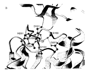
The crystal structure disclosed herein is the first crystal structure of an
al-I domain of an al (31 integrin/ Fab complex. This structure shows the
residues
critical for Fab binding by al -I domain. In addition, the Fab binds in the
putative=
collagen-binding site and inhibits collagen binding. Amino acid residues found
in the
binding site on the a 1-I domain include Asp154, Ser156, Asn157, Ser158,
Tyr160,
G1u192, G1u218, Arg219, G1y220, G1y221, Arg222, G1n223, Thr224, Asp257,
His261, Asn263, Arg291, and Leu294 (crystal numbering). Residues on the Fab
fragment found to bind to the al-I domain include light chain residues Asn30,
Tyr48,
Trp90, Ser91, Asn93 and Trp95, and heavy chain residues Ser30, Arg31, Trp47,
Ser52, G1y53, His56, Tyr58, Phe99, Gly100 and Asp101 (crystal numbering).
This invention also provides a computer for producing a
three-dimensional representation of a molecular complex, where the molecular
complex is defined by the set of structure coordinates of a complex of a
chimeric I
domain of an alp integrin RAH and humanized antibody hAQC2, according to Fig.
19; or a homologue of the molecular complex, the homologue having a root mean
square deviation from the backbone atoms of the amino acids of not more than
0.65
A. The computer includes a machine-readable data storage medium including a
data
storage material encoded with machine-readable data, where the data contains
at least
a portion of the structure coordinates of the complex according to Fig. 19; a
working
memory for storing instructions for processing the machine-readable data; a
central-
processing unit coupled to the working memory and to the machine-readable data
storage medium for processing the machine readable data into the three-
dimensional
CA 02443903 2003-10-10
WO 02/083854 PCT/US02/11521
- 8 -
representations; and a display coupled to the central-processing unit for
displaying the
three-dimensional representation.
This invention further provides a computer for producing a
three-dimensional representation of a molecule or molecular complex including
a
binding site defined by structure coordinates of hAQC2 amino acids including
at least
seven (e.g., 7, 8, 9, 10, 11, 12, 13, 14, 15, or 16) of light chain residues
Asn30, Tyr48,
Trp90, Ser91, Asn93 and Trp95, and heavy chain residues Ser30, Arg31, Trp47,
Ser52, G1y53, His56, Tyr58, Phe99, Gly100 and Asp101 (crystal numbering),
according to Fig. 19; or a homologue of the molecule or molecular complex,
where
the homologue includes a binding site that has a root mean square deviation
from the
backbone atoms of the hAQC2 amino acids of not more than 1.10 A. This
invention
also provides a computer for producing a three-dimensional representation of:
a
binding site defined by structure coordinates of hAQC2 amino acids including
at least
seven (e.g., 7, 8, 9, 10, 11, 12, 13, 14, 15, or 16) of light chain residues
Asn30, Tyr48,
Trp90, Ser91, Asn93 and Trp95, and heavy chain residues Ser30, Arg31, Trp47,
Ser52, G1y53, His56, Tyr58, Phe99, Glyl 00 and Asp101 (crystal numbering),
according to Fig. 19; a binding site of a homologue that has a root mean
square
deviation from the backbone atoms of the hAQC2 amino acids of not more than
1.10
A.
This invention also provides a method for identifying an inhibitor of
an I domain of an integrin including the steps of using structure coordinates
of
hAQC2 amino acids including at least seven (e.g., 7, 8, 9, 10, 11, 12, 13, 14,
15, or
16) of light chain residues Asn30, Tyr48, Trp90, Ser91, Asn93 and Trp95, and
heavy
chain residues Ser30, Arg31, Trp47, Ser52, G1y53, His56, Tyr58, Phe99, Gly100
and
Asp101 (crystal numbering), according to Fig. 19 or a root mean square
deviation
from the backbone atoms of the hAQC2 amino acids not more than 1.10 A, to
generate a three-dimensional structure of a binding site; employing the
three-dimensional structure to design or select a potential antagonist;
synthesizing the
potential antagonist; and contacting the potential antagonist with hAQC2 to
determine
the ability of the potential antagonist to interact with hAQC2, where the
ability of the
potential antagonist to interact with hAQC2 indicates that the potential
antagonist is
CA 02443903 2003-10-10
WO 02/083854 PCT/US02/11521
- 9 -
an inhibitor of the I domain. This invention further provides an inhibitor of
I domain
of integrin identified by this method.
This invention also provides a computer for producing a
three-dimensional representation of a molecule or molecular complex including:
a
binding site defined by structure coordinates of I domain amino acid residues
Asp154,
Ser156, Asn157, Ser158, Tyr160, G1u192, G1n218, Arg219, G1y220, G1y221,
Arg222,
G1n223, Thr224, Asp257, His261, Asn263, Arg291, and Leu294 (crystal
numbering),
according to Fig. 19; or a homologue of the molecule or molecular complex,
where
the homologue includes a second binding site that has a root mean square
deviation
from the backbone atoms of the I domain amino acids not more than 0.65 A. This
invention also provides a computer for producing a three-dimensional
representation
of: a first binding site defined by structure coordinates of I domain amino
acids
residues Asp154, Ser156, Asn157, Ser158, Tyr160, G1u192, G1n218, Arg219,
G1y220,
G1y221, Arg222, G1n223, Thr224, Asp257, His261, Asn263, Arg291, and Leu294
(crystal numbering), according to Fig. 19; or a binding site of a homologue
that has a
root mean square deviation from the backbone atoms of the I domain amino acids
not
more than 0.65 A.
This invention also provides a computer for producing a
three-dimensional representation of a molecule or molecular complex including:
a
binding site defined by structure coordinates of I domain amino acids
including at
least three of residues G1u192, G1n218, Arg219, G1y220, and G1y221 (crystal
numbering), according to Fig. 19; or a homologue of the molecule or molecular
complex, where the homologue includes a second binding site that has a root
mean
square deviation from the backbone atoms of the I domain amino acids not more
than
1.0 A. The invention further provides a computer for producing a three-
dimensional
representation of a binding site defined by structure coordinates of I domain
amino
acids including at least three of residues G1u192, G1n218, Arg219, G1y220, and
Gly221 (crystal numbering), according to Fig. 19; or a binding site of a
homologue
that has a root mean square deviation from the backbone atoms of the I domain
amino
acids not more than 1.0 A.
CA 02443903 2003-10-10
WO 02/083854 PCT/US02/11521
- 10 -
This invention further provides methods for using these three-
dimensional representations to design chemical entities that associate with
the
chimeric domain or the hAQC2 Fab fragment, or portions thereof; and act as
potential
inhibitors of the chimeric domain or the hAQC2 Fab fragment, or portions
thereof.
This invention also relates to compositions including chemical entities, such
as
inhibitors and variants of the chimeric domain or variants of the hAQC2 Fab
fragment, where such chemical entities and variants are rationally designed by
means
of the structure coordinates of the chimeric domain or the hAQC2 Fab fragment,
or
binding sites. The invention further relates to use of the above-identified
chemical
entities to treat or prevent conditions associated with inappropriate or
abnormal alpl
activity in a subject.
This invention further provides a method for identifying an inhibitor
of an I domain of an integrin including the steps of using the structure
coordinates of I
domain amino acids residues Asp154, Ser156, Asn157, Ser158, Tyr160, G1u192,
G1n218, Arg219, G1y220, G1y221, Arg222, G1n223, Thr224, Asp257, His261,
Asn263, Arg291, and Leu294 (crystal numbering), according to Fig. 19, to
generate a
three-dimensional structure of a binding site; employing the three-dimensional
structure to design or select a potential antagonist; synthesizing the
potential
antagonist; and contacting the potential antagonist with I domain to determine
the
ability of the potential antagonist to interact with I domain, where the
ability of the
potential antagonist to interact with the I domain indicates that the
potential antagonist
is an inhibitor of the I domain.
This invention also provides a method for identifying an inhibitor of an
I domain of an integrin including the steps of using the structure coordinates
of at least
three of I domain amino acids including residues G1u192, G1n218, Arg219,
G1y220,
and G1y221 (crystal numbering), according to Fig. 19, or a root mean square
deviation from the backbone atoms of the I domain amino acids not more than
0.65 A,
to generate a three-dimensional structure of a binding site; employing the
three-dimensional structure to design or select a potential antagonist;
synthesizing the
potential antagonist; and contacting the potential antagonist with I domain to
determine the ability of the potential antagonist to interact with I domain of
integrin,
CA 02443903 2013-10-03
= 11
where the ability of the potential antagonist to interact with the I domain
indicates that
the potential antagonist is an inhibitor of the I domain. This invention also
provides an
inhibitor of I domain of integrin identified by this method.
This invention also provides an anti-VLA-1 antibody, or antigen-binding
fragment
thereof, whose light chain complementarity determining regions are defined by
amino
acid residues 24 to 33, 49 to 55, and 88 to 96 of SEQ ID NO:1, and whose heavy
chain
complementarity determining regions are defined by amino acid residues 31 to
35, 50 to
65, and 98 to 107 of SEQ ID NO:2.
This invention also provides a method of determining the level of VLA-1 in a
tissue in vitro, comprising contacting the tissue with the antibody, or the
antigen-binding
fragment thereof, described herein, and detecting the binding of the antibody,
or the
antigen-binding fragment thereof, to the tissue, thereby determining the level
of VLA-1 in
the tissue in vitro.
This invention also provides use of the composition, described herein, in the
manufacture of a medicament for treating an immunological disorder.
This invention also provides use of the composition, described herein, for
treating
an immunological disorder.
This invention also provides the composition described herein, for use in
treating
an immunological disorder.
The invention also provides an anti-VLA-1 antibody, or an antigen-binding
fragment thereof, whose light chain complementarity determining regions are
defined by
amino acid residues 24 to 33, 49 to 55, and 88 to 96 of SEQ ID NO:1, and whose
heavy
chain complementarity determining regions are defined by amino acid residues
31 to 35,
50 to 65, and 98 to 107 of SEQ ID NO:2.
The invention also provides an anti-VLA-1 antibody, or an antigen-binding
fragment thereof, whose light chain complementarity determining regions are
defined by
amino acid residues 24 to 33, 49 to 55, and 88 to 96 of SEQ ID NO:1, and whose
heavy
chain complementarity determining regions are defined by amino acid residues
31 to 35,
50 to 65, and 98 to 107 of SEQ ID NO:2, wherein at least some of the amino
acids in the
CA 02443903 2013-10-03
1 a
framework region of said antibody or antigen binding fragment correspond to
the amino
acids of a human framework region.
The invention also provides an antibody, or an antigen binding fragment
thereof,
whose light chain complementarity determining regions are defined by amino
acid
residues 24 to 33, 49 to 55, and 88 to 96 of SEQ ID NO:1, and whose heavy
chain
complementarity determining regions are defined by amino acid residues 31 to
35, 50 to
65, and 98 to 107 of SEQ ID NO:2, wherein the antibody, or antigen binding
fragment
thereof, is a humanized antibody, or an antigen binding fragment thereof.
The invention also provides compositions comprising the antibody, or antigen
binding fragment thereof, as described above, and a pharmaceutically
acceptable carrier.
The invention also provides an isolated nucleic acid comprising a coding
sequence for SEQ ID NO: l.
The invention also provides an isolated nucleic acid comprising a coding
sequence for SEQ ID NO:2.
The invention also provides an isolated nucleic acid comprising a coding
sequence for SEQ ID NO:l.
The invention also provides an isolated nucleic acid comprising a coding
sequence for SEQ ID NO:2.
The invention also provides an isolated nucleic acid comprising a coding
sequence for SEQ ID NO:3.
The invention also provides an isolated nucleic acid comprising a coding
sequence for SEQ ID NO:4.
The invention also provides an isolated nucleic acid comprising a coding
sequence for SEQ ID NO:5.
The invention also provides an isolated nucleic acid comprising a coding
sequence for SEQ ID NO:6.
The invention also provides an isolated nucleic acid comprising a coding
sequence for residues 1 to 106 of SEQ ID NO:3.
CA 02443903 2013-10-03
1 1 b
The invention also provides an isolated nucleic acid comprising a coding
sequence for residues 1 to 118 of SEQ ID NO:4.
The invention also provides a method of determining the level of VLA-1 in a
tissue in vitro, comprising contacting the tissue with the antibody, or the
antigen-binding
fragment thereof, as described above, and detecting the binding of the
antibody, or the
antigen-binding fragment thereof, to the tissue, thereby determining the level
ofVLA-1 in
the tissue in vitro.
The invention also provides a cell of hybridoma mAQC2 (ATCC accession
number PTA3273).
The invention also provides a cell of cell line hAQC2 (ATCC accession number
PTA3275).
The invention also provides a cell of cell line haAQC2 (ATCC accession number
PTA3274).
The invention also provides a cell of cell line hsAQC2 (ATCC accession number
PTA3356).
The invention also provides a method for identifying an inhibitor of an I
domain
of an integrin comprising the steps of:
(a) using structure coordinates of hAQC2 amino acids comprising at least seven
of light chain residues Asn30, Tyr48, Trp90, Ser91, Asn93 and Trp95 according
to SEQ
ID NO:3, and heavy chain residues Ser30, Arg31, Trp47, Ser52, G1y53, His56,
Tyr58,
Phe99, Gly100 and Asp101 according to SEQ ID NO: 4, or a root mean square
deviation
from the backbone atoms of said hAQC2 amino acids not more than 1.10 A, to
generate a
three-dimensional structure of a binding site, wherein said structure
coordinates are
provided in Figure 19;
(b) employing said three-dimensional structure to design or select a potential
antagonist;
(e) synthesizing said potential antagonist; and
CA 02443903 2013-10-03
=
11c
(d) contacting said potential antagonist with hAQC2 to determine the ability
of
said potential antagonist to interact with hAQC2, wherein the ability of said
potential
antagonist to interact with hAQC2 indicates that the potential antagonist is
an inhibitor of
the I domain.
The invention also provides use of the antibody or antigen binding fragment
and
compositions as described above for treating inflammation in a subject with
rheumatoid
arthritis, inflammatory bowel disease, Crohn's Disease, colitis, gastritis,
irritable bowel
syndrome, psoriasis, systemic lupus erythematosus, colorectal cancer, a
fibrosis,
Hodgkin's Disease, osteoarthritis, sarcoidosis, nephrotic syndrome, graft or
transplant
rejection, or graft versus host disease.
The invention also provides use of the antibody or antigen binding fragment
and
compositions as described above for treating inflammation in a subject with
arthritis,
rheumatoid arthritis, osteoarthtitis or inflammatory bowel disease.
The invention also provides use of a humanized anti-VLA-1 antibody, or an
antigen-binding fragment thereof, that comprises a light chain variable domain
having the
sequence of amino acid residues 1 to 106 of SEQ ID NO:3 and a heavy chain
variable
domain having the sequence of amino acid residues 1 to 118 of SEQ ID NO:4, in
the
manufacture of a medicament for treating an inflammatory disease.
The invention also provides use of a composition comprising a humanized anti-
VLA-1 antibody, or an antigen-binding fragment thereof, that comprises a light
chain
variable domain having the sequence of amino acid residues 1 to 106 of SEQ ID
NO:3
and a heavy chain variable domain having the sequence of amino acid residues 1
to 118
of SEQ ID NO:4, for treating an inflammatory disease.
The invention also provides a composition comprising a humanized anti-VLA-1
antibody, or an antigen-binding fragment thereof, that comprises a light chain
variable
domain having the sequence of amino acid residues 1 to 106 of SEQ ID NO:3 and
a
heavy chain variable domain having the sequence of amino acid residues 1 to
118 of SEQ
ID NO:4, for use in treating an inflammatory disease.
CA 02443903 2013-10-03
= lld
The invention also provides use of a humanized anti-VLA-1 antibody, or an
antigen-binding fragment thereof, that comprises a light chain having the
sequence of
SEQ ID NO:3 and a heavy chain having the sequence of SEQ ID NO:4, in the
manufacture of a medicament for treating an inflammatory disease.
The invention also provides use of a composition comprising a humanized anti-
VLA-1 antibody, or an antigen-binding fragment thereof, that comprises a light
chain
having the sequence of SEQ ID NO:3 and a heavy chain having the sequence of
SEQ ID
NO:4, for treating an inflammatory disease.
The invention also provides a composition comprising a humanized anti-VLA-1
antibody, or an antigen-binding fragment thereof, that comprises a light chain
having the
sequence of SEQ ID NO:3 and a heavy chain having the sequence of SEQ ID NO:4,
for
use in treating an inflammatory disease.
The invention also provides use of a humanized anti-VLA-1 antibody, or an
antigen-binding fragment thereof, that comprises a light chain variable domain
having the
sequence of amino acid residues 1 to 106 of SEQ ID NO:3 and a heavy chain
variable
domain having the sequence of amino acid residues 1 to 118 of SEQ ID NO:4, in
the
manufacture of a medicament for treating inflammation in a subject with
rheumatoid
arthritis, inflammatory bowel disease, Crohn's Disease, colitis, gastritis,
irritable bowel
syndrome, psoriasis, systemic lupus erythematosus, colorectal cancer, a
fibrosis,
Hodgkin's Disease, osteoarthritis, sarcoidosis, nephrotic syndrome, graft or
transplant
rejection, or graft versus host disease.
The invention also provides use of a composition comprising a humanized anti-
VLA-1 antibody, or an antigen-binding fragment thereof, that comprises a light
chain
variable domain having the sequence of amino acid residues 1 to 106 of SEQ ID
NO:3
and a heavy chain variable domain having the sequence of amino acid residues 1
to 118
of SEQ ID NO:4, for treating inflammation in a subject with rheumatoid
arthritis,
inflammatory bowel disease, Crohn's Disease, colitis, gastritis, irritable
bowel syndrome,
psoriasis, systemic lupus erythematosus, colorectal cancer, a fibrosis,
Hodgkin's Disease,
osteoarthritis, sarcoidosis, neplrotic syndrome, graft or transplant
rejection, or graft
versus host disease.
CA 02443903 2013-10-03
1 1 e
The invention also provides a composition comprising a humanized anti-VLA-1
antibody, or an antigen-binding fragment thereof, that comprises a light chain
variable
domain having the sequence of amino acid residues 1 to 106 of SEQ ID NO:3 and
a
heavy chain variable domain having the sequence of amino acid residues 1 to
118 of SEQ
ID NO:4, for use in treating inflammation in a subject with rheumatoid
arthritis,
inflammatory bowel disease, Crohn's Disease, colitis, gastritis, irritable
bowel syndrome,
psoriasis, systemic lupus erythematosus, colorectal cancer, a fibrosis,
Hodgkin's Disease,
osteoarthritis, sarcoidosis, nephrotic syndrome, graft or transplant
rejection, or graft
versus host disease.
The invention also provides use of a humanized anti-VLA-1 antibody, or an
antigen-binding fragment thereof, that comprises a light chain having the
sequence of
SEQ ID NO:3 and a heavy chain having the sequence of SEQ ID NO:4, in the
manufacture of a medicament for treating inflammation in a subject with
rheumatoid
arthritis, inflammatory bowel disease, Crohn's Disease, colitis, gastritis,
irritable bowel
syndrome, psoriasis, systemic lupus erythematosus, colorectal cancer, a
fibrosis,
Hodgkin's Disease, osteoarthritis, sarcoidosis, nephrotic syndrome, graft or
transplant
rejection, or graft versus host disease.
The invention also provides use of a composition comprising a humanized anti-
VLA-1 antibody, or an antigen-binding fragment thereof, that comprises a light
chain
having the sequence of SEQ ID NO:3 and a heavy chain having the sequence of
SEQ ID
NO:4, for treating inflammation in a subject with rheumatoid arthritis,
inflammatory
bowel disease, Crohn's Disease, colitis, gastritis, irritable bowel syndrome,
psoriasis,
systemic lupus erythematosus, colorectal cancer, a fibrosis, Hodgkin's
Disease,
osteoarthritis, sarcoidosis, nephrotic syndrome, graft or transplant
rejection, or graft
versus host disease.
The invention also provides a composition comprising a humanized anti-VLA-1
antibody, or an antigen-binding fragment thereof, that comprises a light chain
having the
sequence of SEQ ID NO:3 and a heavy chain having the sequence of SEQ ID NO:4,
for
use in treating inflammation in a subject with rheumatoid arthritis,
inflammatory bowel
disease, Crohn's Disease, colitis, gastritis, irritable bowel syndrome,
psoriasis, systemic
CA 02443903 2013-10-03
llf
lupus erythematosus, colorectal cancer, a fibrosis, Hodgkin's Disease,
osteoarthritis,
sarcoidosis, nephrotic syndrome, graft or transplant rejection, or graft
versus host disease.
The invention also provides use of a humanized anti-VLA-1 antibody, or an
antigen-binding fragment thereof, that comprises a light chain variable domain
having the
sequence of amino acid residues 1 to 106 of SEQ ID NO:3 and a heavy chain
variable
domain having the sequence of amino acid residues 1 to 118 of SEQ ID NO:4, in
the
manufacture of a medicament for treating inflammation in a subject with
arthritis.
The invention also provides use of a composition comprising a humanized anti-
VLA-1 antibody, or an antigen-binding fragment thereof, that comprises a light
chain
variable domain having the sequence of amino acid residues 1 to 106 of SEQ ID
NO:3
and a heavy chain variable domain having the sequence of amino acid residues 1
to 118
of SEQ ID NO:4, for treating inflammation in a subject with arthritis.
The invention also provides a composition comprising a humanized anti-VLA-1
antibody, or an antigen-binding fragment thereof, that comprises a light chain
variable
domain having the sequence of amino acid residues 1 to 106 of SEQ ID NO:3 and
a
heavy chain variable domain having the sequence of amino acid residues 1 to
118 of SEQ
ID NO:4, for use in treating inflammation in a subject with arthritis.
The invention also provides use of a humanized anti-VLA-1 antibody, or an
antigen-binding fragment thereof, that comprises a light chain having the
sequence of
SEQ ID NO:3 and a heavy chain having the sequence of SEQ ID NO:4, in the
manufacture of a medicament for treating inflammation in a subject with
arthritis.
The invention also provides use of a composition comprising a humanized anti-
VLA-1 antibody, or an antigen-binding fragment thereof, that comprises a light
chain
having the sequence of SEQ ID NO:3 and a heavy chain having the sequence of
SEQ ID
NO:4, for treating inflammation in a subject with arthritis.
The invention also provides a composition comprising a humanized anti-VLA-1
antibody, or an antigen-binding fragment thereof, that comprises a light chain
having the
sequence of SEQ ID NO:3 and a heavy chain having the sequence of SEQ ID NO:4,
for
use in treating inflammation in a subject with arthritis.
CA 02443903 2013-10-03
= llg
The invention also provides use of a humanized anti-VLA-1 antibody, or an
antigen-binding fragment thereof, that comprises a light chain variable domain
having the
sequence of amino acid residues 1 to 106 of SEQ ID NO:3 and a heavy chain
variable
domain having the sequence of amino acid residues 1 to 118 of SEQ ID NO:4, in
the
manufacture of a medicament for treating inflammation in a subject with
rheumatoid
arthritis.
The invention also provides use of a composition comprising a humanized anti-
VLA-1 antibody, or an antigen-binding fragment thereof, that comprises a light
chain
variable domain having the sequence of amino acid residues 1 to 106 of SEQ ID
NO:3
and a heavy chain variable domain having the sequence of amino acid residues 1
to 118
of SEQ ID NO:4, for treating inflammation in a subject with rheumatoid
arthritis.
The invention also provides a composition comprising a humanized anti-VLA-1
antibody, or an antigen-binding fragment thereof, that comprises a light chain
variable
domain having the sequence of amino acid residues 1 to 106 of SEQ ID NO:3 and
a
heavy chain variable domain having the sequence of amino acid residues 1 to
118 of SEQ
ID NO:4, for use in treating inflammation in a subject with rheumatoid
arthritis.
The invention also provides use of a humanized anti-VLA-1 antibody, or an
antigen-binding fragment thereof, that comprises a light chain having the
sequence of
SEQ ID NO:3 and a heavy chain having the sequence of SEQ ID NO:4, in the
manufacture of a medicament for treating inflammation in a subject with
rheumatoid
arthritis.
The invention also provides use of a composition comprising a humanized anti-
VLA-1 antibody, or an antigen-binding fragment thereof, that comprises a light
chain
having the sequence of SEQ ID NO:3 and a heavy chain having the sequence of
SEQ ID
NO:4, for treating inflammation in a subject with rheumatoid arthritis.
The invention also provides a composition comprising a humanized anti-VLA-1
antibody, or an antigen-binding fragment thereof, that comprises a light chain
having the
sequence of SEQ ID NO:3 and a heavy chain having the sequence of SEQ ID NO:4,
for
use in treating inflammation in a subject with rheumatoid arthritis.
CA 02443903 2013-10-03
= 11h
The invention also provides use of a humanized anti-VLA-1 antibody, or an
antigen-binding fragment thereof, that comprises a light chain variable domain
having the
sequence of amino acid residues 1 to 106 of SEQ ID NO:3 and a heavy chain
variable
domain having the sequence of amino acid residues 1 to 118 of SEQ ID NO:4, in
the
manufacture of a medicament for treating inflammation in a subject with
osteoarthritis.
The invention also provides use of a composition comprising a humanized anti-
VLA-1 antibody, or an antigen-binding fragment thereof, that comprises a light
chain
variable domain having the sequence of amino acid residues 1 to 106 of SEQ ID
NO:3
and a heavy chain variable domain having the sequence of amino acid residues 1
to 118
of SEQ ID NO:4, for treating inflammation in a subject with osteoarthritis.
The invention also provides a composition comprising a humanized anti-VLA-1
antibody, or an antigen-binding fragment thereof, that comprises a light chain
variable
domain having the sequence of amino acid residues 1 to 106 of SEQ ID NO:3 and
a
heavy chain variable domain having the sequence of amino acid residues 1 to
118 of SEQ
ID NO:4, for use in treating inflammation in a subject with osteoarthritis.
The invention also provides use of a humanized anti-VLA-1 antibody, or an
antigen-binding fragment thereof, that comprises a light chain having the
sequence of
SEQ ID NO:3 and a heavy chain having the sequence of SEQ ID NO:4, in the
manufacture of a medicament for treating inflammation in a subject with
osteoarthritis.
The invention also provides use of a composition comprising a humanized anti-
VLA-1 antibody, or an antigen-binding fragment thereof, that comprises a light
chain
having the sequence of SEQ ID NO:3 and a heavy chain having the sequence of
SEQ ID
NO:4, for treating inflammation in a subject with osteoarthritis.
The invention also provides a composition comprising a humanized anti-VLA-1
antibody, or an antigen-binding fragment thereof, that comprises a light chain
having the
sequence of SEQ ID NO:3 and a heavy chain having the sequence of SEQ ID NO:4,
for
use in treating inflammation in a subject with osteoarthritis.
The invention also provides use of a humanized anti-VLA-1 antibody, or an
antigen-binding fragment thereof, that comprises a light chain variable domain
having the
CA 02443903 2013-10-03
lli
sequence of amino acid residues 1 to 106 of SEQ ID NO:3 and a heavy chain
variable
domain having the sequence of amino acid residues 1 to 118 of SEQ ID NO:4, in
the
manufacture of a medicament for treating inflammation in a subject with
inflammatory
bowel disease.
The invention also provides use of a composition comprising a humanized anti-
VLA-1 antibody, or an antigen-binding fragment thereof, that comprises a light
chain
variable domain having the sequence of amino acid residues 1 to 106 of SEQ ID
NO:3
and a heavy chain variable domain having the sequence of amino acid residues 1
to 118
of SEQ ID NO:4, for treating inflammation in a subject with inflammatory bowel
disease.
The invention also provides a composition comprising a humanized anti-VLA-1
antibody, or an antigen-binding fragment thereof, that comprises a light chain
variable
domain having the sequence of amino acid residues 1 to 106 of SEQ ID NO:3 and
a
heavy chain variable domain having the sequence of amino acid residues 1 to
118 of SEQ
ID NO:4, for use in treating inflammation in a subject with inflammatory bowel
disease.
The invention also provides use of a humanized anti-VLA-1 antibody, or an
antigen-binding fragment thereof, that comprises a light chain having the
sequence of
SEQ ID NO:3 and a heavy chain having the sequence of SEQ ID NO:4, in the
manufacture of a medicament for treating inflammation in a subject with
inflammatory
bowel disease.
The invention also provides use of a composition comprising a humanized anti-
VLA-1 antibody, or an antigen-binding fragment thereof, that comprises a light
chain
having the sequence of SEQ ID NO:3 and a heavy chain having the sequence of
SEQ ID
NO:4, for treating inflammation in a subject with inflammatory bowel disease.
The invention also provides a composition comprising a humanized anti-VLA-1
antibody, or an antigen-binding fragment thereof, that comprises a light chain
having the
sequence of SEQ ID NO:3 and a heavy chain having the sequence of SEQ ID NO:4,
for
use in treating inflammation in a subject with inflammatory bowel disease.
The invention also provides use of a humanized anti-VLA-1 antibody, or an
antigen-binding fragment thereof, that comprises a light chain variable domain
having the
CA 02443903 2013-10-03
llj
sequence of amino acid residues 1 to 106 of SEQ ID NO:3 and a heavy chain
variable
domain having the sequence of amino acid residues 1 to 118 of SEQ ID NO:4, in
the
manufacture of a medicament for treating arthritis.
The invention also provides use of a composition comprising a humanized anti-
VLA-1 antibody, or an antigen-binding fragment thereof, that comprises a light
chain
variable domain having the sequence of amino acid residues 1 to 106 of SEQ ID
NO:3
and a heavy chain variable domain having the sequence of amino acid residues 1
to 118
of SEQ ID NO:4, for treating arthritis.
The invention also provides a composition comprising a humanized anti-VLA-1
antibody, or an antigen-binding fragment thereof, that comprises a light chain
variable
domain having the sequence of amino acid residues 1 to 106 of SEQ ID NO:3 and
a
heavy chain variable domain having the sequence of amino acid residues 1 to
118 of SEQ
ID NO:4, for use in treating arthritis.
The invention also provides use of a humanized anti-VLA-1 antibody, or an
antigen-binding fragment thereof, that comprises a light chain having the
sequence of
SEQ ID NO:3 and a heavy chain having the sequence of SEQ ID NO:4, in the
manufacture of a medicament for treating arthritis.
The invention also provides use of a composition comprising a humanized anti-
VLA-1 antibody, or an antigen-binding fragment thereof, that comprises a light
chain
having the sequence of SEQ ID NO:3 and a heavy chain having the sequence of
SEQ ID
NO:4, for treating arthritis.
The invention also provides a composition comprising a humanized anti-VLA-1
antibody, or an antigen-binding fragment thereof, that comprises a light chain
having the
sequence of SEQ ID NO:3 and a heavy chain having the sequence of SEQ ID NO:4,
for
use in treating arthritis.
The invention also provides use of a humanized anti-VLA-1 antibody, or an
antigen-binding fragment thereof, that comprises a light chain variable domain
having the
sequence of amino acid residues 1 to 106 of SEQ ID NO:3 and a heavy chain
variable
CA 02443903 2013-10-03
llk
domain having the sequence of amino acid residues 1 to 118 of SEQ ID NO:4, in
the
manufacture of a medicament for treating rheumatoid arthritis.
The invention also provides use of a composition comprising a humanized anti-
VLA-1 antibody, or an antigen-binding fragment thereof, that comprises a light
chain
variable domain having the sequence of amino acid residues 1 to 106 of SEQ ID
NO:3
and a heavy chain variable domain having the sequence of amino acid residues 1
to 118
of SEQ ID NO:4, for treating rheumatoid arthritis.
The invention also provides a composition comprising a humanized anti-VLA-1
antibody, or an antigen-binding fragment thereof, that comprises a light chain
variable
domain having the sequence of amino acid residues 1 to 106 of SEQ ID NO:3 and
a
heavy chain variable domain having the sequence of amino acid residues 1 to
118 of SEQ
ID NO:4, for use in treating rheumatoid arthritis.
The invention also provides use of a humanized anti-VLA-1 antibody, or an
antigen-binding fragment thereof, that comprises a light chain having the
sequence of
SEQ ID NO:3 and a heavy chain having the sequence of SEQ ID NO:4, in the
manufacture of a medicament for treating rheumatoid arthritis.
The invention also provides use of a composition comprising a humanized anti-
VLA-1 antibody, or an antigen-binding fragment thereof, that comprises a light
chain
having the sequence of SEQ ID NO:3 and a heavy chain having the sequence of
SEQ ID
NO:4, for treating rheumatoid arthritis.
The invention also provides a composition comprising a humanized anti-VLA-1
antibody, or an antigen-binding fragment thereof, that comprises a light chain
having the
sequence of SEQ ID NO:3 and a heavy chain having the sequence of SEQ ID NO:4,
for
use in treating rheumatoid arthritis.
The invention also provides use of a humanized anti-VLA-1 antibody, or an
antigen-binding fragment thereof, that comprises a light chain variable domain
having the
sequence of amino acid residues 1 to 106 of SEQ ID NO:3 and a heavy chain
variable
domain having the sequence of amino acid residues 1 to 118 of SEQ ID NO:4, in
the
manufacture of a medicament for treating osteoarthritis.
CA 02443903 2013-10-03
111
The invention also provides use of a composition comprising a humanized anti-
VLA-1 antibody, or an antigen-binding fragment thereof, that comprises a light
chain
variable domain having the sequence of amino acid residues 1 to 106 of SEQ ID
NO:3
and a heavy chain variable domain having the sequence of amino acid residues 1
to 118
of SEQ ID NO:4, for treating osteoarthritis.
The invention also provides a composition comprising a humanized anti-VLA-1
antibody, or an antigen-binding fragment thereof, that comprises a light chain
variable
domain having the sequence of amino acid residues 1 to 106 of SEQ ID NO:3 and
a
heavy chain variable domain having the sequence of amino acid residues 1 to
118 of SEQ
ID NO:4, for use in treating osteoarthritis.
The invention also provides use of a humanized anti-VLA-1 antibody, or an
antigen-binding fragment thereof, that comprises a light chain having the
sequence of
SEQ ID NO:3 and a heavy chain having the sequence of SEQ ID NO:4, in the
manufacture of a medicament for treating osteoarthritis.
The invention also provides use of a composition comprising a humanized anti-
VLA-1 antibody, or an antigen-binding fragment thereof, that comprises a light
chain
having the sequence of SEQ ID NO:3 and a heavy chain having the sequence of
SEQ ID
NO:4, for treating osteoarthritis.
The invention also provides a composition comprising a humanized anti-VLA-1
antibody, or an antigen-binding fragment thereof, that comprises a light chain
having the
sequence of SEQ ID NO:3 and a heavy chain having the sequence of SEQ ID NO:4,
for
use in treating osteoarthritis.
The invention also provides use of a humanized anti-VLA-1 antibody, or an
antigen-binding fragment thereof, that comprises a light chain variable domain
having the
sequence of amino acid residues 1 to 106 of SEQ ID NO:3 and a heavy chain
variable
domain having the sequence of amino acid residues 1 to 118 of SEQ ID NO:4, in
the
manufacture of a medicament for treating inflammatory bowel disease.
The invention also provides use of a composition comprising a humanized anti-
VLA-1 antibody, or an antigen-binding fragment thereof, that comprises a light
chain
CA 02443903 2014-03-03
llm
variable domain having the sequence of amino acid residues 1 to 106 of SEQ ID
NO:3 and a heavy chain variable domain having the sequence of amino acid
residues 1 to
118 of SEQ ID NO:4, for treating inflammatory bowel disease.
The invention also provides a composition comprising a humanized anti-VLA-1
antibody, or an antigen-binding fragment thereof, that comprises a light chain
variable
domain having the sequence of amino acid residues 1 to 106 of SEQ ID NO:3 and
a
heavy chain variable domain having the sequence of amino acid residues 1 to
118 of SEQ
ID NO:4, for use in treating inflammatory bowel disease.
The invention also provides use of a humanized anti-VLA-1 antibody, or an
antigen-binding fragment thereof, that comprises a light chain having the
sequence of
SEQ ID NO:3 and a heavy chain having the sequence of SEQ ID NO:4, in the
manufacture of a medicament for treating inflammatory bowel disease.
The invention also provides use of a composition comprising a humanized anti-
VLA-1 antibody, or an antigen-binding fragment thereof, that comprises a light
chain
having the sequence of SEQ ID NO:3 and a heavy chain having the sequence of
SEQ ID
NO:4, for treating inflammatory bowel disease.
The invention also provides a composition comprising a humanized anti-VLA-1
antibody, or an antigen-binding fragment thereof, that comprises a light chain
having the
sequence of SEQ ID NO:3 and a heavy chain having the sequence of SEQ ID NO:4,
for
use in treating inflammatory bowel disease.
The invention also provides a method for identifying an inhibitor of an 1-I
domain of an integrin comprising the steps of:
(a) using structure coordinates of hAQC2 amino acids comprising at least seven
of light chain residues Asn30, Tyr48, Trp90, Ser91, Asn93 and Trp95 according
to SEQ
ID NO:3, and heavy chain residues Ser30, Arg31, Trp47, Ser52, G1y53, His56,
Tyr58,
Phe99, Gly100 and Asp101 according to SEQ ID NO: 4, or a root mean square
deviation
from the backbone atoms of said hAQC2 amino acids not more than 1.10 A, to
generate a
three-dimensional structure of a binding site, wherein said structure
coordinates are
provided in Figure 19;
CA 02443903 2015-08-13
1 in
(b) employing said three-dimensional structure to design or select a potential
antagonist;
(c) synthesizing said potential antagonist; and
(d) contacting said potential antagonist with an al -I domain of an integrin
to
determine the ability of said potential antagonist to bind to the al -I
domain, wherein the
ability of said potential antagonist to bind to the al-I domain indicates that
the potential
antagonist is an inhibitor of the 1-I domain of an integrin.
The invention further provides an anti-VI,A-1 antibody or antigen-binding
fragment
thereof whose light chain complementarity determining regions (CDRs) are
defined by
amino acid residues 24 to 33, 49 to 55, and 88 to 96 of SEQ ID NO:1, and whose
heavy
CDRs are defined by amino acid residues 31 to 35, 50 to 65, and 98 to 107 of
SEQ ID
NO: 2, wherein said anti-VLA-1 antibody or antigen-binding fragment thereof
comprises
one or more substitutions selected from S24N in the light chain CDR1, G92S in
the light
chain CDR3 and G55S in the heavy chain CDR2.
The invention further provides an anti-VLA-1 antibody, or an antigen-binding
fragment
thereof, comprising a light chain comprising the sequence of SEQ ID NO:3 and a
heavy
chain comprising the sequence of SEQ ID NO:4.
The invention further provides an anti-VLA-1 antibody, or an antigen-binding
fragment
thereof, comprising a light chain comprising the sequence of SEQ ID NO:49 and
a heavy
chain comprising the sequence of SEQ ID NO:42.
The invention further provides an anti-VLA-1 antibody, or an antigen-binding
fragment
thereof, comprising a light chain comprising the sequence of SEQ ID NO:51 and
a heavy
chain comprising the sequence of SEQ ID NO:44.
The invention further provides an anti-VI.A-1 antibody, or an antigen-binding
fragment
thereof, comprising a light chain comprising the sequence of SEQ ID NO:54 and
a heavy
chain comprising the sequence of SEQ ID NO:42.
The invention further provides an anti-VLA-1 antibody, or an antigen-binding
fragment
CA 02443903 2015-08-13
11 o
thereof, comprising a light chain comprising the sequence of SEQ ID NO:58 and
a heavy
chain comprising the sequence of SEQ ID NO:68.
The invention further provides an anti-VI,A-1 antibody, or an antigen-binding
fragment
thereof, comprising a light chain comprising the sequence of SEQ ID NO:70 and
a heavy
chain comprising the sequence of SEQ ID NO:68.
The invention further provides an anti-VLA-1 antibody, or an antigen-binding
fragment
thereof, comprising a light chain comprising the sequence of SEQ ID NO:66 and
a heavy
chain comprising the sequence of SEQ ID NO:42.
The invention further provides an anti-VLA-1 antibody, or an antigen-binding
fragment
thereof, comprising a light chain comprising the sequence of SEQ ID NO:54 and
a heavy
chain comprising the sequence of SEQ ID NO:68.
The invention further provides an anti-VLA-1 antibody, or an antigen-binding
fragment
thereof, comprising a light chain comprising the sequence of SEQ ID NO:47 and
a heavy
chain comprising the sequence of SEQ ID NO:68.
The invention further provides an anti-VLA-1 antibody, or an antigen-binding
fragment
thereof, comprising a light chain comprising the sequence of SEQ ID NO:66 and
a heavy
chain comprising the sequence of SEQ ID NO:68.
The invention further provides use of a humanized anti-VLA-1 antibody, or an
antigen-
binding fragment thereof, that comprises a light chain variable domain having
the
sequence of amino acid residues 1 to 106 of SEQ ID NO:3 and a heavy chain
variable
domain having the sequence of amino acid residues 1 to 118 of SEQ ID NO:4, in
the
manufacture of a medicament for treating inflammation in a subject with
delayed type
hypersensitivity.
The invention further provides use of a composition comprising a humanized
anti-VLA-1
antibody, or an antigen-binding fragment thereof, that comprises a light chain
variable
domain having the sequence of amino acid residues 1 to 106 of SEQ ID NO:3 and
a
heavy chain variable domain having the sequence of amino acid residues 1 to
118 of SEQ
CA 02443903 2015-08-13
llp
ID NO:4, for treating inflammation in a subject with delayed type
hypersensitivity.
The invention further provides a composition comprising a humanized anti-VLA-1
antibody, or an antigen-binding fragment thereof, that comprises a light chain
variable
domain having the sequence of amino acid residues 1 to 106 of SEQ ID NO:3 and
a
heavy chain variable domain having the sequence of amino acid residues 1 to
118 of SEQ
ID NO:4, for use in treating inflammation in a subject with delayed type
hypersensitivity.
The invention further provides use of a humanized anti-VLA-1 antibody, or an
antigen-
binding fragment thereof, that comprises a light chain having the sequence of
SEQ ID
NO:3 and a heavy chain having the sequence of SEQ ID NO:4, in the manufacture
of a
medicament for treating inflammation in a subject with delayed type
hypersensitivity.
The invention further provides use of a composition comprising a humanized
anti-VLA-1
antibody, or an antigen-binding fragment thereof, that comprises a light chain
having the
sequence of SEQ ID NO:3 and a heavy chain having the sequence of SEQ ID NO:4,
for
treating inflammation in a subject with delayed type hypersensitivity.
The invention further provides a composition comprising a humanized anti-VLA-1
antibody, or an antigen-binding fragment thereof, that comprises a light chain
having the
sequence of SEQ ID NO:3 and a heavy chain having the sequence of SEQ ID NO:4,
for
use in treating inflammation in a subject with delayed type hypersensitivity.
The invention further provides use of a humanized anti-VLA-1 antibody, or an
antigen-
binding fragment thereof, that comprises a light chain variable domain having
the
sequence of amino acid residues 1 to 106 of SEQ ID NO:3 and a heavy chain
variable
domain having the sequence of amino acid residues 1 to 118 of SEQ ID NO:4, in
the
manufacture of a medicament for treating inflammation in a subject with
contact
hypersensitivity.
The invention further provides use of a composition comprising a humanized
anti-VLA-1
antibody, or an antigen-binding fragment thereof, that comprises a light chain
variable
domain having the sequence of amino acid residues 1 to 106 of SEQ ID NO:3 and
a
heavy chain variable domain having the sequence of amino acid residues 1 to
118 of SEQ
CA 02443903 2015-08-13
11q
ID NO:4, for treating inflammation in a subject with contact hypersensitivity.
The invention further provides use a composition comprising a humanized anti-
VLA-1
antibody, or an antigen-binding fragment thereof, that comprises a light chain
variable
domain having the sequence of amino acid residues 1 to 106 of SEQ ID NO:3 and
a
heavy chain variable domain having the sequence of amino acid residues 1 to
118 of SEQ
ID NO:4, for use in treating inflammation in a subject with contact
hypersensitivity.
The invention further provides use of a humanized anti-VLA-1 antibody, or an
antigen-
binding fragment thereof, that comprises a light chain having the sequence of
SEQ ID
NO:3 and a heavy chain having the sequence of SEQ ID NO:4, in the manufacture
of a
medicament for treating inflammation in a subject with contact
hypersensitivity.
The invention further provides use of a composition comprising a humanized
anti-VLA-1
antibody, or an antigen-binding fragment thereof, that comprises a light chain
having the
sequence of SEQ ID NO:3 and a heavy chain having the sequence of SEQ ID NO:4,
for
treating inflammation in a subject with contact hypersensitivity.
The invention further provides use a composition comprising a humanized anti-
VLA-1
antibody, or an antigen-binding fragment thereof, that comprises a light chain
having the
sequence of SEQ ID NO:3 and a heavy chain having the sequence of SEQ ID NO:4,
for
use in treating inflammation in a subject with contact hypersensitivity.
The invention further provides use of a humanized anti-VLA-1 antibody, or an
antigen-
binding fragment thereof, that comprises a light chain variable domain having
the
sequence of amino acid residues l to 106 of SEQ ID NO:3 and a heavy chain
variable
domain having the sequence of amino acid residues 1 to 118 of SEQ ID NO:4, in
the
manufacture of a medicament for treating delayed type hypersensitivity in a
subject.
The invention further provides use of a composition comprising a humanized
anti-VLA-1
antibody, or an antigen-binding fragment thereof, that comprises a light chain
variable
domain having the sequence of amino acid residues 1 to 106 of SEQ ID NO:3 and
a
heavy chain variable domain having the sequence of amino acid residues 1 to
118 of SEQ
ID NO:4, for treating delayed type hypersensitivity in a subject.
CA 02443903 2015-08-13
ilr
The invention further provides use a composition comprising a humanized anti-
VLA-1
antibody, or an antigen-binding fragment thereof, that comprises a light chain
variable
domain having the sequence of amino acid residues 1 to 106 of SEQ ID NO:3 and
a
heavy chain variable domain having the sequence of amino acid residues 1 to
118 of SEQ
ID NO:4, for use in treating delayed type hypersensitivity in a subject.
The invention further provides use of a humanized anti-VLA-1 antibody, or an
antigen-
binding fragment thereof, that comprises a light chain having the sequence of
SEQ ID
NO:3 and a heavy chain having the sequence of SEQ ID NO:4, in the manufacture
of a
medicament for treating delayed type hypersensitivity in a subject.165. Use
of a
composition comprising a humanized anti-VLA-1 antibody, or an antigen-binding
fragment thereof, that comprises a light chain having the sequence of SEQ ID
NO:3 and a
heavy chain having the sequence of SEQ ID NO:4, for treating delayed type
hypersensitivity in a subject.
The invention further provides a composition comprising a humanized anti-VLA-1
antibody, or an antigen-binding fragment thereof, that comprises a light chain
having the
sequence of SEQ ID NO:3 and a heavy chain having the sequence of SEQ ID NO:4,
for
use in treating delayed type hypersensitivity in a subject.
The invention further provides use of a humanized anti-VLA-1 antibody, or an
antigen-
binding fragment thereof, that comprises a light chain variable domain having
the
sequence of amino acid residues 1 to 106 of SEQ ID NO:3 and a heavy chain
variable
domain having the sequence of amino acid residues 1 to 118 of SEQ ID NO:4, in
the
manufacture of a medicament for treating contact hypersensitivity in a
subject.
The invention further provides use of a composition comprising a humanized
anti-VLA-1
antibody, or an antigen-binding fragment thereof, that comprises a light chain
variable
domain having the sequence of amino acid residues 1 to 106 of SEQ ID NO:3 and
a
heavy chain variable domain having the sequence of amino acid residues 1 to
118 of SEQ
ID NO:4, for treating contact hypersensitivity in a subject.
The invention further provides a composition comprising a humanized anti-VLA-1
CA 02443903 2015-08-13
I is
antibody, or an antigen-binding fragment thereof, that comprises a light chain
variable
domain having the sequence of amino acid residues 1 to 106 of SEQ ID NO:3 and
a
heavy chain variable domain having the sequence of amino acid residues 1 to
118 of SEQ
ID NO:4, for use in treating contact hypersensitivity in a subject.
The invention further provides use of a humanized anti-VLA-1 antibody, or an
antigen-
binding fragment thereof, that comprises a light chain having the sequence of
SEQ ID
NO:3 and a heavy chain having the sequence of SEQ ID NO:4, in the manufacture
of a
medicament for treating contact hypersensitivity in a subject.
The invention further provides use of a composition comprising a humanized
anti-VLA-1
antibody, or an antigen-binding fragment thereof, that comprises a light chain
having the
sequence of SEQ ID NO:3 and a heavy chain having the sequence of SEQ ID NO:4,
for
treating contact hypersensitivity in a subject.
The invention further provides a composition comprising a humanized anti-VLA-1
antibody, or an antigen-binding fragment thereof, that comprises a light chain
having the
sequence of SEQ ID NO:3 and a heavy chain having the sequence of SEQ ID NO:4,
for
use in treating contact hypersensitivity in a subject.
Other features and advantages of the invention will be apparent from the
following detailed description, and from the claims.
BRIEF DESCRIPTION OF THE DRAWINGS
Figure 1. Collagen-binding integrins al )61 and a2,61 on activated
leukocytes. (A). Flow cytometric analysis of al and a2131 integrin expression
on IL-2-
activated splenocytes (d 11). Cells were labeled with either anti-al mAb, anti-
a2 mAb, or
non-binding control mAb (grey lines), and followed by FITC-anti-hamster
immunoglobulin. (B) Effect of anti-al and anti-a2 mAbs on leukocyte adhesion
to
collagen. 105 IL-2 activated splenocytes were treated with indicated mAbs for
15 min
before plating onto either type IV or type I collagen-coated wells for 1 h at
37 C.
Adhesion was calculated as illustrated in Example 1, and expressed as %
adhesion
CA 02443903 2015-08-13
1 it
relative to control mAb-treated cells. Adhesion assays were done in
triplicate, and at least
three independent experiments were performed. One representative experiment is
shown.
Figure 2. Effect of anti-integrin mAbs on the effector phase of delayed-
type hypersensitivity. SRBC-sensitized mice were injected i.p. with the
indicated mAbs 1
h prior to SRBC challenge. Footpad thickness was measured 20 h after antigen
challenge,
and results shown as % increase in footpad thickness SEM as illustrated in
Example 2.
These data represent a summary of eight experiments with n = 79 (PBS), 68
(control
hamster Ig), 68 (anti-al), 29 (anti-a2), 18 (anti-a 1 + anti-a2), 45 (anti-
a4), 18 (anti-a5),
20 (anti-a6), and 10 (anti-I31). The mAbs used were: Ha4/8 (control hamster Ig
group 2),
Ha31/8 (anti-al), Hal/29 (anti-a2), PS/2 (anti-a4), 5H 10-27 (anti-a5), GoH3
(anti-a6),
and LIMp1-1 (anti-I31).
Figure 3. Effect of anti-integrin mAbs on the effector phase of contact
hypersensitivity. FITC-sensitized mice were injected i.p. with the indicated
mAbs 4 h
prior to FITC challenge. Ear thickness was measured at baseline and 24 h
later, and
results shown as % increase in ear thickness SEM as illustrated in Example
3.
CA 02443903 2003-10-10
WO 02/083854
PCT/US02/11521
- 12 -
These data represent a summary of nine experiments with n = 74 (PBS), 60
(control
hamster Ig), 26 (anti-ICAM-1), 44 (anti-al), 44 (anti-a2), 38 (anti-al -h anti-
a2), 36
(anti-a4), 16 (anti-a5), 26 (anti-a4 + anti-a5), 24 (anti-a6), and 22 (anti-
131). The
hamster mAbs used were: Ha4/8 (control hamster Ig group 2), Ha31/8 (anti-al),
Hal/29 (anti-a2), HM[31-1 (anti-P1), 3E2 (anti-ICAM-1); the rat mAbs used
were:
R35-95 and R35-38 (control rat IgG2a and rat IgG2b, respectively), PS/2 (anti-
a4),
5H10-27 (anti-a5), GoH3 (anti-a6).
Figure 4. Contact hypersensitivity responses in al-deficient inice
compared to wild-type inice. FITC-sensitized mice were injected i.p. with
indicated
mAbs 4 h prior to FITC challenge. Ear thickness was measured at baseline and
24 h
later, and results shown as % increase in ear thickness SEM as illustrated
in
Example 4. Groups of four to five mice per condition were used, and all
experiments
were performed a minimum of three times. One representative experiment is
shown.
Figure 5. Effect of anti-al and anti-a2 mAbs on croton oil-induced
non-specific inflammation. Mice were injected i.p. with indicated mAbs 4 h
prior to
ear painting with croton oil. Ear thickness was measured at baseline and 24 h
later,
and results shown as % increase in ear thickness SEM as illustrated in
Example 5.
Groups of four to five mice per condition were used, and all experiments were
performed a minimum of three times. One representative experiment is shown.
Figure 6. Effect of anti-al and a2 mAbs in collagen niAb-induced
arthritis. Mice were injected i.p. with anti-collagen mAbs at d 0, followed by
LPS on
day 3. Mice were injected i.p. with indicated mAbs every 3rd day starting on d
0.
Clinical arthritis was apparent 2-3 d following LPS injection and continued
for several
weeks. Each limb was evaluated on a 0 to 4 scale every 3rd day as illustrated
in
Example 6 and results are expressed as the mean arthritic score between d 9
and d 15
( SEM) of all four limbs. These data represent a summary of four experiments
with
each experiment consisting of groups of three to four mice per condition.
Figure 7. Effect of anti-al and a2 niAbs in collagen in/lb-induced
arthritis. A. Preventative treatment of mice with either anti-al or anti-a2
mAb
decreases arthritic score. Mice were treated with anti-collagen mAbs at d 0,
followed
by LPS on d 3. Arthritis was apparent by d 6 and continued for several weeks.
Mice
CA 02443903 2003-10-10
WO 02/083854 PCT/US02/11521
- 13 -
were treated with the indicated mAbs every 3' day starting on d 0. Each limb
was
evaluated and scored on a 0 to 4 scale every 3" day. Results are expressed as
the
mean arthritic score between d 9 and d 15 ( SEM) of all four limbs (maximum
score
of 16). Groups of 4 mice per condition were used; the average of 12
experiments is
shown. B. al-deficient mice have a reduced arthritic score comparable to anti-
al
mAb-treated wild-type mice. Experimental details and scoring are as outlined
above..
Groups of 4 mice per condition were used; the average of 2 experiments is
shown.
Figure 8. Development of arthritis is delayed in the absence of
lymphocytes and inhibition of arthritis by anti-al inAb occurs in the absence
of
lymphocytes. Wild-type B6,129 or RAG-1-deficient B6,129 mice were treated with
anti-collagen mAbs at day 0, followed by LPS on day 3. Arthritis was apparent
by
day 6 and continued for several weeks. Mice were treated with the indicated
mAbs
every 3" day starting on day O. Each limb was evaluated and scored on a 0 to 4
scale
every 3rd day. Results are expressed as the mean arthritic score per limb
(maximum
score of 4). Groups of 4 mice per condition were used.
Figure 9. Dose response of anti-al mAb inhibition of arthritis. Wild-
type Balb/c mice were treated with anti-collagen mAbs at day 0, followed by
LPS on
day 3. Arthritis was apparent by day 6 and continued for several weeks. Mice
were
treated i.p. with the indicated dose of either Ha4/8 (isotype control) or
Ha31/8 (anti-
al) mAbs every 3T day starting on day 0. Each limb was evaluated and scored on
a 0
to 4 scale every 3" day. Results are expressed as the mean arthritic score per
limb
(maximum score of 4). Groups of 4 mice per condition were used.
Figure 10. Therapeutic treatment with anti-al mAb can decrease
arthritic score. Wild-type Balb/c mice were treated with anti-collagen mAbs at
day 0,
followed by LPS on day 3. Arthritis was apparent by day 6 and continued for
several
weeks. Mice were treated i.p. with mAbs (250 lig) or Ig fusion protein (200
us) every
day starting on day 4. Mice received either mAb (Ha4/8 isotype control or
Ha31/8
anti-al), Ig fusion protein (Isotype control Ig or TNF-R55-Ig) or a
combination of
both (250 ug Ha31/8 and 200 ug TNF-R55-Ig). Each limb was evaluated and scored
on a 0 to 4 scale every 3' day. Results are expressed as the mean arthritic
score per
limb (maximum score of 4). Groups of 4 mice per condition were used.
CA 02443903 2004-08-27
-14-
Figure 11. Location of the Epitope for the anti-al I domain Blocking
mAbs. A. Amino acid sequence of the rat (top; SEQ ID NO:63) and human (below;
residues 91-96 of SEQ ID NO:64) al-I domain. The residues that comprise the
MIDAS (metal ion dependent adhesion site) motif are shown in bold. The human
amino acids that replaced the corresponding rat residues (RAH) are shown below
the
rat sequence in the boxed region. For clarity, residue numbering in the text
refers to
this figureõ unless otherwise designated, e.g., as crystal numbering. B.
Increasing
concentrations of mAb AJH10 (ATCC No. PTA-3580; deposited under the Budapest
Treaty with the American Type Culture Collection, Manassas, VA, USA on August
2,
2001) were bound to plates coated with 301.1g/m1 human (circles), rat
(triangles) or
RAH (squares) al-I domain. Data shown is representative of three experiments.
Figure 12. Amino acid sequence of the human 1-I domain (SEQ ID
NO:64).
Figure 13. Identification of a blocking mAb to the al-I domain. A.
Increasing concentration of mAbs AEF3 (triangles) or AJH10 (circles) were
bound to
plates coated with 30 pg/m1 al- I domain. B. The al-I domain was treated with
increasing concentrations of mAb AJH10 (diamonds) or mAb BGC5 (squares) and
bound collagen IV (2 l_ig/m1) coated plates. C. K562-al cell were treated with
increasing concentration of mAbs AEF3(triangles) or AJH10 (circles) and bound
to
collagen IV (5 mg/m1) coated plates. 45-50% of cells added to each well
adhered to
collagen IV. Data shown is representative of three independent experiments.
Figure 14. Species Cross-reactivity of the blocking mAbs analyzed by
fluorescence activated cell sorter (FACS). Rabbit vascular smooth muscle cells
were
incubated with either mAb AJH10 (bottom) or murine IgG control (top) and
analyzed
by fluorescence activated cell sorter (FACS).
Figure 15. The al-I domain binds collagen. A. Increasing
concentrations of the human al-I domain were bound to plates previously coated
with
1 pg/m1 collagen I (squares) or collagen IV (circles). Values shown have been
corrected for background binding to BSA. B. 2 ps/ml human al-I domain was
mixed with increasing concentration of an anti- human al integrin antibody
5E8D9
(squares) or an anti- human a2- integrin antibody A2IIE10 (circles), and then
bound
CA 02443903 2004-04-13
Out-09-03 10:23am From- T-998
P0091018 F-440
-15-
to plates previously coated with 1 collagen IV. C. Plates were coated with
1
tig/m1 collagen IV or 3% BSA. al-I domain (2 irg/m1) was subsequenctly bound
to
coated plates plates in the presence o1] mM Mn, 1mM me, or 5 rnIvi EDTA.
Data shown is representative of three independent experiments.
Figure 16. Characterization of Humanized AQC2 Forms. mAQC2
(triangles), chAQC2 (circles), hAQC2 (inverted triangles) and hAQC2' (squares)
were
evaluated.
A. Inhibition of Arlak...1 binding to type IV collagen.
B. Inhibition of c1-I domain binding to type IV collagen.
C. Binding to immobilized al-I domain.
D. Competition with biotinylated mAQC2 for binding to immobilized
al-I domain.
Figure 17. Characterization of Humanized AQC2 Forms by FACS.
Figure 18. Characterization of Humanized AQC2 Forms by FACS.
Figure 19. Atomic structure coordinates for the al-I domain/Fab
complex, as derived by X-ray crystallography from crystals of that complex in
Protein
Data Bank (PDB) format. The coordinates of the two complexes in the asymmetric
unit are listed as follows.
Complex 1: molecule A == I domain of integrin
molecule H ¨ heavy chain of hAQC2 Fab
molecule L light chain of hAQC2 Fab
molecule M = Mn'
Complex 2: molecule B I domain of integrin
molecule X = heavy chain of hAQC2 Fab
molecule Y = light chain of hAQC2 Fab
molecule M Mn
Figure 20. I domain-Fab complex. A. Ribbon diagram of the I
domain-Fab complex. The I domain and the antibody heavy and light chain are
labeled. The Mn+2 ion is shown as a sphere. B. Close-up of the MIDAS (Metal-
Ion-
Dependent-Adhesion-Site) site showing the coordination of the metal ion
(sphere) by
Asp101 (crystal numbering). The
CA 02443903 2004-04-13
Oct-09-03 10:23am From-
T-998 P.010/018 F-440
-16-
protein backbones are shown as ribbons and the side chains in the ball-and-
stick
representation. The cylinders represent interactions between the metal ion and
protein
atoms. The thin lines represent H-bonds. Fig. 20 was made with the software
program RIBBONS (Carson, 1991,J. Appl. Cryst. 24:958-961).
Figure 21. A diagram of a system used to carry out the instructions
encoded by the storage medium of Figs. 22 and 23.
Figure 22. A cross section of a magnetic storage medium.
Figure 23. A cross.section fan optically-readable data storage
medium.
DETAILED DESCRIPTION OF THE INVENTION
It is a discovery of the present invention that an antibody to an integrin
(e.g., VLA-1) and fragment thereof, particularly, an a 1-integrin subunit, can
block the
interaction of pro-inflammatory leukocytes with components of the
extracellular
matrix including, but not limited to collagens, laminin and fibronectin. This
discovery
illustrates the importance of adhesion molecules of the integrin family,
particularly
al 01, in the peripheral tissue environment during conditions related to
inflammation.
It also extends the role of integrins family and fragments thereof in
inflammation
beyond leukocyte attachment and extravasation at the endothelial interface by
highlighting the importance of the matrix-rich peripheral tissue environment
to
immune responses and it reveals peripheral tissues as a new point of
intervention for
adhesion based therapies.
I. Anti-Inteerin Antibodies
The methods of the present invention contemplate the use of antibodies
to integrins where the integrins contemplated include molecules which comprise
a 13
chain, including but not limited to 131, 02, 133, 134, 135, p6, J37, pa, non-
covalently
bound to an a chain, including but not limited to al, a2, a3, a4, a5, a6, ce7,
a8, a9,
a10, aV, aL, aM, aX, aD, aE, allb. Examples of the various integrins
contemplated
for use in the invention include, but are not limited to:
CA 02443903 2003-10-10
WO 02/083854 PCT/US02/11521
- 17 -
a1[31, a2(31, 0131, a4131, a5f31, cx6P1, a7P1, cc8131, a901, al0131,
aVI31, aL[31, aMpl, aX131, aDJ3l, aIlbP1, ccE[31;
a1 J32, a2P2, 0[32, a4[32, cc5P2, a6p2, a7P2, a8132, a9P2, a1 0J32,
(1\7132, aLf32, aMJ32, aX132, alDf32, aIlbf32, ocEf32;
al P3, a2P3, a3133, a4133, a5133, a6133, 0(33, a8133, a9f33, a10133,
aVf33, aLP3, aMf33, aX(33, aDf33, aI11433, ccE[33;
a1134, a2134, 0134, a4134, a5[34, a6P4, cc7f34, a8134, a9134, a10134,
aVP4, aLf34, aM(34, aX[34 aDf34, aIlbf34, aEf34;
alps, a2f35, a3135, a4135, a5f35, a6135, 0135, a8p5, a9[35, a10f35,
aVP5, aLf35, aM135, aX(35, ocDP5, aIlbP5, ccE135;
al P6, a2136, a3(36, a4136, a5(36, a6[36, a7f36, a8136, a9f36, a10f36,
aVf36, aLf36, ocM136, ccXP6, ccDf36, allbf36, ccE06;
al (37, a2(37, a3(37, a4f37, a5P7, a6P7, oc7(37, cc8P7, a9(37, a10(37,
aV[37, cc1437, aM[37, aXI37, aD137, a111137, ccE(37;
al [38, a2(38, a3P8, a4138, a5[38, a6138, a7p8, a8[38, a9f38, a10p8,
aVf38, aLp8, aMf38, aX[38, ccD[38, ccIlbf38, aEf38;
The methods of the present invention also contemplate the use of
antibodies to integrin fragments including for example antibodies to a p chain
alone,
including but not limited to 01, 02, 03, (34, 05, (36, (37, 08, as well as an
a chain
alone, including but not limited to al, a2, a3, a4, a5, a6, a7, a8, a9, al 0,
aV, aL,
aM, ccX, aD, aE, allb. hi addition, the methods of the present invention
further
contemplate the use of antibodies to integrin fragments including for example
CA 02443903 2004-08-27
-18-
antibodies to the I domain of the a chain, including but not limited to the I
domain
from alf31 (Briesewitz et al., 1993, J. Biol. Chem. 268:2989); a2131 ( Takada
and
Hemler, 1989, J Cell Biol 109:397), Lí32 (Larson et al., 1989, J Cell Biol
108:703),
aM(32 (Corbi et al., 1988, J Biol Chem 263:12403), aX132 (Corbi et al., 1987,
EMBO
J6:4023), aDr32 (Grayson et al., 1988, J Exp Med 188:2187), aEfI7 (Shaw et
al.,
1994, J Biol Chem 269:6016). In one embodiment, the al-I domain antigenic
determinant includes an amino acid sequence of at least 6 contiguous amino
acids,
wherein the contiguous sequence is found within the sequence of Fig. 12. In a
related
embodiment, the contiguous sequence is Val-Gln-Arg-Gly-Gly-Arg (residues 91-96
of
SEQ ID NO:64).
Methods for producing integrins for use in the present invention are
known to those of skill in the art (see, e.g., Springer et al., 1990, Nature
346:425-434).
Embodiments of the present invention further include anti-integrin
polyclonal and monoclonal antibodies. Embodiments of the present invention
include
a monoclonal antibody such an anti-al monoclonal antibody. Antibodies for
treatment, in particular for human treatment, include human antibodies,
humanized
antibodies, chimeric antibodies, and antigen-binding fragments of whole
antibodies
such as Fab, Fab', F(ab')2 and F(v) antibody fragments. Some antibodies of
this
invention may also include proteins containing one or more immunoglobulin
light
chains and/or heavy chains, such as monomers and homo-or hetero-multimers
(e.g.,
dimers or trimers) of these chains, where these chains are optionally
disulfide-bonded
or otherwise cross-linked. These antibodies may be capable of binding to one
or more
antigens (e.g., al, a2, a6 or alpha-I domain containing integrin subunits).
An a 1 plfunction blocking antibody as used herein refers to an
antibody that binds to the al-I domain, for example, residues 91-97 of Fig.
12, and
blocks all3lfunction as tested, for example, by their ability to inhibit K562-
al
dependent adhesion to Collagen IV (see Example 15).
The following describes the various methods of making the antibodies
of this invention. Methods that are known in the art but not specifically
described
herein are also within the scope of this invention. For instance, antibodies
of this
CA 02443903 2004-04-13
Oct-00-03 10:24am From- T-998 P
012/010 F-440
-18a-
invention can also be identified using phage-displayed antibody libraries,
such as
those described in Smith, 1985, Science 228:1315-7; U.S. Patents 5,565,332,
CA 02443903 2003-10-10
WO 02/083854 PCT/US02/11521
- 19 -
5,733,743, 6,291,650, and 6,303,313. Additional antibodies of this invention
can be
made by coupling the heavy chains identified herein with a noncognate light
chain,
e.g., a light chain identified by phage display technology.
II. Non-Human Hybridoma Antibodies
The monoclonal antibodies of this invention can be generated by well
known hybridoma technology. For instance, pi animals (e.g., mice, rats or
rabbits)
can be immunized with purified or crude aip, preparations, cells transfected
with
cDNA constructs encoding al, PI or both antigens, cells that constitutively
express
a1f31, and the like. The antigen can be delivered as purified protein, protein
expressed
on cells, protein fragment or peptide thereof, or as naked DNA or viral
vectors
encoding the protein, protein fragment, or peptide. Sera of the immunized
animals are
then tested for the presence of anti-a1131 antibodies. B cells are isolated
from animals
that test positive, and hybridomas are made with these B cells.
Antibodies secreted by the hybridomas are screened for their ability to
bind specifically to VLA-1 (e.g., binding to artransfected cells and not to
untransfected parent cells) and for any other desired features, e.g., having
the desired
CDR consensus sequences, inhibiting (or not inhibiting in the case of
nonblockers) the
binding between collagen and VLA-1.
Hybridoma cells that test positive in the screening assays are cultured
in a nutrient medium under conditions that allow the cells to secrete the
monoclonal
antibodies into the culture medium. The conditioned hybridoma culture
supernatant is
then collected and antibodies contained in the supernatant are purified.
Alternatively,
the desired antibody may be produced by injecting the hybridoma cells into the
peritoneal cavity of an unimmunized animal (e.g., a mouse). The hybridoma
cells
proliferate in the peritoneal cavity, secreting the antibody which accumulates
as
ascites fluid. The antibody may then be harvested by withdrawing the ascites
fluid
from the peritoneal cavity with a syringe.
The monoclonal antibodies can also be generated by isolating the
antibody-coding cDNAs from the desired hybridomas, transfecting mammalian host
cells (e.g., CHO or NSO cells) with the cDNAs, culturing the transfected host
cells,
and recovering the antibody from the culture medium.
CA 02443903 2003-10-10
WO 02/083854 PCT/US02/11521
- 20 -
III. Chimeric Antibodies
The monoclonal antibodies of this invention can also be generated by
engineering a cognate hybridoma (e.g., murine, rat or rabbit) antibody. For
instance, a
cognate antibody can be altered by recombinant DNA technology such that part
or all
of the hinge and/or constant regions of the heavy and/or light chains are
replaced with
the corresponding components of an antibody from another species (e.g.,
human).
Generally, the variable domains of the engineered antibody remain identical or
substantially so to the variable domains of the cognate antibody. Such an
engineered
antibody is called a chimeric antibody and is less antigenic than the cognate
antibody
when administered to an individual of the species from which the hinge and/or
constant region is derived (e.g., a human). Methods of making chimeric
antibodies
are well known in the art. Human constant regions include those derived from
IgG1
and igG4.
Iv. Fully Human Antibodies
The monoclonal antibodies of this invention also include fully human
antibodies. They may be prepared using in vitro-primed human splenocytes, as
described by Boemer et al., 1991, J. Innnunol. 147:86-95, or using phage-
displayed
antibody libraries, as described in, e.g., U.S. Patent 6,300,064.
Alternatively, fully human antibodies may be prepared by repertoire
cloning as described by Persson et al., 1991 , Proc. Nat. Acad. Sci. USA 88:
2432-
2436; and Huang and Stollar, 1991, J Inununol. Methods 141: 227-236. In
addition,
U.S. Patent 5,798,230 (Aug. 25, 1998) describes preparation of human
monoclonal
antibodies from human B cells, wherein human antibody-producing B cells are
immortalized by infection with an Epstein-Barr virus, or a derivative thereof,
that
expresses Epstein-Barr virus nuclear antigen 2 (EBNA2), a protein required for
immortalization. The EBNA2 function is subsequently shut off, resulting in an
increase in antibody production.
Some other methods for producing fully human antibodies involve the
use of non-human animals that have inactivated endogenous 1g loci and are
transgenic
for un-rearranged human antibody heavy chain and light chain genes. Such
transgenic
animals can be immunized with alp and hybridomas are then made from B cells
CA 02443903 2003-10-10
WO 02/083854 PCT/US02/11521
-21 -
derived therefrom. These methods are described in, e.g., the various
GenPhaini/Medarex (Palo Alto, CA) publications/patents concerning transgenic
mice
containing human Ig miniloci (e.g., Lonberg U.S. Patent 5,789,650); the
various
Abgenix (Fremont, CA) publications/patents with respect to XENOMICE (e.g.,
Kucherlapati U.S. Patents 6,075,181, 6,150,584 and 6,162,963; Green et al.,
1994,
Nature Genetics 7:13-21; and Mendez et al., 1997, Nature Genetics 15(2):146-
56);
and the various Kirin (Japan) publications/patents concerning "transomic" mice
(e.g.,
EP 843 961., and Tomizuka et al., 1997, Nature Genetics 16:133-1443).
V. Humanized Antibodies
The monoclonal antibodies of this invention also include humanized
versions of cognate anti-a1(31 antibodies derived from other species. A
humanized
antibody is an antibody produced by recombinant DNA technology, in which some
or
all of the amino acids of a human immunoglobulin light or heavy chain that are
not
required for antigen binding (e.g., the constant regions and the framework
regions of
the variable domains) are used to substitute for the corresponding amino acids
from
the light or heavy chain of the cognate, nonhuman antibody. By way of example,
a
humanized version of a murine antibody to a given antigen has on both of its
heavy
and light chains (1) constant regions of a human antibody; (2) framework
regions
from the variable domains of a human antibody; and (3) CDRs from the murine
antibody. When necessary, one or more residues in the human framework regions
can
be changed to residues at the corresponding positions in the murine antibody
so as to
preserve the binding affinity of the humanized antibody to the antigen. This
change is
sometimes called "back mutation." Humanized antibodies generally are less
likely to
elicit an immune response in humans as compared to chimeric human antibodies
because the former contain considerably fewer non-human components.
The methods for making humanized antibodies are described in, e.g.,
Winter EP 239 400; Jones et al., 1986, Nature 321:522-525; Riechmann et al.,
1988,
Nature 332:323-327 (1988); Verhoeyen et al., 1988, Science 239:1534-1536;
Queen
et al., 1989, Proc. Nat. Acad. Sci. USA 86:10029; U.S. Patent 6,180,370; and
Orlandi
et al., 1989, Proc. Natl. Acad. Sci, USA 86:3833. Generally, the
transplantation of
murine (or other non-human) CDRs onto a human antibody is achieved as follows.
CA 02443903 2003-10-10
WO 02/083854 PCT/US02/11521
- 22 -
The cDNAs encoding heavy and light chain variable domains are isolated from a
hybridoma. The DNA sequences of the variable domains, including the CDRs, are
determined by sequencing. The DNAs encoding the CDRs are transferred to the
corresponding regions of a human antibody heavy or light chain variable domain
coding sequence by site directed mutagenesis. Then human constant region gene
segments of a desired isotype (e.g, yl for CH and k for CL) are added. The
humanized heavy and light chain genes are co-expressed in mammalian host cells
(e.g., CHO or NSO cells) to produce soluble humanized antibody. To facilitate
large
scale production of antibodies, it is often desirable to produce such
humanized
antibodies in bioreactors containing the antibody-expressing cells, or to
produce
transgenic mammals (e.g., goats, cows, or sheep) that express the antibody in
milk
(see, e.g., U.S. Patent 5,827,690).
At times, direct transfer of CDRs to a human framework leads to a loss
of antigen-binding affinity of the resultant antibody. This is because in some
cognate
antibodies, certain amino acids within the framework regions interact with the
CDRs
and thus influence the overall antigen binding affinity of the antibody. In
such cases,
it would be critical to introduce "back mutations" (supra) in the framework
regions of
the acceptor antibody in order to retain the antigen-binding activity of the
cognate
antibody.
The general approach of making back mutations is known in the art.
For instance, Queen et al. (supra), Co et al., 1991, Proc. Nat. Acad. Sci. USA
88:2869-2873, and WO 90/07861 (Protein Design Labs Inc.) describe an approach
that involves two key steps. First, the human V framework regions are chosen
by
computer analysis for optimal protein sequence homology to the V region
framework
of the cognate murine antibody. Then, the tertiary structure of the murine V
region is
modeled by computer in order to visualize framework amino acid residues that
are
likely to interact with the murine CDRs, and these murine amino acid residues
are
then superimposed on the homologous human framework.
Under this two-step approach, there are several criteria for designing
humanized antibodies. The first criterion is to use as the human acceptor the
framework from a particular human immunoglobulin that is usually homologous to
CA 02443903 2003-10-10
WO 02/083854 PCT/US02/11521
- 23 -
the non-human donor immunoglobulin, or to use a consensus framework from many
human antibodies. The second criterion is to use the donor amino acid rather
than the
acceptor if the human acceptor residue is unusual and the donor residue is
typical for
human sequences at a specific residue of the framework. The third criterion is
to use
the donor framework amino acid residue rather than the acceptor at positions
immediately adjacent to the CDRs.
One may also use a different approach as described in, e.g., Tempest,
1991, Biotechnology 9: 266-271. Under this approach, the V region frameworks
derived from NEWM and REI heavy and light chains, respectively, are used for
CDR-grafting without radical introduction of mouse residues. An advantage of
using
this approach is that the three-dimensional structures of NEWM and REI
variable
regions are known from X-ray crystallography and thus specific interactions
between
CDRs and V region framework residues can be readily modeled.
VI. Other Moieties
The monoclonal antibodies of this invention may further include other
moieties to effect the desired functions. For instance, the antibodies may
include a
toxin moiety (e.g., tetanus toxoid or ricin) or a radionuclide (e.g., info or
90Y) for
killing of cells targeted by the antibodies (see, e.g., U.S. Patent
6,307,026). The
antibodies may include a moiety (e.g., biotin, fluorescent moieties,
radioactive
moieties, histidine tag or other peptide tags) for easy isolation or
detection. The
antibodies may also include a moiety that can prolong their serum half life,
for
example, a polyethylene glycol (PEG) moiety, and a member of the
immunoglobulin
super family or fragment thereof (e.g., a portion of human IgG1 heavy chain
constant
region such as the hinge, CH2 and CH3 regions).
VII. Crystallizable Compositions and Crystals
This invention also provides a crystallizable composition containing a
complex of: (1) a rat-human chimeric a 1-I domain (e.g., mutant RAH), or a
portion
thereof (e.g., a polypeptide including 135 to 336 amino acids of the rat-human
chimeric al-I domain); and (2) a Fab fragment of hAQC2, or a portion thereof
(e.g., a
polypeptide including 3 to 213 amino acids of the light chain and/or a
polypeptide
including 3 to 219 amino acids of the heavy chain). An exemplary complex is
shown
CA 02443903 2003-10-10
WO 02/083854 PCT/US02/11521
- 24 -
in Fig. 20. The RAH al-I domain can include, e.g., amino acid residues 145 to
336
(crystal numbering) (SEQ ID NO:59, infra) of the rat al subunit. The hAQC2 Fab
fragments may include light chain amino acid residues 1 to 106 (e.g., 1-213)
of SEQ
ID NO:3 and heavy chain amino acid residues 1 to 118 (e.g., 1-219) of SEQ ID
NO:4.
The hAQC2 Fab fragments may be obtained by papain digestion of the whole
antibody or made by recombinant methods. The Fab fragments include at least an
antigen-binding portion of the variable domains of the light chain and/or the
heavy
chains of hAQC2.
145 TQLDIV
151 IVLDGSNSIY PWESVIAFLN DLLKRMDIGP KQTQVGIVQY
191 GENVTHEFNL NKYSSTEEVL VAANKIVQRG GRQTMTALGI
231 DTARKEAFTE ARGARRGVKK VMVIVTDGES HDNYRLKQVI
271 QDCEDENIQR FSIAILGHYN RGNLSTEKFV EEIKSIASEP
311 TEKHFFNVSD ELALVTIVKA LGERIF
(SEQ ID NO:59)
Some crystallizable compositions and crystals of this invention may
contain a molecule or molecular complex that is homologous to the a 1-I domain
and/or the hAQC2 Fab fragment by amino acid sequence or by three-dimensional
structure. Examples of homologues include, but are not limited to: the a 1 -I
domain
and/or the hAQC2 Fab fragment with mutations, such as conservative
substitutions,
additions, deletions or a combination thereof. "Conservative substitutions"
refer to
replacement residues that are physically similar in size, shape,
hydrophobicity, charge,
and/or chemical properties to the corresponding reference residues. Methods
for
identifying a "corresponding" amino acid are known in the art and are based
upon
sequence, structural alignment, its functional position or a combination
thereof as
compared to the crystal structure solved in the present invention. For
example,
corresponding amino acids may be identified by superimposing the backbone
atoms of
the amino acids in the al-I domain/hAQC2 complex and a a 1 -I domain and/or
hAQC2 homologue using well known software applications, such as QUANTA
(Molecular Simulations, Inc., San Diego, CA 1998,2000). The corresponding
CA 02443903 2003-10-10
WO 02/083854 PCT/US02/11521
- 25 -
amino acids may also be identified using sequence alignment programs such as
the
"bestfit" program available from the Genetics Computer Group, which uses the
local
homology algorithm described by Smith and Waterman in Adv. Appl. Math. 2:482
(1981).
Crystallizable compositions of this invention may further include one
or more components that promote crystallization and/or is compatible with
crystallization conditions. Such components may include, but are not limited
to,
buffer, salts, precipitating agents and other reagents. One component can be
30%
weight/volume Polyethylene Glycol 1500 (PEG1500).
The instant invention also provides methods of making crystals from
crystallizable compositions including a complex of al-I domain and an antigen-
binding portion of hAQC2 (e.g., Fab, Fab' or other fragments, supra). Various
techniques of crystallization can be used in the claimed invention, including,
but not
limited to, vapor-diffusion, dialysis, microbatch, batch, and liquid-liquid
diffusion.
Vapor diffusion methods include, but are not limited too, sitting-drop,
hanging-drop
and sandwich-drop techniques. Vapor-diffusion methods can use techniques to
control the rate of crystallization, such as the addition of oils on the drops
or reservoir
solution. Crystallization methods can include mixing a reservoir solution
containing
precipitating agent with an aqueous solution of a complex of a1-I domain and
an
antigen-binding portion of hAQC2 to produce a crystallizable composition. The
mixture or crystallizable composition may then be crystallized using the
various
above-listed techniques. The crystallizable composition of this invention may
be an
aqueous solution of a complex of al-I domain and an antigen-binding portion of
hAQC2 containing the complex at a concentration of about 1 to 50 mg per mL,
such
as a concentration of about 5 to 15 mg per mL (e.g., 11 mg per mL).
VIII. Crystal Structures and Structure Coordinates
This invention further provides the three-dimensional structure of a
crystal including a complex of mutant RAH, and a hAQC2 Fab fragment at 2.8 A
resolution (Example 24, infra). The three-dimensional structures of other
related
crystals may also be determined using techniques described herein and those
known in
the art. The three-dimensional structure of this complex is defined by a set
of
CA 02443903 2003-10-10
WO 02/083854 PCT/US02/11521
- 26 -
structure coordinates set forth in Fig. 19. These structure coordinates are
Cartesian
atomic coordinates derived from mathematical equations related to the patterns
obtained from diffraction of a monochromatic beam of X-rays by the atoms or
scattering centers of the crystalline complex of the al-I domain and the hAQC2
Fab
fragment. Diffraction data are first used to calculate an electron density map
of the
repeating unit of the crystal. The electron density map is then used to
establish the
positions of individual atoms of the complex.
This invention provides a molecule or a molecular complex defined by
all or part of the structure coordinates of all amino acids set forth in Fig.
19, as well as
a homologue of the molecule or molecular complex, where the homologue has a
root
mean square deviation from the backbone atoms of these amino acids between
0.00 A
and 0.65 A, such as between 0.00 A and 0.60 A (e.g., between 0.00 A and 0.50
A).
The term "root mean square deviation" or "r.m.s. deviation" means the square
root of
the arithmetic mean of the squares of the deviations from the mean. It is a
way to
express the deviation or variation from a trend or object. For purposes of
this
invention, the "root mean square deviation" or "r.m.s. positional deviation"
defines
the variation in the backbone of a protein from the relevant portion of the
backbone of
the polypeptide as defined by the structure coordinates described herein.
A molecule or a molecular complex of this invention may also include
a binding site defined by structure coordinates of at least seven amino acids
of the
hAQC2 Fab fragment selected from the group including of light chain residues
Asn30,
. Tyr48, Trp90, Ser91, Asn93 and Trp95, and heavy chain residues Ser30, Arg31,
Trp47, Ser52, G1y53, His56, Tyr58, Phe99, Gly100 and Asp101 (crystal
numbering)
according to Fig. 19; or a homologue of the molecule or molecular complex,
where
the homologue includes a binding site that has a root mean square deviation
from the
backbone atoms of one or more of these amino acids between 0.00 A and 1.10 A,
such
as between 0.00 A and 1.00 A (e.g., between 0.00 A. and 0.50 A). The term
"binding
site" as used herein, refers to a region of a molecule or molecular complex
that, as a
result of its shape and charge, favorably associates with another chemical
entity. The
term "site" includes, but is not limited to, trench, cleft, channel or pocket.
For
instance, binding sites on the al -I domain may include a collagen-binding
site
CA 02443903 2003-10-10
WO 02/083854 PCT/US02/11521
- 27 -
(Emsley et al., 1997, supra), an antibody-binding site, and an allosteric (or
IDAS)
binding site (Huth et al., 2000, Proc. Natl. Acad. Sci. U.S.A. 97:5231-5236).
The term
"chemical entity" includes, but is not limited to, any molecule, molecular
complex,
compound or fragment thereof. The term "associate with" refers to an
association or
binding in a condition of proximity between a chemical entity, or portions
thereof, and
a binding pocket or binding site on a protein. The association may be non-
covalent --
where the juxtaposition is energetically favored by hydrogen bonding or van
der
Waals or electrostatic interactions -- or it may be covalent.
A molecule or molecular complex of this invention can include a
binding site defined by structure coordinates of c1 -I domain amino acids
selected
from the group consisting of residues Asp154, Ser156, Asn157, Ser158, Tyr160,
G1u192, G1n218, Arg219, G1y220, G1y221, Arg222, G1n223, Thr224, Asp257,
His261, Asn263, Arg291, and Leu294 (crystal numbering), according to Fig. 19,
or a
homologue of the molecule or molecular complex, where the homologue includes a
binding site that has a root mean square deviation from the backbone atoms of
the al-
I domain amino acids between 0.00 A and 0.92 A.
A molecule or molecular complex of this invention also may include a
binding site defined by structure coordinates of al-I domain amino acids
selected
from the group consisting of residues Glul 92, G1n218, Arg219, G1y220, and
Gly221
(crystal numbering), according to Fig. 19; or a homologue of the molecule or
molecular complex, where the homologue includes a binding site that has a root
mean
square deviation from the backbone atoms of the al-I domain amino acids
between
0.00 A and 0.30 A.
Those of skill in the art will understand that a set of structure
coordinates for a polypeptide is a relative set of points that define a shape
in three
dimensions. Thus, it is possible that an entirely different set of coordinates
that define
a similar or identical shape could be generated using mathematical
manipulations of
the structure coordinates in Fig. 19. For example, the structure coordinates
could be
manipulated by crystallographic permutations of the structure coordinates,
fractionalization of the structure coordinates, integer additions or
subtractions to sets
of the structure coordinates, inversion of the structure coordinates, or any
combination
CA 02443903 2003-10-10
WO 02/083854 PCT/US02/11521
- 28 -
thereof. Moreover, slight variations in the individual coordinates will have
little
effect on overall shape.
Alternatively, modification in the crystal structure due to mutations,
such as additions, substitutions, and/or deletions of amino acids, or other
changes in
any of the polypeptide components (e.g., a hAQC2 Fab fragment or a al-I
domain)
that make up the crystal can also account for variations in structure
coordinates. If
such variations are within an acceptable standard error as compared to the
original
coordinates, the resulting three-dimensional shape is considered to be the
same as that
of the unmodified crystal.
It is therefore necessary to determine whether an entity is sufficiently
similar to all or parts of the structure described herein as to be considered
the same.
Such analyses may be carried out using current software applications, such as
QUANTA (Accelrys, Inc. and Molecular Simulations, Inc., San Diego, CA
1998,2000) and 0 (Jones et al., 1991, Acta Clyst. A47:110-119), and
accompanying
User Guides. The Molecular Similarity application of QUANTA and the LSQ
application of 0 permit comparisons between different structures, different
conformations of the same structure, and different parts of the same
structure. The
general procedure used in both applications is to input the structures to be
compared,
define the equivalent atomic positions in these structures, perform a fitting
operation,
and analyze the results.
When each structure is input into the application, it is given a name.
and identified as the fixed structure or a moving structures. Atom equivalency
is
usually defined by equivalent atoms such as protein backbone atoms (N, Co, C
and 0)
for all conserved residues between the two structures being compared. The
moving
structure is translated and rotated to obtain an optimum or least-squares fit
with the
fixed structure. The root mean square difference of the fit over the specified
pairs of
equivalent atom is reported by both programs in angstroms.
For the purpose of this invention, any molecular complex that has a
root mean square deviation of conserved residue backbone atoms (N, Ca, C, 0)
between 0.00 A and 1.50 A, such as between 0.00 A and 1.00 A (e.g., between
0.00 A
and 0.50 A), when superimposed on the relevant backbone atoms described by
CA 02443903 2003-10-10
WO 02/083854 PCT/US02/11521
- 29 -
structure coordinates listed in Fig. 19 are considered identical.
IX. Determining Other Crystal Structures
The structure coordinates set forth in Fig. 19 can also be used to aid in
obtaining structural information about another crystallized molecular entity,
such as
another hAQC2 containing amino acid substitutions in one of its CDRs. This may
be
achieved by any well-known techniques, including molecular replacement, an
especially useful method for determining the structures of mutants and
homologues of
al-I domain/Fab.
The structure coordinates set forth in Fig. 19 can also be used for
determining at least a portion of the three-dimensional structure of molecular
entities
that contain at least some structural features similar to at least a portion
of the al-I
domain or the hAQC2 Fab. Therefore, another embodiment of this invention
provides
a method of utilizing molecular replacement to obtain structural infoimation
about a
crystallized molecule or molecular complex with unknown structure including
the
steps of: (a) generating an X-ray diffraction pattern from the crystallized
molecule or
molecular complex; and (b) applying at least a portion of the structure
coordinates set
forth in Fig. 19 to the X-ray diffraction pattern to generate a three-
dimensional
electron density map of the molecule or molecular complex with unknown
structure.
By using molecular replacement, all or part of the structure coordinates
ILL
___________________________ set forth in Fig. 19 can be used to detet ine
the unknown structure of a crystallized
molecular entity more rapidly and efficiently than attempting to determine
such
information ab initio. Molecular replacement provides an accurate estimation
of the
phases for an unknown structure. Phases are a factor in equations used to
solve
crystal structures that cannot be determined directly. Obtaining accurate
values for the
phases, by methods other than molecular replacement, can often be a time-
consuming
process that involves iterative cycles of approximations and refinements and
greatly
hinders the solution of crystal structures. However, when the crystal
structure of a
protein containing at least a homologous portion has been solved, the phases
from the
known structure can often provide a satisfactory estimate of the phases for
the
unknown structure.
Thus, molecular replacement involves generating a preliminary model
CA 02443903 2003-10-10
WO 02/083854 PCT/US02/11521
- 30 -
of a molecule or molecular complex whose structure coordinates are unknown, by
orienting and positioning the relevant portion of the complex according to
Fig. 19
within the unit cell of the crystal of the unknown molecule or molecular
complex, so
as best to account for the observed X-ray diffraction pattern of the crystal
of the
molecule or molecular complex whose structure is unknown. Phases can then be
calculated from this model and combined with the observed X-ray diffraction
pattern
amplitudes to generate an electron density map of the structure whose
coordinates are
unknown. This, in turn, can be subjected to any well-known model building and
structure refinement techniques to provide a final, accurate structure of the
unknown
crystallized molecule or molecular complex (Lattman, 1985, Meth. Etzzymol.
115:55-77; Rossmann, ed., "The Molecular Replacement Method", Int. Sci. Rev.
Ser.,
No. 13, Gordon & Breach, New York, 1972). The structure of any portion of any
crystallized molecule or molecular complex that is sufficiently homologous to
any
portion of the al-I domain and/or the hAQC2 Fab fragment (according to Fig.
19) can
be solved by this method.
X. Computer and Storage Medium
To use the structure coordinates of this invention, e.g., those set forth
in Fig. 19, it is usually necessary to convert the coordinates into a three-
dimensional
representation or shape. Commercially available graphical software programs
including, but not limited to, 0 (Jones et al., 1991, Acta Cyst. A47:110-119)
and
INSIGHTII (CO Accelrys, Inc. and Molecular Simulations, Inc., San Diego, CA)
are
capable of generating three-dimensional representations of molecules or
molecular
complexes, or portions thereof, from a set of structure coordinates.
In accordance with the present invention, the structure coordinates of
the molecular entities of this invention are stored in a storage medium
readable by
machine (e.g., a computer). Using a computer and appropriate software, such
data
may be used for a variety of purposes, such as drug discovery and X-ray
crystallographic analysis of other protein crystals.
Accordingly, a machine-readable data storage medium may include a
data storage material encoded with machine-readable data including at least a
portion
of the structure coordinates set forth in Fig. 19. The computer may further
include
CA 02443903 2003-10-10
WO 02/083854 PCT/US02/11521
- 31 -
instructions to produce three-dimensional representations of the molecular
complexes
of al-I domain and the hAQC2 Fab fragment by processing the machine-readable
data
of this invention. The computer of this invention may also include a display,
a
graphical interface for displaying, or an input device for moving and
manipulating the
three-dimensional graphical representation of the structure coordinates.
This invention also provides a computer for determining at least a
portion of the structure coordinates corresponding to X-ray diffraction data
obtained
from a molecular complex of a11 integrin and the Fab fragment of hAQC2
antibody,
where the computer includes a machine-readable data storage medium including a
data storage material encoded with machine-readable data, where the data
includes at
least a portion of the structure coordinates of the molecular complex of al-I
domain
and the hAQC2 Fab fragment according to Fig. 19, or X-ray diffraction data
obtained
from the crystalline molecular complex. The computer further includes
instructions
for performing a Fourier transfolin of the machine readable coordinate data,
and
instructions for processing this machine readable diffraction data into
structure
coordinates. This computer may further include: a working memory for storing
instructions for processing the machine-readable data; a central-processing
unit
coupled to the working memory and to the machine-readable data; and
optionally a graphical interface or display coupled to the central-processing
unit for
displaying the three-dimensional graphical representation of the structure
coordinates
of the molecule or molecular complex.
This invention further provides a computer for producing a three-
dimensional representation of: a molecule or a molecular complex defined by at
least
a portion or all of the structure coordinates of all the al-I domain and the
hAQC2 Fab
fragment amino acids set forth in Fig. 19, or a homologue of the molecule or
molecular complex, where the homologue has a root mean square deviation from
the
backbone atoms of the amino acids of between 0.00 A than 1.50 A, such as
between
0.00 A and 1.00 A, (e.g., between 0.00 A and 0.50 A). Further in this
invention the
computer includes: a machine-readable data storage medium including a data
storage
material encoded with machine-readable data, where the data includes at least
a
portion or all of the structure coordinates of all of the al-I domain and the
Fab
CA 02443903 2003-10-10
WO 02/083854 PCT/US02/11521
- 32 -
hAQC2 fragment amino acids set forth in Fig. 19.
A computer of this invention may also produce a three-dimensional
representation of a molecule or molecular complex including a binding site.
The
binding site may be defined by structure coordinates of at least seven amino
acids of:
the hAQC2 Fab fragment selected from the group including light chain residues
Asn30, Tyr48, Trp90, Ser91, Asn93 and Trp95, and heavy chain residues Ser30,
Arg31, Trp47, Ser52, G1y53, His56, Tyr58, Phe99, Gly100 and Asp101 (crystal
numbering) according to Fig. 19; or a homologue of the molecule or molecular
complex, where the homologue includes a binding site that has a root mean
square
deviation from the backbone atoms of the at least one amino acid of the hAQC2
Fab
fragment of between 0.00 A and 1.10 A, such as between 0.00 A and 1.00 A,
(e.g.,
between 0.00 A and 0.50 A). Further, the computer of this invention includes:
a
machine-readable data storage medium including a data storage material encoded
with
machine-readable data, where the data includes the structure coordinates of at
least
seven amino acids of the hAQC2 Fab fragment selected from the group consisting
of
light chain residues Asn30, Tyr48, Trp90, Ser91, Asn93 and Trp95, and heavy
chain
residues Ser30, Arg31, Trp47, Ser52, G1y53, His56, Tyr58, Phe99, Gly100 and
Asp101 (crystal numbering) according to Fig. 19.
This invention also provides a computer for producing a
three-dimensional representation of: a molecule or molecular complex including
a
binding site defined by structure coordinates I domain amino acids selected
from the
group consisting of residues Asp154, Ser156, Asn157, Ser158, Tyr160, G1u192,
G1n218, Arg219, G1y220, G1y221, Arg222, G1n223, Thr224, Asp257, His261,
Asn263, Arg291, and Leu294 (crystal numbering), according to Fig. 19; or a
homologue of the molecule or molecular complex, where the homologue includes a
binding site that has a root mean square deviation from the backbone atoms of
the I
domain amino acids between 0.00 A and 0.92 A. Further in this invention, the
computer includes: a machine-readable data storage medium including a data
storage
material encoded with machine-readable data, where the data includes the
structure
coordinates of I domain amino acids selected from the group consisting of
residues
Asp154, Ser156, Asn157, Ser158, Tyr160, G1u192, G1n218, Arg219, G1y220,
G1y221,
CA 02443903 2003-10-10
WO 02/083854
PCT/US02/11521
- 33 -
Arg222, GIn223, Thr224, Asp257, His261, Asn263, Arg291, and Leu294 (crystal
numbering), according to Fig. 19.
This invention also provides a computer for producing a
three-dimensional representation of a molecule or molecular complex including
a
binding site defined by structure coordinates of I domain amino acids selected
from
the group consisting of residues G1u192, G1n218, Arg219, G1y220, and G1y221
(crystal numbering), according to Fig. 19; or a homologue of the molecule or
molecular complex, where the homologue includes a binding site that has a root
mean
square deviation from the backbone atoms of I domain amino acids between 0.00
A
and 0.30 A. Further in this invention the computer includes: a machine-
readable data
storage medium including a data storage material encoded with machine-readable
data, where the data includes the structure coordinates I domain amino acids
selected
from the group consisting of residues G1u192, G1n218, Arg219, G1y220, and
G1y221
(crystal numbering), according to Fig. 19.
Fig. 21 demonstrates one such embodiment. System 10 includes a
computer 11 including a central-processing unit ("CPU") 20, a working memory
22
which maybe, e.g., RAM (random-access memory) or "core" memory, mass storage
memory 24 (such as one or more disk or tape drives or CD-ROM or DVD-ROM
drives), one or more cathode-ray tube ("CRT") display terminals 26, one or
more
keyboards 28, one or more input lines 30, and one or more output lines 40, all
of
which are interconnected by a conventional bidirectional system bus 50.
Input hardware 36, coupled to computer 11 by input lines 30, may be
implemented in a variety of ways. Machine-readable data of this invention may
be
inputted via the use of a modem or modems 32 connected by a telephone line or
dedicated data line 34. Alternatively or additionally, the input hardware 36
may
include CD-ROM or DVD-ROM drives or tape or disk drives 24. In conjunction
with
display terminal 26, keyboard 28 may also be used as an input device.
Output hardware 46, coupled to computer 11 by output lines 40, may
similarly be implemented by conventional devices. By way of example, output
hardware 46 may include CRT display terminal 26 for displaying a graphical
representation of a binding site of this invention using a program such as
QUANTA as
CA 02443903 2003-10-10
WO 02/083854 PCT/US02/11521
- 34 -
described herein. Output hardware might also include a printer 42, so that
hard copy
output may be produced, or a disk drive 24, to store system output for later
use.
In operation, CPU 20 coordinates the use of the various input and
output devices 36, 46, coordinates data accesses from mass storage 24 and
accesses to
and from working memory 22, and determines the sequence of data processing
steps.
A number of programs may be used to process the machine-readable data of this
invention. Such programs are discussed in reference to the computational
methods of
drug discovery as described herein. Specific references to components of the
hardware system 10 are included as appropriate throughout the following
description
of the data storage medium.
Fig. 22 shows a cross-section of a magnetic data storage medium 100
which can be encoded with machine-readable data that can be carried out by a
system
such as system 10 of Fig. 21. Medium 100 can be a conventional floppy diskette
or
hard disk, having a suitable substrate 101, which may be conventional, and a
suitable
coating 102, which may be conventional, on one or both sides, containing
magnetic
domains (not visible) whose polarity or orientation can be altered
magnetically.
Medium 100 may also have an opening (not shown) for receiving the spindle of a
disk
drive or other data storage device 24.
The magnetic domains of coating 102 of medium 100 are polarized or
oriented so as to encode in manner which may be conventional, machine readable
data
such as that described herein, for execution by a system such as system 10 of
Fig. 21.
Fig. 23 shows a cross-section of an optically-readable data storage
medium 110 which also can be encoded with such machine-readable data, or a set
of
instructions, which can be carried out by a system such as system 10 of Fig.
21.
Medium 110 can be a conventional compact disk or DVD disk read only memory
(CD-ROM or DVD-ROM) or a rewritable medium, such as a magneto-optical disk
which is optically readable and magneto-optically writable. Medium 100 has a
suitable substrate 111, which may be conventional, and a suitable coating 112,
which
may be conventional, usually of one side of substrate 111.
In the case of CD-ROM, as is well known, coating 112 is reflective and
is impressed with a plurality of pits 113 to encode the machine-readable data.
The
CA 02443903 2003-10-10
WO 02/083854 PCT/US02/11521
- 35 -
arrangement of pits is read by reflecting laser light off the surface of
coating 112. A
protective coating 114, which is substantially transparent, is provided on top
of
coating 112.
In the case of a magneto-optical disk, as is well known, coating 112
has no pits 113, but has a plurality of magnetic domains whose polarity or
orientation
can be changed magnetically when heated above a certain temperature, as by a
laser
(not shown). The orientation of the domains can be read by measuring the
polarization of laser light reflected from coating 112. The arrangement of the
domains encodes the data as described above.
XI. Rational Drug Design
The present invention permits the use of structure-based and rational
drug design techniques to design, select, and synthesize or isolate chemical
entities,
such as inhibitors of the al-I domain and to improve known inhibitors of this
domain.
These inhibitors may be capable of blocking the collagen-binding site of VLA-
1. This
invention also permits the use of structure-based and rational drug design
techniques
to design variants that may act as inhibitors of collagen binding.
The three-dimensional representation of this invention can be used
experimentally or computationally to design potential inhibitors, other
chemical
entities, variants of the Fab fragment or combinations of chemical entities
that may
bind to and effect the biological functions of the hAQC2 Fab fragment or the
chimeric
al-1 domain of the current invention.
One skilled in the art can use one of several methods to screen
chemical entities for their ability to associate with the complex of the hAQC2
Fab
fragment or the chimeric al-I domain of the current invention and more
particularly
with a binding site of either the I domain or the Fab fragment. This process
may begin
by visual inspection of, for example, the binding site for either the I domain
or the Fab
fragment on the computer screen, based on the coordinates of the complex in
Fig. 19.
Selected chemical entities may then be positioned in a variety of
orientations, or
docked, within an individual binding site of either the I domain or the Fab
fragment.
Docking may be accomplished using software such as QUANTA, followed by energy
minimization and molecular dynamics with standard molecular mechanics
forcefields,
CA 02443903 2003-10-10
WO 02/083854 PCT/US02/11521
- 36 -
such as CHARMM (Molecular Simulations, Inc., Burlington, MA 1994) and
AMBER (P.A. Kollman, University of California at San Francisco, 1994).
Specialized computer programs may also assist in the process of
selecting chemical entities. These include, inter alia:
1. GRID (Goodford, P.J., 1985,J Med. Chem. 28:849-857). GRID is available
from Oxford University, Oxford, UK.
2. MCSS (Miranker, A. and M. Karplus, 1991, Proteins: Structure,
Function
and Genetics 11:29-34). MCSS is available from Molecular Simulations,
Burlington, MA.
3. AUTODOCK (Goodsell, D.S. and A.J. Olsen, 1990, Proteins: Structure,
Function, and Genetics 8:195-202). AUTODOCK is available from Scripps
Research Institute, La Jolla, CA.
4. DOCK (Kuntz, I.D. et al., 1982, J. Mol. Biol. 161:269-288). DOCK is
available from University of California, San Francisco, CA.
Once suitable chemical entities have been selected, they can be
assembled into a single compound. Assembly may proceed by visual inspection of
the
relationship of the entities to each other on the three-dimensional image
displayed on
a computer screen in relation to the structure coordinates of the complex of
hAQC2
Fab fragment and the chimeric c1 -I domain. This is followed by manual model
building using software such as Quanta or Sybyl.
The above-described evaluation process for chemical entities may be
performed in a similar fashion for compounds or for variants that may bind the
al-I
domain.
Useful programs to aid one of skill in the art in connecting the
individual chemical entities include:
1. CAVEAT (Bartlett, P.A. et al, "CAVEAT: A Program to Facilitate the
Structure-Derived Design of Biologically Active Molecules". In "Molecular
Recognition in Chemical and Biological Problems", Special Pub., 1989, Royal
Chem. Soc., 78:182-196). CAVEAT is available from the University of
California, Berkeley, CA.
2. 3D Database systems such as MACCS-3D (MDL Information Systems, San
Leandro, CA). This area is reviewed in Martin, Y.C., 1992, J. Med. Chem.
35:2145-2154.
CA 02443903 2003-10-10
WO 02/083854 PCT/US02/11521
-37-
3. HOOK (available from Molecular Simulations, Burlington, MA).
Instead of proceeding to build an inhibitor or binding compound in a
step-wise fashion one chemical entity at a time, as described above, binding
compounds may be designed as a whole or "de novo" using either an empty
binding
site (such as a binding site of the al-I domain or the hAQC2 Fab fragment) or
optionally including some portion(s) of a known al-I domain or the hAQC2 Fab
fragment binding compound. These methods include:
1. LUDI (Bohm, H.-J., 1992,J Conzp. Aid. Molec. Design 6:61-78). LUDI
is
available from Biosym Technologies, San Diego, CA.
2. LEGEND (Nishibata, Y. and A. Itai, 1991, Tetrahedron 47:8985). LEGEND
is available from Molecular Simulations, Burlington, MA.
3. LeapFrog (available from Tripos Associates, St. Louis, MO).
Other molecular modeling techniques may also be employed in
accordance with this invention. See, e.g., Cohen, N.C. et al., 1990, J. Med.
Chem.
33:883-894. See also Navia, M.A. and M.A. Murcko, 1992, Curt-. Opin. Struct.
Biol.
2:202-210.
Once an entity has been designed or selected by the above methods, the
efficiency with which that entity may bind to the al-I domain or the hAQC2 Fab
fragment can be tested and optimized by computational evaluation. For example,
a
compound that has been designed or selected to function as a al-I domain
binding
compound can traverse a volume not overlapping that occupied by the binding
site
when it is bound to the chimeric al-I domain. An effective al-I domain binding
compound can demonstrate a relatively small difference in energy between its
bound
and free states (i.e., a small deformation energy of binding). Thus, the most
efficient
a 1-I domain binding compound should be designed with a deformation energy of
binding of not greater than about 10 kcal/mole, e.g., not greater than 7
kcal/mole. al-I
domain binding compounds may interact with the al -I domain in more than one
conformation that is similar in overall binding energy. In those cases, the
deformation
energy of binding is taken to be the difference between the energy of the free
compound and the average energy of the conformations observed when the
compound
binds to the protein.
CA 02443903 2003-10-10
WO 02/083854 PCT/US02/11521
- 38 -
A compound designed or selected as binding to al-I domain may be
further computationally optimized so that in its bound state it would lack
repulsive
electrostatic interaction with the target protein. Such non-complementary
(e.g.,
electrostatic) interactions include repulsive charge-charge, dipole-dipole and
charge-
dipole interactions. Specifically, the sum of all electrostatic interactions
between the
compound and the protein when the compound is bound to al-I domain, should
make
a neutral or favorable contribution to the enthalpy of binding.
Specific computer software is available in the art to evaluate
Compound deformation energy and electrostatic interaction. Examples of
programs
designed for such uses include: Gaussian 92, revision C (M.J. Frisch,
Gaussian, Inc.,
Pittsburgh, PA 1992); AMBER, version 4.0 (P.A. Kollman, University of
California
at San Francisco, 1994); QUANTAJCHARMM (Molecular Simulations, Inc.,
Burlington, MA 1994); and Insight II/Discover (Biosysm Technologies Inc.,
San Diego, CA 1994). These programs may be implemented, for instance, using a
Silicon Graphics workstation. Other hardware systems and software packages
will be
known to those skilled in the art.
One other useful drug design technique enabled by this invention is
iterative drug design. Iterative drug design is a method for optimizing
associations
between a protein and a compound (that compound includes an antibody) by
determining and evaluating the three-dimensional structures of successive sets
of
protein/compound complexes. In iterative drug design, a series of crystals of
a protein
complexed with entities that bind the protein are obtained and then the
three-dimensional structure of each molecular complex is solved. Such an
approach
provides insight into the associations between the proteins and other entities
of each
complex. This is accomplished by selecting chemical entities with inhibitory
activity,
obtaining crystals of these new complexes, solving the three-dimensional
structure of
the complexes, and comparing the associations between the new complexes and
the
previously solved complex. Associations within a complex can be optimized by
observing how changes in the components of the complex affect associations.
In some cases, iterative drug design is carried out by forming
successive complexes and then crystallizing each new complex. Alternatively, a
CA 02443903 2003-10-10
WO 02/083854
PCT/US02/11521
- 39 -
pre-formed protein crystal is soaked in the presence of another chemical
entity,
thereby forming a complex and obviating the need to crystallize each
individual
complex.
XII. Pharmaceutical Compositions
The pharmaceutical compositions of this invention contains one or
more VLA-1 antagonists of the present invention (e.g., anti-VLA-1 antibodies
and the
small molecular VLA-1 antagonists identified by the above-described rational
drug
design methods), or pharmaceutically acceptable derivatives thereof. The
compositions may further contain a pharmaceutically acceptable carrier, such
as an
adjuvant, a vehicle, a buffer, and a stabilizer.
The pharmaceutical compositions of this invention may be given
orally, topically, intravenously, subcutaneously, intraperitoneally,
intramuscularly,
intramedullarily, intraarterially, intra-articularly, intra-synovially,
intrastemally,
intrathecally, intrahepatically, intraspinally, intracranially as desired, or
just locally at
sites of inflammation or tumor growth. The pharmaceutical compositions of this
invention may also be administered by inhalation through the use of, e.g., a
nebulizer,
a dry powder inhaler or a metered dose inhaler, or by implantation of an
infusion
pump or a biocompatible sustained release implant into the subject.
The pharmaceutical compositions may be in the fouli of a sterile
injectable preparation, for example a sterile injectable aqueous or oleaginous
suspension. This suspension may be formulated according to techniques known in
the
art using suitable dispersing, wetting, and suspending agents. If given
orally, the
pharmaceutical compositions can be administered in form of capsules, tablets,
aqueous suspensions or solutions. For topical applications, the pharmaceutical
compositions may be formulated in a suitable ointment.
The dosage and dose rate of the VLA-1 antagonists of this invention
effective to produce the desired effects will depend on a variety of factors,
such as the
nature of the disease to be treated, the size of the subject, the goal of the
treatment, the
specific pharmaceutical composition used, and the judgment of the treating
physician.
Dosage levels of between about 0.001 and about 100 mg/kg body weight per day,
for
example between about 0.1 and about 50 mg/kg body weight per day, of the
active
CA 02443903 2003-10-10
WO 02/083854 PCT/US02/11521
- 40 -
ingredient compound are useful. For instance, an antibody of the invention
will be
administered at a dose ranging between about 0.01 mg/kg body weight/day and
about
20 mg/kg body weight/day, e.g., ranging between about 0.1 mg/kg body
weight/day
and about 10 mg/kg body weight/day, and at intervals of every one to fourteen
days.
In another embodiment, the antibody is administered at a dose of about 0.3 to
1 mg/kg
body weight when administered intraperitoneally. In yet another embodiment,
the
antibody is administered at a dose of about 5 to 12.5 mg/kg body weight when
administered intravenously. In one embodiment, an antibody composition is
administered in an amount effective to provide a plasma level of antibody of
at least 1
mg/ml.
XHI. Diseased Conditions And Animal Models
The VLA-1 antagonists of the invention are useful in the treatment,
including prevention, of a 1j31-mediated diseases such as those enumerated
above. The
treatments of this invention are effective on both human and animal subjects
afflicted
with these conditions. Animal subjects to which the invention is applicable
extend to
both domestic animals and livestock, raised either as pets or for commercial
purposes.
Examples are dogs, cats, cattle, horses, sheep, hogs and goats.
The efficacy of the VLA-1 antagonists of the invention can be tested in
various animal models. For instance, useful psoriasis and arthritis models
include
those described in WO 00/72881. Kidney fibrosis models include those described
in
WO 99/61040, the Alport's syndrome kidney model described in Cosgrove et al.,
2000, Am. J. Path. 157:1649-1659, and the SNF1 mouse model of lupus nephritis
described in Kalled et al., 2001, Lupus 10:9-22. Vascular fibrosis models for
restenosis include a rat carotid balloon injury model described in Smith et
al., 1999,
Circ. Res. 84:1212-1222. Lung fibrosis models for idiopathic pulmonary
fibrosis and
scleroderma-associated pulmonary fibrosis include a bleomycin-induced
pulmonary
fibrosis model described in Wang et al., 1999, Thorax 54:805-812. Liver
cirrhosis
models for hepatitis C- or alcohol-induced cirrhosis include the bile duct
ligation
model described in George et al., 1999, Proc. Natl. Acad. Sci. USA 96:12719-
12724
and the CCL4-induced liver fibrosis model described in Shi et al., 1997, Proc.
Natl.
Acad. Sci. USA 94:10663-10668.
CA 02443903 2003-10-10
WO 02/083854 PCT/US02/11521
-41 -
The efficacy of the treatments of this invention may be measured by a
number of available diagnostic tools, including physical examination, blood
tests,
proteinuria measurements, creatinine levels and creatinine clearance,
pulmonary
function tests, chest X-rays, bronchoscopy, bronchoalveolar lavage, lung
biopsy,
plasma blood urea nitrogen (BUN) levels, observation and scoring of scarring
or
fibrotic lesions, deposition of extracellular matrix such as collagen, smooth
muscle
actin and fibronectin, kidney function tests, ultrasound, magnetic resonance
imaging
(MRI), and CT scan.
XIV. Diagnostic Methods
The antibodies of this invention can be used to diagnose diseased
conditions associated with altered al131 expression levels. A tissue sample
from a
subject, such as a tissue biopsy, body fluid sample or lavage (e.g., alveolar
lavage),
can be tested in an antigen capture assay, ELISA, immunohistochemistry assay,
and
the like using the antibodies. A tissue sample from a normal individual is
used as
control.
Practice of the present invention will employ, unless indicated
otherwise, conventional techniques of cell biology, cell culture, molecular
biology,
microbiology, recombinant DNA, protein chemistry, and immunology, which are
within the skill of the art. Such techniques are described in the literature.
See, for
example, Molecular Cloning: A Laboratory Manual, 2nd edition (Sambrook et al.,
Eds.), 1989; Oligonucleotide Synthesis, (M.J. Gait, Ed.), 1984; U.S. Patent
4,683,195
to Mullis et al.; Nucleic Acid Hybridization, (B.D. Hames and S.J. Higgins),
1984;
Transcription and Translation, (B.D. Hames and S.J. Higgins), 1984; Culture of
Animal Cells (R.I. Freshney, Ed.), 1987; Immobilized Cells and Enzymes, IRL
Press,
1986; A Practical Guide to Molecular Cloning (B. Perbal), 1984; Methods in
Enzymology, Volumes 154 and 155 (Wu et al., Eds.), Academic Press, New York;
Gene Transfer Vectors for Mannnalian Cells (J.H. Miller and M.P. Calos, Eds.),
1987;
Immunocheinical Methods in Cell and Molecular Biology (Mayer and Walker,
Eds.),
1987; Handbook of Experiment Immunology, Volumes I-IV (D.M. Weir and C.C.
Blackwell, Eds.), 1986; Manipulating the Mouse Embryo, 1986.
Unless otherwise defined, all technical and scientific terms used herein
CA 02443903 2010-07-22
- 42 -
have the same meaning as commonly understood by one of ordinary skill in the
art to
which this invention belongs. Exemplary methods and materials are described
below,
although methods and materials similar or equivalent to those described herein
can
also be used in the practice or testing of the present invention. ,
In
case of conflict, the present specification, including definitions, will
control. The
materials, methods, and examples are illustrativt only and not intended to be
limiting.
Throughout this specification and claims, the word "comprise," or variations
such as
"comprises" or "comprising" will be understood to imply the inclusion of a
stated
integer or group of integers but not the exclusion of any other integer or
group of
integers.
The following Examples are provided to illustrate the present
invention, and should not be construed as limiting thereof.
EXAMPLES
Chemical reagents
Fluorescein isothiocyanate (FITC) was purchased from Sigma
Chemical Co. (St. Louis, MO). Croton oil was purchased from ICN Biochemicals
(Aurora, OH). Whole sheep blood in Alsevers solution was obtained from East
Acres
Biologicals (Southbridge, MA). Type I rat tail collagen and type IV mouse
collagen
were purchased from Collaborative Research Inc. (Bedford, MA) and Gibco
(Gaithersburg, MD), respectively.
Balb/c female mice of 6-8 weeks of age were purchased from Taconic
(Germantown, NY) and the alfll integrin-deficient mice on a Balb/c background
were as previously described (3).
Example 1
Monoclonal Antibodies. Function-blocking inAbs to murine antigens
were prepared in an azide-free and low endotoxin format: Ha31/8 (hamster anti-
CD49a; integrin al) (Mendrick et al. 1995. Lab. Invest. 72:367-375), Hal/29
(hamster anti-CD49b; integrin a2)( (31) (Mendrick et al. 1995. Lab. Invest.
72:367-
375; Mendrick, D.L. and D.M. Kelly 1993 Lab. Invest. 69:690-702), hamster
group 11
CA 02443903 2003-10-10
WO 02/083854 PCT/US02/11521
- 43 -
control mAb Ha4/8 (hamster anti-KLH)(Mendrick, D.L. and D.M. Kelly 1993 Lab.
Invest. 69:690-702), and PS/2 (rat anti-CD49d; integrin a4I31 chain) (Miyake
et al.
1991 1 Exp. Med. 173:599-607). In addition, the following function-blocking
mAbs
to murine antigens were purchased as no-azide/low endotoxin preparations from
Pharmingen (San Diego, CA): HMI31-1 (hamster anti-CD29; integrinplchain) (Noto
et al. 1995 Int. Immunol. 7:835-842), Ha2/5 (hamster anti-CD29; integrin
fllchain)(
Mendrick, D.L. and D.M. Kelly 1993 Lab. Invest. 69:690-702), 3E2 (hamster anti-
CD54, ICAM-1)( Scheynius et al.1993 J. Immunol. 150:655-663), 5H10-27 (rat
anti-
CD49e; integrin a5)( Kinashi, T., and T.A. Springer. 1994. Blood Cells. 20:25-
44),
GoH3 (rat anti-CD49f; integrin a6)( Sonnenberg et al. 19871 Biol. Chein.
262:10376-10383), and the rat isotype control mAbs R35-95 (rat IgG2a) and R35-
38
(rat IgG2b).
Adhesion Assay. Splenocytes from Balb/c mice were cultured with 20
ng/ml IL-2 for 7-12 d. Adhesion of cells to type I and type IV collagen was as
previously described (Gotwals et al. 1996 J. Clin. Invest. 97:2469-2477).
Briefly,
96-well Maxisorp plates (Nunc, Napierville, IL) were coated with either 10
ug/mltype
IV or 5 is/m1 type I collagen and non-specific sites blocked with 1% BSA. IL-2
activated splenocytes were labeled with 2 iuM BCECF [2',7'-bis(carboxyethyl)-
5(6)
carboxyl fluorescein penta acetoxymethylester](Molecular Probes, Eugene, OR)
and
incubated with 10 u.g/m1 of indicated mAbs for 15 min. 105 cells in 0.25% BSA
in
RPMI were then added to coated wells and incubated for 60 min at 37 C. Unbound
cells were removed by washing three times with 0.25% BSA in RPMI. Adhesion was
quantified using a CytoFluor 2350 fluorescent plate reader (Millipore,
Bedford, MA).
The ratio of bound cells to input cells was measured and percent adhesion
relative to
control mAb-treated cells (normalized to 100%) calculated. Background values
due
to cell adhesion on wells coated with BSA alone were subtracted.
Expression and functional blockade of al PI and o2P1 on activated
leukocytes. Given the key role leukocytes play in inflammation, we decided to
test
whether anti-al and anti-a2 mAbs were capable of blocking leukocyte adhesion
to
collagens. In order to obtain leukocytes expressing high levels of both al and
a2,
murine T cells were stimulated in vitro with IL-2 for 7-12 d. These cells
expressed
CA 02443903 2003-10-10
WO 02/083854 PCT/US02/11521
- 44 -
high levels of both al and a2 (Fig. 1A), and bound well to both collagen type
IV and
type I-coated surfaces (Fig. 1B). Adhesion to type IV collagen was partially
inhibited
by anti-al mAb alone and was not inhibited by anti-a2 mAb alone. In contrast,
adhesion to type I collagen was completely inhibited by anti-a2 mAb and anti-
al
mAb alone showed only partial inhibition. Both anti-I31 mAb and the
combination of
anti-al and anti-a2 mAbs completely inhibited adhesion to types I and IV
collagen.
Having demonstrated that the al pl and a2 p1 integrins are expressed on
activated T
cells and that anti-al and a2 mAbs are able to functionally block leukocyte
adhesion
to collagens, we used these mAbs to investigate the in vivo role of these
integrins in
animal models of inflammatory disorders.
Example 2
Inhibition of DTH responses by anti-integrin n2Abs. SRBC-induCed
delayed type hypersensitivity (DTH) responses were adapted from a previously
published protocol (Hurtrel et al., 1992, Cell. Inununol. 142:252-263).
Briefly, mice
were immunized s.c. in the back with 2 x 10' SRBC in 100 ul PBS on d 0. The
mice
were challenged on d 5 by injecting 1 x 10⁸ SRBC in 25 ul PBS s.c into the
right hind
footpad. Footpad thickness was measured with an engineer's caliper
(Mitutoyo/MTI,
Paramus, NJ) 20 h after antigen challenge, and the degree of footpad swelling
calculated. Results are reported as the mean percent increase footpad
thickness
SEM and calculated as % increase = [1- (Right footpad thickness 20 h after
antigen
challenge/Uninjected left footpad thickness 20 h after antigen challenge)] x
100. To
block the effector phase of the SRBC-induced DTH response, therapeutic or
control
mAb (100 ug), which were prepared according to the methods described in
Example
1, was given i.p. 1 h prior to antigen challenge on d 5.
SRBC-induced DTH is a well characterized in vivo model of
inflammation, and in particular psoriasis, that has been used to demonstrate
the
importance of a variety of cytokines and adhesion molecules in inflammation
(Tedder
et al., 1995, J. Exp. Med. 181:2259-2264, Terashita et al., 1996, J.
Ininzunol.
156:4638-4643). SRBC-sensitized mice received anti-integrin mAbs 1 h prior to
footpad antigen challenge and inflammation was assessed 20 h later as measured
by
increased footpad thickness. PBS and control hamster Ig-treated mice showed a
60-
CA 02443903 2003-10-10
WO 02/083854 PCT/US02/11521
- 45 -
70% increase in footpad thickness 20 h after antigen challenge (Fig. 2).
Compared to
control hamster Ig treatment, anti-al or anti-a2 mAbs resulted in'a 68% and
60%
inhibition in footpad thickness, respectively. The combination of anti-al and
a2
mAbs resulted in 71% inhibition, demonstrating little additive effect over
anti-al or
anti-a2 mAbs alone. Treatment with other anti-integrin mAbs was also effective
at
inhibiting DTH effector response. The degree of inhibition seen with the
various
mAb treatments was 49% (anti-a4), 23% (anti-a5), and 57% (anti-a6). Lastly,
mAb
blockade of the common pl integrin subunit (mAb HMBI-1) inhibited the effector
DTH response by 67%.
Example 3
Inhibition of CHS effector responses by anti-integrin mAbs. Contact
hypersensitivity (CHS) to FITC was assayed as previously described (Gaspari et
al.,
1991, In Current Protocols in Immunology. J.E. Coligan, A.M. Kruisbeek, D.H.
Margulies, E.M. Shevach, and W. Strober, editors. John Wiley & Sons, New York.
Section 4.2:1). Briefly, mice were sensitized by painting 100 ul 0.5% FITC in
1:1
acetone/dibutylphthalate onto the shaved back on d O. 10 d later, animals were
challenged by applying 5 ul 0.5% FITC onto both sides of each ear. Ear
swelling
response was determined by ear thickness measured with an engineer's caliper
(Mitutoyo/MTI, Paramus, NJ) at the time of antigen challenge (d 10) and 24 h
later,
and the results reported as mean percent increase in baseline ear thickness
SEM.
Increase in ear thickness was calculated as % increase = [1- (Ear thickness 24
h afler
antigen challenge/Ear thickness at the time of antigen challenge)] x 100. To
block the
effector phase of the CHS response, therapeutic or control mAb (250 ug) was
given
i.p. 4 h prior to antigen challenge on d 10. Mice that were antigen-sensitized
and ear
challenged with vehicle only (vehicle control) or mice that were ear
challenged
without prior sensitization (irritant control) served as negative controls
(never
exceeded 2% increase in ear thickness).
Given that CHS is mechanistically distinct from DTH and involves
different effector cells, we investigated what effect anti-integrin mAbs had
on the
effector phase of the CHS response. Mice were hapten-sensitized using FITC
applied
to their shaved backs, followed 10 d later with FITC challenge to the ear
resulting in
CA 02443903 2003-10-10
WO 02/083854 PCT/US02/11521
- 46 -
an inflammatory response the next day. FITC-sensitized mice demonstrated a 60-
70%
increase in thickness 24 h after antigen challenge (Fig. 3). Consistent with
published
results (Scheynius et al., J. Immunol. 150:655-663), anti-ICAM-1 mAb treatment
resulted in 51% inhibition of ear swelling. Compared to control hamster mAb,
treatment of mice with anti-al or anti-a2 mAb 4 h prior to antigen challenge
resulted
in 37% and 57% inhibition in ear swelling, respectively (Fig. 3). The
combination of
anti-al and anti-a2 mAbs resulted in slightly greater inhibition of ear
swelling (65%).
Treatment with other mAbs to 131 integrins revealed that while anti-a4 and
anti-a5
mAbs resulted in no inhibition of FITC-induced CHS effector response when
compared to control rat mAb, treatment with anti-a6 inAb resulted in an 86%
inhibition of effector responses. Lastly, mAb blockade of the common 01
integrin
subunit inhibited CHS effector responses by 74%. Similar CHS results were
obtained
using different strains of mice (C57/BL6, 129/Sv) and a different sensitizing
agent
(oxazolone) (data not shown). Similar to the results seen in the SRBC-induced
DTH
model, histologic analysis of inflammed ears revealed that both edema
formation and
leukocytic infiltration were inhibited by anti-al and anti-a2 mAb treatment.
Consistent with the finding that a1131 and a2f31 can be expressed on
IL-2-activated splenocytes, analysis of lymph nodes from antigen-sensitized
mice
(FITC or oxazolone) revealed al131 and a2131 to be expressed exclusively on
CD44hi
LFA-lhi activated CD4+ and CD8+ T cells (data not shown). Treatment of mice
with
anti-al and anti-a2 mAbs did not result in deletion of these cells, as the
numbers of
activated T cells in both spleen and lymph nodes seen in response to antigen
sensitization in the CHS model was unaffected. In addition, effector cells
were not
functionally deleted as prolonged treatment of antigen-sensitized mice with
anti-al
and anti-a2 mAbs (d 10-16) did not affect the inflammatory response of mice
challenged with antigen at d 20 (data not shown).
Example 4
CHS effector responses are decreased in al f31-deficient mice. To
exclude the possibility that the inhibitory role of cc1131 in the effector
response of
FITC-mediated CHS was mAb-mediated, experiments were carried out in wild-type
and al131-integrin deficient mice (Fig. 4). MAb inhibition of the effector
phase in
CA 02443903 2003-10-10
WO 02/083854 PCT/US02/11521
- 47 -
wild-type mice was consistent with previous results, with 56% inhibition in
ear
thickness seen with anti-al, 56% with anti-a2, and 62% with a combination of
anti-
al and anti-a2. The effector phase of CHS was significantly reduced in
untreated
alf31-deficient mice as compared to untreated wild-type mice (30% vs 71%
increase
in ear thickness, respectively). As expected, the level of ear swelling in
untreated
al 131-deficient mice was equivalent to the level of ear swelling seen in anti-
al mAb-
treated wild-type mice. Lastly, mAb blockade of 0:2[31 in the al131-deficient
mice
resulted in only slightly increased inhibition of ear swelling, consistent
with the results
seen in wild-type mice treated with a combination of anti-al and anti-a2 mAbs.
Example 5
To further exclude the possibility that the inhibitory effect of the anti-
integrin mAbs seen in both the DTH and CHS models of inflammation is caused by
a
general anti-inflammatory effect mediated by the anti-al and anti-a2 mAbs, the
effect
of these mAbs on irritant dermatitis was studied.
To assess irritant demiatitis, mice were painted with 5 ul of 0.8%
croton oil in acetone on both sides of each ear. Therapeutic or control
antibodies were
given 4 h prior to the application of the irritant. Ear swelling was measured
24 h later
as described above and compared to ear thickness prior to croton oil
application.
Results are reported as mean percent increase in baseline ear thickness SEM
as
described above. Mice painted with acetone only (vehicle control) served as a
negative control.
24 h later, ears of mice treated with croton oil showed a significant
increase in ear thickness (48%), when compared to mice receiving vehicle only
(acetone). Toxic ear swelling caused by Groton oil was not significantly
affected in
mice pretreated with anti-al or anti-a2 mAbs when compared to either PBS or
control mAb-treated animals (Fig. 5). Histologic examination of the croton oil-
treated
ears revealed no differences in numbers or types of infiltrating cells or
edema
formation in mice treated with anti-al or anti-a2 mAbs, as compared to control
mAb-
treated mice or PBS-treated mice (data not shown).
CA 02443903 2003-10-10
WO 02/083854 PCT/US02/11521
- 48 -
Example 6
Inhibition of arthritis by al /61 and a2 j31. As a11 is well expressed
on infiltrating cells in the synoviurn of arthritis patients, we decided to
examine
whether anti-al or anti-a2 mAbs would be inhibitory in an accelerated model of
arthritis previously described (Terato et al., 1992, 1 Itnniunol. 148:2103-
2108; Terato
et al., 1995, Autaimmunity 22:137-147).
Arthrogen-CIA Antibody kits were purchased from Stratagene (La
Jolla, CA) and arthritis induced using a well established protocol (Terato et
al., 1992,
J. Immunol. 148:2103-2108; Terato et al., 1995, Autoimmunity 22:137-147).
Briefly,
arthritis was induced through i.p. injection of a cocktail of 4 anti-collagen
type II
mAbs ( 1 mg each) on d 0, followed by i.p. injection of 50 ug LPS on d 3. Over
the
course of the next 3-4 d, the mice developed swollen wrists, ankles and
digits.
Therapeutic or control mAb (250 ug) was administered i.p. 4 h prior to
injection of the
anti-collagen mAbs on d 0, and again 4 h prior to LPS administration on d 3,
and then
continuing every 3 day for the length of the experiment. Beginning on d 3,
mice
were evaluated for the development of arthritis. Severity of arthritis in each
limb was
scored using a four point system. 0=normal; 1¨mild redness, slight swelling of
ankle
or wrist; 2=moderate swelling of ankle or wrist; 3=severe swelling including
some
digits, ankle, and foot; 4=maximally inflamed.
Severe arthritis in Balb/c mice developed within 72 h after LPS injection
and persisted for more than 3 weeks. Neither injection of anti-collagen mAbs
alone nor
LPS alone induced arthritis. Mice receiving control mAb treatment displayed
equally
severe arthritis as than seen in PBS-treated mice (Fig. 6). In contrast,
treatment with anti-
al mAb alone resulted in a marked reduction (78%) in arthritis, lasting the
duration of
the experiment. Treatment with anti-a2 mAb alone also had a beneficial effect,
resulting
in a 32% decrease in the arthritic score as compared to control mAb-treated
mice. The
combination of anti-al and anti-a2 mAbs resulted in a similar degree of
inhibition as
seen with anti-al mAb alone.
CA 02443903 2003-10-10
WO 02/083854
PCT/US02/11521
- 49 -
Example 7
Histological analysis of effect of anti-al and anti-a2 znAb treatnzerzt
on the inflammatory cellular infiltrate. Further histological analysis of the
SRBC-
induced DTH response confirmed the ability of anti-a1 and anti-a2 mAb
treatment to
modulate the elicited inflammatory response. An unchallenged footpad from an
SRBC-sensitized mouse showed virtually no inflammatory cellular infiltrate
when
compared to an SRBC-challenged footpad from the same mouse. Treatment of
SRBC-sensitized mice with anti-al and anti-a2 mAbs either alone or combined
greatly reduced the number of these infiltrating cells found in SRBC-
challenged
footpads when compared to control mAb-treated mice. Closer examination of the
infiltrating cells revealed most cells to be composed of neutrophils, with
some
monocytes and lymphocytes present, and confirmed that anti-al and anti-a2 mAb
treatment greatly decreased the numbers of these cells.
Example 8
Inurzunohistocheinical demonstration of al-expressing cells in the
inflammatory cellular infiltrate. Immunohistochemistry was performed to more
precisely determine the nature of the infiltrating cells and whether they
express
collagen-binding integrins. Infiltrating cells from an inflamed footpad of an
untreated
mouse were examined for expression of al p1 integrin and cell lineage markers.
al í31
integrin was found to be expressed on many infiltrating leukocytes. Dual
immunohistochemistry was utilized to identify the nature of the infiltrating
cells and
the distribution of al pl expression. Using cell lineage markers, the
infiltrate was
found to be composed largely of granulocyte/monocytes (Mac-1+), with many of
these
cells being neutrophils (Grl+), along with a smaller number of T lymphocytes
(CD3+). Expression of al pl integrin was found among all three subsets of
cells,
with al expressed on a subset of Mac-1+ granulocyte/monocytes, a subset of
Gr1+
neutrophils, and on the majority of infiltrating CD3+ T lymphocytes. Detailed
immunohistochemical analysis revealed that although anti-oc1 and anti-a2 mAb
treatment reduced the numbers of infiltrating cells, no change in the cellular
composition of the infiltrate was seen (data not shown). Immunohistochemistry
CA 02443903 2003-10-10
WO 02/083854 PCT/US02/11521
- 50 -
staining with a FITC anti-hamster mAb confirmed the ability of the anti-al and
anti-
a2 mAb to localize to the inflamed footpad (data not shown).
Example 9
Inhibition of arthritis by mAbs to al and a2 l and in al -deficient
Mice. As al (31 is well expressed on infiltrating cells in the synovium of
arthritis
patients, we decided to examine whether anti-al or anti-a2 mAbs would be
inhibitory
in an accelerated model of arthritis previously described (Terato et al.,
1992, J.
Immunol 148:2103-2108; Terato et al., 1995, Autoimmunhy 22:137-147). This
model
involves injection of a cocktail of anti-collagen type II mAbs into mice,
followed later
by LPS administration, resulting in the development of arthritis over the next
3-7 d.
Mice were given mAb every 3 day starting at d 0, and scored for the
development of
arthritis every 3' day. Severe arthritis developed in all mice within 72 h
after LPS
injection and persisted for more than 3 weeks. Neither injection of anti-
collagen
mAbs alone nor LPS alone induced arthritis. Mice receiving control mAb
treatment
displayed equally severe arthritis as than seen in PBS-treated mice (Fig. 7).
In
contrast, treatment with anti-al mAb alone resulted in a marked reduction (79%
and
higher) in arthritis, lasting the duration of the experiment. Treatment with
anti-a2
inAb alone also had a beneficial effect, resulting in a 37% decrease in the
arthritic
score as compared to control mAb-treated mice. The combination of anti-al and
anti-
a2 mAbs resulted in a similar degree of inhibition as seen with anti-al mAb
alone.
Reduction of arthritic score with anti-al mAb treatment was seen in all mice
and
compares favorably with several other mAb-based treatments for arthritis such
as
soluble TNF receptor Ig fusion protein (Mori et al., 1996, J. Immunol.
157:3178-
3182), anti-Mac-1 (Taylor et al., 1996, Immunology. 88:315-321), anti-a4
(Seiffge,
1996, J. Rheumatol. 23:2086-2091), and anti-ICAM-1 (Kakimoto et al., 1992,
Cell
Immunol. 142:326-337). In agreement with mAb-based data showing an important
role for al f31 in arthritis, untreated al-deficient mice showed significant
reduction in
arthritic score when compared to wild-type mice.
CA 02443903 2003-10-10
WO 02/083854 PCT/US02/11521
- 51 -
Example 10
Effect of anti-al mAb treatment on the immunopathology of arthritic
joints. Joints from wild-type arthritic mice (day 8) receiving either control
mAb or
anti-al mAb treatment were compared visually and histologically to joints from
a
normal untreated mouse. Visually, joints from control mAb-treated mice
demonstrated redness and swelling of the entire foot including digits, while
anti-al
mAb-treated mice showed little if any signs of inflammation in either joints
or digits.
Histologic examination showed severe changes in control mAb-treated arthritic
joints,
with extensive infiltration of the subsynovial tissue with inflammatory cells,
adherence of cells to the joint surface, and marked cartilage destruction as
evidenced
by proteoglycan loss. Consistent with previous reports (Terato et al., 1992,
J.
Inzinuizol 148:2103-2108; Terato et al., 1995, Autoin2n2unity 22:137-147), the
majority
of the infiltrating cells in this model are neutrophils. Anti-al mAb treatment
of mice
dramatically reduced the amount of inflammatory infiltrate and the degree of
cartilage
destruction.
Example 11
Development of arthritis is delayed in the absence of lymphocytes and
inhibition of arthritis by anti-al mAb occurs in the absence of lymphocytes.
To
determine what cell types might be important in the collagen mAb-induced
arthritis
model we compared the ability of wild-type B6-129 mice and RAG-1-deficient B6-
129 mice to develop arthritis (Fig. 8). Genetic deletion of the RAG-1
(recombination
activating gene-1) gene results in a complete loss of mature T and B
lymphocytes
(Mombaerts et al., 1992, Cell 68:869-877). Both the wild-type and RAG-1-
deficient
mice developed arthritis, though the kinetics of induction in the RAG-1-
deficient mice
is significantly slower (Fig. 8). These results suggest that while lymphocytes
are
involved in this model of arthritis, they are not required for the development
and
progression of the disease. Published reports examining the effect of the RAG-
1-
deficient mice in other models of arthritis also found that loss of T and B
lymphocytes
delayed the onset of arthritis (Plows et al., 1999, J. Immunol. 162:1018-
1023).
Treatment of either wild-type or RAG-1-deficient mice with anti-al mAb
completely
inhibited arthritis (Fig. 8). These results demonstrate that the effectiveness
of anti-al
CA 02443903 2003-10-10
WO 02/083854 PCT/US02/11521
- 57 -
mAb in this model is not dependent on the presence of lymphocytes, and that as
suggested by previous experiments (Fig. 7), the efficacy of anti-al mAb in
preventing
disease may be through its action on other al-expressing cells, such as
macrophages
and neutrophils.
Example 12
Dose response of anti-al mAb inhibition of arthritis. Given the
striking effects of anti-al mAb treatment on preventing arthritis, we extended
these
studies to include a dose response analysis (Fig. 9). Different doses of mAb
were
admininstered i.p. every 3" day starting at day 0. In agreement with earlier
data, a 250
ug dose of anti-al mAb resulted in near complete prevention of arthritis. A
lower
dose of 100 ug of anti-al mAb was partially effective at preventing arthritis
in this
model, while lower doses did not have any discernable effect on arthritic
score (Fig.
9).
Example 13
Therapeutic treatnzent with anti-al znAb can decrease arthritic score.
Given the effectiveness of anti-al mAb in preventing arthritis, we attempted
to treat
mice that are on their way to develop disease. Arthritis was induced in mice
by
injection of a cocktail of anti-collagen type II mAbs on day 0, followed by
LPS
administration on day 3. Mice were then treated with either anti-al mAb or a
soluble
TNF receptor Ig fusion protein starting on day 4. Progression of arthritis was
completely blocked in mice receiving anti-al mAb starting at day 4, when
compared
to mice receiving control hamster mAb starting at day 4 (Fig. 10). The degree
of
inhibition seen with therapeutic administration of anti-al mAb was complete
and was
equal to that seen with preventative treatment of anti-al mAb (started at day
0) (Fig.
10). In comparison, treatment with TNF receptor Ig fusion protein from day 4
onwards resulted in only a 60-70% inhibition in arthritic score when compared
to
control Ig fusion protein (Fig. 10). Combined treatment of anti-al mAb and TNF
receptor Ig fusion together was effective at completely inhibiting arthritic
score, which
is not surprising given the complete effectiveness of anti-al mAb treatment
alone in
suppressing arthritis. In summary, these results indicate that therapeutic
treatment
CA 02443903 2004-08-27
-53-
with anti-al mAb is effective at inhibiting arthritic score, and compares
favorably to
therapeutic treatment with a TNF antagonist.
Example 14
Cloning and mutagenesis of the al-I domain. Human and rat ali31
integrin I domain sequences were amplified from full length cDNAs (Kern, et
al.,
1994,J. Biol. Chem. 269, 22811-22816; Ignatius et al., 1990,J. Cell Biol. 111,
709-
720) by the polymerase chain reaction (PCR) (PCR CORE Kit; Boehringer
Mannheim, GmbH Germany), using either human specific primers,
5'-CAGGATCCGTCAGCCCCACATTTCAA-3' [forward] (SEQ ID NO:7), and
5'-TCCTCGAGGGCTTGCAGGGCAAATAT-3' [reverse] (SEQ ID NO:8),
or rat specific primers,
5'-CAGGATCCGTCAGTCCTACATTTCAA-3' [forward] (SEQ ID NO:9), and
5'-TCCTCGAGCGCTTCCAAAGCGAATAT-3' [reverse] (SEQ ID NO:10).
The resulting PCR amplified products were purified, ligated into
pGEX4t-i (Pharmacia), and transformed into competent DH5a cells (Life
Technologies). Ampicillin resistant colonies were screened for the expression
of the
¨45 kDa glutathione S-transferase-I domain fusion protein. The sequences from
inserts of plasmid DNA of clones that were selected for further
characterization were
confirmed by DNA sequencing.
A rat/human chimeric a1-I domain (RAH) was generated (MORPH
Mutagenesis kit; 5 prime ¨ 3 prime), exchanging the rat residues G91, R92,
Q93, and
L96 (Fig. 11) for the corresponding human residues, V, Q, R, and R,
respectively.
Clones harboring the RAH I domain were identified by the loss of a diagnostic
Stu 1
restriction enzyme site, and the inserts confirmed by DNA sequencing. The
amino
acid sequence of the human 1-I domain is shown in Fig. 12.
Example 15
Generation of mAbs specific to the al-I domain. Monoclonal
antibodies have proved to be very useful probes in studying the relationship
between
structure and function of integrin subunits. For example, mAbs were used
extensively
to study regions of the 131 subunit associated with an activated conformation
(Qu, A.,
and Leahy, D. J. (1996) Structure 4, 931-942). Thus, to identify potential
probes for
CA 02443903 2010-07-22
- 54 -
conforrnational changes of the al-I domain, we generated a panel of mAbs to
the
human a 1 -1 domain.
Generation of anti-al I domain Monoclonal Antibodies. Female
Robertsonian mice (Jackson Labs) were immunized intraperitoneally (i.p.) with
25 pg
of purified human alill (Edwards et al., 1995,1 Biol. Chem. 270, 12635-12640;
Gotwals et al., 1999, Biochemistry 38:8280-8) emulsified with complete
Fruend's
adjuvant (LifeTechnologies). They were boosted three times i.p. with 25 pg of
alf31
emulsified with incomplete Freunds's adjuvant (LifeTechnologies). The mouse
with
the highest anti-al-I domain titer was boosted i.p. with 100 pg of alp I three
days
prior to fusion, and intravenously with 50 pg of al131 one day prior to
fusion. Spleen
cells were fused with FL653 myeloma cells at a 1:6 ratio and were plated at
100,000
and 33,000 per well into 96 well tissue culture plates.
Supernatants were assessed for binding to the a1131 integrin by single
color FACS. Prior to FACS analysis, supernatants were incubated with
untransfected
K562 cells to eliminate IgG that bound solely to the 13 subunit. Subsequently,
3-5 X
104 K562 cells transfected with the al integrin subunit (K562-al) suspended in
FACS buffer (1% fetal calf serum (FCS) in PBS containing 0.5% NaN3) were
incubated with supernatant for 45 minutes at 4 C, washed and incubated with
anti-
mouse IgG conjugated to phycoerythrin. After washing twice with FACS buffer,
cells
were analyzed in a Becton Dickinson Flow Cytometer.
Supemantants from the resulting hybridomas were screened for
binding to the al-I domain. Briefly, 50 pl of 30 pg/ml human al-I domain-GST
fusion in PBS was coated onto wells of a 96 ¨well plate (Nunc) overnight at 4
C.
The plates were washed with PBS, blocked with 1% BSA in PBS and the hybridoma
supernatant was incubated with the I domain at room temperature for 1 hour.
After
extensive washing with PBS containing 0.03% Tween1.1 20, alkaline phosphatase
linked
anti-mouse IgG (Jackson ImmunoResearch) was added for an additional hour.
After
a final wash, 1 mg/mlp-nitrophenylphosphate (pNPP) in 0.1 M glycine, 1 mM
ZnC12,
and 1 mM MgC12 was added for 30 minutes at room temperature, and the plates
were
read at O.D. 405.
CA 02443903 2003-10-10
WO 02/083854
PCT/US02/11521
-55 -
Selected supernatants were tested for their ability to inhibit K562-a1
dependent adhesion to Collagen IV. K562-a1 cells were labeled with 2 mM 2,7'
(bis-
2-carboxyethy1-5 and 6) carboxyfluorescein penta acetoxymethylester (BCECF;
Molecular Probes) in DMEM containing 0.25% BSA at 37 C for 30 minutes.
Labeled cells were washed with binding buffer (10 m.M Hepes, pH 7.4; 0.9%
NaCl;
and 2% glucose) and resuspended in binding buffer plus 5 mM MgC12 at a final
concentration of 1 X 10⁶ cells/ml. 50 ul of supernatant was incubated with an
equal
volume of 2 X 10⁵ K562-al cells in wells of a 96 well plate. The plate was
then
centrifuged and the supernatants removed. Cells were resuspended in binding
buffer
and transferred to wells of a collagen-coated plate and incubated for 1 hour
at 37 C.
Following incubation, the non-adherent cells were removed by washing three
times
with binding buffer. Attached cells were analyzed on a Cytofluor (Millipore).
We initially identified 19 hybridomas, the supernatants of which bound
to human leukemia K562 cells expressing the al pl integrin (K562-al) and to
the al -
I domain. The immunoglobulins were purified from each of these hybridomas and
tested for the ability to block either K562-a1 or al- I domain binding to
collagen IV.
The mAbs fall into two classes: those that block and those that do not block
a1f31
function. For example, while the mAbs produced by clones AEF3, BGC5, AQC2 and
AJH10 bind the 1-I domain (Fig. 13A, data not shown for BGC5), only mAbs
AJH10 and AQC2 inhibit al-I domain-dependent (Fig. 13B; Fig. 16B) or K562-al
(Fig. 13C; Fig. 16C) adhesion to collagen IV.
Sequencing of the Complementarity Determining Regions. To
establish the clonal origin of this panel of mAbs, we amplified by PCR and
sequenced
the CDRs from 12 of the 19 antibodies (data not shown).
2 ps of mRNA, isolated from 10 hybridomas ( FastTrack mRNA
isolation kit, Invitrogen), was reverse transcribed (Ready-To-Go You Prime
First
Strand Kit, Pharmacia Biotech) using 25 pM each of the following primers:
heavy
chain VH1FOR-2 (Michishita et al., 1993, Cell 72:857-867); light chain,
VK4FOR,
which defines four separate oligos (Kern et al., 1994, J. Biol. Chem.
269:22811-
22816). For each hybridoma, heavy and light chains were amplified in four
separate
PCR reactions using various combination of the following oligos: 1) Heavy
chain:
CA 02443903 201.0-07-22
- 56 -
VH IFRIK (Kamata et al., 1995,1 of Biol. Chem. 270:12531-12535), VH1BACK,
VH1BACK (Baldwin et al.(1998) Structure 6, 923-935), Võfrla, Võfrlb, Võfrl e,
Võfrlf, Vilfrlg (Ignatius et al. (1990) J. Cell BioL 111, 709-720), or VH1FOR-
2
(Michishita, M., Videm, V., and Arnaout, M. A. (1993) Cell 72, 857-867); 2)
Light
chain: VKlBACK (Baldwin et al. (1998) Structure 6, 923-935), VK4FOR,
VK2BACK oligos (Kern et al. (1994)1 Biol. Chem. 269, 22811-22816), or VKfrl a,
Võfrlc, VHfrl e, V,,frlf (Ignatius et al. (1990)J. Cell Biol. 111, 709-720).
Products
were amplified (5 min at 95 C, 50 cycles of 1 min at 94 C, 2 min at 55 C, 2
min at
72 C, and a final cycle of 10 min at 72 C), gel purified (QIAquick, Qiagen),
and
sequenced directly using various of the listed oligos on an ABI 377 Sequencer.
Sequences from clones producing function-blocking mAbs were nearly
identical across all the complementarity-determining regions (CDRs) and the
intervening framework regions suggesting that these hybridomas are clonally
related.
Example 16
Immunoblotting and FACS Analysis. Sequences of the variable regions
of the non-blocking antibodies were markedly different from the clonally
related
family of sequences found for the blocking antibodies. As the blocking
antibodies
appear to originate from a single clone, we chose two (AJH10 and AQC2) to
characterize further.
Immunoblotting The smooth muscle cell layer dissected from sheep
aorta, and K562-a1 cells were extracted with 1% Triton'm X-100 in 50 mM Hepes.
pH
7.5, 150 inM NaC1, 10 mM phenylmethylsulfonyl flouride (PMSF), 20 ps/m1
aprotinin, 10 p.g/ml leupeptin, 10 mM ethylenediaminetetraacetic acid (EDTA).
Samples were subjected to 4-20% gradient SDS-PAGE, and electroblotted onto
nitrocellulose membranes. The blots were blocked with 5% dry milk in TBS;
washed
in TBS containing 0.03% Tween-20, and incubated with antibodies in blocking
buffer
containing 0.05% NaN3 for 2 hours. Blots were then washed as before, incubated
with horseradish peroxidase conjugated anti-mouse IgG for one hour, washed
again
and then treated with ECL reagent (Amersham). Blots were then exposed to film
(Kodak) for 30 to 60 seconds, and developed.
CA 02443903 2010-07-22
- 57 -
Lmmunoblotting and FACS analysis (Fig. 14) demonstrate that AJH10
reacts with human, rabbit, and sheep, but not rat alf31 integrin suggesting
that the
blocking mAbs bind to an evolutionarily conserved, linear epitope. The non-
blocking
mAbs were neither efficient at immunoblotting nor did they react with species
other
than human.
Example 17
Binding of the al- I domain to collagen is divalent cation-dependent
A. Purification of the al- I domains.
The al-I domains were expressed in E. coli as GST (glutathione-S-
transferase) fusion proteins containing a thrombin cleavage site at the
junction of the
sequences. The clarified supernatant from cells lysed in PBS was loaded onto a
glutathione SepharoNel m 4B column (Pharmacia) which was washed extensively
with
PBS. The al-I domain-GST fusion protein was eluted with 50 mM Tris-HC1, pH
8.0,
5 mM glutathione (reduced). For denaturation studies, the I domain was cleaved
with
thrombin in 50 mM Tris, pH 7.5, and purified from the GST fusion partner. DTT
was
added to 2 mM and the sample was loaded on a glutathione Sepharoselm 41i
column.
The flow-through and wash fractions were pooled and loaded onto a Q
Sepharoseim FF
column (Pharmacia). The al-I domain was eluted with 50 mM Tris HC1, pH 7.5, 10
mM 2-mercaptoethanol, 75 mM NaCI. The purified I domain displayed its
predicted
mass (Lee et al. (1995) Structure 3, 1333-1340, 871 Da) by electrospray
ionization-
mass spectrometry (ESI-MS), migrated as a single band by SDS-PAGE, and the
protein eluted as a single peak of appropriate size by size exclusion
chromotography
on a Superose 6 FPLC column (Pharmacia).
B. Functional Analysis
96 well plates were coated overnight at 4 C with 1 u.g/m1 collagen IV
(Sigma) or collagen Type I (Collaborative Biomedical), washed with Triton I"
bu tier
(0.1% Friton'm X-100; 1 mM MnC12; 25 mM Tris-HC1; 150 mM NaC1), and blocked
with 3% bovine serum albumin (BSA) in 25 mM Tris-HCI; 150 mM NaCl (TBS).
Serial dilutions of the al- I domain-GST fusion protein in TBS containing 1 mM
IVInCI, and 3% BSA were incubated on the coated plates at room temperature for
1
hour. and vvasned in Triton' buffer. Bound al- I domain was detected with
serial
CA 02443903 2004-08-27
-58-
additions of 10 pg/mlbiotinylated anti-GST polyclonal antibody (Pharmacia);
ExtrAvidin-horseradish peroxidase (Sigma) diluted 1:3000 in TBS containing 1
mM
MnCl2 and 3% BSA, and 1-Step ABTS (2,2'-Azine-di[3-ethylbenzthiazoline
sulfonate]; Pierce). Plates were read at O.D. 405 on a microplate reader
(Molecular
Devices).
Results.
The human and rat (95% identity to human) al -I domains were
expressed in E. coli as GST-fusion proteins and purified over glutathione
sepharose.
Both proteins were examined for binding to collagen I and IV using a variation
of an
ELISA-based assay previously described (Qu, A., and Leahy, D. J. (1995) Proc.
Natl.
Acad. Sci. USA 92, 10277-10281). The human al-I domain binds collagen IV with
better efficiency than collagen I (Fig. 15A). An antibody specific to the al-I
domain,
but not an antibody specific to the a2-I domain (Fig. 15B) abrogated binding
to both
ligands (data for collagen I is not shown). Both Mn' and Mg"- stimulated
binding,
and EDTA reduced binding to background levels (Fig. 15C). No measurable
differences in ligand binding were detected between the human and rat al-I
domains
suggesting that the sequence differences between species are not functionally
relevant
(data not shown). Thus, the al-I domain, specifically, require cation for
efficient
ligand binding.
Example 18
A Cation-Dependent Epitope Resides near the MIDAS motif. We
exploited the observation that AJH10 recognizes the human, but not the rat al-
I
domain sequences to map the epitope for the al (3 1 function-blocking mAbs.
The
human and rat sequences differ by only 12 amino acids, 4 of which lie in a
stretch of 6
amino acids (aa 91-96, Fig. 11A) adjacent to the critical threonine (Fig. 11A,
aa 98)
within the MIDAS motif. To test the hypothesis that the 6 amino acid residues,
Val-
Gln-Arg-Gly-Gly-Arg (residues 91-96 of SEQ ID NO:64), comprise the epitope for
the blocking mAbs, we constructed a chimeric I domain (RAH), exchanging the
rat
residues G91, R92, Q93, and L96 for the corresponding human residues, V, Q, R,
and
R, respectively. AJH10, along with all the function-blocking mAbs, recognizes
the
chimeric I domain (RAH; Fig. 11B).
CA 02443903 2004-04-13
Oct-09-03 10:24am From¨ T-990
P.014/013 F-440
-58a-
To orient these residues with respect to the MIDAS domain in the
CA 02443903 2010-07-22
- 59 -
tertiary structure of the cc l- I domain, we modeled the 1-I domain using the
coordinates of the crystal structure of the a2 I domain.
A homology model of the human a 1 I ¨domain was built using the X-
ray crystal structure of the human a2 1-domain (Ward et al. (1989) Nature 341,
544-
546). The model was built using the homology modeling module of Insight II
(version 2.3.5; Biosym Technologies). The program CHARMM (Clackson et al.
(1991) Nature 352, 624-628) was used with the all-hydrogen parameter set 22
with a
distant dependent dielectric constant of two times the atom separation
distance. We
first did 1000 steps of steepest descent minimization with mass-weighted
harmonic
positional constraints of lkcal/(mol A') on all atoms of the al-I domain. This
minimization was followed by another 1000 steps of steepest descent and 5000
steps
of Adopted-Basis Newton Raphson with constraints of 0.1 kcal/( mol A2) on the
C-a
atoms of the al-1 domain to avoid significant deviations from the a2-I domain
X-ray
crystal structure.
The al131 and a2131 integrin sequences exhibit 51% identity with no
insertions or deletions, suggesting that the overall structure of the two I
domains will
be similar. The metal coordination site is predicted to be the same in the al-
I domain
as in the a2-I domain, and the residues that comprise the epitope for the
blocking
mAbs lie on a loop between helix a3 and helix a4 which contains the threonine
within the MIDAS motif critical for cation binding. The al-I domain model
predicts
that the amide nitrogen of Q92 (Fig. I 1A) hydrogen bonds with the carbonyl
group of
133, the residue adjacent to S32. Thus, the loop that contains the epitope may
play a
functional role in stabilizing the MIDAS region.
Example 19
Monoclonal antibody AQC2 (i.e., mAQC2; "m" for murine) (Example
15, supra) is an IgG,, kappa antibody. To identify the nucleotide sequences
encoding
the heavy and light chains of this antibody, total cellular RNA from AQC2
murine
hybridoma cells was obtained by using a Q1AGEN RNEASY midi kit in accordance
with the manufacturer's instructions. Then cDNAs encoding the variable regions
of
the heavy and light chains were cloned by RT-PCR from total cellular RNA using
a
GIBCO BR1. Sl'PEIZSCRIPTI" Preamplification System for First Strand cDNA
CA 02443903 2003-10-10
WO 02/083854 PCT/US02/11521
- 60 -
Synthesis following the manufacturer's recommended protocol. Random hexamers
were used for priming.
The heavy chain variable domain of mAQC2 was amplified by PCR
from the first strand cDNA with the primers: 5' TGA GGA GAC GGT GAC CGT
GGC CCT TGG CCC C 3' (SEQ ID NO:11) and 5' AGG TSM ARC TGC AGS AGT
CWG G 3' (S=C/G, M=A/C, RA/G, and W=A/T) (SEQ ID NO:12). The PCR was
subjected to 30 cycles using Clontech's Advantage Taq polymerase: denature 30
sec
at 94 C, anneal 1 min at 50 C, and elongate 1.5 min at 68 C. The mAQC2 light
chain
with its signal sequence was amplified by PCR using the primers: 5' ACT AGT
CGA
CAT GGA TTT WCA GGT GCA GAT TWT CAG CTT C 3' (W=AIT) (SEQ ID
NO:13) and 5' ACT GGA TGG TGG GAA GAT GGA 3' (SEQ ID NO:14). The PCR
was subjected to 30 cycles using Stratagene's cloned Pfu polymerase: denature
1 min
at 94 C, anneal 1 min at 50 C, and elongate 2 min at 72 C. The PCR products
for the
heavy and light chains were gel-purified using a Q1AGEN QIAQUICK gel
extraction
kit following the manufacturer's recommended protocol.
Purified heavy chain product was subcloned into Invitrogen's
pCR2.1-TOPO TA vector using its TOPO TA cloning kit. Purified light chain was
subcloned into Invitrogen's pCRbluntITTOPO vector using its Zero blunt TOPO
cloning kit following the manufacturer's recommended protocol. Inserts from
multiple independent subclones were sequenced. With the exception of
degenerate
positions within the PCR primers, the insert sequences of the independent
subclones
were identical.
The polypeptide sequences of mAQC2 were deduced from their coding
sequences. The N-terminal amino acid sequence for the mature light chain
predicted
by the cDNA sequence from the PCR product amplified with a signal sequence
exactly matched the N-terminal sequence of purified mAQC2 light chain derived
from
Edman degradation (DVKVVESGG; SEQ ID NO:15). BLAST analyses of the
variable domain sequences confirmed their immunoglobulin identity.
The polypeptide sequence of the light chain variable domain of
mAQC2 is shown below:
CA 02443903 2003-10-10
WO 02/083854 PCT/US02/11521
- 61 -
1 Q I VLTQF PAL MSASPGEKVT MTCSASSSVN HMFWYQQKPK
41 S SPKPWIYLT SNLASGVPAR FSGSGSGTSY SLT I SSMEAE
81 DAATYYCQQW SGNPWTFGGG TKLE I K 106
(SEQ ID N0:1)
The CDRs are shown in boldface. The CDRs are defined according to Kabat et
al.,
Sequences of Proteins of Immunological Interest, 5th Edition, The United
States
Depaitment of Health and Human Services, The United States Government Printing
Office, 1991. Using the Kabat numbering system, SEQ ID NO:1 is represented as
follows, where a dash denotes the absence of an amino acid:
1 QIVLTQFPAL MSASPGEKVT MTCSASS - SV NHMFWYQQKP
41 KS SPKPWIYL TSNLASGVPA RFSGSGSGTS YSLT I SSMEA
81 EDAATYYCQQ WS GNPWTFGG GTKLE I K 107
The polypeptide sequence of the heavy chain variable domain of
mAQC2 is:
1 DVKVVESGGG LVKPGGSLKL ACAASGFSFS RYTMSWVRQ I
41 PEKRLEWVAT I S GGGHTYYL DSVKGRFT I S RDNAKNTLYL
81 QMSSLRSEDT AMYYCTRGFG DGGYFDVWGQ GTTVTVSS
(SEQ ID NO:2)
=
The CDRs are shown in boldface. Using the Kabat numbering system, SEQ ID NO:2
is represented as follows, where positions numbers are consecutive numerals
unless
otherwise indicated:
1 DVKVVESGGG LVKPGGSLKL ACAASGFSFS RYTMSWVRQ I
41 PEKRLEWVAT I SGGGHTYYL DSVKGRFTI S RDNAKNTLYL
81 QM
82 a - c SSL
83 RSEDTAMY YCTRGFGDGG
CA 02443903 2003-10-10
WO 02/083854 PCT/US02/11521
- 62 -
100a-b YF
101 DVWGQGTTVT VS S 113
As used herein, residue position numbers of variable domains are
designated in accordance with the Kabat numbering system unless otherwise
indicated.
Example 20
This example describes the generation of a murine-human chimeric
antibody, chAQC2.
The cDNAs encoding the variable regions of the mAQC2 heavy and
light chains were used to construct chAQC2 expression vectors, in which the
mAQC2
variable regions were linked to human IgGi and kappa constant regions.
The heavy chain chimera was constructed as follows. A 0.33 kb
PstI-BstEII fragment from the mAQC2 heavy chain plasmid pAND083 was subcloned
into the phosphatased 2.82 kb PstI-BstEII vector fragment from the 5a8 heavy
chain
plasmid pLCB7, so as to add a murine heavy chain signal-encoding sequence and
a
murine splice donor site to the cDNA of the mAQC2 heavy chain variable region.
5a8 is a molecularly cloned CD4-specific mAb (see, e.g., Boon et al., 2002,
Toxicology 172:191-203). In the mature heavy chain encoded by the resultant
plasmid
(pAND092), the N-terminus differed by five residues from the N-terminus
(DVKVVE; SEQ JD NO:16) of the cognate mAQC2 heavy chain.
To correct the heavy chain N-terminus, pAND092 was subjected to
unique site elimination (USE) mutagenesis using an USE mutagenesis kit
(Arnersham
Pharmacia Biotech) following the manufacturer's recommended protocol. The Q1D,
Q3K, L4V, Q5V, Q6E substitutions were encoded by the mutagenic primer 5' GCA
CCA GGT GCC CAC TCC GAC GTC AAG GTG GTG GAG TCA GGG GGA
GGC TTA GTG 3' (SEQ ID NO:17). Mutated plasmid clones were identified by their
new Aatll and Hinfl sites and eliminated PstI site. The heavy chain coding
sequence
was then confirmed by DNA sequencing. The correctly mutated plasmid was called
pAND094. The 0.43 kb NotI-HindIII fragment from pAND094 and the 1.21 kb
HindIII-NotI fragment from the plasmid pEAG964 (containing a coding sequence
for
CA 02443903 2003-10-10
WO 02/083854 PCT/US02/11521
- 63 -
a human IgGI constant region) were subcloned into the NotI site of pCH269, a
plasmid derived from the pCEP4 EBV expression vector (Invitrogen). The
resultant
plasmid was named pAND099.
The light chain chimera was generated as follows. A 0.46 kb EcoRI
fragment from the mAQC2 light chain variable domain plasmid pAND081 was
subcloned into the phosphatased 2.7 kb vector fragment of the pUC-derived
pNNO9
cloning vector, to add a 5' NotI site. The resulting plasmid, pAND091, was
subjected
to mutagenesis using the Amersham USE kit (supra) to introduce a BglII site at
the 3'
end of the coding sequence. The mutagenic primer had the sequence 5' GGA GGC
ACC AAG CTG GAG ATC TAA CGG GCT GAT GCT GC 3' (SEQ ID NO:18).
The correctly mutated plasmid was identified by its Bg111 and BstYI site
changes. The
light chain coding sequence in the resultant plasmid pAND093 was confirmed by
DNA sequencing. Then the 0.44 kb NotI-BglII light chain variable domain
fragment
from pAND093 and the 0.68 kb Bell-Nod fragment from the plasmid pEAG963
(containing a coding sequence for a human kappa light chain constant domain)
were
subcloned into the NotI site of pCH269 (supra), producing plasmid pAND102. To
create an unblocked kappa light chain (Q1E), pAND093 was subjected to USE
mutagenesis with the mutagenic primer 5' CAT AAT GTC CAG GGG AGA AAT
TGT TCT CAC CCA G 3' (SEQ ID NO:19), to introduce an XmnI site. The mutated
plasmid was identified by screening for an Xmill site change. The light chain
sequence in the resultant plasmid pAND097 was confirmed by DNA sequencing. The
0.44 kb NotI-Bg111 light chain variable domain fragment from pAND097 and the
0.68
kb Bell-Not' fragment from the plasmid pEAG963 (containing a human kappa light
chain constant domain) were subcloned into the NotI site of pCH269, producing
plasmid pAND098.
To generate chAQC2 antibodies, expression vectors (chAQC2 heavy
chain vector pAND099 + chAQC2 light chain vector pAND102, and chAQC2 heavy
chain vector pAND099 + chAQC2 unblocked light chain vector pAND098) were
co-transfected into 293-EBNA cells. The transfectants were tested for antibody
secretion and specificity. The controls were cells transfected with the
corresponding
vectors without an insert or with DNA constructs encoding ch5c8 (a molecularly
CA 02443903 2003-10-10
WO 02/083854 PCT/US02/11521
- 64 -
cloned CD154-specific mAb described in, e.g., Elster et al., 2001,
Transplantation
72:1473-1478) or chCBEll (a molecularly cloned LTI3R-specific inAb described
in,
e.g., Browning et al., 1996, J. Biol. Chem. 271:24934-24938).
Then transfectants with the desired antibody secretion were lysed, and
protein A immunoprecipitation was performed on the lysates and conditioned
medium. Western blot analysis of the precipitates performed with anti-human
heavy
and light chain antibodies indicated that chAQC2-transfected cells synthesized
and
efficiently secreted heavy and light chains at levels similar to ch5c8-
transfected and
chCBEll-transfected cells. Further, huVLA-1-expressing K562a1 cells were
stained
with the conditioned medium from the transfected cells, and FACS analysis was
performed on the stained cells. The results indicated that the chAQC2 antibody
produced staining patterns similar to those of mAQC2, while conditioned media
from
mock-transfected and ch5c8-transfected cells failed to stain K562a1 cells.
Chimeric
AQC2 produced from scaled-up transient transfection was purified and shown to
bind
to VLA-1 by FACS titration. Chimeric AQC2 with either a wildtype or a
genetically
unblocked light chain bound to VLA-1. See also Figs. 16A-D (discussed below).
Example 21
This example describes a method of humanizing the mAQC2
monoclonal antibody.
Analysis of the mAQC2 variable domains. The variable domains in
the light and heavy chains of mAQC2 were compared with the consensus sequences
for mouse and human subgroups (Kabat et al, supra) using the software program
FASTA. The light chain variable domain was found to be a member of mouse
subgroup VI with 89% identity in a 109 amino acid overlap. This domain also
corresponded to human subgroup I with 72% identity in a 113 amino acid
overlap.
The heavy chain variable domain was found to be a member of mouse subgroup II
Id
with 86% identity in a 129 amino acid overlap. This heavy chain variable
domain
also corresponded to human subgroup III with 79% identity in a 130 amino acid
overlap.
The CDRs were categorized into canonical classes according to
Chothia et al., Nature 342, pp. 877-883 (1989). The key residues defining each
CA 02443903 2003-10-10
WO 02/083854 PCT/US02/11521
- 65 -
canonical class determine to a large extent the structural conformation of the
CDR
loop, and thus should be retained in the reshaped antibody. The Ll loop of
mAQC2
fell into canonical class 1 (10 residue loop), L2 into class 1 (7 residue
loop) and L3
into class 1 (9 residue loop). The H1 loop fell into class 1 (5 residue loop)
and the H2
loop into class 1 (16 residue loop) residues. The H3 loop did not seem to
belong to
any canonical class. The canonical residues important for these classes were
all
included in the humanized antibodies.
Unusual framework residues in mAQC2 were determined by analyzing
all mouse and human variable chain sequences in the September 1999 version of
the
Kabat database. It was believed that mAQC2-specific differences might indicate
somatic mutations that enhance binding affinity if these differences were
close to the
binding site. Unusual mAQC2 residues further away from the binding site and
unusual human framework residues were removed in case they would create
immunogenic epitopes in the humanized antibody. Unusual framework residues
found in mAQC2 were 7(F), 10(L), and 41(K) in the light chain; and 4(V),
21(A), and
40(1) in the heavy chain. None of these unusual mouse framework residues were
retained the humanized antibodies.
Modeling the structure of the variable regions. The light and heavy
chains of mAQC2 were aligned against a nonredundant database to determine
which
structural frames to use to construct three-dimensional models of the mAQC2
light
and heavy chains. Using FASTA, the light chain was found to have 82% sequence
identity to monoclonal murine antibody ab57 (1CLOL), whereas the heavy chain
was
found to have 76% sequence identity to murine 6d9 Fab fragment (1HYY). Using
the
molecular modeling software package SYBYL (Tripos Inc.), the approximate three-
dimensional structures of the mAQC2 light and heavy chains were built using
the
light chain of ab57 and the heavy chain of 6d9, respectively. The structural
integrity
of the models was assessed at the console and was found to be reasonable.
Design of the reshaped variable regions. Two approaches were used
to choose human acceptor frameworks to "accept" mAQC2's CDRs. The first
approach was by homology matching and the other by using consensus human Ig
sequences. Under the homology approach, the Kabat database, the nonredundant
CA 02443903 2010-07-22
- 66 -
database from NCBI, ENTREZ (The National Institutes of Health), and the Incyte
database were searched using the software programs FASTA and BLAST. The choice
of human acceptor frameworks was made based on sequence identity between
mAQC2 frameworks and human frameworks (excluding frameworks from previously
humanized antibodies) and the source of the antibody.
The frameworks from an immunoglobulin variable region gene having
a GENBANK accession number of gi:587330 (human kappa subgroup I Vic-1c147)
were eventually chosen for the light chain of the humanized antibody (Welschof
et al.,
J. Immunol. Meth. 179:203-14 (1995)). The frameworks from Amulell (Kabat ID
044469; human subgroup HI) were chosen for the heavy chain of the humanized
antibody (Huang et al.,J. Immunol. 151:5290-300 (1993)).
Back mutations of the human frameworks. Strategies for determining
which back mutations to make are available on the Humanization bY Design web
sites.
= Previous experiments have shown that it is
important to retain canonical residues, interface packing residues and unusual
murine
residues that are close to the binding site. In addition, residues in the
"Vernier Zone,"
which forms a platform on which the CDRs rest (Foote et al., J. Mol. Biol.
224, p. 487
(1992)) and those close to CDR H3 should be considered.
Four reshaped versions were designed for each of the variable light and
heavy chains, as shown in Table 1. Two of the four versions for each chain
were
designed by homology matching (designated huAQC2-hl and -h2) and the other two
versions by consensus matching (huAQC2-cl and -c2). It should be noted that
the
sequences for huAQC-hl heavy chain and huAQC-cl heavy chain are identical.
Table 1. Sequences of mAQC2, huAQC2, and human frameworks
CA 02443903 2004-04-13
=
Oct-09-03 10:25am Fro- T-4116 P.0181018 F-440
LIGHT CRAIN
FR1
Vk-1c147 D--M--S-SSL---V-DR--I--*
huA0C2-h2
huAQC2-hl S-SSL---V-DR--I--
mAQC2 QIVLTQFPALMSASPGEKVTMTC
huAQC2-c1 --Q---S-SSL---V-DR--I--
huAQC2-c2 --Q---S,gSL---V-DR--I--
DR.1 FR2
Vk-1c147 R--Q-ISYLN GKA--LL--
huAQC2-h2 GKA--LL--
huAQC2-hl GKA
mAQC2 SASSSVNHMF WYQQKPKSSFXPWIY
huAQC2-cl GKA
huAQC2-c2 GKA--LL--
gDna FAA
Vk-1c147 AA-S-Q- DFT LQP--F
huAQC2-h2 ---S D-T LQP--F
huAQC2-hl S D T LQP--F
mAQC2 LTSNLAS GVPARFSGSGSGTSYSLTISSMEAEDAATYYC
huAQC2-cl ---S D-T LQP--F
huAQC2-c2 ---$ D-T LQP--F
Framework
c13R3 FR4 chancres
vk-1c147 --SYST-L- 25
huA0C2-h2 v--- 21
huAQC2-hl v--- 19
mAQC2 QOWSGNPWT FGOOTKLEIK** 0
huAQC2-cl 21
huAQC2-c2 23
SEQ ID NOS: 65, 51, 49, 1, 66, and 54, respectively, in
order of appearance.
CA 02443903 2004-08-27
-68-
HEAVY CHAIN:
E. CDR1
AMU1C11 E-QL IQ R-S TV- SNY- -
huAQC2 -h2 E-QL IQ R-S T- -
huAQC2 -hl - -QL Q R-S
mAQC2 DVKVVESGGGLVKPGGS LKLACAAS GFS FS RYTMS
huAQC2 -cl --QL Q R-S
huAQC2 -c2 E-QL Q R-S T- -
FR2 CDR2
AMU1C11 - - --A-G-G-- --S V-YS- -S- - -A
huAQC2 -h2 - - - -A- G - G
huAQC2 -hl - - - -A- G - G
mAQC2 WVRQ I PEKRLEWVA T I SGGGHTYYLDSVKG
huAQC2 -cl - - - -A- G- G
huAQC2 -c2 - - - -A- G- G
FR3 CDR3
AMU1C11 S N---A----V---AS IRFLEWS--Y
huAQC2-h2 S N---A----V
huAQC2-hl S N- - -A- - - -V
mAQC2 RFT I SRDNAKNTLYLQMSSLRSEDTAMYYCTR GFGDGGYFDV
huAQC2 -cl S N- - -A- - - -V
huAQC2-c2 S N- - -A- - --V
FR4 Framework
changes
AMU1C11 L 20
huAQC2 -h2 L 16
huAQC2 -hl L 13
mAQC2 WGQGTTVTVSS*** 0
huAQC2 - cl L 13
huAQC2-c2 L 15
*Dashes indicate identity with the mAQC2 amino acid sequence.
**Part of SEQ ID NO:1; ***Part of SEQ ID NO:2.
SEQ ID NOS: 67, 44, 42, 2, 42 and 68, respectively, in
order of appearance.
Some of the back mutations are discussed below.
(1) light chain:
1 D->Q This mutation was made in all versions since previous reshaping
experiments (e.g. Kolbinger et al, Protein Eng. 6, p. 971 (1993))
suggested its importance for antigen binding.
4 M->L This is a vernier residue and was retained in all versions.
46 L->P This residue is both an interfacial and vernier residue and was
retained only in hl and cl.
47 L->W This is a vernier residue and was retained only in hl and cl.
71 F->Y This residue is in an important canonical position and was retained
CA 02443903 2003-10-10
WO 02/083854 PCT/US02/11521
- 69 -
in all versions.
(2) heavy chain:
1 E->D This back mutation was made in hl (i.e., cl) only.
12 I->I7 The residue I is unusual in human and was retained in the h2 only.
28 T->S This is a vernier residue and was retained in hl only.
29 V->F This is a canonical residue and was retained in all versions.
49 S->A This is a vernier residue and was retained in all versions.
93 A->T This is a vernier residue and interfacial and was retained in all
versions.
94 S->R This is a canonical residue and was retained in both versions.
The huAQC2 variable regions were made by USE mutagenesis as
described above, using the chAQC2 variable domain plasmids as starting
templates.
The human acceptor framework ("FR") cDNA sequences were Kabat #Z37334 for the
light chain and Kabat #U00490 for the heavy chain. To facilitate
identification of
mutated plasmids, silent mutations were introduced to change restriction
sites.
Mutated plasmids were identified by the restriction site changes. The variable
region
cDNA sequences in the resultant plasmids were confirmed by DNA sequencing.
The hl and cl versions of heavy chain (which were identical) were
made by using plasmid pAND094 as template. The mutagenic primers were: FR1
primer 5'GGT GCC CAC TCC GAC GTC CAG CTG GTC GAG TCA GGG GGA
GGC TTA GTC CAG CCT GGA GGG TCC CTG AGA CTC TCC TGT GCA GCC
TCT GGA TTC 3' (SEQ ID NO:20), which introduced TaqI and PvuII sites, and
eliminated a DdeI site; FR2 primer 5' ATG TCT TGG GTT CGC CAG GCT CCG
GGG AAG GGG CTG GAG TGG GTC GCA ACC 3' (SEQ ID NO:21), which
introduced a NciI site, and eliminated BspEI and EarI sites; FR3 primer 5' TTC
ACC
ATC TCC AGA GAC AAT TCC AAG AAC ACC CTG TAC CTG CAG ATG AAC
AGT CTG AGG GCC GAG GAC ACA GCC GTG TAT TAC TGT ACA AGA 3'
(SEQ ID NO:22), which introduced PstI and DdeI sites; and FR4 primer 5' TGG
GGC
CAA GGT ACC CTG GTC ACC GTC TCC TCA GGT GAG 3' (SEQ ID NO:23),
CA 02443903 2003-10-10
WO 02/083854 PCT/US02/11521
- 70 -
which introduced Kpnl and Eco0109I sites. The resultant hl (i.e., cl) heavy
chain
plasmid was designated pAND104.
The c2 version of heavy chain were made by using pAND104 as
template with the following mutagenic primers: FR1 primer 5' TCC TGT GCA GCC
TCT GGA TTC ACC TTC AGT AGG TAT ACT ATG TCT TGG GTT 3' (SEQ ID
NO:24), which introduced an Accl site; and FR1 primer 5' GCA CCA GGT GCG
CAC TCC GAG GTC CAG CTG GTC GAG TCA 3' (SEQ ID NO:25), which
introduced an FspI site and eliminated an Aatll site. The resultant c2 heavy
chain
plasmid was designated pAND115.
The h2 version of heavy chain were made by using pAND115 as
template with the following primer: FR1 primer 5' GAG TCA GGG GGA GGC TTA
ATC CAG CCT GGA GGG TCC CTG 3' (SEQ ID NO:26), which eliminated a DdeI
site. The resultant h2 heavy chain plasmid was designated pAND113.
To generate expression vectors for the huAQC2 heavy chains, the 0.43
kb NotI-HindIll heavy chain variable domain fragment from pAND104, pAND115, or
pAND113, and the 1.21 kb HindIII-Nod fragment from pEAG964 (supra) were
subcloned into the NotI site of pCH269 (supra). The resultant heavy chain
expression
plasmids were designated pAND114 (hl), pAND121 (c2), and pAND124 (h2),
respectively.
The hl version of light chain were made by using plasmid pAND093
as template. The mutagenic primers were: FR1 primer 5' CAA ATT GTT CTC ACC
CAG TCT CCA TCC TCC CTG TCT GCG TCT GTA GGG GAC AGA GTC ACC
ATC ACA TGC AGT GCC AGC TCA 3' (SEQ ID NO:27), which removed BstEII
and PstI sites; FR2 primer 5' TTC TGG TAT CAG CAG AAG CCC GGG AAA GCC
CCC AAA. CCC TGG ATT 3' (SEQ ID NO:28), which introduced an Neil site; FR3
primer 5 GCT TCT GGA GTC CCT TCA CGC TTC AGT GGC AGT GGG TCT
GGG ACA GAT TAC ACT CTC ACA ATC AGC AGC CTG CAA CCT GAA GAT
TTT GCC ACT TAT TAC TGC CAG 3' (SEQ ID NO:29), which introduced a DdeI
site and eliminated Eco0109I and Avail sites; and FR4 primer 5' GGT GGA GGC
ACT AAG GTG GAG ATC TAA CGG GCT 3' (SEQ ID NO:30), which introduced
DdeI and StyI sites. The resultant hl light chain plasmid was designated
pAND103.
CA 02443903 2003-10-10
WO 02/083854 PCT/US02/11521
- 71 -
The h2 version of light chain were made by using pAND103 as
template with the following primer: FR2 primer 5' CCC GGG AAA GCG CCC AAA
CTC CTG ATT TAT CTC ACA TCC 3' (SEQ ID NO:31), which introduced HhaI
and Haell sites. The resultant h2 light chain plasmid was designated pAND116.
The cl version of light chain used plasmid pAND103 template with the
following primers: FR1 primer 5' GCC TCA GTC ATA ATG TCC CGG GGA CAA
ATT CAG CTC ACC CAG TCT CCA TCC 3' (SEQ ID NO:32), which introduced
SmaI, Neil, and Hpall sites; FR4 primer 5' GGT AAC CCG TGG ACG TTC GGT
CAG GGC ACT AAG GTG GAG ATC TAA CGG GCT 3' (SEQ ID NO:33), which
introduced a Bsp1286I site. The resultant cl light chain plasmid was
designated
pAND118.
The c2 version of light chain were made by using plasmid pAND116
template with the following primers: FR1 primer 5' GCC TCA GTC ATA ATG TCC
CGG GGA CAA ATT CAG CTC ACC CAG TCT CCA TCC 3' (SEQ ID NO:34),
which introduced SmaI, NciI, and Hpall sites; FR4 primer 5' GGT AAC CCG TGG
ACG TTC GGT CAG GGC ACT AAG GTG GAG ATC TAA CGG GCT 3' (SEQ ID
NO:35), which introduced a Bsp1286I site. The resultant c2 light chain plasmid
was
designated pAND119.
To generate expression vectors for the huAQC2 light chains, the 0.44
kb NotI-BglII light chain variable domain fragment from pAND103, pAND116,
pAND118, or pAND119, and the 0.68 kb Bell-Noll fragment from pEAG963 (supra)
were subcloned into the NotI site of pCH269 (supra). The resultant light chain
expression vectors were designated pAND117 (hl), pAND120 (h2), pAND122 (c1),
and pAND123 (c2), respectively.
The expression vectors were co-transfected into 293-EBNA cells, and
transfected cells were tested for antibody secretion and specificity. Cells
transfected
with an empty vector served as negative control. The whole cell lysates and
the
conditioned medium were immuno-precipitated with protein A. Western blot
analysis
of the precipitates (developed with anti-human heavy and light chain
antibodies)
indicated that huAQC2-transfected cells synthesized and efficiently secreted
heavy
and light chains at levels similar to chAQC2-transfected cells.
CA 02443903 2003-10-10
WO 02/083854 PCT/US02/11521
- 72 -
FACS analysis of VLA-1-expressing K5621 cells stained with
conditioned medium from the transfected cells was then performed. To do so,
the
K5621 cells were incubated with the conditioned medium on ice for 120 min.
The
cells were then washed three times with a FACS buffer (PBS with 5% FBS and
0.05%
sodium azide). The washed cells were resuspended in the buffer and incubated
with
PE-conjugated anti-human IgG (H + L) (Jackson ImmunoResearch Laboratories,
Inc.)
on ice for 30 min on ice. After the incubation, the cells were washed three
times with
the FACS buffer, and resuspended in the FACS buffer for analysis. The data are
shown in Table 2, in which HuAQC2-hl refers to an mAb consisting of the hl
version
of the huAQC2 heavy chain (HC) and the hl version of the huAQC2 light chain
(LC)
(see Table I). Likewise, huAQC-h2 is an mAb consisting of the h2 versions of
the
heavy and light chains, huAQC2-cl the cl versions, and huAQC2-c2 the c2
versions.
In the table, relative MFI refers to mean MFI normalized to that observed for
chAQC2
blocked. Data shown represents the average from two independent transfections.
These data indicated that the huAQC2-h2 and -c2 mAbs bound less well than
huAQC2-hl and -cl relative to chAQC2.
Table 2. FACS staining of K562c1 cells by chAQC2 and huAQC2
Light chain Heavy chain Relative MFI
chAQC2 pAND102 pAND099 1.00
huAQC2-hl pAND117 pAND114 1.50
huAQC2-h2 pAND120 pAND124 0.64
huAQC2-cl pAND122 pAND114 1.50
huAQC2-c2 pAND123 pAND121 0.68
huAQC2 LC cl/HC c2 pAND122 pAND121 2.21
huAQC2 LC c2/HC cl pAND123 pAND114 0.76
huAQC2 LC unblocked cl/HC c2 pAND150* pAND121 0.75
huAQC2 LC L46P c2/HC c2 pAND133** pAND121 1.50
huAQC2 LC L47W c2/HC c2 pAND132*** pAND121 1.00
*It encodes huAQC2 LC cl with an unblocked N-teuninus Q1D.
**It encodes huAQC2 LC c2 with L46P.
***It encodes huAQC2 LC c2 with L47W.
Co-transfections of 293-EBNA cells with chAQC2 and huAQC2-hl, -
h2, -cl and -c2 were scaled up. Antibodies in the conditioned media were
purified
with Protein A-Sepharose. Purified mAbs were assayed by FACS for activity. The
protocol as follows.
CA 02443903 2003-10-10
WO 02/083854 PCT/US02/11521
- 73 -
1. Count cells from flask that was split 1:4 on the day prior to the assay.
2. Pellet cells and resuspend at 2.5e5 cells/ml in FACS buffer (5% FBS in PBS-
with
0.02% NaAzide).
3. Pipette 100 pi of cells into the wells of a 96 well V bottom plate.
4. Prepare 1:3 serial dilutions of AQC2 starting at 3 gg/ml in FACS buffer.
5. Pellet the cells for 5 minutes at 800 X g and flick plate to remove buffer.
6. Resuspend the cells in 100 ul of the diluted antibody series.
7.Incubate for 2 hours on ice.
8. Wash plate. Pellet the cells for 3 minutes at 800 X g and flick plate to
remove
buffer.
9. Resuspend the cells in 100 gl of secondary antibody (diluted 1:100 in FACS
buffer).
10. Incubate for 30 minutes on ice.
11. Wash plate (see above).
12. Resuspend cells in 25 gl of FACS buffer.
13. Centrifuge the FACS tubes briefly to ensure that the 50 gl is in the
bottom of the
tubes.
14. Vortex each tube vigorously and collect 5000 events.
The data are shown in Fig. 17. These data confirmed that huAQC2-h2
and -c2 bound less well than huAQC2-hl and -cl relative to chAQC2.
The consensus versions of huAQC2 were studied further because they
would be less immunogenic when used to treat patients with chronic
indications.
Mix-and-match cotransfections were performed to identify whether a single
chain was
responsible for the apparent decrease in binding seen with huAQC2-c2. The
co-transfections suggested that the reduction could be attributed to the c2
light chain
(encoded by pAND123), which differed from the cl light chain (encoded by
pAND122) at only two residues in the FR2 region: P46L and W47L.
To examine the individual contributions of each of these two changes,
new c2 light chain expression vectors were constructed. Plasmid pAND125, the
L47W variant of the c2 light chain was made using pAND119 as a template with
the
CA 02443903 2003-10-10
WO 02/083854 PCT/US02/11521
- 74 -
following mutagenic primer: FR2 primer 5' GGG AAA GCA CCC AAA CTC TGG
ATC TAT CTC ACA TCC AAC 3' (SEQ ID NO:36), which introduced HhaI and
HaeII sites. Plasmid pAND126, the L46P variant of the c2 light chain, was made
by
using pAND119 as a template with the following mutagenic primer: FR2 primer 5'
AAG CCC GGG AAG GCG CCC AAA CCC CTG ATT TAT CTC ACA TCC AAC
3' (SEQ NO:37), which introduced BsaHI, Bard, and NarI sites. Expression
vectors for these new huAQC2 light chains were made by subcloning the 0.44 kb
NotI-BglII light chain variable domain fragment from pAND125 or pAND126, and
the 0.68 kb BcII-NotI fragment from pEAG963 (supra) into the NotI site of
pCH269
(supra). The resultant plasmids were designated pAND132 (c2 with L47W), and
pAND133 (c2 with L46P), respectively.
Co-transfections of the new light chain plasmids with each of the
huAQC2 heavy chain plasmids.were performed. VLA-1 binding was examined by
FACS. The data demonstrate that the L47W back mutation failed to improve
binding.
The L46P mutation improved the peak of the binding curve, but the EC50 was
still
right-shifted relative to the behavior of huAQC2 version 1 (Table 2, supra).
These
results suggested that both back mutations were needed for full binding
activity.
A genetically unblocked cl light chain was also made, since the Q1D
variant would be one residue more "humanized." The Q ID mutant, designated
pAND148, was made with the template pAND118 with the following mutagenic
primer: FRI primer 5' GTC ATA ATG TCC CGG GGA GAT ATC CAG CTC ACC
CAG TCT 3' (SEQ 1D NO:38), which introduced a new EcoRI site and removed an
Apor site. An expression vector for this last variant of the huAQC2 light
chain was
made by subcloning the 0.44 kb NotI-Bg111 light chain variable domain fragment
from
pAND148 and the 0.68 kb BcII-NotI fragment from pEAG963 into the NotI site of
pCH269, producing the light chain expression vector pAND150 (c1 with unblocked
N-terminus Q1D). Co-expression of the genetically unblocked light chain with
the c2
heavy chain (i.e., "huAQC2 LC cl unblocked/HC c2"; designated huAQC2-c4) was
equivalent to that of "huAQC2 LC cl/HC c2" (designated as huAQC2-c3). VLA-1
binding was confirmed by FACS on VLA1-expressing K562a1 cells (Table 2).
CA 02443903 2003-10-10
WO 02/083854 PCT/US02/11521
- 75 -
Co-transfections of 293-EBNA cells with chAQC2 and huAQC2-hl, -
h2, -cl , -c2, -c3, and -c4. Antibodies in the conditioned media were purified
on
Protein A-Sepharose. The purified mAbs were assayed for activity (Figs. 17 and
18).
HuAQC2-c3 was chosen as the drug candidate, since its properties were more
similar
to chAQC2. Vectors were then designed for stable expression of huAQC2-c3 in
CHO
cells. The vectors contained a cDNA for the huAQC2 cl LC or c2 HC, with the 5'
and 3' UTRs eliminated and the heavy chain C-terminal lysine genetically
deleted to
ensure product homogeneity. The final vectors were pAND162 (light chain),
pAND160 (heavy chain). As used herein, huAQC2-c3 is also called hAQC2.
The full polypeptide sequences of hAQC2 are as follows.
Light Chain (Plasmid: pAND162)
1 QIQLTQSPSS
LSASVGDRVT ITCSASSSVN HMFWYQQKPG
KAPKPWIYLT
51 SNLASGVPSR
FSGSGSGTDY TLTISSLQPE DFATYYCQQW
SGNPWTFGQG
101 TKVEIKRTVA APSVFIFPPS DEQLKSGTAS VVCLLNNFYP
REAKVQWKVD
151 NALQSGNSQE SVTEQDSKDS TYSLSSTLTL SKADYEKHKV
YACEVTHQGL
201 SSPVTKSFNR GEC
(SEQ ED NO:3)
Heavy Chain (Plasmid: pAND160)
1 EVQLVESGGG
LVQPGGSLRL SCAASGFTFS RYTMSWVRQA
PGKGLEWVAT
51 ISGGGHTYYL DSVKGRFTIS RDNSKNTLYL QMNSLRAEDT
AVYYCTRGFG
101 DGGYFDVWGQ GTLVTVSSAS TKGPSVFPLA PSSKSTSGGT
AALGCLVKDY
151 FPEPVTVSWN SGALTSGVHT FPAVLUSGL YSLSSVVTVP
SSSLGTQTYI
CA 02443903 2003-10-10
WO 02/083854 PCT/US02/11521
-76-
201 CNVNHKPSNT KVDKKVEPKS CDKTHTCPPC PAPELLGGPS
VFLFPPKPKD
251 TLMISRTPEV TCVVVDVSHE DPEVKFNWYV DGVEVHNAKT
KPREEQYNST
301 YRVVSVLTVL HQDWLNGKEY KCKVSNKALP APIEKTISKA
KGQPREPQVY
351 TLPPSRDELT KNQVSLTCLV KGFYPSDIAV EWESNGQPEN
NYKTTPPVLD
401 SDGSFFLYSK LTVDKSRWQQ GNVFSCSVMH EALHNHYTQK SLSLSPG
(SEQ ID NO:4)
Other heavy and light chain polypeptide and nucleotide sequences are
shown below.
A. chAQC2 heavy chain (Pand099) (SEQ ID NOs:39 and 40.
The former No refers to the nucleotide sequence and the
latter to the polypeptide sequence. The same order is
used in the following numbering.)
1
GACGTCAAGGTGGTGGAGTCAGGGGGAGGCTTAGTGAAGCCTGGAGGGTCCOTGAAA
CTC
DVKVVESGGGLVKPGGSL
K L
61
GCCTGTGCAGCCTCTGGATTCAGTTTCAGTAGATATACTATGTOTTGGGTTCGCCAG
ATT
ACAASGFSFSRYTMSWVR
Q I
121
CCGGAGAAGAGGCTGGAGTGGGTCGCAACCATTAGTGGTGGTGGTCACACCTACTAT
CTA
CA 02443903 2003-10-10
WO 02/083854 PCT/US02/11521
- 77 -
PEKRLEWVATISGGGHTY
Y L
181
GACAGTGTGAAGGGCCGATTCACCATCTCCAGAGACAATGCCAAGAACACCCTGTAC
CTG
DSVKGRFTISRDNAKNTL
Y L
241
CAAATGAGCAGTCTGAGGTCTGAGGACACAGCCATGTATTACTGTACAAGAGGTTTT
GGA
QMSSLRSEDTAMYYCTRG
F G
301
GACGGGGGGTACTTCGATGTCTGGGGCCAAGGGACCACGGTCACCGTCTCCTCA
DGGYFDVWGQGTTVTVSS
B. hAQC2 HC hl and c1 (pAND114) (SEQ ID NOs:41 and 42)
1
GACGTCCAGCTGGTCGAGTCAGGGGGAGGCTTAGTCCAGCCTGGAGGGTCCCTGAGA
CTC
DVQLVESGGGLVQPGGSL
R L
61
TCCTGTGCAGCCTCTGGATTCAGTTTCAGTAGATATACTATGTCTTGGGTTCGCCAG
GCT
SCAASGFSFSRYTMSWVR
QA
CA 02443903 2003-10-10
WO 02/083854 PCT/US02/11521
-78-
121
CCGGGGAAGGGGCTGGAGTGGGTCGCAACCATTAGTGGTGGTGGTCACACCTACTAT
CTA
PGKGLEWVATISGGGHTY
Y L
181
GACAGTGTGAAGGGCCGATTCACCATCTCCAGAGACAATTCCAAGAACACCCTGTAC
CTG
DSVKGRFTISRDNSKNTL
Y L
241
CAGATGAACAGTCTGAGGGCCGAGGACACAGCCGTGTATTACTGTACAAGAGGTTTT
GGA
QMNSLRAEDTAVYYCTRG
F G
301
GACGGGGGGTACTTCGATGTCTGGGGCCAAGGTACCCTGGTCACCGTCTCCTCA
DGGYFDVWGQGTLVTVSS
C. hAQIC2 h2 heavy chain (pAND124) (SEQ ID NOs:43 and 44)
GAGGTCCAGCTGGTCGAGTCAGGGGGAGGOTTAATCCAGCCTGGAGGGTCCCTGAGA
CTC
EVQLVESGGGLIQPGGSL
R L
61
TCCTGTGCAGCCTCTGGATTCACCTTCAGTAGGTATACTATGTCTTGGGTTCGCCAG
GCT
CA 02443903 2004-04-13
Oct-00-03 10:25am From-
1-998 P0171018 F-440
-79-
SCAASGPTFSRYTMSIAIVR
QA
121
CCGGGGAAGGGGCTGGAGTGGGTCGCAACCATTAGTGOTGGTGGTCACACCTACTAT
CTA
PGKGLEWVATISGGGHT Y.
Y L
181
GACAGTGTGAAGGGCCGATTCACCATCTCCAGAGACAATTCCAAGAACACCCTGTAC
CTG
DSVKGRFTISRDNSENTL
Y L
241
CAGAIGAACAGTCTGAGGGCCGAGGACACAOCCGTGTATTACTGTACAAGAGGTTTT
GGA
QMNSLRAEDTAVYYCTRG
F G
301
GACGGGGGGTACTTCGATGTCTGGGGCCAAGGTACCCTGGTCACCGTCTCCTCAGG
DGGYPDVWGQGTLVTVSS
D. 1IAQC2 c2 heavy chain (pAND121) (SEQ ID NOs:45 AND 69)
1
GAGGTCCAGCTGGTCGAGTCAGGGGGAGGCTTAGTCCAGCCTGGAGGGTCCCTGAGA
CTC
EVQLVESCOGLVQPGGSL
R L
CA 02443903 2003-10-10
WO 02/083854 PCT/US02/11521
-80-
61
TCCTGTGCAGCCTCTGGATTCACCTTCAGTAGGTATACTATGTCTTGGGTTCGCCAG
GCT
SCAASGFTFSRYTMSWVR
Q A
121
CCGGGGAAGGGGCTGGAGTGGGTCGCAACCATTAGTGGTGGTGGTCACACCTACTAT
CTA
PGKGLEWVATISGGGHTY
Y L
181
GACAGTGTGAAGGGCCGATTCACCATCTCCAGAGACAATTCCAAGAACACCCTGTAC
CTG
DSVKGRFTISRDNSKNTL
Y L
241
CAGATGAACAGTCTGAGGGCCGAGGACACAGCCGTGTATTACTGTACAAGAGGTTTT
GGA
QMNSLRAEDTAVYYCTRG
F G
301
GACGGGGGGTACTTOGATGTOTGGGGCCAAGGTACCCTGGTCACCGTOTCOTCAGG
DGGYFDVWGQGTLVTVSS
E. chAQC2 blocked light chain (Pand102) (SEQ ID NOs:46
and 47)
CA 02443903 2003-10-10
WO 02/083854 PCT/US02/11521
- 81 -
1 CAAATTGTTCTCACCCAGTTTCCAGCACTCATGTCTGCGTCTCCAGGGGAGAAGGTCACC
QIVLTQFPALMSASPGEK
V T
61
ATGACCTGCAGTGCCAGCTCAAGTGTAAATCACATGTTCTGGTATCAGGAGAAGCCA
AAA
MTCSASSSVNHMFWYQQK
P K
121
TCCTCCCCCAAACCCTGGATTTATCTCACATCCAACCTGGCTTCTGGAGTCCCTGCT
CGC
SSPKPWIYLTSNLASGVP
A R
181
TTCAGTGGCAGTGGGTCTGGGACCTCTTACTCTCTOACAATCAGCAGCATGGAGGCT
GAA
FSGSGSGTSYSLTISSME
A E
241
GATGCTGCCACTTATTACTGCCAGCAGTGGAGTGGTAACCCGTGGACGTTCGGTGGA
GGC
DAATYYCQQWSGNPWTFG
G G
301 ACCAAGCTGGAGATCAAA
TKLEIK
F. hAQC2 hl light chain (pAND117) (SEQ ID NOs:48 and 49)
CA 02443903 2003-10-10
WO 02/083854 PCT/US02/11521
-82-
1 CAAATTGTTCTCACCCAGTCTCCATCCTCCCTGTCTGCGTCTGTAGGGGACAGAGTCACC
QIVLTQSPSSLSASVGDR
/ T
61
ATCACATGCAGTGCCAGCTCAAGTGTAAATCACATGTTCTGGTATCAGCAGAAGCCC
GGG
ITCSASSSVNHMFWYQQK
P G
121
AAAGCCCCCAAACCCTGGATTTATCTCACATCCAACCTGGCTTCTGGAGTCCCTTCA
CGC
KAPKPWIYLTSNLASGVP
S R
181
TTCAGTGGCAGTGGGTCTGGGACAGATTACACTCTCACAATCAGCAGCCTGCAACCT
GAA
FSGSGSGTDYTLTISSLQ
P E
241
GATTTTGCCACTTATTACTGCCAGCAGTGGAGTGGTAACCCGTGGACGTTCGGTGGA
GGC
DFATYYCQQWSGNPWTFG
G G
301 ACTAAGGTGGAGATCAAA
TKVEIK
G. hhQC2 h2 light chain (pAND120) (SEQ ID NOs:50 and 51)
CA 02443903 2004-04-13
Oct-09-03 1025am From- T-998 P
018/018 F-440
-83-
1
CAAATTGTTCTCACCCAGTCTCCATCCTCCCTGTCTGCGTCTGTAGGGGACAGAGTC
ACC
QIVLTQSPSELSASVGDR
VT
61
ATCACATGCAGTGCCAGCTCAAGTGTAAATCACATGWCTGGTATCAGCAGAAGCCC
GGG _
ITCSASSSVNUMFWYQQK
P G
121
AAAGCGCCCAAACTCCTGATTTATCTCACATCCAACCTGGCTTCTGGAGTCCCTTCA
CGC
KAPKLLIYLTSNLASGVP
SR
181
TTCAGTGGCAGTGGOTCTGGGACAGATTACACTCTCACAATCAGCAGCCTGCAACCT
GAA
FSGSGSGTDYTLTISSLQ
P E
241
GATTTTGCCACTTATTACTGCCAGCAG'TGGAGTGGTAACCCGTGGACGTTCGGTGGA
GGC
DrATYYCQQWSGNPWTFG
G G
301 ACTAAGGTGGAGATCAAA
TKVEIK
H. hAQC2 c1 light chain (pAND122) (SEQ ID NOs:52 and 70)
CA 02443903 2003-10-10
WO 02/083854 PCT/US02/11521
-84-
1
CAAATTCAGCTCACCCAGTCTCCATCCTCCCTGTCTGCGTCTGTAGGGGACAGAGTC
ACC
QIQLTQSPSSLSASVGDR
V T
61
ATCACATGCAGTGCCAGCTCAAGTGTAAATCACATGTTCTGGTATCAGCAGAAGCCC
GGG
ITCSASSSVNHMFWYQQK
P G
121
AAAGCCCCCAAACCCTGGATTTATCTCACATCCAACCTGGCTTCTGGAGTCCCTTCA
CGC
KAPKPWIYLTSNLASGVP
S R
181
TTCAGTGGCAGTGGGTCTGGGACAGATTACACTCTCACAATCAGCAGCCTGCAACCT
GAA
FSGSGSGTDYTLTISSLQ
P E
241
GATTTTGCCACTTATTACTGCCAGCAGTGGAGTGGTAACCCGTGGACGTTCGGTCAG
GGC
DFATYYCQQWSGNPWTFG
Q G
301 ACTAAGGTGGAGATCAAA
TKVEIK
I. hAQC2 c2 light chain (pAND123) (SEQ ID NOs:53 and 54)
CA 02443903 2004-04-13
- 85 -
1
CAAATTCAGCTCAC CCAGT CTC CATCC TCC CTGTC TGC GT CTGTAGGGGACAGAGTC
ACC
Q I QL TQS P S S L S A S V GDR
V T =
61
ATCACATGCAGTGC CAGC TC.AAGTGTAAATCACATGTTCTGGTATCAGCAGAAGC CC
GGG
I T C S AS S S VNHMF WYQQK
P G
121
AAAGCGCCCAAACTCCTGATTTAT CT CACAT C CAAC CTGG CTTCTGGAGTC CC TTCA
CGC
K A P K L L I Y L T SNL A S GV p
S R
181
T TCAGTGG CAGTGGGT C T GGGACAGATTACAC TCT CACAAT CAG CAGCCTG CAAC CT
GAA
= S GS GS G TD Y TL T I S SLQ
P E
241
GATTTTGC CACT TATTACTGCCAG CAGTGGAGTGGTAACC CGTGGACGTTCGOTCAG
GGC
D F A T Y YCQQW S GNp W T F G
Q G
3 0 1 ACTAAGGTGGAGATCAAA
T K V E I K
CA 02443903 2003-10-10
WO 02/083854 PCT/US02/11521
-86-
J. chAQC2 unblocked light chain (pAND098) (SEQ ID NOs:55
and 56)
1
GAAATTGTTCTCACCCAGTTTCCAGCACTCATGTCTGCGTCTCCAGGGGAGAAGGTC
ACC
EIVLTQFPALMSASPGEK
/ T
61
ATGACCTGCAGTGCCAGCTCAAGTGTAAATCACATGTTCTGGTATCAGCAGAAGCCA
AAA
MTCSASSSVNHMFWYQQK
P K
121
TCCTOCCCCAAACCCTGGATTTATCTCACATCCAACCTGGCTTCTGGAGTCCCTGCT
CGC
SSPKPWIYLTSNLASGVP
A R
181
TTCAGTGGCAGTGGGTCTGGGACCTCTTACTCTOTCACAATCAGCAGCATGGAGGCT
GAA
FSGSGSGTSYSLTISSME
A E
241
GATGCTGCCACTTATTACTGCCAGCAGTGGAGTGGTAACCCGTGGACGTTCGGTGGA
GGC
DAATYYCQQWSGNPWTFG
G G
301 ACCAAGCTGGAGATCAAA
TKLEIK
CA 02443903 2003-10-10
WO 02/083854 PCT/US02/11521
- 87 -
K. huAQC2 unblocked cl light chain (pAND150) (SEQ ID
NOs:57 and 58)
1
GATATCCAGCTCACCCAGTCTCCATCCTCCCTGTCTGCGTCTGTAGGGGACAGAGTC
ACC
DIQLTQSPSSLSASVGDR
/ T
61
ATCACATGCAGTGCCAGCTCAAGTGTAAATCACATGTTCTGGIATCAGCAGAAGCCC
GGG
ITCSASSSVNHMFWYQQK
P G
121
AAAGCCCCCAAACCCTGGATTTATCTCACATCCAACCTGGCTTCTGGAGTCCCTTCA
CGC
KAPKPWIYLTSNLASGVP
S R
181
TTCAGTGGCAGTGGGTCTGGGACAGATTACACTCTCACAATCAGCAGCCTGCAACCT
GAA
FSGSGSGTDYTLTISSLQ
P E
241
GATTTTGCCACTTATTACTGCCAGCAGTGGAGTGGTAACCCGTGGACGTTCGGTCAG
GGC
DFATYYCQQWSGNPWTFG
Q G
301 ACTAAGGTGGAGATCAAA
CA 02443903 2010-07-22
- 88 -
TK VEIK
Example 22
This example describes the characterization of various AQC2
antibodies of the invention.
Solid-phase assay for a1 I domain binding. Fifty ul of 10 mg/ml al I
domain-GST fusion protein was added to a CORNING COSTAR EASY WASH
polystyrene 96-well plate (Gotwals et al., Biochemistry, 38, 8280-8 (1999)).
Following incubation at 4 C for 16 hrs, the plate was washed four times with
350 ul
of 0.1% Tween \1-20 in PBS in a plate washer. The plate was blocked by
addition of
180 ill of 3% BSA in TBS at 25 C for 60 min, and then washed as above.
Dilutions
of antibodies (50 .i1/well) in TBS containing 1 mg/ml BSA (assay buffer) were
prepared in a 96-well roundbottom plate, transferred to the al I domain-coated
plate,
and incubated for 60 min at 25 C. Following a final wash, 100 p.1/well of TMB
reagent (Pierce) was added. After 10 min, 100 ul of 1 M sulfuric acid was
added, and
the absorbance at 450 nm was read on a UV-Vis 96-well spectrophotometer.
Electrochendluminescence assays for binding of al 131 integrin or al I
domain to collagen. Tosyl-activated DYNABEADS'm M-280 (Dynal. Inc.) were
coated
with 100 g/ml type IV collagen (Sigma) according to the manufacturer's
instructions.
Cell lysates from al-transfected K562 cells were prepared as follows. Cells
were
collected by centrifugation, resuspended at 10a cells/ml in a lysis buffer
containing 25
mM Tris, pH 7.4, 1% NP-40, 1 mM CaC12, 1 mM MnC12, 1 mM MgC12, 2% BSA, and
1 mM PMSF, and incubated at 4 C for 60 min. Cell debris was removed by
centrifugation at 12,000 rpm for 30 min and the resulting supernatant was used
in
subsequent experiments. Anti-131 activating antibody TS2/16 and polyclonal
anti-GST antibody (Pharmacia) were labeled with TAG-NHS ester (IGEN
International, Inc., Gaithersburg, MD) according to the manufacturer's
instructions.
Labeled antibodies were purified by gel filtration chromatography on SEPHADEX
G25M (Pharmacia).
To carry out the binding assay, collagen-coated beads (1 mg/ml) were
blocked for 5 min with 8% Lewis rat plasma in an assay buffer containing 50 mM
HEPES, pH 7.5, 150 mM NaC1, and 0.1% Triton X-100. For the o 1131 binding
assay.
CA 02443903 2003-10-10
WO 02/083854 PCT/US02/11521
- 89 -
serial dilutions of antibodies were incubated with 10 p.g of beads, cell
lysate prepared
from 105 al-transfected K562 cells (supra), and 0.1 l_ig/m1 of TAG-TS2/16 in
an assay
buffer containing 1 mM MnC12. For the al I domain binding assay, the
antibodies
were incubated with 10 ug of beads, 0.1 n/m1 al I domain GST fusion protein,
and 1
1..tg/m1 of TAG-anti-GST in an assay buffer containing 1 mM MnC12. After one
to two
hours of agitation at room temperature, 200 j.tl of the assay buffer was added
and the
samples were read on an ORIGEN 1.5 electrochemiluminescence detector (IGEN).
Plots are presented with arbitrary electrochemiluminescence units (ECL) on the
ordinate axis.
Biotinylated nzAQC2 con2petition assay. A 96-well plate was coated
with 50 1 of 51.ig/m1 al I domain GST fusion protein and blocked with 3% BSA
in
TBS as described above. Dilutions of antibodies (60 p,l/well) in the assay
buffer were
prepared in a 96-well roundbottom plate, and 60 ul of 0.1 m/m1 biotinylated
murine
AQC2 in the assay buffer was added. Fifty microliters from each well was
transferred
to the coated plate and incubated for 3 lirs at 25 C. The plate was then
washed as
above, 50 1 of 1 m/m1peroxidase-conjugated EXTRAVIDIN (Sigma) was added,
and the plate was incubated another 2 hrs at 25 C. After a final wash, 100
1/well of
TMB reagent (Pierce) was added. After 10 min, 100 1 of 1 M sulfuric acid was
added, and the absorbance at 450 nm was read on a UV-Vis 96-well
spectrophotometer.
Experimental results. The experimental results are shown in Figs.
16A-D and Table 3. The ability of mAQC2, chAQC2, hAQC2, and hAQC2' (i.e.,
huAQC2-c4; differing from hAQC2 only in that residue 1 of the hAQC2' light
chain
was D instead of Q) to (1) bind to human al- transfected K562 cells (by FACS);
(2)
bind to immobilized al-I domain (by ELISA); (3) compete with mAQC2 for binding
to al -I domain (ELISA); (4) block al-I domain binding to collagen
(Electrochemiluminescence assay); or (5) block al pl integrin binding to
collagen
(Eleetrochemiluminescence assay) was determined. The results are shown in
Figs.
16A-D, and calculated IC50 (for inhibition) or EC50 (for binding) values are
given in
Table 3. In each assay, each of the humanized AQC2 forms showed a similar
ability
to either bind VLA1 (or the al domain) or block binding to collagen (Note that
in
CA 02443903 2003-10-10
WO 02/083854 PCT/US02/11521
- 90 -
panel C, the observed difference in intensity between mAQC2 and the humanized
forms derives from the use of an anti-murine-IgG secondary antibody, instead
of an
anti-human-IgG).
Table 3. Summary of assay results (all values in uM)
Antibody FACS VLA1 a 1 I ELISA Competitio
(EC50) Inhibition Inhibition (EC50) n
with
(IC50) (IC50) biotin-
AQC2
(IC50)
mAQC2 n.d. 0.0726 0.029 0.061 38 ( 8.7)
(+0.014) (+0.011) (+0.015)
Chimera 0.25 0.071 0.027 0.176 30 (+6.9)
(+0.002) (+0.007) (+0.058)
hAQC2 0.29 0.129 0.035 0.190 65 ( 2.2)
(+0.005) ( 0.005) ( 0.010)
hAQC2 0.43 0.125 0.037 0.313 69 (
25.7)
(+0.018) (+0.001) (10.072)
We next tested whether changes at certain conservative residues in the
CDRs could preserve the VLA-1 binding activity of hAQC2. DNA constructs
encoding variants of hAQC2 with the following mutations were made by site-
directed
mutagenesis: (1) G55S in the heavy chain CDR2; (2) S24N in the light chain
CDR1
(introducing an occupied N-linked glycosylation site); (3) G92S in the light
chain
CDR3; (4) a combination of (1) and (2); and (5) a combination of (1) and (3).
The
DNA constructs encoding both the heavy and light chains were then co-
transfected
into 293-EBNA cells, and the conditioned medium of the transfectants was
assayed
for antibody expression by Western blot and ELISA. The results indicated that
the
hAQC2 variants were expressed as efficiently as cognate hAQC2. FACS analysis
using VLA-1-expressing K562 cells further showed that the VLA-1-binding
activities
CA 02443903 2015-08-13
- 91 -
of these variants were similar to hAQC2 itself. In sum, the amino acid
substitutions
did not alter the VLA-1 binding activity of hAQC2. Indeed, X-ray crystal
structure of
the RAH/hAQC2 Fab complex (infra) shows that S24 and G92 of the light chain
and
G55 of the heavy chain are not in the binding pocket that is in contact with
the al-I
domain.
Example 23
The effector functions of an immunoglobulin couple the
immunoglobulin's antigen-binding activity to the inflammatory, cytotoxic and
stimulatory arms of the immune system. Effector functions may impair the
safety and
efficacy of an immunoglobulin therapeutic product. To reduce the potential
effector
functions of hAQC2, mutations of L235A and L236A, were made to its heavy chain
to
generate hsAQC2. For the same reason, a single mutation of N298Q was made in
the
heavy chain of hAQC2 to generate an aglycosylated form of hAQC2, named haAQC2.
Studies can be done to compare their efficacy, residual effector function,
stability and
immunogenicity to cognate hAQC2. Unless otherwise indicated, residue position
numbers in constant regions as used herein are designated in accordance with
the EU
numbering convention.
The heavy chain polypeptide sequence of haAQC2 is as follows
(Plasmid: pAND161):
1 EVQLVESGGG LVQPGGSLRL SCAASGFTFS RYTMSWVRQA
PGKGLEWVAT
51 ISGGGHTYYL DSVKGRFTIS RDNSKNTLYL QMNSLRAEDT
AVYYCTRGFG
101 DGGYFDVWGQ GTLVTVSSAS TKGPSVFPLA PSSKSTSGGT
AALGCLVKDY
151 FPEPVTVSWN SGALTSGVHT FPAVLQSSGL YSLSSVVTVP
SSSLGTQTYI
201 CNVNHKPSNT KVDKKVEPKS CDKTHTCPPC PAPELLGGPS
VFLFPPKPKD
251 TLMISRTPEV TCVVVDVSHE DPEVKFNWYV DGVEVHNAKT
KPREEQYQST
CA 02443903 2003-10-10
WO 02/083854 PCT/US02/11521
- 92 -
301 YRVVSVLTVL HQDWLNGKEY KCKVSNKALP APIEKTISKA
KGQPREPQVY
351 TLPPSRDELT KNQVSLTCLV KGFYPSDIAV EWESNGQPEN
NYKTTPPVLD
401 SDGSFFLYSK LTVDKSRWQQ GNVFSCSVMH EALHNHYTQK SLSLSPG
(SEQ ID NO:5)
The heavy chain polypeptide sequence of hsAQC2 is as follows
(Plasmid: pAND171):
1 EVQLVESGGG LVQPGGSLRL SCAASGFTFS RYTMSWVRQA
PGKGLEWVAT
51 ISGGGHTYYL DSVKGRFTIS RDNSKNTLYL QMNSLRAEDT
AVYYCTRGFG
101 DGGYFDVWGQ GTLVTVSSAS TKGPSVFPLA PSSKSTSGGT
AALGCLVKDY
151 FPEPVTVSWN SGALTSGVHT FPAVLQS5GL YSLSSVVTVP
SSSLGTQTYI
201 CNVNHKPSNT KVDKKVEPKS CDKTHTCPPC PAPEAAGGPS
VFLFPPKPKD
251 TLMISRTPEV TCVVVDVSHE DPEVKFNWYV DGVEVHNAKT
KPREEQYNST
301 YRVVSVLTVL HQDWLNGKEY KCKVSNKALP APIEKTISKA
KGQPREPQVY
351 TLPPSRDELT KNQVSLTCLV KGFYPSDIAV EWESNGQPEN
NYKTTPPVLD
401 SDGSFFLYSK LTVDKSRWQQ GNVFSCSVMH EALHNHYTQK SLSLSPG
(SEQ 10 NO:6)
Example 24
This example describes a method for determining the crystal structure
of the complex of a rat/human chimeric c1-1Z domain of the a 1 pl integrin and
the
hAQC2 Fab fragment.
Preparation of the protein complex
CA 02443903 2003-10-10
WO 02/083854 PCT/US02/11521
- 93 -
The hAQC2 Fab fragment was prepared from hAQC2 antibody using a
variation of the procedure of the IMMUNOPURE Fab preparation kit (Cat# 44885,
Pierce, Rockford, IL). The intact hAQC2 antibody was concentrated to 12 mg/ml
in a
buffer containing 20 mM phosphate, 10 mM EDTA and 25 mM cysteine (pH 7.0).
Immobilized papain was added at an enzyme to substrate ratio of 1:50, and
digestion
was allowed to occur overnight at 37 C. The immobilized papain was removed and
the crude digest was dialyzed against 20 mM sodium acetate buffer (pH 4.5).
The Fab
fragment was separated from residual intact antibody, dimeric Fab fragment,
and Fc
fragment by cation exchange chromatography using a S-column (Poros HS/M,
PERSEPTIVE Biosytems #P042M26) with a shallow salt gradient. The Fab
fragment was then exchanged into 0.1 M Hepes buffer (pH 8.0).
The chimeric al-I domain used in the present invention is a rat/human
chimeric I domain construct (mutant RAH) containing residues Thr145-Phe336 of
the
rat al integyin chain, where residues G1y217, Arg218, G1n219 and Leu222
(crystal
numbering) have been substituted with equivalent human residues Val, Gln, Arg
and
Arg, respectively, in order to restore antibody binding. The amino acid
sequences of
chimeric RAH, rat, and human al-I domains are given below in SEQ ID NOs:59, 60
and 61, respectively. Recombinant al-I domain was expressed in E. coli as a
GST-fusion protein. The RAH al-I domain was cleaved with thrombin and purified
from a Pichia pastoris clone as described previously (Gotwals et al., 1999,
Biochemistry 38:8280-8288).
145 TQLDIV
151 IVLDGSNSIY PWESVIAFLN DLLKRMDIGP KQTQVGIVQY
191 GENVTHEFNL NKYSSTEEVL VAANKIVQRG GRQTMTALGI
231 DTARKEAFTE ARGARRGVKK VMVIVTDGES HDNYRLKQVI
271 QDCEDENIQR FSIAILGHYN RGNLSTEKFV EEIKSIASEP
311 TEKHFFNVSD ELALVTIVKA LGERIF
(SEQ ID NO:59)
145 TQLDIV
151 IVLDGSNSIY PWESVIAFLN DLLKRMDIGP KQTQVGIVQY
CA 02443903 2003-10-10
WO 02/083854 PCT/US02/11521
-94-
191 GENVTHEFNL NKYSSTEEVL VAANKIGRQG GLQTMTALGI
231 DTARKEAFTE ARGARRGVKK VMVIVTDGES HDNYRLKQVI
271 QDCEDENIQR FSIAILGHYN RGNLSTEKFV EEIKSIASEP
311 TEKHFFNVSD ELALVTIVKA LGERIF
(SEQ ID N0:60)
145 TQLDIV
151 IVLDGSNSIY PWDSVTAFLN DLLKRMDIGP KQTQVGIVQY
191 GENVTHEFNL NKYSSTEEVL VAAKKIVQRG GRQTMTALGI
231 DTARKEAFTE ARGARRGVKK VMVIVTDGES HDNHRLKKVI
271 QDCEDENIQR FSIAILGSYN RGNLSTEKFV EEIKSIASEP
311 TEKHFFNVSD EIALVTIVKT LGERIF
(SEQ ID NO:61)
The hAQC2 Fab fragment was mixed with excess chimeric al-I
domain and incubated at 3T C for 15 minutes. The saturated al/Fab complexes
were
separated from uncomplexed al-I domain by size exclusion chromatography using
a
S200 Sephacryl column (Pharmacia, Gibco). The complex was further concentrated
to 11 mg/m1 in a 20 mM Tris (pH 7.4), 150 mM NaCI, 1 mM IVInC12, 5 mM
P-mercaptoethanol.
Preparation of crystals
Crystallization conditions were found using the CRYSTAL
SCREENTM KITs from Hampton Research (Laguna Niguel, CA). Crystals of the
complex described above were grown at 20 C by vapor diffusion using an equal
amount of protein complex solution and a 20-30% PEG 1500 reservoir solution.
Typically, 2 p.L of protein complex was added to 2 IA, of well solution to
yield drops
of 4 L. Crystals grew in two to seven days as hexagonal rods with dimensions
0.8 x
0.05 x 0.05 mrn3. The presence of the al-I domain and hAQC2 Fab fragment was
confirmed by SDS-PAGE analysis of dissolved crystals. In order to reduce the
inherent radiation damage during data collection, X-ray diffraction data was
collected
at approximately 100 K. To prepare the crystals for data collection at this
low
CA 02443903 2003-10-10
WO 02/083854 PCT/US02/11521
- 95 -
temperature, crystals were gradually equilibrated into a cryoprotectant
solution
containing 25% PEG 400 and 30% PEG 1500, and flash cooled in liquid nitrogen.
Structure determination
Native X-ray diffraction data to 2.8 A resolution were collected from a
single crystal at about 100 K using an ADSC Quantum 4 charged-coupled device
detector at beamline X4A of the Brookhaven National Laboratory (BNL) National
Synchrotron Light Source (NSLS). Data was processed using the software
programs
DENZO and SCALEPACK (Otwinowski & Minor, 1997, Methods in Enzymol.
276:307-326). Crystals belonged to the space group P61 or its enantiomorph
P65, with
unit cell dimensions a = b = 255.09 A, c=38.64 A. The data set was 96.6%
complete
and had an R-merge of 8.3%. The Matthews coefficient (Matthews, 1968, J. Mol.
Biol. 33:491-497) was 2.59 A 3 Da-1 with a solvent content of 52.1 %, which
indicated
that there were two complexes in the asymmetric unit. The two complexes in the
asymmetric unit were related by non-crystallographic 2-fold symmetry. Data
statistics
are shown in Table 4.
Molecular replacement searches were done with the program AMoRe
(Navaza, 1994, Acta Oyst. A50:157-163) from the CCP4 program package
(Collaborative Computational Project No.4. The CCP4 Suite: programs for
protein
crystallography. 1994, Acta Oyst. D50:760-763), and molecular graphics
manipulations were done with the program QUANTA. A single al-I domain from the
structure of the rat al-I domain of a131 integrin (Protein Data Bank (PDB)
accession
number lek4; Nolte et al., 1999, FEBS Lett. 452:379-385) was used as a model
or
probe for rotation and translation searches. The translation function search
indicated
that the lst and 9th highest peaks of the rotation function corresponded to
the correct
solutions for the two al-I domains in the asymmetric unit (correlation
coefficient (cc)
= 21.1%, R=53.1 %) and that the space group was P65. Subsequently, searches
for the
hAQC2 Fab fragments were done, keeping the I domain solutions fixed and using
a
model of the Fv domain of the hAQC2 Fab as a search probe. A clear solution
was
found for one of the two Fv domains (cc=22.1%, R=52.6 %), but the second Fv
could
not be located. The position of the second Fv was derived using the non-
.
crystallographic 2-fold symmetry. Rigid body refinement of the two I domains
and
CA 02443903 2003-10-10
WO 02/083854 PCT/US02/11521
- 96 -
two Fv domains reduced the R-factor to 43.6% (R-free -= 42.7 %). An 2Fo-Fc
electron density map showed clear electron density for the constant domain
(Fconst)
of the first Fab fragment, but no density for the Fconst domain of the second
Fab
fragment. A model of the Fconst domain of the first Fab was manually fit in
the
observed electron density. Subsequent rigid body refinement with the software
program CNX (Accelrys Inc., San Diego, CA 2000; Brunger,1998, Acta Clyst.
D54:905-921), using data in the 500-2.8 A resolution range, optimized the
position of
all domains, reducing the R-factor to 39.7 % (R-free = 38.9%).
All subsequent refinement steps were carried out with the CNX
program. To reduce model bias, partial models were used for 2Fo-Fc map
calculation
and model refinement. The initial partial model, was subjected to simulated
annealing
and grouped B-factor refinement with non-crystallographic symmetry restraints.
The
R-working and R-free factors dropped to 28.3% and 32.9%, respectively. Several
cycles consisting of iterative model building, maximum likelihood positional
refinement and B-factor refinement followed. Only model adjustments that
resulted
in a drop in the R-free factor were accepted. A bulk-solvent correction was
employed
after the complete model was built. The R-working and R-free factors of the
final
model are 21.3 % and 2'7.2 %, respectively for the data (F> 2o) in the 500-2.8
A
resolution range.
The final 2Fo-Fc electron density map is of good quality for most of
the complex with the exception of amino acid residues 288-295 of one I domain
fragment (molecule A in Fig. 19) that are associated with weak electron
density and
have not been included in the model. In addition, the entire constant domain
of one
Fab fragment has no visible electron density, which indicates that it is
disordered.
This appears to be consequence of the absence of crystal contacts for the
constant
domain of the Fab fragment due to its position within a large solvent channel.
This
domain was also not included in the final model that consists of 1030 amino
acid
residues, constituting 6 polypeptide chains, and 2 manganese ions. The r.m.s.
positional deviation between equivalent residues from the two complexes in the
asymmetric unit is small (0.37 A for 1660 equivalent main chain atoms).
Stereochemistry statistics were calculated with the software programs PROCHECK
CA 02443903 2003-10-10
WO 02/083854 PCT/US02/11521
- 97 -
(Laskowski et al., 1993, J. Appl. Cryst. 26:283-291; Morris et al., 1992,
Proteins
12:345-364) and CNX. Hydrogen bonds (< 3.6 A) were found with the program
CONTACT (Tadeusz Skarzynski, Imperial College, London, 1.12.88; Collaborative
Computational Project No.4 . The CCP4 Suite: programs for protein
crystallography.
1994, Acta Ciyst. D50, 760-763). All non-glycine residues (except residue
Thr50 of
the L chain that will be discussed below) are in the allowed regions of the
Ramachandran diagram and 86% of the residues are in the most favored regions.
The
average B-factor of the main chain atoms is 38.5 A2. Crystallographic analysis
data
are in Table 4.
CA 02443903 2003-10-10
WO 02/083854
PCT/US02/11521
- 98 -
Table 4: Summary of Data Statistics and Crystallographic Analysis
Data collection
Cell dimensions a, b, c ( A ) 255.09 , 255.09, 38.64
Space group P65
Resolution ( A) 500-2.8 (2.9-2.8)1
Unique reflections 35275
Completeness (%) 96.6 (87.7)1
Average I/s 11.92 (2.29)1
Rm.erge*(%) 8.3 (30.9)1
Model
Number of non-H atoms 7950
Number of protein residues 1030
Contents of asymmetric unit 2 I domains, 1 Fab fragment, 1 Fv domain
Average B-factor (A2) 38.5
Refinement
Resolution range used (F>2o) 500-2.8
R-factor (R-working) (%) 21.3
R-free"(%) 27.2
Stereochemistry
RMS deviations
Bond lengths (A) 0.007
Angles ( ) 1.43
* Rmerge = E hi 1h11h1 / EhiThi
Values for the highest resolution shell given in parenthesis.
if 8% of the data were allocated for the calculation of R-free factor.
CA 02443903 2003-10-10
WO 02/083854 PCT/US02/11521
- 99 -
Example 25
This example describes the crystal structure of the complex of a rat/human
chimeric al-I domain of the al131 integrin and the hAQC2 Fab fragment.
Architecture of crystal Structure
The crystal structure of the complex of the rat/human chimeric al-I
domain of the a 1 pl integrin and the hAQC2 Fab fragment has an elongated
shape
(Fig. 20). The dimensions of the complex are 100 A x 50 A x 35 A.
The Fab fragment exhibits the typical immunoglobulin fold. The light
chain and heavy chains of the Fab fragment each fomi two broad sheets of anti-
parallel l3-strands which pack tightly together to form a scaffold for the
complementarity deteimining region (CDR) loops which extend from the packed
sheets. Both the light chain and the heavy chain contain three CDR loops. The
light
chain loops are called Ll, L2 and L3, while the heavy chain loops are referred
to as
H1, H2 and H3. The complementarity determining region (CDR) loops correspond
to
canonical structure 1 for light chain Ll, L2 and L3 loops and for heavy chain
H1 and
H2 loops (Chothia et al., 1989, Nature 342:877-883). The heavy chain H3 loop
has a
tight 13-hairpin-like conformation that is stabilized by internal hydrogen
bonds as well
as two aromatic residues (Tyr104 and Phel 05) that are packed against the
light chain.
Residue Thr50 of L2 adopts mainchain dihedral angles that fall in the
disallowed
regions of the Ramachandran diagram. The same observation for the
corresponding
residue has been made for other antibodies (Muller et al., 1998, Structure 6,
pp.1153-11567) which indicates that this is a natural characteristic of L2
loops.
The al-I domain in the present invention has a structure very similar to
the uncomplexed al-I domain (PDB accession number lck4; Nolte et al., 1999,
FEBS Lett. 452:379-385; PDB accession code lqc5; Rich et al.,1999, J. Biol.
Chem.
274:24906-24913). The I domain structure exhibits a "dinucleotide-binding" or
"Rossman" fold (Rao & Rossman, 1973, J. Mol. Biol. 76:241-256) in which a
central
sheet of five parallel 13-strands and one small antiparallel-strand is
surrounded on both
sides by a total of seven a-helices. The six (3-strands of the structure in
this invention
will be referred to as pA, pB, pc, PD, PE, and PF and the seven a-helices are
called
al, a2, a3, a4, a5, a6 and a7.
CA 02443903 2003-10-10
WO 02/083854 PCT/US02/11521
- 100 -
Three characteristic structural features exist for I domains. The first
characteristic feature is the presence of an inserted small helix in the f3E-
a6 loop,
termed as the C helix. Most of the C helix loop of molecule A (Fig. 19) in the
present
invention is associated with weak electron density, which suggests disorder.
This
appears to be a consequence of absence of crystal contacts or contacts with
the Fab
that would have stabilized the loop. However, the same loop in molecule B
(Fig. 19)
in the present invention has well-defined electron density and has been
included in the
model. The second characteristic feature of a 1-I domains is the MIDAS or
Metal-Ion-
Dependent-Adhesion-Site where metal ions and ligands are implicated to bind to
the I
domain. Five key residues which form part of the MIDAS are referred to as the
"DxSxS-T-D" motif. These residues, which are completely conserved among I
domains, coordinate the metal ion (Gotwals et al., 1999, Biocheinistly 38:8280-
8288).
The crystals in the present invention were grown in the presence of manganese
and the
MIDAS site of the I domain in this structure is observed to contain a Mn+2
metal ion.
The ion is directly coordinated by the side chains of residues Ser156, Ser158
and
Thr224. The 2Fo-Fc electron density map shows no evidence that MIDAS residues
Asp154 and Asp257 make water-mediated indirect coordination of the metal ion
(Fig.
20). However, the apparent absence of water molecules could be a consequence
of
the limited resolution (2.8 A) of the electron density map. The third feature
of I
domains is that all determined structures of I domains belong to one of two
conformations called "open" and "closed". The differences between the open and
closed conformation include a different mode of metal ion coordination and a
significant (about 10 A) positional shift of the C-terminal helix of the I
domain. The I
domain in the complex in the present invention is in the closed conformation.
In the structure of the complex in the present invention, the Fab
fragment binds to its epitope on the front upper surface of the I domain with
a
footprint 35 A by 30 A. The total buried surface area in the antibody-antigen
interface
is 1534 .ik2 which is typical of other antibody-antigen complexes (Davies et
al., 1996,
Proc. Natl. Acad. Sci. USA 93:7-12; Jones & Thornton, 1996, Proc. Natl. Acad.
Sci.
USA 93:13-20). The surface is 25% hydrophobic and 75% hydrophilic in
character.
The heavy chain contributes 65% of the buried surface area for the complex,
while the
CA 02443903 2003-10-10
WO 02/083854 PCT/US02/11521
- 101 -
remaining 35% is contributed by the light chain. The antibody epitope consists
of
residues located in four loops of the I domain (Emsley et al., 2000, Cell
101:47-56).
Three of the loops faun the MIDAS site: loop 1 (f3A- al) which contains the
conserved DXSXS sequence, loop 2 (a3-a4) which contains the MIDAS Thr224 and
loop 3 (D-x5) that contains MIDAS residue Asp257. The fourth loop is the C-
helix
loop and is involved in only in minor contacts.
The central feature of the antigen-antibody interaction is the
coordination of the MIDAS site metal ion by Asp101 from the CDR H3 of the
antibody (Fig. 20). The distance between the ion and OM of Asp101 is 2.4 A. In
addition, the 082 atom of Asp101 is interacting with His261 of the I domain.
Interestingly, the CDR H3 contains several glycine residues adjacent to Aspl
01
(sequence GFGDGGY)(SEQ ID NO:62), presumably to allow enough flexibility to
the CDR loop to permit proper coordination of the metal ion. The CDR H3
sequence
is essentially invariant in monoclonal antibodies that were raised against the
same
antigen and found to belong in the same class. Most of the antibody residues
that are
involved in antibody-antigen contacts are located in L3, H1, H2 and H3 CDR
loops.
A few residues from the Ll (Asn30) and L2 (Tyr48) loops appear to form minor
Van
Der Waals contacts. L3 primarily contributes to contacts through two large
hydrophobic residues, Trp90 and Trp95. In addition, Asn93 from L3 forms
hydrogen
bonds with G1n223 of the I domain. The side chains of His56 and Tyr58 from the
H2
loop form hydrogen bonds with main chain atoms of loop 2 of the I domain.
Arg31 of
HI is in contact with Arg291 of loop 4 of the I domain. Arg222 from loop 2 of
the I
domain is sandwiched between several antibody residues including Tyr58, Trp95
and
Asn93. This is the only residue out of the four mutated in the RAH I domain,
that is
involved in contacts with the Fab. It is therefore likely to be the only
residue
responsible for restoring the binding of the antibody after the mutagenesis.
Conzparison of the crystal structure of the complex of a rat/hunzan chimeric
td-I
domain and the hAQC2 Fab fragnzent with other I dotnain structures
The chimeric RAH al-I domain has four sequence differences with the
rat al-I domain (rat residues: 217G, 218R, 219Q and 222L), eight sequence
differences with the human al-I domain (human residues: 163D, 166T, 214K,
264H,
CA 02443903 2003-10-10
WO 02/083854 PCT/US02/11521
- 102 -
268K, 288S, 3221 and 380T), and ten sequence differences with the clone used
in the
crystal structure studies of human al-1 domain (clone residues: 163D, 166T,
174E,
214K, 2301, 264H, 268K, 288S, 3221 and 380T). In the unliganded rat a1131 al-I
domain crystal structure (PDB accession code lek4; Nolte et al., 1999, FEBS
Lett.
452:379-385), the al-I domain contains no bound metal ions and adopts the
"closed"
conformation. In the unliganded human al-I domain crystal structure (accession
code
lqc5; Rich et al., 1999, J. Biol. Chem. 274:24906-24913), the al-I domain
contains bound Mg' and similarly adopts the closed confoiniation.
Superimposition
of these two structures with the complexed chimeric al-I domain indicates that
there
are only minor conformational changes upon hAQC2 antibody binding. The r.m.s.
positional deviation between the rat and chimeric al-I domain is 1.04 A for
all 768
main chain atoms. The r.m.s. positional deviation between the human and
chimeric
al-I domain is 0.69 A for all 764 main chain atoms. The biggest differences
(human
and chimeric al-I domain pair) are observed in loop 1 (r.m.s. deviations 1.24
A for
main chain atoms of residues 154-161) and the loop 4 (C helix loop) of the al-
I
domain (r.m.s. deviations 1.55 A for main chain atoms of residues 288-296).
However, these differences can be more accurately described as shifts of the
whole
secondary structure elements rather than complex conformational changes. These
are
likely to be within the normal range of conformational flexibility of
proteins. The
r.m.s. positional deviation between the human and chimeric al-1 domain for
backbone
atoms of amino acid residues G1u192, G1n218, Arg219, G1y220, and G1y221
(crystal
numbering) is 0.33 A. The r.m.s. positional deviation between the rat and
chimeric
al-I domain for backbone atoms of amino acid residues Asp154, Ser156, Asn157,
Ser158, Tyr160, G1u192, G1n218, Arg219, G1y220, G1y221, Arg222, G1n223,
Thr224,
Asp257, His261, Asn263, Arg291, and Leu294 (crystal numbering) is 0.97 A.
The I domain maintains the "closed" I domain conformation that has
been observed only for unliganded I domains crystallized in the absence of
ligands or
pseudo-ligands bound to the MIDAS site. The r.m.s. positional deviation of the
C-terminal helices of the human and chimeric I domains (calculated for the
main
chain atoms of residues 321-335) is 0.64 A. A simulated annealing omit map
calculated for the final refined model unambiguously confirms that the
position of the
CA 02443903 2003-10-10
WO 02/083854 PCT/US02/11521
- 103 -
C-terminal helix and adjacent structural elements are consistent with the
closed
conformation.
In order to investigate the effects of ligand binding to the modes of
metal ion coordination, the structure of the present invention was
superimposed with
the structures of the unliganded a2-I domain (PDB accession code laox; Emsley
et
al., 1997, J. Biol. Chem. 272:28512-28517) and the a2-I domain complexed with
a
collagen peptide (PDB accession code ldzi; Emsley et al., 2000, Cell 101:47-
56).
The coordination of the metal ion by Asp101 from the antibody is remarkably
similar
to the coordination of the metal ion of the a2-I domain by a glutamic acid
from the
collagen peptide. Another feature that is conserved is the simultaneous
interaction of
the acidic group with His261 (His258 in the a2-I domain). All MIDAS residues
of
the I domain-Fab complex except Ser156 and Ser158 adopt conformations very
similar to those observed in the unliganded I domain. In contrast, the side
chains of
Ser156 and Ser158, as well as the metal, adopt conformations similar with
those of
the liganded I domain. It is clear that the coordination of the metal ion by
Asp101
does not allow the ion to maintain the position and coordination distances
that are
observed in the unliganded state. Thus, the metal ion is not directly
coordinated by
Asp257, a fact that permits the ion to maintain high electrophilicity.
Biological Implications
In the present invention, there is no direct coordination of the metal by
Asp257, which may permit high affinity binding by lowering the energy barrier
between a closed (no ligand bound) and open (ligand bound) conformation.
However,
the coordination of the metal by an aspartic acid from the antibody is not
sufficient to
induce the open conformation to the I domain in the present invention. The I
domain
- Fab complex structure indicates that it is possible to have strong binding
to the I
domain that adopts the closed conformation and that coordination of the metal
ion by
an acidic residue from the ligand may be necessary but not sufficient to
induce a
conformational change to the open state. Binding of the antibody is expected
to
stabilize the low affinity state of the integrin and prevent the outside-in
signaling that
would have accompanied integrin binding to collagen.
CA 02443903 2003-10-10
WO 02/083854 PCT/US02/11521
- 104 -
Although the foregoing invention has been described in some detail by
way of illustration and example for purposes of clarity of understanding, it
will be
apparent to those skilled in the art that certain changes and modifications
will be
practiced. Therefore, the description and examples should not be construed as
limiting the scope of the invention.
DEMANDES OU BREVETS VOLUMINEUX
LA PRESENTE PARTIE DE CETTE DEMANDE OU CE BREVETS
COMPREND PLUS D'UN TOME.
CECI EST LE TOME 1 DE 2
NOTE: Pour les tomes additionels, veillez contacter le Bureau Canadien des
Brevets.
JUMBO APPLICATIONS / PATENTS
THIS SECTION OF THE APPLICATION / PATENT CONTAINS MORE
THAN ONE VOLUME.
THIS IS VOLUME 1 OF 2
NOTE: For additional volumes please contact the Canadian Patent Office.