Note: Descriptions are shown in the official language in which they were submitted.
CA 02444878 2010-04-15
50338-61
1
Partial Aortic Occlusion Devices and
Methods For Cerebral Perfusion Augmentation
Field of the Invention
The present invention relates generally to medical devices. More
particularly, the invention relates to methods and devices for augmenting
blood flow to
a patient's vasculature. More particularly, the invention relates to apparatus
and
methods which provide partial obstruction ("coarctation") to aortic blood flow
to
augment cerebral perfusion in patients with global or focal ischemia. The
devices and
methods also provide mechanisms for continuous constriction and variable blood
flow
through the aorta.
Background of the Invention
Patients experiencing cerebral ischemia often suffer from disabilities
ranging from transient neurological deficit to irreversible damage (stroke) or
death.
Cerebral ischemia, i.e., reduction or cessation of blood flow to the central
nervous
system, can be characterized as either global or focal. Global cerebral
ischemia
refers to reduction of blood flow within the cerebral vasculature resulting
from
systemic circulatory failure caused by, e.g., shock, cardiac failure, or
cardiac arrest.
Shock is the state in which failure of the circulatory system to maintain
adequate
cellular perfusion results in reduction of oxygen and nutrients to tissues.
Within
minutes of circulatory failure, tissues become ischemic, particularly in the
heart and
brain.
The two common forms of shock are cardiogenic shock, which results
from severe depression of cardiac performance, and hemorrhagic shock, which
results from trauma. The most frequent cause of cardiogenic shock is
myocardial
infarction with loss of substantial muscle mass. Pump failure can also result
from
acute myocarditis or
CA 02444878 2003-10-23
WO 02/085443 PCT/US02/12582
2
from depression of myocardial contractility following cardiac arrest or
prolonged
cardiopulmonary bypass. Mechanical abnormalities, such as severe valvular
stenosis,
massive aortic or mitral regurgitation, acutely acquired ventricular septal
defects, can
also cause cardiogenic shock by reducing cardiac output. Additional causes of
cardiogenic shock include cardiac arrhythmia, such as ventricular
fibrillation.
Hemorrhagic shock is typically the result of penetrating injuries caused by,
for
example, traffic accidents and gunshot wounds. In this case, cardiac function
is
unimpaired and the cause of shock is blood loss.
Treatment of global cerebral ischemia involves treating the source of the
systemic circulatory failure and ensuring adequate perfusion to the central
nervous
system. For example, treatment of cardiogenic shock due to prolonged
cardiopulmonary bypass consists of cardiovascular support with the combination
of
inotropic agents such as dopamine, dobutamine, and intra-aortic balloon
counterpulsation. Treatment of hemorrhagic shock consists of volume
replacement
and hemostasis. When these measures fail, supracoeceliac aortic clamping is
used.
Vasoconstrictors, such as norepinephrine, are also administered systemically
to
maintain systolic blood pressure (ideally above 80 mmHg). Unfortunately, these
agents
produce a pressure at the expense of flow, particularly blood flow to small
vessels such
as the renal arteries. The use of the vasoconstrictors is, therefore,
associated with
significant side effects, such as acute renal failure, congestive heart
failure, and cardiac
arrhythmias.
Focal cerebral ischemia refers to cessation or reduction of blood flow within
the
cerebral vasculature resulting from a partial or complete occlusion in the
intracranial or
extracranial cerebral arteries. Such occlusion typically results in stroke, a
syndrome
characterized by the acute onset of a neurological deficit that persists for
at least 24
hours, reflecting focal involvement of the central nervous system and is the
result of a
disturbance of the cerebral circulation. Other causes of focal cerebral
ischemia include
vasospasm due to subarachnoid hemorrhage or iatrogenic intervention.
Traditionally, emergent management of acute ischemic stroke consists of
mainly general supportive care, e.g. hydration, monitoring neurological
status, blood
pressure control, and/or anti-platelet or anti-coagulation therapy. Heparin
has been
administered to stroke patients with limited and inconsistent effectiveness.
In some
CA 02444878 2003-10-23
WO 02/085443 PCT/US02/12582
3
circumstances, the ischemia resolves itself over a period of time due to the
fact that
some thrombi get absorbed into the circulation, or fragment and travel
distally over a
period of a few days. In June 1996, the Food and Drug Administration approved
the
use of tissue plasminogen activator (t-PA) or Activase , for treating acute
stroke.
However, treatment with systemic t-PA is associated with increased risk of
intracerebral hemorrhage and other hemorrhagic complications. Vasospasm may be
partially responsive to vasodilating agents. The newly developing field of
neurovascular surgery, which involves placing minimally invasive devices
within the
carotid arteries to physically remove the offending lesion may provide a
therapeutic
option for these patients in the future, although this kind of manipulation
may lead to
vasospasm itself. latrogenic vasospasm and vasospasm caused by subarachnoid
hemorrhage may respond to treatment with aortic constriction.
In both global and focal ischemia, patients develop neurologic deficits due to
the reduction in cerebral blood flow. One treatment may include the use of
devices to
increase blood flow to the cerebral vasculature as the sole therapy.
Alternatively,
treatments include measures to increase blood flow to the cerebral vasculature
to
maintain viability of neural tissue, thereby increasing the length of time
available for
any adjunct interventional treatment and minimizing neurologic deficit while
waiting
for resolution of the ischemia. Augmenting blood flow to the cerebral
vasculature is
not only useful in treating occlusive or vasospastic cerebral ischemia, but
may also be
useful during interventional procedures, such as carotid angioplasty, stenting
or
endarterectomy, which might otherwise result in focal cerebral ischemia, and
also
cardiac procedures which may result in cerebral ischemia, such as cardiac
catheterization, electrophysiologic studies, and angioplasty.
New devices and methods are thus needed for augmentation of cerebral blood
flow in treating patients with either global or focal ischemia caused by
reduced
perfusion, thereby minimizing neurologic deficits.
Summary of the Invention
In one embodiment, the invention provides vascular obstruction, occlusion,
and/or constriction devices and methods for augmenting blood flow to a
patient's
CA 02444878 2003-10-23
WO 02/085443 PCT/US02/12582
4
cerebral vasculature, including the carotid and vertebral arteries. The terms
obstruction, occlusion, and constriction are used interchangeably herein to
refer to
partial or complete blockage of a vessel, and to any of the devices that
provide such
blockage. The devices comprise an obstructing, occluding, or constricting
mechanism
distally mounted on a catheter for delivery to a vessel, such as the aorta.
The
obstructor, occluder, and/or constrictor is collapsed to facilitate insertion
into and
removal from the vessel, and expanded during use to at least partially
obstruct blood
flow.
In one embodiment, the devices comprise an elongate catheter having a
proximal and a distal region. The catheter may also have a lumen extending
between
the proximal and distal regions. An expandable device, e.g., a balloon in
certain cases,
is carried at the distal region of the catheter. The catheter in certain
embodiments may
include a second expandable device carried at the distal region of the
catheter, proximal
the first expandable device. In certain embodiments, the catheter will also
include
blood pressure measuring capabilities distal and/or proximal the first and/or
second
(when present) expandable devices.
In use, the catheter having one expandable device is located in the descending
aorta so that the expandable device is suprarenal or infrarenal. The
expandable device
is then expanded to partially or completely obstruct the descending aorta.
Cerebral
blood flow and cerebral blood pressure rises and is maintained at an increased
level as
desired. Cephalad blood pressure and/or cerebral blood flow may be monitored,
and
the expandable device adjusted as needed. Therapeutic instruments may be
deployed
through the lumen (when present) of the catheter to perform procedures
cephalad.
In another embodiment, the constrictor, when expanded, has a maximum
periphery that conforms to the inner wall of the vessel, thereby providing a
sealed
contact between it and the vessel wall. The constrictor typically has a blood
conduit
allowing blood flow from a location upstream to a location downstream. The
devices
further include a variable flow mechanism in operative association with the
blood
conduit, thereby allowing blood flow through the conduit to be adjusted and
controlled.
The devices can optionally include a manometer and/or pressure limiter to
provide
feedback to the variable flow mechanism for precise control of the upstream
and
downstream blood pressure.
CA 02444878 2010-04-15
50338-61
In certain embodiments, the constrictor includes a second lumen for
passage of other medical devices. Devices, such as an infusion, atherectomy,
angioplasty, hypothermia catheters or devices (selective cerebral hypothermia
with or
without systemic hypothermia, and typically hypothermia will be combined with
5 measures to increase perfusion to overcome the decreased cerebral blood flow
caused by the hypothermia, such that hypothermia and coarctation are
complimentary), or electrophysiologic study (EPS) catheter, can be introduced
through the constrictor to insert in the vessel to provide therapeutic
interventions at
any site rostrally. Where cerebral cooling is desired in combination with
coarctation, a
cooling wire can be introduced through the constrictor to insert into a
desired vessel.
Alternatively, cooling catheter devices can be inserted through the
constrictor to
infuse cool blood selectively into one side of the brain. Devices and methods
described in U.S. Patent Nos. 6,887,227, 6,595,980, 6,555,057, 6,161,547,
6,165,199, and 6,146,370, can be used for cooling or other procedures.
In another embodiment, the expandable constrictor comprises an outer
conical shell and an inner conical shell. Each shell has an apex and an open
base to
receive blood flow. One or a plurality of ports traverses the walls of the two
conical
shells. Blood flows through the open base and through the ports. The inner
shell can
be rotated relative to the outer shell so that the ports align or misalign
with the ports in
the outer shell to allow variable blood flow past the occluder, thereby
providing
adjustable and controlled flow. The inner shell is rotated by a rotating
mechanism,
e.g., a torque cable disposed within the elongate tube and coupled to the
inner shell.
The constrictor can be expanded by, e.g., a resilient pre-shaped ring,
graduated
rings, or a beveled lip formed at the base of the shell, and collapsed by,
e.g., pull
wires distally affixed to the occluder or a guide sheath.
In another embodiment, the outer conical shell includes a plurality of
resilient flaps, which are pivotally affixed to the base or the apex and can
be displaced
to variably control blood flow through the conduit. The flaps can be displaced
by a
plurality of pull wires affixed to the flaps.
CA 02444878 2003-10-23
WO 02/085443 PCT/US02/12582
6
In still another embodiment, the constrictor comprises a first cylindrical
balloon
mounted to a distal end of the catheter, and a second toroidal balloon
disposed about
the cylindrical balloon. The chamber of the first balloon communicates with an
inflation lumen. Blood flow occurs through the cylindrical balloon and through
the
center of the toroidal balloon. The toroidal balloon is expanded by inflation
through a
second and independent inflation lumen to reduce blood flow through the
cylindrical
balloon. In this manner, the first balloon provides an inflatable sleeve and
the second
toroidal balloon provides variable control of blood flow through the sleeve.
Other
embodiments include an expandable sleeve (not a balloon) surrounded by a
toroidal
balloon, or a spring mechanism, for adjustably constricting the flow of blood
through
the cylindrical sleeve.
In use, the obstruction/occlusion/constriction devices described above are
inserted into the descending aorta through an incision on a peripheral artery,
such as the
femoral, subclavian, axillary or radial artery, in a patient suffering from
global or focal
cerebral ischemia, typically stroke, shock or vasospasm, or during cardiac
surgery
(including any operation on the heart, with or without CPB), or during aortic
surgery
(during circulatory arrest, as for aortic arch surgery, repair of an abdominal
aortic
aneurysm, or thoracic aneurysm repair, to reduce perfusion and the amount of
blood
loss in the operating field). The devices can be introduced over a guide wire.
With assistance of transesophageal echocardiography (TEE), transthoracic
echocardiography (TTE), intravascular ultrasound (IVUS), aortic arch cutaneous
ultrasound, or angiogram, the constrictor is positioned downstream from the
takeoff of
the brachiocephalic artery and upstream from the renal arteries. When the
constrictor is
inserted in its preferred position, i.e., below the renal arteries, no
visualization is
necessary with any imaging equipment. The constrictor is expanded to at least
partially
obstruct blood flow in the aorta and maintained during systole, during
diastole, or
during systole and diastole. The constrictor preferably achieves continuous
apposition
to the wall of the vessel, resulting in reduced embolization. The pressure
limiter,
connected to the rotary unit and the pressure monitor, prevents the upstream
and
downstream blood pressure from exceeding, respectively, a set maximum and
minimum pressure differential.
Flow rates can be varied within one cardiac cycle (e.g., 80% during systole,
CA 02444878 2003-10-23
WO 02/085443 PCT/US02/12582
7
20% during diastole, or 70% during systole, 30% during diastole), and every
few cycles
or seconds (e.g., 80% for 6 cycles, 20% for 2 cycles, or 70% for 5 cycles, 10%
for 1
cycle). In certain cases it may be preferred to cycle between lesser and
greater
occlusion so that the brain does not autoregulate. This ensures constant and
continued
increased cerebral perfusion. In this manner, blood in the descending aorta is
diverted
to the cerebral vasculature, thereby increasing cerebral perfusion and
minimizing
neurological deficits. By selectively increasing cerebral blood flow, the use
of
systemically administered vasoconstrictors or inotropic agents to treat shock
may be
reduced or eliminated.
In another method, in patients anticipating a major cardiothoracic surgery,
such
as abdominal aortic aneurysm repair, the device is introduced and deployed
approximately 24 hours prior to surgery, thereby inducing mild artificial
spinal
ischemia. This induces endogenous neuroprotective agents to be released by the
spinal
cord and/or brain in response to the ischemia, thereby protecting the tissue
from
ischemic insult of surgery. This technique is known as "conditioning." The
devices are
inserted into the descending aorta. To induce spinal ischemia, the constrictor
is
positioned downstream from the takeoff of the brachiocephalic artery and
upstream
from the renal artery and expanded to partially occlude blood flow in the
aorta,
resulting in reduction of blood flow to the spinal cord. A similar technique
may be
employed to condition the brain to stimulate production of neuroprotective
agents. To
induce cerebral ischemia, the constrictor is positioned upstream from the
takeoff of the
innominate artery, or between the innominate artery and the left common
carotid artery.
It will be understood that there are many advantages in using the partial
aortic
occlusion devices and methods disclosed herein. For example, the devices can
be used
(1) to provide variable partial occlusion of a vessel; (2) to augment and
maintain
cerebral perfusion in patients suffering from global or focal ischemia; (3) to
condition
the brain or spinal cord to secrete neuroprotective agents prior to a major
surgery which
will necessitate reduced cerebral or spinal perfusion; (4) to prolong the
therapeutic
window in global or focal ischemia; (5) to accommodate other medical devices,
such as
an atherectomy catheter; (6) prophylactically by an interventional
radiologist,
neuroradiologist, or cardiologist in an angiogram or fluoroscopy suite; (7)
for
prevention of cerebral ischemia in patients undergoing procedures, such as
coronary
CA 02444878 2007-04-18
50338-61
8
catheterization or surgery, where cardiac output might fall
as a result of arrhythmia, myocardial infarction or failure;
(8) to treat shock, thereby eliminating or reducing the use
of systemic vasoconstrictors; (9) to prevent hypotensive
neurologic damage during carotid stenting, and (10) to
rescue vasospasm induced by hemorrhage or interventional
procedures.
According to one aspect of the present invention,
there is provided a use of a catheter for increasing
cerebral blood flow, the catheter having a proximal end, a
distal end, a first expandable member mounted on the distal
end, and a second expandable member mounted on the distal
end proximal the first expandable member, wherein: the
catheter is adapted to be inserted into a descending aorta;
the first expandable member is adapted to be locatable
upstream from the takeoff of the renal arteries; the second
expandable member is adapted to be locatable downstream from
the takeoff of the renal arteries; the first expandable
member is adapted to partially occlude blood flow in the
aorta.
According to another aspect of the present
invention, there is provided a medical device for partial
aortic occlusion for cerebral perfusion augmentation,
comprising: an elongate member having proximal and distal
ends and proximal and distal regions, the distal region
having a greater flexibility than the proximal region; a
first expandable member mounted at the distal end of the
elongate member; a second expandable member mounted at the
distal end of the elongate member proximal the first
expandable member; and a pressure measuring device operable
to measure blood pressure in the aorta upstream the second
expandable member, wherein, during use, the flexible distal
region allows the elongate member to conform to the
CA 02444878 2007-04-18
50338-61
8a
descending aorta while the first and second expandable
members are positioned in the descending aorta downstream of
the left subclavian artery and are at least partially
expanded to increase cerebral perfusion, and the proximal
region provides stability to prevent migration during use.
CA 02444878 2007-04-18
50338-61
8b
Brief Description of the Drawings
Fig. 1 illustrates a patient'.s systemic arterial circulation relevant to the
present
invention.
Fig. 2A illustrates an embodiment of the devices constructed according to the
present invention for providing partial occlusion of a vessel.
Fig. 2B illustrates another embodiment of the devices constructed according to
the present invention for providing partial occlusion of a vessel.
Fig. 3 illustrates another embodiment of devices having two expandable
members according to the present invention for providing partial occlusion of
a vessel.
Fig. 4 illustrates deployment of the device shown in Fig. 3 in the aorta.
Fig. 5A illustrates a plot of cerebral blood flow and aortic pressure v.
percent
occlusion area of the descending aorta during use of the devices constructed
according
to the present invention for providing partial occlusion of a vessel.
Fig. 5B illustrates a plot of cerebral blood flow v. time during use of the
devices
constructed according to the present invention for providing partial occlusion
of a
vessel.
Fig. 6 illustrates another embodiment of the devices constructed according to
the present invention for providing partial occlusion of a vessel.
Fig. 6A illustrates a cross-sectional view of the device shown in Fig. 6.
Fig. GB illustrates a hypotube with an atraurnatic tip.
Fig. 6C illustrates a pig-tailed atraumatic tip for a catheter.
Fig. 6D illustrates a cross-sectional view of an alternative design for the
catheter of Fig. 6.
Fig. 6E illustrates a stylet for use in the present invention.
Fig. 6F illustrates a hypotube having a skive.
CA 02444878 2003-10-23
WO 02/085443 PCT/US02/12582
9
Fig. 7 illustrates another embodiment of the devices constructed according to
the present invention for providing partial occlusion of a vessel.
Fig. 7A illustrates a cross-sectional view of the device shown in Fig. 7.
Fig. 8 illustrates another embodiment of the devices constructed according to
the present invention for providing partial occlusion of a vessel.
Fig. 9 illustrates a variable pressure balloon as used in devices constructed
according to the present invention.
Fig. 10 illustrates another embodiment of the devices constructed according to
the present invention having a constricting balloon with centering mechanism.
Fig. IOA illustrates a cross-section view of the device shown in Fig. 10 taken
through section line A-A.
Fig. I OB illustrates a cross-section view of the device shown in Fig. 10
taken
through section line B-B.
Fig. 11 illustrates another embodiment of the devices constructed according to
the present invention having a constricting balloon with centering mechanism.
Fig. 11A illustrates a cross-section view of the device shown in Fig. 11 taken
through section line A-A.
Fig. 12 illustrates another embodiment of the devices constructed according to
the present invention having assorted balloon sizes.
Fig. 13 illustrates another embodiment of the devices constructed according to
the present invention having a control rod and membrane barrier.
Fig. 13A illustrates the membrane barrier with a minimum (20%) cross-
sectional profile.
Fig. 13B illustrates the membrane barrier with an enlarged (40%) cross-
sectional profile.
Fig. 13C illustrates the membrane barrier with a further enlarged (60%) cross-
sectional profile.
Fig. 13D illustrates the membrane barrier with a further enlarged (80%) cross-
sectional profile.
Fig. 14 illustrates another embodiment of the devices constructed according to
the present invention for providing partial occlusion of a vessel.
Fig. 15 illustrates a constrictor of the device depicted in Fig. 14.
CA 02444878 2003-10-23
WO 02/085443 PCT/US02/12582
Fig. 16A illustrates an outer conical shell employed in the constrictor of
Fig. 15.
Fig. 16B illustrates an inner conical shell employed in the constrictor of
Fig. 15.
Fig. 17 illustrates an alternative embodiment of the constrictors of Fig. 15
having elongate rectangular ports.
5 Fig. 18 illustrates another embodiment of the occluder having a beveled lip.
Fig. 19 illustrates another embodiment of the occluder having a plurality of
graduated rings.
Fig. 20 illustrates complete misalignment of the ports on the outer and inner
conical shells.
10 Fig. 21 illustrates partial alignment of the ports on the outer and inner
conical
shells.
Fig. 22 illustrates complete alignment of the ports on the outer and inner
conical
shells.
Fig. 23 illustrates another embodiment of the device for providing partial
occlusion of a vessel.
Fig. 24 illustrates another embodiment of the constrictor employed in the
device
of Fig. 23.
Fig. 25A illustrates a frontal view of the constrictor of Fig. 24 having a
plurality
of preformed flaps extending perpendicular to the longitudinal axis of the
constrictor.
Fig. 25B illustrates a frontal view of the flaps of Fig. 25A under an external
force.
Fig. 25C illustrates a frontal view of the constrictor of Fig. 24 having a
plurality
of preformed flaps extending parallel to the longitudinal axis of the
constrictor.
Fig. 25D illustrates a frontal view of the flaps of Fig. 25C under an external
force.
Fig. 26 illustrates another embodiment of the occluder having flaps included
in
the collar of the outer conical shell.
Fig. 27 illustrates still another embodiment of the device for providing
partial
occlusion of a vessel.
Fig. 28 illustrates an embodiment of the constrictor employed in the device of
Fig. 27.
Fig. 29 illustrates the constrictor of Fig. 28, having an inflated ring-shaped
CA 02444878 2003-10-23
WO 02/085443 PCT/US02/12582
11
balloon for reducing blood flow through a blood conduit.
Fig. 30 illustrates the occluder of Fig. 28, having a deflated ring-shaped
balloon.
Fig. 31 illustrates a suction/atherectomy catheter introduced through the
constrictor of Fig. 28.
Fig. 32 illustrates a perfusion and an EPS catheter introduced through the
constrictor of Fig. 28.
Fig. 33A illustrates the constrictor of Fig. 15 inserted in the aorta
downstream
from the left subclavian artery and partially occluding aortic blood flow.
Fig. 33B illustrates the constrictor of Fig. 26 inserted in the aorta
downstream
from the left subclavian artery and partially occluding aortic blood now.
Fig. 34 illustrates the constrictor of Fig. 15 inserted in the aorta
downstream
from the right brachiocephalic artery and partially occluding aortic blood
flow.
Fig. 35 illustrates a suction/atherectomy catheter introduced through the
constrictor of Fig. 15 and inserted in the left carotid artery proximal to a
thromboembolic occlusion.
Fig. 36 illustrates the constrictor of Fig. 15 inserted in the aorta upstream
from
the lumbar or lumbar or spinal arteries.
Fig. 37 illustrates the constrictor of Fig. 15 inserted in the renal arteries.
Fig. 38 depicts a graph of cerebral blood flow versus time in a stroke induced
rat brain.
Fig. 39 depicts a fluorescent stain of a rat brain section having normal
capillary
perfusion.
Fig. 40 depicts a fluorescent stain of the stroke center in a rat brain
section after
induction of stroke.
Fig. 41 depicts a fluorescent stain of the stroke penumbra in a rat brain
section
after induction of stroke.
Fig. 42 depicts a fluorescent stain of the stroke center in a rat brain
section after
placement of a coarctation device.
Fig. 43 depicts a fluorescent stain of the stroke penumbra in a rat brain
section
after placement of a coarctation device.
Fig. 44A depicts an embodiment of a constrictor that can be removably
CA 02444878 2003-10-23
WO 02/085443 PCT/US02/12582
12
mounted on a standard catheter.
Fig. 44B depicts the inflated constrictor of Fig. 44A.
Fig. 44C depicts another embodiment of a constrictor that can be removably
mounted on a standard catheter.
Fig. 44D depicts the inflated constrictor of Fig. 44C.
Fig. 44E depicts another embodiment of the constrictor having a manometer and
lumen allowing passage of other devices.
Fig. 44F depicts the inflated constrictor of Fig. 44E.
Fig. 44G depicts another embodiment of the constrictor having a spring
mechanism constricting its lumen.
Fig. 44H depicts the constrictor of Fig. 44G with the spring mechanism
relaxed.
Fig. 45A depicts a constrictor mechanism mounted on a stent deployment
catheter.
Fig. 45B depicts the catheter and constrictor of Fig. 45A with the constrictor
expanded.
Fig. 46A depicts another embodiment of a constrictor having an introducer
sheath and an inflatable balloon catheter within the sheath.
Fig. 46B depicts a cross-sectional view of the catheter of Fig. 46A through
sectional line B-B.
Fig. 47A depicts a mechanism for partial obstruction of the aorta.
Fig. 47B depicts another mechanism for partial obstruction of the aorta.
Fig. 47C depicts another mechanism for partial obstruction of the aorta.
Fig. 47D depicts another mechanism for partial obstruction of the aorta.
Fig. 48 depicts another embodiment of the devices constructed according to the
present invention for providing partial occlusion of a vessel.
Fig. 48A depicts a cross-sectional view of the catheter of Fig. 48.
Fig. 48B depicts another embodiment of the devices constructed according to
the present invention for providing partial occlusion of a vessel.
Fig. 48C depicts a cross-sectional view of the catheter of Fig. 48B.
Fig. 48D depicts a guiding catheter for use with the catheter of Fig. 48B.
Fig. 48E depicts the guiding catheter of Fig. 48D disposed within the catheter
of
Fig. 48B.
CA 02444878 2003-10-23
WO 02/085443 PCT/US02/12582
13
Fig. 48F depicts adjustment of the guiding catheter within the catheter of
Fig.
48B.
Detailed Description of the Preferred Embodiments
The devices and methods disclosed herein are most useful in treating patients
suffering from global cerebral ischemia due to systemic circulatory failure,
and focal
cerebral ischemia due to thromboembolic occlusion of the cerebral vasculature.
However, it will be understood that the devices and methods can be used in
other
medical conditions, such as hypertension and spinal cord conditioning.
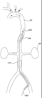
Systemic arterial circulation relevant to the methods of the present invention
is
described in Fig. 1. During systole, oxygenated blood leaving heart 8 enters
aorta 10,
which includes ascending aorta 12, aortic arch 14, and descending aorta 22.
The aortic
arch gives rise to brachiocephalic trunk 16, left common carotid artery 18,
and left
subclavian artery 20. The brachiocephalic trunk branches into right common
carotid
artery 24 and right subclavian artery 26. The right and left subclavian
arteries,
respectively, give rise to right vertebral artery 28 and left vertebral artery
34. The
descending aorta gives rise to a multitude of arteries, including lumbar
(i.e., spinal)
arteries 38, which perfuse the spinal cord, renal arteries 40, which perfuse
the kidneys,
and femoral arteries 42, which perfuse the lower extremities.
In one embodiment as shown in Fig. 2A, the obstruction device comprises
elongate catheter 102 having a proximal end and a distal end, shown here
positioned
within descending aorta 22. The distal end has expandable member 104, e.g., a
balloon. Balloon 104 communicates with inflation lumen 51 through port 105. In
another embodiment, depicted in Fig. 2B, ports 111 are included in the surface
of
catheter 102 to allow blood flow through the distal end of catheter 102 to
pass through
the catheter downstream constrictor 104.
In another embodiment as shown in Fig. 3, the obstruction device comprises
elongate catheter 102 having a proximal end and a distal end. The distal end
has first
expandable member 104 and second expandable member 107, e.g., balloons, and in
certain embodiments elongate balloons, mounted and spaced from each other.
Balloon
104 communicates with inflation lumen 51 through port 105. Balloon 107
CA 02444878 2010-04-15
50338-61
14
communicates with inflation lumen 109 through port 52. Balloon 104 and balloon
107
are thus able to be inflated independent of each other, or, in other
embodiments, are
inflated from a common inflation lumen.
It will be understood that the constrictor, when implemented as a
balloon, can be of any shape that is suitable for use in the aorta. An
elongate balloon
(e.g., balloons 104 and 107 in Fig. 3), elliptical or sausage-shape, is
particularly
desirable because this shape is more stable within rapidly flowing blood. A
spherical
balloon (although useful in the disclosed inventions) will tend to rock within
the aorta,
and rotate and bend the catheter to which it is affixed. The use of an
elongate
balloon, however, reduces the rocking and rotating within the vessel because-
this
shape effectively eliminates one of the degrees of freedom present with a
spherical
balloon.
In certain embodiments, the catheter is equipped with blood pressure
measuring capabilities proximal and/or distal to one or each expandable
member.
The blood pressure measuring capabilities may comprise a manometer mounted on
the catheter or a channel communicating with a transducer at the proximal end
and a
port at the distal end of the catheter. Blood pressure measuring may also be
accomplished by use of a fiber optic in vivo pressure transducer as described
in U.S.
Patent Nos. 5,392,117 and 5,202,939, or a Radi pressure wire as described in
U.S.
Patent Nos. Re 35,648; 5,085,223; 4,712,566; 4,941,473; 4,744,863; 4,853,669;
and
4,996,082.
In use, the catheter is inserted in descending aorta 22, and advanced to
a position such that first constricting balloon 104 is upstream of the renal
arteries,
celiac, and superior mesenteric artery, and second constricting balloon 107 is
downstream of these arteries as shown in Fig. 4. A two-balloon device permits
independent regulation and adjustment of cerebral blood flow and renal blood
flow.
Thus, downstream balloon 107 is first expanded while measuring cerebral blood
flow
until the desired increase over baseline is obtained, e.g., 100% increase.
This step
will also result in increased blood flow to the renal and superior mesenteric
arteries. If
this step results in inadequate cerebral blood flow increase, then upstream
balloon
104 is expanded to constrict upstream the renal and superior mesenteric
arteries until
the desired cerebral blood flow increase is obtained. Deployment of the
upstream
constrictor reduces blood flow to the renal and superior mesenteric arteries
as
compared with blood flow before
CA 02444878 2003-10-23
WO 02/085443 PCT/US02/12582
deployment of the upstream constrictor.
If the deployment of downstream balloon 107 produces the desired increase in
cerebral blood flow, then upstream balloon 104 will not be deployed in certain
procedures. In other procedures, upstream balloon 104 is deployed so that
constriction
5 in downstream balloon 107 can be reduced, thereby partially relieving the
renal and
superior mesenteric arteries of increased flow. It will be understood that
inclusion of a
balloon downstream is desirable in some cases because it allows the surgeon to
maintain renal blood flow at or above baseline while increasing blood flow to
the brain.
It may also be desirable to achieve constriction predominantly downstream of
the renal
10 arteries that supply blood to kidneys 83 to avoid obstructing the spinal
arteries that lie
upstream the renal arteries. It may also be desirable to have both balloons
107 and 104
partially inflated, rather than either balloon fully inflated, to avoid
blocking arteries that
branch from the aorta.
Alternatively, both balloons may be inflated simultaneously until a desired
15 increase in cerebral flow is achieved. In this manner, flow to the renal
arteries will be
maintained at substantially the initial baseline flow. If it is desired to
further adjust
renal blood flow while maintaining the cerebral blood flow and/or increase in
proximal
aortic pressure, the two balloons can be simultaneously adjusted, e.g., one
increased
and one decreased, until the desired renal blood flow is achieved.
It will be understood that one objective for the devices and methods described
herein is to increase cerebral blood flow during stroke. Expansion of a
constrictor in
the descending aorta produces increased blood pressure upstream of the
constrictor,
which leads to increased cerebral blood flow. A small change in upstream blood
pressure, however, can produce a very large change in cerebral blood flow.
Cerebral
blood flow can be measured by transcranial Doppler, functional MRI, CT scan,
PET
scan, SPECT scan, or any other suitable technique known in the art. In certain
procedures therefore, it may be desirable to adjust expansion of constrictors
107 and/or
104 in response to measured cerebral blood flow increase instead of, or in
addition to,
measured blood pressure increase upstream the constrictor and/or measured
blood
pressure decrease downstream the constrictor. If cerebral blood flow is to be
used as a
measure, then a baseline blood flow is measured before expansion of the
constrictor.
The constrictor is then expanded while measuring blood flow until a desired
increase in
CA 02444878 2003-10-23
WO 02/085443 PCT/US02/12582
16
flow is achieved. Typically, the desired increase will be 50 percent or
greater, 60
percent or greater, 70 percent or greater, 80 percent or greater, 90 percent
or greater, or
100 percent or greater of baseline blood flow, or more than 100 percent. The
amount of
increased cerebral blood flow will depend on a variety of factors including
the patient's
baseline blood pressure. If the blood pressure is excessively high, it may be
desirable
to achieve a smaller increase in cerebral blood flow, so as not to increase
the proximal
aortic pressure to an excessive value. In addition, the increase in the amount
of
pressure or flow achievable will also depend on baseline conditions. For
example, the
lower the baseline aortic pressure, the larger the pressure increase
achievable.
A plot of upstream aortic blood pressure and cerebral blood flow versus
percent
occlusion of the cross-sectional area of the descending aorta is shown in Fig.
5A. As
can be seen from these data generated in a model system, a favorable increase
in
cerebral blood flow and aortic blood pressure occurs at 50 percent occlusion
and
greater, at 56 percent occlusion and greater, and at 64 percent occlusion and
greater.
An even more favorable increase occurs at 71 percent occlusion and greater, 76
percent
occlusion and greater, and at 83 percent occlusion and greater. A still more
favorable
increase can be seen at 91 percent occlusion and greater, 96 percent occlusion
and
greater, and at 98 percent occlusion and greater.
It will further be understood that, when constriction is applied, there is a
sharp
increase in cerebral blood flow. The initial percent increase in cerebral flow
is believed
to be significantly higher than the percent increase in upstream aortic
pressure in the
presence of stroke. This appears to be the case for both the ischemic brain
and the
normal brain. A plot of cerebral blood flow versus time as set forth in Fig.
5B,
however, shows that the cerebral blood flow rate decays with time after the
initial
application of the constrictor at time = t1. This decay is possibly due to
autoregulation
within the brain. When the constriction is released, even for a short time
(e.g., 10
seconds, 20 seconds, 30 seconds, 1 minute, or more), and then applied again
(time = t2),
there is again a sharp increase in cerebral blood flow followed by gradual
decay. Thus,
one contemplated treatment regimen would include periodic (every 30 minutes or
one
hour) release of constriction to "reset" the autoregulatory system followed by
re-
expansion of the occluder at time t2. Another contemplated treatment regimen
would
include a gradual increase in constriction with time in order to maintain an
CA 02444878 2003-10-23
WO 02/085443 PCT/US02/12582
17
approximately constant rate of increased cerebral blood flow.
The aorta is a curved vessel that bends as it progresses from the aortic arch
to
the branch at the femoral arteries, as shown in Fig. 4. When one or both of
the
occlusion balloons are inflated, the blood pressure in the aorta upstream of
the
occluder(s) is caused to increase, while the pressure below the occluder(s) is
decreased
from baseline. With significant obstruction, e.g., 85-95 percent diameter
obstruction,
this pressure drop along the length of the occlusion balloon(s) can be
significant, on the
order of 20-150 mmHg. This pressure drop, by acting on the cross-sectional
area of
the occlusion balloon(s) creates a substantial longitudinally directed
compressive force
on the shaft of the catheter. The pressure drop and force are pulsatile in
nature (due to
systole and diastole) and tend to pulsatilly push the occlusion device down
and back up.
To minimize this motion it is desirable to reinforce the catheter shaft. One
way
to reinforce the shaft is to incorporate stiffening mandrel or stylet 240.
This may be
incorporated within the shaft at the point of manufacture or it may be
introduced within
the shaft once the occlusion device is positioned in the aorta. Furthermore,
the mandrel
or stylet 240 may be a solid wire, or may be a hollow tube, such as a
hypotube.
In use, a guidewire is advanced into the aorta. Catheter 102 is advanced over
the guidewire. Once the catheter is in place, the guidewire is removed and
mandrel 240
is advanced into a lumen of the catheter until it reaches the proper position.
In certain
procedures, the mandrel has a curvature at the end to forcibly deflect the
occlusion
balloon(s) to the wall of the aorta. The mandrel is then periodically rotated
to
reposition constrictors 104 and 107 at a new location along the lumenal wall
of
aorta 22. This periodic movement ensures that branching vessels are not
deprived of
blood for too long.
Although the balloon(s) of this embodiment will tend to be deflected to the
wall
of the aorta, the mandrel will further assure that the balloon will be
deflected, resulting
in an eccentric annular flow path for the balloon. Although an eccentric
annulus has
less flow resistance than a concentric annulus, it is desirable to prevent
this non-
centering embodiment from periodically becoming centered, as this would allow
the
flow resistance to vary over time.
A further dual balloon device is illustrated in Fig. 6. Distal balloon 104 and
proximal balloon 107 are both fabricated of an elastomeric material such as
blow
CA 02444878 2003-10-23
WO 02/085443 PCT/US02/12582
18
molded polyurethane. Both are preferably molded to have an initial inflated
diameter
of about 10 mm, with a capability of being inflated to 25 mm with increasing
pressure.
It is anticipated that other sizes could be utilized. For example, the distal
balloon could
be larger than the proximal balloon, with an initial diameter of 15 mm, and a
capability
of being inflated to 35 mm with increasing pressure.
Both balloons may have a body length of from 3-6 cm, preferably about 4 cm.
The distal tapered cone 113 and proximal tapered cone 115 may have a length of
1-3
cm, and about 2 cm. Each balloon has two cylindrical waists 117 and 119 which
are
used in the securing of the balloons to catheter shaft 102. The balloons may
be
adhesively bonded to the catheter shaft, or may be thermally bonded. Other
suitable
means of joining the balloons are also contemplated.
The balloons 104 and 107 are mounted on the distal region of catheter shaft
102. In this embodiment, the catheter shaft structure includes a unitary
extruded multi-
lumen tube (see cross-section in Fig. 6A), which extends for the full length
of the
device, with the exception of a soft tip attached at the distal end. The multi-
lumen tube
is preferably formed of an extrudable polymer, such as Pebax, polyethylene,
polyurethane, polypropylene, or nylon. Alternatively, the shaft structure
could be
fabricated as illustrated in FIG 6D. In this structure, individual thin walled
tubes are
used to define each lumen, and are preferably formed of a material suitable
for very
20, thin walls, such as polyimide or polyimide composite structures. As
illustrated, the
inter-balloon pressure monitoring lumen 161, and the inflation lumens 51 and
109 are
defined by thin polyimide tubes, and the wire lumen is defined by a thin
walled
composite tube of PTFE, braided metal, and polyimide. The four thin walled
tubes 51,
109, 161, and 162 are then encased within an extrusion or coating 163 of a
polymeric
material, such as Pebax, polyurethane, polyethylene, or other suitable
polymer.
There are four lumens within tube 102, wire lumen 162, inter-balloon pressure
monitoring lumen 161, and two inflation lumens 51 and 109, one each for
delivery of
inflation fluid to each balloon. Each balloon is inflated via ports 52 and 105
which
allow fluid communication between the inflation lumen and the balloon
interior. The
portion of the inflation lumens which extend distally of their respective
ports are
occluded by suitable means such as an adhesive plug.
The inter-balloon pressure monitoring lumen 161 is in fluid communication
CA 02444878 2003-10-23
WO 02/085443 PCT/US02/12582
19
with the surrounding blood via a port 160 in the tubing wall. When a suitable
fluid
such as saline resides in this lumen during used of the device, the blood
pressure at the
port is transmitted down the lumen to a pressure transducer. When the device
is
positioned as preferred, with the two balloons spanning the renal arteries,
the renal
blood pressure can be monitored, providing input to influence the degree of
balloon
inflation of the two balloons.
Wire lumen 162 is used during initial placement with a guide wire, which may
be later removed, or may be left in place. The remaining space within the wire
lumen
may be used to monitor the blood pressure upstream from distal balloon 104.
This is
another input which may be used to influence the degree of inflation of one or
both
balloons.
Preferred tubing dimensions for the inflation lumens are between 9 and 60
mils,
more preferably between 1 and 20 mils. Preferred tubing dimensions for the
pressure
lumens are between 5 and 60 mils, more preferably between 8 and 20 mils.
Preferred
tubing dimensions for the main lumens are between 30 and 80 mils, more
preferably
between 35 and 60 mils.
As mentioned, the shaft structure also includes a soft tip. Preferably, this
is a
single lumen tube fabricated of a more flexible material than that of the
multi-lumen
tubing. The tip is attached to the distal end of the multi-lumen tube by
suitable means
such as a thermal or adhesive butt joint. The single lumen within the tip
creates an
extension of the wire lumen. The soft tip is preferably about 2 to 10 cm long,
and
serves as an atraumatic tip facilitating catheter introduction and
positioning, as well as
providing an atraumatic "bumper" to the device during long term indwelling
use. The
tip may be straight, and may further include a tapering dimension on the outer
and inner
diameters. The tip may also be fabricated in a "pigtail" shape (Fig. 6C),
which
straightens in the presence of a guide wire extending through the wire lumen,
but
returns to the curled shape upon removal of the guide wire. A pigtail shape is
relatively
atraumatic.
The device as described is relatively flexible for smooth advancement over a
guidewire, and may be introduced into the aorta without the need for
fluoroscopic
guidance. Radio-opaque markers would nonetheless preferably be provided, in
the
instances where fluoroscopic guidance is utilized, or if a simple plate x-ray
is used to
CA 02444878 2003-10-23
WO 02/085443 PCT/US02/12582
assist in device positioning.
As mentioned previously, when one or both balloons of a dual balloon device
are inflated, significant longitudinal compressive forces can be imposed on
the catheter.
To help stabilize the device, the shaft structure of this embodiment provides
for
5 subsequent introduction of a stiffening element, such as a wire stylet, or a
hypotube. If
a wire stylet is used, the initial delivery guidewire is removed, to make room
for the
stylet. The stylet (Fig. 6E) is preferably tapered, and has a bulbous tip,
facilitating
smooth introduction into the wire lumen. The stylet may be quite large,
occupying
most of the available lumen. However, it is preferable to still maintain a
clearance
10 between the stylet and the wall of the wire lumen, to maintain the ability
to monitor
blood pressure. Alternately, the stylet may incorporate a pressure transducer
mounted
near the tip, in which case, the wire lumen can be fully occupied by the
stylet.
If a hypotube is used as the stiffening element, the initial guide wire need
not be
removed, as long as the inner diameter of the hypotube is large enough to
accommodate
15 the guide wire, which is typically either 0.035 or 0.038 inches in
diameter. Preferably,
the hypotube has a diameter slightly less than the wire lumen diameter, and a
tapering
outer diameter toward the distal end, to facilitate smooth tracking in the
wire lumen.
Hypotube 165 (Fig. 6F) can further incorporate a "skive" to gain further
flexibility near
the distal end to facilitate smooth tracking. Alternately, the distal portion
of the
20 hypotube can have a helical cut of progressively tighter pitch (Fig. 6B),
or other
patterns of removed material in hypotube 165 to facilitate a gradually
increasing
flexibility. The inner lumen of the hypotube can be used as a pressure
monitoring
lumen for the upstream aortic pressure. Preferably the hypotube is coated both
on the
internal surface by a lubricious and non-thrombogenic material, such as a
hydrophilic
coating, PTFE liner, or a paralene coating. With both the wire stylet and the
hypotube
stiffening elements, it is contemplated that they could be incorporated
initially within
the device, as opposed to introduced subsequent to positioning of the
balloons. If the
stiffening element is initially incorporated into the shaft structure, it is
preferred to
connect somewhere in the distal region of the hypotube to the shaft tube, by
suitable
means such as an adhesive or thermal bonding.
Referring again to Fig. 6, at the proximal end of the device, a manifold
structure
is connected to the shaft structure. The manifold structure includes luer
fittings that
CA 02444878 2003-10-23
WO 02/085443 PCT/US02/12582
21
communicate with each of the lumens. Fitting 169 communicates with pressure
monitoring lumen 161, fitting 167 communicates with proximal balloon inflation
lumen
109, and fitting 168 communicates with distal balloon inflation lumen 51. The
entire
shaft structure and balloons are preferably coated with a non-thrombogenic
coating,
such as a hydrophilic coating, and/or a heparin coating. Other anti-
thrombogenic
agents are also possible, such as phospholcholine.
Figure 7 illustrates an additional embodiment for a dual balloon occlusion
device, and utilizes an alternative shaft structure. The shaft structure
comprises two
primary components-multi-lumen polymeric tube 102, and hypotube 165. The
hypotube in this embodiment is fabricated directly into the device. The multi-
lumen
tube has three lumens, as shown in Figure 7A. The main lumen 162 is circular.
The
hypotube resides within this lumen, and the remaining leftover annular space
105
serves as the inflation lumen for the distal balloon. Lumen 109 serves as the
lumen
for inflation of the proximal balloon, and lumen 161 serves as the lumen used
in
connection with inter-balloon pressure monitoring.
Hypotube 165 extends distally of the multi-lumen tube, and preferably
terminates distal of the distal balloon. The hypotube is preferably lined on
the inner
surface, in a manner as described for the hypotube above. The lumen of the
hypotube
serves as the guide wire lumen as well as the pressure monitoring lumen for
the
upstream aortic pressure. The distal balloon is attached to the exterior of
hypotube 165
by suitable means such as adhesive or thermal bonding. The distal end of the
hypotube
can incorporate features as described above to serve as a transition in
stiffness. A soft
tubular tip is preferably attached to the hypotube, creating an atraumatic
tip.
Figure 8 illustrates another embodiment of a dual balloon occlusion device,
which utilizes an alternative shaft structure. The shaft structure is
comprised of three
coaxially positioned tubular components. Outer tube 102 is circular and
polymeric, and
defines lumen 52 which is used for inflation of proximal balloon 107. Middle
tube 170
is circular and polymeric and defines lumen 105 which is used for inflation of
distal
balloon 104. Inner tube 165 is circular, and preferably a hypotube. The tubes
are
arranged such that proximal balloon 107 is attached proximally to outer tube
102, and
distally to middle tube 170. The distal balloon is attached proximally to
middle tube
170, and distally to inner tube 165.
CA 02444878 2003-10-23
WO 02/085443 PCT/US02/12582
22
Hypotube 165 defines lumen 171 which serves as a guide wire lumen, as well as
a lumen for monitoring the upstream aortic blood pressure. The hypotube is
preferably
lined on the inner surface, in a manner as described for the hypotube above.
The distal
end of the hypotube can incorporate features as described above to serve as a
transition
in stiffness. A soft tubular tip is preferably attached to the hypotube,
creating an
atraumatic tip. As with the above embodiments, the entire shaft structure and
balloons
are preferably coated with a non-thrombogenic coating, such as a hydrophilic
coating,
and/or a heparin coating. Other anti-thrombogenic agents are also possible,
such as
phospholcholine.
The balloon constrictors described herein are desirably blow molded from a
material that is elastomeric, such as polyurethane, allowing an adjustable
balloon
diameter, as indicated in Fig 9. The balloons will typically be sized to
achieve full
expansion, i.e., wrinkle-free expansion, at approximately 10 mm diameter in
cross-
section and at a pressure of 0.5-5 psi.. A pressure of 5 psi at the low end of
the
operating range is desirable because the balloon is firm at this pressure and
therefore
resists the tendency to distort its geometry in a rapidly flowing blood
stream. The
balloon material will allow further expansion (beyond 10 mm) upon further
inflation
(e.g., by syringe) to a maximum diameter of approximately 25 mm and at a
pressure of
12-50 psi. An operating range of approximately 10-25 mm balloon diameter is
desirable to accommodate variations in patient anatomy and to allow the
surgeon to
vary constriction to adjust cerebral blood flow rate to the desired level. For
larger
aortas, a balloon of 15-30 mm may be desirable. A wrinkle-free balloon at 10
mm
diameter cross-section is desired because wrinkles will produce unpredictable
and
variable flow properties, and wrinkles will produce a distortion in balloon
material with
material bunching together at the downstream edge of the balloon.
In other embodiments as depicted in Fig. 10, a centering mechanism will be
used to maintain the constricting balloon apart from the vessel wall. Catheter
102
includes balloon 107, and inner sheath 53 includes balloon 104. The centering
mechanism for balloon 107 here is provided by struts 63 mounted (either
slideably or
fixedly) at a proximal end to catheter 102, and at a distal end to inner tube
53. The
centering mechanism for balloon 104 is provided by struts 62 mounted (either
slideably
or fixedly) at a proximal end to inner tube 53, and at a distal end to inner
tube 53. A
CA 02444878 2003-10-23
WO 02/085443 PCT/US02/12582
23
cross-section taken through section line A-A is shown in Fig 10A, and a cross-
section taken through section line B-B is shown in Fig IOB. Alternative
structures,
such as a braid as a centering mechanism, are also contemplated.
An alternative centering mechanism is shown in Fig. 11. Catheter 102 includes
inner shaft 65 having a working channel. Constricting balloon 104 is bonded to
shaft
65 at a distal end thereof Self-expanding wires 66 are bonded at one end to
catheter
102, and at a second end to centering mechanism 64. Here, centering mechanism
64 is
a deployable wire mesh with fabric or polymer cover. A cross-section taken
through
section line A-A is shown in Fig I IA. Wire mesh 64 is surrounded by cover 67.
Distal supporting struts 68 are provided to strengthen the centering mechanism
distally.
By maintaining the constricting balloon centered in the vessel, blood flows
around the balloon on all sides. Thus, all branching vessels are perfused when
this
design is employed. Moreover, the velocity of blood flow increases in the
region of the
constrictor. This increased velocity in combination with the balloon
channeling blood
against the vessel wall can actually increase perfusion of branching vessels
in certain
cases. It will be understood that, in the absence of a centering mechanism and
without
a mandrill, the catheter and the one or more balloons will contact and bear
against the
lumenal wall of the aorta.
In another embodiment as shown in Fig. 12, catheter 102 carries slideable
inner
shaft 53. Shaft 53 includes an inflation lumen and an assortment of
constricting
balloons 104 mounted at different positions. Each of these balloons has a
different
diameter of expansion to accommodate different degrees of constriction and
different
patient anatomy. In use, the first and smallest balloon is advanced from the
distal port
of catheter 102 and deployed. If a larger balloon is needed, then the second,
larger
balloon is advanced out of the catheter and deployed. If needed, the third
balloon can
be advanced into the vessel and deployed. At the proximal end of catheter 102
is outer
sheath 75 and Y-adapter 55 with inflation/deflation port 81 and port 82 for a
guidewire,
for flushing, or for access by any other tools or instruments. Y-adapter 55 is
connected
to sheath 75 by hub 83 that has capabilities for multiple position adjustment.
Fig. 13 depicts occlusion membrane 76 that acts as an occluding member
instead of using a balloon. Occlusion membrane 76 comprises a coated mesh.
Catheter
102, having a flexible outer sheath, carries control rod 77. The distal end of
control rod
CA 02444878 2003-10-23
WO 02/085443 PCT/US02/12582
24
77 is fixed to occlusion membrane 76 at its distal end. When control rod 77 is
extended, occlusion membrane 76 is stretched as shown in Fig. 13, reducing the
cross-
sectional profile at the proximal end of occlusion membrane 76. As control rod
77 is
withdrawn, occlusion membrane 76 progressively expands, increasing the cross-
sectional profile at the proximal end as shown in Figs. 13B, 13C, and 13D. At
the
proximal end, control rod 77 terminates in positioning handle 78 for adjusting
the
cross-sectional profile of the occlusion membrane 76.
Fig. 14 depicts occlusion catheter 100 for use in the methods described
herein.
The device includes elongate catheter 102, distally mounted expandable
constrictor,
i.e., occluder, 104 having distal opening 124 and variable flow mechanism 108.
The
constrictor, when expanded, has maximum periphery 110, which conforms to the
inner
wall of a vessel to form a secure seal with the vascular wall, such that blood
flow
through the vessel can be effectively controlled. Opening 124 receives blood
from
distal the constrictor and controls the passage of blood proximal the
constrictor.
Variable flow mechanism 108, connected to rotary unit 150, operates the
constrictor, .
thereby controlling (1) the flow rate through the occlusion, and (2) upstream
blood
pressure. Preferably, the device includes manometer 112, which is connected to
pressure monitor 156 and pressure limiter 114. Rotary unit 150 receives blood
pressure measurements from the manometer. Pressure limiter 114, connected to
the
rotary unit and the pressure monitor, prevents the upstream and downstream
blood
pressure from exceeding, respectively, a set maximum and minimum pressure
differential. A proximal end of the catheter is equipped with adapter 103,
from which
pull wires 132 can be manipulated for collapsing the occluder and to which the
rotary
unit, pressure monitor, and/or pressure limiter can be connected.
Referring to Fig. 15, the occlusion device comprises catheter 102 and
constrictor 104. The catheter is constructed from a biocompatible and flexible
material,
e.g., polyurethane, polyvinyl chloride, polyethylene, nylon, etc. The catheter
includes
lumen 116 through which various operative elements pass. Alternatively, the
catheter
may include more than one lumen to support various operative elements. The
catheter
also includes proximal adapter 103 (see Fig. 14), which provides an interface
between
the catheter and the various instruments received by the catheter. The
occluding
mechanism consists of outer conical shell 118 and inner conical shell 136,
each having
CA 02444878 2003-10-23
WO 02/085443 PCT/US02/12582
a distal open base and a proximal apex. Pre-shaped ring 130 is affixed to base
120 of
the outer shell to facilitate expansion of the constrictor. The ring is formed
of a
resilient material, capable of expanding the occluder to achieve a maximum
periphery,
which is defined by the outer circumference of the ring. Ring 130, may, in
certain
5 embodiments, further include an anchoring mechanism, such as hooks, bonded
to the
outer circumference of the ring. Expansion of the ring causes the grasping
structure to
engage the surface of the vessel wall, thereby securing the occluder and
preventing
displacement in the vessel due to force exerted by blood flow. In other
embodiments,
the anchoring is provided by an adhesive strip, vacuum, or merely by
frictional
10 engagement of the vessel lumen by the ring.
The constrictor can be collapsed to facilitate insertion into and removal from
a
vessel. A plurality of pull wires 132 (Fig. 14) are disposed within torque
cable 148,
and are distally connected to base 120 of outer shell 118 and proximally
passes through
adapter 103. The constrictor is collapsed by applying a tensile force on wires
132,
15 using torque cable 148 to provide leverage to the pull wires, thereby
drawing the
circumference of the open base 120 towards its center and collapsing the
occluder. A
guide sheath (not shown) can be alternatively used to collapse the
constrictor. Using
this technique, the guide sheath would cover the constrictor and be withdrawn
to
release the constrictor and advanced to collapse the constrictor.
20 Opening 124 is formed in base 138 and 120 of the respective inner and outer
conical shells to provide an inlet for blood flow. Conical interior 106
communicates
with ports 128 of the outer shell. When the constrictor is deployed, blood
flows into
opening 124, through interior 106, and exits through ports 128. The occluding
mechanism comprises inner conical shell 136 (partially shown in phantom in
Fig. 15),
25 which is rotatably disposed within outer shell 118 as shown in Figs. 8, 9,
and 10. The
inner shell can be rotated relative to the outer shell through torque cable
148, which is
disposed in lumen 116 of catheter 102.
Manometer 112 comprises upstream pressure tube 152 and downstream
pressure tube 154, both connected proximally to a pressure monitor to provide
respective blood pressure measurements upstream and downstream the
constrictor. The
upstream pressure tube extends distal to opening 124, or may be attached to
the inner
shell. The downstream pressure tube extends through an orifice in the catheter
proximal
CA 02444878 2003-10-23
WO 02/085443 PCT/US02/12582
26
to the constrictor. The upstream and downstream blood pressure measurements
are
recorded and displayed by the pressure monitor at a proximal end of the
catheter. A
pressure limiter, programmed with a maximum pressure threshold to limit the
upstream
blood pressure and a minimum pressure threshold to limit the downstream blood
pressure, is connected to the pressure monitor to receive pressure
measurements
therefrom, and transmits information to a rotary unit. The limiter thereby
prevents the
rotary unit from rotating the inner shell relative to the outer shell in a
manner that
would cause the upstream blood pressure to exceed the maximum threshold, or
the
downstream blood pressure to fall below the minimum threshold. Without the
rotary
unit, torque cable 148 can also be manually rotated to obtain desired upstream
and
downstream blood pressures. An audible alarm may be incorporated into the
pressure
limiter to sound when blood pressures exceeds the thresholds. The pressure
limiter
may further comprise an interlocking device. The interlocking device, in
operative
association with upstream and downstream tubes 152 and 154, can lock inner
shell 136
with respect to outer shell 118 as blood pressures approach the set
thresholds. It should
be noted that although the rotary unit, pressure monitor, and pressure limiter
are shown
as separate units, they may be incorporated into an integral unit.
Referring to Figs. 16A and 16B, the expanded constrictor comprises outer
conical shell 118 having base 120 and apex 122, and inner conical shell 136
having
base 138 and apex 140. The constrictor is preferably composed of a
biocompatible
material coated with heparin to prevent blood clotting. The conical shape of
the
expanded constrictor minimizes turbulence caused by placement of the occluder
in the
vessel. The outer and inner shells include 2, 3, 4, 5, 6, or any other number
of ports
128 and 144, respectively, in communication with the conical interior to
permit blood
flow through the occluder. The inner shell can be rotated relative to the
outer shell, so
that ports 144 communicate with ports 128. Apices 122 and 140 of the
respective outer
and inner shells further comprise collar 126 and 142. The collars may include
engaging
threads, so that collar 142 can be inserted and secured into collar 126, and
bonded to a
distal end of the torque cable, such that the inner shell is coupled to and
rotates with the
torque cable. A rotary unit, preferably including a stepper motor (not shown),
may be
mechanically coupled to a proximal end of the torque cable to provide precise
rotational position of the inner shell relative to the outer shell, thereby
providing
CA 02444878 2003-10-23
WO 02/085443 PCT/US02/12582
27
variable flow through the occluder.
Instead of having the circular ports in the inner and outer shells as depicted
in
Figs. 16A and 16B, the constrictor may include 2, 3, 4, 5, 6, or any other
number of
ports having other suitable geometric shapes. Fig. 17 depicts constrictor 104
having a
plurality of ports constructed as elongate rectangular slots 175.
Fig. 18 depicts another embodiment of the constrictor, which comprises beveled
lip 140 having distal end 142 and proximal end 141. The proximal end is
affixed to
base 120 of the outer conical shell. The proximal end has a larger diameter
than the
distal end and is everted to prevent the constrictor from being displaced in
the direction
of blood flow, thereby securing the constrictor in the vessel.
Still another embodiment of the occluder may includes 1, 2, 3, 4, 5, or any
other
number of graduated inflatable rings. In Fig. 19, ring 151 is affixed to the
base of the
conical shell. Ring 153, having the smallest inflated diameter, is attached to
ring 152,
which is then attached to ring 151, having the largest inflatable diameter.
The fully
inflated rings will have a thickness of approximately 2 to 3 millimeters.
Similar to the
beveled lip of Fig. 20, the rings prevent the outer conical shell from being
displaced in
the direction of blood flow, thereby securing the constrictor in the vessel.
The flow rate of blood through the constrictor can be easily controlled by
rotating inner conical shell 136 (shown with dotted lines) relative to outer
conical shell
118 as depicted in Figs. 20, 21, and 22. In Fig. 20, the inner shell is
rotated so that
ports 144 and 128 are completely misaligned, thereby achieving no flow through
the
ports and complete vascular occlusion distally. As the inner shell is rotated
clockwise
relative to the second shell in Fig. 21, ports 144 on the inner shell become
partially
aligned with ports 128 on the outer shell, thereby achieving partial flow
through the
ports and partial vascular occlusion. In Fig. 22, with continuing clockwise
rotation of
the inner shell, ports 144 become completely aligned with ports 128, thereby
achieving
maximum flow through the ports. To provide a broader and more predictable
range of
blood flow through the conduit, the ports of the inner and outer shells are
preferably of
equal size and number such that they may align with each other.
Fig. 23 depicts another embodiment of the occlusion device for partial
occlusion
of blood flow in a vessel. Device 200 comprises elongate catheter 202,
distally
mounted expandable constrictor 204 with maximum periphery 210, opening 224,
and
CA 02444878 2003-10-23
WO 02/085443 PCT/US02/12582
28
variable flow mechanism 208 operatively associated with the constrictor. The
catheter
includes adapter 203 at its proximal end. Preferably, the device includes
manometer
212 and pressure limiter 214, and pressure monitor 240. The pressure monitor
records
and displays blood pressure data received from the manometer. Longitudinal
positioning unit 208, receiving signals from pressure limiter 214, and
controls variable
flow mechanism 208 to provide variable blood flow through the constrictor.
Referring to Fig. 24, catheter 202 includes lumen 216. Constrictor 204
comprises hollow conical shell 218 having base 220 and apex 222. The inner
circumference of the base forms opening 224, which provides a distal inlet for
blood
flow through the constrictor. The inner circumference of apex 222 forms collar
228
with proximal opening 226, which provide an outlet for blood flow through the
constrictor. The conical interior, disposed within shell 218, communicates
with
opening 224 distally and opening 226 proximally. When the base of the
constrictor is
positioned upstream in a vessel, blood flows into opening 224, through the
conical .
interior, and exits downstream through opening 226. The catheter is bonded to
collar
228 about a portion of its inner circumference. The constrictor is expanded by
operation of ring 230, a beveled lip, or a series of graduated toroidal
balloons as
described above. The constrictor is collapsed and may be delivered to a vessel
location
by using a guide sheath.
The manometer comprises upstream pressure tube 236 and downstream
pressure tube 238, which are disposed in lumen 216 of the catheter and
connected
proximally to a pressure monitor. The upstream pressure tube extends distal
from the
constrictor or may be bonded to the inner surface of the conical shell,
thereby providing
upstream blood pressure measurement. The downstream pressure tube extends
through
an orifice in the catheter proximal to the constrictor, thereby providing
downstream
blood pressure measurement.
The variable flow mechanism comprises a plurality of flaps 230 pivotally
affixed to base 220. The flaps are preferably made of a resilient material,
such as
Nitinol, to resist movement caused by blood flow through the conduit. A
plurality of
pull wires 232, disposed through lumen 216, are distally connected to flaps
230, such
that applying a tensile force to the wires pivotally displaces flaps 230 from
their
preformed position. Three of the flaps (shown in dotted lines) are displaced
inward.
CA 02444878 2003-10-23
WO 02/085443 PCT/US02/12582
29
Releasing the wires allows the resilient flaps to relax and return to their
preformed
position. The pull wires are coupled proximally to the longitudinal
positioning unit,
which provides precise displacement of the flaps relative to opening 224.
Alternatively, wires 232 can be manually tensed to operate the flaps. The
pressure
limiter receives pressure measurements from the pressure monitor and transmits
signals
to the longitudinal positioning unit to prevent the upstream and downstream
blood
pressures from exceeding the set thresholds.
Figs. 25A, 25B, 25C, and 25D depict frontal views of the constrictor having
flaps in various positions for controlling blood flow. In Fig. 25A, preformed
flaps 230
extend radially inward toward the longitudinal axis of the catheter, as in the
absence of
a displacing force, i.e., an external force other than that created by blood
flow. When
the constrictor is positioned in the descending aorta, for example, the size
of opening
224 and blood flow through the opening is minimized, thereby providing maximal
aortic occlusion. In the presence of a displacing force, such as pulling the
wires to
displace flaps 230 from their preformed position as depicted in Fig. 25B, the
size of
aperture 224 and blood flow through the conduit increases, thereby providing
partial
aortic occlusion.
Alternatively, preformed flaps 230 extend parallel to the longitudinal axis of
opening 224 in the absence of a displacing force as depicted in Fig. 25C. The
size of
opening 224 and blood flow through the conduit are maximized, thereby
providing
minimal blood flow occlusion. In the presence of a displacing force, flaps 230
are
pivotally displaced from their preformed position as depicted in Fig. 25D. The
size of
opening 224 and blood flow through the opening are minimized, thereby
providing
maximal blood flow occlusion. Thus, by pivotally displacing flaps 230 relative
to
opening 224, the size of the opening and flow rate through the constrictor is
controlled
to provide variable vessel occlusion.
The constrictor shown in Fig. 24 can be alternatively mounted on catheter 202,
such that base 220 is proximal to apex 222 as shown in Fig. 26. In this
embodiment,
flaps 230 are formed on open apex 222. When constrictor 204 is inserted
downstream
in the aorta, for example, pressure tube 238 extends distally from opening 226
to
provide downstream blood pressure measurements, whereas pressure tube 236
extends
proximally through an orifice in the catheter to provide upstream blood
pressure
CA 02444878 2003-10-23
WO 02/085443 PCT/US02/12582
measurements.
In Fig. 27, another embodiment of the device comprises catheter 302, a
distally
mounted occluder 304 with maximum periphery 310, blood passage 306 disposed
within the constrictor, and variable flow mechanism 308 in operative
association with
5 the blood conduit. Inflation device 334 communicates with the constrictor,
and
inflation device 338 communicates with the variable flow mechanism. The device
preferably includes proximal adapter 303, manometer 312, and pressure limiter
314.
Pressure monitor 312 records and displays blood pressure data from the
manometer.
The pressure limiter is connected to the pressure monitor and to an
interlocking valve
10 on inflation device 338, such that the blood pressure upstream and
downstream the
constrictor can be controlled to prevent from exceeding set thresholds.
Referring to Fig. 28, constrictor 304 is mounted to a distal end of catheter
302
having lumen 316. The constrictor comprises a sleeve or cylindrical balloon
318
having outer wall 320 and inner wall 322, which enclose chamber 323. The
cylindrical
15 balloon has first end 324 with opening 328 and second end 326 with opening
330.
Catheter 302 is bonded to inner wall 322 of the cylindrical balloon. Inflation
tube 332,
housed within lumen 316 of the catheter, communicates distally with the
cylindrical
balloon and proximally with a syringe or other inflation device. The
cylindrical balloon
can be expanded or collapsed by injecting or removing air, saline, or other
medium.
20 Occlusion is provided by toroidal balloon 334 disposed about the outer or
inner surface
of sleeve 318 and communicating with inflation tube 336 and a syringe. The
inflation
device may include an interlocking valve to prevent unintended deflation.
Lumen 306 communicates with opening 328 distally and opening 328
proximally. When deployed in a vessel, blood flows through lumen 306 and exits
25 downstream opening 330. The constrictor may further include an anchoring
structure,
shown in Fig. 28 as rings 333, which are disposed about outer wall 320 of the
cylindrical sleeve and define maximum periphery 310 of the occluder.
Manometer 312 comprises upstream pressure tube 340 and downstream
pressure tube 342, which are operatively connected proximally to a pressure
monitor.
30 Pressure tube 340 is bonded to the lumen of the cylindrical balloon and
extends distal
to provide upstream blood pressure measurements, while tube 342 emerges from
the
catheter proximal the occluder to provide downstream blood pressure
measurements.
CA 02444878 2003-10-23
WO 02/085443 PCT/US02/12582
31
In Fig. 29, fluid is injected to expand balloon 334, thereby constricting
sleeve
318. As a result, blood flow is constricted. In Fig. 30, balloon deflation
allows sleeve
318 to revert back to its pre-shaped geometry, increasing blood flow
therethrough.
Thus, balloon 334 can be inflated and deflated to vary the cross-sectional
diameter of
lumen 306 to vary flow rate.
The occlusion devices described herein can be employed with a variety of
therapeutic catheters to treat vascular abnormalities. For example, as
depicted in Fig.
31, suction/atherectomy catheter 402 can be inserted through lumen 306, such
that the
suction/atherectomy catheter is independently movable relative to occlusive
device 300.
Catheter 402 includes elongate tube 404 and distally located aspiration port
406, cutting
device 408, and balloon 410 for removing thromboembolic material in a vessel.
In Fig. 32, infusion catheter 502 and EPS catheter 504 are inserted through
opening 206 of occlusion device 200, such that catheter 502 and 504 are
independently
movable relative to occlusion device 200. The infusion catheter, which
includes
elongate tube 506, distally located perfusion port 508, and expandable balloon
510, can
be used to remove thromboembolic material in a vessel. EPS catheter 504, which
includes elongate tube 512 and distally located ablation device 514, may be
used to
map out or ablate an extra conduction pathway in the myocardial tissue, e.g.,
in patients
suffering from Wolff-Parkinson-White syndrome. The occlusion device, capable
of
augmenting cerebral perfusion, is therefore useful not only in facilitating
definitive
treatment but also in cerebral ischemia prevention during EPS and other
cardiac
interventions or cardiac surgery, such as coronary catheterization, where
sudden fall in
cerebral blood flow may occur due to arrhythmia, myocardial infarction, or
congestive
heart failure.
Referring to Fig. 33A, occlusion device 100 described above can be used to
partially occlude blood flow in aorta 10 of a patient suffering from global
cerebral
ischemia due to, e.g., septic shock, congestive heart failure, or cardiac
arrest.
Constrictor 104 can be introduced in its collapsed geometry through an
incision on a
peripheral artery, such as the femoral, subclavian, axillary, or radial
artery, into the
patient's aorta. A guide wire may first be introduced over a needle, and the
collapsed
constrictor is then passed over the guide wire and the needle to position
distal to the
takeoff of left subclavian artery 20 in the descending aorta. The constrictor
is
CA 02444878 2003-10-23
WO 02/085443 PCT/US02/12582
32
expanded, such that maximum periphery 110 of the occluder, formed by
expandable
ring 130, sealingly contacts the inner aortic wall. The position and
orientation of the
collapsed or expanded device can be checked by TEE, TTE, aortic arch cutaneous
ultrasound in the emergency room, or IVUS and angiography in the angiogram
suite.
The expanded constrictor is maintained during systole, during diastole, or
during systole and diastole, during which blood distal to the brachiocephalic
artery is
forced to pass through opening 106, thereby providing a continuous partial
occlusion of
aortic blood flow. Alternatively, partial occlusion of aortic blood flow can
be
intermittent. As a result, blood flow to the descending aorta is partially
diverted to
brachiocephalic artery 16, left subclavian artery 20, and left carotid artery
18, thereby
augmenting blood flow to the cerebral vasculature. In treating global
ischemia, such as
in shock, cerebral perfusion is increased by increasing blood flow through
both carotid
and vertebral arteries. Additionally, blood flow to the aorta is partially
diverted to the
coronary arteries by using the occlusion device, thereby augmenting flow to
the
coronary arteries. Using the partial occlusion methods during systemic
circulatory
failure may, therefore, improve cardiac performance and organ perfusion. By
selectively increasing cerebral and coronary blood flow in this manner, the
dosage of
commonly used systemic vasoconstrictors, such as dopamine and norepinephrine,
may
be reduced or eliminated.
Alternatively, the device of Fig. 26, much like the device used to extinguish
the
flame of a candle, can be introduced through an incision on left subclavian
artery 36 as
depicted in Fig. 33B. Constrictor 204 is inserted in aorta 22 distal to the
takeoff of the
left subclavian artery to provide partial, variable, and/or continuous aortic
occlusion
and is advanced antegrade into the descending aorta. This device is
particularly useful
in situations where peripheral incision cannot be made on the femoral arteries
due to
arteriosclerosis, thrombosis, aneurysm, or stenosis. The device may
alternatively be
inserted into the left or right brachial, left or right subclavian, left or
right radial
arteries, and then advanced into the aorta. It will be understood that these
alternative
approaches do not require a stiffening mandrel because the device is under
tension
rather than compressive loading. Any of the devices described herein can be
used in
these alternative approaches. These alternative approaches may also permit
devices
that are more flexible and smaller in diameter.
CA 02444878 2010-04-15
50338-61
33
The devices and methods described in Figs. 33A and 33B are useful in
treating stroke patients within few minutes of stroke symptom, and the
treatment can
be continued up to 96 hours or more. For example, in treating focal ischemia
due to
a thromboembolic occlusion in the right internal carotid artery the
constrictor may be
position distal to the takeoff of the left subclavian. As a result, blood flow
is diverted
to brachiocephalic artery 16 and left CCA to augment both ipsilateral and
contralateral
collateral circulation by reversing direction of flow across the Circle of
Willis, i.e.,
increasing flow in the right external carotid artery and left common carotid
artery. The
collateral cerebral circulation is further described in details in U.S. Patent
No.
6,165,199.
In treating focal ischemia due to a thromboembolic occlusion in the left
internal carotid artery, for example, the constrictor can be positioned
proximal to the
takeoff of left carotid artery 18 and distal to the takeoff of brachiocephalic
artery 16 as
shown in Fig. 34. Contralateral collateral enhancement is provided by
increasing flow
through the brachiocephalic artery, thereby reversing blood flow in the right
posterior
communicating artery, right PCA, left posterior communicating artery 68 and
anterior
communicating artery, resulting in increased perfusion to the ischemic area
distal to
the occlusion and minimizing neurological deficits. Alternatively, the
constrictor may
be positioned distal to the takeoff of the left subclavian artery to provide
both
ipsilateral and contralateral collateral augmentation. Ipsilateral circulation
is
enhanced by increasing flow through the left external carotid artery and
reversing flow
along the left ophthalmic artery, both of which contribute to increased flow
in the left
ICA distal to the occlusion.
As a result of partially occluding aortic blood flow, blood pressure distal
to the aortic occlusion may decrease, and this may result in a reduction in
renal
output. Blood pressure proximal the aortic occlusion will increase and may
result in
excessive rostral hypertension. The blood pressures, measured by the
manometer,
are monitored continuously, and based on this information the occlusion is
adjusted to
avoid peripheral organ damage. After resolution of the cerebral ischemia, the
constrictor is collapsed and removed, thereby removing the aortic occlusion
and
restoring normal blood flow in the aorta.
CA 02444878 2010-04-15
50338-61
34
In Fig. 35, constrictor 304 is inserted in aorta 10 and can be used to
remove thromboembolic material 72 from left common carotid artery 18, while
augmenting and maintaining cerebral perfusion distal to the occluding lesion.
The
occluder may be introduced through a guide sheath until it is positioned
distal to left
subclavian artery 20. In emergency situations, the constrictor can be inserted
through
a femoral incision in the emergency room, and atherectomy/suction catheter 402
can
be inserted through the constrictor under angioscopic vision in the angiogram
suite
after the patient is stabilized hemodynamically. The atherectomy/suction
catheter,
which includes expandable balloon 410, distal aspiration port 406, and
atherectomy
device 408, is introduced through opening 306 until its distal end is
positioned in left
common carotid artery 18 proximal to the thromboembolic occlusion.
Constrictor 304 is then expanded to partially occlude aortic blood flow,
thereby increasing perfusion to the ischemic region distal to the occluding
lesion by
enhancing ipsilateral collateral flow through left external carotid artery 46
and left
vertebral artery 34 and contralateral collateral flow to right carotid artery
24 and right
vertebral artery 28. The variable flow mechanism of constrictor 304 can be
adjusted
to control blood flow to the cerebral vasculature and the blood pressure.
Balloon 410
of catheter 402 is expanded in the left common carotid artery, thereby
creating a
closed chamber between constrictor 410 and the thromboembolic occlusion.
Suction
can be applied to aspiration port 406 to create a negative pressure in the
closed
chamber, thereby increasing the pressure differential across the
thromboembolic
occlusion, which may dislodge the occluding lesion onto the aspiration port
and
remove the occluding lesion. Thromboembolic material 72 may be further removed
by atherectomy device 408. The methods herein can also be used to remove
thromboembolic occlusion in the vertebral artery. The occlusion device 304,
therefore, not only augments cerebral perfusion in patients suffering from
focal stroke
or global ischemia, but also maintains cerebral perfusion while waiting for
invasive or
noninvasive intervention. The devices and methods of using atherectomy/suction
catheter 102 are further described in U.S. Patent No. 6,165,199.
During abdominal aortic aneurysm (AAA) surgery, lumbar or spinal
arteries, which provide blood supply to the spinal cord, are often dissected
away from
the
CA 02444878 2003-10-23
WO 02/085443 PCT/US02/12582
diseased abdominal aorta, resulting in reduction of blood flow to the spinal
cord. The
devices herein disclosed may be used to condition the spinal cord prior to AAA
repair,
thereby reducing the damage resulting from spinal ischemia during surgery. In
Fig. 36,
constrictor 104 is inserted in aorta 10 and expanded preferably distal to left
subclavian
5 artery 20 and proximal to lumbar arteries 38. As a result, blood flow to the
lumbar or
spinal arteries is reduced. When this device is used in patients anticipating
a major
thoracoabdominal surgery, such as AAA repair, approximately 24 hours prior to
surgery, blood flow to the lumbar arteries can be intentionally reduced to
induce mild
spinal ischemia, thereby conditioning the spinal cord to produce
neuroprotective agents
10 which may protect the spinal cord from more significant ischemic insult
during surgery.
In hypertension, end organ damage often results, e.g., cardiac, renal, and
cerebral ischemia and infarction. The devices and methods herein may be
employed in
hypertension to protect the kidneys from ischemic insult. In Fig. 37,
constrictors 104,
which can be introduced through a femoral artery, are inserted in right renal
artery 80
15 and left renal artery 82. The constrictors are expanded to partially
occlude blood flow
from descending aorta 10 to the renal arteries, thereby reducing blood
pressure distal to
the occlusion. The constrictors can be deployed for the duration of any
systemic
hypertensive condition, thereby protecting the kidneys from damage that might
otherwise be caused by the hypertension.
20 In another embodiment, the constrictor will be provided with capabilities.
for
mounting on a standard catheter, e.g., a standard angioplasty balloon
catheter, a stent
deployment catheter, an ultrasound catheter, or an atherectomy catheter. Such
a device
having capabilities for removable mounting on a standard catheter is depicted
in Figs.
44A-44H. The constrictor shown in Fig. 44A includes elongate tubular member
601
25 having lumen 605 for passage of blood extending from a proximal to a distal
end of
tubular member 601. The device includes constrictor 602, here a balloon
mounted on
tubular member 601 and communicating with inflation lumen 603. Inflation lumen
603
extends proximal, and extends through the incision in the patient so that it
remains
operable outside the patient's body. Tubular member 601 is constructed of a
flexible
30 and deformable material so that inflation of balloon 602 causes a reduction
in the
cross-sectional diameter of lumen 605 as shown in Fig. 44B to reduce blood
flow.
In use, tubular member 601 is positioned in the descending aorta and balloon
CA 02444878 2003-10-23
WO 02/085443 PCT/US02/12582
36
602 is inflated. the outer diameter of balloon 602 expands until it engages
the lumen of
the descending aorta. Further inflation of balloon 602 will cause deformation
of tubular
member 601 to thereby reduce lumenal diameter 605. In this manner, peripheral
blood
flow is reduced, resulting in an increase blood pressure upstream of the
device.
Because the device shown in Figs. 44A and 44B is mounted on a standard
catheter (not
shown), the standard catheter having diagnostic or therapeutic capabilities
extends
beyond tubular member 601 and may access any of the coronary arteries, carotid
arteries, or any other vessels upstream of the descending aorta.
Fig. 44C depicts an alternative mountable constrictor having a shortened
tubular
member 601, shortened by comparison with tubular member 601 shown in Fig. 44A.
Fig. 44D shows the constrictor of Fig. 44C with balloon 602 inflated. It
should be
noted that inflation of balloon 602 proceeds until the outer diameter of
balloon 602
engages the lumen of the aorta, whereupon further inflation causes deformation
of
tubular member 601 inwardly to reduce the diameter of lumen 605, thereby
constricting
blood flow.
In another embodiment, a removably mountable constrictor is provided as
depicted in Figs. 44E and 44F. Referring to Fig. 44E, balloon constrictor 602
communicates with a proximally extending inflation lumen (not shown), and
balloon
602 includes first lumen 620 for passage of blood, second lumen 615 for
passage of a
standard catheter, and manometer 610. Blood flow lumen 620 is equipped with
deformable walls 621, shown in Fig. 44F at three different levels of
deformation.
Lumen 615 is shaped to receive a standard catheter (angioplasty, stent,
ultrasound, or
atherectomy). Lumen 615 is also equipped with a locking mechanism, shown here
as
first and second flexible clips 630 mounted at a position along lumen 615. In
use, clips
630 are operated to clear a passage for advancement of a standard catheter
through
lumen 615. The clips are then released to frictionally engage the catheter and
thereby
ensure that the constrictor maintains a fixed position along the catheter.
In another embodiment shown in Figs. 44G and 44H, elongate tubular member
601 having lumen 605 is constructed of a flexible deformable material. Spring
635 is
disposed about an intermediate portion of tubular member 601. Spring 635 is
operable
between a relaxed configuration (Fig. 44H) and a constricted configuration
(Fig. 44G).
The spring is operable by way of an actuating mechanism, such as a cinch
strap.
CA 02444878 2003-10-23
WO 02/085443 PCT/US02/12582
37
A stand alone coarctation device as depicted in any of Figs. 44A-44H can be
mounted on a standard catheter as depicted in Fig. 45A and 45B. Catheter 641
in Figs.
45A and 45B carries stent 640 at a distal end of catheter 641. In other
embodiments the
catheter may carry an angioplasty balloon, an atherectomy device, and/or
intravascular
ultrasound capabilities. Catheter 641 passes through lumen 605 of elongate
tubular
member 601. Catheter 641 is releasably engaged by tubular member 601 in a
manner
that allows open space for passage of aortic blood through lumen 605. Balloon
602 is
mounted circumferentially about tubular member 601. In use, balloon 602 is
inflated to
engage the lumen of the aorta, and further inflation constricts the diameter
of lumen
605, thereby reducing aortic blood flow. Tubular member 601 is constructed of
a
deformable material that allows inward flexing upon further inflation of
balloon 602.
A balloon having capabilities for purging gas is depicted in Figs. 46A and
46B.
Introducer sheath 650 is disposed about balloon 602 to facilitate entry into a
major
vessel, e.g., a femoral artery. Balloon 602 communicates with first catheter
661 and
second catheter 662, both having a lumen extending to outside the patient's
body.
Saline is injected through catheter 661 and fills balloon 662 through infusion
port 663.
Any gas within balloon 602 is purged through port 664 until balloon 602 is
entirely
filled with saline. Air passes through catheter 662 and exits the patient's
body.
Catheter 662 is then sealed, allowing balloon 602 to be inflated upon infusion
of
additional saline. Blood flow lumen 605 is surrounded by a deformable wall.
Balloon
expansion engages the lumen of the aorta, and further expansion reduces the
diameter
of blood flow lumen 605, increasing blood pressure upstream of the coarctation
device.
Fig. 46B shows a cross-section of the catheter taken through the balloon.
Figs. 47A-47D depict alternative arrangements for partial aortic obstruction
as
contemplated herein. Fig. 47A shows a device that expands radially outward,
and
where blood flows around the expandable member. Fig. 47B shows a device that
expands inward, and where blood flows through the expandable member. Fig. 47C
shows a device that expands outward, and where blood flows through ports in
the
expandable member. Fig. 47D shows a device that expands outward, and where
blood
flows both through and around the expandable members.
Fig. 48 depicts a device that can be used as an adjunctive treatment when
combined with other technology to treat stroke. Catheter 102 includes flexible
distal
CA 02444878 2003-10-23
WO 02/085443 PCT/US02/12582
38
region 172 adapted to access cranial vasculature. Catheter 102 includes a
through
lumen to pass interventional devices, e.g., micro-infusion catheters, pressure
wires,
stent catheters, angioplasty catheters, atherectomy devices, pharmaceuticals,
cooling
mechanisms, and alike. Distal end 172 is sufficiently long to reach the
vessels of the
upper aortic arch. The occlusion mechanism may comprise any of a variety of
expandable members as described in the various embodiments herein. Catheter
102 is
shown in cross-section in Fig. 48A. The catheter includes pressure lumen 161,
proximal balloon inflation lumen 51, and distal balloon inflation lumen 109.
Main
lumen 162 is, in certain cases, Teflon lined at surface 183, and 0.060 inches.
Braid 182
reinforces catheter 102.
In use, as shown in Fig. 48, catheter 102 is positioned with balloon 104
suprarenal, balloon 107 infrarenal, and pressure port 160 in between. The
distal end
172 extends into the right brachiocephalic artery. Interventional instrument
175 passes
through the lumen of catheter 102 and is directed into right common carotid
artery 174
for the purpose of treating a lesion. Distal end 172 of catheter 102 may
alternatively
access right subclavian artery 173, the right vertebral artery, the right
internal carotid
artery, the right external carotid artery, left common carotid artery 176, the
left internal
carotid artery, the left external carotid artery, left brachiocephalic artery
177, and/or the
left vertebral artery.
Figs. 48B-48F depict a further alternative design. Fig. 48B shows catheter 102
having sufficient strength to resist the forces applied by blood flow during
partial
obstruction of the aorta, yet sufficiently flexible to be easily inserted into
the femoral
artery and tracked into the iliac and into the aorta. Fig. 48C is a cross-
sectional view of
catheter 102. Fig. 48D shows guiding catheter 178 with varying stiffness along
its
length. Distal most region 182 is soft, flexible and atraumatic. Intermediate
region 183
is a transitionary stiffness zone for introduction into the body. Proximal
region 185 is
very stiff and stabilizes the system. Bond 179 marks the insertion interface.
Guiding
catheter 178 is slideably interfaced into lumen 162 of catheter 102 depicted
in Fig. 48B.
The catheter 102 of Fig. 48B is, in certain cases, 8 F compatible and
approximately 70
cm in length. The guiding catheter 178 shown in Fig. 48D is, in certain cases,
a 5 F
catheter and approximately 100 - 120 cm in length to facilitate placement in
the
cerebral vasculature. The foregoing ranges are set forth solely for the
purpose of
CA 02444878 2003-10-23
WO 02/085443 PCT/US02/12582
39
illustrating typical device dimensions. The actual dimensions of a device
constructed
according to the principles of the present invention may obviously vary
outside of the
listed ranges without departing from those basic principles.
In use, guiding catheter 178 is inserted into lumen 162 of catheter 102 until
indicator band 179 is flushed with manifold 186 shown in Fig. 48E. A guidewire
is
placed within the aorta near the aortic arch. The assembled device of Fig. 48E
is then
tracked over the guidewire. Contrast media can be injected through guiding
catheter
178 to aid positioning the device. Notably, the transitionary stiffness zone
is within
catheter 102 when assembled as shown in Fig. 48E. When catheter 102 is
properly
positioned in the descending aorta, guiding catheter 178 is further advanced
up the
descending aorta to engage a separate vasculature, e.g., coronary, carotid, or
cerebral
vasculature. Alternatively, at least partial obstruction of the aorta to
increase cerebral
blood flow may begin with the assembly as delivered, and at a later time
guiding
catheter 178 may be advanced.
Example 1
In order to study the efficacy of the coarctation devices disclosed herein, an
experiment was conducted using rats. The rat was placed under anaethesia, and
an
incision was made over one or more of the carotid arteries. The middle
cerebral artery
was ligated and the CCA was clamped using a hemostat to abolish blood flow to
the
ipsilateral cerebral hemisphere, thereby inducing a stroke. The aorta was then
ligated,
thereby causing immediate and sustained elevation in the systolic blood
pressure (SBP),
diastolic blood pressure (DBP), and mean arterial pressure (MAP) proximal to
the
constriction. It was found that the ligation tended to produce doubling of
MAP.
Fig. 38 shows a plot of cerebral blood flow (cc blood/100grams brain
tissue/min) versus time (minutes) in a rate stroke model. Cerebral blood flow
(CBF)
can be measured using Laser Doppler Flow (LDF) measurement. As shown in Fig.
38,
IPSI refers to the cerebral hemisphere where the stroke was induced, CONTRA
refers
to the cerebral hemisphere where the stroke was not induced (i.e., normal
brain), and
MBP refers to the mean blood pressure proximal to the aortic occlusion.
At "initiation," the descending aorta was ligated. CBF is shown in Fig. 38 to
increase immediately following aortic ligation as indicated by the rise in
MBP. The
CA 02444878 2003-10-23
WO 02/085443 PCT/US02/12582
rise in CBF can be seen at any level of placement of the ligation in the
descending
aort a, e.g., infrarenally or suprarenally. Infrarenal placement of the
coarctation devices
may be preferred since renal complications associated with occlusion of the
renal blood
supply are minimized, and prolonged use of the devices in stroke patients is
permitted.
5 The increased CBF tended to fall over a forty-minute time period in some
animals.
The CBF in the ipsilateral hemisphere is also shown in Fig. 38 to increase
immediately following ligation. Ipsilateral CBF on ligation increased by
approximately
two times. The fact that any increase is observed here is an unexpected result
because
this region of the brain represents the penumbra of the stroke. A five fold
increase CBF
10 in the core of the stroke was also observed. This was highly unexpected.
The
increased ipsilateral CBF tends to fall in some animals over a forty-minute
time period
as well.
The CBF in the contralateral hemisphere is also shown to increase immediately
following aortic ligation, as high as up to 500 % of baseline value. The
increased
15 contralateral CBF also tends to fall over a forty-minute time period. The
increase in
perfusion was so marked that the devices described herein may only need to be
inflated
very minimally, and the degree of inflation varied with time.
After "termination", CBF is shown in Fig. 38 to fall immediately after
ligation
is released as indicated by the fall in MBP. The CBF in the ipsilateral and
contralateral
20 hemispheres are also shown in Fig. 38 to fall immediately after ligation is
released.
It was also noted that release followed by re-ligation of the aorta reproduced
the
desired increase in CBF, even when the sequence was repeated several times.
Thus,
changes in MAP induce a hyperperfusional state best maintained by periodic re-
inflation of the device (e.g., twenty-minute periods of inflation with a few
seconds of
25 deflation in between as in the case for prolonged use of the coarctation
device). One
hour of coarctation may be sufficient in treatment of stroke, and thus
repeated inflations
would not be needed. Cerebral autoregulation during aortic ligation was
clearly
overridden in this model. It is conceivable that autoregulatory curves are
quite different
when blood pressure and CBF are increased using aortic constriction as
compared to
30 those obtained using injection of epinephrine, a cerebral vasoconstrictor.
This example further explains the important differences between aortic
ligation
as described herein versus IABP. First, SBP increases when the aorta is
ligated much
CA 02444878 2003-10-23
WO 02/085443 PCT/US02/12582
41
more than SBP increases for IABP. Second, IABP increases DBP but not SBP, and
IABP pulls blood from the brain during systole. By contrast, ligation
increases both
DBP and SBP, and therefore increases cerebral blood flow at all times. Third,
mean
CBF is increased for ligation whereas mean CBF is unchanged for IABP. Ligation
effectively shifts the blood pressure curve upward at all points, systole and
diastole.
Fourth, ligation as described herein increases blood flow in the brain during
stroke by
100% or more, 200% or more, 300% or more, 400% or more, and 500% or more.
IABP by contrast, has been shown to increase CBF by no more than 30-40 % and
in
some studies has caused a decrease in CBF by 10-12%. Fifth, the occlusion
produced
with IABP is inadequate for the purpose of treating stroke, since the increase
in
cerebral perfusion is so marked during total occlusion that, the coarctation
device will
only need to constrict the aortic lumen, rather than occlude it.
Sixth, IABP provides sudden, jerky increases and decreases in blood pressures,
since inflation or deflation is an all or none process. Deflation of IABP is
associated
with sudden, severe drops in MAP to below baseline values and a corresponding
dramatic fall in CBF, thereby causing dangerous hypoperfusion. Cyclical
deflations
and inflations of IABP fail to provide a smooth and manipulable pressure
proximally.
The coarctation device disclosed herein incorporates a mechanism for a very
slow
deflation to avoid the "rebound" hypoperfusion.
Seventh, EKG linkage and external pumping are essential for the operation of
IABP, but not required for the coarctation devices. Eighth, air embolization
is a known
complication of using IABP since air or gas is often used to inflate the
balloon. The
coarctation devices avoid air embolization by using liquid inflation or a
spring
mechanism. Tenth, spinal ischemia, aortic dissection, and renal ischemia are
common
complications associated with high aortic positioning of IABP, e.g., at the
level of
subclavian takeoff. Insertion and positioning of IABP often requires
fluoroscopy in an
angiogram suite. The coarctation devices, on the other hand, can be inserted
either
suprarenally or infrarenally. The infrarenal positioning avoids the
complications
associated with IABP and allows insertion of the coarctation devices in the ER
without
the use of fluoroscopy. Eleventh, IABP is indicated in treatment of heart
failure to
boost coronary perfusion. By contrast, the coarctation devices can be used in
treatment
of stroke and non-cardiogenic shock to boost cerebral perfusion.
CA 02444878 2003-10-23
WO 02/085443 PCT/US02/12582
42
Example 2
Sections of rat brain were taken before and after deployment of the devices
disclosed herein using fluorescent-labeled capillary perfusion techniques.
After
induction of three-vessel stroke, the rat was injected with a red dye,
followed by
deployment of a coarctation device, followed by injection of a fluorescent
green dye
that has affinity for patent capillary. The rat is then sacrificed and
sections of the rat
brain were taken and exposed microscopically under fluorescent light. Fig. 39
depicts a
normal rat brain having numerous fluorescent staining capillaries.
In stroke induced rat model using the method described in Example 1, 10 rats
were used in a control group and 10 rats in a treatment group. In the control
group
(stroke but no coarctation) a fluorescent green dye was injected prior to
sacrificing the
animal as described above. A dramatic reduction in the number of patent
capillaries is
evident in the stroke center as depicted in Fig. 40 and in the stroke penumbra
of the
ipsilateral hemisphere as depicted in Fig. 41. In the rats treated with the
coarctation
devices, the fluorescent dye is injected after constriction of the aorta. No
cerebral
hemorrhages were noted microscopically in the stroke center or the surrounding
tissue.
The number of patent cerebral capillaries is clearly increased in the treated
rats using
the coarctation devices, evident in the stroke center as depicted in Fig. 42
and in the
stroke penumbra as depicted in Fig. 43. A comparison of the stroke control
using
green dye, for which there were eight open blood vessels, to the group treated
with
coarctation, for which there were 20 open vessels, shows that coarctation
opened more
than 100% more capillaries. This example further demonstrates the efficacy of
using
the coarctation devices in improving cerebral blood flow for treatment of
stroke.
Example 3 - Infarct Volume Reduction Using Coarctation Device
Using the TTC technique (technetium stain) to determine infarct (stroke)
volume, one hour of treatment with a coarctation device inflated at the level
of the
kidneys, and started 90 minutes after the onset of stroke (induced by CCA and
MCA
occlusion plus occlusion of contralateral carotid artery plus occlude CCA on
good side
for one hour), reduced stroke volume at 24 hours from 1100 to 400. Thus, one
hour of
treatment achieved a 66% reduction in stroke volume. In certain animals, up to
80%
reduction in stroke volume was achieved, and the stroke was not visible at low
CA 02444878 2003-10-23
WO 02/085443 PCT/US02/12582
43
magnification. Thus, the devices and methods disclosed herein provide for a
reduction
in stroke volume of at least 60%, more preferably at least 70%, more
preferably at least
80%, and most preferably greater than 80%.
Example 4 - Use of Coarctation Device in Dryden Dogs
A coarctation device as depicted in Fig. 2B, consisting of a balloon with a
central passage allowing blood flow through it, was introduced transfemorally
into the
aorta. Correct placement was confirmed by fluoroscopy by injecting dye into
balloon
104. An inflation of 1-3 cc provides incomplete occlusion, allowing blood
passage
through the center and around the edges of the device. At 4-5 cc inflation,
blood only
flows through the center of the device. Blood pressure above and below the
device was
recorded. The effect of the device in different positions (infrarenal,
suprarenal,
supracoeliac, and thoracic) on blood pressure was noted. The effect of changes
in
blood volume on pressure at different levels of constriction were examined
(shock
induced by hemorrhage). Infrarenally, 17-25% increased pressure was noted
starting at
3 cc of inflation, which correlates with incomplete occlusion. This increase
in pressure
is sustainable. Suprarenally, 50-60% increased pressure was noted starting at
3 cc of
inflation, which correlates with incomplete occlusion. This increase in
pressure is
sustainable. Further inflations either suprarenally or infrarenally will not
increase
blood pressure and cause bulging of the aorta outward.
Although the foregoing invention has, for the purposes of clarity and
understanding, been described in some detail by way of illustration and
example, it will
be obvious that certain changes and modifications may be practiced which will
still fall
within the scope of the appended claims.