Note: Descriptions are shown in the official language in which they were submitted.
CA 02446596 2008-07-04
VASCULAR DEVICE WITH VALVE FOR APPROXIMATING VESSEL
WALL
BACKGROUND
Technical Field
This application relates to a vascular device and more particularly to a
vascular
device for approximating the vessel wall and placing a valve for treating
venous valve
insufficiency.
Background of Related Art
Veins in the body transport blood to the heart and arteries carry blood away
from the heart. The veins have one-way valve structures in the form of
leaflets
disposed annularly along the inside wall of the vein which open to permit
blood flow
toward the heart and close to prevent back flow. That is, when blood flows
through the
vein, the pressure forces the valve leaflets apart as they flex in the
direction of blood
flow and move towards the inside wall of the vessel, creating an opening
therebetween
for blood flow. The leaflets, however, do not normally bend in the opposite
direction
and therefore return to a closed position to prevent blood flow in the
opposite, i.e.,
retrograde, direction after the pressure is relieved. The leaflet structures,
when
functioning properly, extend radially inwardly toward one another such that
the tips
contact each other to block backflow of blood.
In the condition of venous valve insufficiency, the valve leaflets do not
function
properly as they thicken and lose flexibility, resulting' in their inability
to extend
sufficiently radially inwardly to enable their tips to come into sufficient
contact with
each other to prevent retrograde blood flow. The retrograde blood flow causes
the
buildup of hydrostatic pressure on the residual valves and the weight of the
blood
dilates the wall of the vessel. Such retrograde blood flow, commonly referred
to as
reflux, leads to swelling and varicose veins, causing great discomfort and
pain to the
1
CA 02446596 2008-07-04
patient. Such retrograde blood flow, if left untreated can also cause venous
stasis ulcers
of the skin and subcutaneous tissue. There are generally two types of venous
valve
insufficiency: primary and secondary. Primary venous valve insufficiency is
typically a
condition from birth, where the vein is simply too large in relation to the
leaflets so that
the leaflets cannot come into adequate contact to prevent backflow. More
common is
secondary venous valve insufficiency which is caused by clots which gel and
scar,
thereby changing the configuration of the leaflets, i.e. thickening the
leaflets creating a
"stub-like" configuration. Venous valve insufficiency can occur in the
superficial
venous system, such as the saphenous veins in the legs, or in the deep venous
system,
such as the femoral and popliteal veins extending along the back of the knee
to the
groin.
A common method of treatment of venous valve insufficiency is placement of
an elastic stocking around the patient's leg to apply external pressure to the
vein,
forcing the walls radially inwardly to force the leaflets into apposition.
Although
sometimes successful, the tight stocking is quite uncomfortable, especially in
warm
weather, as the stocking must be constantly worn to keep the leaflets in
apposition. The
elastic stocking also affects the patient's physical appearance, thereby
potentially
having an adverse psychological affect. This physical and/or psychological
discomfort
sometimes results in the patient removing the stocking, thereby preventing
adequate
treatment.
Another method of treatment has been developed to avoid the discomfort of the
stocking. This method involves major surgery requiring the implantation of a
cuff
internally of the body, directly around the vein. This surgery requires a
large incision,
resulting a long patient recovery time, scarring and carries the risks, e.g.
anesthesia,
inherent with surgery.
Another invasive method of surgery involved selective repairing of the valve
leaflets, referred to as valvuloplasty. In one method, sutures are utilized to
bring the
free edges of the valve cusp into contact. This procedure is complicated and
has the
same disadvantages of the major surgery described above.
U.S. Patent Nos. 6,695,878 and 6,527,800 disclose an advantageous method and
2
CA 02446596 2003-11-07
WO 02/100297 PCT/US01/44572
device to minimally invasively treat venous valve insufficiency without
requiring an
outer stocking or internal cuff. Such device avoids the physical and
psychological
discomfort of an external stocking as well as avoids the risk, complexity and
expense of
surgically implanted cuffs. The device is advantageously inserted minimally
invasively,
i.e. intravascularly, and functions to effectively bring the valve leaflets
into apposition.
This device first expands against the vessel wall to grasp the wall, and then
contracts to
bring the vessel wall radially inwardly so the leaflets can be pulled closer
together to a
functional position. The present application utilizes the device of these
prior applications
for bringing the vessel wall radially inwardly to correct the dilation of the
wall, but rather
than rely on the patient's existing valve leaflets which may be scarred or non-
functional,
contains a replacement valve as a substitute for the patient's leaflets. Thus,
advantageously, venous valve insufficiency can be treated minimally invasively
by
bringing the vessel wall inwardly and replacing the patient's valve.
SUMMARY
The present invention provides a vascular device comprising a plurality of
vessel
engaging members and a valve. The device is movable from a collapsed insertion
position having a first diameter to a second expanded position having a second
diameter
larger than the first diameter. The plurality of vessel engaging members
extend
outwardly from the device for securely engaging the internal wall of a vessel
upon
expansion of the device to the second expanded position, wherein the vessel
engaging
members pull the internal wall of the vessel radially inwardly upon movement
of the
device from the second expanded'position toward a first expanded position
having a third
diameter. This third diameter is greater than the first diameter and less than
the second
diameter. In the first expanded position the valve is movable between an open
position to
allow blood flow therethrough to a closed position to prevent blood flow.
The device is preferably composed of shape memory material and preferably the
first expanded position substantially corresponds to the memorized position of
the device.
The device is expanded to the second expanded position by an expandable
device, such
as a balloon, positioned within the device.
In one embodiment, the device is initially movable from the collapsed position
to
the first expanded position in response to exposure to body temperature, and
is
3.
CA 02446596 2003-11-07
WO 02/100297 PCT/US01/44572
subsequently moved from the first expanded position to the second expanded
position by
an expandable member. In another embodiment, the device is movable from the
collapsed position to the second expanded position by the substantial
simultaneous
exposure to body temperature and expansion by an expandable member.
The present invention also provides a vascular system comprising a balloon
catheter having an elongated shaft and an expandable balloon, a vascular
device mounted
over the expandable balloon and having a first position and a second expanded
position,
and a valve connected to the vascular device and movable between a closed
position to
prevent blood flow and an open position to allow blood flow therethrough. The
vascular
device is expandable to the expanded position to engage the vessel walls and
returnable
substantially to the first position to bring the walls radially inwardly.
The vascular device in one embodiment comprises a shape memory material and
can be expandable first to a memorized condition in response to exposure to
body
temperature and subsequently expanded to the expanded position by inflation of
the
balloon. Alternatively, the vascular device can be expandable to the expanded
position as
the device is substantially simultaneously exposed to body temperature and the
balloon is
inflated. The device in another embodiment can be composed of stainless steel
and is
expandable by the balloon below its elastic limit to enable return of the
device to the first
position.
In the foregoing devices and system, the vascular device can be releasably
connected to the balloon. The valve can be attached to a distal end of the
vascular device
to extend downstream of the device when positioned within a patient.
Alternatively, the
valve can be attached to a proximal end of the vascular device to extend
within a central
portion of the device when positioned within a patient. The valve is
preferably
substantially conical in shape. The valve can alternatively have a duckbill
valve
configuration. In one embodiment, a longitudinal axis of the valve is offset
from a
longitudinal axis of the vascular device. The valve may include a plurality of
blood
drainage openings extending through a side wall. A reinforcement ring can be
provided
adjacent the distal opening.
The present invention also provides a method for treating venous valve
insufficiency comprising:
4
CA 02446596 2003-11-07
WO 02/100297 PCT/US01/44572
inserting a delivery device and a vascular device having a replacement valve
into
a target vessel adjacent the region of the removed portion of leaflets;
deploying the vascular device to an enlarged diameter to securely engage the
internal wall of the vessel; and
reducing the diameter of the vascular device to move the vessel wall radially
inwardly to reduce dilation of the vessel and implant the replacement valve.
The method can further include the step of removing at least a portion of vein
valve leaflets of a patient before inserting the vascular device.
In one embodiment, the method further comprises the step of deploying the
vascular device to a first expanded diameter prior to deploying the device to
the enlarged
diameter, the first expanded diameter being less than the enlarged diameter,
and the step
of reducing the diameter of the vascular device returns the device to a
diameter
substantially equal to the first expanded diameter. In this embodiment, the
step of
deploying the vascular device to a first diameter preferably comprises the
step of
exposing the vascular device from a sheath of the delivery device to enable
the vascular
device to return a shape memorized configuration in response to being warmed
by body
temperature. The step of the deploying the vascular device to an enlarged
diameter in this
embodiment preferably includes the step of inflating a balloon positioned
within the
device.
Alternatively the step of deploying the vascular device to an enlarged
diameter
comprises releasing the vascular device from the delivery device to enable it
to return to a
shape memorized condition and substantially simultaneously inflating a
balloon.
The delivery device can be inserted through the jugular vein or femoral vein
into
the popliteal vein or the saphenous vein.
Replacement Valve
In another aspect, the present invention provides a replacement valve
comprising
a support structure and a valve attached thereto, the valve being
substantially conical in
configuration and having a distal opening facing away from the longitudinal
axis when
the valve is in the closed position and aligned with the longitudinal axis
when the valve is
in the open position.
CA 02446596 2003-11-07
WO 02/100297 PCT/US01/44572
In one embodiment the valve is attached to a proximal end of the support
structure, and in another embodiment the valve is attached to a distal end of
the support
structure. In one embodiment, the valve is offset with respect to the
longitudinal axis of
the support structure. The valve can optionally include a plurality of
drainage openings
formed in a side wall adjacent the proximal end.
BRIEF DESCRIPTION OF THE DRAWINGS
Preferred embodiment(s) of the present disclosure are described herein with
reference to the drawings wherein:
Figure 1 is a perspective view of a first embodiment of the vascular device of
the
present invention shown in the expanded configuration;
Figure 2 is a side view of the vascular device of Figure 1 in the expanded
configuration;
Figure 3, is another side view of the vascular device in the expanded
configuration, rotated 45 degrees with respect to Figure 2;
Figure 4 is a front view of the vascular device of Figure 1 in the expanded
configuration;
Figure 5 is a perspective view of the vascular device of Figure 1 shown in the
collapsed configuration for delivery within the vessel;
Figure 6 is a side view of the vascular device of Figure 1 in the collapsed
configuration;
Figure 7 is another side view of the vascular device in the collapsed
configuration,
rotated 45 degrees with respect to Figure 6;
Figure 8 is a perspective view of an alternate embodiment of the vascular
device
of the present invention shown in the expanded configuration;
Figure 9A is a side view of the vascular device of Figure 8 shown in the
expanded
configuration;
Figure 9B is a side view similar to Figure 9A except showing an alternate
embodiment where the vessel engaging members extend at an angle into the
vessel wall;
Figure 10 is a perspective view of the vascular device of Figure 8 in the
collapsed
configuration for delivery within the vessel;
6
CA 02446596 2003-11-07
WO 02/100297 PCT/US01/44572
Figure 11 is a side view of the vascular device of Figure 8 in the collapsed
configuration;
Figure 12 illustrates one method of insertion of the vascular device of Figure
1
showing the delivery catheter inserted directly into the popliteal vein in an
antegrade
direction;
Figure 13 illustrates an alternate method of insertion of the vascular device
of
Figure 1 through the jugular vein for retrograde insertion into the popliteal
vein;
Figure 14 illustrates another method of insertion of the vascular device of
Figure
1 showing the delivery catheter inserted through the right femoral vein for
retrograde
access to the popliteal vein;
Figure 15 illustrates yet another method of insertion of the vascular device
of
Figure 1 showing a contralateral approach wherein the delivery catheter is
inserted
through the left femoral vein for advancement around the iliac vein for
retrograde
insertion into the right popliteal vein;
Figure 16 shows a side view of the delivery catheter for the vascular device
of
Figure 1, with the vessel wall shown in section, illustrating antegrade
insertion of the
delivery catheter in the popliteal vein;
Figure 17 is a view similar to Figure 16 showing initial withdrawal of the
sheath
in the direction of the arrow to partially expose the vascular device of
Figure 1;
Figure 18 is a view similar to Figure 16 showing the vascular device of Figure
1
expanded within the vessel, upstream (with respect to blood flow) of the valve
leaflets,
after the sheath has been fully withdrawn;
Figure 19 is a view similar to Figure 16, showing the vascular device of
Figure 1
expanded by a balloon so the vessel engaging members penetrate and retain the
vessel
wall;
Figure 20 is a view similar to Figure 16, after the balloon is deflated and
the
catheter withdrawn from the vessel, showing the vascular device returned to
its original
position pulling the vessel wall together and bringing the valve leaflets into
apposition;
Figures 21A-21C are transverse cross-sectional views of the vascular device of
Figure 1 showing its interaction with the vessel wall during delivery and
placement,
wherein
7
CA 02446596 2003-11-07
WO 02/100297 PCT/US01/44572
Figure 21A corresponds to the initial position of the vascular device in
Figure 18 wherein the vessel engaging members have not penetrated the vessel
wall (the balloon has been omitted for clarity);
Figure 21B corresponds to the position of the vascular device in Figure 19
wherein the balloon has been inflated to radially expand the device to a
second
expanded position to enable the vessel engaging members to penetrate the
vessel
wall; and
Figure 21 C corresponds to the position of the vascular device in Figure 20
wherein the balloon has been deflated and the device returns to the first
ex'panded
position bringing the vessel wall radially inwardly;
Figure 22 shows a side view of the delivery device for the vascular device of
Figure 1, with the vessel wall shown in section, illustrating as an
alternative, retrograde
insertion of the delivery device in the popliteal vein;
Figure 23 is a view similar to Figure 22 showing initial withdrawal of the
sheath
in the direction of the arrow to partially expose the vascular device of
Figure 1;
Figure 24 is a view similar to Figure 22 showing the vascular device of Figure
1
expanded within the vessel, upstream of the valve leaflets, after the sheath
has been fully
withdrawn;
Figure 25 is a view similar to Figure 22, showing the vascular device of
Figure 1
expanded by a balloon so the vessel engaging members penetrate and retain the
vessel
wall;
Figure 26 is a view similar to Figure 22, after the balloon is deflated and
the
catheter withdrawn from the vessel, showing the vascular device returned to
its original
position pulling the vessel wall together and bringing the valve leaflets into
apposition;
Figure 27 is a side view of an alternative embodiment of the vascular device
in
the expanded position shown within a vessel (the vessel wall is shown in
section);
Figure 28 is a view similar to Figure 27 showing a balloon expanding the
vascular
device so the hooks penetrate the vessel wall;
Figure 29 is an enlarged view of the hook of the device of Figure 27 embedded
in
the vessel wall;
8
CA 02446596 2003-11-07
WO 02/100297 PCT/US01/44572
Figure 30 shows a side view of the delivery catheter for the vascular device
of
Figure 1, with the vessel wall shown in section, illustrating as another
alternative,
antegrade insertion of the delivery catheter in the popliteal vein for
positioning of the
vascular device downstream of the valve leaflets;
Figure 31 is a view similar to Figure 30 showing iriitial withdrawal of the
sheath
in the direction of the arrow to partially expose the vascular device of
Figure 1;
Figure 32 is a side view of an alternate embodiment of the delivery system of
the
present invention having a restraint, the view being similar to Figure 23 in
showing the
vascular device expanded within the vessel, upstream of the valve leaflets,
after the
sheath has been withdrawn;
Figure 33 is a view similar to Figure 32, showing the vascular device of
Figure 1
expanded by a balloon so the vessel engaging members penetrate and retain the
vessel
wall, and the restraint being severed by expansion of the balloon;
Figure 34 is a transverse cross-sectional view of the vascular device of
Figure 1
with the restraint of Figure 32 shown expanded to the memorized position
substantially
simultaneously with expansion of the balloon;
Figure 35A is a perspective view of the vascular device of the present
invention
having a first embodiment of a replacement valve attached thereto, the device
being
shown in the expanded position and the valve shown in the open position;
Figure 35B is a side view of the, vascular device of Figure 35A in the
collapsed
position;
Figure 36A is a side view'of the vascular device of Figure 35A shown in the
expanded position;
Figure 36B is a side view of the vascular device similar to Figure 36A except
showing the alternate embodiment of the vascular device having angled vessel
engaging
members;
Figure 37A is a transverse cross-sectional view of the vascular device of
Figure
36A;
Figure 37B is a transverse cross-sectional view of the vascular device of
Figure
37A;
9
CA 02446596 2003-11-07
WO 02/100297 PCT/US01/44572
Figure 3 8A is a perspective view of a second embodiment of the replacement
valve of the present invention shown in the closed position to prevent blood
flow
therethrough, the vascular device being shown schematically;
Figure 38B is perspective view of the valve of Figure 38A in the open position
to
enable blood flow;
Figure 39A is a perspective view of a third embodiment of the replacement
valve
of the present invention shown in the closed position to prevent blood flow
therethrough,
the vascular device being shown schematically;
Figure 39B is perspective view of the valve of Figure 39A in the open position
to
enable blood flow;
Figure 40A is a perspective view of a fourth embodiment of the replacement
valve of the present invention shown in the closed position to prevent blood
flow
therethrough, the vascular device being shown schematically;
Figure 40B is perspective view showing the valve of Figure 40A in the open
position to enable blood flow;
Figure 41 A is a perspective view of a fifth embodiment of the replacement
valve
of the present invention having drainage slits formed therein and shown in the
closed
position to prevent blood flow therethrough, the vascular device being shown
schematically;
Figure 41B is perspective view showing the valve of Figure 41A in the open
position to enable blood flow;
Figure 42 is a perspective view of a sixth embodiment of the replacement valve
of the present invention, in the form of a duckbill valve, shown in the closed
position to
prevent blood flow therethrough, the vascular device being shown
schematically;
Figure 43 is perspective view of the valve of Figure 42 in the open position
to
enable blood flow;
Figure 44 is a top view of the valve of Figure 42;
Figure 45 is a schematic view of two vascular devices with the offset valves
of
Figure 41 inserted in the popliteal and femoral vein of a patient;
CA 02446596 2003-11-07
WO 02/100297 PCT/US01/44572
Figures 46A-46C illustrate sequentially the steps of insertion of the vascular
device shown schematically with the offset valve of Figure 41 inserted into
the popliteal
vein wherein
Figure 46A shows advancement of the delivery catheter and valve through
introducer sheath;
Figure 46B shows withdrawal of the pusher from the delivery catheter to
release the vascular device;
Figure 46C shows withdrawal of the delivery catheter for expansion and
placement of the vascular device;
Figures 47A-47C illustrate sequentially the steps of inserting a grasper to
reposition the vascular device wherein
Figure 47A illustrates the grasper and outer tube inserted through the
introducer sheath to access the vascular device;
Figure 47B illustrates advancement of the prongs from the outer tube
towards the vascular device; and
Figure 47C illustrates the vascular device grasped and moved proximally
by the prongs to a different location;
Figures 48A is a cross-sectional view of a seventh embodiment of the
replacement
valve of the present invention having a reinforcement therein, and shown
positioned with
a covered stent; ,
Figure 48B is a perspective view of the replacement valve of Figure 48A, with
a
portion of the covered stent cut away, showing the valve in the closed
position;
Figure 48C is a view similar to Figure 48A except showing the valve in the
open
position;
Figure 49A is a top view of the vascular device and valve of Figure 48;
Figure 49B is a cross-sectional view taking along lines B-B of Figure 49A;
Figure 50 is perspective view of a first embodiment of a vascular device in
the
form of an expandable cylinder and having an eighth embodiment of the
replacement
valve attached thereto, the valve shown in the closed position;
Figure 51 is a perspective view of the valve of Figure 50 in the open position
to
enable blood flow;
11
CA 02446596 2003-11-07
WO 02/100297 PCT/US01/44572
Figure 52 is a perspective view of the vascular device of Figure 50 having a
ninth
embodiment of a replacement valve attached thereto, the valve shown in the
open
position;
Figure 53 is a top view of the vascular device and valve of Figure 52;
Figure 54A is a bottom view of the vascular device of Figure 52 shown in the
expanded position;
Figure 54B is a bottom view of the vascular device in Figure 52 shown in the
retracted position; and
Figure 55 is a cross-sectional view of a tenth embodiment of a replacement
valve
in the form of an expandable cylinder having a duckbill valve.
DETAILED DESCRIPTION OF PREFERRED EMBODIMENTS
Referring now in detail to the drawings where like reference numerals identify
similar or like components throughout the several views, Figures 1-7
illustrate a first
embodiment of the vascular device of the present invention and Figures 8-11
illustrate a
second embodiment of the vascular device of the present invention. The
devices,
designated generally by reference numerals 10 and 100, are expanded to engage
the
internal wall of the vessel and contracted to pull the vessel walls radially
inwardly. By
pulling the vessel wall radially inwardly, the valve leaflets within the
vessel are pulled
closer together to a functional condition.
Figures 1-4 illustrate vascular device 10 of the first embodiment in the
expanded
configuration and Figures 5-7 illustrate vascular device 10 in the collapsed
configuration.
Vascular device 10 is preferably'composed of a shape memory material, such as
a nickel-
titanium alloy commonly known as Nitinol, so that in its memorized
configuration it
assumes the shape shown in Figure 1. This shape memory material
characteristically
exliibits rigidity in the austenitic state and more flexibility in the
martensitic state. To
facilitate passage from the delivery catheter, the shape memory device is
maintained in a
collapsed configuration inside a delivery sheath as described in more detail
below, where
it is cooled by a saline solution to maintain the device below its transition
temperature.
The cold saline maintains the temperature dependent device in a relatively
softer
condition as it is in the martensitic state within the sheath. This
facilitates the exit of
device 10 from the sheath as frictional contact between the device and the
inner wall of
12
CA 02446596 2003-11-07
WO 02/100297 PCT/US01/44572
the sheath would otherwise occur if the device was maintained in a rigid, i.e.
austenitic,
condition. When the device 10 is released from the sheath to the target site,
it is warmed
by body temperature, thereby transitioning in response to this change in
temperature to an
austenitic expanded condition.
Device 10 is preferably formed from a tubular member, preferably by laser
cutting. Device 10 includes a proximal portion 12, and intermediate portion 14
and a
distal portion 16. In the expanded condition, the device 10 has four
substantially
diamond shaped cells 17 forming substantially diamond shaped openings 18 at
the
proximal portion 12 and four substantially diamond shaped cells 15 forming
substantially
diamond shaped openings 20 at the distal portion 16. The end regions 19 of the
cells 18,
and the end regions 21 of the cells 20 are bent outwardly from the plane of
the remainder
of the cell, in a direction away from the longitudinal axis of the vascular
device 10. This
better enables the vessel engaging members, described below, to engage the
vessel walls.
The intermediate portion 14 is formed of four substantially diamond shaped
cells
forming substantially diamond shaped openings 22 arranged around a 360 degree
arc of
the cylindrical tubular member 10, with a longitudinal strip 24 extending
through to
bisect each cell. Thus, four symmetric bisected cells 23 are formed. Each
longitudinal
strip 24 has a vessel engaging member 28 extending therefrom to engage the
vessel wall
as will be described below. In the expanded condition, the longitudinal strip
24 buckles
radially outwardly, away from the longitudinal axis of the vascular device 10,
to enable
the center vessel engaging members 28 (described below) to engage and secure
the
internal vessel wall. '1 1
The geometry of the vascular device 10 can also be appreciated with reference
to
the collapsed configuration of the vascular device 10 shown in Figure 5-7. As
shown, the
device 10 is in the form of a cylinder with a reduced diameter. Each
longitudinal strip 24
has a cutout 27 to form vessel engaging member 28. The longitudinal strip 24
is tapered
in width "w" at its opposing ends 29 which connect to the framework. The
longitudinal
slot 30 on each side of the strip 24 is substantially straight and has
enlarged oval-like
regions 32 at opposing ends. The outer wa1134 of each longitudinal slot 30,
i.e. the wall
of slot 34 spaced fiuther from the longitudinal strip 24, is joined to the
outer wa1134 of an
adjacent longitudinal slot 30 by transverse rib 36. Each rib 36 forms one
vertex of a cell
13
CA 02446596 2003-11-07
WO 02/100297 PCT/US01/44572
15 and one vertex of a cell 17 when expanded. The cell openings 18 and 20 in
the
collapsed configuration as shown in Figure 6, have, respectively, a narrowed
elongated
portion 20a, 18a, and a widened portion 20b, 18b with flared out regions 20c,
18c, to
form the diamond shaped openings having bent end regions 21, 19 when device 10
is
expanded. The flared out regions 20c, 18c enable the formation of such bent
regions 21,
19.
A vessel engaging member extends from the framework of each of the cells 15
and 17. The vessel engaging member is preferably in the form of a hook with a
penetrating tip and a barb.
More specifically, a vessel engaging member 40 extends outwardly and distally
from the frame of each of the four cells 15 at the distal portion 16 of the
device 10. In the
collapsed configuration of device 10, each member 40 preferably extends
generally
parallel to the longitudinal axis of vascular device 10 and in substantially
the same plane
as the corresponding rib 36 at the opposing end.
Similarly, vessel engaging members 42 extend outwardly and proximally from the
framework of each of the four cells 17 at the proximal portion 12 of the
device 10. In the
collapsed configuration of device 10, each member 42 preferably extends
generally
parallel to the longitudinal axis of vascular device 10 and in the same plane
as the
corresponding rib 36 at the opposing end
The four vessel engaging members 28 formed in the middle (intermediate)
portion 14 in the collapsed configuration lie substantially parallel the
longitudinal axis of
the device 10 and in the same plarie as the longitudinal strip 24 from which
it is formed.
Each of the vessel engaging members 28, 40 and 42, are preferably in the form
of
a hook having a penetrating tip 29, 41 and 43 to pierce the vessel wall and a
barb 31, 45
and 47, respectively, to help retain the vessel wall. The sharp penetrating
tips 29, 41, 43
penetrate the vessel wall in a radial direction and hold the vessel against
axial movement
with respect to the device 10; barbs 31, 45, 47, restrict radial movement of
the vessel with
respect to the device 10, thereby together securely retaining (grasping) the
vessel wall for
radial inward movement described below.
It should be understood that although four vessel engaging members 42, 40, 28
are described extending from the proximal and distal cells 17, 15 and from the
center
14
CA 02446596 2003-11-07
WO 02/100297 PCT/US01/44572
longitudinal strips 24, respectively, a fewer or greater number of vessel
engaging
members can be provided as long as they achieve the vessel retaining function
as
described in more detail below.
When the vascular device 10 expands, members 28, 40 and 42 are moved to a
shape memorized orientation bent outwardly at an angle, preferably about 90
degrees,
with respect to the longitudinal axis "A" of the device 10 with regions 19 and
21 bending
out of the plane to increase the distance the members can extend from the
center to the
vessel wall. Longitudinal strips 24 buckle radially outwardly, and members 28
bend
outwardly at an angle, preferably about 90 degrees, with respect to the
longitudinal axis,
to engage the vessel wall. Although 90 degree angles are shown, clearly other
angles are
contemplated. Note that due to the geometry of the device 10, the points at
the outer
edge come inwardly axially, shortening the length of the device, and the
center strut
(strip) 24 buckles radially outwardly. The buckling extends the radial reach
of the device
10. Note also that in the expanded configuration, the tips of the vessel
engaging
members terminate at substantially the same distance from the longitudinal
axis of the
device 10. The length of the end hooks is preferably the same as the length of
the middle
hooks; the bent regions 19, 21 accommodate for the buckling of strut 24. Due
to the laser
cut configuration, foreshortening, i.e. the reduction in length of the device
in response to
expansion, is reduced.
By way of example, for use for instance in an unhealthy dilated vessel of 14
mm.
the length of the vascular device 10 in the collapsed configuration could be
about 3cm
and the outer diameter about 3.5mm. In the memorized expanded configuration,
the
length decreases to about 2.8cm and the transverse cross-sectional dimension
increases to
about 12mm, 15.5 mm if the 1.7mm hooks are included. Note that the length
change is
due mostly to the buckling strip and the bent regions since the amount of
foreshortening
is minimized. These dimensions are provided by way of example as other
dimensions are
clearly contemplated by the present invention and use in different size
vessels is also
contemplated.
An alternate preferred embodiment of the vascular device of the present
invention
is shown in Figures 8-11, with Figures 8 and 9 showing the device in the
expanded
CA 02446596 2003-11-07
WO 02/100297 PCT/US01/44572
configuration and Figures 10-11 showing the collapsed configuration for
delivery to the
vessel.
Turning first to Figures 10 and 11, the device 100 is preferably laser cut
from a
cylindrical tube, forming a series, e.g. ten, of symmetrical longitudinal
strips 102
terminating at opposite ends with vessel engaging members ,110, 112. Each
strip 102 has
a longitudinal slot 104 formed therein having a uniform width throughout its
length.
Adjacent strips 102 are joined by transverse ribs or struts 106, creating a
gap 108, 109 on
either side of the ribs 106 between strips 102. Consequently, the device can
be
considered as forming one centrally located column of slots 104 with ribs 106
in axial
alignment and slots 104 in axial alignment.
The vessel engaging members 110 and 112 are preferably in the form of hooks as
described above in the first embodiment with each vessel engaging member 110
having a
penetrating tip 114 and barb 116 and each member 112 having a penetrating tip
118 and
barb 119. The penetrating tips 114 and 118 penetrate the vessel wall and
prevent axial
movement while the barbs 116, 119 restrict radial movement. In the collapsed
configuration, as shown, the vessel engaging members 110, 112 are
substantially parallel
to the longitudinal axis of device 100, lying in the same plane as the
respective
longitudinal strip 102.
As shown, the cylindrical tubular member is formed into ten longitudinal
strips
102 with ten hooks 110 at the proximal, end 105 and ten hooks 112 at the
distal end 107.
Although ten longitudinal strips and ten vessel engaging members are shown on
each
end, it should be appreciated that'fewer or greater number of longitudinal
strips and
vessel engaging members can be utilized. Moreover, not all of the longitudinal
strips
need to terminate in vessel engaging members, provided a sufficient number of
strips
have vessel engaging members to adequately secure the vessel.
The structure of the vascular device 100 is shown in its first expanded
configuration in Figures 8 and 9. Vascular device 100, like device 10, is
composed of a
shape memory material, such as Nitinol, so that in its memorized configuration
it
assumes the shape shown in Figure 8. The shape memory device is maintained in
a
collapsed configuration inside a sheath as described in more detail below,
where it is
cooled by a saline solution to maintain the device below its transition
temperature. When
16
CA 02446596 2003-11-07
WO 02/100297 PCT/US01/44572
the device 100 is delivered to the target site and released from the sheath,
it is warmed by
body temperature, thereby transitioning in response to this change in
temperature to an
austenitic expanded condition. Maintenance of the device in its softened
martensitic state
within the sheath facilitates delivery to the vessel as frictional contact
between the device
100 and the internal walls of the delivery sheath would otlierwise occur if
the device was
retained within the sheath in its austenitic condition.
When expanded, longitudinal slots 104 form substantially diamond shaped cells
120 with substantially diamond shaped openings 122. Upon expansion, the vessel
engaging members 110 and 112 extend at an angle, preferably about 90 degrees,
to the
longitudinal axis of the vascular device 10 to enable the vessel engaging
members 110
and 112 to engage and secure the vessel wall (see e.g. Figure 9A). However, it
is also
contemplated that the vessel engaging members 110', 112' could extend at a
different
angle, for example about 60 degrees, as shown in the alternative embodiment of
Figure
9B.
As the device moves from the collapsed configuration to the expanded
configuration, it shortens in axial length as the diameter increases. For
example, in one
embodiment the length of the vascular device 100 in the collapsed
configuration is about
1.8cm and the diameter is about 3.5 mm. In the expanded configuration, the
length
decreases to about 1 cm, mainly due to the hooks bending up as foreshortening
is
minimized, and the diameter in the memorized expanded configuration increases
to about
12 mm. (15.5.if the 1.75 mm hook length is included). These diniensions are
provided by
way of example as other dimensions are clearly contemplated.
Turning to the method of use of the vascular devices of the present invention,
the
insertion of vascular device 10 will be described, it being understood that
vascular device
100 would be inserted in the same manner and expanded and retracted in the
same
manner as device 10.
There are several different methods of insertion of the vascular device of the
present invention for treating venous valve insufficiency of the popliteal or
saphenous
vein. Figures 12-15 illustrate examples of some of these approaches by
illustrating
various access vessels for the delivery devices to reach these veins. In
Figure 12, the
catheter 200 is placed into the popliteal vein "P" in the patient's leg "G"
and advanced to
17
CA 02446596 2003-11-07
WO 02/100297 PCT/US01/44572
a region adjacent the leaflets "T" to deploy the vascular device upstream of
the leaflets.
The delivery catheter is thus delivered in an antegrade fashion, with the tip
extending
downstream of leaflets "T" to deploy the device just upstream (defined in
reference to the
direction of blood flow) of the leaflets.
In the approach of Figure 13, the catheter 210 is inserted through the right
jugular
vein "J", where it will be advanced through the superior and inferior vena
cava, past the
iliac vein "I", through the femoral vein "F" and into the popliteal vein "P"
through
leaflets "L" in a retrograde fashion, i.e. opposite the direction of blood
flow. The
delivery catheter 210 would thus extend through the leaflet region just
upstream of the
leaflets. In Figure 14, the catheter 220 is placed in the right femoral vein
"F", where it
will be advanced in a retrograde manner to the popliteal vein "P" in the
manner described
above with respect to Figure 13.
In the contralateral approach of Figure 15, the catheter 230 is inserted
through the
left femoral vein "H" where it will be advanced around the iliac vein "I" and
through the
left femoral vein "F" into the popliteal vein "P."
Each of the delivery catheters 200, 210, 220 and 230 has respective tubing
202,
212, 222 and 232, with a stopcock'204, 214, 224 and 234 to control saline
infusion
through the catheter to maintain the vascular device 10 (or device 100) in the
cooled
martensitic collapsed configuration for delivery. Inflation port 206, 216, 226
and 236
provides for fluid infusion to inflate the, balloon which is mounted on the
catheter shaft
and positioned within the device 10. The outer sheath of the delivery catheter
slides with
respect to the catheter shaft to expose the vascular device. Guidewire port
208, 218, 228
and 238 enables insertion of a conventional guidewire (not shown) to guide the
delivery
catheter intravascularly to the target site. A conventional access or
introducer sheath (not
shown) would be inserted through the skin and into the access vessel, and the
respective
delivery catheter would be inserted into the access vessel through the
introducer sheath.
Figures 16-20 illustrate the method steps of insertion of the vascular device
10 in
an antegrade fashion intravascularly in the popliteal vein "P". Catheter or
delivery sheath
200 is inserted over a conventional guidewire (not shown) so the distal tip
201 of the
catheter shaft extends past, i.e. downstream of the valve leaflets L extending
annularly
from vessel wall "V" as shown in Figure 16. As can be appreciated, since there
is a gap
18
CA 02446596 2003-11-07
WO 02/100297 PCT/US01/44572
"a" between the valve leaflets "L", the valve cannot function properly because
the leaflets
cannot properly close to prevent backflow. Also, due to the malfunctioning of
the valve,
the vessel wall becomes dilated as shown as the weight and pressure of the
backflow
blood pushes out the vessel wall.
Once the position of the sheath 200 is confirmed by venography, intravascular
ultrasound, or other means, the sheath 205 is withdrawn with respect to
catheter tip 201
in the direction of the arrow of Figure 17, exposing the vascular device 10.
When the
sheath 205 has been fully withdrawn to expose the device 10, the device is
warmed by
the body temperature and transitions to its austenitic phase and the first
memorized
expanded configuration of Figure 18.
Next, a balloon member 240 on catheter shaft 209 which is positioned within
device 10 is inflated via introduction of fluid through inflation lumen 206
(Figure 12) to
further expand the device 10 to a second expanded configuration shown in
Figure 19.
That is, the device is expanded to a larger diameter than the diameter in its
memorized
configuration of Figure 18 so that vessel engaging members 28, 40 and 42 will
engage
the vessel wall "V" with the sharp tips and barbs penetrating the vessel wall
to firmly
grasp and secure it. This securement restricts both radial and axial movement
of the
vessel to enhance retention by the device 10.
After retention of the vessel wall as in Figure 19, the balloon is deflated
(and the
catheter 200 removed), resulting in the device 10 contracting from the second
expanded
configuration towards its memorized configuration. Preferably, the device 10
will return
to substantially the same diameter'as the first (memorized) expanded
configuration. As
contracted, the device 10, due to the engagement of the vessel engaging
members with
the internal wall of the vessel, pulls the vessel wall radially inwardly,
thereby pulling the
leaflets radially inwardly to the position of Figure 20 to close gap "a". As
can be
appreciated, the vessel wall is no longer dilated and the valve leaflets are
sufficiently
approximated such that their tips contact to block backflow and their function
is therefore
restored. The device 10 remains inside the vessel, maintaining the
approximation of the
vessel wall to maintain the proper functioning of the leaflets.
The changing diameters of the vascular device 10 can also be appreciated
19
CA 02446596 2003-11-07
WO 02/100297 PCT/US01/44572
by reference to the transverse cross-sectional views of Figure 21 A-21 C. The
delivery
device has been removed for clarity. More specifically, Figure 21A corresponds
to the
initial position of the vascular device 10 in Figure 18 wherein the device 10
has been
delivered to the target vessel, and has expanded to the first expanded
(memorized)
configuration but the vessel engaging members have not penetrated the vessel
wall. It
should be appreciated that in this configuration the vessel engaging members
may or may
not be in contact with the vessel wall, but in either case, do not fully
penetrate and secure
the vessel to the same extent as in the second position. As shown, by way of
example,
the unhealthy dilated vessel can have an internal diameter D 1 of
approximately 14mm.
The balloon is not shown in Figure 21A for clarity.
Figure 21B corresponds to the position of the vascular device in Figure 19
wherein the balloon has been inflated to radially expand the device 10 to a
second
expanded position to enable the vessel engaging members to penetrate and
retain (secure)
the vessel wall. In this configuration, the vessel wall is further expanded to
a diameter
D2 of about 16mm, as the device is expanded to a diameter of about 16mm, with
the
hooks extending an additional 2mm so the device is expanded to 20mm.
Figure 21 C corresponds to the position of the vascular device 10 in Figure 20
wherein the balloon has been deflated and the device contracted to bring the
vessel wall
radially inwardly. The internal vessel wall diameter will preferably be about
12mm to
close the gap between the leaflets. The, diameter of the vascular device 10
preferably
returns to the same diameter as in Figure 21 A, e.g. about 12mm. As can be
seen the _
device 10 abuts the vessel wall V:
Figures 22-26 illustrate retrograde insertion of the vascular device 10. In
this
approach the delivery catheter, e.g. catheter 210, is inserted in a direction
against the
blood flow so tip 211 extends past the valve leaflets "L" in the popliteal
vein "P" and the
catheter 210 is positioned so the device 10 will be deployed, upstream of the
leaflets. The
deployment of the device 10 is otherwise the same as in Figures 16-20. That
is, sheath
215 of the delivery device 210 is retracted in the direction of the arrow of
Figure 23, to
expose the device 10. Full retraction and removal of the sheath 215 to expose
the device
to the warmer body temperature enables it to expand to its memorized (first
expanded)
configuration of Figure 24. Subsequent expansion of balloon 250 (Figure 25)
causes the
CA 02446596 2008-07-04
vessel engaging members 42, 28, 40 to penetrate and retain the vessel wall so
that upon
deflation of the balloon, the device 10 returns to the memorized configuration
of Figure
26 pulling the vessel wall inwardly and bringing the valve leaflets "L" closer
together
into apposition so the tips can contact. The changing diameters would also
correspond
to the aforedescribed transverse cross-sections of Figure 21 A-21 C.
As can be appreciated, device 10 and device 100 are each symmetrical so that
the "proximal" and "distal" portions are identified herein for convenience.
Figures 27-29 illustrate an alternate embodiment of the vascular device
designated generally by reference numeral 300. This shape memory device 300 is
illustrated and described in U.S. Patent Nos. 6,695,878 and 6,527,800. Device
300 is
placed within vessel V, e.g. the popliteal vein, to approximate leaflets "L"
which as
shown in Figure 27 are not functioning properly because the tips Li are spaced
apart.
In its first expanded configuration corresponding to its memorized shape of
Figure 27,
hooks 314 have not penetrated the vessel wall. The device 300 is formed by
struts
302. Hooks 314, affixed to struts 302 at region 304 are crescent shaped and
have
pointed ends 306 with barbed portions 308.
In the expanded configuration of Figure 28, balloon 322 on shaft 324 of the
delivery device has expanded the device 300 so that hooks 314 penetrate and
securely
engage the vessel wall "V". The balloon would then be deflated and the device
300
would return to its first expanded configuration bringing the vessel walls
radially
inwardly and bringing the valve leaflets into apposition in the same manner as
described above with respect to vascular device 10.
Figures 30 and 31 illustrate an alternate method of placement of the vascular
device. In this method, the vascular device 10 (or vascular device 100) is
placed
downstream (with respect to the direction of blood flow) of the valve
leaflets. The
delivery catheter 210' in inserted in the same antegrade manner as described
above
with respect to Figure 16, expect it is advanced sufficiently past the valve
leaflets L to
enable downstream delivery of the device 10. Once positioned as shown in
Figure 31,
the sheath 215' is withdrawn in the direction of the arrow, enabling the
device 10 to
expand to its memorized configuration. Vascular device 10 would then be
further
expanded by a
21
CA 02446596 2003-11-07
WO 02/100297 PCT/US01/44572
balloon and then enabled to contract to its memorized configuration in the
same manner
as in Figures 18-20, the only difference being that the device 10 would grasp
the vessel
wall downstream of the valve leaflets to pull the vessel wall radially
inwardly to bring the
leaflets into apposition.
It should be appreciated that the device 10 or device 100 could also be
delivered
in a retrograde fashion such as shown in Figures 13-15 for positioning of the
device
downstream of the leaflets L.
Figures 32-34 illustrate an alternative delivery system and method for
vascular
device 10 (or device 100 which can be delivered in the same manner). In this
method,
exposure of the vascular device to body temperature and expansion of the
balloon occur
substantially simultaneously. To facilitate placement, a restraint system for
connecting
the vascular device to the balloon is provided.
More specifically, balloon 250' has a pair of sutures 252 attached thereto at
a
proximal and distal portion which wrap around the vascular device 10 forming a
loop of
suture to connect the balloon and the device. Although two sutures, are shown,
it is
contemplated that one suture or more than two sutures can be utilized to
connect the
balloon 250' to the vascular device 10. Additionally, other restraint systems
such as
perforated strips can be utilized.
In the position of Figure 32, the sutures (only one of which is shown, the
other
suture still within the sheath 215') are loosely wrapped around the device. As
the sheath
215' is retracted in the direction of the arrow, the balloon is inflated.
Thus, as the sheath
215' is fully withdrawn, the deviGe expands to the position of Figure 33,
without the
intermediate step required in the methods described above, i.e. without the
step of Figure
24 which first allows the device to expand to the memorized configuration. As
the
balloon expands, the pressure against the sutures 252 breaks the suture loops,
thereby
releasing them from the vascular device 10. This way, when the balloon 250' is
deflated
and withdrawn with the delivery catheter 210' from the body, the sutures 252
are
removed as well. Upon deflation, the vascular device 10 returns to its
memorized
configuration to pull the vessel wall radially inwardly in the manner
described above to
assume a position like that of Figure 26.
22
CA 02446596 2003-11-07
WO 02/100297 PCT/US01/44572
Note that it is also contemplated that the balloon 250' can be inflated first
within
the sheath, followed by withdrawal of the sheath to expose the vascular device
10 to body
temperature.
Additionally, the restraint system can also be utilized with the sequential
method
of deployment of Figures 16-20 and Figures 22-26. The restraint system, e.g.
the sutures,
would help prevent axial movement and help center the balloon with respect to
the
vascular device 10. Other restraint systems, such as a strap, could be used to
releasably
connect the vascular device to the balloon.
As an alternative to shape memory, a stainless steel or polymeric vascular
device.
Such device would be expanded by a balloon below its elastic limit, thus
enabling the
device to return to its smaller configuration after the balloon is deflated.
The vascular
device could also be in the form of a braided structure which can be expanded
to engage
the vessel wall by squeezing or compressing its end(s), and then releasing it
to enable it
to return to its more elongated position of reduced diameter to approximate
the vessel
wall.
Vascular Device with Replacement Valve
The foregoing embodiments of Figures 1-34 describe and show vascular
devices which bring the vessel wall radially inwardly to approximate the
patient's
existing valve leaflets of the patient. In another aspect of the present
invention, instead of
approximating the valve leaflets, the vascular device inserted in the vessel
has a
replacement valve attached thereto. Thus, the vascular device is inserted to
expand and
contract as described above, bring'ing the dilated vessel wall radially
inwardly and
leaving the replacement valve inside the vessel attached to the implanted
vascular device.
This replacement valve can be utilized as a total replacement wherein the
patient's valve
leaflets are removed, or can be placed upstream or downstream of the patient's
leaflets,
leaving the nonfunctioning leaflets in place. Various embodiments of valve
configurations used in conjunction with vascular devices are described in
detail below
and illustrated in Figures 38-48 . Figures 38-48, for simplicity, show the
vascular device
schematically, it being understood that any of the foregoing vascular devices
can be
utilized with the various valve configurations. The valves can be attached at
the proximal
end, distal end, or intermediate the proximal and distal ends of the vascular
devices.
23
CA 02446596 2003-11-07
WO 02/100297 PCT/US01/44572
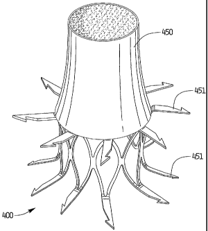
Turning first to Figures 35A-37, vascular device 400 is substantially
identical to
vascular device 100 of Figure 9A, except for the provision of valve 450. For
this reason;
it has been labeled with a different reference numeral. Valve 450 is conically
shaped and
is secured to vascular device 400 by various techniques such as by being
molded onto the
frame or sewn onto the frame. A pair of elongated supports 455 extends from
the device
into the valve 450 which spread to close the valve and move inwardly to open
the valve.
The valve 450 is shown attached to the distal end to extend downstream of the
device
400, with respect to blood flow. The valve 450 is shown in the open position
in the
figures and would collapse to a closed position by spreading of the supports
455,
operating like a duckbill valve.
As an alternative, the supports 455 are not provided and the valve 450
functions
in a similar manner described below with respect to the other conical valves,
e.g. valve
500.
A reinforcement ring as described below could also optionally be provided.
Valve 450 can be multi-layered, with an outer layer 452 composed of one
material and an
inner layer 454 composed of another material as shown in Figure 37A. Possible
valve
materials are discussed below
It should be appreciated that the vessel engaging members 451 can extend
substantially perpendicular as shown in Figure 36A, or can extend at an angle
as
described above with respect to Figure 9B. (See vessel engaging members 451'
of Figure
36B). Also, although the vessel engaging members of Figure 35A are slightly
longer and
are bent at a different region thaii~the device of Figure 9A, it should be
understood that
the device of Figure 9A can be provided with valve 450 or any of the other
valve
configurations described herein.
Turning now to Figures 38-47, the vascular device, since it is shown
schematically for ease of reference, will be referred to in each of the
drawings by
reference letter "D", it being understood that preferably vascular device 100
is utilized,
although device 10 and other support structures could alternatively be used.
With reference first to Figures 38A and 38B, valve 500 is conical in shape and
has
an open proximal end 504 and an open distal=end 502. This conical shape
results in
backflow of blood causing the valve to close. When the valve 500 is in the
position of
24
CA 02446596 2003-11-07
WO 02/100297 PCT/US01/44572
Figure 38A, distal opening faces towards and can press against the vessel wall
to prevent
flow through the valve 500. The force of the blood during systole straightens
the distal
end 502 to the position of Figure 38B to allow blood flow therethrough.
Reinforcement
ring 506 helps to maintain the valve 500 in the open position. As shown, valve
500
extends distally of the device D so it is positioned downstream with respect
to blood flow
of the device.
Figures 39A and 39B illustrate a variation to the valve configuration of
Figure 38
in that it is similar to valve 500 except that the valve 520 is attached to a
proximal end E
of the vascular device D. Valve 520 is attached at points E1, E2, etc. around
the
circumference and extends upwardly through a central portion of the device
"D".
Reinforcement ring 526 functions to help maintain the valve 520 in the open
position of
Figure 39B. Figure 39A shows the valve 520 in the closed position and Figure
39B
illustrates the valve in the open position to enable blood flow therethrough.
In both
positions, the valve extends within vascular device D.
In the embodiment of Figure 40, the valve 550 is attached to the distal end of
vascular device D and has a plurality of leaflets or petals 552 arranged
circumferentially
thereabout. The leaflets fold inwardly towards each other in the closed
position of Figure
40A to prevent blood flow. The pressure of the blood during systole forces the
leaflets
apart to the open position as shown in Figure 40B.
In the embodiment of Figures 41A and 41B, valve 560 is conically shaped like
the
valves of Figures 38 and 39, but is offset from the central longitudinal axis
of vascular
device D. Additionally, eccentric'valve 560 differs from valves 500 and 520 in
that it has
a plurality of slits 562 at a proximal portion to enable drainage of blood to
reduce blood
buildup. That is during the diastole phase, the slits expand to larger holes
as shown in
Figure 41A and the blood draws through the holes. Reinforcement ring 566
functions as
described above to help retain the distal end open.
A duckbill valve 570 is illustrated in the embodiment of Figures 42-44. Valve
570 is attached at the distal end of vascular device D and is moved to the
open position as
shown in Figure 43 by blood flow to enable passage therethrough. The closed
position of
the valve is illustrated in Figures 42 and 44. The proximal region of valve
570 is slightly
CA 02446596 2003-11-07
WO 02/100297 PCT/US01/44572
tapered. As with any of the foregoing valves, valve 570 can be attached at the
proximal
end, distal end, or intermediate portion of the vascular device.
Figures 45-47 illustrate steps for placement of the vascular device of the
present
invention. Figure 45 shows placement in the popliteal vein "P" and the femoral
vein "F"
of the vascular device of Figure 41 by way of example, it being understood
that any of
the vascular devices with any of the valve configurations can be placed in a
similar
fashion. Placement of two vascular devices is shown, although only one
vascular device
D (shown schematically) or more than two can be utilized. In Figure 46A, the
vascular
device and valve 560 are introduced through introducer sheath 600 in a
collapsed
position. The device is retained within a delivery catheter 604. After
introduction of the
catheter 604 through the introducer sheath 600 to the surgical site, the
pusher 606 pushes
the device to the end of the catheter 604, is then retracted, followed by
retraction of the
catheter 604, thereby releasing the device and allowing it to expand to the
memorized
configuration as shown in Figure 46C for retention in the vein. Alternatively,
the pusher
can be used to fully advance the device from the catheter 604.
If it is desired to reposition the device, grasper 610 within delivery
catheter 604 is
inserted through the introducer sheath 602. The prongs or fingers 612 are
advanced from
the outer tube 614 of grasper 610, or the outer tube 614 is moved proximally,
to expose
the prongs 612. The outer tube 614 is then advanced slightly to slightly
clinch the prongs
612 so the prongs 612 can grasp the vascular device D and pull it to a more
proximal
position as shown in Figure 47C. The grasper 610 is then removed. Note valve
560 is
shown in the open position in Figures 47A-47C.
In the embodiment of Figures 48-49, valve 700 is in the form of a duckbill
valve
similar to Figures 42-44, except the valve is reinforced with metal wires or
struts 701.
The vascular device is shown in the form of a covered stent 702, with the
metal stent 703
embedded in the graft material 704. The valve can include blood drainage slits
706 as
shownn.
Replacement Valve
The present invention also contemplates in another aspect use of the various
valve
configurations as replacement valves without the use of a vascular device
which brings
the walls radially inwardly. The patient's valve can be removed or
alternatively left in
26
CA 02446596 2003-11-07
WO 02/100297 PCT/US01/44572
place and the replacement valve of the present invention placed upstream or
downstream
of the patient's valve. In such applications, the valve is attached to a
support structure,
such as a shape memory stent, and is maintained in an open position within the
vessel to
retain the valve. Figures 50 and 51 shown an example of a type of support
structure for
holding the valve.
More specifically, in the embodiment of Figures 50 and 51, instead of a metal
framework, the valve 750 is attached to a rolled up cylindrical ring or metal
band 752.
The ring 752 can be made of shape memory material with its memorized position
being
an expanded position of Figures 50 and 51. The valve 750 is similar to valve
500 of
Figures 38A, except it has a larger reinforcement ring 754, and is cylindrical
instead of
conical in configuration.
In the embodiment of Figures 52-54, the valve 800 has a plurality of drainage
holes 802 which function in a similar manner as drainage slits 562 of valve
560 of
Figures 38-39. The overlapping cylindrical support member 806 is shown in the
contracted delivery position in Figure 54B and in the expanded position in
Figure 54A.
Valve 800 is longitudinally offset with respect to cylindrical member 806.
In the embodiment of Figure 55, valve 850 is in the form of a duckbill valve
and
extends from cylindrical support member 856.
It should be appreciated that the valves 850, 800 and 750 can be used with any
of
the vascular devices described above. Conversely, any of the foregoing valves
can be
used with cylindrical supports 752, 806. Also, the valves can be attached at
the proximal
end, distal end, or intermediate the proximal and distal ends of the vascular
devices.
The foregoing valves can be attached to the vascular devices, the framework
structures and the cylinders, by sewing, molding or other techniques. The
valves can be
composed of a variety of materials such as PET, PTFE, polycarbonate
polyurethane,
swine intestinal submucosa, collagen and other biomaterials. The valve and /or
the
vascular device surface can optionally be coated with anti-platelet or anti-
thrombin/anti-
clotting materials, 2b/2a coating, receptors, heparin coating, endothelial
cell coating, etc..
While the above description contains many specifics, those specifics should
not
be construed, as limitations on the scope of the disclosure, but merely as
exemplifications
of preferred embodiments thereof. For example, instead of a balloon to expand
the
27
CA 02446596 2003-11-07
WO 02/100297 PCT/US01/44572
device to its second expanded diameter/condition, a mechanical means such as
an
expandable wire frame can be utilized. Also, instead of moving the sheath to
expose the
vascular device, the catheter can be advanced with respect to the sheath or
both the
catheter and sheath can move relative to each other in opposite directions.
Those skilled
in the art will envision many other possible variations that`are within the
scope and spirit
of the disclosure as defined by the claims appended hereto.
28