Note: Descriptions are shown in the official language in which they were submitted.
CA 02446741 2008-08-25
WO 03/009784 PCT/US02/23116
OPHTHALIVIIC DRUG DELIVERY DEVICE
Field of the Invention
The present invention generally pertains to biocompatible implants for
delivery of
pharmaceutically active agents to the eye. More particularly, but not by way
of liinitation,
the present invention pertains to biocompatible implants for delivery of
pharmaceutically
active agents to the posterior segment of the eye.
Description of the Related Art
Several diseases and conditions of the posterior segment of the eye threaten
vision.
Age related macular degeneration (ARMD), choroidal neovascularization (CNV),
retinopathies (e.g., diabetic retinopathy, vitreoretinopathy), retinitis
(e.g., cytomegalovirus
(CMV) retinitis), uveitis, macular edema, glaucoma, and neuropathies are
several
examples.
Age related macular degeneration (AR1VID) is the leading cause of blindness in
the
elderly. ARMD attacks the center of vision and blurs it, making reading,
driving, and
other detailed tasks difficult or impossible. About 200,000 new cases of ARMD
occur
each year in the United States alone. Current estimates reveal that
approximately forty
1
CA 02446741 2003-11-07
WO 03/009784 PCT/US02/23116
percent of the population over age 75, and approximately twenty percent of the
population
over age 60, suffer from some degree of macular degeneration. "Wet" ARMD is
the type
of ARMD that most often causes blindness. In wet ARMD, newly formed choroidal
blood vessels (choroidal neovascularization (CNV)) leak fluid and cause
progressive
damage to the retina.
In the particular case of CNV in ARMD, three main methods of treatment are
currently being developed, (a) photocoagulation, (b) the use of angiogenesis
inhibitors,
and (c) photodynamic therapy. Photocoagulation is the most common treatment
modality
for CNV. However, photocoagulation can be harmful to the retina and is
impractical
when the CNV is near the fovea. Furthermore, over time, photocoagulation often
results
in recurrent CNV. Oral or parenteral (non-ocular) administration of anti-
angiogenic
compounds is also being tested as a systemic treatment for ARMD. However, due
to
drug-specific metabolic restrictions, systemic administration usually provides
sub-
therapeutic drug levels to the eye. Therefore, to achieve effective
intraocular drug
concentrations, either an unacceptably high dose or repetitive conventional
doses are
required. Periocular injections of these compounds often result in the drug
being quickly
washed out and depleted from the eye, via periocular vasculature and soft
tissue, into the
general circulation. Repetitive sub-Tenon's capsule injections of these
compounds carry
the potential risk of penetrating the globe and the severe, often blinding,
complications of
retinal detachment and endophthalmitis. In addition, it is difficult to
perform such
injections in a reproduceable manner, and each inj ection may result in a
different
distribution of drug along the scleral surface. Furthermore, many attempts to
inject drug
below the Tenon's capsule actually result in injections into the Tenon's
capsule itself or
the surrounding tissue, which is not desirable. Repetitive intraocular
injections may also
2
CA 02446741 2003-11-07
WO 03/009784 PCT/US02/23116
result in retinal detachment and endophthahnitis. Photodynamic therapy is a
new
teclmology for which the long-term efficacy is still largely unknown.
In order to prevent complications related to the above-described treatments
and to
provide better ocular treatment, researchers have suggested various implants
aimed at
delivery of anti-angiogenic compounds to the eye. U.S. Patent No. 5,824,072 to
Wong
discloses a non-biodegradable polymeric implant with a pharmaceutically active
agent
disposed therein. The pharmaceutically active agent diffuses through the
polymer body of
the implant into the target tissue. The pharmaceutically active agent may
include drugs
for the treatment of macular degeneration and diabetic retinopathy. The
implant is placed
substantially within the tear fluid upon the outer surface of the eye over an
avascular
region, and may, be anchored in the conjunctiva or sclera; episclerally or
intrasclerally
over an avascular region; substantially within the suprachoroidial space over
an avascular
region such as the pars plana or a surgically induced 'avascular region; or in
direct
communication with the vitreous.
U.S. Patent No. 5,476,511 to Gwon et al. discloses a polymer implant for
placement under the conjunctiva of the eye. The implant may be used to deliver
neovascular inhibitors for the treatment of ARMD and drugs for the treatment
of
retinopathies, and retinitis. The pharmaceutically active agent diffuses
through the
polymer body of the implant.
U.S. Patent No. 5,773,019 to Ashton et al. discloses a non-bioerodable polymer
implant for delivery of certain drugs including angiostatic steroids and drugs
such as
cyclosporine for the treatment of uveitis. Once again, the pharmaceutically
active agent
diffuses through the polymer body of the implant.
All of the above-described implants require careful design and manufacture to
permit controlled diffusion of the pharmaceutically active agent through a
polymer body
3
CA 02446741 2003-11-07
WO 03/009784 PCT/US02/23116
or polymer membrane to the desired site of therapy. Drug release from these
devices
depends on the porosity and diffusion characteristics of the matrix or
membrane,
respectively. These parameters must be tailored for each drug moiety to be
used with
these devices. Consequently, these requirements generally increase the
complexity and
cost of such implants.
U.S. Patent No. 5,824,073 to Peyman discloses an indentor for positioning in
the
eye. The indentor has a raised portion that is used to indent or apply
pressure to the sclera
over the macular area of the eye. This patent discloses that such pressure
decreases
choroidal congestion and blood flow through the subretinal neovascular
membrane,
which, in turn, decreases bleeding and subretinal fluid accumulation.
U.S. Patent Nos. 5,725,493 and 5,830,173 both disclose non-bioerodable
implants
that have a drug containing reservoir located outside the globe of the eye and
a drug
delivery tube running from the reservoir and into the vitreous cavity at the
pars plana.
Despite the above-described ophthalmic implants, a need still exists for a
surgically implantable ophthalmic drug delivery device capable of safe,
effective, rate-
controlled, delivery of a wide variety of pharmaceutically active agents. The
surgical
procedure for implanting such a device should be safe, simple, quick, and
capable of
being performed in an outpatient setting. Ideally, such a device should be
easy and
economical to manufacture. Furthermore, because of its versatility and
capability to
deliver a wide variety of pharmaceutically active agents, such an implant
should be
capable of use in ophthalmic clinical studies to deliver various agents that
create a
specific physical condition in a patient. Ideally, such an ophthalmic drug
delivery device
would be capable of localized delivery of pharmaceutically active agents to a
specific
portion of the retina, as well as pan-retinal delivery of pharmaceutically
active agents. In
4
CA 02446741 2003-11-07
WO 03/009784 PCT/US02/23116
addition, such a device should ideally be suitable for delivering two or more
drugs in
combination therapy.
Summary of the Invention
One aspect of the present invention is an ophthalmic drug delivery device
having a
scleral surface, an orbital surface, an injection port on the orbital surface,
and a fluid
conducting passageway disposed within the device. The scleral surface has a
curvature
that facilitates contact with a sclera of an eye. The injection port is for
sealingly engaging
a needle of a syringe, which is for providing a fluid comprising a
pharmaceutically active
agent. The fluid conducting passageway is fluidly coupled to the injection
port and has an
opening for communicating the fluid to an outer surface of the sclera.
Brief Description of the Drawings
For a more complete understanding of the present invention, and for further
objects and advantages thereof, reference is made to the following description
taken in
conjunction with the accompanying drawings in which:
FIG. 1 is a side sectional view schematically illustrating the human eye;
FIG. 2 is detailed cross-sectional view of the eye of FIG. 1 along line 2-2;
FIG. 3 is a perspective view of an ophthalmic drug delivery device according
to a
preferred embodiment of the present invention;
FIG. 4A is an orbital view of the device of FIG. 3 showing a preferred
embodiment of the internal fluid conducting passageways of the device;
FIG. 4B is a side view of the device of FIG. 4A;
FIG. 5 is a perspective view of an ophthalmic drug delivery device according
to a
second preferred embodiment of the present invention;
CA 02446741 2003-11-07
WO 03/009784 PCT/US02/23116
FIG. 5A is a side sectional view of the ophthalmic drug delivery device of
FIG. 5
with the internal fluid conducting passageways of the device not shown for
clarity of
illustration;
FIG. 5B is an enlarged cross-sectional view of the ophthalmic drug delivery
device
of FIG. 5A taken along line 5B-5B;
FIG. 6A is an orbital view of the device of FIGS. 5-5B showing a preferred
embodiment of the internal fluid conducting passageways of the device;
FIG. 6B is a side view of the device of FIG. 6A with the well and inner core
of the
device not shown for clarity of illustration;
FIG. 7A is ain orbital view of the device of FIGS. 5-5B showing a second
preferred
embodiment of the internal fluid conducting passageways of the device;
FIG. 7B is a side view of the device of FIG. 7A with the well and inner core
of the
device not shown for clarity of illustration;
FIG. 8A is an orbital view of the device of FIGS. 5-5B showing a third
preferred
embodiment of the internal fluid conducting passageways of the device;
FIG. 8B is a side view of the device of FIG. 8A with the well and inner core
of the
device not shown for clarity of illustration;
FIG. 9A is an orbital view of the device of FIGS. 5-5B showing a fourth
preferred
embodiment of the internal fluid conducting passageways of the device;
FIG. 9B is a side view of the device of FIG. 9A with the well and inner core
of the
device not shown for clarity of illustration;
FIG. 10A is an orbital view of the device of FIGS. 5-5B showing a fifth
preferred
embodiment of the internal fluid conducting passageways of the device; and
FIG. l OB is a side view of the device of FIG. 1 OA with the well and inner
core of
the device not shown for clarity of illustration.
6
CA 02446741 2003-11-07
WO 03/009784 PCT/US02/23116
Detailed Description of the Preferred Embodiments
The preferred embodiments of the present invention and their advantages are
best
understood by referring to FIGS. 1-lOB of the drawings, like numerals being
used for like
and corresponding parts of the various drawings.
FIGS. 1-4B schematically illustrate an ophthalmic drug delivery device 10
according to a preferred embodiment of the present invention. Device 10 may be
used in
any case where delivery of a pharmaceutically active agent to the eye is
required. Device
is particularly useful for delivery of active agents to the posterior segment
of the eye.
A preferred use for device 10 is the delivery of pharmaceutically active
agents to the
retina for treating ARMD, choroidial neovascularization (CNV), retinopathies,
retinitis,
uveitis, macular edema, and glaucoma. Of course, device 10 may also be
utilized for the
delivery of pharmaceutically active agents to body tissue other than the eye,
if desired.
Referring to FIGS. 1-2, a human eye 52 is schematically illustrated. Eye 52
has a
cornea 54, a lens 56, a sclera 58, a choroid 60, a retina 62, and an optic
nerve 64. An
anterior segment 66 of eye 52 generally includes the portions of eye 52
anterior of a line
67. A posterior segment 68 of eye 52 generally includes the portions of eye 52
posterior
of line 67. Retina 62 is physically attached to choroid 60 in a
circumferential manner
proximate pars plana 70. Retina 62 has a macula 721ocated slightly lateral to
its optic
disk 19. As is well known in the ophthalmic art, macula 72 is comprised
primarily of
retinal cones and is the region of maximum visual acuity in retina 62. A
Tenon's capsule
or Tenon's membrane 74 is disposed on sclera 58. A conjunctiva 76 covers a
short area
of the globe of eye 52 posterior to limbus 77 (the bulbar conjunctiva) and
folds up (the
upper cul-de-sac) or down (the lower cul-de-sac) to cover the inner areas of
upper eyelid
7
CA 02446741 2003-11-07
WO 03/009784 PCT/US02/23116
78 and lower eyelid 79, respectively. Conjunctiva 76 is disposed on top of
Tenon's
capsule 74.
As is shown in FIGS. 1 and 2, and as is described in greater detail
hereinbelow,
device 10 is preferably disposed directly on the outer surface of sclera 58,
below Tenon's
capsule 74 for treatment of most posterior segment diseases or conditions. In
addition, for
treatment of ARMD in humans, device 10 is preferably disposed directly on the
outer
surface of sclera 58, below Tenon's capsule 74, with its distal end 92
proximate macula
72.
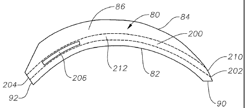
FIGS. 3, 4A, and 4B schematically illustrate device 10 in greater detail.
Device 10
generally includes a body 80 having a scleral surface 82 and an orbital
surface 84. Scleral
surface 82 is preferably designed with a radius of curvature that facilitates
direct contact
with sclera 58. Most preferably, scleral surface 82 is designed with a radius
of curvature
equal to the radius of curvature 91 of an average human eye 52. (See FIG. 1)
Orbital
surface 84 is preferably designed with a radius of curvature that facilitates
implantation
under Tenon's capsule 74. Body 80 preferably has a curved, generally
rectangular three-
dimensional geometry with rounded sides 86 and 88, proximal end 90, and distal
end 92.
Body 80 may have any other geometry that has a curved scleral surface 82 for
contact
with sclera 58. By way of example, body 80 may have a generally cylindrical,
oval,
square, or other polygonal three-dimensional geometry.
Body 80 preferably comprises a biocompatible, non-bioerodable material. Body
80 more preferably comprises a biocompatible, non-bioerodable polymeric
composition.
Said polymeric composition may be a homopolymer, a copolymer, straight,
branched,
cross-linked, or a blend. Examples of polymers suitable for use in said
polymeric
composition include silicone, polyvinyl alcohol, ethylene vinyl acetate,
polylactic acid,
nylon, polypropylene, polycarbonate, cellulose, cellulose acetate,
polyglycolic acid,
8
CA 02446741 2003-11-07
WO 03/009784 PCT/US02/23116
polylactic-glycolic acid, cellulose esters, polyethersulfone, acrylics, their
derivatives, and
combinations thereof. Examples of suitable soft acrylics are more fully
disclosed in U.S.
Patent No. 5,403,901, which is incorporated herein in its entirety by
reference. Said
polymeric composition most preferably comprises silicone. Of course, said
polymeric
composition may also conlprise other conventional materials that affect its
physical
properties, including, but not limited to, porosity, tortuosity, permeability,
rigidity,
hardness, and smoothness. Exemplary materials affecting certain ones of these
physical
properties include conventional plasticizers, fillers, and lubricants. Said
polymeric
composition may comprise other conventional materials that affect its chemical
properties, including, but not limited to, toxicity and hydrophobicity.
Device 10 has a plurality of fluid conducting passageways or cavities within
body
80. FIGS. 4A and 4B show a preferred system of such passageways having a main
passageway 200 having a proximal end 202, a distal opening 204, a first side
opening 206,
and a second side opening 208. Passageway 200 and openings 204, 206, and 208
preferably have a generally rectangular cross-section. Device 10 also has an
injection port
210 located on orbital surface 84 of body 80 near proximal end 202 of main
passageway
200. Injection port 210 is preferably made of a fluid impervious material that
can be
penetrated by a needle and that reseals itself upon removal of the needle. A
preferred
material is silicone rubber. In addition, injection port 210 is preferably
colored or marked
by raised protuberances. Although not shown in the FIGS. 3-4A, passageway 200
may
also have one or more openings to scleral surface 82 of device 10.
A conventional syringe and needle may be used to impart a fluid 212 containing
a
pharmaceutically active agent or agents into passageway 200 via injection port
210. Fluid
212 may comprise a solution, a suspension, an emulsion, an ointment, a gel
forming
solution, a gel, a bioerodable polymer, a non-bioerodable polymer,
microparticles, or
9
CA 02446741 2003-11-07
WO 03/009784 PCT/US02/23116
combinations thereof. Most preferably, fluid 212 is a suspension with or
without
microparticles formed from bioerodable polymers. Fluid 212 includes one or
more
ophthalmically acceptable pharmaceutically active agents, and may also include
conventional non-active incipients. Examples of pharmaceutically active agents
suitable
for fluid 212 are anti-infectives, including, without limitation, antibiotics,
antivirals, and
antifungals; antiallergenic agents and mast cell stabilizers; steroidal and
non-steroidal
anti-inflammatory agents; cyclooxygenase inhibitors, including, without
limitation, Cox I
and Cox II inhibitors; combinations of anti-infective and anti-inflammatory
agents;
decongestants; anti-glaucoma agents, including, without limitation,
adrenergics, ,6-
adrenergic blocking agents, a-adrenergic agonists, parasypathomimetic agents,
cholinesterase inhibitors, carbonic anhydrase inhibitors, and prostaglandins;
combinations
of anti-glaucoma agents; antioxidants; nutritional supplements; drugs for the
treatment of
cystoid macular edema including, without limitation, non-steroidal anti-
inflammatory
agents; drugs for the treatment of ARMD, including, without limitation,
angiogenesis
inhibitors and nutritional supplements; drugs for the treatment of herpetic
infections and
CMV ocular infections; drugs for the treatment of proliferative
vitreoretinopathy
including, without limitation, antimetabolites and fibrinolytics; wound
modulating agents,
including, without limitation, growth factors; antimetabolites;
neuroprotective drugs,
including, without limitation, eliprodil; and angiostatic steroids for the
treatment of
diseases or conditions of posterior segment 68, including, without limitation,
ARMD,
CNV, retinopathies, retinitis, uveitis, macular edema, and glaucoma. Such
angiostatic
steroids are more fully disclosed in U.S. Patent Nos. 5,679,666 and 5,770,592.
Preferred
ones of such angiostatic steroids include 4,9(11)-Pregnadien-17c~21-diol-3,20-
dione and
4,9(11)-Pregnadien-17c~21-diol-3,20-dione-21-acetate. These preferred
angiostatic
steroids are preferably formulated as a suspension. A preferred non-steroidal
anti-
CA 02446741 2003-11-07
WO 03/009784 PCT/US02/23116
inflammatory for the treatment of cystoid macular edema is nepafenac. The
conventional
non-active excipients may include, but are not limited to, ingredients to
enhance the
stability, solubility, penetrability, or other properties of fluid 212. In
particular, hydrolytic
enzymes such as proteases, esterases, hyaluronidases, and collegenases may be
utilized to
enhance the penetration of the pharmaceutically active agents through natural
and newly
formed connective tissue that may encapsulate device 10 after implantation.
Body 80 is
preferably impermeable to fluid 212.
Device 10 may be made by conventional polymer processing methods, including,
but not limited to, injection molding, extrusion molding, transfer molding,
and
compression molding. Preferably, device 10 is formed using conventional
injection
molding techniques.
Device 10 is preferably surgically placed directly on the outer surface of
sclera 58
below Tenon's capsule 74 using a simple surgical technique that is capable of
being
performed in an outpatient setting. The surgeon first performs a peritomy in
one of the
quadrants of eye 52. Preferably, the surgeon performs the peritomy in the
supero-
teinporal or infra-temporal quadrant, about 3 mm posterior to limbus 77 of eye
52. Once
this incision is made, the surgeon performs a blunt dissection to separate
Tenon's capsule
74 from sclera 58, forming an antero-posterior tunnel. Once the tunnel is
formed, the
surgeon uses forceps to hold device 10 with scleral surface 82 facing sclera
58 and distal
end 92 away from the surgeon. The surgeon then introduces device 10 into the
tunnel in a
generally circular motion to position distal end 92 generally above the
desired portion of
retina 62. The surgeon then closes the peritomy by suturing Tenon's capsule 74
and
conjunctiva 76 to sclera 58. After closing, the surgeon places a strip of
antibiotic
ointment on the surgical wound. Alternatively, the surgeon may suture proximal
end 90
11
CA 02446741 2003-11-07
WO 03/009784 PCT/US02/23116
of device 50 to sclera 58 to hold device 10 in the desired location before
closure of the
tunnel.
In the case of ARMD in the human eye, the surgeon preferably utilizes the
above-
described technique to position distal end 92 of device 10 in the supero-
temporal quadrant
of eye 52 directly on the outer surface of sclera 58, below Tenon's capsule 74
with side
openings 206 and 208 positioned directly above macula 72. A surgeon may
position side
openings 206 and 208 of device 10 at this location by moving distal end 92 of
device 10
toward macula 72 along a path generally between the lateral and superior
rectus muscles.
For ARMD, the pharmaceutically active agent of fluid 212 is preferably one of
the
angiostatic steroids disclosed in U.S. Patent Nos. 5,679,666 and 5,770,592.
In the case of ARMD in the human eye, the surgeon preferably utilizes the
above-
described technique to position distal end 92 of device 10 in one. of two
preferred
locations in the infra-temporal quadrant of eye 52. One preferred location is
directly on
the outer surface of sclera 58, below Tenon's capsule 74; with side. openings
206 and 208
positioned proximate to, but not directly above, macula 72. A surgeon may
position side
openings 206 and 208 of device 10 at this location by moving distal end 92 of
device 10
below the inferior oblique muscle in a direction generally parallel to the
lateral rectus
muscle. A second preferred location is directly on the outer surface of sclera
58, below
Tenon's capsule 74, with side openings 206 and 208 positioned directly above
macula 72.
A surgeon may position side openings 206 and 208 of device 10 at this location
by
moving distal end 92 of device 10 toward macula 72 along a path generally
between the
lateral and inferior rectus muscles and below the inferior oblique muscle.
Once device 10 is located in the desired position, the surgeon utilizes a
conventional syringe and needle to inject fluid 212 into passageway 200. The
surgeon
preferably moves lower eyelid 79 downward and instructs the patient to look
upward so as
12
CA 02446741 2003-11-07
WO 03/009784 PCT/US02/23116
to expose proximal end 90 of device 10. Injection port 210 may be visualized
beneath the
Tenon's capsule and any connective tissue encapsulating device 10 due to its
color or
raised protuberances. The surgeon sticks the needle of the syringe into
injection port 210,
injects fluid 212 into passageway 200, and removes the needle from the port
210. Port
210 reseals automatically upon removal of the needle. Fluid 212 is disposed
throughout
passageway 200, and is in communication with sclera 58 via openings 204, 206,
208, and
any openings to scleral surface 82.
It is believed that device 10 can be used to deliver a pharmaceutically
effective
amount of a pharmaceutically active agent through sclera 58 and choroid 60
into retina 62
for many years, depending on the particular physicochemical properties of the
particular
fluid 212 and its pharmaceutically active agent employed. Important
physicochemical
properties include hydrophobicity, solubility, dissolution rate, diffusion
coefficient, and
tissue affinity. In addition, it is believed that device 10 may be used to
deliver both a
localized distribution of drug primarilybeneath distal end 92 of device 10; or
to deliver
drug to substantially the entire retina, depending upon the-particular fluid
212 and its
pharmaceutically active agents and incipients. After passageway 200 no longer
contains
any fluid 212, a surgeon may refill passageway 200 as described hereinabove.
Although
not shown in FIGS. 3-4B, device 10 may also include a sharp surface or edge on
distal
end 92, side 86, or side 88 of body 80. During refilling of passageway 200,
the surgeon
may move device 10 slightly from side to side and/or posteriorly so that such
sharp
surfaces or edges pierce any connective tissue that may encapsulate device 10
after
implantation. Piercing this connective tissue facilitates proper distribution
of fluid 212
via openings 204, 206, and 208. In addition, unlike repetitive sub-Tenon's
capsule
injections of drug formulations, device 10 minimizes the risk of penetrating
the globe of
the eye, always results in fluid 212 being distributed below the Tenon's
capsule 74 on the
13
CA 02446741 2003-11-07
WO 03/009784 PCT/US02/23116
outer surface of sclera 58, and results in a reproduceable distribution of
fluid 212 on a
desired portion of the outer surface of the sclera 58.
FIGS. 5, 5A, 513, 6A, and 6B schematically illustrate an ophthalmic drug
delivery
device 50 according to a second preferred embodiment of the present invention.
Device
50 is similar in construction to device 10 described hereinabove, with several
important
exceptions. First, body 80 of device 50 includes a well or cavity 102 having
an opening
104 to scleral surface 82 and holding an inner core 106. Second, device 50 has
a
preferred system of fluid conducting passageways or cavities 300 within body
80, which
is best illustrated in FIGS. 6A and 6B.
Inner core 106 is preferably a tablet comprising one or more pharmaceutically
active agents. Alternatively, inner core 106 may comprise a conventional
hydrogel having
one or more pharmaceutically active agents disposed therein. A retaining
member 108 is
preferably disposed proximate opening 104. Retaining member 108 prevents inner
core
106 from falling out of well 102. When inner core 106 is a cylindrical tablet,
retaining
member 108 is preferably a continuous rim or lip disposed circumferentially
around
opening 104 having a diameter slightly less than the diameter of tablet 106.
Alternatively,
retaining member 108 may comprise one or more members that extend from body 80
into
opening 104. Although not shown in FIG. 6A, inner core 106 may alternatively
comprise
a suspension, solution, powder, or combination thereof containing one or more
pharmaceutically active agents. In this embodiment, scleral surface 82 is
formed without
opening 104, and the suspension, solution, powder, or combination thereof
diffuses
through the relatively thin portion of scleral surface 82 below inner core 26.
Still further
in the alternative, device 50 may be formed without well 102 or inner core
106, and the
pharmaceutically active agent(s) in the form of a suspension, solution,
powder, or
combination thereof may be dispersed throughout body 80 of device 50, with the
14
CA 02446741 2003-11-07
WO 03/009784 PCT/US02/23116
exception of system of passageways 300. In this embodiment, the
pharmaceutically active
agent diffuses through body 80 into the target tissue.
Body 80 is preferably impermeable to the pharmaceutically active agent of
inner
core 106. When body 80 is made from a generally elastic polymeric composition,
the
diameter of well 102 may be slightly less than the diameter of inner core 106.
This
frictional fit secures inner core 106 within well 102. In this embodiment,
body 80 may be
formed without retaining member 108, if desired.
The geometry and dimensions of device 50 maximize communication between the
pharmaceutically active agent of inner core 106 and the tissue underlying
scleral surface
82. Scleral surface 82 preferably physically contacts the outer surface of
sclera 58.
Although not shown in FIGS. 6A or 6B, inner core 106 may be formed so that
surface
106a physically contacts the outer surface of sclera 58. Alternatively,
scleral surface 82
may be disposed proximate the outer surface of sclera 58. By way of example,
device 50
may be disposed in the periocular tissues just above the outer surface of
sclera 58 or intra-
lamellarly within sclera 58.
Inner core 106 may comprise one or more ophthalmically acceptable
pharmaceutically active agents. Exemplary pharmaceutically active agents
include the
pharmaceutically active agents listed hereinabove for fluid 212. Inner core
106 may also
comprise conventional non-active excipients to enhance the stability,
solubility,
penetrability, or other properties of the active agent.
If inner core 106 is a tablet, it may further comprise conventional excipients
necessary for tableting, such as fillers and lubricants. Such tablets may be
produced using
conventional tableting methods. The pharmaceutically active agent is
preferably
distributed evenly throughout the tablet. In addition to conventional tablets,
inner core
106 may comprise a special tablet that bioerodes at a controlled rate,
releasing the
CA 02446741 2003-11-07
WO 03/009784 PCT/US02/23116
pharmaceutically active agent. By way of example, such bioerosion may occur
through
hydrolosis or enzymatic cleavage. If inner core 106 is a hydrogel, the
hydrogel may
bioerode at a controlled rate, releasing the pharmaceutically active agent.
Alternatively,
the hydrogel may be non-bioerodable but allow diffusion of the
pharmaceutically active
agent.
System of passageways 300 preferably comprises a proximal portion 302, a
longitudinal portion 304 having an opening 306 on distal end 92 of body 80,
and a
longitudinal portion 308 having an opening 310 on distal end 92 of body 80.
Proximal
portion 302 preferably has a generally rectangular cross-section. Longitudinal
portions
304 and 308 and openings 306 and 310 preferably have a generally square cross-
section.
Well 102 and inner core 106 are disposed between longitudinal portions 304 and
308.
Injection port 210 is located on orbital surface 84 of body 80 near proximal
portion 302.
Although not shown in the FIGS. 5-6B, system of passageways 300 may also have
one or
more openings to scleral surface 82 of device 10. A conventional syringe and
needle may
be used to impart fluid 212 into system of passageways 300 via injection port
210.
Device 50 may be made by conventional polymer processing methods, including,
but not limited to, injection molding, extrusion molding, transfer molding,
and
compression molding. Preferably, device 50 is formed using conventional
injection
molding techniques as described hereinabove for device 10.
Device 50 is preferably surgically placed directly on the outer surface of
sclera 58
below Tenon's capsule 74 using the simple surgical technique described
hereinabove in
connection with device 10. In the case of ARMD in the human eye, the surgeon
preferably utilizes the above-described technique to position device 50 in the
supero-
temporal quadrant of eye 52 directly on the outer surface of sclera 58, below
Tenon's
capsule 74, with inner core 106 positioned directly above macula 72. A surgeon
may
16
CA 02446741 2003-11-07
WO 03/009784 PCT/US02/23116
position inner core 106 of device 50 at this location by moving distal end 92
of device 50
toward macula 72 along a path generally between the lateral and superior
rectus muscles.
For ARMD, the pharmaceutically active agent of inner core 106 is preferably
one of the
angiostatic steroids disclosed in U.S. Patent Nos. 5,679,666 and 5,770,592.
In the case of ARMD in the human eye, the surgeon preferably utilizes the
above-
described technique to position inner core 106 of device 50 in one of two
preferred
locations in the infra-temporal quadrant of eye 52. One preferred location is
directly on
the outer surface of sclera 58, below Tenon's capsule 74, with inner core 106
positioned
proximate to, but not directly above, macula 72. A surgeon may position inner
core 106
of device 50 at this location by moving distal end 92 of device 50 below the
inferior
oblique muscle in a direction generally parallel to the lateral rectus muscle.
A second
preferred location is directly on the outer surface of sclera 58, below
Tenon's capsule 74,
with inner core 106 positioned directly above macula 72. A surgeon may
position inner
core 106 of device 50 at this location by moving distal end 92 of device 50
toward macula
72 along a path generally between the lateral and inferior rectus muscles and
below the
inferior oblique muscle.
The physical shape of body 80 of device 50, including the geometry of scleral
surface 82, well 102, opening 104, and retaining member 108, facilitate the
unidirectional
delivery of a pharmaceutically effective amount of the pharmaceutically active
agent from
inner core 106 through sclera 58, choroid 60, and into retina 62. In
particular, the absence
of a polymer layer or membrane between inner core 106 and sclera 58 greatly
enhances
and simplifies the delivery of an active agent to retina 62.
Once device 50 is located in the desired position, the surgeon utilizes a
conventional syringe and needle to inject fluid 212 into system of passageways
300 as
described hereinabove for device 10. Fluid 212 is disposed throughout proximal
portion
17
CA 02446741 2003-11-07
WO 03/009784 PCT/US02/23116
302 and longitudinal portions 304 and 308, and is in communication with sciera
58 via
openings 306, 310, and any openings to scleral surface 82.
It is believed that device 50 can be used to deliver a pharmaceutically
effective
amount of a pharmaceutically active agent through sclera 58 and choroid 60
into retina 62
for many years, depending on the particular physicochemical properties of the
particular
fluid 212, the particular inner core 106, and their pharmaceutically active
agents
employed. Important physicochemical properties include hydrophobicity,
solubility,
dissolution rate, diffusion coefficient, and tissue affinity. In addition, it
is believed that
device 50 may be used to deliver both a localized distribution of drug
primarily beneath
distal end 92 of device 10, or to deliver drug to substantially the entire
retina, depending
upon the particular fluid 212, inner core 106, and their pharmaceutically
active agents and
excipients. After inner core 106 no longer contains active agent, a surgeon
may easily
remove device 50, if desired. The "pre-formed" tunnel facilitates the
replacement of an
old device 50 with a new device 50. After passageway 200 no longer contains
any fluid
212, a surgeon may refill passageway 200 as described hereinabove.
It should be noted that fluid 212 and inner core 106 may contain the same or
different pharmaceutically active agents. Device 50 is especially useful for
combination
drug therapy, and in this case fluid 212 and inner core 106 contain different
pharmaceutically active agents. For example, fluid 212 may contain a
pharmaceutically
active agent(s) that is most easily or best formulated as a fluid, and inner
core 106 may
contain a pharmaceutically active agent(s) that is most easily or best
formulated as a solid
or a semi-solid. In addition, while not wanting to be limited to any
particular theory, it is
believed that fluid 212 may be best for delivery of di-ug to substantially the
entire retina,
while inner core 106 may be best for localized delivery of drug primarily
beneath inner
core 106.
18
CA 02446741 2003-11-07
WO 03/009784 PCT/US02/23116
FIGS. 7A and 7B show a second preferred system of fluid conducting
passageways or cavities 350 within body 80 of device 50. System of passageways
350
preferably comprises a proximal portion 352, a longitudinal portion 354 having
an
opening 356 on side 86 of body 80, a longitudinal portion 358 having an
opening 360 on
side 88 of body 80, and a distal portion 362 having an opening 364 on distal
end 92 of
body 80. Portions 352 and 362 preferably have a generally rectangular cross-
section, and
portions 354 and 358 preferably have a generally square cross-section. Opening
364
preferably has a generally rectangular cross-section, and openings 356 and 360
preferably
have a generally square cross-section. Well 102 and inner core 106 are
surrounded by
system of passageways 350. Injection port 210 is located on orbital surface 84
of body 80
near proximal portion 352. Although not shown in the FIGS. 7A-B, system of
passageways 350 may also have one or more openings to scleral surface 82 of
device 50.
A conventional syringe and needle may be used to impart fluid 212 into system
of
passageways 300,via injection port 210. A device 50 having a system of
passageways 350
is constructed, implanted into the eye, and operated in substantially the same
manner as
described hereinabove with device 50 having a system of passageways 300.
FIGS. 8A and 8B show a third preferred system of fluid conducting passageways
or cavities 400 within body 80 of device 50. System of passageways 400 is
identical to
system of passageways 350 of FIGS. 7A and 7B, with the exception that
longitudinal
portion 354 has an additional opening 366 on side 86 of body 80, and
longitudinal portion
358 has an additional opening 368 on side 88 of body 80. Openings 356, 366,
360, and
368 preferably surround well 102 and inner core 106. Although not shown in
FIGS. 8A-
B, longitudinal portions 354 and 358 may be formed with more than two such
openings, if
desired. Although not shown in the FIGS. 8A-B, system of passageways 400 may
also
have one or more openings to scleral surface 82 of device 50. A device 50
having a
19
CA 02446741 2003-11-07
WO 03/009784 PCT/US02/23116
system of passageways 400 is constructed, implanted into the eye, and operated
in
substantially the same manner as described hereinabove with device 50 having a
system
of passageways 300.
FIGS. 9A and 9B show a fourth preferred system of fluid conducting passageways
or cavities 450 within body 80 of device 50. System of passageways 450 is
identical to
main passageway 200 within body 80 of device 10 of FIGS. 3-4A, with the
exception that
well 102 is formed within main passageway 200 between first side opening 206
and
second side opening 208. Although not shown in the FIGS. 9A-B, system of
passageways
450 may also have one or more openings to scleral surface 82 of device 50. A
device 50
having a systein of passageways 450 is constructed, implanted into the eye,
and operated
in substantially the same manner'as described hereinabove with device 50
having a system
of passageways 300.
FIG. l0A and l OB show a fifth preferred system of fluid conducting
passageways
or cavities 500 within body 80 of device 50. System of passageways 500 is
identical to
system of passageways 300 of FIGS. 6A and 6B, with the exception that
longitudinal
portion 304 has an opening 502 on side 86 of body 80, and longitudinal portion
308 has
an opening 504 on side 88 of body 80. Although not shown in the FIGS. 10A-B,
system
of passageways 500 may also have one or more openings to scleral surface 82 of
device
50. A device 50 having a system of passageways 500 is constructed, implanted
into the
eye, and operated in substantially the same manner as described hereinabove
with device
50 having a system of passageways 300.
From the above, it may be appreciated that the present invention provides
improved devices and methods for safe, effective, rate-controlled delivery of
a variety of
pharmaceutically active agents to the eye. The devices of the present
invention are
especially useful for localized and/or pan-retinal delivery of
pharmaceutically active
CA 02446741 2003-11-07
WO 03/009784 PCT/US02/23116
agents to the posterior segment of the eye to combat diseases such as ARMD,
CNV,
retinopathies, retinitis, uveitis, macular edema, and glaucoma. The devices of
the present
invention are also particularly useful for combination drug therapy. The
surgical
procedure for implanting the devices is safe, simple, quick, and capable of
being
performed in an outpatient setting. The devices are easy and economical to
manufacture.
Furthermore, because of their capability to deliver a wide variety of
pharmaceutically
active agents, such devices are useful in clinical studies to deliver various
agents that
create a specific physical condition in a patient or animal subject.
The present invention is illustrated herein by example, and various
modifications
may be made by a person of ordinary skill in the art. For example, the systems
of fluid
conducting passageways of the present invention may be employed into the
ophthalmic
drug delivery devices having a generally F-shaped geometry, a generally C-
shaped
geometry, or a generally L-shaped geometry as disclosed in U.S. Patent No.
6,416,777.
As another example, well 102 and inner core 106 may have a generally oval,
square, or
other. polygonal three-dimensional geometry. As a further example, different
cross-
sectional geometries and layouts of fluid conducting passageways and their
respective
openings may be utilized than described hereinabove.
It is believed that the operation and construction of the present invention
will be
apparent from the foregoing description. While the apparatus and methods shown
or
described above have been characterized as being preferred, various changes
and
modifications may be made therein without departing from the spirit and scope
of the
invention as defined in the following claims.
21