Note: Descriptions are shown in the official language in which they were submitted.
CA 02447030 2003-11-10
WO 02/092054 PCT/US02/08872
IMMUNE MODULATION DEVICE FOR USE IN ANIMALS
This application claims benefit of provisional patent application 60/290,542
filed May 11, 2001, which is hereby incorporated by reference herein.
FIELD OF THE INVENTION
The present invention relates to an implantable device and method for
modulating the immune response to antigens in mammals. More specifically the
present invention provides a porous, implantable device containing a fibrous
support
and at least one antigen. This device may be used to modulate the immune
system to
provide a robust response against an antigen, or to down regulate an existing
response.
BACKGROUND OF THE INVENTION
Induction of an immune response to an antigen and the magnitude of that
response depend upon a complex interplay among the antigen, various types of
immune
cells, and co-stimulatory molecules including cytokines. The timing and extent
of
exposure of the immune cells to the antigen and the co-stimulatory milieu
further
modulate the immune response. Within the body, these various cell types and
additional factors are brought into proximity in lymphoid tissue such as lymph
nodes.
Of the numerous cell types involved in the process, antigen-presenting cells
(APC),
such as macrophages and dendritic cells, transport antigen from the periphery
to local,
organized lymphoid tissue, process the antigen and present antigenic peptides
to T cells
as well as secrete co-stimulatory molecules. Thus, if antigen reaches lymph
organs in a
localized staggered manner, presenting antigenic epitopes, under the optimal
concentration gradient and under the appropriate environment comprising co-
stimulatory molecules, a response is induced in the draining lymph node.
In this manner, a foreign antigen introduced into the body, such as by means
of a
vaccination, may or may not result in the development of a desirably robust
immune
response. Antigens used for vaccination include attenuated and inactivated
bacteria and
viruses and their components. The success of vaccination depends in part on
the type
CA 02447030 2003-11-10
WO 02/092054 PCT/US02/08872
and quantity of the antigen, the location of the site of immunization, and the
status of
the immune system at the time of vaccination. Not all antigens axe equally
immunogenic, and for poorly immunogenic antigens, there are few alternatives
available to increase the effectiveness of the immunization. Whereas in
experimental
animals numerous techniques are available to enhance the development of the
immune
response, such as conjugating the antigen to a more immunogenic carrier
protein or
biomolecule (e.g., keyhole limpet hemocyanin), or the use of adjuvants such as
Freund's
Adjuvant or Ribi. For human vaccinations such techniques and adjuvants are not
available. Thus, numerous diseases that would otherwise be preventable by
vaccination
before exposure to the infectious agent, or in the case of a therapeutic
vaccine, that may
induce the development of an effective immune response to an existing disease-
causing
agent or cell, such as cancer, are not available to the patient.
Sponge implant studies have been performed in mammals to assess the immune
cell population attracted to a foreign body, which produce what is called a
sterile
abscess, and sponges prior to or after implantation have been loaded with
antigen to
further study the attracted cell population. Vallera et al. (1982, Cancer
Research
42:397-404) implanted sponges containing tumor cells in mice to examine the
composition of cells attracted over a 16 day period, and found that at an
early time,
cytotoxic cell precursors were present, and cytotoxicity peaked at day 16.
Sponges
containing tumor cells implanted in mice that had been previously immunized
with
tumor cells showed a more rapid appearance of cytotoxic cells in the sponge.
In neither
case did cells from the spleen, lymph nodes or peritoneum show cytotoxicity,
which
suggested a highly localized response to the antigen in the sponge.
Zangemeister-
Wittke et al. (I989, J. Immunol. 143:379-385) injected a tumor vaccine into
sponges
implanted in tumor-immune mice, and monitored the generation of a secondary
immune response at the sponge site. No accompanying effect was apparent in
lymph
nodes adj acent to the implanted sponge.
Other devices which overcome some of the limitations of sponges for
immunomodulation have been proposed. US patent 4,919,929 teaches that an
antigen
can be loaded into solid shaped particles, which slowly release the antigen
following
CA 02447030 2003-11-10
WO 02/092054 PCT/US02/08872
implantation. This type of device is envisaged to increase the antibody titers
in the milk
of mammals and thereby confer higher levels of immunity in those who consume
it.
WO application 93/17662 describes a device that consists of an impervious
membrane
surrounding a core, which is a gel loaded with a therapeutically active
ingredient
S (including antigens). There is at least one port in the impervious membrane
that is
capable of releasing the active to the surroundings. The use of the membrane
is shown
to slow the rate of release of the bioactive molecule (including antigens)
relative to the
gel alone. This device therefore primarily serves as a reservoir for slow
release and
does not facilitate the interaction of cells with the bioactive, which
necessarily must
occur outside of the device. In US patent 4,732,1SS, a device is proposed
where there
is a reservoir that provides prolonged release of a chemoattractant, which is
surrounded
by a web of fibers adjacent to the reservoir. Cells are attracted to the
reservoir and
become trapped in the fibrous web. This device is proposed for use in
characterizing
allergic and inflammatory responses to test compounds by allowing controlled
exposure
to the compound and by trapping the cells that respond to it. This device both
incorporates a mechanism for prolonged exposure to an antigen as well as a
mechanism
to facilitate cellular interaction with the antigen. The open web of fibers in
this device;
however, does not enable local retention of the cytokines and chemokines being
secreted by the responding cells since an open web of fibers will not provide
diffusional
resistance to soluble factors.
This design is improved upon in WO 99/44583 which proposes a porous matrix
which is housed in a perforated but otherwise impervious membrane. Antigen is
loaded
within the device and can be present either as native antigen or can be
encapsulated in a
2S slow releasing polymer that provides prolonged presentation of the antigen.
Specific
cells are attracted to the device by diffusion of the antigen from the
perforations in the
device and are also able to enter the device through the perforations, but the
membrane
provides sufficient diffusional resistance that cytokines secreted by cells
become locally
concentrated within the device. The high local densities of cells and
cytokines produce
a much more robust immune response than is seen with an uncontained matrix or
with
simple prolonged release to surrounding tissues.
3
CA 02447030 2003-11-10
WO 02/092054 PCT/US02/08872
The preferred embodiment of the device mentioned above envisages the porous
matrix to be a sponge and the membrane to be a perforated tube. While very
favorable
immunomodulation is seen with the device, it is impractical to miniaturize and
manufacture in large quantities. The primary reason is that it is very
difficult to load a
porous sponge into tubing. Sponges, due to their low bulk densities are
mechanically
weak and tend to tear easily when subj ected to the tensile and compressive
forces of
loading into small diameter tubing. By reducing the bulb density, more
favorable
mechanical properties caxl be encountered however the matrix does not contain
sufficient porosity to attain high cell densities. In addition, it is very
difficult to cut
small cylindrical cores of porous sponges for loading into tubes. The reason
is that the
poor mechanical properties of the porous sponge lead to tearing when the size
of the
piece being cut becomes very small. Consequently, the device envisaged in WO
99/44583 is only practical to make in diameters of greater than 1 mm.
Implantation of
such a large profile device requires a very sizable needle or trochar that
would be very
painful and cause significant local trauma to a patient. An additional problem
with this
device design is that it would be difficult to economically manufacture in
large
quantities. The reason is that each piece of sponge would need to be
individually cut
and stuffed into the tube. This would be very difficult to mechanize and
perform
rapidly.
Accordingly, it would be advantageous to provide an implantable device and
method for modulating an inunune response to specific antigens in mammals,
similar in
concept to the design described in WO 99/44583, whose filling preserves the
porosity
presented by a porous sponge, which is essential for rapid cellular
infiltration, yet
overcomes the mechanical frailties of a sponge.
SUMMARY OF THE INVENTION
The present invention is directed to an implantable immune modulation device
that is suitable for use in modulating an immune response in mammals,
comprising an
impermeable shell having a plurality of pores and said impermeable
biocompatible shell
having an interior lumen, a biocompatible fibrous scaffolding being disposed
within
said interior lumen. The fibrous scaffolding is loaded with single or multiple
antigens
4
CA 02447030 2003-11-10
WO 02/092054 PCT/US02/08872
and optionally one or more biologically active compounds such as cytokines
(e.g.
lymphokines, chemolcines etc.), non-cytokine Ieulcocyte chemotactic agents,
attachment
factors, genes, peptides, proteins, nucleotides, carbohydrates, or cells
depending on the
application. The shell of the device preferably is made from a polymer whose
glass
transition temperature is below physiologic temperature so that the device
will
minimize irntation when implanted in soft tissues. The shell allows cell
ingress but
hinders diffusion of soluble molecules out of the device. This helps to
concentrate
cytokines (e.g. lymphokine and chemokines) secreted by cells which have
entered the
device in response to loaded antigens and other cells which are present in the
device.
This local concentration of cells and cytokines significantly enhances the
immune
response relative to implantation of antigens with standard adjuvants. The
fibrous
scaffolding provides a scaffold for cells to reside on, process the antigens
and interact.
Additional benefits of the fibrous scaffolding disclosed in this invention
include
ease of miniaturization of a device to diameters of less than 1 mm, the
possibility of
rapid insertion into small diameter tubing or even the ability to have tubing
continuously extruded around the matrix.
BRIEF DESCRIPTION OF THE FIGURES
Figure 1 is a perspective drawing of one embodiment of the immune modulating
device described herein.
Figure 2 is a scanning electron micrograph of one embodiment of a textured
fiber suitable for use in the present invention made by the process described
in Example
1.
Figure 3 is a perspective drawing of one embodiment of the-~nunune modulating
device showing one end of the device being sealed.
Figure 4 is a perspective drawing of one embodiment of the immune modulating
device showing a device that is crimped.
CA 02447030 2003-11-10
WO 02/092054 PCT/US02/08872
Figure 5 is a perspective drawing of one embodiment of the immune modulating
device showing one end of the device being crimped and sealed.
DETAILED DESCRIPTION OF THE INVENTION
An immune modulation device is disclosed herein which allows for cell ingress
and concentration of cytokines secreted by cells. A perspective view of the
immune
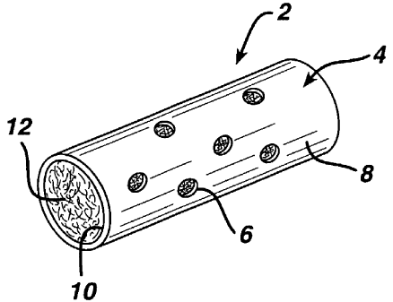
modulation device is provided in Figure 1. The immune modulation device 2 is
comprised of a shell 4 surrounding an interior lumen 10. The shell 4 has pores
6 that
extend from the outer surface 8 to the interior lumen 10. The interior lumen
will have a
volume of at least 1 x 10'8 cm3, preferably will be at least 3 x 10-$ cm3 and
most
preferably the size of the lumen will be sufficient to elicit the desired
immune response
from the animal in which it is implanted (which can be determined by methods
well
known in the art such as ELISA). The shell 2 may have a variety of three
dimensional
shapes (e.g. cylindrical, spherical, rectangular, rhomboidal, etc.). For
example the shell
2 will generally have a longitudinal axis and a cross-section that may be
circular, oval
or polygonal. Preferred for ease of manufacture is a cylindrical shape. A
cylindrically
shaped immune modulation device 2 is illustrated in Figure 1. The ends of the
cylindrically shaped immune modulation device may be capped or left open as
illustrated in Figure 1. The outer surface 8 of the immune modulation device 2
is
preferably impervious to cytokines and immune cells and has numerous pores 6
that
allow for the ingress and egress of immune cells. The number of pores 6 will
generally
be less than 25 percent of the outer surface and preferably will be less than
about 10
percent of the outer surface. The pores 6 size may range from about 10 to
about 500
microns and preferably in the range of from about 100 to about 400 microns.
The
4
interior 10 of immune modulation device 2 will be rilled with a fibrous
scaffolding 12
made of a plurality of fibers (e.g. a yarn or a tow).
The fibrous scaffolding 12 is made from biocompatible fibers, preferably
textured fibers which provide a much lower bulk density filling than non-
texturized
fiber. The low bulk density of textured fibers enables rapid population of the
immune
modulation device 2 with significant numbers of cells and helps to retain the
fibrous
scaffolding 12 within the shell 4. The fibrous scaffolding 12 is loaded with
single or
6
CA 02447030 2003-11-10
WO 02/092054 PCT/US02/08872
multiple antigens and optionally other biologically active or pharmaceutically
active
compounds (e.g. cytokines (e.g. interlukins 1-18; interfexons a, (3, and'y;
growth factors;
colony stimulating factors, chemokines, tumor necrosis factor a and (3, etc.),
non-
cytokine leukocyte chernotactic agents (e.g. CSa, LTB4, etc.), attachment
factors, genes,
peptides, proteins, nucleotides, carbohydrates or synthetic molecules) or
cells
depending on the application.
The shell 4 and the fibrous scaffolding 12 of the device will be made with a
biocompatible material that may be absorbable or non-absorbable. The device
will
preferably be made from biocompatible materials that are flexible and thereby
minimizing irritation to the patient. Preferably the shell will be made from
polymers or
polymer blends having glass transition temperature below physiologic
temperature.
Alternatively the device can be made with a polymer blended with a plasticizer
that
makes it flexible.
In theory but in no way limiting the scope of this invention it is suspected
that
the shell allows cell ingress and egress but hinders diffusion of soluble
molecules out of
the device. This is believed to help to concentrate cytokines secreted by
cells that have
entered the device in response to loaded antigens (e.g. antigen presenting
cells) and
other cells (e.g. helper T cells, B cells etc.) which are present in the
device. The fibrous
scaffolding provides a scaffold for cells to reside on and process the
antigens. This
local concentration of cells and cytokines significantly enhances the immune
response
relative to implantation of antigens with standard adjuvants.
The intended recipient of the implantable device is an animal; preferably a
hmnan, but also including livestock animal, (e.g. sheep, cow, horse, pig,
goat, lama,
emu, ostrich or donkey), poultry (e.g. chicken, turkey, goose, duck, or game
bird), f sh
(e.g. salmon or strugeon), laboratory animal (e.g. rabbit, guinea pig, rat or
mouse)
companion animal (e.g. dog or cat) or a wild animal in captive or free state.
Numerous biocompatible absorbable and nonabsorbable materials can be used
to make the shell or fibrous scaffolding. Suitable nonabsorbable materials for
use in as
7
CA 02447030 2003-11-10
WO 02/092054 PCT/US02/08872
the shell or fibrous scaffolding include, but are not limited to, polyamides
(e.g.
polyhexamethylene adipamide (nylon 6,6), polyhexamethylene sebacamide (nylon
610),
polycapramide (nylon 6), polydodecanamide (nylon 12) and polyhexamethylene
isophthalamide (nylon 61~, copolymers and blends thereof), polyesters (e.g.
polyethylene terephthalate, polybutyl terphthalate (e.g. as described in EPA
287,899
and EPA 448,840), copolymers (e.g. as described in U.S. Pat. No. 4,314,561; Re
32,770; U.S. Pat. Nos. 4,224,946; 5,102,419 and 5,147,382) and blends
thereof),
fluoropolymers (e.g. polytetrafluoroethylene and polyvinylidene fluoride
copolymers
(e.g. as described in U.S. Pat. No. 4,564,013) and blends thereof),
polyolefins (e.g.
polypropylene including atactic but preferably isotactic and syndiotactic
polypropylene
and blends thereof, as well as, blends composed predominately of isotactic or
syndiotactic polypropylene blended with heterotactic polypropylene and
polyethylene),
organosiloxanes (e.g. polydimethylsiloxane rubber such as SILASTIC~ silicone
tubing
from Dow Corning), polyvinyl resins (e.g. polystyrene, polyvinylpyrrolidone,
etc.) and
blends thereof.
Additionally the fibrous scaffolding may be made from natural fibers such as
cotton, linen and silk (although silk is referred to as a nonabsorbable
material, it is
broken down in the human body). Raw silk consists of two filaments that are
held
together by seracin (silk glue). The silk is degummed (the seracin is removed)
and the
resulting single filaments are used to manufacture the fiber. The denier per
filament
(dpf) of individual silk fibers will range from about 0.8 to about 2Ø For
fiber
manufacture it is common to used silk with a dpf of from about 0.8 to about
1.6 and
more preferably a dpf of from about 0.8 to about 1.4. The best grades of silk
are easily
obtainable from suppliers in China and Japan.
Polyesters are also well known commercially available synthetic polymers that
may be used to make the shell or fibrous scaffolding. The most preferred
polyester for
making this device is polyethylene terephthalate. Generally, polyethylene
terephthalate
polymers used to make fibers will have a weight average molecular weight of
greater
than 30,000 preferably greater than 40,000 and most preferably in the range of
from
about 42,000 to about 45,000. The filaments formed from these polymers should
have
8
CA 02447030 2003-11-10
WO 02/092054 PCT/US02/08872
a tenacity of greater than 5 grams/denier and preferably greater than 7
grams/denier.
Polyethylene terephthalate yarns are commonly available from a variety of
commercial
fiber suppliers (such as E.I. DuPont and Hoechst Celanese). Preferred are
commercially
available fibers that may be purchased from Hoechst Celanese under the
trademark
TREVTRA~ High Tenacity type 712 and 787 polyester yarns.
A variety of fluoropolyrners may also be used to make the shell and the
fibrous
scaffolding such as polytetrafluoroethylene and polyvinylidene fluoride (i.e.
as in U.S.
Pat. No. 4,052,550), copolymers and blends thereof Currently the preferred are
the
fluoro polymers blends of polyvinylidene fluoride homopolymer and
polyvinylidene
fluoride and hexafluoropropylene copolymer which is described in U.S. Pat. No.
4,564,013 hereby incorporated by reference herein.
As previously stated the term polypropylene for the purposes of this
application
include atactic but will be preferably isotactic and syndiotactic
polypropylene (such as
is described in U.S. Pat. No. 5,269,807 hereby incorporated by reference
herein) and
blends thereof, as well as, blends composed predominantly of isotactic or
syndiotactic
polypropylene blended with heterotactic polypropylene and polyethylene (such
as is
described in U.S. Pat. No. 4,557,264 issued Dec. 10, 1985 assigned to Ethicon,
Inc.
hereby incorporated by reference) and copolymers composed predominantly of
propylene and other alpha-olefins such as ethylene (which is described in U.S.
Pat. No.
4,520,822 issued Jun. 4, 1985 assigned to Ethicon, hereby incorporated by
reference).
The preferred polypropylene material for making fibers is isotactic
polypropylene
without any other polymers blended or monomers copolymerized therein. The
preferred
method for preparing the flexible polypropylene fibers of the present
invention utilizes
as the raw material pellets of isotactic polypropylene homopolymer having a
weight
average molecular weight of from about 260,00 to about 420,000. Polypropylene
of the
desired grade is commercially available in both powder and pellet form.
A variety of bioabsorbable polymers can be used to make the shell or fibrous
scaffolding of the present invention. Examples of suitable biocompatible,
9
CA 02447030 2003-11-10
WO 02/092054 PCT/US02/08872
bioabsorbable polymers include but are not limited to polymers selected from
the group
consisting of aliphatic polyesters, poly(amino acids), copoly(ether-esters),
polyalkylenes oxalates, polyamides, tyrosine derived polycarbonates,
poly(iminocarbonates), polyorthoesters, polyoxaesters, polyamidoesters,
polyoxaesters
containing amine groups, poly(anhydrides), polyphosphazenes, biomolecules
(i.e.,
biopolymers such as collagen, elastin, bioabsorbable starches, etc.) and
blends thereof.
For the purpose of this invention aliphatic polyesters include, but are not
limited to,
homopolymers and copolymers of lactide (which includes lactic acid, D-, L- and
meso
lactide), glycolide (including glycolic acid), s-caprolactone, p-dioxanone
(1,4-dioxan-2-
one), trimethylene carbonate (1,3-dioxan-2-one), alkyl derivatives of
trimethylene
carbonate, delta-valerolactone, beta-butyrolactone, gamma-butyrolactone, s-
decalactone, hydroxybutyrate, hydroxyvalerate, 1,4-dioxepan-2-one (including
its dimer
1,5,8,12-tetraoxacyclotetradecane-7,14-dione), 1,5-dioxepan-2-one, 6,6-
dimethyl-I,4-
dioxan-2-one, 2,5-diketomorpholine, pivalolactone, gamma, gamma-
diethylpropiolactone, ethylene carbonate, ethylene oxalate, 3-methyl-1,4-
dioxane-2,5-
dione, 3,3-diethyl-1,4-dioxan-2,5-dione, 6,8-dioxabicycloctane-7-one and
polymer
blends thereof. Poly(iminocarbonates), for the purpose of this invention, are
understood to include those polymers as described by Kemnitzer and Kohn, in
the
Handbook of Biodegradable Polymers, edited by Domb, et. al., Hardwood Academic
Press, pp. 251-272 (1997). Copoly(ether-esters), for the purpose of this
invention, are
understood to include those copolyester-ethers as described in the Journal of
Biomaterials Research, Vol. 22, pages 993-1009, 1988 by Cohn and Younes, and
in
Polymer Preprints (ACS Division of Polymer Chemistry), Vol. 30(1), page 498,
1989
by Cohn (e.g. PEO/PLA). Polyalkylene oxalates, for the purpose of this
invention,
include those described in U.S. Patent Numbers 4,208,511; 4,141,087;
4,130,639;
4,140,678; 4,105,034; and 4,205,399 hereby incorporated by reference herein.
Polyphosphazenes, co-, ter- and higher order mixed monomer-based polymers made
from L-lactide, D, L-Iactide, lactic acid, glycolide, glycolic acid, para-
dioxanone,
CA 02447030 2003-11-10
WO 02/092054 PCT/US02/08872
trimethylene carbonate and epsilon-caprolactone such as are described by
Allcock in
The Encyclopedia of Polyrner Science, Vol. 13, pages 31-41, Wiley
Intersciences, John
Wiley & Sons, 1988 and by Vandorpe, et al in the Handbook of Biodegradable
Polymers, edited by Domb, et al, Hardwood Academic Press, pp. 161-182 (1997).
S Polyanhydrides include those derived from diacids of the form HOOC-C6H4 -O-
(CH2)m-O-C6H4-COOH, where m is an integer in the range of from 2 to 8, and
copolymers thereof with aliphatic alpha-omega diacids of up to 12 carbons.
Polyoxaesters, polyoxaamides and polyoxaesters containing amines and/or amido
groups are described in one or more of the following U.S. Patent Nos.
5,464,929;
5,595,751; 5,597,579; 5,607,687; 5,618,552; 5,620,698; 5,645,850; 5,648,088;
5,698,213; 5,700,583; and 5,859,150 hereby incorporated herein by reference.
Polyorthoesters such as those described by Heller in Handbook of Biodegradable
Polymers, edited by Domb, et al, Hardwood Academic Press, pp. 99-118 (1997).
As used herein, the term "glycolide" is understood to include polyglycolic
acid.
Further, the term "lactide" is understood to include L-Iactide, D-lactide,
blends thereof,
and lactic acid polymers and copolymers.
Particularly well suited for use in the present invention are biocompatible
absorbable polymers selected from the group consisting of aliphatic
polyesters,
copolymers and blends which include but are not limited to homopolymers and
copolymers of lactide (which includes D-, L-, lactic acid and D-, L- and meso
lactide),
glycolide (including glycolic acid), epsilon-caprolactone, p-dioxanone (1,4-
dioxan-2-
one which is described in U.S. Pat. No. 4,052,988 incorporated herein by
reference
herein), alkyl substituted derivatives of p-dioxanone (i.e. 6,6-dimethyl-1,4-
dioxan-2-
one which is described in U.S. Pat. No. 5,703,200 assigned to Ethicon and
hereby
incorporated by reference), trimethylene carbonate (1,3-dioxan-2-one), alkyl
substituted
derivatives of 1,3-dioxanone (which are described in U.S. Pat. No. 5,412,068
incorporated herein by reference), delta-valerolactone, beta-butyrolactone,
gamma-
butyrolactone, epsilon-decalactone, hydroxybutyrate, hydroxyvalerate, 1,4-
dioxepan-2-
11
CA 02447030 2003-11-10
WO 02/092054 PCT/US02/08872
one (described in U.S. Pat. No. 4,052,988 and its dimer 1,5,8,12-
tetraoxacyclotetradecane-7,14-dione which is described in U.S. Pat. No.
5,442,032
assigned to Ethicon and hereby incorporated herein by reference), 1,5-dioxepan-
2-one,
and polymer blends thereof. Preferred fiber materials include but are not
limited to
copolymers of trimethylene carbonate, epsilon-caprolactone and glycolide (such
as are
described in U.S. Pat. Nos. 5,431,679 and 5,854,383 hereby herein incorporated
by
reference) and copolymers of p-dioxanone, trimethylene carbonate and glycolide
and
copolymers of lactide and p-dioxanone. Preferred are fibers made from lactide
and
glycolide sometimes referred to herein as simply homopolymers and copolymers
of
Iactide and glycolide and copolymers of glycolide and epsilon-caprolactone
i.e. as
described in U.S. Pat. Nos. 5,133,739; 4,700,704 and 4,605,730 incorporated
herein by
reference), most preferred for use as a fiber is a copolymer that is from
about 80 weight
percent to about I00 weight percent glycolide with the remainder being
Iactide. More
preferred are copolymers of from about 85 to about 95 weight percent glycolide
with
the remainder being lactide.
The molecular weight of the polymers used in the present invention can be
varied as is well know in the art to provide the desired performance
characteristics.
However, it is preferred to have aliphatic polyesters having a molecular
weight that
provides an inherent viscosity between about 0.5 to about 5.0 deciliters per
gram (dl/g)
as measured in a 0.1 g/dl solution of hexafluoroisopropanol at 25 °C,
and preferably
between about 0.7 and 3.5 deciliters per gram (dl/g).
As mentioned above, the outer surface 8 of shell 4 will be perforated with
pores
6, which provide a passageway for the ingress and egress of cells to the
interior lumen
10 of the immune modulation device 2. At the time of implantation the shell 2,
is
substantially impermeable to diffusion of water through the non-perforated
walls of the
shell. The shell 2 is preferably made from one or more absorbable polymers
that may
become more permeable to aqueous media as they degrade. Absorbable polymers
can
either be of natural or synthetic origin. The absorbable polymers for the
membrane
most preferably have a glass transition temperature below physiologic
temperature and
would therefore be less irritating when implanted in soft tissues. Preferred
polymers for
12
CA 02447030 2003-11-10
WO 02/092054 PCT/US02/08872
the shell would include copolymers with a significant content (at least 30
weight
percent) of epsilon-caprolactone or pare-dioxanone. A particularly desirable
composition includes an elastomeric copolymer of from about 3S to about 4S
weight
percent epsilon-caprolactone and from about SS to about 6S weight percent
glycolide,
S lactide (or lactic acid) and mixtures thereof. Another particularly
desirable composition
includes pare-dioxanone homopolymer or copolymers containing from about 0 to
about
80 weight percent pare-dioxanone and from about 0 to about 20 weight percent
of either
lactide, glycolide and combinations thereof. The degradation time for the
membrane in
vivo is preferably longer than 1 month but is shorter than 6 months and more
preferably
is longer than 1 month but less than 4 months.
The shell 4 can be of any shape into which the fibrous scaffolding can be
placed.
The shell can initially have openings that may be later sealed following
placement of
the fibrous scaffolding 12. The shell 4 can be made by conventional polymer
processing
1S techniques including molding, welding, casting, extrusion, injection
molding,
machining process or combinations thereof. These conventional procedures axe
well
known in the art and described in the Encyclopedia of Polymer Science and
Engineering, incorporated herein as reference. Melt extrusion is the preferred
method of
process as it is rapid, inexpensive, scalable, and can be performed solvent-
free for many
polymers of interest. Processing aides and plasticizers can be added to the
polymer to
decrease the processing temperature and/or modify the physical properties of
the
construct. Processing aides, such as solvents, can be added to decrease the
processing
temperature by decreasing the glass transition temperature of the polymer.
Subsequently, the aide can be removed by either heat and/or vacuum or by
passing the
2S extruded construct through a secondary solvent in which the polymer has
minimal
solubility but is miscible with the processing aide. For example halogenated
solvents
such as methylene chloride or chloroform can be added to homo- and copolymers
of
lactide and epsilon-caprolactone. After extrusion, the solvent can be removed
through
evaporation, vacuum, and/or heat. These solvents could also be extracted by
passing
the extrudate through a secondary solvent such as alcohol, which has
miscibility with
the halogenated solvent. Plasticizers can also be incorporated into a polymer
to
increase its workability, flexibility, or distensibility. Typically these
materials work by
13
CA 02447030 2003-11-10
WO 02/092054 PCT/US02/08872
increasing the free volmne ofthe polymer. For example many citrates, malates
and
caprilates will work to plasticize many aliphatic polyesters. Oligomers of a
given
polymer or copolymer can also be used to plasticize a system.
The preferred shapes of the shell are those with a minimal diameter in one
dimension to facilitate placement using a small gauge needle. A most preferred
shape
is a cylinder with an outer diameter preferably less than 1 millimeter and
most
preferably less than 750 microns. This shape and size facilitates implantation
of the
device using an 18 gauge needle or smaller. For this embodiment it is
preferred that the
wall thickness is preferably less than 250 microns and most preferably is less
than 150
microns. The pores 6 in the shell 4 generally are large enough to provide for
the ingress
and egress of cells. The pores are preferably larger than about 10 microns but
smaller
than about 500 microns in cross-sectional diameter and more preferably are
from about
100 to about 400 microns in cross-sectional diameter. The density of
perforations
preferably does not exceed 25% of the outer surface area of the device and
more
preferably is below 10% of the outer surface area of the shell of the immune
modulation
device. The pores can be formed using any appropriate drilling technique (e.g.
using a
hypodermic needle, mechanical or laser) or alternatively by including a
solvent or water
soluble solid in the wall polymer which later can be leached out by immersing
the tube
in the solvent to generate the hole. Alternatively, if biocompatible water
soluble
particles such as sugars, amino acids, polymers such as PVP, proteins such as
gelatin,
carbohydrates such as hyalyronic acid and certain carboxy methylcelluloses are
used,
the device can be implanted with the particles present. Upon exposure to body
fluids
the pore forming particles can leach out or degrade forming pores. Most of the
pore
must extend completely through the wall of the device and provide a pathway
for cells
involved in the immune response to ingress into the interior lumen 10 of the
device as
well as for antigen and cytokines to diffuse out of the interior lumen 10 of
the immune
modulation device 2. If the immune modulation device 2 has one or more open
ends 14
of the immune modulation device can either be sealed with layer 16 or left
open, but are
preferably left open. One embodiment of an immune modulation device with one
sealed end is illustrated in Figure 3.
14
CA 02447030 2003-11-10
WO 02/092054 PCT/US02/08872
In another embodiment of the present invention two portions of the interior
surface 1$ may contact the fibrous scaffolding 12 to restrain movement of the
fibers in
the immune modulation device 2. For example if the immune modulation device 2
were cylindrical a portion of the device could be crimped about the fibrous
scaffolding
12. The crimping could be performed with heating to permanently reshape a
portion of
the shell 4. One embodiment of a crimped device is illustrated in Figure 4.
Alternatively, the crimping could be performed with cutting and sealing one
end of the
immune modulation device 2 to form a cylindrical device with one sealed end
20. One
embodiment of this device with a sealed end is illustrated in Figure 5.
Fibers suitable for use in the present device can be made using conventional
spinning processes such as melt spinning processes or solution spinning. After
spinning
the yarns may be quenched, treated with a spin finish, drawn and annealed as
is known
in the art. The fibrous scaffolding made from these fibers should have a
porosity of
greater than 20%, more preferably from about 25% to about 95%, and most
preferably
from about 30% to about 90% to the fibers.
The fibrous scaffold should be made up of filaments having a denier in the
range of from about 0.2 to about 10 and preferably a denier from about 0.$ to
about 6
and more preferably a denier of from about 1 to about 3. The filaments are
commonly
extruded in bundles (yarns) having a denier in the range of from about 20 to
about 400
denier and preferably about 50 to about 100 denier. The fibers need to be
treated to
develop the bulk density or porosity need for a fibrous scaffold. The
preferred yarns for
this application are textured yarns. There are many forms of textured yarns
that may be
used to form a fibrous scaffolding such as bulked yarns, coil yarns, core
bulked yarns,
crinkle yarns, entangled yarns, modified stretch yarns, nontorqued yarns, set
yarns,
stretch yarns and torqued yarns and combinations thereof. Methods for making
these
yarns are well known and include the false-twisted method, entanglement (e.g.
rotoset
or air jet entangled), crimping (e.g. gear crimped, edge crimped or stuffer
box crimped),
and knit-de-knit. Preferably the fibers will be textured by false-twisting
method, the
stuffer box method or knit-de-knit method of textile texturing. The filaments
are
texturized to provide a high degree of permanent crimping or random looping or
CA 02447030 2003-11-10
WO 02/092054 PCT/US02/08872
coiling. Crimped fibers are currently preferred. Crimping causes the
orientation of the
filament to change angle at the crimping points. The angle change is
preferably greater
than 10 degrees at each crimp point. The crimping can be accomplished through
a
variety of processes but is most easily generated by feeding the extruded
filaments
through a stuffer box.
The fibrous scaffolding is preferably a texturized fiber made from an
absorbable
polymer that can either be of natural or synthetic origin. Each fiber filament
preferably
has a diameter of less than 20 microns and most preferably less than 15
microns. This
imparts to the filaments sufficient flexibility to completely fill the lumen
of the tube and
provide a suitable surface for cells to colonize in the lumen of the shell.
The fibers
preferably will take longer than 1 month to biodegrade (via hydrolysis andlor
enzymatic
activity) in a normal subcutaneous implantation but will completely be
biodegraded
within 6 months and more preferably between 1 and 4 months. .An example of a
good
polymer for making a fibrous scaffolding is a copolymer of 90% glycolide (or
glycolic
acid) and 10% lactide (or lactic acid) having an inherent viscosity between
about 0.7 to
about 1.5 deciliters per gram (dl/g) as measured in a 0.1 g/dl solution of
hexafluoroisopropanol at 25°C.
The most significant advantage with the use of fibrous scaffolding is that the
fibers can be easily placed within the shell. For example, a textured fiber
can be
stretched and then the shell extruded, molded or otherwise coated of shaped
around
them. Following placement of the shell around the stretched fibers, the
tension can be
relaxed which allows the fibers to assume their crimped shapes and fill the
space inside
the shell. Unlike sponges that can also be compressed, the textured fibers can
be
wound onto spools in very long lengths, which can be continuously fed as a
core in a
core-sheath or wire coating extrusion process. The sheath can be a molten
polymer that
is co-extruded and drawn with the stretched fibers. Individual units could be
created by
cutting the core sheath constructs to a desired length. Perforations can be
created by
piercing the tubing wall to form small holes. Open pore sponges are very
difficult to
produce in a continuous form and hence would require the shell be formed as
small
discrete units into which the sponge can be stuffed.
16
CA 02447030 2003-11-10
WO 02/092054 PCT/US02/08872
An additional advantage of fibrous scaffolding over sponges in processing is
that the spool of fibers will be strong while an open cell sponge will be weak
and will
tear easily. This is an important consideration in miniaturization of the
device. Small
bunches of fibers can be stretched, compressed or otherwise exposed to robust
mechanical processing. In contrast, small dimension sponges tear or break
easily and
can only be subj ected to gentle processing. Formation of sub-millimeter
devices
necessarily subjects the filling to significant stresses in order to fit
within the small
dimensions of the shell. Miniaturization is very important in minimizing
patient pain
and discomfort following implantation of the device. Hence the use of fibers,
which
can be compressed more substantially that an open-cell sponge, enables a
smaller
device which is preferable from the patient's standpoint.
At first glance it may appear desirable to fill the shell with simple straight
fibers.
However, straight fibers would settle and bunch in the shell over time and
would not
provide a hospitable environment for ingress of large numbers of cells.
Additionally,
straight fiber would require that the device be modified to prevent the fibers
from fall
out of the device during handling. If the fibers were densely packed or
braided so as to
provide an interference fit in the shell there would not be sufficient
porosity for cell
colonization. Texturizing the fibers allows them to effectively fill space
while
maintaining porosities needed for colonization with high cell number
densities. This
low bulk density property of the texturized fibers enables an interference fit
with the
walls of the shell without having to worry about compaction of the filling
during
storage and handling.
The textured fibers can either be filled into a preformed tube or the tube can
be
extruded around the filaments. During the filling process it may be desirable
to stretch
the filaments to a straight orientation. This radially compresses the fibers
to a much
smaller diameter than they occupy when in a relaxed state. The void volume in
the
lumen of the tube is preferably greater than 30% and more preferably greater
than 50%.
Once relaxed the textured filaments should completely fill the lumen of the
device and
17
CA 02447030 2003-11-10
WO 02/092054 PCT/US02/08872
should stay in place in the lumen due to the compressive force exerted by the
tubing
walls on the filling.
A preferred process for generating the textured fiber filled tubes consists of
extruding the tubing around the stretched filaments in a continuous manner.
This can
be accomplished by having the textured fiber wound on a spool and fed under
tension
through the lumen of an extruder die as a core around which a sheath of wall
polymer is
continuously extruded. Perforations can later be drilled through the wall of
the polymer
either mechanically or using electromagnetic radiation (e.g. laser ablation).
It is
especially desirable to adjust the depth of drilling so that the wall is
completely
punctured but the filling is not damaged. With electromagnetic radiation this
can be
accomplished by provided just enough focused energy to ablate through the wall
of the
tube. Alternatively it is possible to fill a preformed tube by tying the
textured fiber to a
thin wire or needle and then dragging the textured filaments under tension
through the
tubing. Additionally, it is possible to fill a preformed tube by using a
pressure
differential (e.g. vacuum or blown air) to pull the textured filament through
the tubing.
In this configuration the perforations in the tube can be created either pre
or post filling
of the lumen. The length of the textured fiber filled tube is cut to be
greater than a few
millimeters and more preferably greater than 5 millimeters.
The Iumen of the device is filled with an antigen, mixture of antigens and
optionally one or more cytokines, prior to implantation. The antigen can
either be in a
dry or wet form. Potential antigens include peptides, proteins, nucleotides,
carbohydrates or even cells or cell fragments. The antigen or antigens can be
bioavailable at the time of implantation (for immediate release with
optionally a portion
in a sustained release form) or designed to be bioavailable after implantation
(e.g. 3
days after). The antigen or antigens can be supplied in a sustained release
form, such as
encapsulated in microparticles, can be supplied in a naked form or in
combinations
thereof. One method by which antigen can be loaded is to suspend it in a
suitable liquid
which is then injected or pumped into the Iurnen of the filled tube. The
textured fiber
filling must be under sufficient compression as to stay in place through the
convection
of the fluid. The fluid filled device can then be implanted or the filling
fluid can be
18
CA 02447030 2003-11-10
WO 02/092054 PCT/US02/08872
dehydrated or lyophilized prior to implantation leaving behind in the lumen of
the filled
device the desired antigen or antigens. Alteniatively the textured fiber may
be
impregnated with the antigen etc. prior to insertion into the shell. The
dehydrated
system will rehydrate following implantation that will present the antigen in
a suitable
form for generating the desired immunomodulatory response. A particularly
convenient
site of implantation is subcutaneous insertion directly beneath the skin,
however any
site which offers access to antigen presenting cells, macrophages and other
cells of the
immune system is acceptable. Desired immunomodulatory responses can include
either
generation of humoral and/or cellular immunity against the desired antigen or
alternatively desensitization towards particular allergen or cell types.
Any specific antigen or combination of synthetic or natural antigens may be
employed as the antigenic substance for incorporation in the immune modulation
device
and subsequent implantation in the animal. The antigens can be from bacterial,
fungal,
viral, cellular (e.g. from parasites or in autoimmune treatments from animal
tissue) or
synthetic sources which contain at least one epitope to which the immune
system of the
animal will respond. In immunization the antigen is desired to induce
protective
immunity to the animal to which it is administered. The antigen source can be
preparations of killed microorganisms; living weakened microorganisms;
inactivated
bacterial toxins (toxoids); purified macromolecules; recombinantly produced
macromolecules and the like. Preferably for mammals, the antigen or mixtures
of
antigens will be derived from bacterial or viral sources with polyvalent
antigenic
domains being present. Suitable bacterial antigen sources include, but are not
limited
to, Actinobacillus equuli, Aetinobacillus lignie>"esi, Actinobaccilus
semizzis, Aerobacter
aeYOgenes, BoYrelia burgdorferi, Borrelia gai~inii, Borrelia afzelii, Babesia
micz°oti,
Klebsiella pneunzoniae, Bacillus ce~eus, Bacillus a~cth~acis, Bo~detella
peYtussis,
Brucella abo~tus, B~ucella melitensis, Brucella ovis, Brzzcella suis, B~ucella
cams,
Campylobacter fetus, Campylobactez- fetz~s intestinalis, Chlamydia psittaci,
Clzlamydia
tz~achonzatis, Clost>"idium tetarzi, Cotynebacteriuzn acne Types 1 and 2,
Corynebacterium diphtheriae, Cozyfzebacte~ium equi, Corynebacte>"iunz
pyogezzes,
Cofynebacteriuzn Yefzale, Coxiella buz-netii, Diplococcus pzzeumozziae,
Esche~ichia coli,
Eh~liclaia plzagocytoplzila, Ehrlichia equi, Francisella tularensis,
Fusobacterium
19
CA 02447030 2003-11-10
WO 02/092054 PCT/US02/08872
necrophorum, Giardia lambia, Granuloma ittguinale, Haemophilus influenzae,
Haemophilus vaginalis, Group b Hemophilus ducreyi, Lymplaopathia vettereuna,
Leptospira porrtona, Listeria monocytogenes, Microplasma hominis, Moraxella
bovis,
Mycobacterium tuberculosis, Mycobacterium laprae, MycoplasnZa bovigertitalium,
Neisseria gortorr~hea, Neisseria meningitidis, Pseudontonas maltophiia,
Pasteurella
multocida, Pasteurella harnenaolytica, Proteus vulgaris, Pseudomonas
aerugittosa,
Plasmodium berghei, Plasmodium falciparutn, Plasrrtodiuna malariae, Plasmodium
ovals, Plasmodium vivax, Rickettsia prowazekii, Rickettsia mooseri, Rickettsia
rickettsii, Rickettsia tsutsugan2ushi, Rickettsia akaf°i, Salmonella
abortus ovis,
Salmonella abortzts equi, Salmonella dublin, Salmonella enteritidis,
Salmonella
heidleberg, Salmonella paratyphi, Salmonella typhimur iurrt, Shigella
dysenteriae,
Staphylococcus aureus, Streptococcus ecoli, Staphylococcus epidermidis,
Streptococcus pyrogenes, Streptococcus mutans, Streptococcus Group B,
Streptococcus
bovis, Streptococcus dysgalactiae, Streptococcus equisimili, Streptococcus
uberis,
Streptococcus viridans, Treponema pallidum, Tlibrio cholerae, Yersina pesti,
Yersinia
enterocolitica and combinations thereof. Suitable fungi antigen sources
including, but
are not limited to, Aspergillus funaigatus, Blastomyces dermatitidis, Candida
albicans,
Crytococcus neofornaatts, Coccidioides immitis, Histoplasma eapsulatunt and
combinations thereof. Suitable viral antigen sources from viral sources
include, but are
not limited to, influenza, HIV, hanta virus (e.g. Sin Nombre virus), Mumps
virus,
Rubella virus, Measles virus, Smallpox virus, Hepatitis virus, (e.g. A, B, C,
D, E), Rift
Valley Fever (i.e. Plebovirus), viral encephalitis, (e.g. Eastern equine
encephalitic virus,
St. Louis encephalitic virus, Western equine encephalitic virus, West Nile
Virus),
human papilloma virus, cytomegalovirus, polio virus, rabies virus, Equine
herpes virus,
Equine arteritis virus, IBR--IBP virus, BVD--MD virus, Herpes virus (humonis
types 1
and 2) and combinations thereof Suitable parasite antigen sources include, but
are not
limited to, Schistosoma, Oftchocerca, parasitic amoebas and combinations
thereof.
Preferred infectious diseases that this device and method may provide
prophylaxis
against include viruses such as influenza, HIV, human papillorna, hepatitis,
cytomegalovirus, polio and rabies; bacteria for example E. coli, PseudonZOnas,
Shigella,
Treponenza pallidutrt, Mycobacterium (tuberculosis and laprae), Chlarnydia,
CA 02447030 2003-11-10
WO 02/092054 PCT/US02/08872
Rickettsiae, and Neisseria; fungi such as Aspe~gillus and Candida; and
parasitic
multicellular pathogens.
Suppression of the immune response may also be desirable to treat conditions,
S such as allergies, or to prepare patients for the exposure to foreign
antigens, such as for
transplant. Inappropriate immune~responses are believed to be the underlying
etiology
in a number of autoimmune and other diseases, such as type I diabetes,
rheumatoid
arthritis, multiple sclerosis, uveitis, systemic lupus erythematosus,
myasthenia gravis,
and Graves' disease. By implanting in an individual a device of the present
invention
containing the suspect antigen, entry of cells primed to recognize the antigen
can be
induced to u~ldergo apoptosis, and be eliminated from the immune system.
Elimination
of progenitor antigen-specific cells can permit the later transplant of
foreign antigens
without rej ection.
Further utilities of the present invention include improvements in the
generation
of polyclonal antibodies (immune serum) and nonclonal antibodies in laboratory
animals and obtaining the desired isotype of antibody so generated. In one
embodiment, a procedure for preparing polyclonal (iinmune serum) and
monoclonal
antibodies against an antigen available only in minute quantities can be
performed by
the device of the present invention. The device can be provided with a small
amount of
the rare antigen, in order to immunize the animal, after which spleen cells
can be
harvested. This procedure offers an improvement over current tedious and
unpredictable method of introducing the rare antigen directly into the spleen.
Furthermore, the need for a boost immunization may be obviated by use of the
device
of the present invention, and, in addition, an immune response will be
generated more
quickly. A shortened time required to immunize animals will allow the
generation of
monoclonal antibodes more rapidly. In another embodiment, immune cells for the
production of hybridomas can be harvested from the device after immunization
of an
animal with an antigen provided within the device. This procedure can also be
used to
generate human monoclonal antibodies, by implanting a device of the present
invention
into an individual, loading the device with antigen, and then harvesting
immune cells
from the device for the production of hybridomas. The above-mentioned
polyclonal
21
CA 02447030 2003-11-10
WO 02/092054 PCT/US02/08872
antibodies (immune serum) and monoclonal antibodies can be used for diagnosis,
basic
research, imaging and/or therapy. In another embodiment, human monoclonal
antibodies can be generated using the device of the present invention
implanted in a
severe combined immunodeficiency (SCID) mouse, by the following procedure.
First,
S human peripheral blood lymphocytes are injected into a SCID mouse, wherein
the
human lymphocytes populate the murine immune system. After implantation of a
device of the present invention comprising the desired antigen which is
bioavailable
after implantation, subsequent harvesting of cells from the device will
provide human B
lymphocytes cells which can then be used to prepare hybridomas which secrete
human
antibodies against the desired antigen.
A further utility of the device of the present invention is in collection of
immune
cells from a mammal for later reintroduction into the mammal. Cells can be
removed
from the device, for example, by aspiration from the implanted device or
collection
1 S from the device after removal from the body by dissolving the polymer
matrix,
subsequent storage of the cells, for example by cryopreservation, and
reintroduction
into the mammal at a later time. This can be particularly useful for mammals
undergoing whole body radiation therapy. A device of the present invention,
without
containing antigen, can be implanted and maintained for a time Buff dent to
allow
immune cells to migrate into the device, (e.g. seven to ten days).
Subsequently the
device or its contents are removed and the cells contained therein
cryopreserved.
Following radiation therapy, the mammal can have the cells reintroduced into
the body,
whereby the cells will reconstitute the immune system. In another embodiment
of this
utility, co-stimulatory factors such as cytokines which induce the
proliferation of
2S immune cells can be introduced into the device to increase the yield of
cells within the
device, before harvesting. In a further embodiment, immune cells collected
from a
device provided with antigen can be used for active immunization, wherein the
cells
can be stored and then reintroduced into the mammal after, for example, a
course of
chemotherapy or other therapeutic manipulation. In a still further embodiment,
cells
collected from a device can be cyropreserved, and at a later time be exposed
to the
antigen (for example, a cancer antigen) for ex-vivo propagation of T cells
prior to
introduction into the body, for adoptive immunotherapy.
22
CA 02447030 2003-11-10
WO 02/092054 PCT/US02/08872
EXAMPLES
The following examples illustrate the construction of a textured fiber filled
S device for generating an immunomodulatory response. Those skilled in the art
will
realize that these specific examples do not limit the~scope of this invention
and many
alternative forms of an antigen loaded textured fiber filled device could also
be
generated within the scope of this invention.
EXAMPLE 1
TEXTURED FIBROUS FILLING
Fiber texturing was performed using a Techtex ° HDC10 texturizer
(Techniservice, 738 West Cypress Street, I~ennett Square, PA 19348-0817). Nine
1 S spools of S6 denier natural 90/10 glycolide-co-lactide (IV of about 1.1
deciliters per
gram (dl/g) as measured in a 0.1 g/dl solution of hexafluoroisopropanol at 2S
°C. The
filaments had been drawn about SX (original length compared to final length).
The
filaments were placed on the creel and combined into a single S04 denier tow
by
running the drawn yarns together through a common eyelet. The individual yarn
filament diameters were between 12-20 pm. A pretension of S-7 grams was used
for
each yarn by passing them through the gate tensioner. The large yarn tow was
then°
passed over a heated godet with the separator roller (1S wraps) with the
heated godet
being set to a temperature of 130°C. This yarn tow was then fed into
the stuffer box by
two crimper rolls. The clearance between the stuffer box and rollers was 0.012
inches
2S and the temperature in the stuffer box was about SO°C (the box was
not heated, the
elevated temperature of SO°C came from the yarn, heated on the godet).
Uniformity of
crimp texture is maintained through accurate control of the crimped column
height in
the stuffer box. The column height control is provided by the optical sensor
located in
the stuffer box and signaling the take up winder inverter to speed up/slow
down. The
stuffer box optical sensor was set to hole no. 8 from the top of the box.
After the stuffer
box, the textured yarn tow passed through the gate tensioner set at S grams
for
combining and keeping all yarns in the tow under the same tension. The crimped
yarn
23
CA 02447030 2003-11-10
WO 02/092054 PCT/US02/08872
then passed the overfeed rolls to reduce high yarn tension prior to winding on
the take
up winder. The take up winder speed was set at 170 m/min. An image of the
resulting
textured fiber is shown in Figure 2.
EXAMPLE 2
'S MEMBRANE FORMATION
Membranes were formed from both poly(para-dioxanone) (PDO) and a
copolymer of 35/65 epsilon-caprolactone/glycolide (CAP/GLY). The inherent
viscosity
(dl/g) of the PDO and CAP/GLY, as measured in a O.lg/dl solution of
hexafluoroisopropanol (HFIP) 25°C, were 1.80 and 1.30, respectively.
All membranes
were formed by extrusion using a 3/4-inch Brabender single-screw extruder
(C.W.
Brabender~ Instruments, Inc., So. Hackensack, NJ) under flowing nitrogen.
Membranes with several inner and outer dimensions were formed. Extrusion
conditions for the extruded membranes are shown in Table 1. Tmmediately
following
exit from the die, all membranes were run through a 12-foot cooling trough
filled with
chilled water at a temperature of 5-10°C. For the CAP/GLY membranes,
short
segments (~2-3 ft.) were cut and hung from one end at room temperature to
allow
solidification and crystallization of the polymer.
Table 1: Extrusion conditions
POly2iierDle TzonelTzone2Tzone3Tadapt.Tdie PhlockPair SCreWTdke-OD
slZe
Die (~C) (C) (C) (C) (C) (Psi)(psi)speedoff (mm)
x tip
(mil) (rpm}(FTM)
35/65 170 140 145 145 145 140 1900 0.1 12 20 2.0
x 138
CAP/GLY
35/65 102 140 145 145 145 145 4480 0 4 18 1.03
x 83
CAP/GLY
35/65 53 x 140 145 145 145 140 4300 0.1 3 14 0.9
40
CAP/GLY
35/65 56 X 140 145 150 150 150 2470 0.3 4 34 0.65
40
CAP/GLY
PDO 102 130 135 135 135 l35 5000 0 5 20 1.03
X 83
PDO 102 145 150 150 150 150 3750 0 5 20 0.65
X 83
After extrusion, the membranes were cut to the desired length (2-2.5 cm) using
a razor blade. Membrane perforations were formed at Resonetics, Inc. (Nashua,
NH)
using an excimer laser (Lambda-Physik EMG201MSC Excimer Laser) operating at a
wavelength of 193 nm. The laser was coupled to a Resonetics engineering
workstation
consisting of a mask projection imaging beam delivery system and a three-axis
24
CA 02447030 2003-11-10
WO 02/092054 PCT/US02/08872
(XYtheta) computerized motion control system. Hole sizes ranging between 100
and
500 microns were formed through the membrane walls. Drilling parameters for
the
different tubing are shown in Table 2.
Table 2: Laser drilling conditions
Polymer OD/ID (mm/mm)Fluence (J/cmPulse rate - Etch rate
) (Hz) (~m/pulse
35/65 CAP/GLY2.0x1.5., 10 50 0.63
0.9x0.7
35/65 CAP/GLY2.0x1.5 3.5 50 0
.56
35/65 CAP/GLY2.0x1.5 0.7 10 _
0.5
35/65 CAP/GLY1.03x0.83, 2 25 0.67
0.65x0.45
PDO 1.03x0.83, 2.6 50 0.5
0.65x0.45
EXAMPLE 3
VLN CONSTRUCT FORMATION
The textured fiber filling from Example 1 was placed inside the membranes
discussed in Example 2 as follows. Textured fiber was attached to a small
needle or
thin filament of wire and pulled through the membrane. The fiber was cut to
the length
of the membrane. Available porosity was calculated from the volume of the
inner
1 S lumen of the membrane, weight of textured yarn placed inside of the
membrane, and
the density of the fibers used. Table 3 shows several of the construct
geometries and
resultant porosities.
CA 02447030 2003-11-10
WO 02/092054 PCT/US02/08872
Table 3: Absorbable VLN constructs containing textured fiber.
Membrane OD/ID/lengthHole diameter# Fiber ~ PercentSample
Composition(mm/mm/mm) _~ holesweight porosity#
(mg)
CAP/GLY 2.0/1.5/25 300 20 12 80% 1
CAP/GLY 2.0/1.5/20 300 16 10 80% 2
CAP/GLY 2.0/1.5/20 300 12 10 80% 3
CAP/GLY 2.0/1.5/20 300 8 10 80% 4
CAP/GLY 2.0/1.5/20 300 4 10 80% 5
CAP/GLY 2,0/1.5/20 not a licable0 10 80% 6
CAP/GLY 2.0/1.5/25 300 16 10 83% 7
CAP/GLY 2.0/1.5/25 300 16 15 75% 8
CAP/GLY 2.0/1.5/20 300 20 8 83% 9
CAP/GLY 2.0/1.5/20 300 20 92 75% 10
CAP/GLY 0.65/0.45/25150 4 2 65% 11
CAP/GLY 0.65/0.45/25150 12 2 65% 12
CAP/GLY 0.65/0.45/25150 20 2 65% 13
PDO 0.65/0.45/25150 4 1.3 75% 14
PDO 0.65/0.45/25150 8 1,3 75% 15
PDO 0.65/0.45/25150 12 1.1 80% 16
PDO 0.65/0.45/25150 16 1.3 75% 17
EXAMPLE 4
Prior art (WO 99/44583) has demonstrated that a nonabsorbable device using a
25 mm length of silicone tubing with an internal diameter of 1.5 rnm and outer
diameter
of 2 mm, fitted with a 25 mm-long segment of hydroxylated polyvinyl acetate
sponge
induces a more robust immune response to the influenza vaccine (in BALB/c
mice)
than traditional intramuscular injections with and without the use of
traditional
adjuvants such as Ribi. Similarly the device of the present invention such as
the
absorbable, fiber-filled device described in Example 3 (Sample #1) could be
loaded
with 100 ng of influenza antigen (FLUSHIELD° influenza virus vaccine,
trivalent,
Types A & B; obtained from Henry Schein~, Melville NY). Female BALB/c mice (6-
8
weeks old) would be anesthetized with Avertin. One device per animal could be
inserted through a 0.5-cm dorsal midline incision on day 1.
At appropriate intervals post-immunization the mice could be bled and the sera
tested for influenza-specific humoral response, using conventional ELISA or
other
appropriate protocols to determine immune response. The optimum dosage of
antigen
of the device could be determined by developing dose response curves at
appropriate
26
CA 02447030 2003-11-10
WO 02/092054 PCT/US02/08872
time intervals post implantation. Similarly, the cell population in the device
could be
determined at appropriate intervals (e.g. days 3, 7, 10 etc.) to verify the
migration of
cells into the device, cell types in the device and optimum configuration of
holes etc. to
provide the most advantageous conditions for immune modulation in any animal
with a
particular antigen (or antigens).
27