Note: Descriptions are shown in the official language in which they were submitted.
CA 02448695 2003-11-27
WO 02/098502 PCT/US02/17393
APPARATUS AND METHODS FOR
FACILITATING WOUND HEALING
[0001] The present invention relates generally to
apparatus and methods for facilitating wound healing
through the use of electrical stimulation, and more
particularly to apparatus and methods for providing a
voltage gradient and a pattern of current flow that
envelopes and permeates the wound.
Background of the Invention
[0002] Connective tissue wound healing typically
occurs in three distinct phases. Although these phases
intertwine and overlap, each has a specific sequence of
events that distinguishes it. During the initial, or
inflammatory phase, the body begins to clean away
bacteria and initiate hemostasis. The inflammatory
phase has three subphases: hemostasis~ leukocyte and
macrophage migration; and epithelialization. This
phase typically lasts for about four days.
[0003] The second phase, the proliferative phase, is
characterized by a proliferation of fibroblasts,
collagen synthesis, granulation, and wound contraction.
The proliferative phase typically begins about 48 hours
after the wound is inflicted and can extend anywhere
from two hours up to a week. In this phase, the
CA 02448695 2003-11-27
WO 02/098502 PCT/US02/17393
fibroblast cells begin the synthesis and deposition of
the protein collagen, which will form the main
structural matrix for the successful healing of the
wound.
[0004] In the third phase, the remodeling phase, the
collagen production slows. The collagen that is formed
in this stage is more highly organized than the
collagen formed in the proliferative phase.
Eventually, the remodeled collagen increases the
tensile strength in the wound and returns the wound to
about 800 of the skin's original strength.
[0005] This is the general process that occurs in
healthy human beings. Patients that suffer from
conditions which limit the flow of blood to the wound
site are unfortunately not able to exhibit the normal
wound healing process as described. In some patients
this process can be halted. Factors that can
negatively affect this normal wound healing process
include diabetes, impaired circulation, infection,
malnutrition, medication, and reduced mobility. Other
factors such as traumatic injuries and burns can also
impair the natural wound healing process.
[0006] Poor circulation, for,varying reasons, is the
primary cause of chronic wounds such as venous stasis
ulcers, diabetic ulcers, and decubitus foot ulcers.
Venous stasis ulcers typically form just above the
patient's ankles. The blood flow in this region of the
legs in elderly or incapacitated patients can be very
sluggish, leading to drying skin cells. These skin
cells are thus oxygen starved and poisoned by their own
waste products and begin to die. As they do so, they
leave behind an open leg wound with an extremely poor
chance of healing on its own. Diabetic foot ulcers
CA 02448695 2003-11-27
WO 02/098502 PCT/US02/17393
- 3 -
form below the ankle, in regions of the foot that have
very low levels of circulation.
[0007] Similarly, decubitus ulcers form when skin is
subjected to constant compressive force without
movement to allow for blood flow. The lack of blood
flow leads to the same degenerative process as
described above. Paraplegics and severely immobile
elderly patients which lack the ability to toss and
turn while in bed are the main candidates for this
problem.
[0008] Traditional approaches to the care and
management of these types of chronic non-healing wounds
have included passive techniques that attempt to
increase the rate of repair and decrease the rate of
tissue destruction. Examples of these techniques
include antibiotics, protective wound dressings,
removal of mechanical stresses from the affected areas,
and the use of various debridement techniques or agents
to remove wound exudate and necrotic tissue.
[0009] For the most part, these treatment approaches
are not very successful. The ulcers can take many
months to heal and in some cases they may never heal or
they may partially heal only to recur at some later
time.
[0010] Active approaches have been employed to
decrease the healing time and increase the healing
rates of these ulcers. These approaches may include
surgical treatment as well as alterations to the wound
environment. These alterations may include the
application of a skin substitute impregnated with
specific growth factors or other agents, the use of
hyperbaric oxygen treatments, or the use of electrical
stimulation. It has also been shown experimentally
CA 02448695 2003-11-27
WO 02/098502 PCT/US02/17393
- 4 -
(both in animal and clinical trials) that specific
types of electrical stimulation will alter the wound
environment in a positive way so that the normal wound
healing process can occur or in some cases occur in an
accelerated fashion.
[0011] Therapeutic Electrostimulation
[0012] The relationship between direct current
electricity and cellular mitosis and cellular growth
has become better understood during the latter half of
the twentieth century. Weiss, in Weiss, Daryl S., et.
al., Electrical Stimulation and Wound Healing, Arch
Dermatology, 126:222 (Feb. 1990), points out that
living tissues naturally possess direct current
electropotentials that regulate, at least in part, the
wound healing process. Following tissue damage, a
current of injury is generated that is thought to
trigger biological repair. This current of injury.has
been extensively documented in scientific studies., It
is believed that this current of injury is instrumental
in ensuring that the necessary cells are drawn to the
wound location at the appropriate times during the
various stages of wound healing. Localized exposure to
low levels of electrical current that mimic this
naturally occurring current of injury has been shown to
enhance the healing of soft tissue wounds in both human
subjects and animals. It is thought that these
externally applied fields enhance, augment, or take the
place of the naturally occurring biological field in
the wound environment, thus fostering the wound healing
process.
CA 02448695 2003-11-27
WO 02/098502 PCT/US02/17393
- 5 -
[0013] Weiss continues to explain, in a summary of
the scientific literature, that intractable ulcers have
demonstrated accelerated healing and skin wounds have
resurfaced faster and with better tensile properties
following exposure to electrical currents. Dayton and
Palladino, in Dayton, Paul D., and Palladino, Steven
J., Electrical Stimulation of Cutaneous Ulcerations - A
Literature Review, Journal of the American Podiatric
Medical Association, 79(7):318 (July 1989), also state
that the alteration of cellular activity with
externally applied currents can positively or
negatively influence the status of a healing tissue,
thereby directing the healing process to a desired
outcome.
[0014] Furthermore, research conducted by Rafael
Andino during his graduate tenure at the University of
Alabama at Birmingham, also demonstrated that the
presence of electrical fields (in this case induced by
the application of pulsating electromagnetic fields)
dramatically accelerated the healing rates of wounds
created in an animal model. This research found that
the onset and duration of the first two phases of the
wound healing process, the inflammatory and
proliferative phases, had been markedly accelerated in
the treated wounds while the volume of collagen which
had been synthesized by the fibroblasts was also
markedly increased in the treated wounds. This
resulted in the wounds healing in a much shorter amount
of time. Similar findings from other researchers can
be found in other wound healing literature.
[0015] U.S. Pat. No. 5,433,735 to Zanakis et al. and
U.S. Pat. No. 4,982,742 to Claude describe various
electro-stimulation apparatus and techniques for
CA 02448695 2003-11-27
WO 02/098502 PCT/US02/17393
- 6 -
facilitating the regeneration and repair of damaged
tissue. However, each of these references suffers from
the disadvantage that the pattern of current flow
generated with these electrode devices does not pass
through all portions of the wound and thus, certain
portions of the wound site may not be exposed to the
beneficial effects of electrostimulation.
[0016] U.S. Pat. No. 4,911,688 to Jones describes a
wound cover that includes a chamber that encloses fluid
around the wound. One electrode is located in the
chamber and another electrode is placed away from the
wound on the skin. By using conductive liquid within
the chamber, a circuit is completed allowing current to
flow from the electrode in the chamber, through the
liquid, wound, and surrounding tissue and skin to the
other electrode. The liquid is introduced into the
chamber and replaced using two ports, one port is used
to introduce the liquid while at the same time the
other port is used to remove the gas (when the wound
cover is originally applied to the wound) or fluid.
within the chamber. This wound cover, however, is
complicated to use and involves a delicate process of
adding and replacing the conductive liquid.
[0017] In view of the foregoing, it is an object of
the present invention to provide improved apparatus and
methods for easily providing a voltage gradient and a
pattern of current flow that envelops and permeates the
entire wound site.
Summary of the Invention
[0018] This and other objects of the invention are
accomplished in accordance with the principles of the
present invention by providing an electrode system that
CA 02448695 2003-11-27
WO 02/098502 PCT/US02/17393
includes two electrodes that are adapted for connection
to a power source sufficient to cause a current to flow
between them. The electrodes are shaped and oriented
to cause a pattern of current flow that envelops and
permeates the entire wound site. Such shapes~and
orientations may include a circular first electrode
located at and covering the wound site and a second
electrode shaped as a ring fully encircling the first
electrode. The second electrode may be located outside
or partially within the wound site. Other suitable
shapes of the electrodes may include electrodes that
are ovally shaped, rectangularly shaped, triangularly
shaped or any other suitable shape where one electrode
encircles the other electrode. The shape of the
electrode may conform to the shape of the wound.
[0019] The two electrodes of the electrode system
may be mounted to an oxygen-permeable top layer that is
impermeable to water and water vapor. The top layer
may provide support for the electrodes and may allow
the wound site to breathe.
(0020] The electrode system may also include an
electrically insulative element that is disposed
between the two electrodes. The insulative element may
ensure that most if not all of the current flow between
the electrodes passes through the damaged and healthy
surrounding tissue.
[0021] The power supply for applying a voltage
potential across the electrodes may be local to or
remote from the electrode system. In one suitable
arrangement, the power supply is attached to the top
layer of the electrode system. The power -supply can be
configured to provide a constant or varying voltage, a
constant or varying current, or any other suitable
CA 02448695 2003-11-27
WO 02/098502 PCT/US02/17393
_ g _
electrical output to the electrodes to facilitate wound
healing. For example, the power supply may be
configured to provide the desired current or voltage to
the electrodes at different time intervals with the
same electrode system in place. Tn one suitable
embodiment, the power supply is a battery. In another
suitable embodiment, the power supply is electronic
circuitry that is configured to provide the desired
current or voltage.
[0022] In another suitable embodiment of the
invention, the two electrodes of the electrode system
are comprised of oppositely charged polymers of
sufficient voltage differential and charge capacity to
cause a current to flow from the first elec rode to the
second electrode through the wound.
[0023] The electrode system can be designed and
fabricated to be either disposable or reusable.
[0024] The electrode system according to the various
embodiments described herein is capable of generating a
voltage gradient and a pattern of current flow that
envelops and permeates the entire wound site. Such a
pattern of current flow maximizes the recruitment of
the necessary cells to the wound location at the
appropriate times during the various stages of wound
~5 healing.
Brief Description of the Drawings
[0025] The above and other objects and advantages of
the invention will be apparent upon consideration of
the following detailed description, taken in
conjunction with the accompanying drawings, in which
like reference characters refer to like parts
throughout, and in which:
CA 02448695 2003-11-27
WO 02/098502 PCT/US02/17393
- 9 -
[0026] FIG. 1 is a cross-sectional view of an
illustrative electrode system in accordance with the
present invention taken generally along the line 1-1 of
FIG. 2.
[0027] FIG. 2 is a cross-sectional view of the
electrode system of FIG. 1 taken generally along the
line 2-2 of FTG. 1
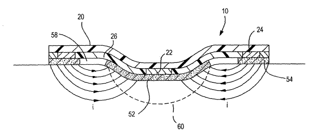
[0028] FIG. 3 is a cross-sectional view of the
electrode system of FIG. 1 as applied to a wound that
illustrates the pattern of current flow generated by
the electrode system in accordance the present
invention.
[0029] FIG. 4 is a perspective view of an
illustrative electrode system placed over a wound site
in accordance with the present invention.
Detailed Description of the Preferred Embodiments
[0030] FIG. 1 is a cross-sectional view of electrode
system 10. The view in FIG. 1 is taken along the line
1-1 of FIG. 2. FIG. 2 shows a simplified cross-
sectional view of electrode system 10 taken alone the
line 2-2 of FIG. 1. As illustrated in FIG. 1,
electrode system 10 includes.top overlay layer 20 to
which electrodes 22 and 24, electrically insulative
element 26, and end material 28 are attached.
Electrode 22 is located towards the center of top
overlay layer 20. Electrically insulative element 26
surrounds electrode 22 and electrode 24 surrounds
electrically insulative element 26. Attached to the
other side of electrodes 22 and 24, electrically
insulative element 26, and end material 28 are adhesive
layers 52 and 54. As illustrated in FIG. 2,
electrically conductive lead 32 connects electrode 22
CA 02448695 2003-11-27
WO 02/098502 PCT/US02/17393
- 10 -
to terminal 42 of power supply 40 and electrically
conductive lead 34 connects electrode 24 to terminal 44
of the power supply 40.
[0031] Top overlay layer 20 may serve several
different purposes. First, top overlay layer 20
provides the mechanical integrity of electrode system
10, thus providing structural support for electrodes 22
and 24. Second, top overlay layer 20 should be
flexible enough to allow electrode system 10 to conform
to the contours of the skin surface to which it is
adhered. Third, top overlay layer 20 should be oxygen
permeable to allow the wound site to breathe. Finally,
top overlay layer 20 should be water impermeable so
that the wound site remains moist. In some
embodiments, all of these characteristics may not be
necessary. For example, a separate water impermeable
layer may be used to keep the wound site moist. Top
overlay layer 20 may be comprised of any suitable
material or structure that exhibits these
characteristics. For example, top overlay layer-20 may
be comprised of a mesh structure of polypropylene,
polyethylene, polyurethane, polytetrafluoroethylene
(PTFE), or any other suitable material. In one
embodiment, top overlay layer 20 can be electrically
insulative to prevent current from flowing between
electrodes 22 and 24, which are attached to top overlay
layer 20. In another suitable embodiment, the adhesive
or binding agent (not shown) used to adhere electrodes
22 and 24 to top overlay layer 20 can be electrically
insulative to prevent current from flowing between
electrodes 22 and 24.
[0032] Electrodes 22 and 24 may be thin metal,
metallic paint or pigment deposition, metallic foil,
CA 02448695 2003-11-27
WO 02/098502 PCT/US02/17393
- 11 -
conductive hydrogels, or any other suitable conductive
material. Hydrogels are generally clear, viscous gels
that protect the wound from dessicating. In one
suitable approach, conductive hydrogels may be used as
the material for electrodes 22 and 24 because of their
permeability to oxygen and ability to retain water.
Both oxygen and a humid environment is required for the
cells in a wound to be viable. In addition, hydrogels
can be easily cast into any shape and size. Various
types of conductive hydrogels may be employed,
including cellulose, gelatin, polyacrylamide,
polymethacrylamide, polyethylene-co-vinyl acetate),
poly(N-vinyl pyrrolidone), polyvinyl alcohol), HEMA,
HEEMA, HDEEMA, MEMA, MEEMA, MDEEMA, EGDMA, mathacrylic
acid based materials, and siliconized hydrogels. PVA-
based hydrogels are inexpensive and easy to form. The
conductivity of such hydrogels can be changed by
Varying the salt concentration within the hydrogels.
By increasing the salt concentration within a hydrogel,
the conductivity of the hydrogel increases.
[0033] Insulative element 26 prevents the flow of
current between electrodes 22 and 24 above the wound
surface such as by moisture trapped under the top
overlay layer. Insulative element 26 may be composed
of any high resistance material such as polythylene,
poly(tetrafluoroethylene) (TEFLON), polyurethane,
polyester, a hydrogel made to be an insulator or any
other suitable insulative material. In addition,
insulative element 26 may be formed of a material or
designed to have gaps or openings within its body to
prevent the flow of current or greatly increase the
current resistance above the wound surface.
CA 02448695 2003-11-27
WO 02/098502 PCT/US02/17393
- 12 -
[0034] End material 28 surrounds electrode 24. End
material 28, in combination with the outer edge of top
overlay layer 20, forms the outer edge of electrode
system 10. End material 28 may be comprised of any
suitable material flexible enough to allow electrode
system 10 to conform to the contours of the skin
surface to which it is adhered. In one embodiment, end
material 28 may be composed of the same material as top
overlay layer 20. In one suitable approach, end
material 28 may be a part of and seamless with top
overlay layer 20.
[0035] Conductive adhesive layers 52 and 54 are
attached to the underside of electrode system 10,
contacting electrodes 22 and 24, respectively and
electrically insulative element 26. Adhesive layers 52
and 54 should be separated from each other by a
suitable space or gap 58 to prevent short-circuiting of
the electrodes. Adhesive layers 52 and 54 may be a
hydrogel, fibrin, conductively transformed
cyanoacrylates or can be comprised of any suitable
electrically conductive material capable of attaching
electrode system 10 to the skin and wound surfaces.
Adhesive layer 52 can be arranged to distribute
substantially the same voltage of electrode 22 to the
entire surface of the wound. Similarly, adhesive layer
54 can be arranged to distribute substantially the same
voltage of electrode 24 to the skin surrounding the
wound. In another suitable approach, adhesive layer 52
can be arranged so that the center of adhesive layer 52
applies a voltage substantially similar to electrode 22
to the center of the wound and that the outer edge of
adhesive layer 52 applies a voltage that is between the
voltages of electrodes 52 and 54 to the outer edge of
CA 02448695 2003-11-27
WO 02/098502 PCT/US02/17393
- 13 -
the wound. The voltage applied to the wound may be
varied, for example, by varying the thickness of
adhesive 52 or by any other suitable method.
[0036] As illustrated in FIG. 1, adhesive layer 52
extends beyond electrode 22. In another suitable
arrangement, adhesive layer 52 may be the same size as
or smaller than electrode 22. Adhesive layer 54 as
illustrated is larger than electrode 24. In another
suitable arrangement, adhesive layer 54 may be the same
size as or smaller than electrode 24.
[0037] In another suitable embodiment, conductive
adhesive layers 52 and 54 may be omitted from electrode
system 10. In this embodiment, electrodes 22 and 24
are themselves adhesive and capable of attaching
electrode system 10 to the wound site. Conductive
hydrogels can be fashioned to have the requisite
adhesive properties, thereby eliminating the need for
separate adhesive layers. One type of highly
conductive hydrogel that is sufficiently tacky and
adhesive to adhere to the skin is described in U.S.
Pat. No. 4,989,607 to Keusch et al. Electrodes 22 and
24 may be comprised of any suitable conductive adhesive
material capable of attaching electrode system 10 to
the wound site.
[0038] Backing layer 60 is attached to conductive
adhesives 52 and 54 to protect the adhesive layer prior
to the use of electrode system 10, Backing layer 60
may be peeled off of adhesives 52 and 54 to expose the
adhesive layer prior to contacting electrode system 10
to the wound site. Backing layer 60 may protrude out
from underneath top overlay layer 20 in. one area, such
as area 60' as shown in FIG. 2, to allow the user to
CA 02448695 2003-11-27
WO 02/098502 PCT/US02/17393
- 14 -
easily remove backing layer 60 from electrode system
10.
[0039] In use, electrode system 10 is positioned
over the wound site such that electrode 22 is located
at approximate the center of the wound site and
adhesive layer 52 can be sized to cover the entire
wound. Electrode system 10 is provided in a family of
sizes appropriate for wounds of various sizes.
Electrode 24 and adhesive layer 54 are generally in the
shape of a ring and are located a distance away from
electrode 22. In one arrangement, the diameters of the
inner edges of electrode 24.and adhesive layer 54 are
greater than the diameter of the wound. In another
words, the size of the wound determines the minimum
inner diameter of electrode 24 and adhesive layer 54.
In another suitable arrangement, adhesive layer 52 can
be sized to cover the inner portion of the wound and
the inner diameters of the inner edges of electrode 24
and adhesive layer 54 may be the same or less than the
size of the wound.
[0040] FIG. 3 is a cross-sectional view of electrode
system 10 as applied to wound 60. As shown in FIG. 3,
the pattern of current flow generated by electrode
system 10 is toroidal in shape. A toroid is generally
formed by rotating a circular disk about an axis, where
the axis lies in the plane of the disk, but outside of
the disk. Here, the pattern of current flow is similar
to a semicircle rotated about an axis, where the axis
lies in the plane of the semicircle and the axis is
near the edge of the semicircle. The current generally
flows tangential to the radial lines of the semicircle.
Because electrode 24 surrounds electrode 22, the
pattern of current flow is similar to the semicircular
CA 02448695 2003-11-27
WO 02/098502 PCT/US02/17393
- 15 -
disk rotated completely around the axis. Therefore,
the pattern of current flow is toroidal in shape. The
pattern of current flow as illustrated in FIG. 3 would
therefore generally be the same regardless of the angle
of the cross-section cut through electrode system 10
with respect to reference direction 65 of Fig. 2. More
specifically, as illustrated, electrode 22 is
negatively charged and electrode 24 is positively "
charged. The lines of current flow extend from
adhesive 54 through wound 60 to adhesive 52 in an
arcuate shape. The lines of current pass through the
entire wound 60, thereby enveloping and permeating the
entire wound and the adjoining unwounded tissue. If
the voltage that is applied to the wound from adhesive
52 is varied, as described above, then the current
density at different portions of wound 60 can be
increased or decreased accordingly. Electrode system
10 can produce a current density within the wound that
is generally between 1 pA/cm2 and 10, 000 p.A/cm~.
Depending on the size and nature of the wound,
electrode system 10 may be configured to produce a
current density within the wound that is less than of 1
~aA/cm2 or greater 10, 000 pA/cmz .
[0041] Referring to FIG. 2, conductive leads 32 and
34, which connect electrodes 22 and 24 respectively to
power supply 40, may be comprised of metal, conductive
ink or any other suitable conductive material. In one
suitable arrangement, leads 32 and 34 are comprised of
conductive carbon ink that is screened onto top overlay
layer 20. In such an arrangement, electrodes 22 and 24
are formed in place over conductive leads 32 and 34,
respectively.
CA 02448695 2003-11-27
WO 02/098502 PCT/US02/17393
- 16 -
[0042] Power supply 40 generates a voltage that is
applied to electrodes 22 and 24 through leads 32 and
34, respectively. Power supply 40 may be configured to
apply a voltage that is anywhere between 1 mV and 9 V.
The resulting current flow that flows through the wound
may be between 1 ~.tA and 50 mA. Depending on the size
and nature of the wound, power supply 40 may be
configured to apply a voltage that is less than 1 mV or
greater than 9 V. The resulting current flow may
therefore be less than 1 pA or greater than 50 mA.
Power supply 40 may be attached to the upper portion of
top overlay layer 20 or any other suitable location on
electrode system 10 or may be located remote from
electrode system 10. In one suitable embodiment, power
supply 40 is a battery. Power supply 40 may be any
suitable battery such as an alkaline, nickel cadmium,
or lithium battery. In one suitable arrangement, power
supply 40 is a lithium polymer stack. The battery may
be arranged so that terminal 42 is negative and
terminal 44 is positive. Thus, electrode 22 functions
as an anode and electrode 24 functions as a cathode.
As described above, current will flow along outward
radial lines from electrode 24 through the wound to
electrode 22. In another suitable approach, the
battery can be arranged so that terminal 42 is positive
and terminal 44 in negative. In such an approach, the
lines of current are reversed and directed outward from
electrode 22 to electrode 24.
[0043] Tn another suitable embodiment, power supply
40 is comprised of electronic circuitry that is
configured to provide a constant or varying voltage, a
constant or varying current, or any other suitable
electrical output. The current density within the
CA 02448695 2003-11-27
WO 02/098502 PCT/US02/17393
- 17 -
wound site may therefore be constant or time varying.
When power supply 40 varies the voltage or current,
electrodes 22 and 24 may change polarities at a
constant or at a time varying frequency. In another
suitable electrical output, power supply 40 can be
configured to pulse electrodes 22 and 24 to provide
other possible therapeutic benefits.
[0044] In one suitable arrangement, the electrical
circuitry can be configured to provide a constant
current source using a current-to-voltage converter.
The current to voltage converter may be probed at test
points to check the current accuracy. The constant
current source may be implemented with an operational
amplifier (Op-amp). The Op-amp compares a precision
voltage reference source to the output of a current-to-
voltage converter and adjusts the output current until
the reference and the converter are equal. The output
voltage is limited to the battery voltage minus a
certain predetermined amount used for operational
purposes.
[0045] The circuit may be built with surface mount
integrated circuits and other surface mount components
and may be powered, for example, by lithium coin cell
batteries.
[0046] The electrode system 10 herein described may
not require a switch to be activated for current to
commence flowing between electrodes 22 and 24. Rather,
current may begin to flow following conductive contact
of electrodes 22 and 24 to the wound site. Such
contact completes a circuit between the electrodes and
results in current flow between the electrodes. In
another suitable embodiment, a switch may be located on
CA 02448695 2003-11-27
WO 02/098502 PCT/US02/17393
- 18 -
electrode system 10 that may allow the user to engage
and disengage power supply 40 to electrodes 22 and 24.
[0047] Electrode system 10 may contain within its
circuitry a visual indicator to allow the user to
determine whether or how well the electrode system is
functioning. The visual indicator may be a light
emitting diode (LED), a series of LEDs, a basic current
meter, or any other suitable visual indicator.
[0048] FIG. 4 demonstrates a view of electrode
system 10 placed over wound 60. In this embodiment,
electrode system 10 is a disposable, one-time-use
bandage that uses a battery and associated circuitry as
power supply 40, which is attached to electrode system
10. Appropriate electrical parameters may be selected
such that the current generated by the internal
circuitry will last for a desired period of time. For
example, the desired period of time may be at least as
long as the typical amount of time a normal bandage is
used on the wound. For users with chronic ulcers, this
amount of time may typically be 1 to 2 days.
Therefore, after electrode system 10 is activated by
placement over the wound, an electrical current may
last for 1 to 2 days. When it is time for electrode
system 10 to be replaced, a new electrode system will
be applied and the treatment will continue as required
by the individual user and the type of wound present.
[0049] While electrode system 10 has been described
as being generally circular in shape, it is understood
that electrode system 10 may also be provided in other
shapes as well. For example, electrode system 10 may
be provided in an oval shape, rectangular shape,
triangular shape, or any other suitable shape. The
resulting pattern of current flow would therefore be
CA 02448695 2003-11-27
WO 02/098502 PCT/US02/17393
- 19 -
similar to the toroidal shape described above which has
been stretch from a circle to an oval shape,
rectangular shape, triangular shape, or any other
suitable shape of electrode system 10. Electrode
system 10 is preferably provided in different shapes
appropriate for wounds of different shapes. For
example, if the wound is a long gash wound, a
rectangular or oval shaped electrode system may be the
appropriate shape for the wound. In one suitable
approach, a preferred electrode system shape for a
wound is a shape that will allow adhesive 52 to cover
the entire wound and that will minimize the amount of
area that adhesive 52 covers exterior to the wound.
This will maximize the current flow through the wound.
[0050] In another suitable electrode system
embodiment, electrodes 22 and 24 are electrically
charged polymers. In this embodiment, power supply 40
and leads 32 and 34, as illustrated in FIGS. 1 and 2
are not required. In addition, top overlay layer 20
may not be required and electrodes 22 and 24 may be
separately applied. Electrodes 22 and 24 can be
oppositely charged polymers (e.g., hydrogel or any
other suitable material for holding a charge) of
sufficient differential voltage potential and of
sufficient charge densities to cause a current to flow
between the electrodes. In one suitable arrangement,
electrode 22 is negatively charged and electrode 24 is
positively charged. This would cause current to flow
through the wound to negative electrode 22 from
positive electrode 24. In another suitable
arrangement, electrode 22 is positively charged and
electrode 24 is negatively charged. This would cause
current to flow from positive electrode 22 through the
wound to negative electrode 24.
CA 02448695 2003-11-27
WO 02/098502 PCT/US02/17393
- 20 -
[0051] The foregoing is merely illustrative of the
principles of this invention and various modifications
can be made by those skilled in the art without
departing from the scope and spirit of the invention.