Note: Descriptions are shown in the official language in which they were submitted.
CA 02448902 2003-11-28
WO 02/100251 PCT/US02/19053
-1-
SELF OPTIMIZING LANCING DEVICE WITH ADAPTATION MEANS
TO TEMPORAL VARIATIONS 1N CUTANEOUS PROPERTIES
TECHNICAL FIELD
Lancing devices are well known in the medical health-care products industry
for
piercing the slcin to produce blood for analysis. Biochemical analysis of
blood samples
is a diagnostic tool for determining clinical information. Many point-of care
tests are
performed using capillary whole blood, the most common being monitoring
diabetic
blood glucose level. Other uses for this method include the analysis of
coagulation
based on Prothrombin time measurement. Typically, a drop of blood for this
type of
analysis is obtained by making a small incision in the fingertip, creating a
small wound,
wlich generates a small blood droplet on the surface of the skin.
BACKGROUND ART
Early methods of lancing included piercing or slicing the skin with a needle
or
razor. Current methods utilize lancet drivers that contain a multitude of
spring, cam and
mass actuators to drive the lancet. These include cantilever springs,
diaphragms, coil
springs, as well as gravity plmnbs used to actuate the lancet. Typically, the
device is
pre-cocked, or the user cocks the device. The device is held against the skin
and the
user, or pressure from the users skin, mechanically triggers the ballistic
launch of the
lancet. The forward movement, and depth of shin penetration of the lancet is
determined
by a mechanical stop and/or damping, as well as a spring or cam which retract
the
lancet.
Current devices generally rely on adjustable mechanical stops or damping to
control the lancet's depth of penetration to compensate for skin thickness and
hydration.
Such devices have the possibility of multiple strikes due to recoil, in
addition to
vibratory stimulation of the severed nerves as the driver impacts the end of
the launcher
stop. Cams may offer rough control of lancet velocity in and out of the shin,
but do not
allow for compensation for shin thickness and hydration. Variations in shin
thickness
CA 02448902 2003-11-28
WO 02/100251 PCT/US02/19053
_2_
and hydration may yield different results in terms of pain perception, blood
yield and
success of obtaining blood from different users of the lancing device.
DISCLOSURE OF INVENTION
Embodiments of the present invention are related to medical health-care
products
and to methods for obtaining body fluids for chemical analysis. More
particularly,
embodiments of the invention relate to devices and methods for piercing the
skin
(lancing) using an electrically driven lancet having user definable lancet
parameters such
as lancet displacement, velocity of incision, retraction, acceleration, and
tissue dwell
time. A device having features of the invention can compensate for long-term
changes
in skin physiology, nerve function, and peripheral vascular perfusion such as
occurs in
diabetes, as well as diurnal variation in skin tensile properties.
Alternatively, a device
having features of the invention can compensate for skin differences between
widely
differing populations such as pediatric and geriatric patients.
An embodiment of the invention is directed to a lancing device which controls
the advancement and retraction of a lancet by monitoring the position of the
lancet in
conjunction with a control feedback for modulating the lancet driver to follow
a
predetermined profile.
BRIEF DESCRIPTION OF DRAWING
The objects, advantages and features of this invention will be more readily
appreciated from the following detailed description, when read in conjunction
with the
accompanying drawing, in which:
Figures 1A and 2A illustrate the displacement over time profile of a harmonic
spring/mass system and a controlled lancet.
Figures 1B and 2B illustrate the velocity over time profiles of a harmonic
spring/mass system and a controlled lancet.
Figure 3 illustrates a controlled actuator using an electromagnetic actuator
to
drive the lancet.
Figure 4 is a flowchart illustrating a controlled feed-back loop.
CA 02448902 2003-11-28
WO 02/100251 PCT/US02/19053
-3-
Figure 5 is a graph of force vs. time during the advancement and retraction of
a lancet showing the characteristic phases of the lancing cycle.
BEST MODE FOR CARRYING OUT THE INVENTION
Lancing device is generally defined to mean any self contained device for
puncturing the skin for the purpose of obtaining a body fluid sample. Lancing
devices
are typically disposable and reusable in their entirety, or in part. For
example, some
lancing devices are disposed of as biohazards after one usage. Other lancing
devices
dispose of only the portions that come in contact with the shin.
Lancet is generally defined to mean any sharp or blunt member used to puncture
the skin for the purpose of cutting blood vessels and allowing blood to flow
to the
surface of the skin. The lancet has certain parameters such as diameter to
define the
cross-sectional area of the member, and geometry to define the shape of the
distal or
front lancing end of the member.
Lancet driver is generally defined to mean any means for controlling the
advancement and retraction of the lancet. Examples of lancet drivers can
include spring-
actuated drivers, electromagnetic drivers and piezoelectric drivers. Examples
of
electromagnetic drivers include solenoids, linear induction motors, and linear
reluctance
motors.
Feedback loop is generally defined to mean a feedback control loop where
information is collected about the current behavior of the lancet (such as
relative lancet
position, rate and direction of lancet motion, resistance to lancet motion,
etc.) and is
used to modulate the drive power applied to the lancet.
Processor is generally defined to mean a high-speed digital processor
containing
memory and calculation capabilities. Such processor is used to modulate the
lancet
driver. Modulate is generally defined to mean controlling the profile of the
lancet.
Profile is generally defined to mean a displacement, velocity or acceleration
versus time plot or table.
Typically, the lancet and the lancet driver are configured so that lancet
velocity
is high at the moment of first contact with the skin, decelerates to zero at
the
predetermined penetration depth, and immediately retracts from the skin,
leaving at
CA 02448902 2003-11-28
WO 02/100251 PCT/US02/19053
-4-
approximately the same velocity that it entered. The energy required for
lancet actuation
is initially stored as potential energy, as in the actuators discussed above.
During the
lancing cycle, the stored energy is transferred into the lcinetic energy of
the lancet, which
is then transferred to potential energy at the. apex of the trajectory, and is
immediately
transferred back into kinetic energy by the retraction mechanism. The
actuation and
retraction velocities are similar, though opposite in sign. The devices which
employ
spring or cam driving methods have a symmetrical actuation displacement and
velocity
profile on the advancement and retraction of the lancet. In most of the
available lancet
devices, once the launch is initiated, the stored energy determines the
velocity profile
until the energy is dissipated. Piezoelectric assisted cutting methods have
also been
described; however, the launching mechanism is spring driven, and no feedback
is
described for controlling lancet motion. Variations in skin properties require
controlling
impact, retraction velocity, and dwell time of the lancet within the tissue.
Advantages are achieved by taking into account that tissue dwell time is
related
to the amount of skin deformation as the lancet tries to puncture the surface
of the skin
and variance in skin deformation from patient to patient based on shin
hydration with
regard to dwell time and the necessity to achieve at least 100 microns of shin
depth to
successfully sample blood.
Pain reduction can be achieved through both the rapid lancet cutting speed and
light weight of the proposed lancet. The rapid cutting minimizes the shock
waves
produced when the lancet strikes the skin in addition to compressing the shin
for
efficient cutting. Due to the very light mass of the lancet and lack of
mechanical stop,
there is insubstantial or no vibrational energy transferred to the finger
during cutting.
Lancing devices such as the spring and cam driven devices typically yield 70 -
80 % success rate in obtaining a blood droplet, as some lancing events are
unsuccessful.
Success rate is dependent on reaching the blood capillaries and venuoles,
which yield
the blood sample. Due to variation in skin thickness and hydration, some skin
will
deform more before cutting starts, and hence the actual depth of penetration
will be less,
resulting in less capillaries and venuoles cut. An electronic feedback
mechanism yields
accurate measurement of skin resistance, and therefore depth of penetration
and thus
directly improves the success rate of blood yield.
CA 02448902 2003-11-28
WO 02/100251 PCT/US02/19053
-5-
Spontaneous blood yield occurs when blood from the cut vessels flows up the
wound tract to the surface of the shin, where it can be collected and tested.
Tissue
o elasticity parameters may force the wound tract to close behind the
retracting lancet
preventing the blood from reaching the surface. If however, the lancet were to
dwell
before being retracted, and or be withdrawn slowly from the wound tract, thus
keeping
the wound open, blood could flow up the patent channel, as described in a
copending
application (Attorney Docket Number 38187-2556, Inventors: Boeclcer, et al.,
entitled
"METHOD AND APPARATUS FOR IMPROVING SUCCESS RATE OF BLOOD
YIELD FROM A FINGERSTICK") submitted on the same day and assigned to the same
assignee as the present application. Said copending application is
incorporated by
reference in its entirety herein.
The ability to control the lancet speed into and out of the wound is critical
as it
allows the device to compensate for changes in skin thickness and variations
in skin
hydration to achieve spontaneous blood yield with maximum success rate while
minimizing pain. This is done by taking into consideration the skin
deformation to
achieve a desirable tissue dwell time and depth of penetration.
This ability to control velocity and depth of penetration therefore requires
an
actuation mechanism where feedback is an integral part of driver control. An
example
of such a driver is the electromagnetic actuator design as described in a
copending
application (Attorney Docket Number 38187-2551, Inventors: Don Alden, et al.,
entitled
"ELECTRIC LANCET ACTUATOR") submitted on the same day and assigned to the
same assignee as the present application. Said copending application is
incorporated by
reference in its entirety herein. Such drivers can control either metal or
polymeric
lancets. The dynamic control of such a driver is shown in Figure 2A which
illustrates
the controlled displacement profile and Figure 2B which illustrates the
controlled
velocity profile. These are compared to Figures 1A and 1B which illustrate
.the
displacement and velocity profiles, respectively, of a harmonic spring/mass
system.
It is, accordingly, an advantage to control the lancet displacement, velocity,
and
acceleration at several steps in the lancing cycle. Such control increases the
success rate
of obtaining an acceptable sample volume of blood and the ability to obtain a
spontaneous blood sample, and decreases the pain perceived by the patient
during the
CA 02448902 2003-11-28
WO 02/100251 PCT/US02/19053
-6-
lancing procedure. Reduced pain is achieved because of fast entry of the
lancet into the
tissue. Reduced lancet velocity with increased lancet dwell time in the tissue
at a point
where the lancet intersects the venuoles and capillary mesh, allows the blood
to pool,
promoting uninhibited flow into the exit channel. Retraction of the lancet at
a low
velocity following the sectioning of the venuole/capillary mesh allows the
blood to flood
the wound tract and flow freely to the surface, thus using the lancet to beep
the channel
open during retraction. Low-velocity retraction of the lancet near the wound
flap
prevents the wound flap from sealing off the channel. Thus, the ability to
slow the
lancet retraction directly contributes to increasing the success rate of
obtaining blood.
Increasing the sampling success rate to near 100% is considered an essential
prerequisite
to combine sampling and acquisition into an integrated sampling module (e.g.
an
integrated glucose sampling module which incorporates a glucose test strip).
Reference will now be made to exemplary embodiments of the invention. In the
first embodiment, a lancing device contains a lancet and lancet driver. The
Lancet and
Lancet driver are configured so that feedback control is based on lancet
displacement,
velocity, or acceleration. The feedback control information relating to the
actual lancet
path is returned to a processor that regulates the energy to the lancet
driver, thereby
precisely controlling the lancet throughout its advancement and retraction.
The lancet
driver may be driven by electric current which includes direct current and
alternating
current. Figure 3 shows an electromagnetic type lancet driver that is capable
of driving
an iron core mounted to the lancet assembly using a direct current (DC) power
supply.
The solenoid is divided into three separate coils along the path of the
lancet, two end
coils and a middle coil. Direct current is applied to the coils to advance and
retract the
lancet. The coils are used in pairs to draw the iron core into the solenoid.
As one of
the drive coils is switched on, the corresponding induced current in the adj
scent coil is
monitored. The strength of this induced current is related to the degree of
magnetic
coupling provided by the iron core, and can be used to infer the position of
the core.
After a period of time, the drive voltage is turned off, allowing the coils to
relax, and
then the cycle is repeated. The degree of magnetic coupling between the coils
is
converted electronically to a proportional DC voltage that is supplied to an
analog-to-
digital converter. The digitized position signal is then processed and
compared to a
CA 02448902 2003-11-28
WO 02/100251 PCT/US02/19053
_7_
desired "nominal" position by a central processing unit (CPU). Error between
the actual
and nominal positions is used by the CPU to set the level and/or length of the
next
power pulse to the solenoid coils.
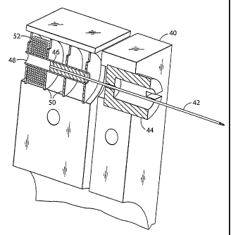
Referring to FIG. 3, the stationary housing (40) contains the solenoid whose
first
coil (52) is separated by a magnetically permeable spacer (50) from the
adjacent coil.
The housing (40) is made from a magnetically permeable material, and a
magnetically
permeable spacer is assembled outside of the first coil. The spacers and
housing form
a magnetic circuit that focuses the magnetic field produced by the coil
between the inner
diameter edges of the spacers. The same is true of each of the other coils,
the housing,
and their spacers. The inner guide tube (48) isolates the lancet (42) and iron
core (46)
from the solenoid coils (52). The lancet (42) and iron core (46) are centered
by the
lancet guide (44). The lancet (42) is advanced and retracted by alternating
the current
between the first coil (52), the middle coil (not shown), and the third coil
(not shown),
singly or in combination, to advance or retract the iron core (46). The lancet
guide (44)
also serves as a stop for the iron core (46) mounted to the lancet (42).
In another embodiment, the solenoid comprises three coils consisting of a
central
driving coil flanked by balanced detection coils built into the driver
assembly so that
they surround the actuation region with the region centered on the middle coil
at mid-
stroke. When a current pulse is applied to the central coil, voltages are
induced in the
adjacent sense coils. If the sense coils are connected together so that their
induced
voltages oppose each other, the resulting signal will be positive for
deflection from mid-
stroke in one direction, negative in the other direction, and zero at mid-
stroke. This
measuring technique is commonly used in Linear Variable Differential
Transformers
(LVDT). Lancet position is determined by measuring the electrical balance
between the
two sensing coils.
In another embodiment, the feedback loop uses a commercially available
LED/photo transducer module such as the OPB703 (manufactured by Optek
Technology,
Inc., 1215 W. Crosby Road, Carrollton, Texas, 75006 (972) 323-2200) to
determine the
distance from the fixed module on the stationary housing to a reflective
surface or target
mounted on the lancet assembly. The LED acts as a light emitter to send light
beams
to the reflective surface which in turn reflects the light back to the photo
transducer
CA 02448902 2003-11-28
WO 02/100251 PCT/US02/19053
_g_
which acts as a light sensor. Distances over the range of 4mm or so are
determined by
measuring the intensity of the reflected light by the photo transducer.
In another embodiment, the feed-back loop uses a magnetically permeable region
on the lancet shaft itself as the core of a Linear Variable Differential
Transformer
(LVDT). A permeable region created by selectively annealing a portion of the
lancet'
shaft, or by including a component in the lancet assembly, such as ferrite,
with sufficient
magnetic permeability to allow coupling between adjacent sensing coils. Coil
size,
number of windings, drive current, signal amplification, and air gap to the
permeable
region axe specified in the design process.
In another embodiment, the feedback control supplies a piezoelectric driver,
superimposing a high frequency oscillation on the basic displacement profile.
The
piezoelectric driver provides improved cutting efficiency and reduces pain by
allowing
the lancet to "saw" its way into the tissue or to destroy cells with
cavitation energy
generated by the high frequency of vibration of the advancing edge of the
lancet. The
drive power to the piezoelectric driver is monitored for an impedance shift as
the device
interacts with the target tissue. The resulting force measurement, coupled
with the
known mass of the lancet is used to determine lancet acceleration, velocity,
and position.
Figure 4 shows the operation of the feedback Ioop using the processor. The
processor (60) stores profiles (62) in non-volatile memory. A user inputs
information
(64) about the desired circumstances for the lancing event. The processor (60)
selects
a profile (62) from a set of alternative profiles that have been preprogrammed
in the
processor (60) based on typical device performance determined through testing
at the
factory. The processor (60) may customize by either scaling or modifying the
profile
based on additional user input information (64). Once the processor has chosen
and
customized the profile, the processor (60) is ready to modulate the power from
the
power supply (66) to the lancet driver (68) through an amplifier (70). The
processor
(60) measures the location of the lancet (72) using a position sensing
mechanism (74)
through an analog to digital converter (76). Examples of position sensing
mechasusms
have been described in the embodiments above. The processor (60) calculates
the
movement of the lancet by comparing the actual profile of the lancet to the
predetermined profile. The processor (60) modulates the power to the lancet
driver (68)
CA 02448902 2003-11-28
WO 02/100251 PCT/US02/19053
-9-
through a signal generator (78), which controls the amplifier (70) so that the
actual
profile of the lancet does not exceed the predetermined profile by more than a
preset
error limit. The error limit is the accuracy in the control of the lancet.
After the lancing event, the processor (60) allows the user to rank the
results of
the lancing event. The processor (60) stores these results and constructs a
database (80)
for the individual user. Using the database (80), the processor (60)
calculates the profile
traits such as degree of painlessness, success rate, and blood volume for
various profiles
(62) depending on user input information (64) to optimize the profile to the
individual
user for subsequent lancing cycles. These profile traits depend on the
characteristic
phases of lancet advancement and retraction. The processor (60) uses these
calculations
to optimize profiles (62) for each user. In addition to user input information
(64), an
internal clock allows storage in the database (80) of information such as the
time of day
to generate a time stamp for the lancing event and the time between lancing
events to
anticipate the user's diurnal needs. The database stores information and
statistics for
each user and each profile that particular user uses.
In addition to varying the profiles, the processor calculates the appropriate
lancet
diameter and geometry necessary to realize the blood volume required by the
user. For
example, if the user requires a 1-5 microliter volume of blood, the processor
selects a
200 micrometer lancet diameter to achieve these results. For each class of
lancet, both
diameter and lancet tip geometry, is stored in the processor to correspond
with upper and
lower limits of attainable blood volume based on the predetermined
displacement and
velocity profiles.
The lancing device is capable of prompting the user for information at the
beginning and the end of the lancing event to more adequately suit the user.
The goal
is to either change to a different profile or modify an existing profile. Once
the profile
is set, the force driving the lancet is varied during advancement and
retraction to follow
the profile. The method of lancing using the lancing device comprises
selecting a
profile, lancing, determining lancing profile traits for each characteristic
phase of the
lancing cycle, and optimizing for subsequent lancing events.
Figure 5 shows the characteristic phases of lancet advancement and retraction
on
a graph of force versus time illustrating the force exerted by the lancet
driver on the
CA 02448902 2003-11-28
WO 02/100251 PCT/US02/19053
-10-
lancet to achieve the desired displacement and velocity profile. The
characteristic phases
are the lancet introduction phase A-C where the lancet is longitudinally
advanced into
the skin, the lancet rest phase D where the lancet terminates its longitudinal
movement
reaching its maximum depth and becoming relatively stationary, and the lancet
retraction
phase E-G where the lancet is longitudinally retracted out of the skin. The
duration of
the lancet retraction phase E-G is longer than the duration of the lancet
introduction
phase A-C, which in turn is longer than the duration of the lancet rest phase
D.
The introduction phase further comprises a lancet launch phase prior to A when
the lancet is longitudinally moving through air toward the skin, a tissue
contact phase
at the beginning of A when the distal end of the lancet makes initial contact
with the
skin, a tissue deformation phase A when the skin bends depending on its
elastic
properties which are related to hydration and thickness, a tissue lancing
phase which
comprises when the lancet hits the inflection point on the skin and begins to
cut the skin
B and the lancet continues cutting the skin C. The lancet rest phase D is the
limit of
the penetration of the lancet into the skin. Pain is reduced by minimizing the
duration
of the lancet introduction phase A-C so that there is a fast incision to a
certain
penetration depth regardless of the duration of the deformation phase A and
inflection
point cutting B which will vary from user to user. Success rate is increased
by
measuring the exact depth of penetration from inflection point B to the limit
of
penetration in the lancet rest phase D. This measurement allows the lancet to
always,
or at least .reliably, hit the capillary beds which are a lmown distance
underneath the
surface of the skin.
The lancet retraction phase further comprises a primary retraction phase E
when
the skin pushes the lancet out of the wound tract, a secondary retraction
phase F when
the lancet starts to become dislodged and pulls in the opposite direction of
the skin, and
lancet exit phase G when the lancet becomes free of the skin. Primary
retraction is the
result of exerting a decreasing force to pull the lancet out of the skin as
the lancet pulls
away from the finger. Secondary retraction is the result of exerting a force
in the
opposite direction to dislodge the lancet. Control is necessary to keep the
wound tract
open as blood flows up the wound tract. Blood volume is increased by using a
uniform
velocity to retract the lancet during the lancet retraction phase E-G
regardless of the
CA 02448902 2003-11-28
WO 02/100251 PCT/US02/19053
-11-
force required for the primary retraction phase E or secondary retraction
phase F, either
of which may vary from user to user depending on the properties of the user's
skin.
Other embodiments of the invention will be apparent to those slcilled in the
art
from consideration of the specification and practice of the invention
disclosed herein.
It is intended that the specification and examples be considered as exemplary
only, with
a true scope and spirit of the invention being indicated by the following
claims.