Note: Descriptions are shown in the official language in which they were submitted.
CA 02449015 2003-11-27
WO 02/096508 PCT/US02/16700
1
ULTRASOUND FEEDBACK IN MEDICALLY-TREATED PATIENTS
-Th~prescnt-appiicatian claims priority of~S~~visiorial- Application
Serial No. 60/294,135 filed May 29, 2001, the entire disclosure of which is
incorporated herein by reference.
Field of the Invention
The present invention relates generally to ultrasound, and more
particularly to an ultrasound medical system and/or to an ultrasound medical
method.
Background of the Invention
Known ultrasound medical systems and methods include using
ultrasound imaging of patients to identify patient tissue for medical
treatment
and include using ultrasound to medically destroy identified patient tissue by
heating the tissue. Imaging is done at lower power and medical treatment is
done at higher power. Low power imaging ultrasound will not medically affect
patient tissue. High power medical-treatment ultrasound, when focused at a
focal zone a distance away from the ultrasound source, will substantially
medically affect patient tissue in the focal zone. However, focused medical-
treatment ultrasound will not substantially medically affect patient tissue
outside the focal zone such as patient tissue located between the source and
the
focal zone.
In one known example, a transducer assembly includes a single
ultrasound transducer having a single transducer element, or an array of
transducer elements acting together, to ultrasonically image the patient and
to
ultrasonically ablate identified patient tissue. It is known to convert
ultrasound
imaging data into temperature imaging data for ultrasound-treated patient
tissue
to monitor the ultrasound treatment. A known transducer element includes a
transducer element having a concave shape or an acoustic lens to focus
END-862
CA 02449015 2003-11-27
WO 02/096508 PCT/US02/16700
2
ultrasound energy. A known array of transducer elements includes a planar,
concave, or convex array of transducer elements to focus ultrasound energy. A
irnowrr-array~of-transducer-elements-imclndes-a~array who~~sdu~er-
elements are electronically or mechanically controlled together to steer and
focus the ultrasound emitted by the array to a focal zone (which may be large
or
which may be as small as, for example, a grain of rice) to provide three-
dimensional medical ultrasound treatment of patient tissue. In some
applications, the transducer is placed on the surface of patient tissue for
ultrasound imaging and/or ultrasound medical treatment of areas within the
patient tissue. In other applications, the transducer is surrounded with a
balloon
which is expanded to contact the surface of patient tissue by filling with a
fluid
such as a saline solution to provide acoustic coupling betweem the transducer
and the patient tissue.
Known ultrasound medical systems and methods include deploying an
end effector having an ultrasound transducer outside the body to break up
kidney stones inside the body, endoscopically inserting an end effector having
an ultrasound transducer in the colon to medically destroy prostate cancer,
laparoscopically inserting an end effector having an ultrasound transducer in
the
abdominal cavity to medically destroy a cancerous liver tumor, intravenously
inserting a catheter end effector having an ultrasound transducer into a vein
in
the arm and moving the catheter to the heart to medically destroy diseased
heart
tissue, and interstitially inserting a needle end effector having an
ultrasound
transducer needle into the tongue to medically destroy tissue to reduce tongue
volume to reduce snoring. Known methods for guiding an end effector within a
patient include guiding the end effector from x-rays, from MRI images, and
from ultrasound images obtained using the ultrasound transducer. Known
ultrasound imaging includes Doppler ultrasound imaging to detect blood flow,
and a proposed known use of ultrasound includes using an ultrasound
transducer outside the body to stop internal bleeding (by sealing ruptured
blood
vessels) of a patient brought to an emergency room of a hospital.
END-862
CA 02449015 2003-11-27
WO 02/096508 PCT/US02/16700
3
A Mammotome~ Breast Biopsy System manufactured by Ethicon
Endo-Surgery, Inc. (a Johnson & Johnson Company) inserts a tube into breast
tissue; whereinrthe-tube contains an~~d effect-or havin a iopsy cutting took:
A
known electromagnetic transponder and three-receiver system for calculating
S the position of the transponder and for guiding the transponder (which is
attached to a heart catheter for monitoring the heart) inside a patient is the
CARTOTM EP Navigation System used with a NAVI-STAR~ catheter
manufactured by Biosense Webster (a Johnson & Johnson Company). Further,
it is known that changes in patient tissue because of medical treatment of
patient
tissue, such as ultrasound medical treatment, affect the amplitude and/or
phase
of ultrasound imaging signals.
What is needed is an improved ultrasound medical system andlor an
improved ultrasound medical method. This invention addresses those needs
lacking in an ultrasonic medical system and/or an ultrasonic medical method.
Summary of the Invention
One method of the invention is for ultrasound imaging of patient tissue
and includes steps a) through c). Step a) includes obtaining a first signal of
a
first imaging ultrasound wave which has been reflected back from, a location
in
the patient tissue at a first time. Step b) includes obtaining a second signal
of a
second imaging ultrasound wave which has been reflected back from the
location in the patient tissue at a later second time wherein the patient has
received at least some medical treatment by the second time. Step c) includes
creating an image of the location using the first signal and the second
signal. In
one example, steps a) through c) are repeated for different locations to image
the patient tissue, wherein the image of the patient tissue includes medically-
treated locations and medically-untreated locations.
The present invention has, without limitation, application in
conventional endoscopic and open surgical instrumentation as well as
application in robotic-assisted surgery.
END-862
CA 02449015 2003-11-27
WO 02/096508 PCT/US02/16700
4
Brief Description of the Drawings
~igure~-is-a perspectivewi~~f a-fir embo~c iment~o a presen
invention showing an ultrasound medical treatment system which includes a
tissue-retaining device;
Figure 2 is an enlarged view of the end effector of the ultrasound
medical treatment system of Figure 1;
Figure 3 is a view of the end effector of Figure 2 retaining an
intervertebral disk of a patient;
Figure 4 is a perspective view of a first alternate end effector which can
be used in the ultrasound medical treatment system of Figure 1;
Figure 5 is a perspective view of a second alternate end effector which
can be used in the ultrasound medical treatment system of Figure 1;
Figure 6 is a perspective view of a third alternate end effector which can
be used in the ultrasound medical treatment system of Figure 1;
Figure 7 is a side elevational view of a second embodiment of the
present invention showing another ultrasound medical treatment system which
includes a tissue-retaining device;
Figure 8 is an enlarged, partially-cutaway view of the end effector of the
ultrasound medical treatment system of Figure 7;
Figure 9 is a perspective view of a third embodiment of the present
invention showing an ultrasound medical system which includes flexible
fingers, wherein each finger includes an ultrasound transducer;
Figure 10 is an enlarged view of the tube and the flexible fingers of the
ultrasound medical system of Figure 9 showing the flexible fingers in a
deployed fan-like state;
Figure 11 is a view of the flexible fingers of Figure 10 shown in a
stowed state;
Figure 12 is a perspective view of an alternate flexible finger
arrangement which can be used in the ultrasound medical system of Figure 9,
END-862
CA 02449015 2003-11-27
WO 02/096508 PCT/US02/16700
showing the flexible fingers in a deployed claw-like state surrounding patient
tissue;
liigure 1-3-is a perspec me mew of a four h~odiment o a present
invention showing an ultrasound medical system which includes an ultrasound
5 transducer assembly which includes at least two ultrasound transducers;
Figure 14 is an enlarged view of the ultrasound transducer assembly of
the ultrasound medical system of Figure 13;
Figure 15 is a cross-sectional view of the transducer assembly of Figure
14;
Figure 16 is a cross-sectional view of a first alternate transducer
arrangement which can be used in place of the arrangement of Figure 1 S;
Figure 17 is a cross-sectional view of a second alternate transducer
arrangement which can be used in place of the arrangement of Figure 15;
Figure 18 is a perspective view of a fifth embodiment of the present
invention showing an ultrasound medical treatment system which includes a
cutting tool and an ultrasound medical-treatment transducer assembly;
Figure 19 is an enlarged, cross-sectional view of the tube of Figure 18
showing a cutting tool that has been introduced into the lumen of the tube;
Figure 20 is an enlarged, cross-sectional view of the tube of Figure 18
showing an ultrasound medical-treatment transducer assembly that has been
introduced into the lumen of the tube;
Figure 21 is a block diagram of an eighth method of the present
invention which includes ultrasound staging of medical treatment of patient
tissue in the gastrointestinal area;
Figure 22 is a block diagram of an eleventh method of the present
invention which includes ultrasound medical treatment of a lesion on or in the
lung of a patient;
Figure 23 is a block diagram of a thirteenth method of the present
invention which includes ultrasound medical treatment of a blood vessel to
stop
the supply of blood to a lesion from the blood vessel;
END-862
CA 02449015 2003-11-27
WO 02/096508 PCT/US02/16700
6
Figure 24 is a perspective view of a sixth embodiment of the present
invention showing a portion of an ultrasound medical treatment system which
i~clt~zte-s-rec~~ers ~cating the position o a ans ucer assembly o~he
system;
Figure 25 is a perspective view of a seventh embodiment of the present
invention showing a portion of another ultrasound medical treatment system
which includes receivers for locating the position of the transponder of the
system;
Figure 26 is a block diagram of a seventeenth method of the present
invention which includes aiming the transducer assembly; and
Figure 27 is a block diagram of a twentieth method of the present
invention which includes creating an image after starting medical treatment
using an imaging ultrasound wave before medical treatment and an imaging
ultrasound wave after starting medical treatment.
Detailed Description of the Invention
Before explaining the present invention in detail, it should be noted that
the invention is not limited in its application or use to the details of
construction
and arrangement of parts illustrated in the accompanying drawings and
description. The illustrative embodiments of the invention may be implemented
or incorporated in other embodiments, variations and modifications, and may be
practiced or carried out in various ways. Furthermore, unless otherwise
indicated, the terms and expressions employed herein have been chosen for the
purpose of describing the illustrative embodiments of the present invention
for
the convenience of the reader and 'are not for the purpose of limiting the
invention.
It is understood that any one or more of the following-described
embodiments, expressions of embodiments, examples, methods, etc. can be
combined with any one or more of the other following-described embodiments,
expressions of embodiments, examples, methods, etc. For example, and
END-862
CA 02449015 2003-11-27
WO 02/096508 PCT/US02/16700
7
without limitation, any of the end effectors can be used in any of the
methods,
any of the transducer arrangements can be used in any of the end effectors,
and
-any appropriate-methods cari he combined-su~i~ as~o~birri~g the seventeenth-
and twentieth methods, etc.
Ultrasound Medical Treatment Using Tissue-Retaining Devices
Tissue-Retaining System for Ultrasound Medical Treatment
Referring now to the drawings, Figures 1-3 illustrate a first embodiment
of the present invention. A first expression of the first embodiment of the
present invention is for an ultrasound medical treatment system 10 including
an
end effector 12 insertable into a patient 14. The end effector 12 includes a
tissue-retaining device 16. The tissue-retaining device 16 includes a first
tissue
retaining member 18 having an (i.e., at least one) ultrasound medical-
treatment
transducer 20 (also called "transducer 20") and includes a second tissue-
retaining member 22. The first and second tissue-retaining members 18 and 22
are operatively connected together to retain patient tissue 24 between the
first
and second tissue-retaining members 18 and 22 and to release patient tissue 24
so retained.
It is noted that an ultrasound medical-treatment transducer is an
ultrasound transducer adapted at least for ultrasound medical treatment of a
patient such as, but not limited to, a human patient. An ultrasound medical-
treatment transducer includes either a single ultrasound medical-treatment
transducer element or an array of ultrasound medical-treatment transducer
elements, as is known to those skilled in the art. An ultrasound medical-
treatment transducer may or may not also be adapted for ultrasound imaging of
a patient. Likewise, an ultrasound imaging transducer is an ultrasound
transducer adapted at least for ultrasound imaging of a patient and may or may
not also be adapted for ultrasound medical-treatment of a patient.
Advantages of retaining patient tissue between two tissue-retaining
members during ultrasound medical treatment by one of the tissue-retaining
END-862
CA 02449015 2003-11-27
WO 02/096508 PCT/US02/16700
8
members include having a single instrument which ultrasonically medically
treats patient tissue and at the same time immobilizes patient tissue against
undesired movement~uring the treatment Ibis also noted~in one
application the tissue-retaining device is a clamp which retains and holds
tissue
and that in another application the tissue-retaining device retains tissue
against
movement, but does not hold tissue, and therefore is not a clamp.
In one variation, not shown, the second tissue-retaining member 22 has
an ultrasound imaging and/or medical treatment transducer. In the same or a
different variation, not shown, the tissue-retaining device 16 has at least
one
additional tissue-retaining member. Mechanisms, not shown, for remotely
moving two (or more) members toward and away from each other are within the
ordinary level of skill of the artisan and include, without limitation, the
use of
pivotal member attachments and the use of cables or motors. In the same or a
different variation, the retained patient tissue 24 is retained between the
ultrasound medical-treatment transducer 20 and the second tissue-retaining
member 22. In the same or a different variation, the ultrasound medical-
treatment transducer 20 focuses ultrasound energy, such focusing being known
to those skilled in the art. In the same or a different variation, not shown,
the
second tissue-retaining member 22 is substantially ultrasonically non-
reflective.
A second expression of the first embodiment of the present invention is
for an ultrasound medical treatment system 10 including an end effector 12
insertable into a patient 14. The end effector 12 includes a tissue-retaining
device 16. The tissue-retaining device 16 includes a first tissue-retaining
member 18 having an (i.e., at least one) ultrasound imaging and medical-
treatment transducer 26 (also called "transducer 26") and includes a second
tissue-retaining member 22. The first and second tissue-retaining members 18
and 22 are operatively connected together to retain patient tissue 24 between
the
first and second tissue-retaining members 18 and 22 and to release patient
tissue
24 so retained.
It is noted that an ultrasound imaging and medical-treatment transducer
is an ultrasound transducer adapted at least for both ultrasound imaging and
END-862
CA 02449015 2003-11-27
WO 02/096508 PCT/US02/16700
9
ultrasound medical treatment of a patient. An ultrasound imaging and medical-
treatment transducer includes either a single ultrasound imaging and medical-
-treatment-transducer elemen or an array ultras~~ic~ducer
elements (including an array having at least one separate element for imaging
and at least one separate element for medical treatment or an array having at
least two elements each adapted for both imaging and medical treatment), as is
known to those skilled in the art. In one variation, the retained patient
tissue 24
is retained between the imaging and medical-treatment transducer 26 and the
second tissue-retaining member 22. In the same or a different variation, the
ultrasound imaging and medical-treatment transducer 26 focuses ultrasound
energy. In the same or a different variation, not shown, the second tissue-
retaining member 22 is substantially ultrasonically non-reflective.
A third expression of the first embodiment shown in Figures 1-3 is for
an ultrasound medical treatment system 10 including an end effector 12
insertable into a patient 14. The end effector 12 includes a tissue-retaining
device 16. The tissue-retaining device 16 includes a first tissue-retaining
member 18 having an (i.e., at least one) ultrasound medical-treatment
transducer 20 and includes a second tissue-retaining member 22 having an
(i.e.,
at least one) ultrasound reflector 28. The first and second tissue-retaining
members 18 and 22 are operatively connected together to retain patient tissue
24
between the first and second tissue-retaining members 18 and 22 and to release
patient tissue 24 so retained.
Advantages of retaining patient tissue between two tissue-retaining
members during ultrasound medical treatment by an ultrasound medical-
treatment transducer of a first tissue-retaining member and an ultrasound
reflector of a second tissue-retaining member include having a single
instrument
which ultrasonically medically treats patient tissue by direct ultrasound,
which
enhances the ultrasound medical treatment by reflected ultrasound, and which
at
the same time immobilizes patient tissue against undesired movement during
the treatment.
END-862
CA 02449015 2003-11-27
WO 02/096508 PCT/US02/16700
It is noted that an ultrasound reflector 28 is a material which reflects
ultrasound at least to a degree that would substantially medically affect
patient
tissue over a treatment peno~c-by direc~ltrasound wluc is emg reflec a -back-
by the ultrasound reflector. Choices of ultrasound reflecting materials
include,
5 without limitation, acoustically-rigid materials such as stainless steel
(which
reflects about 100%) and aluminum (which reflects about 80%) and
acoustically-softer materials such as corporene (which reflects about 90%). An
ultrasound reflecting material is contrasted with an ultrasound absorbing
material such as, without limitation, rubber or plastic. In one variation, the
10 retained patient tissue 24 is retained between the ultrasound medical-
treatment
transducer 20 and the ultrasound reflector 28. In the same or a different
variation, the ultrasound medical-treatment transducer 20 and the ultrasound
reflector 28 each focus ultrasound energy, such ultrasound reflector focusing
being accomplished by the shape of, or by shaping, the reflector surface as is
within the ordinary level of skill of the artisan.
A fourth expression of the first embodiment shown in Figures 1-3 is for
an ultrasound medical treatment system 10 including an end effector 12
insertable into a patient 14. The end effector 12 includes a tissue-retaining
device 16. The tissue-retaining device 16 includes a first tissue-retaining
member 18 having an (i.e., at least one) ultrasound imaging and medical-
treatment transducer 26 and includes a second tissue-retaining member 22
having an (i.e., at least one) ultrasound reflector 28. The first and second
tissue-
retaining members 18 and 22 are operatively connected together to retain
patient tissue 24 between the first and second tissue-retaining members 18 and
22 and to release patient tissue 24 so retained. In one variation, the
retained
patient tissue 24 is retained between the ultrasound imaging and medical-
treatment transducer 26 and the ultrasound reflector 28. In the same or a
different variation, the ultrasound imaging and medical-treatment transducer
26
and the ultrasound reflector 28 each focus ultrasound energy.
In one example of the previously-described third and fourth expressions
of the first embodiment, the ultrasound reflector 28 is disposed to receive
END-862
CA 02449015 2003-11-27
WO 02/096508 PCT/US02/16700
11
ultrasound energy from the transducer 20 and 26 and is oriented to reflect the
received ultrasound energy back into patient tissue 24 retained by the tissue-
-retainirrg~eviee-16: Iri the sam~or a~iiffe~en examp e, a uI O d reflector
28 is oriented to reflect the received ultrasound energy away from the
transducer 20 and 26 when the patient tissue 14 is retained by the tissue-
retaining device 16. An advantage of this arrangement is that it avoids damage
to the transducer from the reflected ultrasound. In the same or a different
example, one of the first and second tissue-retaining members 18 and 22 is
controllably orientatable relative to the other of the first and second tissue-
retaining members 18 and 22 such as, without limitation, by being orientatable
along the double-headed arrows shown in Figure 2. In one modification, the
second tissue-retaining member 22 is controllably orientatable relative to the
first tissue-retaining member 18 to reflect the received ultrasound energy
back
along different directions. A first alternate end effector 30 is shown in
Figure 4
wherein the second tissue-retaining member 32 is controllably orientatable
relative to the first tissue-retaining member 34 as shown by the double-headed
arrows in Figure 4. Mechanisms, not shown, for remotely controlling the
orientation of one member relative to another member are within the ordinary
level of skill of the artisan and include, without limitation, the use of
pivotal
member attachments and the use of cables or motors. In one application, the
transducer 20 and 26 generates wide-focused ultrasound (shown by the two
single-headed arrows coming from the first tissue-retaining member 18 in
Figure 3) and the ultrasound reflector 28 generates narrow-focused ultrasound
(shown by the two single-headed arrows coming from the second tissue
retaining member 22 in Figure 3).
In one example of the previously-described first through fourth
expressions of the first embodiment, the end effector 12 is an open-surgery
end
effector, an endoscopic end effector, a laparoscopic end effector (as shown in
Figure 1), a catheter end effector (such as, but not limited to, an
intravascular
catheter end effector), or a needle end effector, as can be appreciated by
those
skilled in the art. In one application, the end effector 12 is used to retain
a
END-862
CA 02449015 2003-11-27
WO 02/096508 PCT/US02/16700
12
blood vessel and then to ultrasonically treat the blood vessel to seal the
blood
vessel stopping the flow of blood in the retained blood vessel. In another
application, the end~fector 1~ is used to retain patient tissue an then to
ultrasonically ablate at least a portion of the retained patient tissue.
In one design of the previously-described first through fourth
expressions of the first embodiment, the end effector 12 has a longitudinal
axis
35, and one of the first and second tissue-retaining members 18 and 22 at all
times faces along a direction which is substantially perpendicular to the
longitudinal axis 35. If the one tissue-retaining member were planar, this
means
that the longitudinal axis would be substantially parallel to the plane of the
one
tissue-retaining member. In one enablement, the one tissue-retaining member is
the first tissue-retaining member 18. A second alternate end effector 36 has
first
and second tissue-retaining members 38 and 40 which are hinged together to
relatively move as indicated by the double-headed arrow and which are shown
in a partially open configuration in Figure S. The second alternate end
effector
36 has a longitudinal axis 42, and one of the first and second tissue-
retaining
members 38 and 40 at all times faces along a direction which is substantially
parallel to the longitudinal axis 42. If the one tissue-retaining member were
planar, this means that the longitudinal axis would be substantially
perpendicular to the plane of the one tissue-retaining member. In one
enablement, the one tissue-retaining member is the first tissue-retaining
member
38. A third alternate end effector 37 having first and second tissue-retaining
members 39 and 41 with one member longitudinally movable with respect to
the other member (as indicated by the double-headed arrow) is shown in Figure
6. The third alternate end effector 37 has a longitudinal axis 43, and one of
the
first and second tissue-retaining members 39 and 41 at all times faces along a
direction which is substantially parallel to the longitudinal axis 43. In one
enablement, the one tissue-retaining member is the first tissue-retaining
member
39.
In one enablement, as shown in Figure 1, the ultrasound medical
treatment system 10 also includes a handpiece 44 operatively connected to the
END-862
CA 02449015 2003-11-27
WO 02/096508 PCT/US02/16700
13
end effector 12 and to an ultrasound controller 46 operatively connected to a
foot-pedal power switch 47, as can be appreciated by those skilled in the art.
A first method o~'the invention is for a rasoun me ica ~ea en of a
patient and uses the ultrasound medical treatment system as previously
described in the first, second, third or fourth expression of the first
embodiment
with or without the previously-described variations, etc. thereof. The first
method includes steps a) through e). Step a) includes endoscopically inserting
the end effector into an ear, nose, or throat of the patient. Step b) includes
guiding the end effector in the patient. Step c) includes identifying patient
tissue for medical treatment such as optionally at least in part from
ultrasound
imaging using the transducer. Other ways of identifying patient tissue for
medical treatment include, without limitation, using x-rays and/or MRI
imaging,
as are known to the artisan. Step d) includes retaining the identified patient
tissue using the tissue-retaining device. Step e) includes medically treating
the
retained patient tissue with ultrasound using the transducer or using the
transducer and the ultrasound reflector. In one implementation, one tissue-
retaining member at all times faces. along a direction. which is substantially
parallel to the longitudinal axis of the end effector (as seen in Figures S
and 6).
A second method of the invention is for ultrasound medical treatment of
a patient and uses the ultrasound medical treatment system as previously
described in the first, second, third or fourth expression of the first
embodiment
with or without the previously-described variations, etc. thereof. The second
method includes steps a) through c). Step a) includes inserting the end
effector
12 into the patient. Step b) includes retaining an intervertebral disk 48 (see
Figure 3) of the patient with the tissue-retaining device, wherein the
intervertebral disk 48 includes tissue. Step c) includes medically treating
the
retained intervertebral disk 48 with ultrasound to shrink the tissue using the
transducer or using the transducer and the ultrasound reflector. In one
implementation, one tissue-retaining member at all times faces along a
direction
which is substantially perpendicular to the longitudinal axis of the end
effector
END-862
CA 02449015 2003-11-27
WO 02/096508 PCT/US02/16700
14
(as seen in Figures 2 and 4). In one application of the second method of the
invention, the intervertebral disk 48 includes connective and nerve tissue.
A third-methazh~tlr~inventio~or a around me ica ea men o a
patient and uses the ultrasound medical treatment system as previously
described in the first, second, third or fourth expression of the first
embodiment
with or without the previously-described variations, etc. thereof. The third
method includes steps a) through c). Step a) includes inserting the end
effector
into the patient. Step b) includes retaining a joint of the patient with the
tissue-
retaining device, wherein the joint includes tissue. Step c) includes
medically
treating the retained joint with ultrasound to shrink the tissue using the
transducer or using the transducer and the ultrasound reflector. In one
implementation, one tissue-retaining member at all times faces along a
direction
which is substantially perpendicular to the longitudinal axis of the end
effector
(as seen in Figures 2 and 4). In one application of the third method of the
invention, the joint includes connective and nerve tissue.
As previously mentioned, one application of the ultrasound medical
treatment system 10 of the previously-described first through fourth
expressions
of the first embodiment uses the tissue-retaining device to retain a blood
vessel
and uses the transducer, or the transducer and the ultrasound reflector, to
substantially stop the flow of blood within the blood vessel.
Refernng again to the drawings, Figures 7-8 illustrate a second
embodiment of the present invention which is an ultrasound medical treatment
system SO including an end effector 52 insertable into a patient. The end
effector 52 includes a tissue-retaining device 54. The tissue-retaining device
54
includes a first tissue-retaining member 56 having an ultrasound imaging and
medical-treatment transducer 58 and includes a second tissue-retaining member
60 having an ultrasound reflector 62. The first and second tissue-retaining
members 56 and 60 are operatively connected together to retain patient tissue
between the first and second tissue-restraining members and to release patient
tissue so retained. The first and second tissue-retaining members 56 and 60
always maintain a substantially parallel alignment.
END-862
CA 02449015 2003-11-27
WO 02/096508 PCT/US02/16700
Advantages of having a substantially parallel alignment between the
tissue-retaining members include, in one example, having the transducer and
the
ultras reflector maintain a substantially para a a ignmen or improve
reflected ultrasound medical treatment enhancement for any thickness of
patient
5 tissue retained by the tissue-retaining members.
In one example of the second embodiment, the first tissue-retaining
member 56 is a distal end portion 64 of a first tube 66. The ultrasound
medical
treatment system 50 also includes a second tube 68, first and second link
members 70 and 72, and a cable 74. The second tube 68 is oriented
10 substantially parallel to the first tube 66. The first and second link
members 70
and 72 are pivotally attached to the second tissue-retaining member 60 and to
the second tube 68 at pivot points 76-82 creating a hinged parallelogram
defined
by a proximal portion 84 of the second tissue-retaining member 60, a distal
portion 86 of the second tube 68, and the first and second link members 70 and
15 72. The ultrasound reflector 62 is disposed at a distal portion 88 of the
second
tissue-retaining member 60 and faces the transducer 58. The cable 74 is
operatively connected to the hinged parallelogram to move the second tissue-
retaining member 60 toward and away from the first tissue-retaining member
56.
In one variation, the ultrasound medical treatment system 50 also
includes an outer tube 90. The cable 74 and the first and second tubes 66 and
68 are disposed in the outer tube 90. In one modification, the ultrasound
medical treatment system 50 also includes a handpiece 92. The cable 74 and
the first, second, and outer tubes 66, 68 and 90 are operatively connected to
the
handpiece 92. In one design, the orientation of the first tube 66 about the
longitudinal axis of the first tube 66 is controlled by a step motor (not
shown)
disposed in, and actuated by, the handpiece 92. In the same or another design,
the first tube 66 is a hollow tube allowing for transducer wiring (not shown),
and the second tube is a solid tube (not shown). Depending on use, the tubes
66, 68, and 90 may be rigid or flexible which also is true for any tube
arrangement (specifically disclosed as rigid or flexible, or not so
specifically
END-862
CA 02449015 2003-11-27
WO 02/096508 PCT/US02/16700
16
disclosed) of any end effector and for any end effector itself of any of the
previous or following embodiments of the invention.
Ultrasound Medical Treatment Using Specific Transducer Arrangements
Deployable Ultrasound Medical Transducers
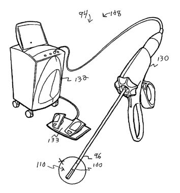
Refernng to the drawings, Figures 9-11 illustrate a third embodiment of
the present invention. A first expression of the third embodiment of the
present
invention is for an ultrasound medical system 94 including a tube 96 and a
plurality of resiliently flexible fingers 98. The tube 96 has a distal end 100
insertable into a patient and has a lumen 102 with a distal opening 104. The
fingers 98 are extendable out of the distal opening 104 of the lumen 102
creating a deployed state (seen in Figure 10) and which are at-least-partially
retractable into the distal opening 104 of the lumen 102 creating a stowed
state
1 S (seen in Figure 11 ). Each finger 98 includes an ultrasound transducer
106. The
distance between the ultrasound transducers 106 of adj acent fingers 98 is
greater in the deployed state than in the stowed state. It is noted that an
ultrasound medical system is a medical system which at least provides
ultrasound imaging or ultrasound medical treatment of a patient.
Advantages of the tube and extendable/retractable flexible-finger array
arrangement include, when the transducers are ultrasound medical-treatment
transducers having a common focal zone in the deployed state, providing faster
medical treatment times by allowing for more transducer ultrasound-emitting
surface area which can be simply stowed into a compact shape for transport
within a patient to and from the site of patient tissue receiving ultrasound
medical treatment.
In one variation, the fingers 98 are only partially retracted into the distal
opening 104 of the lumen 102 in the stowed state (as seen in Figure 11). In
another variation, not shown, the fingers 98 are completely retracted into the
distal opening 104 of the lumen 102 in the stowed state. By the forgers 98
being extendable out of the distal opening 104 of the lumen 102 creating the
END-862
CA 02449015 2003-11-27
WO 02/096508 PCT/US02/16700
17
deployed state and being at-least-partially retractable into the distal
opening 104
of the lumen 102 creating the stowed state means the fingers 98 protrude more
out -of the distal opening~0~f the lumen 102-in a ex ende s a a an i at
all) in the stowed state. Mechanisms, not shown, for remotely extending and
retracting fingers in a tube include, without limitation, a common shaft
attached
to the proximal ends of the fingers, disposed in the lumen of the tube, and
spring-biased to move forward upon squeezing of a handpiece and to return
backward upon relaxing of the handpiece, as is within the ordinary level of
skill
of the artisan. In one modification, the distal opening 104 of the lumen 102
coincides with the distal end 100 of the tube 96. In another modification, not
shown, the distal opening of the lumen is spaced apart from the distal end of
the
tube. In one implementation, the distal opening 104 of the lumen 102 faces in
the same direction as the distal end 100 of the tube 96. Other implementations
are left to the artisan, such as, without limitation, the distal opening of
the
1 S lumen facing perpendicular to the distal end of the tube. In one example,
at
least one of the transducers 106 is an ultrasound imaging transducer. In the
same or a different example, at least one of the transducers 106 is an
ultrasound
medical-treatment transducer. In the same or a different example, at least one
of the transducers 106 is an ultrasound imaging and medical-treatment
transducer.
A second expression of the third embodiment is for an ultrasound
medical treatment system 108 including a tube 96 and including an end effector
110 having a plurality of fingers 98. The tube 96 has a distal end 100
insertable
into a patient and has a lumen 102 with a distal opening 104. The fingers 98
are
extendable out of the distal opening 104 of the lumen 102 creating a deployed
state (seen in Figure 10) and are at-least-partially retractable into the
distal
opening 104 of the lumen 102 creating a stowed state (seen in Figure 11). Each
finger 98 includes an ultrasound medical-treatment transducer 112. The
distance between the ultrasound medical-treatment transducers 112 of adjacent
fingers 98 is greater in the deployed state than in the stowed state.
END-862
CA 02449015 2003-11-27
WO 02/096508 PCT/US02/16700
18
A third expression of the third embodiment is for an ultrasound medical
treatment system 108 including a tube 96 and including an end effector 110
having a plurality of~ngers 98-The tube 96-has a dis~a en T0~0-insertable into
a patient and has a lumen 102 with a distal opening 104. The fingers 98 are
extendable out of the distal opening 104 of the lumen 102 creating a deployed
state (seen in Figure 10) and are at-least-partially retractable into the
distal
opening 104 of the lumen 102 creating a stowed state (seen in Figure 11). Each
finger 98 includes an ultrasound imaging and medical-treatment transducer 114.
The distance between the ultrasound imaging and medical-treatment transducers
114 of adjacent fingers 98 is greater in the deployed state than in the stowed
state.
It is noted that the variations, modifications, and implementations, etc.
previously discussed for the first expression of the third embodiment are
equally
applicable to the second and third expressions of the third embodiment.
In one example of the first, second and third expressions of the third
embodiment, the transducers 106, 112 and 114 each have an ultrasound-
emitting concave surface 116. In another example, not shown, the transducers
have a planar ultrasound-emitting surface. In one arrangement, each concave
surface 116 is concave as one moves along the corresponding finger 98 (as best
seen in Figure 10). In another arrangement, not shown, each concave surface is
concave as one moves across the corresponding finger or is concave as one
moves both along and across the corresponding finger (such as, for example,
with a hemispherically-concave surface). In one design, the concave surfaces
116 together have a substantially common focal zone when the fingers 98 are in
the deployed state. The end effector 110 is seen with its fingers 98 facing
the
patient tissue 119 in Figure 10. In another design, not shown, the focal zones
are not common. In one configuration, the fingers 98 define an open-hand
finger array 118 in the deployed state. An alternate flexible finger
arrangement
in the form of a substitute end effector 120 is shown in Figure 12, wherein
the
fingers 122 define a clawed-hand finger array 124 in the deployed state. The
substitute end effector 120 is seen with its fingers 122 surrounding the
patient
END-862
CA 02449015 2003-11-27
WO 02/096508 PCT/US02/16700
19
tissue 126 for imaging and/or medical treatment by the ultrasound transducers
128 in Figure 12. In other transducer arrangements, not shown, one or more or
all of the ultrasound transducers ace outwar rather an acing inward.
In the same or another example of the first, second and third expressions
of the third embodiment, the fingers 98 are at least four in number. In the
same
or yet another example of the second and third expressions of the third
embodiment, the end effector 110 (as well as the substitute end effector 120)
is
an open-surgery end effector, an endoscopic end effector, a laparoscopic end
effector (as shown in Figure 9), a catheter end effector (such as, but not
limited
to, an intravascular catheter end effector), or a needle end effector, as can
be
appreciated by those skilled in the art.
In one enablement, as shown in Figure 9, the ultrasound medical
treatment system 108 also includes a handpiece 130 operatively connected to
the end effector 110 and to an ultrasound controller 132 operatively connected
to a foot-pedal power switch 133, as can be appreciated by those skilled in
the
art.
Faceted Ultrasound Medical Transducer Assembly
A fourth embodiment of the present invention is shown in Figures 13
15. A first expression of the fourth embodiment of the present invention is
for
an ultrasound medical system 134 including an ultrasound transducer assembly
136 insertable into a patient. The ultrasound transducer assembly 136 has a
longitudinal axis 138. The ultrasound transducer assembly 136 includes a
plurality P of ultrasound transducers 140. Each transducer 140 has an
ultrasound-emitting surface 142 oriented at an angle of substantially 360/P
degrees apart from the ultrasound-emitting surface 142 of an adjacent
transducer 140 when viewed in a cross section (see Figure 15) of the
transducer
assembly 136 taken by a cutting plane which is perpendicular to the
longitudinal
axis 138.
Advantages of such a transducer configuration include, in one example,
providing directed or focused medical-treatment ultrasound which is not
END-862
CA 02449015 2003-11-27
WO 02/096508 PCT/US02/16700
possible with a cylindrical ultrasound transducer, as can be appreciated by
those
skilled in the art.
It-is-n~t~d-tlhat~lt~oun~ducer assem y rose able m~
patient is an ultrasound imaging transducer assembly, an ultrasound medical-
5 treatment transducer assembly, or an ultrasound imaging and medical-
treatment
transducer assembly. An ultrasound imaging transducer assembly has at least
one ultrasound imaging transducer, and an ultrasound medical-treatment
transducer assembly has at least one ultrasound medical-treatment transducer.
An ultrasound imaging and medical-treatment transducer assembly has at least
10 one ultrasound imaging transducer and at least one ultrasound medical-
treatment transducer or has at least one ultrasound imaging and medical-
treatment transducer.
A second expression of the fourth embodiment of the present invention
is for an ultrasound medical-treatment system 144 including an end effector
146
15 insertable into a patient. The end effector 146 includes an ultrasound
medical-
treatment transducer assembly 148. The ultrasound medical-treatment
transducer assembly 148 has a longitudinal axis 138. The ultrasound medical-
treatment transducer assembly 148 includes a plurality P of ultrasound.medical-
treatment transducers 150. Each transducer 150 has an ultrasound-emitting
20 surface 142 which faces away from the longitudinal axis 138 and which is
oriented at an angle of substantially 360/P degrees apart from the ultrasound-
emitting surface 142 of an adjacent transducer 150 when viewed in a cross
section (see Figure 15) of the transducer assembly 148 taken by a cutting
plane
which is perpendicular to the longitudinal axis 138. In one example, at least
one
of the ultrasound medical-treatment transducers 150 is also adapted for
ultrasound imaging.
A fourth method of the present invention is for ultrasound medical
treatment of a patient and uses the ultrasound medical treatment system 144 as
previously described in the second expression of the fourth embodiment. The
fourth method includes steps a) through b). Step a) includes inserting the end
effector 146 into the liver of the patient. Step b) includes medically
treating a
END-862
CA 02449015 2003-11-27
WO 02/096508 PCT/US02/16700
21
lesion in the liver with ultrasound from the ultrasound medical-treatment
transducer assembly 148. In one example, step a) interstially inserts the end
effector mto a esion. anot er example, step a en oscopicaIty inserts
the end effector 146 into the liver through the hepato-biliary duct system.
A third expression of the fourth embodiment of the present invention is
for an ultrasound medical treatment system 144 including an end effector 146
insertable into a patient. The end effector 146 includes an ultrasound imaging
and medical-treatment transducer assembly 152. The ultrasound imaging and
medical-treatment transducer assembly 152 has a longitudinal axis 138. The
ultrasound imaging and medical-treatment transducer assembly 152 includes a
plurality P of ultrasound imaging and medical-treatment transducers 154. Each
transducer 154 has an ultrasound-emitting surface 142 which faces away from
the longitudinal axis 138 and which is oriented at an angle of substantially
360/P degrees apart from the ultrasound-emitting surface 142 of an adjacent
transducer 154 when viewed in a cross section (see Figure 15) of the
transducer
assembly 152 taken by a cutting plane which is perpendicular to the
longitudinal
axis 138.
A fifth method of the present invention is for ultrasound medical
treatment of a patient and uses the ultrasound medical-treatment system 144 as
previously described in the third expression of the fourth embodiment. The
fourth method includes steps a) through c). Step a) includes inserting the end
effector 146 into the liver of the patient. Step b) includes identifying a
lesion in
the liver for medical treatment at least in part from ultrasound imaging using
the
ultrasound imaging and medical-treatment transducer assembly 152. Step c)
includes medically treating the lesion with ultrasound from the ultrasound
imaging and medical-treatment transducer assembly 152. In one example, step
a) interstially inserts the end effector 146 into the lesion. In another
example,
step a) endoscopically inserts the end effector 146 into the liver through the
hepato-biliary duct system.
In one example of the previously-described first, second and third
expressions of the fourth embodiment, the transducer assembly 136, 148, and
END-862
CA 02449015 2003-11-27
WO 02/096508 PCT/US02/16700
22
152 has a distal tip 156 and has a tip transducer 158. In one design, the tip
transducer is a forward facing tip transducer. In another design, the tip
trans ucer is a si eways facing tip transducer. In one vanation, the tip
transducer is an ultrasound imaging tip transducer. In another variation, the
tip
transducer is an ultrasound medical-treatment tip transducer. In a further
variation, the tip transducer is an ultrasound imaging and medical-treatment
tip
transducer. In an additional variation, the tip transducer is a transponder
which
emits electromagnetic waves or mechanical waves or both.
In the same or a different example of the previously-described first,
second and third expressions of the third embodiment, each ultrasound-emitting
surface 142 is substantially straight when viewed in the cross section, as
seen in
Figure 15. In one variation, as seen in Figure 14, each ultrasound-emitting
surface 142 has a substantially concave shape as one moves along the
ultrasound-emitting surface 142 in a direction parallel to the longitudinal
axis
1 S 138, and each ultrasound-emitting surface 142 has a focal zone. In a first
alternate transducer arrangement seen Figure 16, each ultrasound-emitting
surface 162 has a substantially planar shape. In a second alternate transducer
arrangement seen in Figure 17, each ultrasound-emitting surface 164 has a
substantially concave shape when viewed in the cross section, and each
ultrasound-emitting surface 164 has a focal zone. In one modification, each
ultrasound-emitting surface 164 also has a substantially concave shape as one
moves along the ultrasound-emitting surface 164 in a direction parallel to the
longitudinal axis (such as, for example, by the ultrasound-emitting surface
164
having a hemispherically-concave shape). Such ultrasound-emitting surface
shapes are equally applicable to any ultrasound transducer mentioned in any
other embodiment of the invention.
In the same or a different example of the previously-described first,
second and third expressions of the third embodiment, P is no greater than
four.
In one variation, P equals three as seen in Figures 15 and 17. In another
variation, P equals two as seen in Figure 16.
END-862
CA 02449015 2003-11-27
WO 02/096508 PCT/US02/16700
23
In the same or a different example of the previously-described second
and third expressions of the third embodiment, the end effector 146 is an open-
surgery end~fector; an endoscopic end effector, a Iaparoscopic~ a ector as
shown in Figure 13), a catheter end effector (such as, but not limited to; an
intravascular catheter end effector), or a needle end effector, as can be
appreciated by those skilled in the art. In one enablement, as shown in Figure
13, the ultrasound medical treatment system 144 also includes a handpiece 166
operatively connected to the end effector 146 and to an ultrasound controller
168 operatively connected to a foot-pedal power switch 169, as can be
appreciated by the artisan.
Ultrasound Medical Treatment Applications
Excisional And Ultrasound Medical treatment System
A fifth embodiment of the present invention is shown in Figures 18-20.
In a first expression of the fifth embodiment of the present invention, an
ultrasound medical treatment system 170 includes a tube 172, a first end
effector 174, and a second end effector 176. The tube 172 has a distal end 178
~insertable into a patient 180 and has a lumen 182. The first end effector 174
has
a cutting tool 184, is introducible into the lumen 182 of the inserted tube
172
from outside the patient 180, and is translatable through the lumen 182 of the
inserted tube 172 to inside the patient 180. The second end effector 176 has
an
ultrasound medical-treatment transducer assembly 186, is introducible into the
lumen 182 of the inserted tube 172 from outside the patient 180, and is
translatable through the lumen 182 of the inserted tube 172 to inside the
patient
180. In one variation, the first and second end effectors are introduced into
the
lumen through separate openings in the lumen or through separate branch
channels leading to the lumen. In another variation, the first and second end
effectors are introduced into the lumen through the same opening in the lumen.
In one modification, a lumen opening is disposed at the end of the tube. In
another modification, a lumen opening is spaced apart from the end of the
tube.
END-862
CA 02449015 2003-11-27
WO 02/096508 PCT/US02/16700
24
A second expression of the fifth embodiment of the present invention is
for an ultrasound medical treatment system 170 including a tube 172, a first
end
effecto~4, and a second end effector 176The a as a istaTend-1-78-
insertable into a patient 180 and has a lumen 182 with a distal opening 188
and
a proximal opening 190. The first end effector 174 has a cutting tool 184, is
introducible into the proximal opening 190, and is translatable through the
lumen 182 to the distal opening 188. The second end effector 176 has an
ultrasound medical-treatment transducer assembly 186, is introducible into the
proximal opening 190, and is translatable through the lumen 182 to the distal
opening 188.
In one example of the first and second expressions of the fifth
embodiment of the present invention, the lumen 182 is sized to allow
introduction of only one of the first and second end effectors 174 and 176 at
a
time. In the same or another example, the distal end 178 of the tube 172 is
1 S interstitially insertable into patient tissue 192 of the patient 180. In
the same or
a different example, the cutting tool 184 is a biopsy cutting tool 194 or
other
excisional cutting tool.
A third expression of the fifth embodiment of the present invention is for
an ultrasound medical treatment system 170 including a tube 172, a first end
effector 174, and a second end effector 176. The tube 172 has a distal end 178
interstitially insertable into breast tissue 196 of a patient 180 and has a
lumen
182 with a distal opening 188 and a proximal opening 190. The first end
effector 174 has a biopsy cutting tool 194 (or other excisional cutting tool),
is
introducible into the proximal opening 190, and is translatable through the
lumen 182 to the distal opening 188. The second end effector 176 has an
ultrasound medical-treatment transducer assembly 186, is introducible into the
proximal opening 190, and is translatable through the lumen 182 to the distal
opening 188. The lumen 182 is sized to allow introduction of only one of the
first and second end effectors 174 and 176 at a time. In one design, the first
end
effector also includes a suction mechanism to draw in patient tissue to be
biopsied by the biopsy cutting tool 194. In one application, the tube 172 and
the
END-862
CA 02449015 2003-11-27
WO 02/096508 PCT/US02/16700
first end effector 174 (with the biopsy cutting tool 194 including a suction
mechanism) are based on components of a Mammotome~ Breast Biopsy
-Systern~n-anufactur~d-by-Etl~con Endo=Surgery, Inc. (ado mson o nson
Company).
S A sixth method of the invention is for ultrasound medical treatment of a
patient 180 and uses the ultrasound medical treatment system 170 as previously
described in the third expression of the fifth embodiment of the present
invention. The sixth method includes steps a) through h). Step a) includes
identifying possibly cancerous breast tissue 196 of the patient. Step b)
includes
10 interstitially inserting the distal end 178 of the tube 172 into the
patient 180 with
the distal opening 188 disposed proximate the breast tissue 196 and with the
proximal opening 190 disposed outside the patient. Step c) includes
introducing
the first end effector 174 into the proximal opening 190 and translating the
first
end effector 174 through the lumen 182 to the distal opening 188. Step d)
15 includes obtaining a biopsy sample of the breast tissue 196 with the biopsy
cutting tool 194. Step e) includes removing the first end effector 174 from
the
lumen 182, Step f) includes introducing the second end effector 176 into the
proximal opening 190 and translating the second end effector 176 through the
lumen 182 to the distal opening 188. Step g) includes identifying an area of
20 hemorrhaging in the breast tissue where the biopsy sample was obtained.
Step
h) includes medically treating the identified area with ultrasound using the
transducer assembly 186 to substantially stop the hemorrhaging. In one
application, the sixth method of the invention also includes the steps of
testing
the biopsy sample for cancer and substantially ablating any remaining cancer
in
25 the breast tissue with ultrasound using the transducer assembly 186.
Advantages of such an ultrasound medical treatment system and method include
the ease of obtaining a breast biopsy and the control of hemorrhaging caused
by
the biopsy procedure coupled together in a minimally invasive manner.
In a fourth expression of the fifth embodiment of the present invention,
an ultrasound medical treatment system 170 includes a tube 172, a first end
effector 174, and a second end effector 176. The tube 172 has a distal end 178
END-862
CA 02449015 2003-11-27
WO 02/096508 PCT/US02/16700
26
insertable into a patient 180 and has a lumen 182. The first end effector 174
has
a cutting tool 184, is introducible into the lumen 182 of the inserted tube
172
from outside the patient 1-80; and-is translatable-through~he Iumen 182 of~he
inserted tube 172 to inside the patient 180. The second end effector 176 has
an
ultrasound imaging and medical-treatment transducer assembly 198, is
introducible into the lumen 182 of the inserted tube 172 from outside the
patient
180, and is translatable through the lumen 182 of the inserted tube 172 to
inside
the patient 180. In one variation, the first and second end effectors are
introduced into the lumen through separate openings in the lumen or through
separate branch channels leading to the lumen. In another variation, the first
and second end effectors are introduced into the lumen through the same
opening in the lumen. In one modification, a lumen opening is disposed at the
end of the tube. In another modification, a lumen opening is spaced apart from
the end of the tube.
1 S A fifth expression of the fifth embodiment of the present invention is for
an ultrasound medical treatment system 170 including a tube 172, a first end
effector 174, and a second end effector 176. The tube has a distal end 178
insertable into a patient 180 and has a lumen 182 with a distal opening 188
and
a proximal opening 190. The first end effector 174 has a cutting tool 184, is
introducible into the proximal opening 190, and is translatable through the
lumen 182 to the distal opening 188. The second end effector 176 has an
ultrasound imaging and medical-treatment transducer assembly 198, is
introducible into proximal opening 190, and is translatable through the lumen
182 to the distal opening 188.
In one example of the fourth and fifth expressions of the fifth
embodiment of the present invention, the lumen 182 is sized to allow
introduction of only one of the first and second end effectors 174 and 176 at
a
time. In the same or another example, the distal end 178 of the tube 172 is
interstitially insertable into patient tissue 192 of the patient 180. In the
same or
a different example, the cutting tool 184 is a biopsy cutting tool 194 or
other
excisional cutting tool.
END-862
CA 02449015 2003-11-27
WO 02/096508 PCT/US02/16700
27
A sixth expression of the fifth embodiment of the present invention is
for an ultrasound medical treatment system 170 including a tube 172, a first
end
~'fector I74, an a secon en~effector I76. The~7~ has a usta en ~'7$-
interstitially insertable into breast tissue 196 of a patient 180 and has a
lumen
182 with a distal opening 188 and a proximal opening 190. The first end
effector 174 has a biopsy cutting tool 194 (or other excisional cutting tool),
is
introducible into the proximal opening 190, and is translatable through the
lumen 182 to the distal opening 188. The second end effector 176 has an
ultrasound imaging and medical-treatment transducer assembly 196, is
introducible into the proximal opening 190, and is translatable through the
lumen 182 to the distal opening 188. The lumen 182 is sized to allow
introduction of only one of the first and second end effectors 174 and 176 at
a
time. In one application, the tube 172 and the first end effector 174 (with
the
biopsy cutting tool 194 including a suction mechanism) are based on
components of a Mammotome~ Breast Biopsy System manufactured by
Ethicon Endo-Surgery, Inc. (a Johnson & Johnson Company).
A seventh method of the invention is for ultrasound medical treatment of
a patient 180 and uses the ultrasound medical treatment system 170 as
previously described in the sixth expression of the fifth embodiment of the
present invention. The seventh method includes steps a) through h). Step a)
includes identifying possibly cancerous breast tissue 196 of the patient. Step
b)
includes interstitially inserting the distal end 178 of the tube 172 into the
patient
180 with the distal opening 188 disposed proximate the breast tissue 196 and
with the proximal opening 190 disposed outside the patient. Step c) includes
introducing the first end effector 174 into the proximal opening 190 and
translating the first end effector 174 through the lumen 182 to the distal
opening
188. Step d) includes obtaining a biopsy sample of the breast tissue 196 with
the biopsy cutting tool 194. Step e) includes removing the first end effector
174
from the lumen 182, Step f) includes introducing the second end effector 176
into the proximal opening 190 and translating the second end effector 176
through the lumen 182 to the distal opening 188. Step g) includes identifying
END-862
CA 02449015 2003-11-27
WO 02/096508 PCT/US02/16700
28
an area of hemorrhaging in the breast tissue where the biopsy sample was
obtained from ultrasound imaging using the transducer assembly 198. Step h)
inclu es medically treating the idenh~ied area wit ultra usWng~t a
transducer assembly 198 to substantially stop the hemorrhaging. In one
application, the seventh method of the invention also includes the steps of
testing the biopsy sample for cancer and substantially ablating any remaining
cancer in the breast tissue with ultrasound using the transducer assembly 198.
Advantages of such an ultrasound medical treatment system and method include
the ease of obtaining a breast biopsy and the imaging and control of
hemorrhaging caused by the biopsy procedure coupled together in a minimally
invasive manner.
In one enablement, as shown in Figure 18, the ultrasound medical
treatment system 170 also includes a handpiece 199 which is attached to the
tube 172, which contains the first end effector 174 for extending the cutting
tool
184 into, and withdrawing it from, the lumen 182, and which is operatively
connected to an ultrasound controller 201 via a first cable 203. The second
end
effector 176, in this enablement, is operatively connected to the ultrasound
controller 201 via a second cable 205 and is inserted into the lumen 182 from
outside the handpiece 199 as shown in Figure 18.
Staging Medical Treatment Using Ultrasound
An eighth method of the invention is shown in block diagram form in
Figure 21 and is for medical treatment of a patient. The eighth method
includes
steps a) through f). Step a) is labeled "Obtain Transducer Assembly" in block
200 of Figure 21. Step a) includes obtaining an ultrasound imaging transducer
assembly. Step b) is labeled "Insert Assembly Into Gastrointestinal Area" in
block 202 of Figure 21. Step b) includes inserting the transducer assembly
into
a gastrointestinal area of the patient. Step c) is labeled "Guide Assembly" in
block 204 of Figure 21. Step c) includes guiding the transducer assembly
within the gastrointestinal area. Step d) is labeled "Identify Patient Tissue
For
Treatment" in block 206 of Figure 21. Step d) includes identifying patient
END-862
CA 02449015 2003-11-27
WO 02/096508 PCT/US02/16700
29
tissue in the gastrointestinal area for medical treatment. Step e) is labeled
"Stage Treatment From Ultrasound Imaging" in block 208 of Figure 21. Step e)
in udes staging the me ica treatment from a trasound imaging using the
transducer assembly. Step f) is labeled as "Medically Treat Patient" in block
210 of Figure 21. Step f) includes medically treating the patient tissue
according to the staging of step e). It is pointed out that in the eighth
method
the medical treatment need not include ultrasound medical treatment with the
transducer assembly used for staging and/or need not include ultrasound
medical treatment with any other ultrasound transducer assembly. In one
procedure depending on the pathology size and site, a first transducer
assembly
is used endoscopically to stage the medical treatment in step e) and a second
transducer assembly is used laparoscopically to medically treat the patient
tissue
with ultrasound in step f). In one variation, the first transducer assembly is
used
laparoscopically to stage the medical treatment in step e) and the second
transducer assembly is used endoscopically to medically treat the patient
tissue
with ultrasound in step f). In another procedure, the medical treatment in
step f)
is radio-frequency, laser, microwave, or chemical ablation medical treatment.
Other types of medical treatment are left to the artisan.
It is noted that the gastrointestinal (GI) area of a human patient includes,
without limitation, the esophagus and the stomach of the upper GI area and the
rectum and the colon of the lower GI area. It further is noted that the liver
is
also considered to be in the GI area for purposes of this method.
By "staging the medical treatment from ultrasound imaging" is meant at
least using ultrasound images to determine the three-dimensional size and
shape
of the patient tissue that is to receive medical treatment. For example, and
without limitation, upper and lower GI tumors can be visualized with high
frequency (6-30 MHz) ultrasound imaging using a cylindrical, side-firing, or
half convex ultrasound array or single-element transducer introduced
endoscopically into the GI tract. All layers of the GI tract can be visualized
including all layers of the esophagus, stomach, duodenum, colon, etc. In one
procedure, a three-dimensional representation of the GI structures is created
by
END-862
CA 02449015 2003-11-27
WO 02/096508 PCT/US02/16700
collating a series of two-dimensional scans generated by axially advancing the
ultrasound transducer. Any neoplastic growth, its morphological
~ara~teri-sti~c~; as-wolf-as-the~r'-s size~nd shape can easi y a a ermine -
from the three-dimensional representation.
5 Advantages of such medical-treatment staging from ultrasound imaging
include, in one example, providing a non-invasive medical-treatment staging
technique which has greater resolution and which is more practical compared to
conventional extracorporeal medical-treatment staging techniques such as using
x-rays or MRI imaging or compared to using conventional endoscopic optical
10 techniques.
A ninth method of the invention is for ultrasound medical treatment of a
patient and includes steps a) through f). The ninth method uses the same block
diagram of Figure 21 as does the eighth method but with "end effector"
replacing "transducer assembly" in block 200 and with "end effector" replacing
1 S "assembly" in blocks 202 and 204. Step a) includes obtaining an end
effector
having an ultrasound imaging and medical-treatment transducer assembly. Step
b) includes inserting the end effector into a gastrointestinal area of the
patient.
Step c) includes guiding the transducer assembly within the gastrointestinal
area. Step d) includes identifying patient tissue in the gastrointestinal area
for
20 medical treatment. Step e) includes staging the medical treatment from
ultrasound imaging using the transducer assembly. Step f) includes medically
treating the patient tissue with ultrasound using the transducer assembly
according to the staging of step e).
A tenth method of the invention is for ultrasound medical treatment of a
25 patient and includes steps a) through f). The tenth method uses the same
block
diagram of Figure 21 as does the eighth method but with "end effector"
replacing "transducer assembly" in block 200 and with "end effector" replacing
"assembly" in blocks 202 and 204. Step a) includes obtaining an end effector
having an ultrasound imaging and medical-treatment transducer assembly. Step
30 b) includes inserting the end effector into a gastrointestinal area of the
patient.
Step c) includes guiding the transducer assembly within the gastrointestinal
END-862
CA 02449015 2003-11-27
WO 02/096508 PCT/US02/16700
31
area. Step d) includes identifying patient tissue in the gastrointestinal area
for
medical treatment at least in part from ultrasound imaging using the
transducer
assembly: Step e) includes staging the medic ea . en tom ultrasoun3-
imaging using the transducer assembly. Step f) includes medically treating the
patient tissue with ultrasound using the transducer assembly according to the
staging of step e). In one procedure, large GI tumors are staged through a
laparoscopic access to the GI area, whereby the tumors are identified, staged
and treated using an end effector having an ultrasound imaging and medical-
treatment transducer assembly.
In one example of the ninth and tenth methods of the invention, the
patient tissue is gastroesophageal tissue containing a lesion, and step f)
ultrasonically substantially ablates the lesion. ' In one modification, the
gastroesophageal tissue contains a blood vessel supplying blood to the lesion,
and step f) ultrasonically treats the blood vessel to substantially stop the
supply
of blood to the lesion from the blood vessel.
In another example of the ninth and tenth methods of the invention, the
patient tissue is liver tissue containing a lesion and a blood vessel
supplying
blood to the lesion, and step f) ultrasonically treats the blood vessel to
substantially stop the supply of blood to the lesion from the blood vessel.
In an additional example of the ninth and tenth methods of the invention,
the patient tissue is liver tissue containing a lesion, and step f)
ultrasonically
substantially ablates the lesion. In one modification, the liver tissue
contains a
blood vessel supplying blood to the lesion, and step f) also ultrasonically
treats
the blood vessel to substantially stop the supply of blood to the lesion from
the
blood vessel. In one procedure, an end effector having an ultrasound imaging
and medical-treatment transducer assembly is introduced endoscopically into
the GI tract, is advanced retrogradely through the ampulla of Vater up the
common bile duct, and is advanced further into the hepatic duct system where
liver parenchyma requiring medical treatment (such as cholangio-carcinomas)
are identified, staged, and treated using the end effector.
END-862
CA 02449015 2003-11-27
WO 02/096508 PCT/US02/16700
32
Treatment Of Lung Lesions Using Ultrasound
An eleventh method of the invention is shown in block diagram form in
-Figure 22 and is of r ultraso~ medical trea en o a pa lentT~levent~
method includes steps a) through f). Step a) is labeled "Obtain End Effector"
in
block 212 of Figure 22. Step a) includes obtaining an end effector having an
ultrasound medical-treatment transducer assembly. Step b) is labeled "Insert
End Effector" in block 214 of Figure 22. Step b) includes inserting the end
effector into the patient. Step c) is labeled "Guide End Effector To Lung" in
block 216 of Figure 22. Step c) includes guiding the end effector within the
patient to a lung of the patient. Step d) is labeled "Identify Lesion" in
block 218
of Figure 22. Step d) includes identifying a lesion on or in the lung for
medical
treatment. Step e) is labeled "Position Transducer Assembly" in block 220 of
Figure 22. Step e) includes positioning the transducer assembly on or in the
lesion. Step f) is labeled "Medically Treat Lesion" in block 222 of Figure 22.
1 S Step f) includes medically treating the lesion with ultrasound using the
transducer assembly.
A twelfth method of the invention is for ultrasound medical treatment of
a patient and includes steps a) through f). The twelfth method uses the same
block diagram of Figure 22 as does the eleventh method. Step a) includes
obtaining an end effector having an ultrasound imaging and medical-treatment
transducer assembly. Step b) includes inserting the end effector into the
patient.
Step c) includes guiding the end effector within the patient to a lung of the
patient. Step d) includes identifying a lesion on or in the lung for medical
treatment at least in part from ultrasound imaging using the transducer
assembly. Step e) includes positioning the transducer assembly on or in the
lesion. Step f) includes medically treating the lesion with ultrasound using
the
transducer assembly.
In one example of the eleventh and twelfth methods, step f)
ultrasonically substantially ablates the lesion. In one application, the end
effector is an endoscopic end effector and step b) transbronchial-
endoscopically
inserts the end effector into the patient. In another application, the end
effector
END-862
CA 02449015 2003-11-27
WO 02/096508 PCT/US02/16700
33
is a needle end effector and step b) interstitially inserts the end effector
into the
patient. In one implementation, step e) positions the transducer assembly on
the
lesion.~n another i~ementation, step a positions tea transducer assembly in
the lesion. In one practice of the eleventh and twelfth methods, step c) a
bronchoscope is used to guide the end effector to a lung of the patient.
Ultrasound medical treatment of the lung has conventionally been
avoided because such ultrasound is prevented from reaching a lesion within the
lung by the alveoli of the lung which contain air which reflect back most of
the
ultrasound preventing the ultrasound from effectively penetrating the lung to
the
lesion. Using higher power ultrasound for effective penetration of the lung to
reach the lesion would injure or destroy the alveoli which are needed for
breathing. Applicants theorized that positioning the ultrasound transducer on
or
in a lesion of the lung would allow ultrasound medical treatment of the lesion
(such as a tumor or an infarct) without injury to the alveoli. It is noted
that
Applicants' method is applicable to surface lesions as well as non-surface
lesions. Advantages of Applicants' eleventh and twelfth methods for ultrasound
medical treatment include, in one example, the destruction of lung cancer
lesions in cases which otherwise would be inoperable or incurable.
Ultrasound-Based Occlusive Procedure For Medical Treatment
A thirteenth method of the invention is shown in block diagram form in
Figure 23 and is for ultrasound medical treatment of a patient. The thirteenth
method includes steps a) through e). Step a) is labeled "Obtain End Effector"
in
block 224 of Figure 23. Step a) includes obtaining an end effector having an
ultrasound medical-treatment transducer assembly. Step b) is labeled "Insert
End Effector" in block 226 of Figure 23. Step b) includes inserting the end
effector into the patient. Step c) is labeled "Guide End Effector" in block
228
of Figure 23. Step c) includes guiding the end effector within the patient to
a
region of patient tissue containing a lesion. Step d) is labeled "Identify
Blood
Vessel Supplying Lesion" in block 230 of Figure 23. Step d) includes
identifying a blood vessel in the region which supplies blood to the lesion.
Step
END-862
CA 02449015 2003-11-27
WO 02/096508 PCT/US02/16700
34
e) is labeled "Stop Blood Supply Using Ultrasound" in block 232 of Figure 23.
Step e) includes medically treating the blood vessel with ultrasound from the
transducer assemb~ly~sub~t~t~i y sea . a oo vesse -to stop the p y of-
blood to the lesion from the blood vessel. One implementation of the
thirteenth
method of the invention also includes the step of medically treating the
lesion
with ultrasound from the transducer assembly to substantially ablate the
lesion.
A fourteenth method of the invention is for ultrasound medical treatment
of a patient and includes steps a) through g). The fourteenth method is
similar
to the thirteenth method. Step a) includes obtaining an end effector having an
ultrasound imaging and medical-treatment transducer assembly. Step b)
includes inserting the end effector into the patient. Step c) includes guiding
the
end effector within the patient to a region of patient tissue containing a
lesion.
Step d) includes identifying the lesion at least in part from ultrasound
imaging
using the transducer assembly. Step e) includes identifying a blood vessel in
the
region which supplies blood to the lesion from ultrasound imaging using the
transducer assembly. Step f) includes medically treating the blood vessel with
ultrasound from the transducer assembly to substantially seal the blood vessel
to
substantially stop the supply of blood to the lesion from the blood vessel.
Step
g) includes medically treating the lesion with ultrasound from the transducer
assembly to substantially ablate the lesion. It is noted that Doppler
ultrasound
imaging alone, gray-scale ultrasound imaging alone, and a combination of
Doppler and gray-scale ultrasound imaging are known ultrasound techniques to
image blood flow in blood vessels.
In one application of the thirteenth and fourteenth methods, the end
effector is an open-surgery end effector. In another application, the end
effector
is an endoscopic end effector. In a further application, the end effector is a
laparoscopic end effector. In an additional application, the end effector is a
catheter end effector (such as, but not limited to, an intravascular catheter
end
effector). In a different application, the end effector is a needle end
effector.
A broadened thirteenth method of the invention eliminates the inserting
into and guiding within steps of the above-described thirteenth method and
END-862
CA 02449015 2003-11-27
WO 02/096508 PCT/US02/16700
includes steps a) through c). Step a) includes obtaining an end effector
having
an ultrasound medical-treatment transducer assembly. Step b) includes
-iden ymg a b~o~lco vesse in the patient wfiich s p ies blo~o to a esion. tep
c) includes medically treating the blood vessel with ultrasound from the
5 transducer assembly to substantially seal the blood vessel to substantially
stop
the supply of blood to the lesion from the blood vessel.
A broadened fourteenth method of the invention eliminates the inserting
into and guiding within steps of the above-described fourteenth method and
includes steps a) through e). Step a) includes obtaining an end effector
having
10 an ultrasound imaging and medical-treatment transducer assembly. Step b)
includes identifying a lesion in the patient at least in part from ultrasound
imaging using the transducer assembly. Step c) includes identifying a blood
vessel which supplies blood to the lesion from ultrasound imaging using the
transducer assembly. Step d) includes medically treating the blood vessel with
15 ultrasound from the transducer assembly to substantially seal the blood
vessel to
substantially stop the supply of blood to the lesion from the blood vessel.
Step
e) includes medically treating the lesion with ultrasound from the transducer
assembly to substantially ablate the lesion.
In one example of the broadened thirteenth and fourteenth methods, the
20 end effector is an extracorporeal end effector. In another example, the end
effector is an intracorporeal end effector. In a further example, the end
effector
can be used in both an extracorporeal mode and in an intracorporeal mode.
Advantages of Applicants' thirteenth and broadened thirteenth methods
for ultrasound medical treatment include, in one example, the indirect
25 destruction of cancer lesions by ultrasound hemostasis in blood vessels
supplying the cancer lesions in cases which otherwise would be inoperable or
incurable because the location of the cancer lesions prevents medical
treatment
of the lesions themselves. Advantages of Applicants' fourteenth and broadened
fourteenth methods for ultrasound treatment include, in one example, direct
30 destruction of cancer lesions by ultrasound ablation of the cancer lesions
together with the indirect destruction of any cancer lesions missed in the
END-862
CA 02449015 2003-11-27
WO 02/096508 PCT/US02/16700
36
ultrasound ablation step by ultrasound hemostasis in blood vessels supplying
blood to the missed cancer lesions.
Guiding and Targeting Ultrasound End Effectors
Guiding Ultrasound End Effector for Medical Treatment
A sixth embodiment of the present invention is shown in Figure 24. In a
first expression of the sixth embodiment of the present invention, an
ultrasound
medical treatment system 234 (only a portion of which is shown in Figure 24)
includes an end effector 236 and at least three receivers 238. The end
effector
236 has a transducer assembly 240 including a transducer 242 having at least
one transducer element 244 adapted for emitting medical-treatment ultrasound
waves and for emitting mechanical waves. It is noted that the terminology
"mechanical waves" includes ultrasound and non-ultrasound compression
(acoustic) waves and ultrasound and non-ultrasound shear waves, and that
waves include wave pulses. The receivers 238 are spaced apart from the
transducer assembly 240, and the receivers 238 are adapted to receive the
emitted mechanical waves for use in locating the position of the transducer
assembly 240. Conventional methods (including triangulation methods) for
locating the position of a transponder emitting waves which are received by
three receivers are well known. A second expression of the sixth embodiment is
identical to the first expression of the sixth embodiment except that the at-
least-
one transducer element 244 is also adapted for emitting imaging ultrasound
waves. In one variation of the first and second expressions of the sixth
embodiment, the end effector and the receivers are disposable outside
(including in one modification on) the patient. In another variation, the end
effector is insertable into the patient and the receivers are disposable
outside
(including in one modification on) the patient.
A seventh embodiment of the present invention is shown in Figure 25.
In a first expression of the seventh embodiment of the present invention, an
ultrasound medical treatment system 246 (only a portion of which is shown in
END-862
CA 02449015 2003-11-27
WO 02/096508 PCT/US02/16700
37
Figure 25) includes an end effector 248 and at least three receivers 250. The
end effector 248 has an ultrasound medical-treatment transducer assembly 252
has a transpon er 23~The transponder 234 is~apted to ermt waves, an
the waves include electromagnetic waves or mechanical waves or both. The
receivers 250 are spaced apart from the transducer assembly 252, and the
receivers 250 are adapted to receive the emitted waves for use in locating the
position of the transponder 254. In a second expression of the seventh
embodiment, the ultrasound medical-treatment transducer assembly 252 is an
ultrasound imaging and medical-treatment transducer assembly 256.
In one application of the first and second expressions of the seventh
embodiment, the end effector 248 is insertable into a patient, the transponder
254 is adapted to emit electromagnetic waves, and the receivers 250 are
disposable outside the patient. In one variation, the receivers 250 are
disposable
on the patient. In another application, the end effector is disposable outside
(including in one modification on) the patient and the receivers are
disposable
outside (including in one modification on) the patient.
In one example of the first and second expressions of the seventh
embodiment, the end effector 248 is an endoscopic end effector, a laparoscopic
end effector, a catheter end effector (such as, but not limited to, an
intravascular
catheter end effector), or a needle end effector. In one design of the first
and
second expressions of the seventh embodiment, the end effector 248 has a
distal
tip 260, and the transponder 254 is disposed at the distal tip 260 of the end
effector 248. In one variation, the transducer assembly 252 and 256 is
disposed
proximate the transponder 254.
A fifteenth method of the invention uses the ultrasound medical
treatment system of the first expression of the seventh embodiment and
includes
steps a) through h). Step a) includes inserting the end effector 248 into the
patient. Step b) includes disposing the receivers 250 outside the patient.
Step c)
includes emitting electromagnetic waves from the transponder 254. Step d)
includes receiving the electromagnetic waves with the disposed receivers 250.
Step e) includes calculating the position of the transponder 254 from the
END-862
CA 02449015 2003-11-27
WO 02/096508 PCT/US02/16700
38
received electromagnetic waves. Step f) includes guiding the end effector
within the patient to a desired location from the calculated position of the
an~sponder 254-Step inclu-lc es, after s ep , identifying patient fiss~or
medical treatment. Step h) includes medically treating the identified patient
tissue with ultrasound using the transducer assembly 252.
A sixteenth method of the invention uses the ultrasound medical
treatment system of the second expression of the seventh embodiment and
includes steps a) through h). Step a) includes inserting the end effector 248
into
the patient. Step b) includes disposing the receivers 250 outside the patient.
Step c) includes emitting electromagnetic waves from the transponder 254.
Step d) includes receiving the electromagnetic waves with the disposed
receivers 250. Step e) includes calculating the position of the transponder
254
from the received electromagnetic waves. Step f) includes guiding the end
effector within the patient to a desired location from the calculated position
of
the transponder 254. Step g) includes, after step f), identifying patient
tissue for
medical treatment at least in part from ultrasound imaging using the
transducer
assembly 256. Step h) includes medically treating the identified patient
tissue
with ultrasound using the transducer assembly 256.
A known electromagnetic transponder and three-receiver system for
calculating the position of the transponder and for guiding the transponder
(which is attached to a heart catheter for monitoring the heart) inside a
patient is
the CARTOTM EP Navigation System used with a NAVI-STAR~ catheter
manufactured by Biosense Webster (a Johnson & Johnson Company).
Advantages of an end effector with ultrasound medical treatment and
position-location capabilities include, in one example, more accurately
guiding
the end effector inside a patient to patient tissue for ultrasound medical
treatment of the patient tissue.
Method For Aiming Ultrasound For Medical Treatment
A seventeenth method of the invention is shown in block diagram form
in Figure 26 and is for ultrasound medical treatment of a patient. The
END-862
CA 02449015 2003-11-27
WO 02/096508 PCT/US02/16700
39
seventeenth method includes steps a) through f). Step a) is labeled "Obtain
End
Effector" in block 262 of Figure 26. Step a) includes obtaining an end
effector
aving ari ultrasound m--~e ical=treatmen~trans ucer assem less la a a
"Aim Transducer Assembly" in block 264 of Figure 26. Step b) includes
aiming the transducer assembly to focus ultrasound energy at a desired focal
zone of patient tissue. It is noted that, in one example, to aim a transducer
assembly means to focus ultrasound energy at a particular distance from the
transducer assembly and along a particular direction. Step c) is labeled
"Activate Transducer Assembly" in block 266 of Figure 26. Step c) includes
activating the aimed transducer assembly to emit ultrasound energy sufficient
to
achieve a temperature increase in the patient tissue essentially without
medically affecting the patient tissue. Step d) is labeled "Detect Actual
Focal
Zone" in block 268 of Figure 26. Step d) includes after step c) detecting,
from
reflected ultrasound energy, an actual focal zone of patient tissue having a
temperature increase. Step e) is labeled "Correct For Any Aiming Error" in
block 269 of Figure 26. Step e) includes correcting for any error between the
desired focal zone and the actual focal zone. Step f) is labeled "Medically
Treat
Patient Tissue" in block 270 of Figure 26. Step f) includes after step e),
medically treating the patient tissue with ultrasound using the transducer
assembly. In one application, step d) uses one or more additional ultrasound
transducer assemblies, separate from the ultrasound transducer assembly used
in
steps a) through c) and e) through f), to detect, from reflected ultrasound
energy,
the actual focal zone. In another application, the same ultrasound transducer
assembly is used for steps a) through f). In one example of the seventeenth
method, the end effector is an extracorporeal end effector. In another
example,
the end effector is an intracorporeal end effector. In a further example, the
end
effector can be used in both an extracorporeal mode and in an intracorporeal
mode.
An eighteenth method of the invention is for ultrasound medical
treatment of a patient and includes steps a) through f). The eighteenth method
uses the same block diagram of Figure 26 as does the seventeenth method. Step
END-862
CA 02449015 2003-11-27
WO 02/096508 PCT/US02/16700
a) includes obtaining an end effector having an ultrasound imaging and
medical-treatment transducer assembly. Step b) includes aiming the transducer
asse~ly to of cus ultrasound energy aft a esired foca o f patient tissue.
Step c) includes activating the aimed transducer assembly to emit ultrasound
5 energy sufficient to achieve a temperature increase in the patient tissue
essentially without medically affecting the patient tissue. Step d) includes
after
step c) detecting, from reflected ultrasound energy using the transducer
assembly, an actual focal zone of patient tissue having a temperature
increase.
Step e) includes correcting for any error between the desired focal zone and
the
10 actual focal zone. Step ~ includes after step e), medically treating the
patient
tissue with ultrasound using the transducer assembly. In one example, the end
effector is an extracorporeal end effector. In another example, the end
effector
is an intracorporeal end effector. In a further example, the end effector can
be
used in both an extracorporeal mode and in an intracorporeal mode.
15 A nineteenth method of the invention is for ultrasound medical treatment
of a patient and includes steps a) through i). The nineteenth method uses the
same block diagram of Figure 26 as does the seventeenth method but with three
extra steps added between block 262's step a) and block 264's step b) of the
seventeenth method. In the nineteenth method, step a) includes obtaining an
20 end effector having an ultrasound imaging and medical-treatment transducer
assembly. Step b) includes inserting the end effector into the patient. Step
c)
includes guiding the end effector inside the patient. Step d) includes
identifying
a desired focal zone of patient tissue at least in part from ultrasound
imaging
using the transducer assembly. Step e) includes aiming the transducer assembly
25 to focus ultrasound energy at the desired focal zone of patient tissue.
Step f)
includes activating the aimed transducer assembly to emit ultrasound energy
sufficient to achieve a temperature increase in the patient tissue essentially
without medically affecting the patient tissue. Step g) includes after step f)
detecting, from reflected ultrasound energy using the transducer assembly, an
30 actual focal zone of patient tissue having a temperature increase. Step h)
includes correcting for any error between the desired focal zone and the
actual
END-862
CA 02449015 2003-11-27
WO 02/096508 PCT/US02/16700
41
focal zone. Step i) includes after step h), medically treating the patient
tissue
with ultrasound using the transducer assembly.
one examp ~ fhe seventeenth thiouhg nine een me ods; the end
effector is an endoscopic end effector. In another example, the end effector
is a
laparoscopic end effector. In a further example, the end effector is a
catheter
end effector (such as, but not limited to, an intravascular catheter end
effector).
In an additional example, the end effector is a needle end effector.
It is noted that the achieved temperature increase will decrease over time
so that the detected temperature increase may not exactly equal the achieved
temperature increase. In one, implementation of the seventeenth through
nineteenth methods, the temperature increase detected in the detecting step is
equal substantially to the temperature increase achieved in the activating
step.
In one application of the seventeenth through nineteenth methods, the detected
temperature increase is not greater than about five degrees Celsius. , In one
variation, the detected temperature increase is not greater than about two
degrees Celsius.
It is noted that conventional methods are known to the artisan to convert
ultrasound image data into temperature images. In one variation of the
seventeenth through nineteenth methods, the correcting step is performed
automatically by a feedback control on the same mechanism used to aim the
transducer assembly in the aiming step, as can be appreciated by the artisan.
As
previously noted, mechanisms for aiming an ultrasound medical-treatment
transducer assembly include conventional electronic and/or mechanical
techniques as are known to those skilled in the art.
Advantages of correcting for any error between the desired and actual
focal zones before medical treatment include more precise ultrasound medical
treatment of patient tissue. In one example, better targeting maximizes the
ablation of a lesion (and any appropriate margin) while minimizing medical
treatment of patient tissue outside the lesion (and outside any appropriate
margin).
END-862
CA 02449015 2003-11-27
WO 02/096508 PCT/US02/16700
42
Ultrasound Imaging Of Patient Tissue
UItraso~-Feedback-In~dically--'I rested atients
A twentieth method of the invention is shown in block diagram form in
Figure 27 and is for ultrasound imaging of patient tissue of a patient. The
twentieth method includes steps a) through c). Step a) is labeled "Obtain A
First Signal From A Location At A First Time" in block 272 of Figure 27. Step
a) includes obtaining a first signal of a first imaging ultrasound wave which
has
been reflected back from a location in the patient tissue at a first time.
Step b) is
labeled "Obtain A Second Signal From The Location At A Later Second Time"
in block 274 of Figure 27. Step b) includes obtaining a second signal of a
second imaging ultrasound wave which has been reflected back from the
location in the patient tissue at a later second time wherein the patient has
received at least some medical treatment by the second time. Step c) is
labeled
"Create An Image Of The Location Using The Two Signals" in block 276 of
Figure 27. Step c) includes creating an image of the location using the first
signal and the second signal. It is understood that the terminology "creating
an
image" includes, without limitation, creating an image in visual form
displayed,
for example, on a monitor and creating an image in electronic form which, for
example, is used by a computer without being displayed in visual form on a
monitor. In one enablement of the twentieth method of the invention, the image
of the location is visually displayed at a pixel location on a monitor.
In one example of the twentieth method of the invention, step c)
includes creating an image of the location using at least the amplitude of the
first signal and the amplitude of the second signal. In one variation, step c)
calculates the difference in the amplitudes between the first and second
signals.
In one modification, step c) uses the calculated amplitude difference and uses
one of the amplitudes of one of the first and second signals. In one
implementation, step c) calculates the sum of the one amplitude and a fimction
of the calculated amplitude difference. In one illustration for a first signal
amplitude of 6 and a second signal amplitude of 7, step c) calculates the
END-862
CA 02449015 2003-11-27
WO 02/096508 PCT/US02/16700
43
amplitude difference, adds the difference to the second signal amplitude
creating a processed amplitude of 8, and creates the image of the location
using
the processed amplitude. Other algorithms for using the amplitude of the first
and second signals to enhance any amplitude difference in creating the image
of
the location after medical treatment are left to the artisan.
In another example of the twentieth method of the invention, step c)
includes creating an image of the location using at least the phase of the
first
signal and the phase of the second signal. In one variation, step c)
calculates the
difference in the phase between the first and second signals. In one
modification, step c) uses the calculated phase difference and uses one of the
phases of one of the first and second signals. In one implementation, step c)
calculates the sum of the one phase and a fimction of the calculated phase
difference. In one illustration of a first signal phase of 6 degrees and a
second
signal phase of 7 degrees, step c) calculates the phase difference, adds the
difference to the second signal phase creating a processed phase of 8 degrees,
and creates the image of the location using the processed phase. Other
algorithms for using the phase of the first and second signals to enhance any
phase difference in creating the image after medical treatment are left to the
artisan.
In an additional example of the twentieth method of the invention, step
c) includes creating an image of the location using at least the amplitude and
the
phase of the first signal and the amplitude and phase of the second signal. In
one variation step c) combines the discussions in the previous two paragraphs,
as is within the ordinary level of skill of the artisan.
In one application of the twentieth method and examples, etc. thereof,
the first signal of step a) has a first frequency (e.g., a first center
frequency
having a sigma) and the second signal of step b) has a second frequency (e.g.,
a
second center frequency having a sigma) which is different from the first
frequency (meaning, for example, that the center frequencies are different).
In
the same or a different application, the medical treatment is ultrasound
medical
treatment. In the same or a different application, steps a) through c) are
END-862
CA 02449015 2003-11-27
WO 02/096508 PCT/US02/16700
44
repeated for different locations to image the patient tissue, wherein the
image of
the patient tissue includes medically-treated locations and medically-
untreated
locations. In one ena em t of the twentieth meted of t~mvention; the
image of the patient tissue is visually displayed on a monitor. In another
enablement, the image remains as an image map in a computer without being
displayed on a monitor. In one extension of the twentieth method, additional
signals are obtained between steps a) and b) which are also used in creating
the
image of the location in step c).
Applicants were the first to realize that changes in patient tissue because
of medical treatment of patient tissue, such as ultrasound medical treatment,
which affect the amplitude and/or phase of ultrasound imaging signals can be
used to enhance the ultrasound image differences of medically-treated patient
tissue from surrounding untreated tissue. Applicants have theorized that using
different frequencies for the two signals can enhance amplitude and/or phase
differences for medically treated and untreated tissue and can be used to
enhance the ultrasound image differences of medically-treated patient tissue
from surrounding untreated tissue. Advantages of the twentieth method and
examples, etc. thereof include, in one application, better ultrasound image
contrast between treated and untreated patient tissue providing better
monitoring during patient treatment.
Other medical treatments applicable to the twentieth method include,
without limitation, other thermal ablation techniques such as radio-frequency,
laser, and microwave medical treatments and chemical ablation techniques such
as ethanol and chemo-therapeutics (including anti-cancer drugs). Other
optional
steps in the twentieth method include using signal smoothing techniques, as
are
known to those skilled in the art.
It is understood that any one or more of the previously-described
embodiments, expressions of embodiments, examples, methods, etc. can be
combined with any one or more of the other previously-described embodiments,
expressions of embodiments, examples, methods, etc. For example, and
END-862
CA 02449015 2003-11-27
WO 02/096508 PCT/US02/16700
without limitation, any of the end effectors can be used in any of the
methods,
any of the transducer arrangements can be used in any of the end effectors,
and
any appropriate methods can be combined such as combining the seventeenth
and twentieth methods, etc.
5 The foregoing description of several expressions of embodiments and
methods of the invention has been presented for purposes of illustration. It
is
not intended to be exhaustive or to limit the invention to the precise forms
and
procedures disclosed, and obviously many modifications and variations are
possible in light of the above teaching. For example, as would be apparent to
10 those skilled in the art, the disclosures herein of the ultrasonic systems
and
methods have equal application in robotic assisted surgery taking into account
the obvious modifications of the invention to be compatible with such a
robotic
system. It is intended that the scope of the invention be defined by the
claims
appended hereto.
END-862