Note: Descriptions are shown in the official language in which they were submitted.
CA 02449283 2011-07-19
WO 02/102238 PCT/US02/19422
Functional Brain Imaging for Detecting and Assessing Deception and Concealed
Recognition, and Cognitive/Emotional Response to Information
FIELD OF THE INVENTION
This invention relates generally to the field of utilizing measured changes in
the brain
activity of an individual by functional brain imaging methods for
investigative purposes,
e.g., detecting and assessing whether an individual is being truthful or
deceptive, whether an
individual has a prior knowledge of a certain face or object, as well as
determining the
cognitive/emotional response of an individual to media messages.
BACKGROUND OF T1-I E INVENTION
Recent progress in medical brain imaging, computing and neuroscience allows
the
creation of an accurate and objective method based on automated analysis of
the
measurements of brain activity by functional brain imaging for identification
of cognitive
activities of particular practical importance, namely 1) detection of
deception and concealed
prior knowledge and 2) assessment of the impact of the audiovisual media on
target
audiences.
Deception has major legal, political and business implications. Thus, there is
a
strong general interest in objective methods for determining with a high
degree of certainty
when one is intentionally lying (Holden, Science 291:967 (2001)). According to
the
traditional approach, deception of another individual is the intentional
negation of subjective
truth (Eck, In Lies and Truth, McMillan, New York (1970)). This concept
suggests that
alteration of truthful response is a prerequisite of intentional deception.
Muftichannel physiological recording (polygraph) is currently the most widely
used
technology for the detection of deception. The polygraph examination relies on
the
peripheral manifestations of anxiety (skin conductance, heart rate, and
respiration), which
deception is expected to induce (Office of Technology Assessment, 1983). The
accuracy of
this technique is limited by the variability of the association between
deception and anxiety
across individuals and within the same individual at different points in time
(Steinbrook, N.
CA 02449283 2003-12-02
WO 02/102238 PCT/US02/19422
Scalp-recorded event-related potentials (ERPs) have also been used
experimentally to
detect deception. The P-300 (P-3) wave of the ERP appears in response to rare,
meaningful
stimuli with a 300- to 1000-ms latency (Rosenfeld, In Handbook of Polygraphy
(Kleiner,
ed.), pp. 265-286, Academic Press, New York, 2001). These series of voltage
oscillations,
which reflect the neuronal activity associated with a sensory, motor, or
cognitive event,
provide high temporal resolution, but their source in the brain cannot be
uniquely localized
(Hillyard et al., Proc. Natl. Acad. Sci. USA 95:781-787 (1998)). As a result,
ERP reflect
cortical activity with a high temporal, but poor spatial resolution. Although
amplitude and
latency of the P-300 wave of the ERP have been associated with deception in
the lab, this
finding has not been successfully translated into a reliable lie-detection
technology
(Rosenfeld, 2001). Thus, a need remains in the art for the development of a
consistent,
reputable and effective method and system for detecting deception in an
individual by
objective, rather than subjective means. Since deception-induced mood and
somatic states
appear to vary across individuals, a search for a marker of deception
independent of anxiety
or guilt is justified.
Medical Brain Imaging: All brain-imaging devices use energy to probe the area
of
interest and create a digital image that can be displayed graphically and
manipulated
statistically. In Magnetic Resonance Imaging (MRI) the type of energy used to
construct
images is radio-frequency electromagnetic wave. The focus of medical brain
imaging is
either brain structure or brain function. Structural imaging emphasizes high
spatial
resolution and is used to detect stable anatomical changes in the brain, such
as those
occurring after strokes or degenerative diseases of the brain (e.g.,
Alzheimer's disease). The
high spatial resolution is achieved at the expense of temporal (time)
resolution, i.e., the
detection of rapid brain changes during cognitive or other activity is not
possible with
structural imaging.
Both functional and structural imaging yields digital 2 or 3-dimensional maps
of the
brain that reflect tissue density (gray matter, white matter, fluid, tumor,
etc.) or a measure of
brain activity (e.g., rate of blood flow or metabolism). Functional brain
imaging is
performed with the same imaging equipment as structural imaging, to detect
reversible
changes in the brain that occur during cognitive, motor or sensory activity,
such as finger
tapping, remembering or deceiving. This requires a rate of acquisition of
individual brain
images in the order of magnitude of seconds (whole brain) or tens of
milliseconds (single
brain slice) that is much faster than is possible using structural imaging.
2
CA 02449283 2003-12-02
WO 02/102238 PCT/US02/19422
Functional magnetic resonance imaging (fMRI) comprises a group of MRI methods
characterized by rapid acquisition of radiofrequency signals reflecting one of
the parameters
of regional neuronal activity in the brain, such as increased regional
cerebral blood flow
(rCBF) or change in the proportion of oxygenated hemoglobin associated with
increased
metabolic activity of a group of brain cells performing a certain motor,
sensory or cognitive
activity. The advantage that fMRI offers over EEG is that it can localize the
source of
changed signal with a spatial resolution in the order of 3 mm, while the
source of signal in
EEG can not be established with certainty.
Blood Oxygenation Level Dependent (BOLD) MRI is a variant of fMRI that is
sensitive to the change in the ratio between oxygenated to deoxygenated
hemoglobin
(Oxy/Deoxy Hgb) in the small blood vessels supplying clusters of brain
neurons. However,
BOLD fMRI measures only the change in Oxy/Deoxy Hgb ratio, but not the
absolute rCBF
itself. This feature of BOLD fMRI demands that a baseline condition to which
the brain
activity during the condition of interest is to be compared, must be included
in every BOLD
fiVERI experiment. This ratio is closely coupled to the neuronal rate of
metabolism, which is
in turn highly correlated with neuronal activity (Chen 1999). Thus, the change
in
Oxy/Deoxy Hgb is an indicator of neural activity in the brain.
Currently BOLD is the most commonly used fMRI technique, however other fMRI
techniques, such as Arterial Spin Echo Labeling (ASL) fMRI may be used
interchangeably
with BOLD (Aguirre et al., Neuroitnage 15: in press (2002)). In other fMRI
techniques,
absolute measures of the rCBF can be obtained.
Recent advances in computing speed and storage permit acquisition of an image
of a
single 4-mm slice of the brain in less than 100 mseconds. Twenty 4-mm slices
cover most
of the brain cortex, permitting acquisition of a whole brain image every 2
seconds. The
pattern of the change in the Oxy/Deoxy Hgb is similar across a variety of
cognitive and
sensory tasks and is called Hemodynamic Response Function (HRF). Acquiring
whole brain
images every few (1-6) seconds allows monitoring and mapping of the HRF
response to
single stimuli during cognitive processes.
Unlike the ERP, the spatial resolution of functional magnetic resonance
imaging
(fMRI) exceeds that of any other brain imaging technique, while the temporal
resolution is
sufficient to resolve rCBF or Oxy/Deoxy Hgb changes occurring in response to
either groups
(blocks) or single cognitive events (e.g., a response to a question flashed on
a screen). (Chen
3
CA 02449283 2003-12-02
WO 02/102238 PCT/US02/19422
et al., In Functional MR, B. P. Moonen and Bandettini, eds., pp. 103-114,
Springer-Verlag,
New York, 1999).
The frequency and order of the stimuli which comprise an event-related fMRI
task
affects the statistical power of the test. Until recently, the frequency of
the brain
hemo dynamic response function (HRF, 1 cycle per approximately 15 seconds)
limited the
rate of stimuli presentation to 1 per 15 seconds. Recent work demonstrated a
Fourier
transform-based method to deconvolve the HRF response to individual stimuli
that are
presented at rates faster than the HRF frequency, if the inter-stimulus
interval is variable.
Such paradigms are termed "fast jittered event-related fMRI" (Burock et al.,
NeuroReport
9:3735-3739 (1998)). This approach permits an order of magnitude increase in
the number
of stimuli presented per unit time, thus increasing the statistical power.
Paradigms that are
effective at a 1 per 15-second stimulus presentation rate can be converted
into a fast jittered
event-related fMRI paradigm to maximize the statistical power by these
techniques.
Functional MRI imaging yields 2-dimensional maps of "raw" MRI signal, which
are
meaningless unless subtracted from the baseline or comparison condition
(Friston et al.,
1995a, 1995b). For example, in studying a response to light, activity in the
occipital cortex
during light is subtracted from activity in that region during darkness. The
resolution of the
system determines the dimensions of the smallest 3-D imaging unit, which is
determined a
"voxel" and is usually a 3 to 4-mm cube. The key steps in fMRI image analysis
include
motion correction, 3-D reconstruction of the 2-D data, "morphing" of the brain
image of
each individual to a standard template using a mapping coordinate system
(Talairach et al.,
1998). The resulting statistical image allows unique localization, and then
comparisons
between baseline and target conditions within and across subjects. The
comparisons are
voxel-by-voxel subtractions of the MRI signal in any two conditions (e.g.,
activity while
seeing a familiar vs. unfamiliar face) made throughout the entire brain. The
significance of
the differences is determined using familiar two tailed t-tests, ANOVA or
MANOVA,
depending on the presence of additional non-imaging covariates of interest,
such as
polygraphic variables, gender, left-or-right-handedness, or ¨ in this
application ¨ native
language. The area commonly included in the analysis is often in the order of
magnitude of
20-30,000 voxels, which requires a correction for multiple comparisons. The
end result of
this process is usually a map of above-threshold differences between two
conditions
expressed as t or F values.
4
CA 02449283 2003-12-02
WO 02/102238 PCT/US02/19422
Additional development in fMRI-research of higher cognitive functions is the
ability
of fMRI to distinguish brain activity pattern in response to a familiar vs.
novel face or object
(Opitz etal., Cereb. Cortex 9:379-391 (1999); Senior et al., Cognitive Brain
Research
10:133-144 (2000); Wiser etal., J. Cogn. Neurosci.12:255-266 (2000)). Studies
indicate
that this effect takes place even in the absence of awareness (Milner, Philos.
Trans. R. Soc.
Lond. B. Biol. Sci. 352(1358):1249-1256 (1997); Berns etal., Science 276:1272-
1275
(1997)). Moreover, different parts of the brain are activated in response to
exposure to
audiovisual stimuli (e.g., media) of different semantic categories, e.g.,
faces vs. furniture
(Ishai et al., I Cogn. Neurosci. 12:35-51 (2000); Haxby etal., Science
293:2425-2430
(2001); Haxby et al., Biol. Psychiaby 51:59-67 (2002)).
Assessment of the impact of audiovisual media on target populations is of
interest to
the producers of such media (advertisers, filmmakers). Presently, such
assessments are
usually made by large scale and costly surveys of the subjective impressions
of the target
populations by following viewership (Nielsen's ratings) and also empirically.
Such
techniques are costly and limited in their ability to predict response.
Moreover, they do not
allow objective testing prior to the completion of the media segment by the
time assessment
that would permit adjustments in the content and form during production.
Recently, the first
attempt to use EEG/ERP to gauge brain response to media was made by Rossiter,
Advertising Res. 41 (Mar-Apr 2001)). However, the limitations of the method
described
above for detection of deception with EEG limits the utility of this approach
to the
assessment of the media impact. As a result, there has been a need in the art
for a reliable,
yet simple and non-invasive method or system for predicting the impact of
media messages
on the public or sectors of the public.
The Guilty Knowledge Test (GKT): GKT is a method of polygraph interrogation
that facilitates psychophysiological detection of prior knowledge of crime
details that would
be known only to a suspect involved in the crime (Lykken et al., Integr.
PhysioL Behav. Sci.
26:214-222 (1991); Elaad etal., J. App!. Psycho!. 77:757-767 (1992)). The GKT
has been
adapted to model deception in psychophysiological (Furedy et al.,
Psychophysiology
28:163-171 (1991); Furedy etal., Int. J. PsychophysicaL 18:13-22 (1994); Elaad
et al.,
Psychophysiology 34:587-596 (1997)) and ERP research (Rosenfeld et al., Int.
J. Neurosci.
42:157-161 (1988); Farwell etal., Psychophysiology 28:531-547 (1991); Allen
etal.,
Psychophysiology 29:504-522 (1992)). In a typical laboratory GKT, the subject
is
instructed to answer "No" in response to a series of questions or statements,
the answer to
5
CA 02449283 2003-12-02
WO 02/102238 PCT/US02/19422
some of which is known to be "Yes" to both the investigator and the
participant; however,
the participant may be unaware of investigator's knowledge. An important
distinction
between the forensic and the laboratory GKT is that in the latter, the
deception is endorsed
by the investigator (Furedy et aL, 1991).
While still conforming to the traditional definition of deception, committing
experimental deception may not be perceived by the subject as an immoral act
and is less
likely to invoke guilt or anxiety than the forensic version. Consequently, a
method that is
sensitive to deception under experimental conditions is likely to be
independent of anxiety
and thus free of the limitations of the polygraph.
SUMMARY OF THE INVENTION
It is an object of the present invention, particularly in light of recent
terrorist
activities against the United States, to provide a system and method or marker
that permits
the objective detection of deception by an individual; thus, permitting the
reliable detection
of criminal intent and conspiracies before innocent parties are harmed by the
deception.
Information about individuals or networks of individuals conspiring to commit
acts of terror
or drug trafficking is the single most important factor in protecting society
by combating and
preventing their activities. The principles of democracy limit the means
available to law
enforcement agencies for the interrogation of suspects and their
collaborators, while
intentional deception reduces the value and reliability of any information
that is obtained.
Presently, polygraph is the only objective interrogative device in common use.
But,
as previously indicated, the validity and accuracy of polygraph results has
been questioned
because the polygraph monitors only the peripheral manifestations of the
nervous system.
However, the human brain, not the peripheral nervous system, is the ultimate
location of the
information sought by investigators. Moreover, variability in polygraph
results can also
arise from the association of emotional arousal (guilt or anxiety) with
deliberate lying.
False-positive results are common in anxious subjects in the setting of
screening large
numbers of largely innocent individuals, such as those taking place in
relation to the anthrax
attacks investigation. False negative results are especially likely with
suspects trained in
polygraph countermeasure techniques, and those with abnormal anxiety response
to stress.
Individuals with antisocial personality disorder, which is common in career
criminals, may
have reduced level of anxiety response to a variety of stimuli, including
interrogation.
Thus, it is a primary object of the present invention to provide a general lie
detection
system and method based on an automated or semi-automated analysis of brain
activity data
6
CA 02449283 2003-12-02
WO 02/102238 PCT/US02/19422
acquired with direct imaging and mapp-ing of individual brain activities by
fMRI or other
methods of measurement of brain blood flow and oxygenation.
It is also an object of the present invention to provide a method and system
that apply
the principles set forth in the fMRI deception paradigm to deception regarding
acquaintanceship, e.g., to facial recognition. Specifically, this system and
method will
determine whether an individual is telling the truth or lying, and whether the
subject is
previously acquainted with another individual or is familiar with a particular
object.
The test study presented in Example 1, provides a paradigm which is then
subject to
modification, and for which normative values are generated to establish the
effects of
relevant types of human variability (e.g., gender, mother-tongue language,
handedness, and
the like) on the brain response patterns established in the presented study.
The thus-provided
prototype is useful for the testing of "real life" suspects. Results of the
prototype testing
indicate that (a) cognitive differences between deception and truth have
neural correlates
detectable in an individual fMRI; (b) alteration of a truthful response is a
basic component of
intentional deception; (c) the anterior cingulate and the prefrontal cortices
of the brain are
components of the basic neural circuitry activated during deception in humans;
and (d) MR1
is a promising and effective tool in the study of deception and other
cognitive process,
relevant to lie detection, such as recognition of previously seen objects,
which offers a
significant new tool to the defense and criminal justice system and for use in
many other
areas in which detecting deception is of value.
The test study presented in Example 3, provides a paradigm which is then
subject to
modification, and for which normative values are generated to establish the
effects of
relevant types of individual variability (e.g., gender, socioeconomic status,
age and the like)
on the brain response patterns established in the presented study. The thus-
provided
prototype is useful for the testing of actual media segments. Results of the
prototype testing
indicate that (a) cognitive differences between two media segments of
different semantic and
emotional relevance have neural correlates detectable by fMRI; (b) MIRE signal
is correlated
with subjective emotions induced by a media segment; and (c) MRI is a
promising and
effective tool in the study of group and individual response to media and in
the manipulation
of media content and form to achieve optimal desired and minimize the
undesired response
and impact.
Additional objects, advantages and novel features of the invention will be set
forth
in part in the description, examples and figures which follow, and in part
will become
7
CA 02449283 2003-12-02
WO 02/102238 PCT/US02/19422
apparent to those skilled in the art on examination of the following, or may
be learned by
practice of the invention,
DESCRIPTION OF THE DRAWINGS
The foregoing summary, as well as the following detailed description of the
invention, will be better understood when read in conjunction with the
appended drawings.
For the purpose of illustrating the invention, there are shown in the
drawings, certain
embodiment(s) which are presently preferred. It should be understood, however,
that the
invention is not limited to the precise arrangements and instrumentalities
shown.
FIG. 1 depicts a segment from the computerized GKT adapted for event-related
fMRI. Each "Truth" (2 of Hearts), "Lie" (5 of Clubs), and "Control" (10 of
Spades) was
presented 16 times, each Non-Target card was presented twice. Stimulus
presentation time
was 3 seconds, inter-stimulus interval was12 seconds, total number of
presentations was 88.
Order of presentation was pseudorandom (randomly predetermined).
FIG. 2 depicts a SPM{t} map projected over standard MRI template demonstrating
significant increase in fMRI signal after "Lie" is compared with "Truth" in
the ACC, the
medial right SFG, the border of the left prefrontal cortex, the left dorsal
premotor cortex, and
the left anterior parietal cortex. Threshold of p was less than 0.01;
corrected for spacial
extent at p<0.05.
FIG. 3 depicts the average of statistically significant rCBF differences in 3
opiate-
dependent patients when viewing a video containing heroin-related segments vs.
neutral
media segments, as demonstrated with ASL fMRI.
FIG. 4 depicts a high level of positive correlation between the reported
subjective
emotion of craving to use a drug and the strength of the MRI signal in the
midbrain of
patients addicted to the drug.
DESCRIPTION OF PREFERRED EMBODIMENTS OF THE INVENTION
Deception, specifically "intentional deception," is an act intended to create
in the
mind of the individual being deceived, a perception of reality which is
different from the
individual causing the deception, and in fact, usually different from
objective reality. This
invention provides a system and method by which regional brain activity in the
deceiving
individual, as elicited by that individual's inhibition of the truth response,
comprises a
marker for intentional deception. The invention is recognizes at least the
following: (1) the
difference in brain activity in an individual who is lying, and the same
individual telling the
8
CA 02449283 2003-12-02
WO 02/102238
PCT/US02/19422
- truth can be detected and localized with fMRI; and (2) in normal adult
human beings, a
paradigm modeling deception, such as the GKT, activates parts of the cingulate
and
= prefrontal cortex associated with altering the truth response into the
deceptive response.
Although a detailed disclosure of the test study used to form the paradigm is
presented by Example 1, a brief overview follows. A task was prepared that
offers a formal,
multiple choice type method of questioning an individual, wherein deception is
modeled as
intentional denial of the facts the individual believes to be true. For
example if applied to a
crime suspect, knowledge of the facts, and hence deceptions relating to those
facts, indicates
direct or indirect involvement (including witnessing) the crime. Results were
generated
using an event-related GKT and BOLD fMRI on a 4-Tesla (4-T) General Electric
MRI
scanner to compare MRI signals during deception and truthful responses in a
representative
sample of the population that performed the GKT. Data was analyzed
automatically with
statistical parametric mapping (SPM99).
Briefly, the approach is as follows. The rate and duration of stimulus
presentation
and the rate of acquisition of fMRI images of the brain (time of repetition
(TR)) are
synchronized via an electronic pulse emitted by the scanner at the start of
each TR interval,
which triggers presentation of the visual stimulus (e.g., photograph or a
card) at a rate which
is a multiple of the TR. There is thus a direct correspondence between
individual stimuli and
the fMRI images. Stimulus-dependent activation is assessed, for each
individual voxel, via
multiple regression of the time series of activation versus a set of lagged
stimulus sequences,
under the assumption that signal changes elicited by adjacent stimuli are
linearly additive
(Maccotta et al., 2001). This technique is termed "event-related fMRI"
(Aguirre, In
Functional MRI (Moonen and Bandettini, eds.), pp. 369-381, Springer-Verlag,
New York,
1999). Mapping of the brain rCBF response to longer (20-30 seconds) trains
(blocks) of
closely spaced repeated stimuli is also possible and such paradigms are termed
"block-
design fMRI."
MRI is the most established method for non-invasive imaging of brain activity,
however additional experimental methods of measurement of regional cerebral
blood flow
and oxygenation, such as Near Infrared Spectroscopy (Villringer et al., Trends
Neurosei.
20:435-442 (1997)), which, once commercialized, could be used by an average
practitioner
in the present invention in the same fashion as fMRI. Nonetheless, fMRI is the
technique of
most relevance for the current purposes because it allows repeat studies of
the same
individual, is non-invasive (e.g., requires no IV lines or radiation exposure)
and is a mature
9
CA 02449283 2003-12-02
WO 02/102238 PCT/US02/19422
technology. The fMRI studies for the present invention utilized at high
magnetic field
scanner (4T, rather than 1.5T) because of the improved signal-to-noise ratio
improvement
over the conventional 1.5T scanner (Maldjian et al., 1999). Alternative
scanning
mechanisms may be substituted therefor.
Standard approaches employing parametric statistics (Statistical Parametric
Mapping
or SPM99') within the General Linear Model have already been developed and
statistical
packages for fMRI image analysis are commercially available. Statistical power
analysis in
MRI experiments is an area of intense investigation because its effects in
cognitive. MRI
experiments are not well established, but it usually is in the 2-5% range.
The present invention is exemplified by a test version of the GKT, variations
of
which have been well validated as a model of deception, but have never before
been
combined with MRI measurements to detect the deception. Nor has any other type
of
deception model been previously combined with MRI to detect deception.
However, when
fMRI analysis was applied in the present invention, increased activity in the
anterior part of
the cingulate gyrus (further named Anterior Cingulate Cortex or ACC), the
tight superior
frontal gyrus (SFG) and a contigious area extending from the left lateral
prefrontal to the left
anterior parietal cortex (further named left lateral prefrontal cortex or the
left PFC) were
found to be specifically associated with deceptive responses. Thus, the
results confirm that
(a) cognitive differences between deception and truth have neural correlates
detectable by
fMRI imaging; and (b) ACC, SFG and PFC are components of the basic neural
circuitry in
an individual practicing deception.
The ACC and the dorsolateral prefrontal cortex (DLPFC) activation has been
reported in executive function tasks involving inhibition of a "prepotent"
(e.g., basic)
response, divided attention, or novel and open-ended responses (Carter et al.,
Science
280:747-749 (1998)). Recent fMRI studies manipulating the Stroop task, a
response
inhibition paradigm, have narrowed the role of the ACC to monitoring the
conflicting
response tendencies, and showed that the degree of right ACC activation is
proportional to
the degree of response conflict and inversely related to the left DLPFC
activation (Carter et
al., Proc. Natl. Acad. Sci. USA 97:1944-1948 (2000); MacDonald etal., Science
288:1835-
1838 (2000)). Increased activation of the right ACC, during the "Lie" response
indicates
that a conflict with the prepotent response (Truth) and its' alteration are
taking place.
Differential activation in the brain during the "Lie" also included the aspect
of the
right SFG (BA 8) contiguous with the ACC, suggesting functional continuity
during the
CA 02449283 2003-12-02
WO 02/102238 PCT/US02/19422
GKT deception (Koski et al., Exp. Brain Res. 133:55-65 (2000)). Primate
studies have
demonstrated rich projections between the BA 8 and the ACC as well as the
inhibitory role
of BA 8 in previously learned forelimb movements (Oishi et al.,Neurosci. Res.
8:202-209
(1990); Bates et al., J. Comp. Neurol. 336:211-228 (1993)). Consequently,
increased
activity at the junction of the left dorsal premotor and prefrontal cortices
and the anterior
parietal cortex may be related to increased demand for motor control directing
right thumb to
the appropriate response button during the "Lie" button press. This increase
in activation
appears to reflect additional effort needed to "overcome" the inhibited true
response.
Importantly, the aforementioned brain regions were found to be more active
during
"Lie" than "Truth," but no brain regions were more active during "Truth" than
"Lie." This
indicates that "Truth" is the baseline cognitive state and deception indeed
requires
performing a cognitive procedure on the truth, which leads to extra brain
activation during
"Lie" but not "Truth," as described above.
In the present invention the GKT was designed to minimize anxiety response,
while
maintaining the motivation to deceive with modest positive reinforcement (in
this case by a
small monetary reward). None of the participants reported any symptoms of
subjective
anxiety during or after the GKT scan. Similarly, the clinicians conducting the
study found
no activation of the regions frequently associated with positive skin
conductance response,
anxiety, or emotion (orbitofrontal cortex, lingual and fusiform gyrus,
cerebellum, insula, and
amygdala) (Gur et al., J. Cereb. Blood Flow Metab. 7:173-177 (1987); Chua et
al.,
Neurahnage 9:563-571 (1999); Critchley et al., J. Neurosci. 20:3033-3040
(2000)). Thus,
ACC activation does not appear to be a correlate of anxiety. Nevertheless,
because parts of
the ACC may be involved in emotional information processing, the present data
alone can
not definitively exclude anxiety or emotion-related activation (Whalen et al.,
Biol.
Psychiatry 44:1219-1228 (1998)).
Consequently, the present test study has certain recognized limitations
stemming
from paradigm design and the constraints imposed by the MRI environment, for
which
compensating considerations have been added.
First, under "field" conditions, deception involves elements of choice and
more
elements of risk and emotion than is the case in the test situation that
follows. Recognizing
that supplementing the GKT with a paradigm that allows the participant a
choice in
manipulating risk could reveal additional regions of deception-specific
activation, such as
the orbitofrontal cortex (Bechara et al., Cereb. Cortex 10:295-307 (2000).
Moreover,
11
CA 02449283 2003-12-02
WO 02/102238 PCT/US02/19422
because a susceptibility artifact limits BOLD fMRI imaging of the
orbitofrontal cortex,
alternative imaging sequences offer certain advantages.
Second, the 12-second inter-trial interval of the event-related test design
limited the
number of stimuli that could be presented in a single session, and thus the
statistical power
of the findings. Consequently, the repetition of the Lie and Truth stimuli was
necessary to
amplify the inherently low power of event-related BOLD fMRI paradigms
(Aguirre, 1999).
However, even using a polygraph, Elaad reported no decline in the accuracy of
detection of
deception with repetitive GKT stimuli (Elaad et al., 1997). The present test
GKT was
controlled for both habituation and the "oddball" effect by equal repetition
of all stimuli
included in the analysis (Control, Lie, Truth). A modified event-related
paradigm with faster
stimuli presentation rate and variable inter-trial interval ("jitter") could
allow an even greater
reduction in repetition of salient stimuli (Burock et al., 1998).
Third, the Truth and Lie cards (FIG. 1) differed in both suit and number.
Shape and
color discrimination have been associated with parietal and occipital, but not
cingulate
activation, making the graphic differences between the Truth and the Lie cards
unlikely
causes of ACC activation (Farah et al., Trends Cognit. Sci. 3:179-186 (1999)).
A proposal
to resolve this question involves replication of the present findings with a
GKT using
playing cards that differ in number only, or that are simple number cards.
Finally, the present MRI data have not been correlated with ERP or polygraph
recordings because of the limited reliability of polygraphy (Office of
Technology
Assessment, 1990). Simultaneous ERP and MRI recording is hampered by the
strong
magnetic field and is a focus of current research (Goldman et al., Clin.
Neurophysiol.
111:1974-1980 (2000)).
Although the system and method of the present invention are set forth in
detail in the
Examples, many of the variables may be substituted or altered so long as the
changes are in
keeping with the general principles defining the claimed invention. For
instance, images of
suspected collaborators or physical evidence can be substituted for the cards
used in the
Example. Other computer or scanner models or brands may be substituted if they
perform
similar functions to those that were used in the Examples. Such changes and
substitution
would be within the capability of the average clinician or practitioner of
such assays and
within the scope of the present invention.
12
CA 02449283 2003-12-02
WO 02/102238 PCT/US02/19422
In addition to detecting deception or concealed knowledge in defense and law
enforcement, the applications of the present technology include civil law,
commerce
psychiatry and psychology. For example, it can be used for:
1) asserting innocence in civil, as well as criminal investigations (e.g.,
screening of
thousands of federal employees in relation to the anthrax attacks
investigations);
2) medicolegal applications, such as evaluating claims for psychiatric and
other
medical disability against private and government insurers; or
3) psychiatric diagnosis and objective assessment of the progress of
psychotherapy as
evidenced by an increase in brain activity characteristic of intentional
denial instead of
unconscious suppression, which is unlikely to produce deception-type brain
response and
assessment for false vs. true "recovered" memories (Schacter et al., Neuron
17:267-274
(1996).
EXAMPLES
The invention is further described by example. The examples, however, are
provided
for purposes of illustration to those skilled in the art, and are not intended
to be limiting.
Moreover, the examples are not to be construed as limiting the scope of the
appended claims.
Thus, the invention should in no way be construed as being limited to the
following
examples, but rather, should be construed to encompass any and all variations
which become
evident as a result of the teaching provided herein.
Example 1: A GKT test study
Twenty-three (23) healthy right-handed participants (11 men and 12 women) ages
22
to 50 years (average 32), education 12-20 years (average 16), were recruited
from the
University of Pennsylvania community. Participants were screened with Symptom
Checklist-90 - Revised (SCL-90-R) and a DSM-IV-based interview (American
Psychiatric
Association Diagnostic and Statistical Manual, 4th Edition (DSM-IV))-based
interview to
assure psychological normalcy before the scan. They were also questioned about
symptoms
of anxiety, if any, experienced during and/or after the scan {SCL-90-R items
2, 4, 12, 17, 23,
31, 39, 55, 57, 72, 78} (see survey published by Derogatis, et al., Br. J.
Psychiatry 128:280-
289 (1976)).
A "high-motivation" version of the GKT described by Furedy et al., 1991, was
adapted as follows: (1) instead of handmade cards with written numbers,
numbered playing
cards (FIG. 1) were used, (2) two non-salient card types were added to ensure
alertness and
13
CA 02449283 2003-12-02
WO 02/102238 PCT/US02/19422
attention and to control for the effect of repetition of the salient cards.
The need for the
multiple repetition of the salient stimuli and thus a special effort to
maintain participants'
alertness was dictated by the event-related fMRI paradigm design (Aguirre,
1999). Four (4)
categories of cards were used: 5 of Clubs ("Lie"), 11 different numbered
playing cards
("Non-Target"), 2 of Hearts ("Truth"), and 10 of Spades ("Control").
The Lie, Non-Target, and Truth cards carried the question: "Do you have this
card?"
The Control was accompanied by a question "Is this the 10 of Spades?" to
detect
indiscriminate "No" responses. The Control forced the participants to read the
questions on
top of all cards, rather than give an indiscriminate "No" response. The Non-
Target
introduced an appearance of randomness and reduced habituation and boredom
that is
expected if only three cards were repeatedly presented over 22 minutes. Truth
was presented
the same number of times as Lie to control for the effect of repetition
(habituation).
Participants were told that if they lied about any card other than the one
hidden in
their pocket the reward would be forfeited. This amounted to endorsing the
truth about not
having the Non-Target and Truth cards, denying the truth (lying) about not
having the Lie
card, and endorsing the truth about the Control being the 10 of Spades. Lie,
Truth, and
Control were presented 16 times, and each Non-Target was presented only twice,
for a total
of 88 stimuli. A random numbers generator was used to order the stimuli, which
were
presented for 3 seconds each. The inter-stimulus interval was 12 seconds
(Aguirre, 1999),
and thus the entire session lasted 1320 seconds (22 minutes).
PowerLab software (Chute et al., Behav. Res. Methods Instruments Comput.
28:311-
314 (1996) (MacLaboratory, Inc., Devon, PA) was used to assemble the GKT from
scanned
images of selected numbered playing cards and add-on graphics (FIG. 1).
All participants were familiar with card games, but had no history of problem
gambling. Participants were asked to pick one of three sealed envelopes, all
of which
contained a $20 bill and a 5 of Clubs playing card. Participants did not know
that all
envelopes held the same contents. Participants were asked to secretly open the
envelope,
memorize the card, put it back in the envelope, and hide it in their pocket.
Participants were
told that they would be able to keep the $20 if they succeeded in concealing
the identity of
their card from a "computer" that would administer the GKT and analyze their
brain activity
during the MRI session. Participants were then positioned in a high field MR
scanner (4
Tesla MRI scanner, GE Signa), equipped for echo-planar imaging.
14
CA 02449283 2003-12-02
WO 02/102238 PCT/US02/19422
A computer (Apple) running PowerLab and interfaced with a video projector was
used to back-project the GKT onto a screen at the participants' feet, visible
through a mirror
inside the radiofrequency head coil. "Yes" or "No" responses were made with a
right-thumb
press on a two-button fiber-optic response pad (Current Designs, Philadelphia,
PA).
Responses were fed back to the Apple computer and recorded by the PowerLab.
Image
acquisition was synchronized with stimuli presentation in an event-related
fashion. Sagittal
Ti-weighted localizer and a T1 -weighted acquisition of the entire brain were
performed in
the axial plane (24 cm FOV, 256 x 256 matrix, 3-mm slice thickness). This
sequence was
used both for anatomic overlays of the functional data and spatial
normalization of the data
sets to a standard atlas.
Functional imaging was performed in the axial plane using multislice gradient-
echo
echo-planar imaging (21 slices, 5 mm thickness, no skip, TR 5 3000, TE 5 40,
and effective
- voxel resolution of 3.75 x 3.75 3 4 mm. The fIVIRI raw echo amplitudes
were saved and
transferred to a memory source (Sun Ultrasparc 10, Sun Microsystems, Mountain
View, CA)
for offline reconstruction. Correction for image distortion and alternate k-
space line errors
on each image was based on the data acquired during phase-encoded reference
imaging
(Alsop, Radiology 197:388 (1995).
Statistical analysis was perfonned as described by Friston et al., (Hum. Brain
Mapping 2:165-189 (1995a); Hum. Brain Mapping 2:189-210 (1995b) using 5PM99
(Wellcome Department of Cognitive Neurology, UK) implemented in Matlab (The
Mathworks, Inc., Sherborn, MA), with an Interactive Data Language (DL)
(Research
Systems, Inc., Boulder, CO) interface developed in-house. The Ti-weighted
images were
normalized to a standard atlas (Talairach et al., In Co-planar Sterotaxic
Atlas of the Human
Brain. 3-Dimensional Proportional System: An Approach to Cerebral Imaging,
Thieme, New
York, 1988) within SPM99. Slice-acquisition timing correction was performed on
the
functional data using sync interpolation. Functional data sets were then
motion corrected
within SPM99 using the first image as the reference. Functional data sets were
normalized
to Talairach space using image header information to determine the 16-
parameter affine
transform between the data sets and the Ti-weighted images (Maldjian et al.,
J. Comput.
Assisted Tomogr. 21:910-912 (1997), in combination with the transform computed
within
SPM99 for the Ti-weighted anatomic images in Talairach space. The normalized
data sets
were resampled to 4 x 4 x 4 mm within Talairach space using sync-
interpolation. The data
CA 02449283 2003-12-02
WO 02/102238 PCT/US02/19422
sets were smoothed using a 12 x 12 x 12-mm full width at half-maximum Gaussian
smoothing kernel.
For the statistical parametric mapping (SPM) analysis, a canonical hemodynamic
response function with time and dispersion derivatives was employed as a basis
function,
with proportional scaling of the image means. Temporal smoothing, detrending,
and high
pass filtering were performed as part of the SPM analysis. SPM projection maps
(SPMs)
were generated using the general linear model (GLM) within SPM99. Within-
subject
contrasts between GLM regression coefficients were generated within SPM99 for
the main
contrast: "Lie vs Truth."
A second-level analysis was performed to generate group SPMs using a random-
effects model within SPM99 with the individual contrast maps (Holmes et al.,
NeuroImage
7:S754 (1988). The resulting SPM{t} maps of distribution of the values of T
was
transformed to the unit normal distribution SPM {Z} Both Z and T are basic
statistical
values available from standard tables expressing the difference between the
observed
frequency of an event and the an event is expected to occur by chance in a
given number of
trials. The higher the value of Z and T, the less likely the event to occur at
random. P is the
probability of certain value of Z or T, and thresholded at a P of 0.01,
corrected for spatial
extent (P <0.05), using the theory of Gaussian fields as implemented in SPM99.
Anatomic
regions were automatically defined using a digital MRI atlas (ICikinis et al.,
IEEE Trans.
Visualization Comput. Graph. 2:2223-2241 (1996)), which had been previously
normalized
to the same SPM99 Talairach template for use with the present fMRI data. The
resultant
thresholded SPM was overlaid on a standard Ti template with MEDx (MEDx 3.3;
Sensor
Systems, Inc., Sterling, VA) software.
Subjects were excluded from analysis if they made more than two errors
responding
to the Truth or Lie stimulus or more than three errors total on the GKT.
Participants were
also excluded from analysis if their individual Z maps contained nonanatomical
curvilinear
change in Z values, indicating a motion artifact (distortion of the image by
subjects' motion
during the scan) (Hajnal et al., Magn. Reson. Med. 31:283-291 (1994)). In
fact, during the
analysis, four participants were excluded because of motion artifact, and one
because of a
100% error rate on the GKT. The correct response rate was 97 to 100%. In a
total of 88
trials, nine participants made no errors, four made one error, three made two
errors, and two
made three errors. None made more than two errors on the Lie, Truth, or
Control cards.
Therefore, the final number of participants included in the analysis was 18.
16
CA 02449283 2003-12-02
WO 02/102238 PCT/US02/19422
Montreal Neurological Institute coordinates (SPM99 output) were converted into
stereotactic Talairach coordinates (referred to as {x;y;z}) using a nonlinear
transform
(Duncan et al., Science 289:457-460 (2000)) and anatomical and Brodmann areas
(BA)
determined from the Talairach atlas (Talairach et al., 1988). Within SPM99, a
"contrast"
between condition A and condition B returns only positive differences (an
increase); to
detect a decrease a reversed subtraction (B minus A) was performed.
Results:
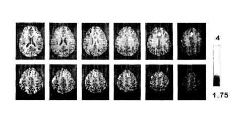
In the "Lie vs. Truth" contrast (Table 1, Fig. 2), there are two clusters of
significant
BOLD signal increase. The first is a 146-voxel cluster extending from the left
anterior
cingulate gyms (ACC) to the medial aspect of the right superior frontal gyms
(SFG),
including BA 24,32, and 8, global activity peak at Talairach {x;y;z}
coordinates {0;21;28}
and local peaks at {4;33;43} and {0;26;47}. The second is a 91-voxel cluster,
U-shaped
along the craniocaudal axis, extending from the border of the prefrontal to
the dorsal
premotor cortex (BA 6, bordering on BA 3 and 4) and also involving the
anterior parietal
cortex from the central sulcus to the lower bank of the intraparietal sulcus
(BA 1-3 to the
edge of BA 40), with a global activity peak at {-63;-17;45} and local peaks at
{-59;-10;41}
and {-55;3;51}. There were no regions with significant signal decrease. See
FIG. 2.
Table 1. Talairach coordinates, gyms (Talairach et al., 1988) and Brodmann
Area (BA)
locations of the peaks of activity within clusters (FIG. 2) of significant
fMRI signal
differences between "Lie" and "Truth" conditions.
Talairach coordinates
Cluster Z x Y z BA Gyms
size
(voxels)
146 3.8 -1 16 29 24;32 Anterior
cingulate
3.17 3 28 43 6;8 Right superior
frontal
3.15 0 24 52 8 Superior frontal
91 3.58 -57 -23 41 1;2;3;40 Left
postcentral
3.40 -54 -15 38 3;4;6 Left pre- and
postcentral
3.19 -50 -3 49 6 Left precentral
17
CA 02449283 2003-12-02
WO 02/102238 PCT/US02/19422
Note. Voxel level threshold T = 2.57, P < 0.001 uncorrected and 0.05 corrected
for
multiple comparisons, spatial extent threshold >80 voxels. Bold numbers
correspond to a
global peak of the cluster; italics represent local peaks within same
contiguous cluster.
Conclusions:
The results demonstrate that there are measurable difference between lying and
telling the truth using event-related fMRI and the GKT model of deception.
This finding
indicates that there is a neurophysiological difference between deception and
truth at the
brain activation level that can be detected with fMRI. The anatomical
distribution of
deception-related activation indicates that deception involves conflict with,
and alteration of,
the prepotent (truthful) response. Further refinements of the paradigm design
and image
analysis methodology involving e.g., testing the effect of handedness,
language or gender, or
creating grades of deception based upon familiarity in the GKT, or testing the
effect of
implemented counter-measures by the subject (such as, nor responding to
questions or
commands in response to the presented stimuli) could further increase the
salience and the
statistical power of the simulated deception paradigms and establish an
activation pattern
predictive of deception on an individual level.
Example 2: Recognition of Familiar Faces.
A conspiracy suspect trying to intentionally deceive an investigator about
being
acquainted with another individual (e.g., a co-conspirator) exhibits two
parameters of brain
function detectable by fMRI. The first is intentional denial of recognizing
the co-conspirator
(or his/her image). The second is response to a familiar face or object, which
is different
from the response to a novel face or object.
Studies of brain activity patterns during facial recognition have shown
significant
differences in the brain response to familiar vs. novel faces as well as the
effect of the degree
of prior familiarity with the displayed face (Haxby, 2002; Glahn et al., 1997;
Henson et al.,
2001; Schlack et al., 2001, Gobbini et al., 2001). Thus, when the principles
of Example 1
are applied to the question of whether an individual recognizes a face or not,
the present data
indicates that when faces are used as stimuli in a GKT type paradigm a
response is as strong
or stronger (in amplitude and/or spatial distribution) than the GKT paradigm
established
with playing cards.
Studies indicate that this effect takes place even in the absence of awareness
[Milner,
1997 #111; Berns et al. Science 276:1272-1275 (1997). Ishai et al., J. Cogn.
Neurosci.
12:35-51 (2000);Haxby et al., Biol. Psychiatry 51:59-67 (2002). Consequently,
the
18
CA 02449283 2003-12-02
WO 02/102238 PCT/US02/19422
principles set forth in the fMRI deception paradigm of Example 1 are
applicable to deception
regarding acquaintanceship and are combinable sequentially or serially with
mapping the
brain activity associated with novel vs. familiar facial or object recognition
without
deception.
Example 3: Brain Response to Media Information.
The principles set forth in the fMRI deception paradigm of Example 1 may also
be
applied to individuals viewing media information, such as movies, video film
clips, or
advertising. Although in this case, rather than examining for deception, the
data is used to
interpret the effect of the information on the individual. This uses the known
patterns of
brain response, e.g., aversive, pleasurable, exciting or memory-evoking
stimuli to adjust
media content to achieve a desirable impact. This study explores the use of
magnetic
resonance signal as a marker of cognitive (e.g., attention) and emotional
(e.g., arousal)
responses to commercial audiovisual media. Subjects are selected and analyzed
as in
Example 1 with certain modifications in the presentation and evaluation of the
signals and
resulting data.
Data acquisition:
Subjects view the baseline media segment (control material) followed by the
target
media segment of same duration. (While randomizing the order of the drug and
neutral
videos would remove the risk of systematic error due to MRI system drift, data
acquired by
the inventors indicates significant carry-over effects from the drug to the
neutral cue). The
target film used depicts two male heroin users engaged in drug-specific
dialogue while
preparing and injecting simulated heroin. The baseline film is a nature film
about the life of
hummingbirds. FIG. 3 depicts an averaging of the rCBF differences between the
brain
response to a movie about heroin use and a movie about hummingbirds in 3
opiate-
dependent patients as determined by with ASL IIVIRI projected over Ti IVIRI in
Talairach
space. Both films have been validated by correlation with skin conductance
response and
used in several previous studies at the inventor's laboratory.
Imaging consists of a sagittal scout scan (TR/TE=500/10 mseconds, 128 x 256, 5
mm
thick, 2minutes), an anatomical scan using 3D inversion recovery (IR) prepared
spoiled
GRASS (TRITE/T1-33/7/400 mseconds, 192 x 256, 124 slices, 1.5 mm thick),
followed by
the fMRI using the arterial spin labeling (ASL) perfusion sequence
(TR/TE=3400/18
mseconds, 64 x 40, 10 slices, 50 mseconds acquisition time/slice, 8mm
thick/2mm sp,
resolution 3.75 x 3.75 x lOmm, FOV 24cm, 180 repetitions, 10mins). The ASL
sequence
19
CA 02449283 2003-12-02
WO 02/102238 PCT/US02/19422
consists of interleaved global (control) and slice-selective (label) inversion
recovery gradient
echo echoplanar acquisitions. A specific sharp-edge pulse (FOCI) is applied
for spin
labeling to minimize the system error between acquisitions. The duration of
the tagging
bolus is defined by playing out a saturation pulse at the tagging region at
800 ms after the
FOCI pulse, followed by a 1-second post-labeling delay before image
acquisition. The total
time in the scanner is about 30 minutes. Heart rate is obtained continuously
and sampled
every 30 seconds with a pulse oxymeter attached to subject's finger.
Assessment of the desire to use drugs depicted in the target segment and other
subjective feelings, such as aversion, sexual arousal and remembering, are
performed at
fixed intervals or continuously throughout the session. Subjects use a
response pad with
multiple buttons, which permit them to communicate the degree to which they
experience
the above feelings to the investigator. Additional parameters such as skin
conductance,
penile tumescence, heart and respiratory rate and blood pressure are also
collected as needed.
Procedures:
=
After informed written consent, subjects are placed in the scanner. Video
segments
are projected onto a screen at the subject's feet and viewed with the aid of
prism glasses
attached to the inside of the radio frequency head coil. The sound is
delivered by air
conduction through plastic tubes threaded through earplugs that attenuate
scanner noise.
Videos are 10 minutes in duration, and are preceded and followed by a 4-minute
blank gray
screen during which VAS is administered and MRI is halted. VAS is used to
index the
change in cue-induced heroin craving. Subjects respond using a fibro-optic
response pad.
Table 2. MM session timeline indicating onset of the variables in terms of
time elapsed from
beginning of the imaging session. (x) indicates alternative (counterbalanced)
order.
Elapsed time 0 6 16 20 30
(min)
Structural MRI x
fiVIRI
Target
segment
Non-target
segment
Subjective
symptoms
CA 02449283 2003-12-02
WO 02/102238 PCT/US02/19422
Data Analysis:
Data is reconstructed offline, corrected for motion artifacts and smoothed
using
SPM99' (28, http://www.fillon.ucLac.uld). The series of label images are
shifted in time by
one TR using linear or sync interpolation. Perfusion contrast images are
generated by
pairwise subtraction between the time-matched label and control images. FIG. 4
depicts the
correlation between the change in the desire to use heroin and the change in
rCBF in the
midbrain area. Conversion to CBF values are effected using the general PASL
perfusion
model. CBF signals during the drug and non-drug video are compared within
subjects using
SPM99.
Individual activation maps (either beta or correlation coefficient) are
normalized to
Talairach space and correlated with methadone plasma levels and the heart rate
to detect the
brain areas associated with opiate craving and physiological parameters within
both the
patients and the controls. ANOVA analysis is performed on the normalized
individual data
to study the effects of drug cue and testing population, followed by region-of-
interest
analysis to further study the temporal evolvement of the time-course of the
CBF change in
these detected brain regions.
Results:
1) Media segment of high emotional value for the target population elicits a
different brain response than a media segment of neutral value in the
midbrain, the thalamus,
the insula and the amygdala. This effect was not observed in control subjects
who were not
addicted to heroin, nor in brain regions that were not involved in the
mediation of the reward
and motivation, such as the occipital cortex.
2) Brain response in some of these regions (midbrain) is correlated with the
subjective emotions of the audience.
3) Perfusion fMRI, at 4-T is a promising technique for the study of media
impact on
target populations, as well as individuals.
The method herein described is, therefore, useful for the effective
manipulation of the
content of the media segments to achieve maximal desired impact in target
populations or on
specific individuals.
Each and every patent, patent application and publication that is cited in the
foregoing specification is herein incorporated by reference in its entirety.
21
CA 02449283 2003-12-02
WO 02/102238 PCT/US02/19422
While the foregoing specification has been described with regard to certain
preferred
embodiments, and many details have been set forth for the purpose of
illustration, it will be
apparent to those skilled in the art that the invention may be subject to
various modifications
and additional embodiments, and that certain of the details described herein
can be varied
considerably without departing from the spirit and scope of the invention.
Such
modifications, equivalent variations and additional embodiments are also
intended to fall
within the scope of the appended claims.
22