Note: Descriptions are shown in the official language in which they were submitted.
CA 02449290 2003-12-02
WO 02/098914 PCT/EP02/06161
Mutants of IGF binding proteins and methods of production of
antagonists thereof
The present invention relates to a complex of an IGF binding protein fragment
(IGFBP) with IGF, its uses and to novel IGFBP mutants with a higher affinity
than
natural IGFBPs for IGF as well as to methods for the production of antagonists
for
IGFBPs which hinder or reverse complex formation between IGFBPs and IGF.
Introduction
Insulin-like growth factors I and II (hereafter also referred to as IGFs or
IGF) are
members of the insulin superfamily of hormones, growth factors and
neuropeptides
whose biological actions are achieved through binding to cell surface
receptors. IGF
actions are regulated by IGF binding proteins (IGFBPs) that act as
transporters of
IGFs, protect them from degradation, limit their binding to receptors, and
maintain
a "reservoir" of biologically inactive IGF (Martin, J.L., and Baxter, R.C.,
IGF binding
proteins as modulators of IGF actions; in: Rosenfeld, R.G., and Roberts, C.T.
(eds.),
The IGF system, Molecular Biology, Physiology, and Clinical Applications (
1999),
Humana Press, Totowa, pp. 227-255; Jones, J.L., and Clemmons, D.R., Endocr.
Rev. 12 (1995) 10-21; Khandwala, H.M., et al., Endocr. Rev. 21 (2000) 215-244;
Hwa, V., et al., The IGF binding protein superfamily, In: Rosenfeld, R.G., and
Roberts, C.T. (eds.), The IGF system, Molecular Biology, Physiology, and
Clinical
Applications ( 1999), Humana Press, Totowa, pp. 315-327). The IGF and growth
hormone (GH) axis plays a large part in regulating fetal and childhood somatic
growth and several decades of basic and clinical research have demonstrated
that it
also is critical in maintaining neoplastic growth (Khandwala, H.M., et al.,
Endocr.
Rev. 21 (2000) 215-244). High circulating IGF-I concentrations may also be an
important determinant of cancer incidence (Hankinson, S.E., et al., Lancet 351
(1998) 1393-1396; Holly, J., Lancet 351 (1998) 1373-1374; Wolk, A., Lancet 356
(2000) 1902-1903). Virtually every level of the IGF system mediating response
on
the tumor tissues (IGFs, IGFBPs, IGF receptors) can be targeted for
therapeutic
approaches (Khandwala, H.M., et al., Endocr. Rev. 21 (2000) 215-244; Fanayan,
S.,
et al., J. Biol. Chem. 275 (2000) 39146-39151; Imai, Y., et al., J. Biol.
Chem. 275
(2000) 18188-18194). It should also be mentioned here that IGFBP-3 has IGF-
independent anti-proliferative and proapoptotic effects (Wetterau, L.A., et
al., Mol.
CA 02449290 2003-12-02
WO 02/098914 PCT/EP02/06161
-2-
Gen. Metab. 68 (1999) 161-181; Butt, A.J., et al., J. Biol. Chem. 275 (2000)
39174-
39181).
IGF-I and IGF-II are 67% identical single polypeptide chains of 70 and 67
amino
acids, respectively, sharing with insulin about 40% sequence identity and
presumed
structural homology. The first 29 residues of IGFs are homologous to the B-
chain
of insulin (B region, 1-29), followed by 12 residues that are analogous to the
C-
peptide of proinsulin (C region, 30-41), and a 21-residue region that is
homologous
to the A-chain of insulin (A region, 42-62). The carboxy-terminal octapeptide
(D
region, 63-70) has no counterpart in insulins and proinsulins (Murray-Rust,
J., et
al., BioEssays 14 (1992) 325-331; Baxter, R.C., et al., J. Biol. Chem. 267
(1992) 60-
65). The IGFs are the only members of the insulin superfamily in which the C
region is not removed proteolytically after translation. The 3D structure of
insulin
has been studied intensively since the first crystal structure determination
in the
1960s (Adams, M.J., et al., Nature 224 (1969) 491-492). There are now
structures of
insulins in several oligomeric states, for insulins crystallized in different
solvent
conditions, and for many variants from different species and chemical
modifications. This is in stark contrast to IGFs (and proinsulins) for which
no high
definition structure has been available prior to this report. Instead, the
tertiary
structure of IGF-I has been modeled after porcine insulin (Blundell, T.L.,
Proc.
Natl. Acad. Sci. USA 75 (1978) 180-184). 2D NMR studies of IGF-I have
confirmed
that the solution structure is consistent with the model (Cooke, R.M., et al.,
Biochemistry 30 ( 1991 ) 5484-5491; Sato, A., et al., Int. J. Pept. Protein
Res. 41
(1993) 433-440). However, NMR studies of IGF-I have yielded structures only of
low resolution, probably because IGF-I is soluble at the concentrations
required for
NMR only at pH values below 3 (Cooke, R.M., et al., Biochemistry 30 (1991)
5484-
5491; Sato, A., et al., Int. J. Pept. Protein Res. 41 (1993) 433-440). More
recently,
better defined structures have been obtained for IGF-II (Terasawa, H., et al.,
EMBO
J. 13 ( 1994) 5590-5597; Torres, A.M., et al., J. Mol. Biol. 248 ( 1995) 385-
401 ) and
for a Glu-3 to Arg variant of IGF-I (long-[Arg3]IGF-I) that additionally
possesses a
13-amino acid extension at the N-terminus (Laajoki, L.G., et al., J. Biol.
Chem. 275
(2000) 10009-10015).
IGFBPs (insulin-like growth factor binding proteins -1 to -6) are proteins of
216 to
289 residues, with mature IGFBP-5 consisting of 252 residues (Wetterau, L.A.,
et
al., Mol. Gen. Metab. 68 (1999) 161-181). All IGFBPs share a common domain
CA 02449290 2003-12-02
WO 02/098914 PCT/EP02/06161
-3-
organization. The highest conservation is found in the N- (residues 1 to ca.
100)
and C- (from residue 170) terminal cysteine rich regions. Twelve conserved
cysteines are found in the N-terminal domain and six in the C-terminal domain.
The central, weakly conserved part (L-domain) contains most of the cleavage
sites
for specific proteases (Chernausek, S.D., et al., J. Biol. Chem. 270 (1995)
11377-
11382). Several different fragments of IGFBPs have been described and
biochemically characterized so far (Mazerbourg, S., et al., Endocrinology 140
(1999) 4175-4184). Mutagenesis studies suggest that the high affinity IGF
binding
site is located in the N-terminal domain (Wetterau, L.A., et al., Mol. Gen.
Metab. 68
(1999) 161-181; Chernausek, S.D., et al., J. Biol. Chem. 270 (1995) 11377-
11382)
and that at least IGFBP-3 and IGFBP-2 contain two binding determinants, one in
the N- and one at the C-terminal domains (Wetterau, L.A., et al., Mol. Gen.
Metab.
68 (1999) 161-181). Recently, a group of IGFBP-related proteins (IGFBP-rPs)
which bind IGFs with lower affinity than IGFBPs have been described (Hwa, V.,
et
al., The IGF binding protein superfamily, In: Rosenfeld, R.G., and Roberts,
C.T.
(eds.), The IGF system, Molecular Biology, Physiology, and Clinical
Applications
(1999), Humana Press, Totowa, pp. 315-327). IGFBPs and IGFBP-rPs share the
highly conserved and cysteine-rich N-terminal domain which appears to be
crucial
for several biological actions, including their binding to IGFs and high
affinity
binding to insulin (Hwa et al., 1999). N-terminal fragments of IGFBP-3,
generated
for example by plasma digestion, also bind insulin and physiologically are
thus
likely relevant for insulin action. Beyond the N-terminal domain, there is a
lack of
sequence similarity between the IGFBPs and IGFBP-rPs.
The sequences of human IGFBP-1 to -6 are described in detail in the SwissProt
Database (http://www.expasy.ch) and identified by the following Accession
Nos.:
Name Accession No.
IGFBP-1 P 08833
IGFBP-2 P 18065
IGFBP-3 P 17936
IGFBP-4 P 22692
IGFBP-5 P 24593
IGFBP-6 P 24592
CA 02449290 2003-12-02
WO 02/098914 PCT/EP02/06161
-4-
The amino acid positions described in the following refer to the sequence of
the
mature forms the human IGF binding proteins (sequence after removal of the
signaling peptide starts with amino acid in position 1, see also Tables 1 to
6).
The association of insulin-like growth factors with neoplasia indicates that
inhibition of the IGF signaling pathway in tumors might be a successful
strategy in
cancer therapy. Such modulation might be accomplished, for example, through
exogenous administration of recombinant inhibitory IGFBPs and effective
fragments thereof. Additionally, tumor cell IGFBP production, inhibition or
degradation may be controlled by agents such as tamoxifen and ICI 182,780 that
modify tumor IGFBP production (Khandwala, H.M., et al., Endocr. Rev. 21 (2000)
215-244). The consequent alteration in IGFBP-3 levels appears in certain
instances
to inhibit IGF-I-stimulated cell proliferation (Khwandala et al., 2000). There
is also
recent evidence that IGFBP-3 may be a p53-independent effector of apoptosis in
breast cancer cells via its modulation of the Bax:Bcl-2 protein ratio (Butt,
A.J., et al.,
J. Biol. Chem. 275 (2000) 39174-39181; Wetterau, L.A., et al., Mol. Gen.
Metab. 68
(1999) 161-181).
IGFBPs show a significant inhibition of tumor cell proliferation in vitro but
only
very high doses result in inhibition of tumor growth in vivo (van den Berg,
C.L., et
al., Eur. J. Cancer 33 (1997) 1108-1113). Van den Berg therefore covalently
coupled
IGFBP-1 to polyethylene glycol, which leads to a prolonged serum half life.
However, the inhibitory effects of the pegylated IGFBP-1 is still not
sufficient for
therapeutic intervention in humans because only partial response is observed
even
if pegylated IGFBP-1 is given in doses of 1 mg/dose daily in mice. This
corresponds to a dose of 50 mg/kg x day which can not be administered to
humans
by established procedures and can not be produced economically.
IGF releasing peptides are described by Loddick, S.A., et al., Proc. Natl.
Acad. Sci.
USA 95 ( 1998) 1894-1898 and Lowman, H.B., et al., Biochemistry 37 ( 1998)
8870-
8878. The described molecules which are able to displace IGFs from their
binding
proteins are either mutants of IGF-I which bind to IGFBPs but are not able to
stimulate the IGF-IR or a 14 amino acid peptide with similar properties
derived
from a phage-display library. The biological activities of the peptides were
shown by
administration either by injection into the lateral ventricle of the brain or
infused
by a minipump.
CA 02449290 2003-12-02
WO 02/098914 PCT/EP02/06161
-5-
Mutagenesis studies for IGFs indicated that IGF amino acid residues Glu 3, Thr
4,
Gln 15 and Phe 16 of IGF-I and Glu 6, Phe 48, Arg 49 and Ser 50 in IGF-II are
important for binding to IGFBPs (Baxter, R.C., et al., J. Biol. Chem. 267
(1992) 60-
65; Bach, L.A., et al., J. Biol. Chem. 268 (1993) 9246-9254; Luethi, C., et
al., Eur. J.
Biochem. 205 ( 1992) 483-490; Jansson, M., et al., Biochemistry 36 ( 1997)
4108-
4117). Baxter et al. (1992) suggested that the IGF-I amino acid residues Glu
3, Thr
4, Gln 15 and Phe 16 are crucial for interaction with IGFBP-3, whereas
residues Phe
49, Arg 50 and Ser 51 are of secondary importance. It also was suggested that
Phe
26 of IGF-II plays a role in changing the local structures of IGFs but does
not
10~ directly bind to IGFBPs (Terasawa, H., et al., EMBO J. 13 ( 1994) 5590-
5597).
Kalus, W., et al., in EMBO J. 17 (1998) 6558-6572, describe proteolytic
studies of
human IGFBP-5 and the cloning and expressing of the domain of IGFBP-5 between
residues 40-92 (mini-IGFBP-5); this domain binds IGF-I and IGF-II with KD
values
of 37 nM and 6 nM, respectively, as well as the determination of the solution
structure of uncomplexed mini-IGFBP-5 by NMR. Kalus et al. identified some IGF
binding sites which are residues Va149, Tyr50, Pro62 and Lys68 to Leu75 of
IGFBP-5.
Imai, Y., et al., in J. Biol. Chem. 275 (2000) 18188-18194, describe an IGFBP-
3
variant and an IGFBP-5 variant, each with a five-fold substitution pattern at
amino
acid positions hypothesized by Kalus et al. as IGF binding sites. Imai et al.
found
that a substantial alteration of the amino acid residues simultaneously at
positions
68, 69, 70, 73 and 74 results in a 1000-fold or larger reduction in the
affinity for
IGF-I in relation to the affinity of wild-type IGFBP-5.
Conover, C.A., et al., in J. Biol. Chem. 270 ( 1995) 4395-4400, describe
protease-
resistant mutants of IGFBP-4. All four IGFBP-4 mutants around the putative
cleavage site at Met135-Lys136 and the wild-type protein bind IGFs with
equivalent
affinities.
Byun, D., et al., in J. Endocrinology 169 (2001) 135-143, postulate several
regions
involved in IGF binding by IGFBP-4. Deletion of segments Leu72-Ser 91 or Leu72
His74 results in loss of IGF binding. Also mutation of certain cysteine
residues
significantly reduces the binding of IGFs.
CA 02449290 2003-12-02
WO 02/098914 PCT/EP02/06161
-6-
Thus, these described mutant forms of insulin-like growth factor binding
proteins
have reduced or equivalent affinities for IGF-I and/or IGF-II. Mutants of
IGFBPs
with a significantly higher affinity and a therefore improved effectiveness
have not
been known heretofore and there exists a need for such molecules as well as
for
methods for identifying IGFBP antagonists.
Summary of the Invention
The invention provides a crystal suitable for X-ray diffraction, comprising a
complex of insulin-like growth factor I or II and a polypeptide consisting of
the
amino acids 39-91 of IGFBP-1, the amino acids 55-107 of IGFBP-2, the amino
acids
47-99 of IGFBP-3, the amino acids 39-91 of IGFBP-4, the amino acids 40-92 of
IGFBP-5, or the amino acids 40-92 of IGFBP-6 or a fragment thereof consisting
at
least of the 9'}' to 12~' cysteine of IGFBP-l, IGFBP-2, IGFBP-3, IGFBP-4, or
IGFBP-5 or at least of the 7'~ to 10'1' cysteine of IGFBP-6 (such polypeptides
and
fragments are hereinafter also referred to as "mini-IGFBPs).
Such a crystal is suitable for determining the atomic coordinates of the
binding sites
of IGF-I, IGF-II, and IGFBPs, and therefore allows the optimization of these
molecules to identify and improve stabilizing interactions between IGF-I or
IGF-II
and IGFBPs. Preferably, the crystal effectively diffracts X-ray for the
determination
of the atomic coordinates of said complex to a resolution of 1.5 to 3.5 ~. The
crystal
is arranged in the cubic space group P213 having unit cell dimensions of
74.385 t1 x
74.385 t~ x 74.385 A.
The invention further provides a method for producing a crystal suitable for X-
ray
diffraction, comprising
(a) contacting IGF-I or IGF-II with a polypeptide consisting of the amino
acids
39-91 of IGFBP-1, the amino acids 55-107 of IGFBP-2, the amino acids 47-99
of IGFBP-3, the amino acids 39-91 of IGFBP-4, the amino acids 40-92 of
IGFBP-5, or the amino acids 40-92 of IGFBP-6 or a fragment thereof
consisting at least of the 9'1' to 12'}' cysteine of IGFBP-1, IGFBP-2, IGFBP-
3,
IGFBP-4, or IGFBP-5 or at least of the 7'1' to 10'1' cysteine of IGFBP-6, to
form
a complex which exhibits restricted conformation mobility, and
(b) obtaining a crystal from the complex so formed suitable for X-ray
diffraction.
CA 02449290 2003-12-02
WO 02/098914 PCT/EP02/06161
_7_
Using this crystal, the atomic coordinates of the complex can be determined.
The invention further comprises a method for identifying a mutant of IGFBP or
a
mutant of a fragment thereof consisting at least of the 9~' to 12'h rysteine
of
IGFBP-1, IGFBP-2, IGFBP-3, IGFBP-4, or IGFBP-5 or at least of the 7'h to 10'h
rysteine of IGFBP-6, and having enhanced binding affinity for IGF-I and/or IGF-
II
comprising
(a) constructing a three-dimensional structure of the complex of IGF-I or IGF-
II
and a polypeptide consisting of the amino acids 39-91 of IGFBP-l, the amino
acids 55-107 of IGFBP-2, the amino acids 47-99 of IGFBP-3, the amino acids
39-91 of IGFBP-4, the amino acids 40-92 of IGFBP-5, or the amino acids 40-
92 of IGFBP-6 or a fragment thereof consisting at least of the 9th to 12'h
cysteine of IGFBP-1, IGFBP-2, IGFBP-3, IGFBP-4, or IGFBP-5 or at least of
the 7~h to 10'~ cysteine of IGFBP-6, based on the atomic coordinates of a
crystal consisting of IGF-I or IGF-II and said polypeptide;
(b) employing said three-dimensional structure and modeling methods to
identify said mutant in which an amino acid residue within a distance of 5 t1
to a hydrophobic amino acid residue of IGF-I or IGF-II is modified in that
the hydrophobic interaction between IGF-I or IGF-II and said mutant of
IGFBP is enhanced;
(c) producing said mutant;
(d) assaying said mutant to determine said enhanced binding affinity for IGF.
The invention further comprises a method for identifying a mutant of IGFBP-5
with enhanced binding affinity for IGF-I, said method comprising
(a) constructing a three-dimensional structure of the complex of IGF-1 and
IGFBP-5 defined by the atomic coordinates shown in Figs. 5 and 6;
(b) employing said three-dimensional structure and modeling methods to
identify an amino acid residue in IGFBP-5 within a distance of 5 t1 or shorter
to an amino acid residue of IGF-I, wherein said residue of IGFBP-5 can be
modified hydrophobically in that the hydrophobic interaction between IGF
and IGFBP-5 is enhanced;
(c) producing said mutant;
CA 02449290 2003-12-02
WO 02/098914 PCT/EP02/06161
_g_
(d) assaying said mutant to determine said enhanced binding affinity for IGF.
The amino acid residues) in which IGFBP(s) is/are modified is/are preferably
selected from the amino acids 39-91 of IGFBP-l, the amino acids 55-107 of
IGFBP-2, the amino acids 47-99 of IGFBP-3, the amino acids 39-91 of IGFBP-4,
the
amino acids 49-92 of IGFBP-5, or the amino acids 40-92 of IGFBP-6.
Especially preferred IGFBP mutants are modified at amino acid residues 49, 70
and/or 73 corresponding to IGFBP-5 sequence alignment and according to Table
7.
The invention therefore provides mutant IGFBPs ("IGFBPs" as used herein means
IGFBP-1, IGFBP-2, IGFBP-3, IGFBP-4, IGFBP-5 and/or IGFBP-6) with enhanced
affinity (preferably about 3-fold to 10-fold increased affinity to the
corresponding
wild-type IGFBP) for IGF ("IGF" as used herein means IGF-I and/or IGF-II),
improved .inhibitory potenry for the activity of IGF in vitro and in vivo and
therefore improved therapeutic effectiveness.
The invention further provides a method for identifying a compound capable of
binding to IGFBP, comprising
(a) constructing a three-dimensional structure of a complex of IGF-I or IGF-II
and a polypeptide consisting of the amino acids 39-91 of IGFBP-1, amino
acids 55-107 of IGFBP-2, amino acids 47-99 of IGFBP-3, amino acids 39-91
of IGFBP-4, amino acids 40-92 of IGFBP-5, amino acids 40-92 of IGFBP-6 or
a fragment thereof consisting at least of the 9'~ to 12~' cysteine of IGFBP-1,
IGFBP-2, IGFBP-3, IGFBP-4, or IGFBP-5 or at least of the 7'1' to 10'}'
rysteine
of IGFBP-6, based on the atomic coordinates of a crystal consisting of IGF - I
and said IGFBP;
(b) employing said three-dimensional structure and modeling methods to
identify a compound forming a complex with said IGFBP by hydrophobic
binding with amino acids 49, 50, 70, 71 and 74 in the case of IGFBP-5 and in
the case of IGFBP-1, IGFBP-2, IGFBP-3, IGFBP-4 and IGFBP-6 with
corresponding amino acids according to Table 7;
(c) producing said compound;
(d) determining the binding between the compound and IGFBP.
CA 02449290 2003-12-02
WO 02/098914 PCT/EP02/06161
-9-
The invention further provides a method of inhibiting the binding of IGF to
the
IGFBP in a subject, preferably a human subject, comprising administering an
effective amount of an above-described mutant of IGFBP to the subject.
Detailed Description of the Invention
The present invention provides methods for co-crystallizing IGF-I or IGF-II
with a
truncated N-terminal fragment of IGFBP, preferably of IGFBP-5 (mini-IGF),
where
the crystals diffract X-rays with sufficiently high resolution to allow
determination
of the three-dimensional structure of said complex, including atomic
coordinates.
The three-dimensional structure (e.g. as provided on computer-readable media)
is
useful for rational drug design of IGFBP mutants with modified affinity for
IGF-I
or IGF-II, preferably with an improved affinity. There is specifically
provided a
method for co-crystallizing IGF-I with a polypeptide consisting of an isolated
folded domain of IGFBPs (mini-IGFBPs), which is formed by the amino acids
between the 9'~ and the 12~' cysteine of IGFBP-1 to IGFBP-5 or the 7~' and
10th
cysteine of IGFBP-6 and additionally including up to 7 amino acids N-terminal
of
this fragment and up to 5-20 amino acids C-terminal to this fragment. The
amino
acids 39-9lof BP-1, the amino acids 55-107 of IGFBP-2, the amino acids 47-99
of
IGFBP-3, the amino acids 39-91 of IGFBP-4, the amino acids 40-92 of IGFBP-5,
or
the amino acids 40-92 of IGFBP-6 or fragments thereof are especially suitable
to
form a complex with IGF-I or IGF-II which exhibits restricted conformational
mobility and high suitability for X-ray diffraction.
Such a complex co-crystallizes in a manner sufficient for the determination of
atomic coordinates by X-ray diffraction. The crystal effectively diffracts X-
ray for
the determination of the atomic coordinates of the complex to a resolution of
1.5 or
at least better (less) than 3.5 fir. Said IGFBP fragments are able to form a
compact
and globular structure whose scaffold is secured by an inside packing of two
rysteine bridges and stabilized further by a three-stranded f3-sheet. The
folded
fragments are still able to bind IGF-I and IGF-II with high affinities. Other
forms of
the IGFBPs such as full-length IGFBPs, the isolated C-terminal domain of
IGFBPs
or fragments without N-terminal truncation do not co-crystallize with IGF in a
suitable manner for X-ray-based determination of the structure at high
resolution.
CA 02449290 2003-12-02
WO 02/098914 PCT/EP02/06161
-10-
Knowledge of the crystal structure enables the production of specific IGFBP
mutants which develop improved interaction with, thereby exhibiting enhanced
affinity for, IGF and, as a consequence, have improved therapeutic efficacy as
IGF
antagonists. Such IGFBP mutants with increased affinity for IGF are capable of
preventing the formation of the complex between naturally occurring IGF and
IGF-
I receptor (IGF-IR) in vitro and in vivo and, thereby, of effecting an
decrease in the
concentration of biologically active, free IGF. Such rational designed IGF
antagonists are therefore capable of inhibiting tumor growth and inducing
apoptosis in tumor cells more efficient than natural IGFBPs. As a result,
lower
doses of the optimal designed IGFBP mutants with enhanced affinity are needed
for
achieving an effect comparable to that of naturally occurring IGFBPs.
A further embodiment of the invention is the identification and optimization
of
non-proteinaceous compounds which bind to the IGF binding site of IGFBPs and
prevent the formation of an inhibitory complex between IGFs and IGFBPs and
therefore activates the anabolic action of IGF. Such "IGF-releasing compounds"
can
be identified according to the invention on the basis of the crystal data,
using
protein-ligand docking programs such as FlexX (Kramer, B., et al., Proteins:
Structure, Functions and Genetics 37 ( 1999)' 228-241 ).
The X-ray diffraction patterns of the invention have a sufficiently high
resolution to
be useful for three-dimensional modeling of an IGF releasing compound.
Preferably, the resolution is in the range of 1.5 to 3.5 t1, preferably 1.5 to
3.0 t~.
Three-dimensional modeling is performed using the diffraction coordinates from
these X-ray diffraction patterns. The coordinates are entered into one or more
computer programs for molecular modeling as known in the art. Such molecular
modeling can utilize known X-ray diffraction molecular modeling algorithms or
molecular modeling software to generate atomic coordinates corresponding to
the
three-dimensional structure of at least one IGF releasing compound.
Such a compound shows affinity for IGFBP based on stereochemical
complementary relative to naturally occurring IGFs. Such stereochemical
complementary according to the present invention is characterized by a
molecule
that matches intra-site surface residues that form the contours of IGFBPs as
enumerated by the coordinates set out in Figs. 5 and 6. The residues that
define the
contours are depicted in Figs. 5 and 6. Matching according to the invention
means
CA 02449290 2003-12-02
WO 02/098914 PCT/EP02/06161
-11-
that the identified atoms or atom groups interact with the IGFBP surface
residues,
for example via hydrogen bonding or by enthalpy-reduced van der Waals
interactions which prevent or reduce the interaction between IGFBP and IGFs
and
thereby promote the release of the biologically active compound from the
binding
site. In general, the design of a molecule possessing stereochemical
complementary
to the contours of IGFBPs can be accomplished by means of techniques that
optimize either chemically or geometrically the fit between a molecule and a
target
receptor. Known techniques of this sort are reviewed by Sheridan, R.P., and
Venktaraghavan, R., Acc. Chem. Res. 20 ( 1987) 322; Goodford, P.J., J. Med.
Chem.
27 ( 1984) 557; Verlinde, C., and Hol, W., Structure 2 ( 1994) 577; and
Blundell, T.L.
et al., Nature 326 (1987) 347. The design of optimized IGFBP ligands based on
the
invention is preferably done by the use of software such as GRID (Goodford,
P.J., J.
Med. Chem. 28 (1985) 849-857), a program that determines probable interaction
sites between probes with various functional group characteristics and the
protein
surface - is used to analyze the surface sites to determine structures of
similar
inhibiting proteins or compounds.
The program DOCK (Kuntz, LD., et al., J. Mol. Biol. 161 (1982) 269-288) can
also
be used to analyze an active site or ligand binding site and suggest ligands
with
complementary steric properties. Several methodologies for searching three-
dimensional databases to test pharmacophore hypotheses and select compounds
for
screening are available. These include the program CAVEAT (Bacon et al., J.
Mol.
Biol. 225 ( 1992) 849-858) which uses databases of ryclic compounds which can
act
as spacers to connect any number of chemical fragments already positioned in
the
active site. The program LUDI (Bohm, H.J., et al., J. Comput. Aided Mol. Des.
6
(1992) 61-78 and 593-606) defines interaction sites into which both hydrogen
bonding and hydrophobic fragments fit.
Programs suitable for searching three-dimensional databases to identify also
non-
proteinaceous molecules bearing a desired pharmacophore include: MACCS-3D
and ISIS/3D (Molecular Design Ltd., San Leandro, CA), ChemDBS-3D (Chemical
Design Ltd., Oxford, U.K.), and Sybyl/3DB Unity (Tripos Associates, St. Louis,
MO).
CA 02449290 2003-12-02
WO 02/098914 PCT/EP02/06161
-12-
Programs suitable for pharmacophore selection and design include: DISCO
(Abbott Laboratories, Abbott Park, IL), Catalyst (Bio-CAD Corp., Mountain
View,
CA), and ChemDBS-3D (Chemical Design Ltd., Oxford, U.K.).
Databases of chemical structures are available from a number of sources
including
Cambridge Crystallographic Data Centre (Cambridge, U.K. ) and Chemical
Abstracts Service (Columbus, OH).
De novo design programs include Ludi (Biosyrn Technologies Inc., San Diego,
CA),
Sybyl (Tripos Associates) and Aladdin (Daylight Chemical Information Systems,
Irvine, CA).
Those skilled in the art will recognize that the design of such compounds may
require slight structural alteration or adjustment of a chemical structure
designed
or identified using the methods of the invention.
Non-proteinaceous compounds and IGFBP mutants with increased binding affinity
for IGF can be identified by incubating said compounds or mutants with an
IGF-I/IGFBP-5 complex and measuring the binding of released IGF-I to IGF-I
receptor expressing cells. Due to the binding of IGF-I to its cell-bound
receptor, the
receptor is activated and autophosphorylated. Alternatively, radiolabeled IGF-
I can
be used and its binding to its receptor after release from the complex can be
determined.
Formation of the IGF-I mini-IGFBP-5 complex buries a binding surface totalling
about 550 ~rZ. Of the eleven IGFBP-5 amino acid residues within 5 t~ of IGF,
six are
hydrophobic, three of which are surface-exposed leucines and valines and are
of
primary importance for hydrophobic interaction to IGFs (Figures 1 to 4). On
the
IGF side, four of the eleven amino acid residues within 5 1~ of mini-IBFBP-5
are
hydrophobic (Figures 1 to 4).
The IGFBPs bind to IGF-I and IGF-II by presenting a binding surface
complementary to that of IGF. The IGF binding surface consists of a relatively
flat
hydrophobic surface, a small hydrophobic depression, two hydrophobic
protruberances, and surrounding polar residues. Identification of the IGF
binding
surface itself (Figure 3) enables the design of binding partners in general,
and
CA 02449290 2003-12-02
WO 02/098914 PCT/EP02/06161
-13-
optimization of known binding partners in particular. General binding partners
will have at least two of the following features 1 to 4:
1. Non-polar atoms lying approximately in a plane defined by atoms Leu74 CD 1
and CD2, Va149 CG1 and CG2, Leu70 CB, and Tyr 50 CB, within a perimeter
defined by IGF residues Glu9, Glu3, Leu54, Phe 16 and by BP5 atom Tyr 50
OH and depicted in Figure 3 such that they present an approximately planar
and hydrophobic molecular surface of at least 20 square Angstroms.
2. A non-polar atom or atoms near the positions of Leu 70 CG, CD1 relative to
IGF, filling the depression of IGF as seen in the complex structure.
3. Hydrophobic and/or aromatic interactions with the side chains of Phel6,
Va117, and/or Leu54 of IGF as defined by a net buried surface area in the
complex of at least 20 square Angstroms.
4. Polar (hydrogen bonding and/or charge complementary) interactions, either
directly or via bridging solvent molecules, with one or more of the following
IGF atoms: Aspl2 OD1,2; Glu9 OE1,2; Glu3 OE1,2; G1u58 OE1,2; Thr4
O,OGI; Cys520; Ser51 OG; Asp530D1,2; Arg55NH1,2,NE; Arg21NH1,2,NE;
Va1170; Cys180; Asp200D1,2,N; G1n150,OD1,ND2.
Abbreviations: Letters corresponding to standard amino acid atom naming
(according to the International Union of Physicists and Chemists-IUPAC-
naming).
CG: Carbon Cy
CB: Carbon C(3
OE: Oxygen OE
OH: Oxygen O'q
OD: Oxygen 08
O: Backbone Oxygen
NH: Nitrogen N~
NE: Nitrogen NE
N: Backbone Nitrogen
ND: Nitrogen Nb
CA 02449290 2003-12-02
WO 02/098914 PCT/EP02/06161
-14-
The principal IGF/IGFBP interaction, shown in the example of IGF-I mini-IGFBP-
interaction, is a hydrophobic sandwich that consists of interlaced protruding
side chains of IGF-I and solvent exposed hydrophobic side chains of the mini-
s IGFBP-5 (Figures 1 to 4). The side-chains of IGF-I Phe 16, Leu 54 and also
Glu 3,
are inserted deep into a cleft on the mini-IGFBP-5 (Figures 1 to 4). This
cleft is
formed by side chains of Arg 53, Arg 59 on the solvent exposed side of the
molecule
and by Val 49, Leu 70, Leu 74 on the opposite inner side, with a base formed
by
residues Cys 60 and Leu 61. Phe 16 makes direct contacts with the backbone and
side chain of Val 49, and with Cys 60 of mini-IGFBP-5. The hydrophobic cluster
is
closed on the solvent side by side chains of Glu 3 and Glu 9 of IGF-I and His
71 and
Tyr 50 of mini-IGFBP-5. These residues form a network of hydrogen bonds; in
addition Arg 59 of mini-IGFBP-5 makes hydrogen bonds with Glu 58 (Figures 2 to
4).
Arg 53 and Arg 59 of mini-IGFBP-5 isolate the hydrophobic sandwich from the
solvent close to the C-terminus. In the full length IGFBP-5, the segment
corresponding to the C-terminus of mini-IGFBP-5 is followed by nine
hydrophilic
residues and then by at least 30 residues of mixed types. Thus, the
conformations
seen in the structure of the complex near the C-terminus of mini-IGFBP-5 are
likely to be preserved in the complex of IGF-I with the full length-IGFBP-5.
The
mini-IGFBP-5 domain begins preferably at residue 40 of full length IGFBP-5.
The hydrophobic residues Val 49, Leu 70 and Leu 73 of IGFBP-5 are crucial for
binding to IGFs. Since these residues are highly conserved among all IGFBPs,
these
hydrophobic interactions dominate the IGF binding properties of all IGFBPs.
The increased inhibitory potency of the mutant IGFBPs and fragments thereof
results in the inhibition of the binding to and autophosphorylation of the IGF-
IR
(as described by Kalus, W., et al., in EMBO J. 17 (1998) 6558-6572) at
significantly
lower concentrations than observed for the wildtype proteins and the
corresponding fragments. The higher potency of the mutant IGFBPs furthermore
can be shown by the inhibition of the growth of tumor cells in vitro and in
vivo.
The growth of several tumor cell lines is known to be significantly reduced by
IGFBPs. IGFBP-1 for example inhibits the growth of MCF-7 and MDA-MB-435A
cells in vitro and the growth of tumors formed MDA-MB-231 cells in vivo in
mice
CA 02449290 2003-12-02
WO 02/098914 PCT/EP02/06161
-15-
(van den Berg, C.L., et al., Eur. J. Cancer 33 (1997) 1108-1113). IGFBP
mutants
with increased affinity inhibit the growth of these tumor cells at lower
concentrations than the wild type proteins.
The following mutations of IGFBPs are preferred for enhancing binding affinity
to
S IGF (numbering according to IGF-BP5 as aligned in Fig. 1) (standard one-
letter
abbreviation for amino acids used):
Table 1:
IGFBP-1
Amino acid No. Original amino acid Preferred mutationsl~
48 V L,I,M,F,Y,W
49 A Y,R,K
52 R W,Y,M,F,H
60 R Y,W,F
69 L Y,W,M,I,F
72 L I,Y,W,M,F
73 T V,L,Y,W,M,I,F
74 R H,D
82 E R,K,H,N,Q,S,T,A,G
CA 02449290 2003-12-02
WO 02/098914 PCT/EP02/06161
- 16-
Table 2:
IGFBP-2
Amino acid No. Original amino acid Preferred mutations'
~
64 V L,I,M,F,Y,W
65 Y R,K
68 R W,Y,M,F,H
76 Y W,F
85 L Y,W,M,I,F
86 Q T,S,R,K,N,H,Y,C
88 L I,Y,W,M,F
89 V L,I,Y,W,M,F
90 M H,D
Table 3:
IGFBP-3
Amino acid No. Original amino acid Preferred mutationsl~
56 I L,V,M,F,Y,W
57 Y R,K
60 R W,Y,M,F,H
68 Q L,Y,W,F
75 R Q
77 L Y,W,M,I,F
78 Q T,S,R,K,N,H,Y,C
80 L I,Y,W,M,F
81 L Y,W,M,I,F
CA 02449290 2003-12-02
WO 02/098914 PCT/EP02/06161
-17-
Table 4:
IGFBP-4
Amino acid No. Original amino acid Preferred mutationsl~
48 V L,I,M,F,Y,W
49 Y R,K
52 R W,Y,M,F,H
60 Y W,F
67 K Q
69 L Y,W,M,I,F
72 L I,Y,W,M,F
73 M Y,W,I,F
74 H D
Table 5:
IGFBP-5
Amino acid No. Original amino acid Preferred mutations'
49 V L,I,M,F,Y,W
50 Y R,K
53 R W,Y,M,F,H
61 L Y,W,F
68 K Q
70 L Y,W,M,I,F
73 L I,Y,W,M,F
74 L Y,W,M,I,F
75 H D
83 E R,K,H,N,Q,S,T,A,G
CA 02449290 2003-12-02
WO 02/098914 PCT/EP02/06161
-18-
Table 6:
IGFBP-6
Amino acid No. Original amino acid Preferred mutations'
49 V L,I,M,F,Y,W
50 Y R,K
53 N R,W,Y,M,F,H
61 H L,Y,W,F
68 A K,Q
70 L Y,W,M,I,F
71 R T,S,H,K,N,Q,Y,C
73 L I,Y,W,M,I,F
74 L Y,W,M,I,F
75 L H,D
Amino acids are given in the standard one-letter amino acid code and are to
be understood as alternative amino acid exchanges (or). For instance, the last
line
of Table 6 means that amino acid residue 75 of IGFBP-6, which is leucine (L),
can
preferably be modified to be either histidine (H) or aspartic acid (D). Table
6 is
additionally to be interpreted such that amino acids 49, 50, 53, 61, 68, 70,
73, 74
and/or 75 can be exchanged in order to improve affinity. Especially preferred
are
IGFBP mutants with single point mutations. Most preferred are IGFBP mutants
having a single point mutation from the bold face residues. This applies
correspondingly to the other tables.
CA 02449290 2003-12-02
WO 02/098914 PCT/EP02/06161
- 19-
Table 7:
Sequence alignment
showing corresponding amino acids of IGFBP-1 to -6
Amino Acid
No.
IGFBP-1 IGFBP-2 IGFBP-3 IGFBP-4 IGFBP-5 IGFBP-6
48 64 56 48 49 49
49 65 57 49 50 50
52 68 60 52 53 53
60 76 68 60 61 61
67 83 75 67 68 68
69 85 77 69 70 70
70 86 78 70 71 71
72 88 80 72 73 73
73 89 81 73 74 74
74 90 82 74 75 75
The presented structure enables in silico screens for small IGFBP ligand
inhibitors
with the potential to release "free" bioactive IGF. Displacement of IGF from
their
binding proteins are therapeutically useful in treating a variety of potential
indications, including short stature, renal failure, type I and type II
diabetis, stroke
and other neuro-degenerative diseases.
The compounds and IGFBP mutants of the present invention can be formulated
according to methods for the preparation of compositions, preferably
pharmaceutical compositions, which methods are known to the person skilled in
the art. Preferably, such a compound and IGFBP mutant is combined in a mixture
with a pharmaceutically acceptable carrier. Such acceptable carriers are
described
in, for example, Remington's Pharmaceutical Sciences, 18~' ed., 1990, Mack
Publishing Company, edited by Oslo et al. (e.g. pp. 1435-1712). Typical
compositions contain an effective amount of a non-proteinaceous compound or
IGFBP mutant according to the invention, for example from about 1 to 10 mg/ml,
together with a suitable amount of a carrier. The compounds and IGFBP mutants
may be administered preferably parenterally.
CA 02449290 2003-12-02
WO 02/098914 PCT/EP02/06161
-20-
The invention further provides pharmaceutical compositions containing a
non-proteinaceous compound or IGFBP mutant according to the invention. Such
pharmaceutical compositions contain an effective amount of a compound and
IGFBP mutant of the invention, together with pharmaceutically acceptable
diluents,
preservatives, solubilizers, emulsifiers, adjuvants and/or carriers. Such
compositions include diluents of various buffer contents (e.g., acetate,
phosphate,
phosphate-buffered saline), pH and ionic strength, additives such as
detergents and
solubilizing agents (e.g., Tween~80, polysorbate, Pluronic°F68),
antioxidants (e.g.,
ascorbic acid, sodium metabisulfite), preservatives (Timersol°, benzyl
alcohol) and
bulking substances (e.g., saccharose, mannitol).
Compositions and pharmaceutical compositions according to the invention are
manufactured in that the substances in pure lyophilized form are dissolved at
a
concentration up to from 1 to 20 mg/1 in PBS or physiological sodium chloride
solution at a neutral pH value. For better solubility it is preferred to add a
detergent.
Typically, in a standard cancer treatment regimen, patients are treated with
dosages
in the range of between 0.5 to 10 mg substance/kg weight per day.
The following examples, references, sequence listing and figures are provided
to aid
the understanding of the present invention, the true scope of which is set
forth in
the appended claims. It is understood that modifications can be made in the
procedures set forth without departing from the spirit of the invention.
Desc ~ntion of the Fi~u~ res
Figure 1A: Sequence alignment of IGF-I and IGF-II. Bold underlined
residues of IGF-I make contacts with mini-IGFPBS. Residues
responsible for binding to the IGF-I receptor (IGF-IR) are
marked with an asterisk above the sequence.
Figure 1B: Multiple sequence alignment of the N-terminal domains of
human IGF-BPs 1-6. The mini-BP construct, numbered
according to BP5 numbering, is marked above the aligned
residues with "m", including GS which indicate additional
CA 02449290 2003-12-02
WO 02/098914 PCT/EP02/06161
-21-
residues from the cloning vector. (After position 81, mini-BPS
was disordered in the X-ray structure; this is indicated with
italics.) BP5 residues that interact with IGF-I are shown
underlined and in bold face. The degree of conservation of the
residues is marked under the alignment with * for strict
conservation, : for strict conservation of residue type, and . for
relatively high conservation. The consensus sequence uses the
following code to depict level of strict conservation:
o alcohol, 1 aliphatic, a aromatic, c charged, h hydrophobic,
negative, p polar, + positive, s small, a tiny, t turnlike).
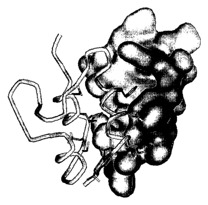
Figure 2: The overall structure of the IGF-I (tube model) mini-IGFBPS
(molecular surface) complex. Side chains plotted show the IGF
residues in contact with BPS. Particularly important is Phel6,
seen filling a hydrophobic depression on the BP5 surface.
Figure 3: Similar to Figure 2, whereby the IGF is depicted with its
molecular surface and BP5 is depicted as a tube model. Side
chains of BP5 responsible for binding to IGF are also depicted.
The surface of IGF Phel6 is prominent, as is the relatively flat
hydrophobic IGF surface contributing to the interface.
Figures 4A
and 4B: Summary of IGF-BPS and IGF-I contacts. Interactions
contributing to the binding affinity consist of hydrophobic
interactions (a) (involving especially residues Leucines 70, 73, and
74 of BP5 and Phel6 of IGF-I) and also polar interactions (b).
Enhancement of BP-IGF binding relies especially on the
enhancement of hydrophobic interactions, either by increasing
the intermolecular contact surface with these or with additional
residues, or by the introduction of further polar contacts.
(A) Packing contacts between IGFBP-5 and IGF-I. Contacts are
denoted according to nearest distances, whereby the closest
contacts include polar interactions.
(B) Polar contacts between IGFBP-S and IGF-I. Abbreviations
denote hydrogen bonds (HB), CH-O hydrogen bonds (CHB), salt
CA 02449290 2003-12-02
WO 02/098914 PCT/EP02/06161
-22-
bridge (SB), and side chain (SC) or main chain (MC)
interactions.
Figure S: Atomic coordinates of IGF-I in the complex with mini-IGFBP-5.
Figure 6: Atomic coordinates of mini-IGFBP-5 in the complex with IGF-I.
Figure 7: Binding of radioactive J-125 IGF-I to NIH 3T3 cells expressing
the IGF-IR in the absence and in the presence of IGFBP-5 and
compounds potentially interfering with complex formation
between IGF-I and IGFBP-5
Figure 8: IGF-I induced autophosphorylation of the IGF-IR expressed by
NIH 3T3 cells in the absence and in the presence of IGFBP-5 and
compounds potentially interfering with complex formation
between IGF-I and IGFBP-5
Sequence Listing
SEQ ID NO:1 Primer FBPSLY.
SEQ ID N0:2 Primer RBPSLY.
SEQ ID N0:3 Primer FBPSLM.
SEQ ID N0:4 Primer RBPSLM.
SEQ ID N0:5 Primer IBP4NdeI.
SEQ ID N0:6 Primer IBP4BamHI.
SEQ ID N0:7 Peptide GSALA.
SEQ ID N0:8 Peptide GSHMDEAIH.
CA 02449290 2003-12-02
WO 02/098914 PCT/EP02/06161
-23-
xa a
Crystallization, data collection and derivatization
Mini-IGFBP-5 was produced as described by Kalus, W., et al., in EMBO J. 17
( 1998) 6558-6572 and in Example 6, and IGF-I was obtained from OvoPepi,
Australia. Crystallization was successful with 10% Jeffamine M-600, 0.1 M
sodium
citrate, 0.01 M ferric chloride, pH 5.6. Within 11 days, crystals appeared at
4 °C,
growing to a final size of about 0.3 x 0.2 x 0.2 mm3. They belong to the cubic
space
group P213 and have unit cell dimensions a, b, c = 74.385 t~, with one complex
molecule per asymmetric unit. Soaking in precipitation buffer with heavy atom
compounds yielded a derivative K2PtC14 (2.7 mM, 3 d) which was interpretable.
All
diffraction data were collected using a 300 mm MAR Research (Hamburg,
Germany) image plate detector mounted on a Rigaku (Tokyo, Japan) RU300
rotating anode X-ray generator with graphite monochromatized CuKa radiation.
All image plate data were processed with MOSFLM (Leslie, A.G.W., Molecular
Data
in Processing, in: Moras, D., Podjarny, A.D., and Thierry, J.C. (eds.),
Crystallographic Computing 5 ( 1991 ), Oxford University Press, Oxford, UK,
pp.
50-61) and the CCP4 program suite (Collaborative Computational Project,
Number 4 1994).
x a
Phase calculation, model building and refinement
The structure of the IGF/mini-IGFBP-5 complex was solved by the single
isomorphous replacement (s.i.r.) method using one heavy atom derivative
described above. Derivative data was analyzed with the native data set, first
using
isomorphous difference Patterson maps and employing the Patterson vector
superposition methods implemented in SHELX-97 (Sheldrick, G., Tutorial on
automated Patterson interpretation to find heavy atoms, in: Moras, D.,
Podjarny,
A.D., and Thierry, J.C. (eds.), Crystallographic Computing 5 (1991), Oxford
University Press, Oxford, UK, pp. 145-157). The 2 heavy sites locations were
confirmed by difference Fourier methods with appropriate initial single site
s.i.r.
phases using CCP4 programs. The refinement of heavy atom parameters and
calculation of s.i.r. phases were done with SHARP (de la Fortelle, E., and de
Bricogne, G., Methods Enzymol. 276 (1997) 472-494). The final parameters are
given in Table 8. The initial s.i.r. phases were improved with SOLOMON
CA 02449290 2003-12-02
WO 02/098914 PCT/EP02/06161
-24-
(Abrahams, J.P., and Leslie, A.G.W., Acta. Cryst. D52 (1996) 30-42) using an
solvent fraction of 45%, resulting in a 2.1 A electron density map that was
interpretable. Refinement was performed by conjugate gradient and simulated
annealing protocols as implemented in CNS 1.0 (Briinger, A.T., et al., Acta
Crystallogr. D54 (1998) 905-921. All protocols included refinement of
individual
isotropic B-factors and using the amplitude based maximum likelihood target
function. The R-factor dropped to 21.8 % (Rfree= 26.2 %, resolution range 16.2
-
2.1 t~) for the final model including 47 water molecules. The water model was
calculated using ARP and verified by visual inspection. The final refinement
statistics are shown in Table 8.
CA 02449290 2003-12-02
WO 02/098914 PCT/EP02/06161
-25-
Table 8:
Statistics from the crystallographic analysis
native KZPtCI4
Resolution (fir) 16.2 - 2.1 18.6 - 2.5
Measurements 45345 32833
Uni ue measurements 8035 4925
% Com fete (last shell/t~)99.3 (96.9/2.23 - 99.9 (95.4/2.64-2.5)
2.11)
RS m (%) (last shell)8.2 (44.8) 8.8 (49.5)
Rc~~u~5-~so - 0.77
p;so - 1.48
Res. for hase talc. - 18.6 - 2.5
(~)
Mean FOM - 0.41
Refinement statistics:
Resolution ran a (t~)16.2 - 2.1
reflections in workin7522
set
reflections in test 501
set
R St (%) 21.8
R ~e (%) 26.2
Protein atoms (non-H)765
Solvent atoms (non-H)47
Avera a B-factor (t12)38.1
r.m.s. 0B (21~ cutoff)3.4
Deviations from ideality
(r.m.s.):
Bond len the (t1) 0.013
Bond an les () 1.7
R.,Yn, -
Rc~~ttr5-r5o = r.m.s. lack of closure / r.m.s isomorphous difference
PASO (Phasing power) _ ~IF,,I~ / r.m.s. lack of closure for all reflections
Mean FOM = mean figure of merit
CA 02449290 2003-12-02
WO 02/098914 PCT/EP02/06161
-26-
R~,~,St = Crystallographic R-factor for reflections used in refinement
R~.e~ = Crystallographic R-factor for reflections not used in refinement
r.m.s. = Root mean square
a e3
Determination of the binding affinity of IGFBP mutants
The IGF-binding properties of wildtype and mutant fragments and full-length
IGFBPs were quantitatively analyzed by BIAcore biosensor measurements. BIAcore
2000, Sensor Chip SA and HBSwere obtained from BIAcore AB (Uppsala, Sweden).
All experiments were performed at 25°C and HBS (20 mM HEPES, 150 mM
NaCI, 3
mM EDTA, pH 7.5) was used as a running buffer and for the dilution of ligands
and
analytes. Biotinylated IGF-I was immobilized at a concentration of 5 nM and 10
nM
in HBS at a flow rate of 5 l.~I/min to the strepavidin coated sensor chip
resulting in
signals of 40 and 110 resonance units (RU). Biotinylated IGF-II was
immobilized at a
concentration of 5 nM in HBS resulting in a signal of 20 RU. An empty flow
cell was
used as control for unspecific binding and bulk effects. The low ligand
concentration
was necessary to limit mass transport limitations and rebinding. For the same
reason
all kinetic experiments were performed at the highest possible flow rate of
100 E~l/min.
Each protein (wildtype and mutant IGFBPs or fragments of these proteins) was
injected at four concentrations (250, 50, 10, and 2 nM). Each sample was
injected for 2
min followed by dissociation in buffer flow for 4 min. After the dissociation
phase the
sensor chip was regenerated by injection of 10 ~.~I 100 mM HCl at a flow rate
of 5
~.~I/min. The kinetic parameters were calculated using the BIAevaluation 3.0
software
(BIAcore AB). After subtraction of the blank sensorgram the kinetic rate
constants
were calculated from a general fit of an overlay of the sensorgrams of all
concentration
of one analyte using the method called "1:1 binding with mass transfer". IGF-I
and
IGF-II were biotinylated with a five-fold molar excess of D-biotinyl-~-
aminocaproic
acid-N-hydroxysuccinimide ester using the reagents and the operation
instructions of
the Biotin Protein Labelling Kit (Roche Diagnostics GmbH, DE). After blocking
with
lysine, the reaction was dialyzed against SO mM Na-phosphate, 50 mM NaCI, pH
7.5.
CA 02449290 2003-12-02
WO 02/098914 PCT/EP02/06161
-27-
Example 44
Inhibition of IGF-I-induced IGF-IR phosphorylation by IGFBP mutants
Confluent monolayers of NIH3T3 cells stably expressing human IGF-IR in 3.5 cm
dishes were starved in DMEM containing 0.5% dialyzed fetal calf serum. After
48 h,
cells were incubated without any hormone or with 5 x 10⁻⁹ M IGF-I or 1 x 10⁻⁸
M
IGF-II; each sample was preincubated with increasing concentrations of
different
IGF-binding proteins or fragments thereof at room temperature for 1 h. After a
10
min stimulation at 37°C, the medium was removed and cells were lysed
with 250 E.~l
of lysing buffer (20 mM Hepes, pH 7.5, 150 mM NaCI, 10% glycerol, 1% Nonidet
P40, 1.5 mM MgCl2, 1 mM EGTA (ethylene glycol-bis(2-aminoethyl)-N,N,N',N'-
tetraacetic acid, Aldrich, USA), 10 mM sodium orthovanadate, and protease
inhibitor cocktail Complete (Roche Diagnostics GmbH, DE) for 10 min on ice.
Subsequently, cells were scraped off the plate and the insoluble material was
separated by centrifugation for 20 min at 4°C. The protein
concentration of the
supernatant was determined using the BCA kit from Pierce, Rockford, USA
according to the manufacturer's instructions. Equal protein concentration was
incubated with the SDS sample buffer (63 mM Tris-HCI, pH 6.8, 3% SDS, 10%
glycerol, 0.05% bromphenolblue, 100 mM DTT), boiled for 5 min and loaded on a
7.5% SDS polyacrylamide gel. After electrophoresis the proteins were
transferred on
a nitrocellulose membrane which first was blocked for 1 h with the 3 % BSA
containing PBST (phosphate buffered saline-Tween°), then overnight
incubated
with 1 p,g/ml monoclonal anti-phosphotyrosine antibody 3-365-10 (Roche
Diagnostics GmbH, DE) in PBST that contained 3% BSA. Unbound antibody was
removed by extensive washing. The blot was then incubated with 1:10000 diluted
anti-mouse IgG-specific antibody conjugated with horse raddish peroxidase
(Roche
Diagnostics GmbH, DE). The immunoblot was developed using the ECL kit from
Amersham and the concentration of IGFBP which results in 50 % inhibition of
the
IGF-I receptor phosphorylation was determined.
xa a 5
Determination of the inhibition of tumor cell growth by IGFBP mutants
MCF-7 cells (from ATCC, American type Culture Collection, Rockville, Maryland,
U.S.A., HTB22) were used to investigate the inhibitory effect of IGFBP mutants
on
tumor cells. 1000 MCF-7 cells were seeded per well in medium containing 2.5
CA 02449290 2003-12-02
WO 02/098914 PCT/EP02/06161
-28-
FBS (fetal bovine serum). The cells were cultured in the presence of various
concentrations of IGFBPs for 48 h. The percentage of surviving cells was
determined by MTT ((3-[4,5-dimethylthiazol-2y1]-2,5-diphenyltetrazolium
bromide) assay and the concentration of binding protein which results in
reduction of cell survival by 50 % was determined.
xa 6
Mutagenesis, expression and purification of mini-IGFBP-5s and subcloning of
IGFBP-4 into Pet-28a (+)
6.1 Buffers and media
Cell growth media:
LB-medium per 1 liter: peptone 10 g, yeast extract 5 g, NaCI 10 g,
adjusted to pH 7.
LB-agar per 1 liter: peptone 10 g, yeast extract 5 g, NaCI 10 g, bacto
agar 15 g, adjusted to pH 7.
Minimal medium per 1 liter: 0.5 g NaCI, 1 g citric acid monohydrate, 36 mg
ferrous citrate (pre-dissolved in conc. HCl), 4.02 g
KHZP04, 7.82 g KZHP04, 1g 'SN-NH4C1, 1.3 ml trace
elements solution (per liter of the stock solution: 2.5 g
H3B03, 2.0 g CoCl2, 1.13 g CuCl2, 9.8 g MnCl2, 2.0 g
NazMo04), 1 ml Zn-EDTA solution (per ml of the stock
solution: 5 mg EDTA, 8.4 mg zinc acetate), adjusted to pH
7, autoclaved. Added afterwards: 25 ml autoclaved 20%
(w/v) glucose, 560 E.~l sterile filtered 1% (w/v) thiamine,
2m1 1M MgS04.
Antibiotic stocks:
Ampicillin 50 mg/ml in dist. water, 0.22 ~,m filtrated, stored at -
20°C.
Kanamycin 25 mg/ml in dist. water, 0.22 ~,m filtrated, stored at -
20°C.
Chloramphenicol 35 mg/ml in 96 % ethanol, stored at -20 °C.
CA 02449290 2003-12-02
WO 02/098914 PCT/EP02/06161
-29-
Agarose-gel electrophoresis:
TAE-buffer (50x) 2 M Tris-HCl (pH 8.0), 2 M glacial acetic acid and 50 mM
EDTA.
Loading buffer (3x) 0.13 % bromophenol blue, 0.13 % xylene cyanol, 30
glycerol.
Et-Br-solution 10 mg/ml ethidiumbromide in dd H20.
SDS-PAGE:
Sample buffer (5x) 125 mM Tris-HC1 (pH 6.8), 10 % SDS, 760 mM 2-
mercaptoethanol, 0.13 % bromophenol blue, 50 % glycerol
and 2 mM EDTA.
Staining solution 0.125 % CBB-8250 in 500 ml 96 % ethanol and 500 ml 10
% acetic acid.
Distaining solution 96 % ethanol, 10 % acetic acid and dest. H20 in 4:3:3
proportion.
Tricine gels:
Cathode (top)
running buffer ( 10x) 1 M Tris-HCl (pH 8.25), 1 M Tricine and 1 % SDS.
Anode (bottom)
running buffer ( 10x) 2 M Tris-HCl (pH 8.9).
Separation buffer 3 M Tris-HCl (pH 8.9) and 0.3 % SDS.
Stacking buffer 1 M Tris-HCl (pH 6.8) and 0.3 % SDS.
Separation acrylamide 48 % (w/v) acrylamide, 1.5 % (w/v) N,N'-methylene-bis-
acrylamide.
Stacking acrylamide 30 % (w/v) acrylamide, 0.8 % (w/v) N,N'-methylene-bis-
acrylamide.
APS 10 % ammonium persulphate in dd HzO.
Separation gel (main) for 2 70x80x0.75 mm mini-gels: 1.675 ml HzO, 2.5 ml
separation buffer, 2.5 ml separation acrylamide, 0.8 ml
glycerol, 25 ~1 APS and 2.5 X1.1 TEMED.
CA 02449290 2003-12-02
WO 02/098914 PCT/EP02/06161
-30-
Separation gel
(intermediate) 1.725 ml HzO, 1.25 ml separation buffer, 0.75 ml separation
acrylamide, 12.5 ~1 APS and 1.25 X11 TEMED.
Stacking gel 2.575 ml H20, 0.475 ml stacking buffer, 0.625 ml stacking
acrylamide, 12.5 u1 0.5 M EDTA (pH 8.0), 37.5 E.il APS and
1.9 E.~l TEMED.
Protein purification:
Buffer A 6 M guanidinium-HCI, 100 mM NaHzP04, 10 mM Tris and
10 mM 2-mercaptoethanol, pH 8Ø
Buffer B 6 M guanidinium-HCI, 100 mM NaH2P04, 10 mM Tris and
10 mM 2-mercaptoethanol, pH 6.5
Buffer C 6 M guanidinium-HCI, 100 mM Na-acetate and 10 mM 2-
mercaptoethanol, pH 4Ø
Buffer D 6 M guanidinium-HCI, pH 3Ø
Buffer E 200 mM arginine, 1 mM EDTA, 100 mM Tris-HCI, 2 mM
reduced glutathione, 2 mM oxidised glutathione, pH 8.4.
PB(0) 10 mM Na2HP04, 1.8 mM KHzP04 and 0.05 % NaN3, pH
7.2.
PB(1000) 10 mM Na2HP04, 1.8 mM KHZP04, 0.05 % NaN3 and 1 M
NaCI, pH 7.2.
PBS 140 mM NaCI, 27 mM KCI, 10 mM Na2HP04, 1.8 mM
KHzP04 and 0.05% NaN~.
Thrombin cleavage
buffer 60 mM NaCI, 60 mM KCI, 2.5 mM CaClz, 50 mM Tris, pH
8Ø
6.2 Cloning of mini-IGFBP-5
Mini-IGFBP-5 (residues 40-92 of IGFBP-5) was subcloned from a vector
containing the complete sequence of IGFBP-5 into the BamHI and PstI
restriction
sites of the pQE30-vector (Qiagen, Hilden, Germany). Restriction sites, a stop
codon and 21 bases encoding an N-terminal thrombin cleavage site were
introduced by means of PCR (Kalus, W., et al., EMBO J. 17 ( 1998) 6558-6572).
CA 02449290 2003-12-02
WO 02/098914 PCT/EP02/06161
-31-
6.3 Mutagenesis of mini-IGFBP-S
For introduction of mutations leading to substitution of Leu61 by Tyr and
Leu~4 by
Met, in vitro mutagenesis was performed using QuickChangeTM site-directed
mutagenesis kit (Stratagene, La Jolla, Canada). Two sets of the following
mutagenic
oligonucleotide primers were designed for amplification of plasmid DNA and
introduction of the desired point mutations:
FBPSLY: 5'-G GGG CTG CGC TGC TAC CCC CGG CAG GAC G-3';
(SEQ ID NO:1)
RBPSLY: 5'-C GTC CTG CCG GGG GTA GCA GCG CAG CCC C-3';
(SEQ ID N0:2)
FBPSLM: 5'-CG CTG CAC GCC CTG ATG CAC GGC CGC GGG G-3';
(SEQ ID N0:3)
RBPSLM: 5'-C CCC GCG GCC GTG CAT CAG GGC GTG CAG CG-3'
(SEQ ID N0:4).
The changed codons (CTC into TAC in L61Y mutant and CTG into ATG in L~4M
mutant) are presented in bold. Degenerated bases are underlined.
The reactions were set up according to the instructions found in the
mutagenesis kit
manual. The PCR mixtures (50 ~.l) contained app 50 ng of the template (pQE30
(mini-IGFBP-5), prepared by means of mini prep spin columns kit, Qiagen) and
125 ng of each of the two oligonucleotide primers. Cycling parameters for the
reactions were as follows: 30 seconds at 95°C followed by 13 rycles of
95°C for 30
seconds, 55°C for 1 minute and 68°C for 7.5 min. The DpnI
digestion and XL1-
Blue supercompetent cells transformation was carried out strictly according to
the
supplier's guidelines.
Two clones of each mutant were subjected to verification by automated double
stranded sequencing, which proved the existence of the expected substitutions
in all
4 cases.
CA 02449290 2003-12-02
WO 02/098914 PCT/EP02/06161
-32-
6.4 Expression of the mutant mini-IGFBP-5s
Electrocompetent cells BL21 were transformed with the construct carrying the
mutation. From a fresh plate, a 3-ml LB culture was started and grown overday
(6-7
h) in the presence of 300 p,g ampicillin per ml at 37°C. From this
culture 50 p1 were
used to inoculate 20 ml of MM. This culture was grown overnight (9-llh). 11
culture was inoculated in 1:50 proportion. Expression of the protein was
induced at
OD6oo = 0.8 by addition of IPTG ( 1 mM final concentration). Cells were
harvested
after 3 h (6000 xG, 20 min at 4°C).
6.5 Purification of mini-IGFBP-5
Harvested cells were resuspended in buffer A (30 ml of the buffer was used to
resuspend cells from 11 culture) and incubated at room temperature with
vigorous
shaking (280 RMP) for 1 h to overnight. The cells were opened by sonification
(macrotip, 50 % duty rycle, output control 70, 2x4 min). The cell extract was
then
centrifuged to pellet cell debris (65 000 xG, 1h at room temp.). The pH of the
supernatant was adjusted to the value of app. 8Ø The supernatant was then
mixed
with pre-equilibrated with buffer A Ni-NTA Superffow matrix (Qiagen) ,
incubated
with agitation for 1 h to overnight and then loaded onto an empty column (3 ml
bed volume for 1 1 culture). The column was washed with buffer A followed by
buffer B until a stable W-absorption base line. Bound proteins were
fractionated
with 100 ml pH gradient of buffer B and C. Collected fractions were analysed
by
tricine gel electrophoresis (prior electrophoresis, the proteins were
precipitated
with 5 % (w/v) TCA). Fractions containing mini-IGFBP-5 were pooled,
concentrated on Amicon YM3 to 2-4 ml, and dialysed against 2 1 of buffer D
overnight ( 100 p1 excess of 2-mercaptoethanol was added to the sample prior
dialysis).
To promote refolding, the dialysed sample was diluted in 100 p1 portions into
freshly prepared, ice-cold buffer E, with vigorous stirring (in proportion 1
ml
sample per 50 ml of buffer E), and left at 4°C for 2-3 days with
stirring.
The sample was concentrated on Amicon YM3 to 15-25 ml, centrifuged to get rid
of
a precipitated material, and dialysed overnight into 4 1 of buffer PB
containing 30
mM NaCI.
CA 02449290 2003-12-02
WO 02/098914 PCT/EP02/06161
-33-
The solution was subsequently loaded onto pre-equilibrated with buffer PB (0)
MonoS 5/5 HR cation-exchanger column (app. 1 ml) (Amersham Pharmacia,
Uppsala, Sweden) at a flow rate of 1 ml/min. The column was washed with buffer
PB (0). Proteins were eluted by 45 ml linear gradient of 0-70 % NaCI, 1 ml
fractions
were collected.
The fractions containing mini-IGFBP-5 (as determined on the basis of tricine
gel
electrophoresis) were pooled, concentrated to 2-3 ml and loaded onto a pre-
equilibrated with PBS Superdex 75 HiLoad 26/60 (app. 320 ml) gel-filtration
column (Pharmacia) at a flow rate of 0.6 ml/min. Mini-IGFBP-5 was eluted as a
symmetrical, single pick. Fractions containing the protein were pooled and
concentrated on centricon YM3.
6.6 Subcloning into pET-28a (+)
The reason for overall low expression of the proteins from the pQE30 might be
the
fact that this vector is not well optimised for expression in E. coli. For
instance, the
vector-encoded sequences contain a cluster of 3 rare codons just downstream
from
the initiator codon AUG (namely, AGA, GGA and TCG, encoding Arg, Gly and Ser,
respectively). The number of studies has indicated that excessive rare codon
usage
in a target gene may be a cause for low level expression. The impact seems to
be
most severe when multiple rare codons occur near the amino terminus and when
they appear consecutively. Especially presence of the Arg codons AGG and AGA
can have severe effects on the level of protein production. The system seems
to be
also not well repressed (no extra copies of a gene encoding Lac repressor),
and the
leaky expression may cause the observed plasmid instability. The vector
carries not
very efficient selective marker, AmpR gene (bla), what makes possible rapid
over-
growing of a culture at a certain stage by cells lacking the unstable plasmid.
The
vector pET-28a (+) (Novagen) was then chosen as an alternative for pQE30. The
plasmid is well optimised for expression of genes in E. coli, carries a strong
selective
marker (KanR) and is stable due to high level of repression of the target gene
expression in the absence of IPTG (in a non-DE3 lysogenic strain even in the
presence of the inducer).
To subclone mini-IGFBP-5 wild type, L61Y and L~4M from pQE30 to pET-28a, the
relevant fragments were excised from the vector with BamHI and HindIII
(HindIII
CA 02449290 2003-12-02
WO 02/098914 PCT/EP02/06161
-34-
cleavage site exists in pQE30 downstream from PstI site). The excision was
performed as double-digestion. Digested pET vector was 5'-dephosphorylated.
Reaction mixtures were electrophorized and bands corresponding to app. 200 by
fragments excised from pQE30 (mini-IGFBP-5 wt, L61Y and L~4M) and app 5000 by
fragment of pET-28a were cut from 1 % agarose gel and purified (gel extraction
kit,
Qiagen). The fragments were ligated (Ligation kit, Fermentas) and XL-1 Blue
Supercompetent cells were transformed with the ligation mixture.
Restriction assay carried out subsequently on isolated plasmid DNA revealed
presence of fragments of expected size (restriction enzymes NcoI and PstI were
used, double digestion was performed. PstI restriction site was introduced
into the
pET vector together with the fragment encoding mini-IGFBP-5).
Pilot-scale expression and purification experiment showed that expression of
the
protein of interest (mini-IGFBP-5 L6~Y in this case) is higher than the
expression of
the wild-type protein when pQE30 vector was used.
The proteins are expressed as double-fusions: they carry His-tag followed by
T7-tag.
It is possible to remove both tags by thrombin cleavage. Mini-IGFBP-5 after
cleavage by thrombin comprises the following N-terminal amino acid sequence:
GSALA (SEQ ID N0:7) (N-terminus of mini-IGFBP-5 starting from as 40 with to
additional as from cloning with thrombin cleavage site). Vector-derived amino
acids are underlined.
6.7 Subcloning of IGFBP4 from pKK177-3HB to pET-28a(+)
For subcloning of IGFBP4-2 into the NdeI and BamHI restriction sites of the
pET-
28a vector in-frame to a His-tag, following oligonucleotides were designed for
amplification of DNA by PCR:
IBP4NdeI: 5'-CGG AGG AAA AAC ATA TGG ATG AAG C-3'
(SEQ ID N0:5)
IBP4BamHI: 5'-GCC AAG CTT GGA TCC AGG TCG AC-3'
(SEQ ID N0:6)
CA 02449290 2003-12-02
WO 02/098914 PCT/EP02/06161
-35-
The restriction sites recognized by NdeI and BamHI are presented in bold.
Degenerated bases are underlined.
The PCR mixture (50 ~tl) contained 124 ng of mixture of pKK177-3HB and
Pfdx500 repressor plasmid, 130 ng of each of the primers, 1 1.L1 dNTP mix and
2.5 U
Pfu Turbo DNA polymerase (Strategene). After initial step of 30 sec. At
95°C, the
reaction was cycled 30x at 95°C for 30 seconds, 55°C for 1 min
and 68°C for 2 min.
The product of PCR was purified (PCR purification kit, Qiagen), double-
digested
and electrophorised. The bands corresponding to cleaved pET-28a and PCR
product were excised from the gel and purified.
XL-1 Blue Supercompetent cells were transformed with the ligation mixture.
IGFBP4-2 is expressed as a N-terminal His-tag fusion protein. After thrombin
cleavage, the protein comprises the following amino acid sequence:
GSHMDEAIH... (SEQ ID N0:8). Vector derived amino acids are underlined.
The same purification routine will be used for His-tagged IGFBP-4 as for mini-
IGFBP-5.
a 1e
Identification of chemical non-proteinaceous compounds binding to IGFBP-5 or
IGF-I by using the coordinates of the crystal structure of the complex of both
molecules
FlexX version 1.9.0 was used to screen a substance library of ca. 90,000
compounds
in an ACD (Available Chemicals Directory; ACD-3D 2000), choosing compounds
with a molecular weight of less than 550 Daltons and containing at least one
of the
atoms {N, O, F, or S}. Docking searches were conducted on both molecular
surfaces
of the IGFBP-5 interface. Top scoring hits as judged by the FlexX standard
scoring
function and the proximity to binding site protein atoms were selected and
tested
for activity.
The top scoring compounds selected according to these these criteria for
release of
IGF-I from IGFBP-5 were:
CA 02449290 2003-12-02
WO 02/098914 PCT/EP02/06161
-36-
Compound 1: Nl-(3,4-Dichlorophenyl)-2-[2-[5-(3,5-dichlorophenyl)-2H-
1,2,3,-tetraazol-2YL]A (MF: C16H11C14N70S; MW: 491,1890
Da)
Compound 2: F-MOC-Tyr(P03H2)-OH (C24H22N08P; MW: 483.4110)
Compound 2A: Na-FMOC-O-tert-butyl-L-tyrosine
Compound 2B: Na-FMOC-L-phenylalanine
Compound 2C: Na-FMOC-N-BOC-L-tryptophan
Compound 2D: Na-FMOC-L-leucine
Compound 3: 4-(2,5-Dichlorophenylazo)-4'fluorosulfonyl-1-hydroxy-2-
naphthanilide (MF: C23H14C12FN3O4S; MW: 518.3510)
Compound 4B: 5-Amino-2[(4-amino-2-carboxyphenyl)thio]benzoic acid
(C14H12N2O4S; MW 304.3250)
Compound 4C: 5-Amino-2[(2-carboxyphenyl)thio]benzoic acid (C14H11N04S;
MW 289.3100)
am 8
Release of IGF-I from the complex with IGFBP-5 by selected compounds
measured by IGF-I binding to IGF-IR expressing cells
Kalus, W., et al., in EMBO J. 17 (1998) 6558-6572, describe the inhibition of
the
binding of IGF-I to IGF-IR expressing NIH 3T3 cells by formation of an
inhibitory
complex. This assay was used to investigate the release of IGF-I from the
inhibitory
complex with IGFBP-5.
NIH 3T3 cells stably expressing human IGF-IR were grown in culture dishes in
Dulbecco's modified Eagle's medium (DMEM) containing 10% fetal calf serum.
Cells were washed carefully with PBS and incubated with 5 ml of 50 mM EDTA in
PBS for 45 min. Cells were removed from the plate, washed once with PBS and
once with binding buffer ( 100 mM HEPES pH 7.6, 120 mM NaCI, 5 mM KCI, 1.2
mM MgSO 4 , 1 mM EDTA, 10 mM glucose, 15 mM sodium acetate, 1% dialysed
BSA), and resuspended in binding buffer to determine the cell number. 5 pM 'ZS
I-
IGF-I (Amersham) was preincubated with either 10 or 100 nM IGFBP-5 alone or in
CA 02449290 2003-12-02
WO 02/098914 PCT/EP02/06161
-37-
combination with 33 ~M of the different compounds 1,2,3,4B and 4C at
4°C for 1
h. Then 400 ~1 of the cell suspension corresponding to 2x10⁵ cells were added
to
give a total volume of 500 E.il. After 12 h incubation at 4°C, cells
were washed with
binding buffer (at 4°C). Free hormone was removed by repeated
centrifugation and
resuspension in the binding buffer. The 125 I radioactivity bound to the cells
was
determined in a gamma-counter.
As shown in figure 7 the labeled IGF-I binds to NIH 3T3 cells in the absence
of
IGFBP-5 and cell binding is inhibited by the addition of IGFBP-5.
Preincubation of
the complex of IGFBP-5 and IGF-I with the selected compounds results in
release
of IGF-I from the complex by compound 3 and consequently binding of IGF-I to
the IGF-IR expressing cells.
e9
Release of IGF-I from the complex with IGFBP-5 by selected compounds
measured by IGF-IR activation
Kalus, W., et al., in EMBO J. 17 (1998) 6558-6572, describe the inhibition of
the
activation and autophosphorylation of the IGF-IR by IGF-I in the presence of
IGFBP-5. This assay was used to further investigate the release of IGF-I from
the
inhibitory complex with IGFBP-5 by compound 3. Binding of compound 3 to
IGFBP-5 and dissociation of the complex of the binding protein with IGF-I
should
result in an activation and autophosphorylation of the IGF-IR in the presence
of
IGFBP-5.
Confluent monolayers of the NIH 3T3 cells stably expressing human IGF-IR in
3.5
cm dishes were starved in DMEM containing 0.5% dialysed fetal calf serum.
After
48 h, cells were incubated without any hormone or with 10 nM IGF-I. Samples
were preincubated with 100 nM IGFBP-5 and increasing concentrations of
compound 3 from 0 to 50 ~M at room temperature for 1 h. After a 10 min
stimulation at 37°C, the medium was removed and cells were lysed with
250 ~l of
lysing buffer (20 mM HEPES pH 7.5, 150 mM NaCI, 10% glycerol, 1% NP-40, 1.5
mM MgCI 2 , 1 mM EGTA), 10 mM sodium orthovanadate, and protease inhibitor
cocktail Complete (Roche Molecular Biochemicals) for 10 min on ice.
Subsequently, cells were scraped off the plate and the insoluble material was
separated by centrifugation for 20 min at 4°C. The protein
concentration of the
CA 02449290 2003-12-02
WO 02/098914 PCT/EP02/06161
-38-
supernatant was determined using the BCA kit from Pierce according to the
manufacturer's instructions. Equal protein concentration was incubated with
the
SDS sample buffer (63 mM Tris-HCl pH 6.8, 3% SDS, 10% glycerol, 0.05%
bromophenolblue, 100 mM DTT), boiled for 5 min and loaded on a 7.5% SDS-
polyacrylamide gel. After electrophoresis the proteins were transferred on a
nitrocellulose membrane which first was blocked for 1 h with the 3% BSA
containing phosphate-buffered saline-Tween (PBST), then incubated overnight
with 1 mg/ml monoclonal anti-phosphotyrosine antibody 4610 (Upstate
Biotechnology), polyclonal anti-phospho-AKT antibody (New England Biolabs) or
polyclonal anti- IGF-IR (C-20, Santa Cruz Biotechnology) in PBST that
contained
3% BSA. Unbound antibody was removed by extensive washing. The blot was then
incubated with 1:10 000 diluted anti-mouse IgG-specific antibody or 1:5000
diluted
anti-rabbit specific antibody conjugated with horse radish peroxidase (both
Roche
Molecular Biochemicals). The immunoblot was developed using the ECL kit from
Amersham.
As shown in Fig. 8 the autophosphorylation of IGF-IR by IGF-I is inhibited in
the
presence of IGFBP-5. The addition of compound 3 to the inactive complex of
IGFBP-5 and IGF-I results in an increased autophosphorylation of the receptor
at
50 uM compound 3.
a a
Detection of ligand binding
Ligand binding was detected by acquiring 15N-HSQC spectra. All NMR spectra
were acquired at 300 K on Bruker DRX600 spectrometer. The samples for NMR
spectroscopy were concentrated and dialyzed against PBS buffer. Typically, the
sample concentration was varied from 0.3 to 1.0 mM. Before measuring, the
sample
was centrifuged in order to sediment aggregates and other macroscopic
particles.
450 E.~l of the protein solution were mixed with 50 ~l of D20 (5-10%) and
transferred to an NMR sample tube. The stock solutions of compounds were 100
mM either in water or in perdeuterated DMSO. pH was maintained constant
during the whole titration. The binding was monitored by observation of the
changes in the ESN-HSQC spectrum. Dissociation constants were obtained by
monitoring the chemical shift changes of the backbone amide of several amino
acid
residues (Table 9) as a function of ligand concentration. Data were fit using
a single
CA 02449290 2003-12-02
WO 02/098914 PCT/EP02/06161
-39-
binding site model. In the same way dissociation constants for derivatives of
compound 2 are estimated (Table 10).
Table 9:
Dissociation constant calculations for compound 2 or DMSO binding to IGFBP-5
using data from distinct amino acid residues
residue ligand in DMSO ligand in PBS DMSO KD [mM]
KD KD
[mM] [mM]
Y50 1.58 0.09 1.82 0.95 648 370
L73 1.31 0.17 2.93 1.41 541 306
S85 1.38 0.10 2.33 0.94 650 373
Y86 1.90 0.17 1.72 0.99 783 498
R87 1.64 0.12 2.36 1.00 921 662
K91 2.42 0.18 2.12 1.03 719 434
average: 1.71 0.37 2.21 0.40 710 120
Table 10:
Dissociation constants calculated for compound 2 and its derivatives binding
to
IGFBP-5 using changes in chemical shift for the residue L81
compound chemical name KD [mM]
2 Na-FMOC-O-phospho-L-tyrosine2.78 0.30
2A Na-FMOC-O-tert-butyl-L-tyrosine0.718 0.079
2B Na-FMOC-L-phenylalanine 1.075 0.507
2C Na-FMOC-N-BOC-L-tryptophan0.0432 0.0115
2D Na-FMOC-L-leucine 1.088 0.519
CA 02449290 2003-12-02
WO 02/098914 PCT/EP02/06161
-40-
List of References
Abrahams, J.P., and Leslie, A.G.W., Acta. Cryst. D52 (1996) 30-42
Adams, M.J., et al., Nature 224 ( 1969) 491-492
Bach, L.A., et al., J. Biol. Chem. 268 (1993) 9246-9254
Bacon et al., J. Mol. Biol. 225 (1992) 849-858
Baxter, R.C., et al., J. Biol. Chem. 267 ( 1992) 60-65
Blundell, T.L., et al., Nature 326 ( 1987) 347
Blundell, T.L., Proc. Natl. Acad. Sci. USA 75 (1978) 180-184
Bohm, H.J., et al., J. Comput. Aided Mol. Des. 6 (1992) 61-78 and 593-606
Briinger, A.T., et al., Acta Crystallogr. D54 (1998) 905-921
Butt, A.J., et al., J. Biol. Chem. 275 (2000) 39174-39181
Byun, D., et al., J. Endocrinology 169 (2001) 135-143
Chernausek, S.D., et al., J. Biol. Chem. 270 (1995) 11377-11382
Conover, C.A., et al., in J. Biol. Chem. 270 ( 1995) 4395-4400
Cooke, R.M., et al., Biochemistry 30 ( 1991 ) 5484-5491
de la Fortelle, E., and de Bricogne, G., Methods Enzymol. 276 ( 1997) 472-494
Fanayan, S., et al., J. Biol. Chem. 275 (2000) 39146-39151
Gill, R., et al., Prot. Eng. 9 (1996) 1011-1019
Goodford, P.J., J. Med. Chem. 27 (1984) 557
Goodford, P.J., J. Med. Chem. 28 (1985) 849-857
Hankinson, S.E., et al., Lancet 351 (1998) 1393-1396
Holly, J., Lancet 351 ( 1998) 1373-1374
Hwa, V., et al., The IGF binding protein superfamily, In: Rosenfeld, R.G., and
Roberts, C.T. (eds.), The IGF system, Molecular Biology, Physiology,
and Clinical Applications ( 1999), Humana Press, Totowa, pp. 315-327
Imai, Y., et al., J. Biol. Chem. 275 (2000) 18188-18194
Jansson, M., et al., Biochemistry 36 ( 1997) 4108-4117
Jones, J.L., and Clemmons, D.R., Endocr. Rev. 12 (1995) 10-21
Kalus, W., et al., EMBO J. 17 ( 1998) 6558-6572
Khandwala, H.M., et al., Endocr. Rev. 21 (2000) 215-244
Kramer, B., et al., Proteins: Structure, Functions and Genetics 37 (1999) 228-
241
Kuntz, LD., et al., J. Mol. Biol. 161 ( 1982) 269-288
Laajoki, L.G., et al., J. Biol. Chem. 275 (2000) 10009-10015
CA 02449290 2003-12-02
WO 02/098914 PCT/EP02/06161
-41-
Leslie, A.G.W., Molecular Data in Processing, in: Moras, D., Podjarny, A.D.,
and
Thierry, J.C. (eds.), Crystallographic Computing 5 (1991), Oxford
University Press, Oxford, UK, pp. 50-61
Loddick, S.A., et al., Proc. Natl. Acad. Sci. USA 95 (1998) 1894-1898
Lowman, H.B., et al., Biochemistry 37 ( 1998) 8870-8878
Luethi, C., et al., Eur. J. Biochem. 205 ( 1992) 483-490
Martin, J.L., and Baxter, R.C., IGF binding proteins as modulators of IGF
actions;
in: Rosenfeld, R.G., and Roberts, C.T. (eds.), The IGF system,
Molecular Biology, Physiology, and Clinical Applications ( 1999),
Humana Press, Totowa, pp. 227-255
Mazerbourg, S., et al., Endocrinology 140 (1999) 4175-4184
Murray-Rust, J., et al., BioEssays 14 (1992) 325-331
Remington's Pharmaceutical Sciences, 18a' ed., 1990, Mack Publishing Company,
edited by Oslo et al. (e.g. pp. 1435-1712)
Sato, A., et al., Int. J. Pept. Protein Res. 41 (1993) 433-440
Sheldrick, G., Tutorial on automated Patterson interpretation to find heavy
atoms,
in: Moras, D., Podjarny, A.D., and Thierry, J.C. (eds.), Crystallographic
Computing 5 ( 1991 ), Oxford University Press, Oxford, UK, pp. 145-157
Sheridan, R.P., and Venktaraghavan, R., Acc. Chem. Res. 20 ( 1987) 322
SwissProt Database (http://www.expasy.ch)
Terasawa, H., et al., EMBO J. 13 ( 1994) 5590-5597
Tomes, A.M., et al., J. Mol. Biol. 248 (1995) 385-401
van den Berg, C.L., et al., Eur. J. Cancer 33 (1997) 1108-1113
Verlinde, C., and Hol, W., Structure 2 (1994) 577
Wetterau, L.A., et al., Mol. Gen. Metab. 68 (1999) 161-181
Wolk, A., Lancet 356 (2000) 1902-1903
CA 02449290 2003-12-02
PATENT COOPERAT10N TREATY
PST
INTERNATIONAL PRELIMINARY EXAMINATION REPORT
(PCT Article 36 and Rule 70)
(Rationalised Report according to the Notice of the President of the EPO
published in the OJ1I/2001)
Applicant's or agent's file reference
FOR FURTHER ACTION See Notification of Transmittal of International
20909 WO-SR Preliminary Examination Report (Form PCT/IPEA/416)
International application No. International filing date (dayJmonth/year)
Priority date (day/rnonth/year)
PCT/EP 02/ 06161 05/06(2002 07/06/2001
International Patent Classification (IPC) or national classification and IPC
C07K14/47
Applicant
F. HOFFMANN-LA ROCHE AG et al.
1. This international preliminary examination report has been prepared by this
International Preliminary Examining
Authority and is transmitted to the applicant according to Article 36.
2. This REPORT consists of a total of 2 sheets, including this cover sheet.
This report is also accompanied by ANNEXES, i.e., sheets of the description,
claims and/or drawings which have
been amended and are the basis for this report and/or sheets containing
rectifications made before this Authority
(see Rule 70.16 and Section 607 of the Administrative Instructions under the
PCT).
These annexes consists of a total of sheets.
3. This report contains indications relating to the following items:
L ~X Basis of the report
LI ~ Priority
ILI ~ Non-establishment of opinion with regard to novelty, inventive step and
industrial applicability
IV ~ Lack of unity of invention
V ~ Reasoned statement under Article 35(2) with regard to novelty, inventive
step or industrial applicability;
citations and explanations supporting such statement
VI ~ Certain documents cited
VII ~ Certain defects in the international application
VIII a Certain observations on the international application
Date of submission of the demand Daze of completion of this report
17/12/2002 23/04/2003
P~SCHES PA~LY
Name and mailing address of the IPEA/ Authorized officer ''
'"c
European Patent Office MEATS S M g
'~~ D-80298 Munich ~ a
Tel. (+49-89) 2399-0, Tx: 523656 epmu d ~ a
Fax: (+49-89) 2399-4465 Tel. (+49-89) 2399 2828
Form PCT/IPEAj409 (cover sheet) P204?6 (October 2002)
CA 02449290 2003-12-02
WO 02/098914 PCT/EP02/06161
-1-
SEQUENCE LISTING
<110> F. HOFFMANN-LA ROCHE AG
<120> Mutants of IGF binding proteins and methods of
production of antagonists thereof
<130> 20909410-Sr
<140>
<141>
<150> EP01112958.2
<151> 2001-06-07
<160> 8
<170> PatentIn Ver. 2.1
<210> 1
<211> 29
<212> DNA
<213> Artificial Sequence
<220>
<223> Description of Artificial Sequence: primer FBPSLY
<400> 1
ggggctgcgc tgctaccccc ggcaggacg 29
<210> 2
<211> 29
<212> DNA
<213> Artificial Sequence
<220>
<223> Description of Artificial Sequence: primer RBPSLY
<400> 2
cgtcctgccg ggggtagcag cgcagcccc 29
<210> 3
<211> 30
<212> DNA
<213> Artificial Sequence
<220>
<223> Description of Artificial Sequence: primer FBPSLM
<400> 3
cgctgcacgc cctgatgcac ggccgcgggg 30
CA 02449290 2003-12-02
WO 02/098914 PCT/EP02/06161
-2-
<210> 4
<211> 30
<212> DNA
<213> Artificial Sequence
<220>
<223> Description of Artificial Sequence: primer RBPSLM
<400> 4
ccccgcggcc gtgcatcagg gcgtgcagcg 30
<210> 5
<211> 25
<212> DNA
<213> Artificial Sequence
<220>
<223> Description of Artificial Sequence: primer IBP4NdeI
<400> 5
cggaggaaaa acatatggat gaagc 25
30
<210> 6
<211> 23
<212> DNA
<213> Artificial Sequence
<220>
<223> Description of Artificial Sequence: primer
IBP4BamHI
<400> 6
gccaagcttg gatccaggtc gac 23
<210> 7
<211> 5
<212> PRT
<213> Artificial Sequence
<220>
<223> Description of Artificial Sequence: peptide GSALA
<400> 7
Gly Ser Ala Leu Ala
1 5
<210> 8
<211> 9
<212> PRT
<213> Artificial Sequence
CA 02449290 2003-12-02
WO 02/098914 PCT/EP02/06161
-3-
<220>
<223> Description of Artificial Sequence: peptide
GSHMDEAIH
<400> 8
Gly Ser His Met Asp Glu Ala Ile His
1 5