Note: Descriptions are shown in the official language in which they were submitted.
CA 02449795 2008-09-29
-1-
PERCUTANEOUS ACCESS
Technical Field
The invention relates generally to percutaneous access, and more specifically
to
devices associated with percutaneous access.
Background Information
Long term access to a patient's bloodstream (longer than one month, for
example) is required for many medical treatments including antibiotic therapy,
hemodialysis access, chemotherapy regimens, and other treatments that require
repeated
infusion or blood processing. In some cases, internal access to the patient is
required for
years. Current devices and methods generally negatively impact the quality of
the
patient's life, and the patient sometimes develops complications as a result
of the long
term access. Vascular access devices used for longer term treatments include
tunneled
central catheters (including dialysis catheters), implanted infusion ports
(including
dialysis ports), dialysis grafts, and fistulas. A cuffed catheter can be used
for non-
vascular access, such as to the abdominal cavity for peritoneal dialysis to
prevent
infection.
Tunneled catheters can cause infection of the bloodstream or peritoneum and
the
skin entry site. The external portion of the catheter can fracture or
otherwise fail due to
its movement after placement. Also, the placed catheter can be accidentally or
intentionally removed from the body, causing the patient pain and other
complications.
There is also the possibility of increased wear, damage, or disassembly caused
by the
patient "playing with" the placed device. The skin entry site requires
constant
maintenance and clamps are required to prevent bleeding through the catheter
and to
prevent air embolus. The portion of the catheter external to the patient's
body frequently
is uncomfortable for the patient. The external catheter and the skin entry
site can
prevent the patient from bathing normally or engaging in normal physical
activities.
CA 02449795 2008-09-29
-2-
Subcutaneously implanted access ports may require the use of needles to access
the port through the patient's skin. Using needles, such as the large needles
used for
dialysis ports, creates the potential for infection and causes the patient
pain. The access
port reservoir has the potential of accumulating debris and harboring
infection. In the
event an internally-connected catheter connecting to this type of port needs
to be
replaced, a surgical procedure is required.
Grafts and fistulas on the patient's arm are disfiguring, and they require
frequent
access with large bore needles which causes pain and eventually destroys the
access
route. Grafts and fistulas also require invasive vascular surgery to be
created and
revised. Additionally, interluminal declotting is often necessary.
With respect to medical devices that are permanently implanted into a patient,
such as a pacemaker for example, access is limited to surgical means in order
to reach
the device to replace batteries or repair components. Electrical leads that
pass through
the skin to supply power and control for the internally-implanted device can
cause
infection.
Summary of the Invention
The invention relates generally to percutaneous access, and more specifically
to
devices associated with percutaneous access. In one embodiment, an access
device
allows physicians and other medical personnel to obtain long term percutaneous
access
to a patient's body and may comprise the following features:
- The access device reduces the opportunity for infection by completely
shielding
fluid connections (that extend into the interior of the patient's body) from
the
patient's skin and from the external environment.
- The access device has no protruding external elements, and can be protected
by a
low-profile cover that is substantially flush with the patient's skin.
- The access device thus is cosmetically appealing and allows substantially
normal
physical activity.
- The cover is difficult to remove accidentally or intentional from the access
device.
CA 02449795 2008-09-29
-3-
- The access device allows access to the interior of the patient without
requiring a
needle to pierce the skin.
- Further, internal components, such as a catheter or a valve, can be replaced
without
a surgical procedure.
More specifically, according to the present invention, there is provided a
medical
device comprising: a housing defining a cavity, a first opening into the
cavity, and a
second opening into the cavity, the housing being implantable in a patient to
dispose the
cavity subcutaneously within the patient, the first opening being
substantially flush with
the surface of the skin of the patient and creating a percutaneous passageway
from the
exterior of the skin of the patient into the cavity, the second opening
creating a
passageway from the cavity into the interior of the patient; and a connector
coupled to
the second opening and disposed substantially within the cavity, the connector
for
allowing a connection between a first device and a second device disposed
within the
interior of the patient.
According to non-restrictive illustrative embodiments:
- The housing defines a flange for extending subcutaneously into the patient
to anchor
the housing in the patient.
- The medical device includes a cover that is removably couplable to the
housing to
selectively seal and expose the first opening, the cover being substantially
coplanar
with the surface of the skin of the patient when sealing the first opening and
being
removable to allow the first and second devices to be connected via the
connector.
- The cover may include a locking mechanism to prevent the cover from being
inadvertently removed.
- The cover is canoe or elliptically shaped.
- The cover includes an electrical connector.
- The cover includes a display.
- The connector includes a luer connector.
- The medical device includes a valve.
- The medical device includes a cap removably coupled to the luer connector to
selectively seal and expose the luer connector.
- The cap removably couples to the luer connector with a threaded connection.
CA 02449795 2008-09-29
-4-
- The luer connector is telescopic and capable of being extended out of the
cavity
when the cover is removed from the first opening.
- The luer connector includes a pivoting luer connector which opens a fluid
path
through the second opening when pivoted to a first position and seals the
fluid path
through the second opening when pivoted to a second position.
- The connector is releasably couplable to the second opening.
- The first device includes a connection tube and the second device includes a
catheter.
- The catheter includes a single lumen catheter or a multilumen catheter.
- The first device includes an infusion device for infusing medication into
the patient.
- The first device includes a device for removing bodily fluids of the
patient.
- The first device includes a device for removing, purifying, and
reintroducing blood
into the patient.
- The connector includes an electrical connector.
- The electrical connector of the connector is releasably couplable to a
battery
disposable entirely within the cavity for supplying power to the second
device.
- The electrical connector of the connector is releasably couplable to a
control device
disposable entirely within the cavity for supplying control signals to the
second
device.
- The housing is canoe or elliptically shaped.
The medical device can be used in a method of obtaining percutaneous access to
the interior of a patient. The method includes making a straight incision in
the patient
and implanting in the patient through the straight incision the medical
device. The
housing is implantable in the patient to dispose the cavity subcutaneously
within the
patient. The first opening is substantially flush with the surface of the skin
of the patient
and creates a percutaneous passageway from the exterior of the skin of the
patient into
the cavity. The second opening creates a passageway from the cavity into the
interior of
the patient. The method further includes mating a connector to a proximal end
of a
catheter and inserting a distal end of the catheter through the second
opening. The
method further includes sliding the catheter through the second opening into
the interior
of the patient and coupling the proximal end of the catheter and the connector
to the
CA 02449795 2008-09-29
~
-5-
second opening thereby disposing the connector substantially within the cavity
and
sealing the second opening and creating a fluid path from the interior of the
patient to
the connector. The method further includes connecting a first device external
to the
patient to the connector through the first opening.
The method may further include anchoring the housing within the patient with
sutures. The sutures may include subcutaneous sutures.
The method may further include anchoring the housing within the patient with
subcutaneous hooks.
The foregoing and other objects, advantages and features of the present
invention will become more apparent upon reading of the following non-
restrictive
description of illustrative embodiments thereof, given by way of example only
with
reference to the accompanying drawings.
Brief Description of the Drawings
In the drawings, like reference characters generally refer to the same parts
throughout the different views. Also, the drawings are not necessarily to
scale, emphasis
instead generally being placed upon illustrating the principles of the
invention.
Fig. 1 is an illustrative perspective side view of a percutaneous access
device
according to one embodiment of the invention.
Fig. 2 is an illustrative top view of the percutaneous access device shown in
Fig.
1.
Fig. 3 is an illustrative perspective view of the percutaneous access device
shown in Fig. 1.
Fig. 4 is an illustrative perspective front view of the percutaneous access
device
shown in Fig. 1.
Fig. 5 is an illustrative cross-sectional side view of a percutaneous access
device
according to another embodiment of the invention.
Fig. 6 is an illustrative cross-sectional side view of the percutaneous access
device of Fig. 5, implanted in a patient.
CA 02449795 2003-12-05
WO 03/002171 PCT/US02/17229
-6-
Fig. 7 is an illustrative cross-sectional side view of the percutaneous access
device shown
in Fig. 5, implanted in a patient and connected to an external medical device.
Fig. 8A is an illustrative cross-sectional view of a connector-catheter
connection
including a valve in a closed position according to one embodiment of the
invention.
Fig. 8B is an illustrative cross-sectional view of the connector-catheter
connection shown
in Fig. 8A, including the valve in an open position.
Fig. 9A is an illustrative cross-sectional view of a percutaneous access
device including a
pivoting luer connector in an open position, according to one embodiment of
the invention.
Fig. 9B is an illustrative cross-sectional view of the percutaneous access
device shown in
Fig. 9A, with the pivoting luer connector in a closed position.
Fig. l0A is an illustrative cross-section view of a percutaneous access device
including a
telescopic luer connector in an extended position, according to one embodiment
of the invention.
Fig. 1 OB is an illustrative cross-section view of a percutaneous access
device including a
telescopic luer connector in a retracted position, according to one embodiment
of the invention.
Fig. 11 is an illustrative top view of a percutaneous access device according
to another
embodiment of the invention.
Fig. 12 is an illustrative cross-sectional side view of the percutaneous
access device of
Fig. 11.
Fig. 13 is an illustrative top view'of the percutaneous access device
including two
connectors, according to another embodiment of the invention.
Fig. 14 is an illustrative cross-sectional side view of the percutaneous
access device
shown in Fig. 11, implanted in a patient and connected to internally-implanted
medical devices.
Fig. 15A is an illustrative top view of a housing cover, according to one
embodiment of
the invention.
CA 02449795 2003-12-05
WO 03/002171 PCT/US02/17229
-7-
Fig. 15B is an illustrative cross-sectional side view of a housing including
the housing
cover shown in Fig. 15A.
Fig. 16A is an illustrative diagram of a percutaneous access device implanted
in a patient
and connected to internal and external medical devices, according to one
embodiment of the
invention.
Fig. 16B is an illustrative diagram of the percutaneous access device shown in
Fig. 16A,
implanted in a patient and connected to internal medical devices.
Fig. 17A is an illustrative top view of a housing cover with an elliptical
shape, according
to one embodiment of the invention.
Fig. 17B is an illustrative top view of a housing cover with an canoe shape,
according to
another embodiment of the invention.
Fig. 17C is an illustrative top view of a housing cover with an almond shape,
according
to yet another embodiment of the invention.
Description
The invention relates generally to percutaneous access, and more specifically
to methods
and devices associated with percutaneous access. In one embodiment, an access
device allows
physicians and/or other medical personnel to obtain long term percutaneous
access to the interior
of a patient's body. The access device reduces the opportunity for infection
by completely
shielding fluid connections (that extend into the interior of the patient's
body) from the patient's
skin and from the external environment. The access device has no protruding
external elements,
and can be protected by a low-profile cover that is substantially flush with
the patient's skin.
The access device thus is cosmetically appealing and allows substantially
normal physical
activity. The cover is difficult to remove accidentally or intentionally from
the access device.
The access device allows access to the interior of the patient without
requiring a needle to pierce
CA 02449795 2003-12-05
WO 03/002171 PCT/US02/17229
-8-
the skin. Further, internal components, such as a catheter or a valve, can be
replaced without a
surgical procedure.
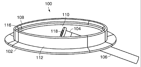
Referring to Figs. 1-4, in one embodiment, a medical device for allowing
percutaneous
access to a patient's body is an access device 100 which includes a housing
112, a cavity 110, a
first opening 116, a flange 102, a second opening 114, and a connector 104.
The housing 112
defines the cavity 110, the first opening 116 (which leads into the cavity
110), and the second
opening 114 (which also leads into the cavity 110).
The housing 112 is implanted in a patient to dispose the cavity 110
subcutaneously
within the patient. After the housing 112 is implanted in the patient, the
first opening 116 is
substantially flush with the surface of the skin of the patient and creates a
percutaneous
passageway from the exterior of the skin of the patient into the cavity 110.
The second opening
114 creates a passageway from the cavity 110 into the interior of the patient.
The connector 104
is coupled to the second opening 114 and is disposed substantially within the
cavity 110. The
connector 104 allows a first device which is external to the patient, such as
an infusion pump for
example, to be connected to a second device disposed within the interior of
the patient, such as a
catheter 106 for example. The flange 102, which is coupled to the housing 112,
holds the
housing 112 in place once the housing 112 is implanted in a patient. In one
embodiment, the
housing 112 is made of a bio-compatible material such as Polysulfone or
Titanium. The housing
112 can also be made of a molded bio-compatible plastic material. In another
embodiment, the
housing 112 can made of a soft material that can be penetrated by sutures or
needles. In some
embodiments, the housing 112 is canoe shaped, elliptically shaped, or almond
shaped, as
indicated in Figs. 17A, 17B, and 17C. In other embodiment, the housing 112
includes a concave
bottom, as indicated in Fig 5 and in still other embodiments the housing 112
include a flat
bottom, as indicated in Fig. 1.
CA 02449795 2003-12-05
WO 03/002171 PCT/US02/17229
-9-
Referring to Fig. 5, in one embodiment, the connector 104 is a luer connector
and is
coupled to the second opening 114 and the catheter 106. A proximal end 516 of
the catheter 106
is first positioned over a distal end 506 of the connector 104. The catheter
is held in place over
the connector 104 by a plurality of barbs 504 (or a raised ring) on the distal
end 506 of the
connector 104. The distal end 506 of the catheter 106 is fed through the
opening 114 until the
plurality of barbs 504 on the distal end 506 of the connector 104 engage a
plurality of barbs 512
within the second opening 114. The connector 104 is secured in place by
engaging the plurality
of barbs 504 with the plurality of barbs 512. After the connector 104 is
secured in place, the
connector 104 is positioned such that the connector 104 is disposed
substantially within the
cavity 110. Specifically, in some embodiments, a small portion of the
connector 104 can extend
out of the first opening 116. However, in other embodiments, no portion of the
connector 104
extends out of the first opening 116 and is disposed entirely within the
cavity 110. In some
embodiments, the connector 106 is sealable when not in use. For example, the
connector 106
can have a threaded or friction fit sealing cap that is removed during use and
replaced when not
in use. The cap can also include a penetrable surface, such as rubber or
silicone for example,
which can be penetrated by a needle. Further, the connector 106 can include a
valve 518 which
opens when the connector 106 is connected to an external device and closes
when the connector
106 is disconnected from the external device. In some embodiments, the value
518 can be a slit
valve made of foam or rubber. The connector 106 is also compatible with
typical medical luer
attachments. In other embodiments, the connector 104 and the catheter 106 can
include a single
lumen or multiple lumens.
Referring to Figs. 1 and 6, in one embodiment, the percutaneous access device
100 is
implanted into a patient 604 as follows. First, a linear incision is made in
the patient. Such an
incision is less traumatic to the patient (as opposed to coring). A distal end
of a guidewire is
inserted through the incision, into the patient 604, and into an area in which
the catheter 106 is to
CA 02449795 2003-12-05
WO 03/002171 PCT/US02/17229
-10-
be placed, such as vein for example. If necessary, a dilator may be placed
over the guidewire to
dilate the area where the catheter 106 is to be inserted. A proximal end of
the guidewire is
inserted through the second opening 114 and the housing 112 is then inserted
into the patient 604
through the incision. The housing 112 is positioned so that the second opening
114 is axially
aligned with the guidewire, the flange 102 is subcutaneous, and the first
opening 116 is
substantially flush with the surface of the patient's skin 604. The flange 102
promotes stability
of the housing 112 and adhesion of the skin and subdermal layers immediately
adjacent to the
incision site. In some embodiments, subcutaneous sutures sewn through holes in
the flange 102
can be used to anchor the subdermal layers to the flange 102. In other
embodiments
subcutaneous hooks may be used to anchor the subdermal layers to the flange
102. In still other
embodiments, the flange 102 can be coated with materials that promote tissue
growth to provide
better sealing of the incision, such as collagen or other tissue growth
catalysts, for example.
Materials that promote ingrowth of cells, such as a permeable fabric, a
textured polymer, or a
steel mesh can also be bonded to or embedded in the flange 102. The added
ingrowth materials
cause the skin surrounding the flange 102 to bond securely with the flange
102. The surface of
the patient's skin 604 may also be secured to the housing 112 by using glue,
such as Dermabond
(a trademark of and a product commercially available from Closure Medical
Corporation of
Raleigh, N.C.) or medical tape around the incision site.
After the housing 112 is anchored in place, the distal end 514 of the catheter
106 is
inserted through the second opening 114 over the guidewire and fed into the
patient. Next, the
guidewire is removed and the proximal end 516 of the catheter 106 is coupled
to the distal end
506 of the connector 104 and the distal end 506 of the connector 104 is fed
through the opening
114 and secured in place (as previously described) thereby sealing the opening
114 and creating
a fluid path 502 from the interior of the patient to the connector 104.
CA 02449795 2003-12-05
WO 03/002171 PCT/US02/17229
-11-
The implanted access device 100 can then be covered with a temporary dressing
or
Tegaderm (a trademark of and a product commercially available from 3M Health
Care Ltd. of
Loughborough, UK) which is a skin-like bandage. The cavity I 10 can also be
filed with gauze
and/or antimicrobial agents. In another embodiment, the housing 112 can be
covered with a low-
profile housing cover 602, which can be shaped to conform to the contour of
the patient's skin.
The housing cover 602 couples to an edge 108 of the housing 112 and creates a
watertight seal
and protects the connector 106 and the cavity 110 from debris and damage from
the
environment. In some embodiments, the housing cover 602 includes a locking
mechanism
which prevents the housing cover 602 from being accidentally or intentionally
removed by the
patient. For example, the housing cover 602 can be secured to the housing 112
by using a
friction fit or a thread fit. The housing cover 602 can also be secured to the
housing 112 using
clamps that clamp onto the edge 108. The clamps can be configured to
selectively engage and
disengage the edge 108 when a key is inserted into the housing cover and
turned. In other
embodiments, the housing cover 602 can be coupled to the housing 112 with a
wire or a hinge,
for example. Additionally, gauze can be placed around the first opening 116
between the
patient's skin and the housing cover 602.
In another embodiment, after the linear incision is made, the guidewire is
inserted into
the vein (or other organ) and then the distal end 514 of the catheter 106 is
inserted into the vein
over the guidewire. Next, the proximal end 516 of the catheter is inserted
into the second
opening 114 and fed into the housing 112. The guidewire is removed and the
distal end 506 of
the connector 104 is then coupled to the proximal end 516 of the catheter and
then fed through
the opening 114 and secured. The housing 112 is then implanted into the
patient using the
procedure previously described.
Referring to Figs. 6 and 7, the connector 104 is accessed by first removing
the housing
cover 602. Next, an external medical device, such as a connection tube 702 to
an infusion pump,
CA 02449795 2003-12-05
WO 03/002171 PCT/US02/17229
-12-
is connected to the connector 104 creating a fluid connection 502 through
opening 118. After
the procedure utilizing the infusion pump has been completed, the connection
tube 702 is
disconnected from the connector 104 and the housing cover 602 is placed back
on the housing
112 to seal the cavity 110 and protect the connector 104. The access device
100 can be used
with a variety of other medical devices, such as body fluid removal devices
and blood
purification devices, for example.
The access device 100, after initial surgical implantation, enables physicians
and/or other
medical personnel to repeatedly (and without further surgery) access various
internal regions of
the patient, such as veins, arteries, and various organs for example. The
connector 104 and fluid
connection that extends into the patient's body is shielded from the patient's
skin and from the
external environment, and thereby reduces opportunity for infection. The
access device 100 has
no protruding external elements, and can be protected by the low-profile
housing cover 602
which is substantially flush with the patient's skin and thereby allows the
patient to engage in
substantially normal physical activity.
Referring again to Figs. 5 and 6, if the access device 100 remains implanted
for an
extended period of time, the connector 104 and/or the catheter 106 may need to
be replaced.
Replacement of these components can be achieved without surgery. First, the
housing cover 602
is removed from the housing 112. Next, the connector 104 is removed from the
second opening
114 and the connector 104 is decoupled from the catheter 106. A guidewire is
fed through the
catheter into the patient. The catheter 106 is then removed from the patient.
A new catheter 106
is inserted into the patient over the guidewire through the second opening 114
and the guidewire
is then removed. A new connector 104 is coupled to the catheter 106 and
secured in the second
opening 114 as previously described. A benefit of this feature is that the
connector 104 and/or
the catheter 106 can be replaced without surgery, resulting in less trauma to
the patient and
reduced chance of infection.
CA 02449795 2003-12-05
WO 03/002171 PCT/US02/17229
- 13 -
Referring to Figs. 8A and 8B, in another embodiment, the connector 104 is a
luer
connector and is coupled to the second opening 114 and the catheter 106. A
proximal end 516 of
the catheter 106 is first positioned over a distal end 506 of the connector
104. The catheter 106
is held in place over the connector 104 by a plurality of barbs 804 (or a
rings) on the distal end
506 of the connector 104. The distal end of the catheter 106 is fed through
the opening 114 until
the plurality of barbs 504 on the distal end 506 of the connector 104 meet a
plurality of 0-rings
802. The connector 104 is secured in place by engaging the plurality of barbs
504 with the
plurality of 0-rings 802. After the connector 104 is secured in place, the
connector 104 is
positioned such that the entire connector 104 is disposed entirely within the
cavity I 10. The
connector 104 also includes a threaded locking cap 808 which engages threads
806. The locking
cap 808 is used to secure a connection between the connector 104 and an
external medical
device. The connector 104 can also include a valve 810 which remains sealed
when no external
medical device is connected to the connector 104 and opens when the connector
104 is
connected to an external medical device, such as a connection tube 812 to an
infusion pump.
Referring to Figs. 9A and 9B, in another embodiment, the access device 100
includes a
pivoting luer connector 902. The pivoting luer connector 902, when pivoted to
a first position,
opens the fluid path 502 through the second opening 114 and, when pivoted to a
second position,
closes the fluid path 502 through the second opening 114. In operation, when
the pivoting luer
connector 902 is not in use, the pivoting luer connector 902 is pivoted to the
second position
thereby keeping the fluid path 502 closed. The pivoting luer connector 902 is
only pivoted to the
first position after an external medical device has been connected to the
pivoting connector 902.
Referring to Fig. l0A and 10B, in still another embodiment, the access device
100
includes a telescopic luer connector 1002. The telescopic connector 1002, when
not in use, is
disposed entirely within the cavity 110. However, when the telescopic
connector 1002 is in use,
the telescopic connector 1002 can be extended out of the cavity 110 to allow a
physician or other
CA 02449795 2008-09-29
-14-
medical personnel to connect an external medical device more easily. In
another
embodiment, the telescopic connector 1002 includes a stop or plug disposed
inside the
connector 1002. The stop is coaxial with the opening 118 and acts as a valve
which
seals the opening 118 when the telescopic connector 1002 is retracted. When
the
telescopic connector 1002 is extended, the seal between the opening 118 and
the stop is
broken thereby allowing fluid to flow past the stop and through the opening
118.
Referring to Fig. 13, in other embodiments, the access device 100 includes two
luer connectors 1304a and 1304b and two corresponding catheters 1302a and
1302b. In
this configuration, blood, for example, can be easily drawn out of a patient,
purified,
and put back into the patient. In another embodiment, the access device 100
includes
two luer connections that both connect to a single catheter. The single
catheter can be a
single lumen catheter or multilumen catheter.
In other embodiment, the access device 100 includes a luer connector with a
pressure responsive slit valve. The valve includes a diaphragm including a
slit which is
flexed in one direction by hydrostatic pressure and flexed in an opposite
direction by
negative pressure to selectively open the slit. Examples of such a pressure-
responsive
slit valves are shown in U.S. Patent 5,205,834, U.S. Patent 5,201,722, and
U.S. Patent
5,169,393.
Referring to Figs. 11, 12, and 14, in another embodiment, the cavity 110 of
the
access device 100 is used to store a small printed circuit board 1102
including
electronics 1108 and/or a battery 1106 used in conjunction with one or more
medical
devices implanted in a patient, such as a pacemaker 1402 and/or a sensor 1404,
for
example. In this configuration, the connector 104 is an electrical connector.
The
connector 104 is positioned such that the connector 104 is disposed
substantially within
the cavity 110. Specifically, in some embodiments, a small portion 25 of the
connector
104 can extend out of the first opening 116. However, in other embodiments,
CA 02449795 2003-12-05
WO 03/002171 PCT/US02/17229
- 15-
no portion of the connector 104 extends out of the first opening 116 and is
disposed entirely
within the cavity 110.
Wires (or optical fiber) 1202 from the connector 104 extend subcutaneously
from the
housing 112 and connect to the pacemaker 1402 and/or sensor 1404. Wires (or
optical fiber)
1104 extending from the connector 104 inside the cavity 110 connect to a
connector 1110 on the
printed circuit board 1102. The printed circuit bard 1102 is coupled to the
housing 112 inside
the cavity I 10 by mounting posts 1204.
Referring to Fig. 16B, in one embodiment, an infusion pump 1616 and a
medication
reservoir 1618 can be housed in the cavity 110 of the access device 100. The
medication
reservoir 1618 supplies medication to the infusion pump 1616 through tube
1620. The infusion
pump 1616 pumps medication though tube 1622, through luer connector 1626, and
through
catheter 1628 and into the patient's body 604. Electronics 1614 (housed in the
cavity 110) can
include a battery to power the infusion pump 1616 and control circuitry to
control the infusion
pump 1616. In addition to the luer connector 1626, the access device 100 can
also include one
or more electronic connectors, such as electrical connector 1630. Electronic
connector 1630 can
be used to connect power and control electronics to a sensor 1624, or other
device (via wires or
optical fiber 1626) implanted in the patient, for example. In another
embodiment, the entire
cavity 110 can be used as a medication reservoir.
Referring to Fig. 14, in another embodiment, the printed circuit board 1102
(housed in
the cavity 110) can include control circuitry 1108 and a battery 110 to
control and power a
pacemaker 1402 implanted in a patient's body 604. In this configuration, the
battery 1110 can
be replaced without surgery by simply removing the housing cover 602,
replacing the battery
1110, and then replacing the cover 602. Similarly, the electronics 1108
controlling the
pacemaker 1402 can also be repaired and/or adjusted with surgery. In other
embodiments, the
wires 1202 extending into the patent's body 604, can fan out to connect to
multiple medical
CA 02449795 2003-12-05
WO 03/002171 PCT/US02/17229
-16-
devices such as the pacemaker 1402 and one or more sensors 1404. In still
other embodiments,
the access device 100 can include multiple electronic connectors 104.
As previously described, the electronics 1108 on the printed circuit board
1102 can
include control and memory electronics for various sensors, such as pressure
sensors and urine
pH sensors for example. In another embodiment, these sensors (along with
control circuitry and
power) can be housed in the cavity 100, and the fluid to be analyzed (blood or
urine, for
example) is brought into the cavity 100 via an inlet luer connector and pumped
back into the
body via an outlet luer connector.
Referring to Fig. 16A, in still another embodiment, the access housing 100 can
include
any combination of connectors. For example, the access housing 100 can include
an inlet luer
connector 1638 and an outlet luer connector 1636. The inlet connector 1638 is
connected to a
catheter 1632 which is also connected to a vein 1602. The outlet connector
1636 is connected to
a catheter 1634 which is also connected to the vein 1602. The inlet connector
1638 is also
connected to tube 1606 which is connected to a blood purification device 1608
external to the
patient 604. The outlet connector 1636 is also connected to a tube 1604 which
is also connected
to the blood purification device 1608. In operation, the blood purification
device 1608 draws
blood through the catheter 1632, through the inlet connector 1638, through the
tube 1606 and
into the blood purification device. After the blood is purified, the blood
purification device 1608
pumps the purified blood through tube 1604, through outlet connector 1636,
through catheter
1634 and back into the vein 1602. Further, the access device 100 can include
an electronic
connector 1640 which connects control and power electronics 1612 to a medical
device (via
Wires or optical fiber 1642) such as a blood press sensor 1610 implanted in
the patient 604.
In another embodiment, the cavity 110 of the access device 100 can be
configured to
house various electro-mechanical components of an artificial heart implanted
in a patient. In this
CA 02449795 2003-12-05
WO 03/002171 PCT/US02/17229
-17-
embodiment, the electro-mechanical parts are accessible (without requiring
surgery) by
removing the housing cover 602.
In other embodiments, the access device 100 can include electronics capable of
wireless
communication. In this embodiment, physicians and/or medical personnel can
wirelessly
communicate with electronics stored in the cavity 110 (without removing the
cover 602) to
download data from various sensors implanted.in a patient, for example. The
physician can also
download a status of a medication reservoir or a status of battery power.
Further, the electronics
housed in the cavity used to communicate with and control various implanted
medical devices
can do so wirelessly. For example, a sensor used for sensing the pressure in a
particular artery
can transmit sensor data wirelessly to an electronic storage element in the
cavity 110, or control
circuitry used for controlling a pacemaker can transmit control signals
wirelessly to the
pacemaker.
In another embodiment, sensor signals can be transmitted through a fluid. For
example, a
pressure sensor is housed within the cavity 110. The sensor is in physical
communication with a
proximal portion of an elongated membrane that contains a fluid. The elongated
membrane
extends outside the cavity 110 into the interior of the patient. A pressure
change in the patient
causes pressure on a distal portion of the membrane which, in turn, causes the
fluid within the
membrane to flow back to the proximal portion of the membrane and be detected
by the pressure
sensor.
Referring to Figs. 15A and 15B, in another embodiment, the housing cover 602
can
include low-profile electronic connectors 1506 and/or fiber optics connectors
1508 which can be
used to access and read out sensor data stored in a memory chip on the printed
circuit board 1102
without having to remove the housing cover 602. The housing cover 602 can also
include a low-
profile luer connector 1510 which enables a fluid connection to the cavity
110. Such a fluid
connection enables a physician and/or medical personnel to access a medical
device implanted in
CA 02449795 2003-12-05
WO 03/002171 PCT/US02/17229
- 18-
the patient (through the cavity 110) or refill a medication reservoir within
the cavity 110 without
having to remove the housing cover 602. Further, the housing cover 602 can
also include
indicator LEDs 1502 to indicate low battery power or low medication reservoir
levels, for
example. The housing cover 602 can also include a battery connector 1502 to
enable recharging
of a battery 1110 stored in the cavity 110 without having to remove the cover
602. The housing
cover 602 can also include a low-profile liquid crystal or LED display for
reading sensor data,
providing a status of battery power, or providing a status of a medication
reservoir stored in the
cavity 110, for example. Moreover, the housing cover 602 can include an
infrared or wireless
communication port 1514 to allow wireless communication with electronics
stored within the
cavity 110 and/or medical devices implanted in the patient.
Some of the benefits of utilizing the access device 100 to store such
electronics and/or
batteries include nonsurgical accessibility of the electronics for repair
and/or replacement,
nonsurgical battery replacement, patient comfort, and reduced chance of
infection from
electronic components.
Variations, modifications, and other implementations of what is described
herein will
occur to those of ordinary skill in the art without departing from the spirit
and the scope of the
invention. Accordingly, the invention is not to be defined solely by the
preceding illustrative
description.
What is claimed is: