Note: Descriptions are shown in the official language in which they were submitted.
CA 02449934 2003-12-08
WO 02/100328 PCT/US02/17553
TREATING PAIN BY TARGETING HYPERPOLARIZATION-ACTIVATED,
CYCLIC NUCLEOTIDE-GATED CHANNELS
Field of the Invention
The present invention relates to treatment of pain.
More particularly, the present invention relates to using
hyperpolarization-activated, cyclic nucleotide-gated (HCN
pacemaker) channels as therapeutic targets for the
treatment of neuropathic pain and inflammatory pain.
Background of the Invention
Pain can be devastating to the sufferer. The causes
of pain can include inflammation, injury, disease, muscle
spasm and the onset of a neuropathic event or syndrome.
Generally, pain is experienced when bodily tissues are
subjected to mechanical, thermal or chemical stimuli of
sufficient intensity to be capable of producing tissue
damage. Pain resolves when the stimulus is removed or the
injured tissue heals. However, under conditions of
inflammatory sensitization or damage to actual nerve
tissue, spontaneous pain may become chronic or permanent
despite apparent tissue healing. Pain may be felt in the
absence of an external stimulus and the pain experienced
due to stimuli may become disproportionately intense and
persistent.
Inflammatory pain can result from surgery, an adverse
physical, chemical or thermal event, infection by a
biologic agent, and/or idiopathic/autoimmune processes.
Causes of inflammatory pain are numerous and include, but
are not limited to, infections, burn pain, rheumatoid
arthritis, inflammatory arthritis, ankylosing spondylitis,
1
CA 02449934 2003-12-08
WO 02/100328 PCT/US02/17553
osteoarthritis, colitis, irritable bowel disease,
carditis, dermatitis, myositis, neuritis and collagen
vascular diseases, as well as cancer. Current methods for
treating inflammatory pain have many drawbacks and
deficiencies. For example, Corticosteroids, which are
commonly used to suppress destructive autoimmune
processes, can result in undesirable side effects
including, but not limited to, vulnerability to infection,
weakening of tissues and loss of bone density leading to
fractures, and ocular cataract formation.
Neuropathic pain is defined as pain induced by injury
or disease of the peripheral or central nervous system.
NeuropathiC pain conditions are heterogeneous and include,
but are not limited to, mechanical nerve injury, e.g.,
carpal tunnel syndrome, radiculopathy due to
intervertebral disk herniation; post-amputation syndromes,
e.g. stump pain, phantom limb pain; metabolic disease,
e.g., diabetic neuropathy; neurotropic viral disease,
e.g., herpes zoster, human immunodeficiency virus (HIV)
disease; cancer, e.g. tumor infiltration, irritation or
compression of nervous tissue; radiation neuritis, as
after cancer radiotherapy; neurotoxiCity, e.g., caused by
exogenous substances such as chemotherapy of cancer, HIV
or tuberculosis; inflammatory and/or immunologic
mechanisms, e.g., multiple sclerosis, paraneoplastiC
syndromes; nervous system focal ischemia, e.g., thalamic
syndrome (anesthesia dolorosa); multiple neurotransmitter
system dysfunction, e.g., complex regional pain syndrome
(CRPS); and idiopathic causes, e.g., trigeminal neuralgia.
The long-term treatment of chronic pain of any
etiology may be very challenging. Although pain may
respond to conventional analgesics, the side effects may
2
CA 02449934 2003-12-08
WO 02/100328 PCT/US02/17553
not be tolerable, or tolerance to the analgesic effects of
the drug in question may render therapy problematic.
Therapy with ibuprofen and aspirin (both nonsteroidal
anti-inflammatory drugs) may be limited by
gastrointestinal side effects. Chronic therapy with
opiate drugs (morphine, codeine, hydrocodone, oxycodone,
etc. and derivatives) may be unacceptable to either the
patient or the physician due to side effects (sedation,
Constipation, etc.), the difficulties of pain management
associated with drug tolerance or withdrawal phenomena,
and to social factors (the stigma of opiate consumption,
concerns about substance abuse potential, drug diversion,
loss of productivity, etc).
It is well known to both preclinical investigators
and Clinicians that neuropathiC pain is particularly
difficult to treat. Commonly used analgesics such as
opiates and nonsteroidal anti-inflammatory drugs are often
ineffective to alleviate neuropathiC pain. For morphine-
like drugs (opiates), perceived efficacy may have to do
with sedation (i.e., the patient is too sedated to care
about pain). Also, the use of opiates to treat
neuropathiC pain may be more likely to be associated with
tolerance and escalating dose requirements that render
therapy problematic. Therefore, the analgesic effects of
these Compounds may be transient. The vast majority of
patients treated with these analgesics Continue to
experience pain and may not experience pain relief at all.
A number of so-called "adjuvant" analgesics, drugs not
typically thought of as pain relievers, such as the
tricycliC antidepressants (e. g. amitriptyline,
nortriptyline, desipramine, imipramine), certain
anticonvulsants (e. g. carbamazepine, gabapentin,
phenytoin, lamotrigine), the antiarrythmiC drugs
3
CA 02449934 2003-12-08
WO 02/100328 PCT/US02/17553
mexiletine, lidocaine, and tocainide, and various
miscellaneous drugs such as baclofen (GABA-B agonist) and
clonidine (alpha2 adrenergic agonist) have become the
mainstays of neuropathic pain therapy. These agents,
however, also suffer from limited efficacy or significant
side effects ranging from sedation to cardiovascular
effects to life-threatening bone marrow suppression. A
number of invasive treatments exist, both pharmacological
(nerve blocks, spinal injections, implantable drug
delivery devices) and non-pharmacological (e. g.
implantable nerve/spinal cord stimulators, neuroablative
procedures); all suffer from both limited efficacy and the
drawback of the known potential for complications due to
the respective procedures. Limitations of the current
armamentarium of analgesics call for development of novel
methods and strategies with original mechanisms for the
treatment of neuropathic pain.
Despite the diversity of etiologies, many neuropathic
pain syndromes share common clinical characteristics.
Symptoms of neuropathic pain include unusual sensations of
burning, tingling, electricity, pins and needles,
stiffness, numbness in the extremities, feelings of bodily
distortion, allodynia (pain evoked by innocuous
stimulation of the skin), hyperalgesia (lowered threshold
for pain, e.g. mild thermal stimuli cause pain)
hyperpathia (an elevated pain threshold however with an
exaggerated pain response once the threshold is surpassed)
summation (cumulative exacerbation of pain with repetitive
mild stimuli), and pain in the absence of other sensory
function in the affected area. These observations have
led to the proposal that many neuropathic pain syndromes
may share common mechanisms.
4
CA 02449934 2003-12-08
WO 02/100328 PCT/US02/17553
Experiments using various animal models have
suggested that spontaneous activity in the peripheral
and/or central nervous system could be a mechanism by
which pain can be explained. A consistent observation
S from animal model studies is that primary afferent neurons
in the dorsal root ganglia of affected spinal levels
demonstrate spontaneous discharges. These discharges are
predominantly associated with Af3 and A8 fibers although
enhanced C fiber activity may also be involved.
Additional studies have demonstrated spontaneous or
abnormally easily evoked discharges in second-order
neurons in the spinal cord dorsal horn upon which these
primary afferent neurons synapse. Consequently, it is
held that treatments that suppress the spontaneous
discharges or abnormal excitability will thereby decrease
pain (Gold, (2000) Pain 84: 117-20). In particular, drugs
or compounds that selectively suppress
spontaneous/hyperexcitable discharges without interfering
with other normal neuronal transmission are likely to be
useful in the treatment of pain syndromes.
Summary of the Invention
This invention teaches the role of a hithertofore
unknown cellular component involved in pain: HCN
pacemaker channels, which can serve as a specific
therapeutic target for developing novel treatment for
pain, preferably neuropathic or inflammatory pain.
In one aspect, the present invention relates to a
method for preventing the onset of pain in a subject in
need thereof, comprising administering to the subject a
prophylactically effective dose of a composition that
5
CA 02449934 2003-12-08
WO 02/100328 PCT/US02/17553
decreases the current mediated by an HCN pacemaker
channel, or the expression of an HCN subunit, in a
sensory cell of the subject, in the presence or absence
of one or more other analgesics.
In another aspect, the present invention relates to
a method for treating pain in a subject in need thereof,
comprising administering to the subject a
therapeutically effective dose of a composition that
decreases the current density of a current mediated by
an HCN pacemaker channel, or the expression of an HCN
subunit, in a sensory cell of the subject, in the
presence or absence of one or more other analgesics.
In another aspect, the present invention relates to
a method of identifying a compound useful for treating
pain, comprising the steps of:
(a) contacting a test compound with an HCN
pacemaker protein; and
(b) determining the ability of the compound to
decrease HCN-pacemaker channel-mediated currents.
Optionally the method can be further confirmed through
the addition of an additional step comprising:
administering the compound to an animal model for pain.
The present invention relates to.another method of
identifying a compound useful for treating pain,
comprising the steps of:
(a) contacting a test compound with a regulatory
sequence for an HCN pacemaker gene or a protein
that binds to the regulatory sequence for an HCN
pacemaker gene; and
6
CA 02449934 2003-12-08
WO 02/100328 PCT/US02/17553
(b) determining whether the test compound
decreases the expression of the HCN gene
controlled by said regulatory sequence.
Optionally the method can be further confirmed through
the addition of an additional step comprising:
administering the compound to an animal model for pain.
The present invention relates to yet another method
of identifying a compound useful for treating pain,
comprising the steps of:
(a) combining a test compound, a measurably labeled
ligand for an HCN pacemaker protein, and an HCN
pacemaker protein; and
(b) measuring binding of the compound to the HCN
pacemaker protein by a reduction in the amount of
labeled ligand binding to the HCN pacemaker protein.
Optionally the method can be further confirmed through
the addition of an additional step comprising:
administering the compound to an animal model for pain.
Also included in the present invention is an
antibody that binds specifically to the carboxy-terminus
of a HCN protein.
Other features and advantages of the invention will
be apparent from the following detailed description and
claims.
Brief Description of the Drawings
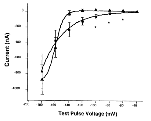
Figure 1. Hyperpolarization-activated currents were
elicited in Xenopus oocytes previously injected with
human HCNl cRNA (HCNl) or water (control). The figure
7
CA 02449934 2003-12-08
WO 02/100328 PCT/US02/17553
shows the mean +/- SEM inward current at the indicated
test pulse potential at the end of an 800 msec voltage
step. Inward currents were elicited in HCN1-injected
oocytes by hyperpolari~ation steps equal to or greater
than -60 mV (squares; n=4 oocytes from 2 separate
batches of oocytes). There were no detectable time-
dependent inward currents until ~-140 mV in control
sister oocytes (triangle; n=8 oocytes from 2 separate
batches of oocytes). At voltage steps in the
physiological range, there was a significant difference
(asterisks indicate p <0.05; Student's t-test).
Reproducible results were obtained from two separate
oocyte batches and the data were pooled.
Figure 2. Low threshold hyperpolarization-activated
currents were elicited in HEK293 cells stably expressing
human HCN3. (a). Slowly activating inward currents were
elicited by the voltage protocol shown in (b). The
current-voltage relationship (c) reveals a threshold
voltage for activation near -84 mV (note that the voltage
axis includes the junction potential correction of -14
mV). (d). No inward currents are observed in control
cells (same voltage protocol as shown in (b)).
Intracellular solution: K gluconate IS; Extracellular
solution: Tyrode's. Holding potential was -64 mV.
Figure 3. The figure illustrates data obtained using an
in-continuity preparation of excised dorsal root, dorsal
root ganglion and spinal nerve from rats having been
prepared with L4/5 spinal nerve ligation (SNL) 1-3 weeks
previously. Spontaneous discharges were recorded in vitro
in an ACSF bath (see Example 4) . In panels (a) and (b) ,
examples of the effect of bath application of 100
micromolar ZD7288 (a specific blocker of Ih; (BoSmith et
8
CA 02449934 2003-12-08
WO 02/100328 PCT/US02/17553
al., (1993) Br J Pharmacol 110: 343-9)) on spontaneous
firing of Af3 and A8 neurons (distinguished by conduction
velocity) are illustrated. Panel (a): Histogram (y-axis=
spikes/second) for single fiber in vitro recording from a
typical Aid fiber before and after application of ZD7288
100 micromolar shows that complete suppression of ectopic
firing was achieved after 3-4 minutes. The horizontal bar
above the histogram indicates the timing and duration of
application of ZD7288 to the preparation. Inset (a)(i)
shows an enlarged view of a one-second recording period
prior to drug application, illustrating baseline spike
frequency; inset (a)(ii) shows a one-second recording
period after ZD7288 application, illustrating the
reduction in firing. The conduction velocity for the
depicted fiber was 31.3 m/sec (Aft range)
Panel (b): Single fiber recording as in (a) from an A8
fiber shows attenuation of firing after ZD7288
application. The horizontal bar above the histogram
indicates the timing and duration of application of ZD7288
to the preparation. Inset (b)(i) shows an enlarged view
of a one-second recording period prior to drug
application, illustrating baseline spike frequency; inset
(b)(ii) shows a one second recording period after ZD7288
application, illustrating the degree of reduction in
firing. The conduction velocity for the depicted fiber
was 7.8 m/sec (A2 range).
Figure 4. This graph shows the time course (x-axis) of
percent change in firing from baseline (y-axis), in the
single fiber recording experiments illustrated in. Figure 3
(above), after ZD7288 application. Horizontal bar between
0-5 minutes indicates timing and duration of ZD7288 100
micromolar application. Data points and error bars
9
CA 02449934 2003-12-08
WO 02/100328 PCT/US02/17553
indicate mean +/- SEM for 7-8 fibers per group. Symbols:
Filled squares= ACSF control (Af3 and A8 fibers combined) ;
open squares= A8 fibers; open circles= Aft fibers. *_
P<.05, 1-way ANOVA, followed by Dunnett's multiple
comparisons.
Figure 5. Allodynia exhibited by SNL rats was blocked in
a dose dependent manner by i.p. administration of ZD7288,
1 mg/kg (open squares), 3 mg/kg (open circles), or 10
mg/kg (filled squares), compared with saline vehicle
(filled circles). (a) Y-axis shows 50% threshold for paw
withdrawal from von Frey hairs; X-axis shows a redacted
time course illustrating pre-ligation baseline paw
threshold (normal), immediate pre-drug baseline threshold
(maximum allodynia, "base") and post-drug administration
time points. The timing of drug administration is
indicated by the arrow. (b) the same data analyzed as a
dose-response curve showing the ED50 of ZD7288 to be ~3
mg/kg for allodynia suppression. To compare dose and drug
effects, raw paw thresholds were normalized as percent of
maximum possible drug effect (oMPE, Y-axis) using the
following formula: o MPE= [post-drug threshold (g)-predrug
allodynia baseline threshold (g)]/[Pre-ligation baseline
threshold (g)]-predrug allodynia baseline threshold (g)] x
100. Pre-drug maximum allodynia (baseline) thresholds
were assumed to reflect 0% drug effect (no suppression of
allodynia) and pre-ligation threshold values were
designated as 100% effect, i.e., a drug effect causing
return of the paw threshold to a normal, pre-ligation
baseline was taken to represent complete suppression of
allodynia.
CA 02449934 2003-12-08
WO 02/100328 PCT/US02/17553
Figure 6. Graph illustrates the minimal effect of i.p.
ZD7288 10 mg/kg (filled circles) on acute thermally-evoked
pain, as determined using the 55 °C hot plate test, in
which the latency to demonstrating escape behavior
(hindpaw licking) from a noxious thermal stimulus is
timed, in normal rats, with and without drug treatment.
Filled circles; ZD7288 10 mg/kg, i.p. (N=8); open circles;
saline vehicle i.p. (N=8). * = P < 0.05, t-test for 75 min
timepoint only; N= 8 per group.
Figure 7. In the rat complete Freund's adjuvant (CFA) paw
model of inflammatory pain, allodynia was suppressed by
HCN blockade with ZD7288, as well as by treatment with.
morphine and ibuprofen, but not by gabapentin. ,All drugs
were administered i.p. Symbols: ZD7288 10 mg/kg, filled
circles; ibuprofen 30 mg/kg, open circles; morphine 3
mg/kg, filled triangles; gabapentin 100 mg/kg, open
triangles. Y-axis: paw withdrawal threshold (g). X-axis:
Normal baseline thresholds, maximum allodynia timepoint
after CFA administration, and timepoints after drug
treatment. N= 6 per group.
Figure 8. Spontaneous pain behaviors were blocked in the
rat mild thermal injury model. Drugs (morphine, ZD7288,
or saline) were administered 10 minutes after the mild
thermal injury. The total amount of time during which
spontaneous pain behaviors were displayed (paw lifting,
paw shaking, guarding posture of paw) during two separate
l0-minute intervals, 30 and 60 minutes after
intraperitoneal vehicle or drug administration, was
recorded.
Panel a: Raw data are presented. Y-axis: cumulative
spontaneous pain behavior time (seconds). X-axis: time
post drug administration (hrs). Both morphine (hatched
11
CA 02449934 2003-12-08
WO 02/100328 PCT/US02/17553
bars, n=3) and ZD7288 (filled bars, n=6) showed near
complete suppression of spontaneous pain behaviors
compared to saline (open bars, n=9) at both 30 and 60 min
after administration (*= P< .0001, one-way ANOVA).
Panel b: Data were converted to percent efficacy (versus
saline). Mean percent efficacy (Y-axis) (0= no effect,
1000= complete suppression of spontaneous pain) was
calculated as (1-(observed pain score / mean overall
saline pain score))x100; percent efficacy for the two
timepoints was averaged. Percent overall efficacy for
morphine was 89.6 +/- 2.1 (mean +/- SEM), for ~D7288 89.1
+/-15.7; * - P<.0001 vs. saline, 1 way ANOVA with Fisher's
PLSD.
Figure 9. Quantitative RT-PCR analysis of HCN mRNAs in
the cell bodies of primary afferent neurons of nerve-
injured (SNL) and control (Sham) rats. The relative
abundance of the four HCN subtypes was simultaneously
measured in whole L5/6 DRGs from 1-week nerve ligated
versus sham control rats. The Y-axis represents relative
mRNA copy number as detected by fluorescence, normalized
to the housekeeping gene Cyclophilin A. Panel (A): A
significant decrease in HCN1 mRNA in SNL samples, compared
to sham controls, was seen using primers that amplified a
region toward the 3' end of the coding sequence (labeled
3' in figure), whereas no significant change in the
abundance of a 5' region amplicon (labeled 5' in figure)
spanning the region of intron #1(Ludwig et al., (1999)
Embo J 18: 2323-9) was seen. Panel (B): A significant
decrease in HCN2 mRNA was seen in SNL samples for the
amplicon in the region of intron #1 (as above). Panel (C)
No significant difference between SNL and control was
observed for HCN3, again, using an amplicon in the region
12
CA 02449934 2003-12-08
WO 02/100328 PCT/US02/17553
of intron #1. Panel (D): No significant difference
between SNL and control was observed for an amplicon in
this region for HCN4. N= 8 SNL and 8 sham control rats.
Asterisks indicate P< .02, unpaired t-test.
Figure 10. Ih was detected in both control (hatched bars)
and SNL (solid bars) L5 large neurons at a step to -114
mV. The distribution of Ih peak current in large DRG
neurons is shown, normalized to cell size (current
density). A much larger population of neurons expressed
high levels of Ih in SNL operated rats compared to sham
controls.
Figure 11. The voltage dependence of Ih activation was
determined using tail current analysis in which the
current through channels that were opened by a previous
voltage step was measured before they deactivated. Open
circles represent sham neurons, and filled circles
represent SNL neurons. Tail currents were determined
from a step to -64 or -54 mV after > 2 sec duration pre-
pulses to a series of voltages between -44 and -154 mV
in -10 mV increments. The voltages at which tail
currents were measured (-64 or -54 mV) were chosen
because tail currents were large enough to provide
accurate measurements and there was little contamination
by other voltage-gated channel currents. The data were
normalized to the maximum tail current observed after
the most hyperpolarizing prepulses (y axis: 1 is maximum
current), then were fit by a Boltzmann function, and the
voltage to half maximal activation (Vo_5) and slope of
the curve were determined. The threshold for activation
was estimated from these plots and was similar to the
values determined by measuring current at the end of >2
sec test steps where the threshold in neurons of SNL
13
CA 02449934 2003-12-08
WO 02/100328 PCT/US02/17553
rats was significantly more positive (Mean +/- SEM: -
64.3 +/- 1.0 mV, n=44) compared to controls (73.9 +/-
1.9 mV, n=35; p« 0.001) . ,V0.5 was calculated to be -
82.5 +/- 2.9 mV in SNL neurons (N=17); this was also
significantly different from controls, -91.0 +/- 2.6
(P<.05, t-test). Slopes, however, did not differ
significantly, at 9.5 +/- 1.1 for SNLs (N=15) and 9.3
+/-1.1 for controls (N=11).
Figure 12. The effect of bath-applied lidocaine-HC1 at
neutral pH on native Ih in normal rat L4 dorsal root
ganglion neurons (large neurons, diameter >42 microns) is
illustrated. Y-axis: percent inhibition of current at -134
mV. X-axis: concentration of lidocaine-HCl (M) expressed
in log. Concentration-dependent block of Ihwas seen with
an ED50 of 23 micromolar. Data were obtained from 3 cells.
Detailed Description of the Invention
The present invention relates to the treatment of
pain. Particularly, the present invention provides a new
therapeutic target, the HCN pacemaker channel, for
developing novel methods and strategies for treatment. of
pain, preferably neuropathic pain or inflammatory pain.
HCN pacemaker channels are involved in pain.
Hyperpolarization-activated, cyclic nucleotide-
gated (HCN) channels have recently been identified as a
family of pacemaker channels responsible for fast
rhythmic oscillations inherent in cardiac and neuronal
depolarizations.
14
CA 02449934 2003-12-08
WO 02/100328 PCT/US02/17553
The pacemaker current is a hyperpolarization-
activated, cation-selective, inward current that
modulates the firing rate of cardiac and neuronal
pacemaker cells. This current is oftentimes abbreviated
as Ih ( "hyperpolarization" ) , If ("funny" ) , or Iq
("queer"). Ih contributes to normal pacemaking in the
sinoatrial node and atrioventricular node of the heart
and Purkinje fibers in the ventricle (DiFrancesco,
(1995) Acta Cardiol 50: 413-27), and to abnormal
automatic activity of cardiac myocytes under
pathological conditions (Opthof, (1998) Cardiovasc Res
38: 537-40.). Ihalso mediates repetitive firing in
neurons and oscillatory behavior in neuronal networks.
In addition, it acts to set the resting potential of
certain excitatory cells, and may function in synaptic
plasticity, and in the activation of sperm (Page, (1996)
Annu Rev Physiol 58: 299-327).
The pacemaker current Ihhas unusual
characteristics, including activation upon
hyperpolarization, a tiny single-channel conductance,
modulation by intracellular cyclic nucleotides,
permeability to both K+ and Na+, and poor permeability to
Li+. Ih is mediated by both Na~ (inward flux at a
resting membrane potential near -70 mV) and K+ (outward
flux at a resting membrane potential near -70 mV), and
has a reversal potential around -30 or -40 mV under
physiologic conditions (Ho et al. (1994), Pflugers Arch
426:68-74)(Mercuri et al., (1995) Eur J Neurosci 7: 462-
9 ) .
Four genes encoding ion channels that conduct
pacemaker currents have recently been cloned. These
genes belong to the HCN family, and have been designated
CA 02449934 2003-12-08
WO 02/100328 PCT/US02/17553
as HCN1, HCN2, HCN3, and HCN4, respectively. HCN
channels share structural features with voltage-gated K+
channels. These features include a GYG K+ channel
signature sequence in the pore loop, and a highly
positively charged S4 domain that is the putative
voltage sensor (Gauss et al., (1998) Nature 393: 583-7;
Ludwig et al., (1998) Nature 393: 587-91; Santoro et
al., (1997) Proc Natl Acad Sci U S A 94: 14815-20;
Santoro et al., (1998) Cell 93: 717-29). HCN channels
are most homologous to the eag family of K+ channels
(for example, erg, eag, elk) and the KAT1 family of
plant K+ channels (Biel et al., (1999) Rev Physiol
Biochem Pharmacol 136: 165-81) in that they possess six
transmembrane domains, and incorporate an intracellular
Cyclic nucleotide binding domain that can modulate the
voltage dependence of activation. For instance, binding
of CAMP to HCN2 shifts the activation curve at least 20
mZT to the right, thus enhancing channel activity at the
resting membrane potential. These four HCN channels
share substantial homology, but have different
activation kinetics and degrees of responsiveness to
cyclic AMP.
A significant feature of the increased spontaneous
discharges observed in rodent neuropathic pain models is
rhythmicity, whether rhythmic firing or rhythmic burst
firing. This feature suggests underlying non-random
processes for generation of the increased spontaneous
discharges. In the present invention, we investigated
the possible role of HCN pacemaker channels in
neuropathic pain and other types of pain.
As used herein, a "HCN pacemaker channel" refers to a
membrane channel, which is a hyperpolarization-activated,
16
CA 02449934 2003-12-08
WO 02/100328 PCT/US02/17553
Cyclic nucleotide-gated channel. A "HCN pacemaker
channel" conducts both Na+ (inward flux from extracellular
milieus to cytosol) and K+ (outward flux) , and has a
reversal potential around -30 or -40 mV under physiologic
conditions. The single channel conductance for mammalian
channels is thought to be quite low (Pape, (1996) Annu Rev
Physiol 58: 299-327). An HCN pacemaker channel likely
comprises tetramers of HCN pacemaker channel subunits. An
HCN pacemaker channel may be heteromeric, when it is made
of at least two different HCN pacemaker channel subunits,
or homomeric, when it is made of the same HCN pacemaker
channel protein subunits. An HCN pacemaker channel might
also contain other subunits as accessories, such as Mirp1
(Yu et al . , (2001) Circ Res 88 : E84-7 . ) .
As used herein, "HCN polypeptide" or "HCN subunit"
refers to a polypeptide that is a subunit or monomer of a
hyperpolarization-activated, cyclic nucleotide-modulated
channel, a member of the HCN gene family. When an HCN
polypeptide, e.g., HCN1, HCN2, HCN3, or HCN4, is part of
an HCN pacemaker channel, either a homomeric or
heteromeric potassium channel, the channel has
hyperpolarization-activated, cyclic nucleotide-gated
activity. The term HCN polypeptide therefore refers to
polymorphic variants, alleles, mutants, and interspecies
homologs that: (1) have a sequence that has greater than
about 60% amino acid sequence identity, preferably about
65, 70, 75, 80, 85, 90, or 95 % amino acid sequence
identity, to an HCN pacemaker channel family member
polypeptide such as human HCNl (SEQ ID NO: 4), human HCN2
(GenBank Protein Id: NP 001185), human HCN3 (SEQ ID No:
10), and human HCN4 (GenBank Protein Id: NP 005468); (2)
bind to antibodies, e.g., polyclonal or monoclonal
17
CA 02449934 2003-12-08
WO 02/100328 PCT/US02/17553
antibodies, raised against an immunogen comprising an HCN
pacemaker channel family member polypeptide, such as
described above, and conservatively modified variants
thereof; (3) encoded by a DNA molecule that specifically
hybridizes under stringent hybridization conditions to a
HCN pacemaker channel family member polynucleotide, such
as human HCN1 (SEQ ID N0: 3), human HCN2 (GenBank
Accession No: NM 001194), human HCN3 (SEQ ID No: 9), and
human HCN4 (GenBank Accession No: NM 005477); or (4)
encoded by a DNA molecule that can be amplified by primers
that specifically hybridize under stringent hybridization
conditions to an HCN pacemaker channel family member
polynucleotide, such as described above.
Exemplary high stringency or stringent hybridization
conditions include: 50o formamide, 5x SSC and 1 o SDS
incubated at 42 °C or 5x SSC and 1% SDS incubated at 65 °C,
with a wash in 0.2x SSC and 0.1o SDS at 65 °C.
As used herein, a "HCN pacemaker gene" refers to a
DNA molecule that (1) encodes a protein having a sequence
that has greater than about 60% amino acid sequence
identity, preferably about 65, 70, 75, 80, 85, 90, or 95
amino acid sequence identity, to an HCN pacemaker channel
family member polypeptide such. as human HCN1 (SEQ ID NO:
4), human HCN2 (GenBank Protein Id: NP 001185), human HCN3
(SEQ ID No: 10), and human HCN4 (GenBank Protein Id:
NP-005468); (2) encodes a protein capable of binding to
antibodies, e.g., polyclonal or monoclonal antibodies,
raised against an immunogen comprising an HCN pacemaker
channel family member polypeptide, such as described
above, and conservatively modified variants thereof; (3)
specifically hybridizes under stringent hybridization
conditions to an HCN pacemaker channel family member
18
CA 02449934 2003-12-08
WO 02/100328 PCT/US02/17553
polynucleotide, such as human HCN1 (SEQ ID NO: 3), human
HCN2 (GenBank Accession No: NM_001194), human HCN3 (SEQ ID
No: 9), and human HCN4 (GenBank Accession No: NM-005477);
or (4) can be amplified by primers that specifically
hybridize under stringent hybridization conditions to an
HCN pacemaker family polynucleotide, such as described
above.
As used herein, the term "HCN pacemaker channel
family" is intended to mean two or more proteins or
nucleic acid molecules having a common structural domain
and having sufficient amino acid or nucleotide sequence
identity to a known HCN pacemaker member, such as HCN1,
HCN2, HCN3, or HCN4. Family members can be from either
the same or different species. For example, a family
can comprises two or more proteins of human origin, or
can comprise one or more proteins of human origin and
one or more of non-human origin.
In the present invention, we investigated levels of
the mRNA and protein of HCN subunits, and whole cell
current mediated by HCN pacemaker subunits in dorsal
root ganglion (DRG) neurons from animal models of pain
compared to those from the Control animals.
As used herein, "control animal(s)" include a variety
of preclinical animals that do not exhibit pain syndromes.
"Animal models for pain" include a variety of preclinical
animals that exhibit pain syndromes. Commonly studied
rodent models of neuropathic pain include: the chronic
constriction injury (CCI or Bennett) model; neuroma or
axotomy models; the spinal nerve ligation (SNL or Chung)
model; and the partial sciatic transection or Seltzer
19
CA 02449934 2003-12-08
WO 02/100328 PCT/US02/17553
model (Shir et al . , (1990) Neurosci Lett 115 : 62-7) .
Neuropathic pain models include, but are not limited to,
several traumatic nerve injury preparations (Bennett et
al., (1988) Pain 33: 87-107; Decosterd et al., (2000) Pain
87: 149-58; Kim et al., (1992) Pain 50: 355-363; Shir et
al., (1990) Neurosci Lett 115: 62-7), neuroinflammation
models (Chacur et al., (2001) Pain 94: 231-44; Milligan et
al., (2000) Brain Res 861: 105-16) diabetic neuropathy
(Calcutt et al., (1997) Br J Pharmacol 122: 1478-82),
virally induced neuropathy (Fleetwood-Walker et al.,
(1999) J Gen Virol 80: 2433-6.), vincristine neuropathy
(Aley et al., (1996) Neuroscience 73: 259-65; Nozaki-
Taguchi et al., (2001) Pain 93: 69-76.), and paclitaxel
neuropathy (Cavaletti et al., (1995) Exp Neurol 133: 64-
72). Commonly studied rodent models of inflammatory pain
include: the complete Freund's adjuvant (CFA)-induced
inflammation model, experimental burn injury models, the
carrageenan inflammatory hyperalgesia model, the formalin
test, and the rat inflamed knee and ankle joint models.
Assessment of pain and therapeutic responses to
pharmacological and other interventions is done in a
variety of ways, including behavioral and
electrophysiological assessment, the latter providing
"surrogate" outcomes. "Surrogate" assessments attempt to
correlate physiological findings with behavior. Among the
best-studied surrogate responses are electrophysiological
responses of 1) primary afferent neurons, and 2)
spinothalamic tract neurons in the dorsal horn of the
spinal cord.
1. Two full-length human HCN pacemaker channel cDNAs were
isolated and characterized.
CA 02449934 2003-12-08
WO 02/100328 PCT/US02/17553
Two full-length human HCN pacemaker cDNA sequences
(SEQ ID NO: 3 (hHCN1) and SEQ ID NO: 9 (hHCN3) were
isolated and cloned from human spinal cord cDNA and
human Marathon ready brain cDNA, respectively. The two
DNA molecules encode two polypeptides, SEQ ID N0: 4 and
SEQ ID NO: 10, respectively (Example 1).
We have demonstrated that the two newly isolated
full length human HCN pacemaker cDNA sequences encode
protein products that form functional HCN pacemaker
channels in either an oocyte expression system (Fig. 1
and Example 2) or a mammalian expression system (Fig. 2
and Example 3).
We subsequently found that similar sequences had
been isolated and disclosed elsewhere (see Wo0063349,
Wo0190142, Wo0202630, Wo0212340, and Wo9932615).
2. Specific blockade of HCN channels suppressed
spontaneous firing of injured primary afferents and the
tactile allodynia in an animal neuropathic pain model.
We performed extracellular recording in vitro on
peripheral nerve fibers in control or previously ligated
L4 or L5 (SNL) excised nerve-DRG preparations (Example 4).
Spontaneous discharges arose from Aa/i~ neurons and some A~
neurons (distinguished by their conduction velocity) in
DRGs 1 to 3 weeks post injury (Fig. 3). Spontaneous
action potentials tended to be rhythmic. Discharges from
Aft fibers were completely suppressed by bath application
of 100 ~,M ZD7288, which is a specific blocker of Ih current
(BoSmith et al., (1993) Br J Pharmacol 110: 343-9) but has
no selectivity between. HCN channel family members, for the
21
CA 02449934 2003-12-08
WO 02/100328 PCT/US02/17553
duration of the extended period of observation (Fig. 3a,
Fig. 4). Fiber identities (Aid, A8) were determined by
evoked action potentials after data collection; in
distinction to the suppression of spontaneous discharge,
there was no conduction blockade of evoked action
potentials observed after the application of ZD7288.
Thus, these data illustrate that (1) Ih is critical to the
generation of spontaneous firing of injured primary
afferents, and that (2) blockade of Ih only suppresses
spontaneous activity, but does not cause generalized
failure of neuronal conduction (or nerve block).
We also tested the role of I,, in animal behavioral
studies. Allodynia, or pathological sensitivity to touch,
is among the most troublesome of neuropathic symptoms, and
is thought to arise from abnormal responses of large
myelinated sensory fibers (Af3) to stimulation. In the
present invention, we used the SNL (unilateral L5/6
ligation) model to study the role of Ih in tactile
allodynia (Example 5). We observed that ZD7288 (10 mg/kg)
suppressed the tactile allodynia exhibited by awake SNL
rats, without evidence of total sensory blockade or
numbness and without overt adverse effects on behavior, in
a dose-dependent manner (Fig. 5). Clearly, Ihcontributes
to pathologic neuronal activity manifested as tactile
allodynia.
Results described supra demonstrated, for the first
time, that Ih is critical to the generation of spontaneous
firing of injured primary afferents, and that blockade of
I,, ameliorates neuropathic pain behavior associated with
such abnormal firing.
22
CA 02449934 2003-12-08
WO 02/100328 PCT/US02/17553
3. Specific blockade of HCN channels did not yield
analgesia of a clinically relevant magnitude against acute
thermal stimuli.
To test whether the antiallodynic effect seen with
specific blockade of HCN channels, ZD7288, is a general
analgesic effect independent of neuropathic pain, the hot
plate test was performed to evaluate effects of ZD7288 on
an acute thermally induced pain state with normal rats
(Example 6). No statistically significant difference was
seen between treatment with ZD7288 and saline at 45 or 60
min; a statistically significant, only minor difference
was seen at 75 min (approximately 15%) (Fig.6).
These results demonstrate that specific blockade of
HCN channels does not yield analgesia of a clinically
relevant magnitude against acute thermal stimuli.
Therefore, the antiallodynic effects in the SNL model do
not represent a generalized impairment of sensory
function. Tn addition, these results demonstrate that
ZD7288 does not impair the ability of rats to respond to
perceived noxious stimuli; thus, the effect of ZD7288 on
allodynia thresholds is not due to inhibition of motor
responses or cognitive depression.
4. Specific blockade of HCN channels suppressed the
tactile allodynia in an animal inflammatory pain model.
We also tested the role of HCN in inflammatory pain,
which differs mechanistically and pharmacologically from
neuropathic pain (Example 7). After injection of complete
Freund's adjuvant (CFA) into one hind-paw of rats, animals
developed marked tactile allodynia as measured using von
Frey hairs (baseline, Figure 7). Similar to morphine, a
23
CA 02449934 2003-12-08
WO 02/100328 PCT/US02/17553
drug that is known to be effective against inflammatory
pain, ZD7288 suppressed allodynia in CFA-injected rats, as
shown by causing return of the paw threshold toward a
normal, pre-CFA injection level (Figure 7). Ibuprofen
also showed some efficacy.
However, blockade of HCN channels with ZD7288 at 10
mg/kg, i.p., had no effect on thermal hyperalgesia
(measured using a modified Hargreaves apparatus) in two
different models of inflammatory pain: the rat carrageenan
model of hindpaw inflammation, and the rat complete
Freund's adjuvant (CFA) model of hindpaw inflammation. As
shown supra, HCN blockade also had little effect on acute
thermally evoked pain in the hot plate test.
Our data indicate that although specific blockade of
HCN channels suppressed tactile allodynia in both
neuropathic pain and inflammatory pain animal models,
temperature sensation even in the presence of nociceptor
sensitization in the periphery (e. g. skin) is not affected
by Ih blockade. These results highlight the
pharmacological differences between mechanical/tactile
sensation and thermal perception, including thermal
hyperalgesia, and appear consistent with our observation
that the effect of ZD7288 is much more extensive on Aid
fibers (responsible for transducing mechanical/tactile
sensation, not known to play a role in thermal sensation)
than on A8 fibers (responsible for transducing the "fast'
component of heat evoked pain). Our results suggest that
specific blockade of HCN channels can be effective to
suppress pain responding to mechanical stimuli in general.
24
CA 02449934 2003-12-08
WO 02/100328 PCT/US02/17553
5. Specific blockade of HCN channels suppressed
spontaneous pain in an animal model of burn injury pain.
We further tested the role of HCN channels in
spontaneous pain (Example 8). Both morphine and ZD7288
suppressed spontaneous pain in an animal model of burn
injury pain (Figure 8). No adverse behavioral effects
were noted. Thus, Ih blockade is highly effective against
spontaneous pain elicited by a first-degree burn injury.
Our data indicate that spontaneous, ongoing post-burn
pain, which is experienced long after removal from the
actual thermal contact, does not rely on the same
transduction mechanisms as immediate thermal perception,
and the two types of pain can be pharmacologically
differentiated. Since post-burn pain is an obvious result
of tissue damage, our results clearly suggest that tissue
damage such as by a burn injury leads to the activation of
resident HCN channels.
6. Specific measurement of HCN mRNA or protein level.
The mRNA or protein level of a HCN in the DRG is
measured by contacting the DRG with a compound or an agent
capable of detecting the HCN mRNA or protein specifically.
A preferred agent for detecting HCN mRNA is a labeled
nucleic acid probe capable of hybridizing specifically to
the mRNA. For example, the nucleic acid probe specific
for HCN1 mRNA can be, a full-length cDNA, such as the
nucleic acid of SEQ ID NO: 3, or a portion thereof, such
as an oligonucleotide of at least Z5, 30, 50, 100, 250 or
500 nucleotides in length of SEQ ID NO: 3 and sufficient
to hybridize to a HCN1 mRNA under stringent conditions.
CA 02449934 2003-12-08
WO 02/100328 PCT/US02/17553
Preferably, a nucleic acid probe specific for HCN1 mRNA
will only hybridize to HCNl mRNA, not the mRNA of HCN2,
HCN3, or HCN4 under stringent conditions.
A preferred agent for detecting a HCN protein is an
antibody capable of binding specifically to the
polypeptide, preferably a labeled antibody with a
detectable label. Antibodies can be polyclonal or
monoclonal. An intact antibody, or a fragment thereof
(e. g., Fab or F(ab)2) can be used.
The term "labeled", with regard to the nuclei acid
probe or antibody, is intended to encompass direct
labeling of the probe or antibody by coupling (i.e.,
physically linking) a detectable substance to the probe or
antibody, as well as indirect labeling of the probe or
antibody by reactivity with another reagent that is
directly labeled. Examples of indirect labeling include
detection of a primary antibody using a fluorescently
labeled secondary antibody and end-labeling of a DNA probe
with biotin such that it can be detected with
fluorescently labeled streptavidin.
HCN protein and mRNA in DRG can be assayed in vitro
as well as in vivo. For example, in vitro techniques for
detection of mRNA include Northern hybridizations, DNA
microarray, and RT-PCR. In vitro techniques for detection
of a polypeptide include enzyme linked immunosorbent
assays (ELISAs), Western blots, immunoprecipitations and
immunofluorescence. In vivo techniques for detection of
mRNAs include transcriptional fusion described infra. In
addition, as described in Example 9, HCN mRNA or proteins
can also be assayed by in-situ hybridization and
immunohistochemistry (to localized messenger RNA and
26
CA 02449934 2003-12-08
WO 02/100328 PCT/US02/17553
protein to specific subcellular compartments and/or within
neuropathological structures associated with the disease
such as neurofibrillary tangles and amyloid plaques).
In addition, quantitative methods, such as positron
emission tomography (PET) imaging make possible the
assessment by noninvasive means of the changes of HCN
proteins in the living human brain (Sedvall G, et al,
(1988), Psychopharmacol Ser; 5:27-33). Tracer amounts of
the HCN-binding radiotracers are injected intravenously
into the subject, and the distribution of the
radiolabeling in the brain of the subject can be imaged.
Procedures for PET imaging are know to those skilled in
the art.
7. Abnormal function of HCN pacemaker channels in
sensory neurons of a neuropathic pain animal model.
The present invention further demonstrated abnormal
expression and activity of HCN pacemaker channels in
sensory neurons of a neuropathic pain animal model.
First, quantitative real-time PCR (Example 9)
comparison of mRNA levels for the four HCN subtypes in
whole L5/6 DRGs revealed that, in sham operated DRGs, the
rank order abundance of transcripts was HCN1 » HCN2 >
HCN3, HCN4 (Figure 9). These results differ slightly from
the relative abundance described in murine whole DRG,
where no HCN3 was detected. In the DRGs from nerve-
ligated rats, we observed significant decreases in HCN1
mRNA using a primer pair directed toward the 3' end of the
coding sequence. Of note, no significant decrease was
observed using a primer pair spanning a 5' region
containing intron #1 (Ludwig et al., (1999) Embo J 18:
27
CA 02449934 2003-12-08
WO 02/100328 PCT/US02/17553
2323-9). No significant changes were observed in mRNA for
HCN3 and HCN4 (Figure 9). In-situ hybridization using
unique probes directed toward the 3' end of the coding
sequence showed that the decreases in HCN1 and HCN2 mRNA
were generalized across all neurons and not confined to
any specific neuronal subpopulation.
Second, immunohistochemical analysis (Example 9)
showed that after nerve injury, changes in the amounts of
detected HCN channel proteins mirrored changes seen in the
amounts of HCN mRNA. Immunohistochemical staining of
adjacent 10 ~,m sections revealed that HCN1, HCN2 and HCN3
were co-localized in the membrane region of predominantly,
but not exclusively, larger neuronal profiles. Two
different antibodies, directed toward either the N- or the
C-terminus of HCN1, both revealed reduced membrane
delineation in large neurons from nerve-ligated rats. The
decrease in HCNl immunoreactivity was quantified by
Western blot: mean band density of HCNl normalized to
tubulin was lower, at 10.1 +/-1.1 (dimensionless, +/- SEM)
in injured DRGs in comparison to controls, at 16.3 +/- 1.7
(P<.02, unpaired t-test). Marked decreases in HCN2
immunoreactivity were also apparent in injured DRGs
compared to controls, again, in keeping with. the PCR and
in-situ data. While the distribution of HCN3
immunoreactivity suggested denser juxtamembranous staining
in large neurons after injury, these changes were not
clear enough to be considered definitive. No specific
HCN4 immunoreactivity could be distinguished from
background in either control or injured DRGs, likely due
to low protein expression levels.
Third, whole cell patch clamp recordings (Example 10)
from dissociated DRG neurons revealed a shift toward
2~
CA 02449934 2003-12-08
WO 02/100328 PCT/US02/17553
higher Ih current density in the nerve-ligated neurons. We
compared I,, in single, acutely dissociated large neurons
from the L5 DRGs of nerve-ligated (SNL) or sham-operated
rats using the whole cell configuration of the patch clamp
method. Nearly all large neurons (diameter 50 ~ 1 ~,m,
mean ~ SEM) in both groups expressed currents consistent
with Ih, as evidenced by their voltage and time dependent
activation and their sensitivity to Cs~ (3 mM) and ZD7288
(50 /~M). However, the distribution of current densities
measured at -114 mV differed markedly between the two
groups of neurons. A striking finding in SNL large L5
neurons was a shift toward a higher Ih current density
distribution such that ~92o expressed Ih greater than 4
pA/pF (Fig 10, solid bars), compared with -.420 of control
neurons (Fig 10, hatched bars). As used herein, the term
"Th current density" refers to the steady state inward
current elicited by a voltage step normalized to the
membrane capacitance, a measure of cell surface area.
After nerve injury, the population of neurons having low Ih
current density was significantly decreased, and the
population of neurons expressing high current density was
significantly increased (Figure 10). This result is
likely due to an increase in Ih expressed in injured
neurons and not due to loss of a population of low
expressing neurons since there is no evidence of DRG cell
loss in the SNL injury model at this timepoint (Lekan et
al., (1997) Neuroscience 81: 527-34). The cells from
control and injured ganglia were indistinguishable with
respect to cell size, and thus current density increases
were due to an increase in expressed Ih and not a decrease
in. SNL neuron surface area as determined in dissociated
ganglia preparations.
29
CA 02449934 2003-12-08
WO 02/100328 PCT/US02/17553
The observed shift toward higher I,, current density in
the nerve-ligated neurons could be due to a number of
parameters including an increase in open probability (po)
(e. g., as would result from a shift in the voltage
dependence of Ih activation), an increase in the number of
functional channels, or an increase in the current that a
single channel fluxes. As used herein, the "activation
threshold" refers to the voltage at which current is first
detected. As used herein, "open probability (pa)" refers
to the percentage of time that a channel is in the open
conducting state.
Tndeed, we found that the activation threshold of Ih
was significantly more positive in DRG cells from the SNL
rat compared to controls (Example 10 and Figure 11).
Furthermore, we found that the resting membrane potential
was significantly more positive in SNL neurons, at -64.8
~1.0 mV, (n=22), compared to controls, at -71.9 ~ 1.9 mV,
(n=14; P<.005), consistent with a larger contribution of Ih
to the resting potential of SNL neurons. There was a
tendency for the SNL DRG neurons to have faster kinetics
of activation when activated by voltage steps to less than
-100 mV (Fig 11). This difference is likely related to
the shift in threshold for activation of Ih to more
depolarized values.
8. I,, is blocked by lidocaine.
Systemically administered lidocaine has been known to
be a useful treatment for neuropathic pain for some time
(for review, see [Chaplan, (1997) Anesthesia: Biologic
Foundations (eds. Biebuyck, J. et al.) Raven Press, New
York]. When administered systemically so as to attain
plasma drug concentrations within the range considered
CA 02449934 2003-12-08
WO 02/100328 PCT/US02/17553
safe and therapeutic against cardiac dysrhythmias,
lidocaine shows specific anti-hyperalgesic activity in
neuropathiC pain states, whereas it does not appear to be
useful as a general analgesic in experimental or clinical
acute pain states. The anti-hyperalgesiC activity occurs
selectively without blockade of normal sensory function;
specifically, the concentrations required for this effect
are very much below the concentrations necessary to attain
conduction blockade of peripheral nerves.
These same selective anti-hyperalgesiC effects are
demonstrable in preclinical models of neuropathic pain
(again, see Chaplan, 1997 supra; also Abram et al., (1994)
Anesthesiology 80: 383-391; Chaplan°et al., (1995)
Anesthesiology 83: 775-785). However, anti-hyperalgesiC
effects are not a general property of sodium channel
blocking compounds in preclinical models: for example,
bupivacaine, which is structurally similar to lidocaine,
does not possess any anti-hyperalgesiC activity [Chaplan
(1999) Opioid sensiti vi ty of chronic non-cancer pain (eds.
E., K. & Wiesenfeld-Hallin, Z.) (IASP Press)]. In these
same models, it has been amply demonstrated that
systemically administered lidocaine also stops the ectopic
firing in injured peripheral nerves (Devor et al., (1992)
Pain 48: 261-268), in a manner similar to the data shown
here for ZD7288.
Since lidocaine is generally considered a sodium
channel blocker, the mechanistic basis of the
antihyperalgesic effect has until now been attributed to
sodium Channel blockade. The present invention has shown
that lidocaine blocks I,, in acutely dissociated rat dorsal
root ganglion neurons, in a Concentration dependent manner
(Example 12 and Fig. 12). This blockade oCCUrs over a
31
CA 02449934 2003-12-08
WO 02/100328 PCT/US02/17553
concentration range that is approximately similar to the
range in which lidocaine blocks sodium channels(Gold et
al . , (2001) J Pharmacol Exp Ther 299: 705-11. ) . Lidocaine
has previously been reported to block I,, in a different
preparation, the rabbit sinoatrial node(Rocchetti et al.,
(1999) J Cardiovasc Pharmacol 34: 434-9.); the ED50 of
38.2 micromolar previously reported is comparable to the
ED50 of 23 micromolar described here. Similarly, QX-314,
the extracellularly restricted quaternary amide analog of
lidocaine, blocks Ih when applied intracellularly at 5 or
10 mM (Perkins et al., (1995) J Neurophysiol 73: 911-5).
Thus, the therapeutic effect of systemically
administered lidocaine in neuropathic pain may reside
wholly or in part in blockade of HCN channels by lidocaine
rather than sodium channel blockade. This provides
additional demonstration, with examples in the clinical
literature, of the potential for utility of compounds
directed at HCN channels in neuropathic pain, and in
addition provides demonstration of another Ih blocking
compound identified by our screening techniques.
The present invention demonstrated for the first time
that abnormal function of HCN pacemaker channels
contributes importantly to spontaneous electrical behavior
and abnormal resting membrane potential after painful
nerve injury. Specific blockade of HCN channels
suppressed pain resulting from nerve injury, inflammation
and mechanical stimulation, as well as spontaneous pain.
The effects of HCN blockade appear to be sensory-modality
specific, as opposed to model specific. For example, in
the same inflammatory CFA pain model, specific
pharmacological blockade of HCN channels by administering
ZD7288 to the animal had no effect on thermal hyperalgesia
32
CA 02449934 2003-12-08
WO 02/100328 PCT/US02/17553
but markedly suppressed tactile allodynia. The modalities
most affected are spontaneous pain and tactile allodynia,
which are the two most troublesome complaints of patients
with neuropathic pain in clinical studies. This
observation has important implications for the ability of
a pharmacological treatment to selectively stop pain,
without causing a generalized loss of normal sensation.
The present invention provides an entirely new
synthesis of the pathophysiology governing pain syndromes
in general, enabling directed research toward useful
interventions to prevent or treat these disorders. By
analogy, insights gained from neuronal dysregulation
leading to spontaneous activity manifested as pain, can
illuminate other disorders involving ectopic or excessive
spontaneous electrical activity or dysregulation of
spontaneous activity, including but not limited to
epileptiform disorders, psychiatric illnesses, cardiac
arrhythmias, tinnitus, Tourette's syndrome, hemiballismus,
choreoathetosis, sleep apneas, sudden infant death
syndrome, irritable bowel syndrome, and restless leg
syndrome.
Antibody that specifically binds the carboxy-terminus of a
HCN protein
The present invention encompasses antibodies that
specifically bind the carboxy(C)-terminus of a HCN
protein. The term "antibody" as used herein refers to
immunoglobulin molecules and immunologically active
portions of immunoglobulin molecules, i.e., molecules that
contain an antigen binding site which specifically binds
an antigen, such as the C-terminus of a HCN polypeptide.
A molecule which specifically binds an antigen, binds only
33
CA 02449934 2003-12-08
WO 02/100328 PCT/US02/17553
the antigen, but does not substantially binds other
molecules in a sample, e.g., a biological sample, which
naturally contains the antigen polypeptide. Examples of
immunologically active portions of immunoglobulin
molecules include Fab and F(ab)2 fragments which can be
generated by treating the antibody with an enzyme such as
pepsin.
In various embodiments, the substantially purified
antibodies of the invention, or fragments thereof, can be
human, non-human, chimeric and/or humanized antibodies.
Such antibodies of the invention can be, but are not
limited to, goat, mouse, rat, sheep, horse, chicken, or
rabbit antibodies. In addition, such. antibodies of the
invention can be polyclonal antibodies or monoclonal
antibodies. The term "monoclonal antibody" or "monoclonal
antibody composition", as used herein, refers to a
population of antibody molecules that contain only one
species of an antigen-binding site capable of
immunoreacting with a particular epitope. The term
"polyclonal antibody" refers to antibodies directed
against a polypeptide or polypeptides of the invention
capable of immunoreacting with more than one epitope.
Particularly preferred polyclonal antibody preparations
are ones that contain only antibodies directed against a
polypeptide or polypeptides of the invention.
The term "antigen" as used herein refers to a
molecule containing one or more epitopes that will
stimulate a host's immune system to make a humoral and/or
cellular antigen- specific response. The term is also
used herein interchangeably with "immunogen".
34
CA 02449934 2003-12-08
WO 02/100328 PCT/US02/17553
The term "epitope" as used herein refers to the site
on an antigen or hapten to which a specific antibody
molecule binds. The term is also used herein
interchangeably with "antigenic determinant" or "antigenic
determinant site."
The term "the carboxy-terminus of a HCN protein" or
"the C-terminus of a HCN protein" as used herein refers to
the fragment of a HCN protein which comprises the end of
the HCN protein having a free carboxyl (-COOH) group, but
does not include the six transmembrane segments of the HCN
protein. Examples of the C-terminus of a HCN protein can
be the linker region between the last transmembrane
segment and the cyclic nucleotide-binding domain (CNBD),
the CNBD, the extreme C-terminus including the last 50
amino acid residues of the HCN, or the combination
thereof .
An isolated C-terminus of a HCN protein can be used
as an immunogen to generate antibodies using standard
techniques for polyclonal and monoclonal antibody
preparation. The immunogen comprises at least 8
(preferably 20, 30, or more) amino acid residues of the C-
terminus of a HCN protein and encompasses an epitope of
the protein such that an antibody raised against the
peptide forms a specific immune complex with the protein.
Preferred epitopes encompassed by the antigenic peptide
are regions that are located on the surface of the
protein, e.g., hydrophilic regions of the proteins of the
invention. Hydrophobic or hydrophilic regions on a
protein can be identified using hydrophobicity plotting
software programs. The immunogen can be obtained using
protein expression and isolation techniques known to those
skilled in the art, such as recombinant expression from a
CA 02449934 2003-12-08
WO 02/100328 PCT/US02/17553
host cell, chemical synthesis of proteins, or in vitro
transcription/translation. Particularly preferred
immunogen compositions are those that contain no other
animal proteins such as, for example, immunogen
recombinantly expressed from a non-animal host cell, i.e.,
a bacterial host cell.
Polyclonal antibodies can be raised by immunizing
suitable subject animals such as mice, rats, guinea pigs,
rabbits, goats, horses and the like, with rabbits being
preferred. Preimmune serum is collected prior to the
first immunization. Each animal receives between about
0.001 mg and about 1000 mg of the immunogen either with or
without an immune adjuvant. Acceptable adjuvants include,
but'are not limited to, Freund's complete, Freund's
incomplete, alum-precipitate, water in oil emulsion
containing Coryne.bacterium parvum and tRNA. The initial
immunization consists of the polypeptide in, preferably,
Freund's complete adjuvant at multiple sites either
subcutaneously (SC) , intraperitoneally (IP) or both. Each
animal is bled at regular intervals, preferably weekly, to
determine antibody titer. The animals may or may not
receive booster injections following the initial
immunization. Those animals receiving booster injections
are generally given an equal amount of the antigen in
Freund's incomplete adjuvant by the same route. Booster
injections are given at about three-week intervals until
maximal titers are obtained. At about 7 days after each
booster immunization or about weekly after a single
immunization, the animals are bled, the serum collected,
and aliquots are stored at about -20°C.
Monoclonal antibodies (mAb) are prepared by
immunizing inbred mice, preferably Balb/c, with the
36
CA 02449934 2003-12-08
WO 02/100328 PCT/US02/17553
immunogen. The mice are immunized by the IP or SC route
with about 0.001 mg to about 1.0 mg, preferably about 0.1
mg, of HCN C-terminus polypeptide in about 0.1 ml buffer
or saline incorporated in an equal volume of an acceptable
adjuvant, as discussed above. Freund's adjuvant is
preferred, with Freund's complete adjuvant being used for
the initial immunization and Freund's incomplete adjuvant
used thereafter. The mice receive an initial immunization
on day 0 and are rested for about 2 to about 30 weeks.
Immunized mice are given one or more booster immunizations
of about 0.001 to about 1.0 mg of the immunogen in a
buffer solution such as phosphate buffered saline by the
intravenous (IV) route. Lymphocytes, from antibody
positive mice, preferably splenic lymphocytes, are
obtained by removing spleens from immunized mice by
standard procedures known in the art. Hybridoma cells are
produced by mixing the splenic lymphocytes with an
appropriate fusion partner, preferably myeloma cells,
under conditions that will allow the formation of stable
hybridomas. Fusion partners may include, but are not
limited to: mouse myelomas P3/NS1/Ag 4-1; MPC-ll; S-194
and Sp2/0, with Sp2/0 being generally preferred. The
antibody producing cells and myeloma cells are fused in
polyethylene glycol, about 1000 mol. wt., at
concentrations from about 30o to about 50%. Fused
hybridoma cells are selected by growth in hypoxanthine,
thymidine and aminopterin supplemented Dulbecco's Modified
Eagles Medium (DMEM) by procedures known in the art.
Supernatant fluids are collected from growth positive
wells on about days 14, 18, and 22 and are screened for
antibody production by an immunoassay such as solid phase
immunoradioassay (SPIRA) using polypeptide of the
invention as the antigen. The culture fluids are also
tested in the Ouchterlony precipitation assay to determine
37
CA 02449934 2003-12-08
WO 02/100328 PCT/US02/17553
the isotype of the mAb. Hybridoma cells from antibody
positive wells are cloned by a technique such as the soft
agar technique of MacPherson (Soft Agar Techniques, in
Tissue Culture Methods and Applications, Kruse and
Paterson, Eds., Academic Press, 1973 or by the technique
of limited dilution).
Monoclonal antibodies can be produced in vivo by
injection of pristane primed Balb/c mice, approximately
0.5 ml per mouse, with about 1 x 10⁶ to about 6 x 10⁶
hybridoma cells at least about 4 days after priming.
Ascites fluid is collected at approximately 8-12 days
after cell transfer and the monoclonal antibodies are
purified by techniques known in the art.
Monoclonal Ab can also be produced in vitro by
growing the hydridoma in tissue culture media well known
in the art. High density in vitro cell culture may be
conducted to produce large quantities of mAbs using hollow
fiber culture techniques, air lift reactors, roller
bottle, or spinner flasks culture techniques well known in
the art. The mAb are purified by techniques known in the
art.
Antibody titers of ascites or hybridoma culture
fluids are determined by various serological or
immunological assays which include, but are not limited
to, precipitation, passive agglutination, enzyme-linked
immunosorbent antibody (ELISA) technique and
radioimmunoassay (RIA) techniques.
The antibody molecules can be isolated from the mammal
(e. g., from the blood) or culture cells and further
purified by well-known techniques, such as protein A
chromatography to obtain the IgG fraction. Alternatively,
3~
CA 02449934 2003-12-08
WO 02/100328 PCT/US02/17553
can be selected for (e. g., partially purified) or purified
by, e.g., affinity chromatography. For example, a
recombinantly expressed and purified (or partially
purified) immunogen of the invention is produced as
described herein, and covalently or non-covalently coupled
to a solid support such as, for example, a chromatography
column. The column can then be used to affinity purify
antibodies specific for the proteins of the invention from
a sample containing antibodies directed against a large
number of different epitopes, thereby generating a
substantially purified antibody composition, i.e., one
that is substantially free of contaminating antibodies. By
a substantially purified antibody composition is meant, in
this context, that the antibody sample contains at most
only 300 (by dry weight) of contaminating antibodies
directed against epitopes other than those on the
immunogen of the invention, and preferably at most 20%,
yet more preferably at most 10o, and most preferably at
most 50 (by dry weight) of the sample is contaminating
antibodies. A purified antibody composition means that at
least 99o of the antibodies in the composition are
directed against the desired immunogen of the invention.
Additionally, recombinant antibodies, such as
chimeric and humanized monoclonal antibodies, comprising
both human and non-human portions, which can be made using
standard recombinant DNA techniques, are within. the scope
of the invention. A chimeric antibody is a molecule in
which different portions are derived from different animal
species, such as those having a variable region derived
from a murine mAb and a human immunoglobulin constant
region (See, e.g., U.S. Patent No. 4,816,567; and U.S.
Patent No. 4,816397). Humanized antibodies are antibody
molecules from non-human species having one or more
39
CA 02449934 2003-12-08
WO 02/100328 PCT/US02/17553
complementarily determining regions (CDRs) from the non-
human species and a frame work region from a human
immunoglobulin molecule (See, e.g., U.S. Patent No.
5,585,089). Such chimeric and humanized monoclonal
antibodies can be produced by recombinant DNA techniques
known in. the art, for example using methods described in
PCT Publication No. WO 87/02671; European Patent
Application 184,187; PCT Publication No. WO 86/01533; and
U.S. Patent No. 4,816,567.
Completely human antibodies are particularly
desirable for therapeutic treatment of human patients.
Such antibodies can be produced, for example, using
transgeniC mice which are incapable of expressing
endogenous immunoglobulin heavy and light chains genes,
but which can express human heavy and light chain genes.
The transgeniC mice are immunized in the normal fashion
with a selected antigen, e.g., the immunogen of the
invention. Monoclonal antibodies directed against the
antigen can be obtained using conventional hybridoma
technology. The human immunoglobulin transgenes harbored
by the transgenic mice rearrange during B Cell
differentiation, and subsequently undergo class switching
and somatic mutation. Thus, using such a technique, it is
possible to produce therapeutically useful IgG, IgA and
IgE antibodies. For an overview of this technology for
producing human antibodies, see Lonberg and Huszar
((1995), Int. Rev. Immunol. 13:65-93). For a detailed
discussion of this technology for producing human
antibodies and human monoclonal antibodies and protocols
for producing such antibodies, see, e.g., U.S. Patent
5,625,126; U.S. Patent 5,633,425; U.S. Patent 5,569,825;
U.S. Patent 5,661,016; and U.S. Patent 5,545,806.
CA 02449934 2003-12-08
WO 02/100328 PCT/US02/17553
An antibody directed against an HCN can be used to
isolate the HCN polypeptide by standard techniques, such
as affinity chromatography or immunoprecipitation.
Moreover, such an antibody can be used to detect the
protein (e. g., in a cellular lysate or cell supernatant)
in order to evaluate the abundance and pattern of
expression of the polypeptide. A detectable substance can
be coupled to the antibody to facilitate protein
detection. Such detectable substance can be various
enzymes, prosthetic groups, fluorescent materials,
luminescent materials, bioluminescent materials, or
radioactive materials. Examples of suitable enzymes
include horseradish peroxidase, alkaline phosphatase,
beta-galactosidase, or acetyleholinesterase; examples of
suitable prosthetic group complexes include
streptavidin/biotin and avidin/biotin; examples of
suitable fluorescent materials include umbelliferone,
fluorescein, fluorescein isoth.iocyanate, rhodamine,
dichlorotriazinylamine fluorescein, dansyl chloride or
phycoerythrin; an example of a luminescent material
includes luminol; examples of bioluminescent materials
include luciferase, luciferin, and aequorin, and examples
of suitable radioactive material include 12s1, 13~f, 3sS or
3H .
Further, an antibody (or fragment thereof) of the
invention can be conjugated to a therapeutic moiety such
as a therapeutic agent or a radioactive metal ion for
modifying a given biological response, such as inhibiting
the conductance of current through a HCN channel. The
therapeutic moiety is not to be construed as limited to
classical chemical therapeutic agents. For example, the
drug moiety may be a protein or polynucleotide possessing
a desired biological activity. Techniques for conjugating
41
CA 02449934 2003-12-08
WO 02/100328 PCT/US02/17553
such therapeutic moiety to antibodies are well known, see,
e.g., Amon et al., (1985), Monoclonal Antibodies And
Cancer Therapy, Reisfeld et al . (eds. ) , pp. 243- 56 (Alan
R. Liss, Inc.); Hellstrom et al., (1987), Controlled Drug
Delivery (2nd Ed.), Robinson et al. (eds.), pp. 623-53
(Marvel Dekker, Inc.); and Thorpe et al., (1982), Immunol.
Rev., 62:119-58.
Method of reducing pain by targeting HCN Pacemaker
channels
The present invention provides new methods for
reducing pain, preferably neuropathic pain or inflammatory
pain, by targeting HCN pacemaker channels.
The term "pain" as used herein refers to all
categories of pain, including pain that is desvribed in
terms of stimulus or nerve response, e.g., somatic pain
(normal nerve response to a noxious stimulus) and
neuropathiv pain (abnormal response of a injured or
altered sensory pathway, often without clear noxious
input); pain that is categorized temporally, e.g., chronic
pain and acute pain; pain that is categorized in terms of
its severity, e.g., mild, moderate, or severe; and pain
that is a symptom or a result of a disease state or
syndrome, e.g., inflammatory pain, cancer pain, AIDS pain,
arthropathy, migraine, trigeminal neuralgia, cardiac
ischemia, and diabetic neuropathy (see, e.g., Harrison's
Principles of Internal Medicine, pp. 93-98 (Wilson et al.,
eds., 12th ed. 199 1); Williams et al., (1999) J of
Medicinal Chem. 42:1481-1485), herein each incorporated by
reference in their entirety).
42
CA 02449934 2003-12-08
WO 02/100328 PCT/US02/17553
As used herein "neuropathiC pain" refers to pain
induced by injury or disease of the peripheral or central
sensory pathways, where the pain often occurs or persists
without an obvious noxious input. It is selected from the
group consisting of carpal tunnel syndrome, central pain,
complex regional pain syndrome (CRPS), diabetic
neuropathy, opioid resistant pain, phantom limb pain,
postmastectomy pain, thalamic syndrome (anesthesia
dolorosa), lumbar radiculopathy; cancer related
neuropathy, herpetic neuralgia, HIV related neuropathy,
multiple sclerosis, and pain caused by immunologiC
mechanisms, multiple neurotransmitter system dysfunction,
nervous system focal ischemia, and neurotoxicity.
As used herein "inflammatory pain" refers to pain
induced by inflammation. Such types of pain may be acute
or chronic and can be due to any number of conditions
characterized by inflammation including, without
limitation, sunburn, rheumatoid arthritis, osteoarthritis,
colitis, carditis, dermatitis, myositis, neuritis and
collagen vascular diseases.
The term "subject" as used herein, refers to an
animal, preferably a mammal, most preferably a human, who
has been the object of treatment, observation or
experiment.
The term "control individual" as used herein, refers
to the same animal as that of the subject to whom it
compares with, who has no pain syndromes.
The term "prophylactically effective dose" refers to
that amount of active compound or pharmaceutical agent
that inhibits in a subject the onset of a pain as being
43
CA 02449934 2003-12-08
WO 02/100328 PCT/US02/17553
sought by a researcher, veterinarian, medical doctor or
other clinician, the delaying of the pain is mediated by
the modulation of an HCN pacemaker channel activity.
Methods are known in the art for determining the
prophylactically effective dose of an. active compound or
pharmaceutical agent.
In another aspect, the present invention relates to
a method for treating pain, preferably neuropathic pain
or inflammatory pain, in a subject in need thereof,
comprising administering to the subject a
therapeutically effective dose of a composition that
decreases the current mediated by an HCN pacemaker
channel in a sensory cell of the subject.
The invention further provides a combination therapy
for preventing the onset of or treating pain, preferably
neuropathic pain or inflammatory pain, in a subject in
need of, by administering to the subject a
prophylactically or therapeutically effective dose of a
composition that decreases the current mediated by an HCN
pacemaker channel in a sensory cell of the subject, in
combination with one or more other analgesics or
adjuvants, such as morphine or other opiate receptor
agonists; nalbuphine or other mixed opioid
agonist/antagonists; tramadol; baclofen; clonidine or
other alpha-2 adrenoreceptor agonists; amitriptyline or
other tricyclic antidepressants; gabapentin or pregabalin,
carbamazepine, phenytoin, lamotrigine, or other
anticonvulsants; and/or lidocaine, tocainide, or other
local anesthetics/antiarrhythmics.
The term "therapeutically effective dose" refers
to that amount of an active composition alone, or
44
CA 02449934 2003-12-08
WO 02/100328 PCT/US02/17553
together with other analgesics, that produces the
desired reduction of pain. In the case of treating a
condition characterized by a higher current density of
I,, from the sensory neurons of the subject, the desired
reduction of pain is associated with decreased current
density of Ih from the sensory neurons of the subject to
a level that is within a normal range found in a control
individual not suffering from pain.
As used herein, the term "composition" is intended to
encompass a product comprising the specified ingredients
in the specified amounts, as well as any product which
results, directly or indirectly, from combinations of the
specified ingredients in the specified amounts, provided
that the specified ingredients in the specified amounts
have not been previously used in a method for treating
pain, preferably neuropathic pain or inflammatory pain, in
a subject in. need thereof. For example, the term
"composition" as used herein shall not include compounds
lidocaine, clonidine, and any other inhibitor for HCNs
that have been used in a method for treating pain
previously. The term "inhibitor" for HCN is as defined
infra .
In one embodiment, the present invention provides a
method for treating pain, preferably neuropathic pain or
inflammatory pain, in a subject in need thereof, by
administering to the subject a therapeutically effective
dose of a composition that decreases the open
probability of HCN channels, for example by blocking the
pore, stabilizing non-conducting states, or by shifting
the voltage dependence of Ih activation in the sensory
cells of the subject. Preferably, the composition
blocks current flux through the channel or shifts the
CA 02449934 2003-12-08
WO 02/100328 PCT/US02/17553
activation threshold of HCN pacemaker channels in
sensory neurons toward more negative potentials. One
example of such compounds is ZD7288; others can be
identified by methods described infra.
In another embodiment, the present invention
provides a method for treating pain, preferably
neuropathic pain or inflammatory pain, in a subject in
need thereof, by administering to the subject a
therapeutically effective dose of a composition that
decreases ion conductance of HCN channels in sensory
cells of the subject. Examples of such compositions
include but are not limited to, ZD7288, ZM-227189 (Astra
Zeneca), Zatebradine, DK-AH268, alinidine (Boehringer
Ingelheim), ivabradine (Servier). More compounds that
decrease single HCN channel conductance can be
identified using methods described infra.
In yet another embodiment, the present invention
provides a method for treating a pain, preferably a
neuropathic pain or inflammatory pain in a subject in need
thereof, by administering to the subject a therapeutically
effective dose of a composition that decreases the number
of functional HCN channels in sensory cells of the
subject. Preferably, the method involves a composition
that decreases the expression of HCN pacemaker proteins,
in sensory cells of the subject. Examples of such a
composition include compounds that repress HCN
transcription or translation, which can be identified by
methods described infra. In addition, antisense nucleic
acids or small interfering RNAs (siRNAs) can also be used
to reduce the expression of HCN pacemaker proteins through
gene therapy.
46
CA 02449934 2003-12-08
WO 02/100328 PCT/US02/17553
The invention is amenable to antisense nucleic acids
or siRNA based strategies by reducing expression of HCN
pacemaker proteins in sensory cells of a subject. The
principle of antisense nucleic acids strategies is based
on the hypothesis that sequence-specific suppression of
gene expression can be achieved by intracellular
hybridization between mRNA and a complementary antisense
species. The formation of a hybrid RNA duplex may then
interfere with the processing/transport/translation and/or
stability of the target HCN mRNA. Hybridization is
required for the antisense effect to occur. Antisense
strategies may use a variety of approaches including the
use of antisense oligonucleotides, injection of antisense
RNA and transfection of antisense RNA expression vectors.
Phenotypic effects induced by antisense effects are based
on changes in criteria such. as protein levels, protein
activity measurement, and target mRNA levels.
An antisense nucleic acid can be complementary to an
entire coding strand of an HCN pacemaker gene, or to only
a portion thereof. An antisense nucleic acid molecule can
also be complementary to all or part of a non-coding
region of the coding strand of an HCN pacemaker gene. The
non-coding regions ("5' and 3' untranslated regions") are
the 5' and 3' sequences that flank the coding region and
are not translated into amino acids. Preferably, the non-
coding region is a regulatory region for the transcription
or translation of the HCN pacemaker channel gene. The
term "regulatory region" or "regulatory sequence" is
intended to include promoters, enhancers and other
expression control elements (e. g., polyadenylation
signals, and ribosome binding site (for bacterial
expression) and, an operator). Such regulatory sequences
are described and can be readily determined using a
47
CA 02449934 2003-12-08
WO 02/100328 PCT/US02/17553
variety of methods known to those skilled in the art (see
for example, in Goeddel, Gene Expression Technology:
Methods in Enzymology 185, Academic Press, San Diego, CA
(1990). Regulatory sequences include those that direct
constitutive expression of a nucleotide sequence in many
types of host cell and those that direct expression of the
nucleotide sequence only in certain host cells (e. g.,
tissue-specific regulatory sequences).
An antisense oligonucleotide of the invention can be,
for example, a length of about 15, 20, 25, 30, 35, 40, 45
or 50 nucleotides or more that is complementary to the
nucleotide sequence of human HCN 1 (SEQ ID N0:3), human
HCN2 (GenBank Accession No: NM 001194), human HCN3 (SEQ ID
N0:9), or human HCN4 (GenBank Accession No:NM 005477). An
antisense nucleic acid can be constructed using chemical
synthesis and enzymatic ligation reactions using
procedures known in the art. For example, an antisense
nucleic acid (e.g., an antisense oligonucleotide) can be
chemically synthesized using naturally occurring
nucleotides or variously modified nucleotides designed to
increase the biological stability of the molecules or to
increase the physical stability of the duplex formed
between the antisense and sense nucleic acids, e.g.,
phosphorothioate derivatives and acridine substituted
nucleotides can be used. Examples of modified nucleotides
that can be used to generate the antisense nucleic acid
include 5-fluorouracil, 5- bromouracil, 5-chlorouracil, 5-
iodouracil, hypoxanthine, xanthine, 4-acetylcytosine, 5-
(carboxyhydroxylmethyl) uracil, 5-
carboxytnethylaminomethyl-2-thiouridine, 5-
carboxymethylaminomethyluracil, dihydrouracil, beta-D-
galactosylqueosine, inosine, N6-isopentenyladenine, I-
methylguanine, 1-methylinosine, 2,2-dimethylguanine, 2-
- 48
CA 02449934 2003-12-08
WO 02/100328 PCT/US02/17553
methyladenine, 2- methylguanine, 3-methyleytosine,.5-
methylcytosine, N6-adenine, 7- methylguanine, 5-
methylaminomethyluracil, 5-methoxyaminomethyl-2-
thiouracil, beta-D- mannosylqueosine, 5'-
methoxycarboxymethyluracil, 5-methoxyuracil, 2-
methylthio-N6- isopentenyladenine, uracil-5-oxyacetic acid
(v), wybutoxosine, pseudouracil, queosine, 2-
thiocytosine, 5-methyl-2-thiouracil, 2-thiouracil, 4-
thiouracil, 5- methyluracil, uracil-5- oxyacetic acid
methylester, uracil-5-oxyacetic acid (v), 5-methyl-2-
thiouracil, 3-(3- amino-3-N-2-carboxypropyl) uracil,
(acp3 )w, and 2,6-diaminopurine. An antisense nucleic
acid molecule can be a CC-anomeric nucleic acid molecule.
A CC-anomeric nucleic acid molecule forms specific double-
stranded hybrids with complementary RNA in which, the
strands run parallel to each other (Gaultier et al. (1987)
Nucleic Acids Res. 15:6625-664 1). The antisense nucleic
acid molecule can also comprise a 2'-0-
methylribonucleotide (moue et al. (1987) Nucleic Acids
Res. 15:6131-6148) or a chimeric RNA-DNA analogue (moue
et al. (1987) FEBS Lett. 215:327-330).
Alternatively, the antisense nucleic acid can also be
produced biologically using an expression vector into
which a nucleic acid has been subcloned in an antisense
orientation (i.e., RNA transcribed from the inserted
nucleic acid will be of an antisense orientation to a
target nucleic acid of interest). That is, a DNA molecule
is operably linked to a regulatory sequence in a manner
that allows for expression (by transcription of the DNA
molecule) of an RNA molecule that is antisense to the mRNA
encoding an HCN pacemaker protein. Regulatory sequences
operably linked to a nucleic~acid cloned in the antisense
orientation can be chosen that direct the continuous
49
CA 02449934 2003-12-08
WO 02/100328 PCT/US02/17553
expression of the antisense RNA molecule in a variety of
cell types, for instance viral promoters and/or enhancers,
or regulatory sequences can be chosen that direct
constitutive, tissue specific or cell type specific
expression of antisense RNA. The antisense expression
vector can be in the form of a recombinant plasmid,
phagemid or attenuated virus in which antisense nucleic
acids are produced under the control of a high efficiency
regulatory region, the activity of which can be determined
by the cell type into which the vector is introduced. for
a discussion of the regulation of gene expression using
antisense genes see Weintraub et al, ((1986), Reviews -
Trends in Genetics, Vol. 1 (1) ) .
The antisense nucleic acid molecules of the invention
are typically administered to a subject or generated in
situ such that they hybridize with or bind to cellular
mRNA and/or genomic DNA encoding an HCN protein to thereby
inhibit expression of the protein, e.g., by inhibiting
transcription and/or translation. The hybridization can be
by conventional nucleotide complementarity to form a
stable duplex, or, for example, in the case of an
antisense nucleic acid molecule that binds to DNA
duplexes, through specific interactions in the major
groove of the double helix. Antisense nucleic acid
molecules can be administered to the subject via direct
injection or surgical implantation in the proximity of the
damaged tissues or cells in order to circumvent their
exclusion from the central nervous system (CNS) by an
intact blood-brain barrier. Successful delivery of
nucleic acid molecules to the CNS by direct injection or
implantation has been documented (See e.g., Otto et al.,
(1989), J. Neurosci. Res. 22: 83-91; Goodman & Gilman's
The Pharmacological Basis of Therapeutics, 6th ed, pp 244;
CA 02449934 2003-12-08
WO 02/100328 PCT/US02/17553
Williams et al., (1986), Proc. Natl. Acad. Sci. USA 83:
9231-9235; and Oritz et al., (1990), Soc. Neurosci. Abs.
386: 18) .
Alternatively, antisense nucleic acid molecules can
be modified to target selected cells and then administered
systemically. For example, for systemic administration,
antisense molecules can be modified such that they
specifically bind to receptors or antigens expressed on a
selected cell surface, e.g., by linking the antisense
nucleic acid molecules to peptides or antibodies that bind
to cell surface receptors or antigens.
The antisense nucleic acid molecules can also be
generated in situ by expression from vectors described
herein harboring the antisense sequence. To achieve
sufficient intracellular concentrations of the antisense
molecules, vector constructs in which the antisense
nucleic acid molecule is placed under the control of a
strong pol II or pol III promoter are preferred.
In a preferred embodiment, the method of treating a
pain in a subject in need thereof involves the use of
small interfering RNA (siRNA). In several organisms,
introduction of double-stranded RNA has proven to be a
powerful tool to suppress gene expression through a
process known as RNA interference. Many organisms possess
mechanisms to silence any gene when double-stranded RNA
(dsRNA) corresponding to the gene is present in the cell.
The technique of using dsRNA to reduce the activity of a
specific gene was first developed using the worm C.
elegans and has been termed RNA interference, or RNAi
(Fire, et al . , (1998) , Nature 391 : 806-811) . RNAi has
51
CA 02449934 2003-12-08
WO 02/100328 PCT/US02/17553
since been found to be useful in many organisms, and
recently has been extended to mammalian cells in culture
(see review by Moss, (2001), Curr Biol 11: 8772-5).
An important advance was made when RNAi was shown to
involve the generation of small RNAs of 21-25 nucleotides
(Hammond et al., (2000) Nature 404: 293-6; Zamore et al.,
(2000) Cell 101: 25-33). These small interfering RNAs, or
siRNAs, may initially be derived from a larger dsRNA that
begins the process, and are complementary to the target
RNA that is eventually degraded. The siRNAs are
themselves double-stranded with short overhangs at each
end; they act as guide RNAs, directing a single cleavage
of the target in the region of complementarity (Elbashir
et al., (2001) Genes Dev 15: 188-200; Zamore et al.,
(2000) Cell 101: 25-33).
Methods of producing siRNA, 21-23 nucleotides (nt) in
length from an in vitro system and use of the siRNA to
interfere with mRNA of a gene in a cell or organism were
described in W00175164 A2, the contents of which is
entirely incorporated herein by reference.
The siRNA can also be made in vivo from a mammalian
cell using a stable expression system. For example, a
vector system, named pSUPER, that directs the synthesis of
small interfering RNAs (siRNAs) in mammalian Cells, was
recently reported (Brummelkamp et al., (2002) Science 296:
550-3.), and the contents of which is incorporated herein
by reference.
On the pSUPER, the H1-RNA promoter was cloned in
front of the gene specific targeting sequence (19-nt
sequences from the target transcript separated by a short
52
CA 02449934 2003-12-08
WO 02/100328 PCT/US02/17553
spacer from the reverse complement of the same sequence)
and five thymidines (T5) as a termination signal. The
resulting transcript is predicted to fold back on itself
to form a 19-base pair stem-loop structure, resembling
that of C. elegans Let-7. The size of the loop (the short
spacer) is preferably 9 bp. A small RNA transcript
lacking a poly-adenosine tail, with a well-defined start
of transcription and a termination signal consisting of
five thymidines in a row (T5) was produced. Most
importantly, the cleavage of the transcript at the
termination site is after the second uridine yielding a
transcript resembling the ends of synthetic siRNAs, that
also contain two 3' overhanging T or U nucleotides. The
siRNA expressed from pSUPER is able to knock down gene
expression as efficiently as the synthetic siRNA.
The present invention provides a method of treating
pain in a subject in need thereof, comprising the steps of
(a) introducing siRNA that targets the mRNA of the HCN
gene for degradation into the cell or organism; (b)
maintaining the cell or organism produced (a) under
conditions under which siRNA interference of the mRNA of
the HCN gene in the cell or organism occurs. The siRNA
can be produced chemically via nucleotide synthesis, from
an in vitro system similar to that described in W00175164,
or from an in vivo stable expression vector similar to
pSUPER described herein. The siRNA can be administered
similarly as that of the anti-sense nucleic acids
described herein.
During treatment, the therapeutically effective dose
of the composition will depend on the particular condition
being treated, the severity of the condition, the
individual patient parameters including age, physical
53
CA 02449934 2003-12-08
WO 02/100328 PCT/US02/17553
condition, size and weight, the duration of the treatment,
the nature of the particular agent thereof employed and
the concurrent therapy (if any), the specific route of
administration and like factors within the knowledge and
expertise of the health practitioner. A physician or
veterinarian of ordinary skill can readily determine and
prescribe the effective amount of the drug required to
treat or prevent the progress of the condition. Optimal
precision in achieving concentrations of drug within the
range that yields efficacy without toxicity requires a
regimen based on the kinetics of the drug's availability
to target sites. This involves a consideration of the
distribution, equilibrium, and elimination of a drug. It
is preferred generally that a maximum dose be used, that
is, the highest safe dose according to sound medical
judgment. It will be understood by those of ordinary
skill in the art, however, that a patient may insist upon
a lower dose or tolerable dose for medical, psychological
or other reasons.
The daily dosage of administration of the active
composition may be varied over a wide range from 0.01 to
1,000 mg per patient, per day. For oral administration,
the compositions are preferably provided in the form of
scored or unscored tablets containing 0.01, 0.05, 0.1,
0.5, 1.0, 2.5, 5.0, 10.0, 15.0, 25.0, and 50.0
milligrams of the active ingredient for the symptomatic
adjustment of the dosage to the patient to be treated.
An effective amount of the drug is ordinarily supplied
at a dosage level of from about 0.0001 mg/kg to about
100 mg/kg of body weight per day. The range is more
particularly from about 0.001 mg/kg to 10 mg/kg of body
weight per day.
54
CA 02449934 2003-12-08
WO 02/100328 PCT/US02/17553
Advantageously, active compounds of the present
invention may be administered in a single daily dose, or
the total daily dosage can be administered in divided
doses of two, three or four times daily. Furthermore,
the composition can be administered topically or via
transdermal routes, using, for example, transdermal
patches, as are well known to those of ordinary skill in
that art. Preferably, active compounds of the present
invention may be administered over an extended time
period to produce analgesic therapy by a transdermal
controlled release-rate mechanism as described in
US5914131. Dosage may also be administered
intravenously, by intramuscular injection, or by
injection in the vicinity of a nerve, ganglion or the
spinal cord. The active compound may also be
administered as a diagnostic test to evaluate whether a
subject suffers from dysfunction of HCN pacemaker
channels.
The active compounds of the present invention may
also be administered by continuous infusion either from an
external source, for example by intravenous infusion or
from a source of the compound placed within the body.
Internal sources include implanted reservoirs containing
the compound to be infused which is continuously released,
for example, by osmosis and implants which may be: (a)
liquid-based such as an oily suspension of the compound to
be infused for example in the form of a very sparingly
water-soluble derivative such as a dodecanoate salt or a
lipophilic ester; or (b) solid in the form of an implanted
support, for example a synthetic resin or waxy material,
for the compound to be infused. The support may be a
single body containing all the compound or a series of
several bodies each containing part of the compound to be
CA 02449934 2003-12-08
WO 02/100328 PCT/US02/17553
delivered. The amount of active compound present in an
internal source should be such that a therapeutically
effective amount of the compound is delivered over a long
period of time.
The active composition disclosed herein may be used
alone at appropriate dosages defined by routine testing
in order to obtain optimal treatment of pain while
minimizing any potential toxicity. In addition, Co-
administration or sequential administration of other
analgesics described supra may be desirable. For
combination treatment with more than one active
compounds, where the active compounds are in separate
dosage formulations, the active compounds can be
administered concurrently, or they each can be
administered at separately staggered times. The dosages
of administration are adjusted when several agents are
combined to achieve desired effects. Dosages of these
various agents may be independently optimized and
combined to achieve a synergistic result wherein the
pathology is reduced more than it would be if either
agent were used alone.
Identification of compounds that are useful for treating
pain.
The invention further provides efficient methods of
identifying compounds that are useful for pain treatment.
Generally, the methods involve identifying compounds that
increase or decrease: 1) the expression of an HCN
pacemaker protein; 2) the open probability of an HCN
pacemaker channel; or 3) the ionic conductance of a HCN
pacemaker channel, Preferably, the methods further
involve the step of administering the identified compound
56
CA 02449934 2003-12-08
WO 02/100328 PCT/US02/17553
into an animal pain model to test its therapeutic effect
on pain.
The compound identification methods can be in
conventional laboratory format or adapted for high
throughput. The term "high throughput" refers to an assay
design that allows easy screening of multiple samples
simultaneously, and capacity for robotic manipulation.
Another desired feature of high throughput assays is an
assay design that is optimized to reduce reagent usage, or
minimize the number of manipulations in order to achieve
the analysis desired. Examples of assay formats include
96-well or 384-well plates, levitating droplets, and "lab
on a chip" microchannel chips used for liquid handling
experiments. It is well known by those in the art that as
miniaturization of plastic molds and liquid handling
devices are advanced, or as improved assay devices are
designed, that greater numbers of samples may be performed
using the design of the present invention.
Candidate compounds encompass numerous chemical
classes, although typically they are organic compounds.
Preferably, they are small organic compounds, i.e., those
having a molecular weight of more than 50 yet less than
about 2500. Candidate compounds comprise functional
chemical groups necessary for structural interactions with.
polypeptides, and typically include at least an amine,
carbonyl, hydroxyl or carboxyl group, preferably at least
two of the functional chemical groups and more preferably
at least three of the functional chemical groups. The
candidate compounds can comprise cyclic carbon or
heterocyclic structure and/or aromatic or polyaromatic
structures substituted with one or more of the above-
identified functional groups. Candidate compounds also
57
CA 02449934 2003-12-08
WO 02/100328 PCT/US02/17553
can be biomolecules such as peptides, saccharides, fatty
acids, sterols, isoprenoids, purines, pyrimidines,
derivatives or structural analogs of the above, or
combinations thereof and the like. Where the compound is a
nucleic acid, the compound typically is a DNA or RNA
molecule, although modified nucleic acids having non-
natural bonds or subunits are also contemplated.
Candidate compounds are obtained from a wide variety
I0 of sources including libraries of synthetic or natural
compounds. For example, numerous means are available for
random and directed synthesis of a wide variety of organic
compounds and biomolecules, including expression of
randomised oligonucleotides, synthetic organic
combinatorial libraries, phage display libraries of random
peptides, and the like. Candidate compounds can also be
obtained using any of the numerous approaches in
combinatorial library methods known in the art, including:
biological libraries; spatially addressable parallel solid
phase or solution phase libraries: synthetic library
methods requiring deconvolution; the "one-bead one-
compound" library method; and synthetic library methods
using affinity chromatography selection (Lam (1997)
Anticancer Drug Des. 12:145). Alternatively, libraries
of natural compounds in the form of bacterial, fungal,
plant and animal extracts are available or readily
produced. Additionally, natural and synthetically
produced libraries and compounds can be readily modified
through conventional chemical, physical, and biochemical
means .
Further, known pharmacological agents may be
subjected to directed or random chemical modifications
such as acylation, alkylation, esterification,
5~
CA 02449934 2003-12-08
WO 02/100328 PCT/US02/17553
amidification, etC. to produce structural analogs of the
agents. Candidate compounds can be selected randomly or
can be based on existing compounds that bind to and/or
modulate the function of HCN pacemaker channels. Examples
include: ZD7288, ZM-227189 (Astra Zeneca), Zatebradine,
DK-AH268, alinidine (Boehringer Ingelheim), ivabradine
(Servier), clonidine, and lidocaine. Therefore, a source
of candidate agents is libraries of molecules based on the
known HCN pacemaker channel activators or inhibitors, in
which the structure of the compound is changed at one or
more positions of the molecule to contain more or fewer
chemical moieties or different chemical moieties. The
structural changes made to the molecules in creating the
libraries of analog activators/inhibitors can be directed,
IS random, or a combination of both directed and random
substitutions and/or additions. One of ordinary skill in
the art in the preparation of combinatorial libraries can
readily prepare such libraries based on the existing HCN
pacemaker channel activators/inhibitors.
A variety of other reagents also can be included in
the mixture. These include reagents such as salts,
buffers, neutral proteins (e. g., albumin), detergents,
etc. that may be used to facilitate optimal protein-
protein and/or protein-nucleic acid binding. Such a
reagent may also reduce non- specific or background
interactions of the reaction components. Other reagents
that improve the efficiency of the assay such as
protease inhibitors, nuclease inhibitors, antimicrobial
agents, and the like may also be used.
1. Identify compounds that increase or decrease the HCN
pacemaker protein expression.
59
CA 02449934 2003-12-08
WO 02/100328 PCT/US02/17553
As used herein, "compounds that increase or
decrease the HCN pacemaker protein expression" include
compounds that increase or decrease HCN pacemaker gene
transcription and/or translation. The invention
S provides a method of identifying such a compound, which
comprises the steps of contacting a compound with a
regulatory sequence of the HCN pacemaker gene or a
cellular component that subsequently binds to the
regulatory sequence; and determining the effect of the
compound on the expression of a gene controlled by the
regulatory sequence; wherein the regulatory sequence of
the HCN pacemaker gene is either within a host cell or
in a cell-free system. The term "regulatory sequence"
is as defined supra.
In a preferred embodiment, the method involves a
regulatory sequence of the HCN pacemaker gene within a
host cell. The cell-based assay comprises the step of:
(1) contacting a compound with a cell having a
regulatory sequence for an HCN pacemaker gene or a
cellular component that binds to the regulatory sequence
for an HCN pacemaker gene; (2) measuring the effect of
the compound on the expression of an HCN or reporter
gene controlled by the regulatory sequence; and (3)
comparing the effect of the compound with that of a
reference control. The host cell can be a native HCN
host Cell, or a recombinant host cell. The reference
Control contains only the vehicle in which the testing
compound is dissolved. Several assay methods Can be
used to measure the effect of the compound on the
expression of the HCN or reporter gene inside a Cell.
For example, gene or protein fusions Comprising the
regulatory sequence for an HCN pacemaker linked to a
reporter gene Can be used. As used herein, "a reporter
CA 02449934 2003-12-08
WO 02/100328 PCT/US02/17553
gene" refers to a gene encoding a gene product which can
be measured using conventional lab techniques. Such
reporter genes include but are not limited to genes
encoding green fluorescent protein (GFP), (3-
galactosidase, luciferase, chloramphenicol
acetyltransferase, (3-glucuronidase, neomycin
phosphotransferase, and guanine xanthine phosphoribosyl-
transferase. The gene fusion is constructed such that
only the transcription of the reporter gene is under
control of the HCN pacemaker regulatory sequence. The
protein fusion is constructed so that both the
transcription and translation of the reporter gene
protein are under control of the HCN pacemaker
regulatory sequence. Preferably, a second gene or
protein fusion comprising the same reporter gene but a
different regulatory sequence (i.e., a regulatory
sequence for a gene unrelated to HCN pacemaker family)
can be used to increase the specificity of the assay.
The effect of the compound on the expression of the
reporter gene, such as GFP, can be measured by methods
known to those skilled in the art. For example, the
effect of the compound on expression of GFP can be
measured as the effect of the compound on emissions of
green fluorescence from the cell using a fluorometer.
Alternatively, a cellular phenotype attributed to an HCN
pacemaker channel, such as a characteristic voltage and
time dependent activation profile, or a specific range
of sensitivity to Cs+ or ZD7288, can also be used to
measure the effect of the compound on the expression of
the HCN pacemaker protein. In addition, the effect of
the compound can be assayed by measuring the amount of
HCN or reporter mRNA or protein inside the cell directly
61
CA 02449934 2003-12-08
WO 02/100328 PCT/US02/17553
using methods described supra (i.e., Northern Blot, RT-
PCR, SDS-PAGE, Western Blot, etc).
Note that the cell-based method described supra not
only identifies compounds that regulate HCN expression
directly via binding to the regulatory sequence of an
HCN gene, but also identifies compounds that regulate
HCN expression indirectly via binding to other cellular
components whose activities influence the HCN
expression. For example, compounds that modulate the
activity of a transcriptional activator or inhibitor for
HCN genes can be identified using the method described
herein.
Tn another embodiment, the method involves a
regulatory sequence of the HCN pacemaker gene in a cell-
free assay system. The cell-free assay comprises the
step of: (1) contacting a compound to the regulatory
sequence for an HCN pacemaker gene or a cellular
component that binds to the regulatory sequence for an
HCN pacemaker gene in a cell-free assay system; (2)
measuring the effect of the compound on the expression
of the HCN or reporter gene controlled by the regulatory
sequence; and (3) comparing the effect of the compound
with that of a reference control. The reference control
contains only the vehicle in which the testing compound
is dissolved. Examples of the cell-free assay system
include the in vitro translation and/or transcription
system, which are known to those skilled in the art.
For example, the full length HCN pacemaker cDNA,
including the regulatory sequence, can be cloned into a
plasmid. Then, using this construct as the template,
HCN pacemaker protein can be produced in an in vitro
transcription and translation system. Alternatively,
62
CA 02449934 2003-12-08
WO 02/100328 PCT/US02/17553
synthetic HCN pacemaker mRNA or mRNA isolated from HCN
pacemaker protein producing cells can be efficiently
translated in various cell-free systems, including but
not limited to wheat germ extracts and reticulocyte
extracts. The effect of the compound on the expression
of the HCN or reporter genes controlled by the
regulatory sequence can be monitored by direct
measurement of the quantity of HCN or reporter mRNA or
protein using methods described supra.
Methods of identifying an inhibitor or activator of an
HCN pacemaker channel.
"Inhibitors" or "blockers", "activators" or
"openers," and "modulators" of HCN pacemaker channels
refer to inhibitory or activating molecules identified
using in vitro and in vivo assays for HCN channel
function. In particular, "inhibitors" or "blockers",
refer to compounds that decrease, block, prevent, delay
activation, inactivate, desensitize or down regulate
channel activity, or speed or enhance deactivation of the
channel. "Activators°' or "openers" are compounds that
increase, open, activate, facilitate, enhance activation,
sensitize or upregulate channel activity, or delay or slow
inactivation. "Modulators" include both the "inhibitors"
and "activators".
The invention further provides a method of
identifying an inhibitor or an activator of an HCN
pacemaker channel. The method comprises the steps of
contacting a test compound with an HCN pacemaker subunit;
and determining the effect of the compound on the function
of an HCN pacemaker channel.
63
CA 02449934 2003-12-08
WO 02/100328 PCT/US02/17553
The amount of time necessary for cellular contact
with the compound is empirically determined, for example,
by running a time course with an HCN pacemaker modulator,
such as ZD7288, and measuring cellular changes as a
function of time .
The term "function" as used herein refers to the
expression of an HCN pacemaker characteristic activity.
For example, but not by way of limitation, the function of
an HCN channel may be measured by the Ih current conducted
by the channel, the voltage- and time-dependent activation
of the channel, and the sensitivity of channel to Cs+ and
ZD7288.
A variety of assay methods can be used to determine
the effect of the compound on the function of an HCN
pacemaker channel. Some of the screening methods are
illustrated herein in examples 13-15 without limiting the
scope of the invention. In one preferred embodiment,
compounds that increase or decrease the Ih current density
can be identified by contacting a test compound with an
HCN channel, and measuring Ih current with patch-clamp
techniques or voltage-clamp techniques under different
conditions, or by measuring ion flux with radioisotope or
non-radioisotope flux assays, or fluorescence assays using
voltage-sensitive dyes (See, e.g., Vestergarrd-Bogind et
al., (1988), J. Membrane Biol., 88: 67-75; Daniel et al.,
(1991), J. Pharmacol. Meth., 25:185-193; Holevinsky et
al., (1994), J. Membrane Biology, 137: 59-70).
Preferably, recombinant host cells that express
recombinant HCN subunit, cell membranes prepared from the
recombinant host cells, or substantially purified HCN
protein incorporated into lipid bilayers are used for the
assay. As used herein "recombinant HCN subunit " refers
64
CA 02449934 2003-12-08
WO 02/100328 PCT/US02/17553
to an HCN subunit produced by recombinant DNA techniques;
i.e., produced from cells transformed by an exogenous DNA
construct encoding the HCN subunit. Alternatively, native
host cells expressing endogeneous HCN channels, such as
DRG cells, or membrane proteins from the native host cell,
can also be used for the assay. Convenient reagents for
such assay methods are known in the art. Exemplary assays
are described herein.
To examine the extent of inhibition, samples or
assays comprising an HCN channel are treated with a
potential activator or inhibitor compound and are compared
to control samples without the test compound. Control
samples (untreated with test compounds) are assigned a
relative HCN activity value of 100%. Inhibition of
channels comprising an HCN subunit is achieved when the
HCN activity value relative to the control is about 75%,
preferably 500, more preferably 25-0%. Activation of
channels comprising an HCN subunit is achieved when the
HCN activity value relative to the control is 110%, more
preferably 1500, most preferably at least 200-500% higher
or 1000% or higher.
The measurement means of the method of the present
invention can be further defined by comparing two cells,
one containing an HCN channel subunit and a second cell
originating from the same clone but lacking the HCN
channel subunit. After both cells are contacted with the
same test compound, differences in HCN activities between
the two cells are compared. This technique is also useful
in establishing the background noise of these assays. One
of ordinary skill in the art will appreciate that these
control mechanisms also allow easy selection of cellular
CA 02449934 2003-12-08
WO 02/100328 PCT/US02/17553
changes that are responsive to modulation of functional
HCN channels.
The term "cell" refers to at least one cell, but
includes a plurality of cells appropriate for the
sensitivity of the detection method. Cells suitable for
the present invention may be bacterial, yeast, or
eukaryotic.
In another preferred embodiment, binding assays can
be used to identify to a compound that binds to an HCN
subunit, and potentially is capable of inhibiting or
activating the function of an HCN channel comprising such
an HCN subunit. One exemplary method comprising the steps
of: (a) incubating an HCN subunit with a labeled ligand
for an HCN subunit, such as a radioactive ZD7288, and a
test compound, and where the contact is for sufficient
time to allow the labeled ligand to reach equilibrium
binding to the HCN subunit; (b) separating the HCN subunit
from unbound labeled ligand; and (c) identifying a
compound that inhibits ligand binding to the subunit by a
reduction in the amount of labeled ligand binding to the
HCN subunit. Preferably, an HCN host cell (recombinant or
native) that expresses the HCN subunit can be used for the
binding assay. More preferably, membranes prepared from
the HCN host cell can be used for the binding assay.
Further preferably, a substantially purified HCN subunit
protein can be used for the binding assay.
As used herein, the term "substantially purified"
means that the protein or biologically active portion
thereof is substantially free of cellular material or
other contaminating proteins from the cell or tissue
source from which the protein is derived, or substantially
66
CA 02449934 2003-12-08
WO 02/100328 PCT/US02/17553
free of chemical precursors or other chemicals when
chemically synthesized. The language "substantially free
of cellular material" includes preparations of protein in
which the protein is separated from cellular components of
the cells from which it is isolated or recombinantly
produced. Thus, protein that is substantially free of
cellular material includes preparations of protein having
less than about 300, 20%, 100, or 5% (by dry weight) of
heterologous protein (also referred to herein as a
"contaminating protein"). When the protein or
biologically active portion thereof is recombinantly
produced, it is also preferably substantially free of
culture medium, i.e., culture medium represents less than
about 20%, 100, or 5 0 of the volume of the protein.
preparation. When the protein is produced by chemical
synthesis, it is preferably substantially free of chemical
precursors or other chemicals, i.e., it is separated from
chemical precursors or other chemicals that are involved
in the synthesis of the protein. Accordingly such
preparations of the protein have less than about 300, 200,
100, 5% (by dry weight) of chemical precursors or
compounds other than the polypeptide of interest.
Separation of the HCN subunit from unbound labeled
ligand can be accomplished in a variety of ways.
Conveniently, at least one of the components is
immobilized on a solid substrate, from which the unbound
components may be easily separated. The solid substrate
can be made of a wide variety of materials and in a wide
variety of shapes, e.g., microtiter plate, microbead,
dipstick, resin particle, etc. The substrate preferably
is chosen to maximize signal to noise ratios, primarily to
minimize background binding, as well as for ease of
separation and cost.
67
CA 02449934 2003-12-08
WO 02/100328 PCT/US02/17553
Separation may be effected for example, by removing a
bead or dipstick from a reservoir, emptying or diluting a
reservoir such as a microtiter plate well, or rinsing a
bead, particle, chromatographic column or filter with a
wash solution or solvent. The separation step preferably
includes multiple rinses or washes. Fox example, when the
solid substrate is a microtiter plate, the wells may be
washed several times with a washing solution, that
typically includes those components of the incubation
mixture that do not participate in specific bindings such
as salts, buffer, detergent, non-specific protein, etc.
Where the solid substrate is a magnetic bead, the beads
may be washed one or more times with a washing solution
and isolated using a magnet.
A wide variety of labels can be used to label the HCN
ligand, such as those that provide direct detection (e. g.,
radioactivity, luminescence, optical or electron density,
etc), or indirect detection (e.g., epitope tag such as the
FLAG epitope, enzyme tag such as horseradish peroxidase,
etc . ) .
A variety of methods may be used to detect the label,
depending on the nature of the label and other assay
components. For example, the label may be detected while
bound to the solid substrate or subsequent to separation
from the solid substrate. Labels may be directly detected
through optical or electron density, radioactive
emissions, nonradiative energy transfers, etc. or
indirectly detected with antibody conjugates,
streptavidin-biotin conjugates, etc. Methods for
detecting the labels are well known in the art.
6~
CA 02449934 2003-12-08
WO 02/100328 PCT/US02/17553
Yet another assay for identifying compound that
increases or decreases ion flux through an HCN pacemaker
channel involves "virtual genetics". The method of which
is described in WO 98/11139 and is fully incorporated
herein.
The following examples illustrate the present
invention without, however, limiting the same thereto.
EXAMPLE 1
Clon3.ng o~ Human HCN1 and HCN3 eDNA
1. Cloning of human HCN1 cDNA.
We used the partial rat HCN1 coding region sequence
(Genbank accession ID AF155163) to query the human genome
draft sequence to identify putative human HCN1 translation
start and stop sites. These were identified within
GenBank htgs contigs # AC013384 and AC026621. Two
primers, SEQ ID NO: 1, 5' ACG TAA GCT TGC CAC CAT GGA AGG
AGG CGG CAA GCC CAA C 3' and SEQ ID NO: 2, 5' ACG TAG GCG
GCC GCT CAT AAA TTT GAA GCA AAT CGT GGC T 3', were used to
PCR amplify the human HCN1 coding region using human
spinal cord cDNA as template. A 2.7 kb PCR fragment was
cloned into pcDNA 3.1/~eo and the complete human HCN1 cDNA
was sequenced. The nucleotide sequence of the complete
human HCN1 is depicted in SEQ ID NO: 3, and the deduced
amino acid sequence of human HCN1 protein is shown in SEQ
ID NO: 4.
2. Cloning of human HCN3 cDNA
We used the rat HCN3 cDNA sequence (#AF247452) to
query the Genbank DNA database to identify putative human
HCN3 cDNA. A partial cDNA (AB040968) encoding KIAA1535
protein with high homology to the 3' end of rat HCN3 was
69
CA 02449934 2003-12-08
WO 02/100328 PCT/US02/17553
identified. Two primers, SEQ ID NO: 5,
5'CCTCCTCCACCACGATGCCCGTTCGGAAGTGAG 3' (designed from
AB040968) and SEQ ID NO: 6, 5'CCATCCTAATACGACTCACTATAGGGC
3' (an adaptor) were used to PCR amplify the 5' end of
human HCN3 using human brain Marathon-ready cDNA
(Clontech) as template. The resulting amplicon was
sequenced to obtain the 5' end sequence of human HCN3.
Two primers, SEQ ID NO: 7:
5'ATCAAAGCTTGCCACCATGGAGGCAGAGCAGCGGCCGGCGG 3' and SEQ ID
NO: 8: 5'ACGTACGCGGCCGCTTACATGTTGGCAGAAAGCTGGAGAC 3'were
then used to amplify the complete human HCN3 cDNA. The
resulting 2.3 kb PCR fragment was cloned into a mammalian
expression vector, pcDNA 3.1/Zeo (Invitrogen), and an
oocyte expression vector, pGEMHE, and the complete human
HCN3 cDNA was sequenced. The nucleotide sequence of the
complete human HCN3 is depicted in SEQ ID NO: 9, and the
deduced amino acid sequence of human HCN3 protein is shown
in SEQ ID NO: 10.
The 2325 base pairs nucleotide sequence of human HCN3
revealed a single large open reading frame encoding a
polypeptide of 774 amino acids. The first in-frame
methionine was designated as the initiation codon for an
open reading frame that predicts a human
hyperpolarization-activated ration-nonselective cyclic-
nucleotide modulated protein with an estimated molecular
mass (Mr) of about 86 kDa.
EXAMPLE 2
Characterization of functional protein encoded by HCN1
in Xenopus oocytes
Xenopus laevis oocytes were prepared and injected
using standard methods previously described and known in
CA 02449934 2003-12-08
WO 02/100328 PCT/US02/17553
the art [Fraser et al. (1993). Electrophysiology: a
practical approach. D. I. Walk s, IRL Press at Oxford
University Press, Oxford: 65-86]. Ovarian lobes from
adult female Xenopus laevis (Nasco, Fort Atkinson, WI)
were teased apart, rinsed several times in nominally Ca-
free saline containing: 82.5mM NaCl, 2.5mM KC1, 1mM MgCl2,
5 mM HEPES, adjusted to pH 7.0 with NaOH (OR-2), and
gently shaken in OR-2 containing 0.2% collagenase Type 1
(ICN Biomedicals, Aurora, Ohio) for 2-5 hours. When
approximately 50% of the follicular layers were removed,
Stage V and VI oocytes were selected and rinsed in media
consisting of 75% OR-2 and 25% ND-96. The ND-96
Contained: 100 mM NaCl, 2 mM KCl, I mM MgCl2, 1.8 mM
CaCl2, 5 mM HEPES, 2.5 mM Na pyruvate, gentamicin (50
ug/ml), adjusted to pH 7.0 with NaOH. The extracellular
Ca*2 was gradually increased and the Cells were maintained
in ND-96 for 2-24 hours before injection. For in vitro
transcription, pGEM HE (Liman et al., (1992) Neuron 9:
861-71) containing human HCNl was linearized with Nhel and
transcribed with T7 RNA polymerase (Promega) in the
presence of the cap analog m7G(5')ppp(5')G. The
synthesized cRNA was precipitated with ammonium acetate
and isopropanol, and resuspended in 50,1 nuclease-free
water. cRNA was quantified using formaldehyde gels (10
agarose, lxMOPS , 3% formaldehyde) against 1,2 and 5 ~,l
RNA markers (Gibco BRL, 0.24 - 9.5 Kb).
Oocytes were injected with 50 n1 of human HCN1 RNA
(1-10 ng). Control oocytes were injected with 50 n1 of
water. Oocytes were incubated for 2 days in ND-96 before
analysis for expression of human HCN1. Incubations and
collagenase digestion were Carried out at room
temperature. Injected ooCytes were maintained in 48 well
Cell Culture Clusters (Costar; Cambridge, MA) at l8oC.
71
CA 02449934 2003-12-08
WO 02/100328 PCT/US02/17553
Whole cell voltage-activated currents were measured 2 days
after injection with a conventional two-electrode voltage
clamp (GeneClamp500, Axon Instruments, Foster City, CA)
using standard methods previously described and known in
the art (Dascal et al. , (1987) Pflugers Arch 409: 512-20) .
The microelectrodes, which had resistances of ~ 1 MSS, were
filled with 3 M KC1. Cells were continuously perfused
with ND96 at ~10 ml/min at room temperature. Membrane
voltage was clamped at -30 mV unless indicated.
Oocytes were challenged with a series of
hyperpolarizing voltage steps from a holding potential of
-30 mV. Voltage steps (800 msec duration) to -40 through
-180 mV in 20 mV intervals were applied to oocytes at a
sampling rate of 0.1 Hz. Hyperpolarizing voltage steps
activated an inward current having a threshold of about -
60 mV. Control-injected oocytes revealed no inward
currents at potentials below about -120 mV, however, an
endogenous inward current was activated in both HCN- and
control-injected oocytes having a threshold near -120 mV.
The inward currents observed in HCN1-injected oocytes at -
60, -80 and -100 mV were significantly larger than the
currents observed in control oocytes (p < 0.05) (Figure
1). Cs+ (3 mM added to ND-96 as CsCl) reversibly blocked
the low threshold inward currents in HCN1-injected oocytes
(n = 3) however, Cs+ had no effect on the endogenous
currents. Cs+ blocked HCN1 currents by 94, 92 and 90
when Ih was evoked by stepping to -80, -100 and -120 mV,
respectively. Thus, HCN1-injected oocytes expressed a
hyperpolarization-activated inward current that was
sensitive to external Cs+ and was consistent with an Ih
current.
EXAMPLE 3
72
CA 02449934 2003-12-08
WO 02/100328 PCT/US02/17553
Functional characterization of human HCN3 subunit in a
mammalian expression system by whole cell patch clamp
1. Expression of the functional cloned human HCN3 in
HEK293 cells
Stable transfection: Semi-adherent, confluent wild-type
Human Embryonic Kidney (HEK) cells were incubated in the
presence of 0.25% trypsin/1mM EDTA-4Na until dislodged,
diluted in HEK medium [DMEM (Gibco, Grand Island, NY) +
20o fetal bovine serum (FBS) + 1:200 pen/strep] and
plated in l0cm dishes at a sufficient density to ensure
75o cell confluence following overnight incubation at 37°C.
Transfections were performed using Superfect (Qiagen,
Chatsworth, CA) according to the manufacturer's protocol.
A transfection mixture comprising 10 micrograms of hHCN3-
pCDNA3.1 Zeo plasmid DNA diluted in serum-free DMEM medium
(Gibco) (a final volume of 0.3 ml) and 0.06m1 of Superfect
reagent (Qiagen) was vortexed for 10 seconds and incubated
at room temperature for 10 minutes to facilitate
liposome/DNA complex formation. Then, adherent cells
washed once in serum-free DMEM medium were added to the
transfection mixture (supplemented with 3mls of DMEM
medium). After 2 hours incubation at 37°C, the cells in
the transfection mixture were grown overnight at 37°C in
fresh HEK medium. Forty-eight hours following
transfection, the cells were passaged into l5cm culture
dishes and maintained under zeocin (400 ,ug/ml) selection
(Invitrogen, Carlsbad, CA), and individual cell colonies
were selected and tested electrophysiologically for the
expression of HCN currents.
Confirmation of hHCN3 plasmid-containing cell clones by
PCR: hHCN3 transfected cell lines revealing
73
CA 02449934 2003-12-08
WO 02/100328 PCT/US02/17553
hyperpolarization-activated currents and untransfected HEK
293 cells were grown in l0cm culture dishes until 800
confluent. Total RNA was isolated from the cells with
Trizol reagent (Gibco) according to the manufacturer's
protocol. Following spectrophotometric quantification, 1
microgram of total RNA, isolated from both untransfected
and potential hHCN3-expressing HEK293 cells, was reverse-
transcribed into cDNA with Superscript II reverse
transcriptase (Gibco) according to the manufacturer's
protocol. Synthesized cDNAs were diluted 1:5 in nuclease-
free H20 supplemented with poly-inosine to a final
concentration of 10 nanograms per ml, heated at 70°C for
five minutes and placed on ice for an additional 2
minutes. Diluted cDNA was used as template for
LightCycler~ PCR (Roche, Indianapolis, IN) in accordance
with. user-defined protocols. Primer sequences used in
LightCycler PCR were selected to permit positive
identification of hHCN3 plasmid-derived transcripts as
well as to reveal possible endogenous HEK 293 hHCN3
expression. These primer sequences included, SEQ ID NO:
11, 5' AGCTTCGTCACTGCAGTTCTCACC 3'(hHCN3 gene-specific
sense oligo), SEQ ID NO: 12, 5' AGCCATGTCTCTGTCATGTTGCACC
3'(hHCN3 gene-specific antisense oligo), and SEQ ID NO:
13, 5' AGTGGCACCTTCCAGGGTCAA 3' (pcDNA3.1 Zeo plasmid-
specific antisense oligo) .
PCR products were fractionated by ethidium bromide
agarose gel electrophoresis and visualized under
ultraviolet light. Amplicons of the predicted molecular
weight were subcloned into the pCR4-TOPO TA cloning vector
according to the manufacturer's protocol and sequenced to
confirm sequence identity. Indeed, hHCN3 gene specific
74
CA 02449934 2003-12-08
WO 02/100328 PCT/US02/17553
sequences were successfully amplified and identified from
stably transfected cells but not control cell lines.
2. Characterization of human hyperpolarization-activated
non-selective cation channel HCN3 in the human HEK293 cell
line.
Patch clamp: The whole cell patch clamp technique (Hamill
et al., (1981) Pflugers Arch 391: 85-100) was used to
record voltage-activated currents from HEK293 stably
expressing human HCN3 obtained above. The transfected
cells were maintained for more than 1 day on 12 mm
coverslips. Cells were visualized using a Nikon Diaphot
300 with DIC Nomarski optics. Cells were continuously
perfused in physiological solution (~0.5 ml/min) unless
otherwise indicated. The standard physiological solution
used (1Ca Tyrode's ("Tyrode's") contained: 130 mM NaCl, 4
mM KC1, 1 mM CaCl2, l.2mM MgCl2, and lOmM hemi-Na-HEPES
(pH 7.3, 295-300 mOsm as measured using a Wescor 5500
vapor-pressure (Wescor, Inc., Logan, UT)). Recording
electrodes were fabricated from borosilicate capillary
tubing (R6; Garner Glass, Claremont, CA), the tips were
coated with dental periphery wax (Miles Laboratories,
South Bend, IN), and had resistances of 1-2 MSS when
containing the following intracellular solution: 100 mM K-
gluconate, 25 mM KC1, 0.483 mM CaCl2, 3 mM MgCl2, 10 mM
hemi-Na-HEPES and 1 mM K4-BAPTA (100nM free Ca+Z); pH 7.4,
with dextrose added to achieve 290 mOsm). Liquid junction
potentials were -14 mV using standard pipette and bath
solutions, as determined both empirically and using the
computer program JPCalc(Barry, (1994) J Neurosci Methods
51: 107-16). All voltages shown were corrected for liquid
junction potential. Current and voltage signals were
detected and filtered at 2 kHz with an Axopatch 1D patch-
CA 02449934 2003-12-08
WO 02/100328 PCT/US02/17553
clamp amplifier (Axon Instruments, Foster City, CA),
digitally recorded with a DigiData 1200B laboratory
interface (Axon Instruments), and PC compatible computer
system and stored on magnetic disk for off-line analysis.
Data acquisition and analysis were performed with PClamp
software.
Parameter determination: The total membrane capacitance
(Cm) was used to normalize currents to cell size. Cm was
determined as the difference between the maximum current
after a 30 mV hyperpolarizing voltage ramp from -64 mV
generated at a rate of 10 mV/ms and the steady state
current at the final potential (-94 mV)(Dubin et al.,
(1999) J Neurosci 19: 1371-81; Dubin et al., (1999) J Biol
Chem 274: 30799-810). Since Ih develops slowly, the
current reached steady state prior to the onset of the
hyperpolarization-induced currents.
Whole cell hyperpolarization-induced currents were
determined as the difference between the initial baseline
current at the end of the capacitative transient and the
maximum inward current at the end of a 1-3 sec voltage
step. The values determined were not significantly
different from the current that was blocked by 3 mM CsCl,
which. completely blocks Ih, in the same cells. Membrane
potential was held at -64 mV between voltage pulses and
the current required to clamp the cells at -64 mV was
continuously monitored. The voltage protocol for Ih
activation included a step to -54 mV prior to the family
of voltage steps to activate Ih.
The kinetics of activation were determined using
Chebyshev with 4-pt smoothing filter fit in the Pclamp
76
CA 02449934 2003-12-08
WO 02/100328 PCT/US02/17553
CLAMPFIT suite of programs (Axon Instruments). The
currents could be best described by a 2,exponential fit.
Apparent reversal potentials (Vrev: the voltage at
which there was no current) of hyperpolarization activated
conductance changes were determined using either a
voltage-ramp protocol (Dubin et al., (1999) J Neurosci 19:
1371-81; Dubin et al., (1999) J Bio1 Chem 274: 30799-8I0)
or tail current analysis. Membrane potential was held at
-64 mV between voltage pulses. Every 2 sec the membrane
potential was stepped to -164 mV for 450 mseC to nearly
fully activate Ih followed by a voltage ramp from -164 mV
to +36 mV at a rate of 0.5 mV/msec. The resulting whole
cell voltage step- and ramp-induced currents were recorded
without on-line leak subtraction in the presence and
absence of 3 mM CsCl. Vre" was the voltage at which the Cs-
sensitive current was 0 (the voltage at which the current
voltage relationships in the presence and absence of Cs
crossed each other) or the voltage at which the ramp-
induced currents merged. Tail currents were measured by
activating Ih and then stepping to -84 to -14 mV in
increments of 10 mV. In tail current analysis, Vrev was
the voltage at which the deactivating current reversed
sign. Controls were done in the presence of extracellular
Cs+ to block I,, to show that the currents elicited by
voltage steps to -30 and more positive were not confounded
by the activation of endogenous outward currents. At this
low~Cs~ concentration there was little or no effect on
endogenous outward K+ currents and Cs+ strongly blocked the
currents elicited at membrane potentials more negative
than about -40 mV.
Results: Currents with the activation, kinetic and
pharmacological characteristics of Ih were observed in
77
CA 02449934 2003-12-08
WO 02/100328 PCT/US02/17553
human HCN3-transfected cell lines. These currents were
not observed in control lines. Hyperpolarizing voltage
pulses activated a slowly developing inward current in
HCN3/HEK cells (Fig 2). The threshold of activation of
the currents was -87 +/- 2 mV (n= 10). The threshold
values were similar for 5 independent cell lines tested.
The hyperpolarization-induced inward currents ("Ih") were
completely blocked by 3 mM CsCl. The effect of Cs+ was
rapidly reversible. At very negative voltages, a noisy
current developed that was not sensitive to Cs+. The
specific antagonist ZD7288 (Tocris Cookson Inc, Ballwin,
MO), (50 ~,M) blocked the currents by 98 +/-2% (n=3) The
slow development of block was consistent with previous
reports and is likely due to ZD7288 binding to the
intracellular pore vestibule (Shin et al., (2001)
Biophysical Journal 80: 337a); the effect of ZD7288 did
not reverse during a washout period of at least 15 min.
When new cells were selected for recording that had been
previously exposed to bath application of ZD7288 (50 ~.M)
and subsequently washed, it was found that 3 of 3 cells
showed no detectable I,,, whereas previously 6 of 6 cells
tested expressed Ih. Thus, HCN3 mediated currents were
sensitive to both Cs+ and ZD7288. Hyperpolarization
activated currents in HCN3/HEK cells had a reversal
potential similar to that reported in the literature and
consistent with. mediation by both K+ and Na+. By tail
current analysis, Vrev was -44 +/- 4 mV (n=4). Similar
results were obtained from Cs+ sensitive currents measured
using a voltage ramp protocol (-41 +/- 5 mV; n=3).
78
CA 02449934 2003-12-08
WO 02/100328 PCT/US02/17553
Example 4
Specific blockade of HCN channels suppressed spontaneous
firing of injured primary afferents in an animal
neuropathic pain model
Male Sprague Dawley rats (Harlan, Indianapolis, IN),
weighing 120-150g, were used for the experiments. The
animals were housed in groups of two in plastic cages,
with corn chip bedding under a 22/12 hour reversed light-
dark cycle (light cycle was 9:00 PM to 9:00 AM), with a
constant ambient temperature and free access to food and
water.
Surgery for the spinal nerve ligation was performed as
previously described (Kim et al., (1992) Pain 50: 355-363)
with the modification for electrophysiological study of
ligation of L4 and L5 instead of L5 and L6;
electrophysiology methods were as previously published,
and as summarized below (Lee et al., (1999) J Neurophysiol
81: 2226-33).
Single unit recordings were made from the L4 or L5
dorsal root filaments at a time between postoperative day
7 and 23. Under isoflurane anesthesia, the L4 and L5
DRGs, along with dorsal roots and spinal nerves, were
removed. The DRG was placed in an in vitro recording
chamber with separate compartments for the DRG and the
spinal nerve versus the dorsal root. The DRG/spinal nerve
compartment was perfused with oxygenated (95% OZ and 5%
CO~) artificial cerebrospinal fluid (ACSF; composition in
mM : NaCl 13 0 , KCl 3 . 5 , NaH2P04 1 . 2 5 , NaHC03 24 , Dextrose
10, MgCl2 1.2, CaCl2 1.2, pH=7.3) at a rate of 4-5 ml/min.
The dorsal root compartment was filled with mineral oil.
The temperature was kept at 34 ~ 1°C through a temperature
79
CA 02449934 2003-12-08
WO 02/100328 PCT/US02/17553
controlled water bath. Ectopic discharges were recorded
from the teased dorsal root fascicles and the spinal nerve
was stimulated using a tungsten bipolar (1mm gap)
electrode. Fiber types were classified according to their
conduction velocity: > 14 m/sec for A(3, 2 -14 m/sec for
A8, and < 2 m/sec for C fibers.
Fine filaments were dissected until single spontaneous
units (>1 Hz) could be isolated on the basis of the
amplitude and waveform. Neural activity was amplified with
an AC-coupled amplifier (WPI, ISO-80A) and the output then
fed to a window discriminator (WPI, N-750). The output of
the window discriminator was used to construct
peristimulus time histograms (PSTHs) by a data acquisition
system (CED-1401, spike 2).
Unit recordings were made from teased dorsal root fibers.
Once a spontaneously active unit was found, baseline
activity was recorded for at least 10 minutes. If the
activity was not stable (continuous increase or decrease)
during this baseline measurement, the baseline period was
extended until a full 10 minutes of stable activity was
recorded or the unit was discarded and another fiber was
teased. During the baseline recording, the action
potential was sampled and stored into a digital
oscilloscope (Tektronix,) for comparison to electrically
evoked activity at the end of recording for determination
of conduction velocity. Once a stable baseline was
obtained, ZD7288 dissolved in ACSF was added to the
perfusate for 5 minutes. For control experiments, ACSF
was applied for 5 minutes through the same route as with
ZD7288 application. After beginning the drug application,
unit activity was monitored for 30 minutes. Conduction
velocity (CV) was measured at the end of experiment
because electrical stimulation often changed the firing
~0
CA 02449934 2003-12-08
WO 02/100328 PCT/US02/17553
pattern of the units. At the end of the experiment,
ectopic discharge was usually still present, although the
firing rate was decreased. However, some units exposed to
the highest dose of ZD7288 (100uM) were still silent at
the end of the 30 min observation period. If the unit was
totally lost, CV was measured by referring to the
digitally stored action potential.
The numbers of spikes during 5 minute bins were
calculated over a 40 minute period (10 minutes baseline
and remaining 30 minutes) (Figure 3). Each number was
transformed to percentage of change from the firing
frequency during the first 10 minutes (Figure 4). Data
were expressed as mean ~ standard error of the mean
(S.E.M.). Statistical analyses were performed by one-way
ANOVA followed by Dunnett's multiple comparisons in each
time point.
Example 5
Specific pharmacological blockade of HCN channels
selectively suppresses the neuropathic pain behavior seen
in the SNL (Chung) model.
ZD7288 (BoSmith et al., (1993) Br J Pharmacol 110:
343-9) has been reported to suppress Ih in peripheral
nerves(Takigawa et al., (1998) Neuroscience 82: 631-4) and
DRG neurons(Cardenas et al., (1999) J Physiol (Lond) 518:
507-23; Yagi et al., (1998) J Neurophysiol 80: 1094-104).
Suppression of repetitive action potentials in in vitro
DRG preparations with attached sciatic nerve fragments
from previously sciatic nerve ligated rats with ZD7288 has
also recently been reported (Magi et al., (2000)
~1
CA 02449934 2003-12-08
WO 02/100328 PCT/US02/17553
Proceedings of the 9th World Congress on Pain 16: 109-
1l7 ) .
Preparation of SNL (spinal nerve ligation) model: All
studies were conducted in keeping with the guidelines of
the Institutional Animal Care Committees of the University
of California, San Diego and RWJPRT. Male Harlan Sprague-
Dawley rats, 100-150 g, were housed in cages with solid
bottoms and sawdust bedding, with a 12/12h reversed light
cycle (lights on 2100-900), and allowed free access to
food pellets and water. Animals were housed in groups of
2 after surgical interventions. A surgical neuropathy was
created as follows, to create a model commonly referred to
as the SNL, or spinal nerve ligation model, also commonly
referred to as the Chung model. Under isoflurane/oxygen
anesthesia, a dorsal midline incision was made from
approximately L3-S2. Using a mixture of sharp and blunt
dissection, the left L6/S1 posterior interarticular
process was exposed and reseCted to permit adequate
visualisation of the L6 transverse process, which was
gently removed. Careful teasing of the underlying fascia
exposed the left L4 and L5 spinal nerves distal to their
emergence from the intervertebral foramina. The nerves
were gently separated, and the L5 and in some cases either
the L4 (for in vitro electrophysiology recordings) or the
L6 nerve firmly ligated with 6-0 silk suture material.
The wound was then inspected for hemostasis and closed in
two layers. Animals with thresholds greater than 4 g were
considered unsuccessful preparations(Chaplan et al.,
(1994) Journal of Neuroscience Methods 53: 55-63). Note
that in some versions of this procedure, both L5 and L6
are ligated; however, it has been shown that behavioral
outcomes with L5 section or ligation alone are comparable
82
CA 02449934 2003-12-08
WO 02/100328 PCT/US02/17553
to ligation of both L5 and L6 (Kinnman et al., (1995)
Neuroscience 64: 751-67).
Behavioral assessment: Behavioral signs of allodynia were
S documented as follows. Briefly, rats were transferred to
a testing cage with a wire mesh bottom and allowed to
acclimate for 10-15 minutes. Von Frey filaments
(Stoelting, Wood Dale IL) were used to determine the 50%
mechanical threshold for foot withdrawal, using the up-
down method of (Dixon, (1980) Annual Review of
Pharmacological Toxicology 20: 441-462)Dixon as adapted by
Chaplan et al (Chaplan et al., (1994) Journal of
Neuroscience Methods 53: 55-63). A series of calibrated
filaments, designated 3.61,3.84, 4.08, 4.17, 4.31, 4.56,
1S 4.74, 4.93, 5.18 by the manufacturer (Stoelting, Wood
Dale, IL) starting with one that possessed a buckling
weight of approximately 2.5 g, was applied in sequence to
the plantar surface of the left hindpaw with a pressure
that caused the filament to buckle. Lifting of the paw
was recorded as a positive response and the next lightest
filament chosen for the next measurement. Absence of a
response after 5 seconds prompted use of the next filament
of increasing weight. This paradigm was continued until
four measurements had been made after an initial change in
2S the behavior or until five consecutive negative (given
the score of 15g) or positive (score of 0.35g) scores had
occurred. The resulting sequence of positive and negative
scores was used to interpolate the 50o response threshold
as previously described(Chaplan et al., (1994) Journal of
Neuroscience Methods 53: 55-63).
Drug administration: After baseline documentation of
allodynia, ZD7288 diluted in physiologic saline was
administered to groups of rats at 10, 3 and 1 mg/kg, i.p.
83
CA 02449934 2003-12-08
WO 02/100328 PCT/US02/17553
Paw thresholds were tested at 0.5, 1, 2, 4 and 24 hours
after the administration. To compare dose and drug
effects, raw paw thresholds were normalized as percent of
maximum possible drug effect (%MPE) using the following
formula: o MPE= [post-drug threshold(g)-predrug allodynia
baseline threshold (g)l/[Pre-ligation baseline threshold
(g)]-predrug allodynia baseline threshold(g)] x 100. Pre-
drug maximum allodynia (baseline) thresholds were assumed
to reflect Oo drug effect (no suppression of allodynia)
and pre-ligation threshold values were designated as 1000
effect, i.e., a drug effect causing return of the paw
threshold to a normal, pre-ligation baseline was taken to
represent complete suppression of allodynia.
Results: ZD7288 suppressed allodynic responses in a dose-
dependent manner, with an efficacy of 75.7 +/- 15.40 and
an ED50 of approximately 3 mg/kg. No untoward behavioral
effects were seen; rats had normal motor function. See
Fig. 5.
Example 6
The antiallodynic effects of ZD7288 are not due to
numbness or motor deficits and ZD7288 is not a general
analgesic
Behavioral assessment: To evaluate drug effects on an
acute thermally induced pain state, the hot plate test was
performed, by placing the rat on a surface of 55 °C and
observing the latency to paw lick, in seconds. A cutoff
of 20 seconds of thermal surface exposure was employed to
prevent tissue damage.
Drug administration: Normal rats were administered ZD7288,
10 mg/kg, or an equivalent volume of saline, i.p. at time
~4
CA 02449934 2003-12-08
WO 02/100328 PCT/US02/17553
0. Behavioral assessments were performed at 45, 60 and 75
minutes after drug administration.
Results: No statistically significant difference was seen
between treatment with ZD7288 and saline at 45 or 60 min;
a statistically significant, but very minor, difference
was seen at 75 min (approximately 15%). Thus, specific
blockade of HCN channels does not yield analgesia of a
clinically relevant magnitude against acute thermal
stimuli; the antiallodynic effects in the SNL model are
selective. In addition, these results demonstrate that
ZD7288 does not impair the ability of rats to respond to
perceived noxious stimuli; thus, the effect of ZD7288 on
allodynia thresholds is not due to inhibition of motor
responses or cognitive depression. See Fig. 6.
Example 7
CFA-induced tactile allodynia was blocked by specific
pharmacological blockade of HCN channels
A total of 25 male Sprague Dawley rats (Harlan,
Indianapolis, IN), weighing 230-2808, were used for the
experiments. The animals were housed in groups of two in
plastic cages, with corn chip bedding under a 12/12 hour
reversed light-dark cycle (light cycle was 9:00 PM to 9:00
AM), with a constant ambient temperature and free access
to food and water. Following baseline testing of
mechanical withdrawal thresholds (see below), complete
Freund's adjuvant (CFA; 50%/100u1, dissolved in saline,
Sigma, St. Louis, MO, U.S.A.) was injected (s.c.) in the
plantar surface of the left hind paw under gaseous
anesthesia with isoflurane in 02. Twenty-four hours later,
mechanical sensitivity was again measured by determining
the median 50% foot withdrawal threshold for von Frey
CA 02449934 2003-12-08
WO 02/100328 PCT/US02/17553
filaments using the up-down method (Chaplan et al., 1994).
The rats were placed under a plastic cover (9 x 9 x 20 cm)
on a metal mesh floor. The area tested was the middle
glabrous area between the footpads of the plantar surface
of the CFA-injected hind paw. The plantar area was touched
with a series of 12 von Frey hairs with approximately
logarithmic incremental bending forces (von Frey values:
3.61, 3.80, 4.00, 4.20, 4.61, 4.80, 5.00, 5.20, 5.40,
5.60, 5.80; equivalent to: 0.41, 0.63, 1, 1.58, 2.51,
4.07, 6,31, 10, 15.8, 25.1, 39.8 and 63.1g). The von Frey
hair was presented perpendicular to the plantar surface
with sufficient force to cause slight bending against the
plantar surface, and held for approximately 2-3 seconds.
Abrupt withdrawal of the foot (paw flinching) was recorded
as a response. Immediately after the baseline
measurement, vehicle (saline) or one of ZD7288 (l0mg/kg),
ibuprofen (30mg/kg), morphine (3mg/kg) was administered
intraperitoneally. The von Frey test was repeated every
30 min. or 1 hr up to 4 hrs after the compound
administration.
Example 8
Spontaneous pain in the rat mild thermal injury model
was blocked by specific pharmacological blockade of HCN
channels
A standardized first-degree burn injury was induced
in rats (Lofgren et al., (1998) Neuropeptides 32: 173-
177). Under deep volatile anesthesia with a mixture of
isoflurane (4%) in O~, an 84 g weight was placed on the
dorsum of the animal's left hind foot while the plantar
surface was contacted atop a moistened hotplate (56°C) for
20 seconds. Ten minutes after this burn injury, vehicle
86
CA 02449934 2003-12-08
WO 02/100328 PCT/US02/17553
(saline), morphine (3 mg/kg) or ZD7288 (lOmg/kg) was
injected intraperitoneally.
Spontaneous pain was assessed 0.5 and 1 hour after
the compound or vehicle injection in each group. To
assess spontaneous pain, the animal was placed under a
transparent plastic cover on a metal mesh floor. Ten
minutes were allowed for acclimatization. Following
acclimatization, the cumulative amount of time during
which the foot was lifted off the floor, or held in a
guarded posture, was measured during specified 10-minute
intervals as above. Foot lifts associated with locomotion
or grooming were not counted. At 3 mg/kg, efficacy of
morphine suppression of spontaneous flinching and guarding
was about 89.6 +/- 2.10 (average of 30 min and 60 min time
points: mean +/- SEM; P<.0001 vs. saline, 1 way ANOVA with
Fisher's PLSD). Similarly, efficacy of ZD7288 was about
89.1 +/-15.7% (P<.0001 vs. saline, 1 way ANOVA with
Fisher's PLSD) (Figure 8).
Example 9
Alterations in levels of HCN message RNAs and proteins in
the DRG of the SNL model of neuropathic pain.
Methods: Rats were prepared with SNL (L5/6) or sham
ligation as detailed above. Behavioral testing was
carried out as above to document the presence of allodynia
in neuropathic rats, and absence in sham rats.
RNA quantification: One week after surgery, total RNA was
extracted from left L5/L6 DRGs for each rat (RNEasy,
Qiagen). Conventional first strand cDNA synthesis was
performed on 1/10t'' of the yield using Superscript II (Life
Technologies); 1/16t'' of the resulting preparation was used
87
CA 02449934 2003-12-08
WO 02/100328 PCT/US02/17553
as template per PCR reaction. Samples were simultaneously
analyzed using an iCycler~ (BioRad, Inc.), with Qiagen Taq
Master Mix (Qiagen, Valencia, CA) with 1:1000 Sybr Green
(Molecular Probes, Inc.) per reaction. Forward and
reverse primers (Genset, La Jolla, CA) were as follows:
HCN1: bases 308-329 and 548-570 of Genbank# AF247450
(NM 053375); HCN2: 332-349 and 464-492 of Genbank#
AF247451; HCN3: 140-157 and 318-337 of Genbank# AF247452
(NM 053685); and HCN4: 589-610 and 777-805 of Genbank#
AF247453. These PCR amplicons spanned large introns to
preclude genomic DNA amplification. In addition, a 3'
directed primer pair was used to study HCN1 (Genbank#
AF247450/ NM 053375) consisting of nucleotides 2391-2413
and 2589-2620. Primers used to amplify cyclophilin A
(peptidylprolyl isomerase A) were: 157-182 and 496-521 of
Genbank# NM 017101. Products were cloned into pCR~4-TOPO
vector (Invitrogen) and sequenced. Relative fluorescence
was compared during the log-linear phase of amplification
and copy number was calculated based on plasmid standard
dilutions. Samples were normalized for differences in RNA
extraction efficiency using simultaneously measured
cyclophilin, by dividing measured cyclophilin values by
the value for the largest amount of cyclophilin measured
(recovered) per run, assumed to represent 100% extraction,
thus converting cyclophilin values to a fraction of 1.
Test samples were then divided by their respective
cyclophilin fraction. Cyclophilin values did not vary
significantly between control and SNL DRGs.
In-situ hybridization and Immunohistochemistry: Left
(injured) and right (uninjured) L5 dorsal root ganglia
were embedded in the same cryomold and processed
simultaneously. A digoxigenin based detection system was
88
CA 02449934 2003-12-08
WO 02/100328 PCT/US02/17553
used for in-situ hybridization (Braissant et al., (1998)
Biochemica 1: 10-16). Labeled antisense and sense cRNA
probes of HCN1, HCN2, HCN3 and HCN4 corresponded to bases
2391-2602, 1448-1880, 1907-2232 and 3459-3815 of sequences
with GenBank accession numbers AF247450 (NM 053375)
(HCN1), AF247451 (HCN2), AF247452 (NM-053685) (HCN3), and
AF247453 (HCN4), respectively.
For immunohistochemistry, post-fixed sections were
blocked in 5o normal goat serum then incubated with rabbit
anti HCN antibodies overnight at 4 °C (anti-HCN1, 1:2000;
Alomone Labs, 1:500; anti-HCN2, 1:500, Alomone Labs; anti-
HCN3 1:1000). After secondary antibody application,
sections were developed with a Vectastain Elite ABC kit
(Vector Laboratories) and visualized with 3,3'-
diaminobenzidine-tetrhydrochloride. Peptide or fusion
protein pre-absorption and omission of primary antibodies
were performed as negative controls.
Results: Quantitative real-time PCR comparison of mRNA
levels for the four HCN subtypes in whole L5/6 DRGs
revealed that, in sham operated DRGs, the rank order
abundance of transcripts was HCN1 » HCN2 > HCN3, HCN4.
In the DRGs from nerve-ligated rats, we observed
significant decreases in the amplicon in the 3' end of the
HCN1 molecule, but not the 5' end of the HCNl molecule.
We also observed significant decreases in HCN2 mRNA,
however HCN3/4 mRNA levels did not change (Fig 9). In-
situ hybridization using a 3' directed probe sequence
showed that the decreases in QPCR-detected HCN1 mRNA (3'
end) were reflected in decreases in visualized HCN1
message in neurons. Decreases in HCN2 mRNA were
distinctly seen. The decreases in HCN1 and HCN2 message
89
CA 02449934 2003-12-08
WO 02/100328 PCT/US02/17553
were not confined to any specific neuronal subpopulation,
and the cellular distribution of HCN3 was unaltered.
Tmmunohistochemical staining of adjacent 10 ~,
sections revealed that HCN1, 2 and 3 are co-localized in
the membrane region of predominantly, but not exclusively,
larger neuronal profiles. After nerve injury, changes in
the distribution of immunoreactivity mirrored those seen
in mRNA levels. An antibody directed toward the C-terminus
of HCN1 revealed reduced membrane delineation in large
neurons from nerve ligated rats in comparison to controls.
An antibody directed toward the N-terminus also revealed
reduced HCNl immunoreactivity compared to controls.
Marked decreases in HCN2 immunoreactivity were also
apparent in injured DRGs compared to controls, in keeping
with the PCR and in-situ data. While the distribution of
HCN3 immunoreactivity suggested denser juxtamembranous
staining in large neurons after injury, these changes were
not clear enough to be considered definitive.
Example 10
Abx~.ormal Activity of HCN Pacemaker Channels in SNL rats
vs. Sham Controls
Methods: Rats were prepared according to the above SNL
model (L5 ligation). Sham rats were prepared identically,
but without resection of the transverse process to avoid
nerve trauma, and without nerve ligation. After 7 days,
rats were killed by cervical dislocation and the
ipsilateral L4 (no ligation) and L5 DRGs were quickly
excised with fine forceps under a stereomicroscope and
placed in ice cold Tyrode's containing pen/strep
antibiotics.
CA 02449934 2003-12-08
WO 02/100328 PCT/US02/17553
Dissociation and culture of DRG neurons: DRGs from the L5
level of SNL, sham ligated and naive rats, and the L4
level of SNL rats were removed and maintained in ice-cold
Tyrode's solution (140 mM NaCl, 4 mM KCl, 2 mM CaCl2, 1.3
mM MgCl2, 10 mM D-glucose, 10 mM HEPES, pH adjusted to 7.4
with NaOH) with additional 2 mM Ca+2 (Tyrode's) prior to
dissociation. Ganglia were transferred (1-2 per well)
into 24-well tissue culture plates containing freshly
prepared collagenase/protease solution (2 mg/ml
collagenase (Sigma, type 1A) and 1 mg/ml protease
(Sigma, type XIV) in Tyrode's containing pen/strep and
gentamicin to dissociate the ganglia. After 45 min
incubation in enzymes (37°C; 5% C02), ganglia were
extensively washed 5 times at RT, for 5 min each wash, in
0.5 ml Tyrode's solution; separate Pasteur pipettes were
used for each experimental condition. Ganglia were
individually transferred into Eppendorf tubes containing 1
ml of DMEM (Gibco #11965-092) supplemented with 10% FBS
(HyClone, #SH30070.03) and 1% pen/strep (Gibco 15070-063),
and gently triturated to encourage dispersion of cells
with fire-polished Pasteur pipettes of decreasing
diameters. Cell suspensions (50-100 ~,l) were dropped on
the center of freshly coated poly-D-lysine coverslips and
incubated 30 min at 37 deg C (5oC02). Culture medium
(0.5 ml) wa's then added to the wells. Just prior to
plating, ~ 50 ~,l sterile filtered poly-D lysine solution
(300K, 1 mg/ml in water) was spread on the surface of 12
mm round #1 cover glass (VWR) and after 15 min at RT, was
removed by extensive rinsing in water.
Patch clamp recordings: The whole cell patch clamp
technique(Hamill et al., (1981) Pflugers Arch 391: 85-100)
was used to record voltage-activated currents from acutely
91
CA 02449934 2003-12-08
WO 02/100328 PCT/US02/17553
dissociated DRG neurons with round or oval cell bodies
without processes between 4 hours and 2 days after
plating. Cells were visualized using a Nikon Diaphot 300
with DIC Nomarski optics. Cells were identified as small
(16-31 Vim) , medium (32-42 ~.m) or large (>42 ~,m)
(Villiere et al., (1996) J Neurophyszol 76: 1924-41) using
a reticule in the eyepiece of the microscope. Only cells
with diameters >42 ~m were included in this study. The
extracellular solution was Tyrode's. Recording electrodes
were fabricated from borosilicate capillary tubing (R6;
Garner Glass, Claremont, CA), the tips were coated with
dental periphery wax (Miles Laboratories, South Bend, IN),
and had a resistance of 2-2.5 MSZ when containing the
following intracellular solution: 130 mM K-gluconate, 10
mM KCl, 3 mM MgCl2, 10 mM hemi-Na-HEPES, 2 mM Mg-ATP, and
0.1 mM EGTA; pH 7.4, with dextrose added to achieve 290
mOsm as measured using a Wescor 5500 vapor-pressure
(Wescor, Inc., Logan, UT)). Tyrode's containing CsCl (3
mM) was bath applied to show inhibition of the
hyperpolarization-activated current. ZD7288 was applied
at 50 ~M to determine the sensitivity of the current to
this antagonist.
Current and voltage signals were detected and
filtered at 2 kHz with an Axopatch 1D patch-clamp
amplifier (Axon Instruments, Foster City, CA), digitally
recorded with a DigiData 1200B laboratory interface (Axon
Instruments), and PC compatible computer system and stored
on magnetic disk for off-line analysis. Data acquisition
and analysis were performed with. PClamp software.
Modulators of currents were applied by bath addition or
from nearby puffer pipettes situated 2-3 cell diameters
away. The puffer pipettes contained 0.050 fast green dye
92
CA 02449934 2003-12-08
WO 02/100328 PCT/US02/17553
to indicate the extent of the plume upon pressure ejection
of the contents.
Parameter determination: The same as described above in
Example 3.
Results: Nearly all large neurons (>42 ~.m diameter) in
control and SNL ganglia expressed currents consistent with
Ih as demonstrated by activation by hyperpolarization in a
voltage- and time-dependent manner, and efficacious block
by extracellular Cs+ (3 mM) and ZD7288 (50 ~,M) . The
reversal potential of Ih currents was similar to previously
reported values in SNL (-31.3 ~ 3.8, n=4) and control
cells (-34.3 ~ 4.0, n=6). Large neurons from control DRG
expressed Ih ranging from 0 to -21.3 pA/pF (normalized for
cell capacitance). Most 0580) expressed less than 4
pA/pF (Fig 10, hatched bars). A striking finding in SNL
large L5 neurons was a shift in the Ih Current density
distribution such that only ~8% expressed Ih < 4 pA/pF
(Fig 10, solid bars). A population of neurons having low
expression under Control conditions appeared to have
shifted to a high level of expression after insult. The
threshold voltage for activation and the resting membrane
potential were shifted to significantly more positive
potentials in SNL neurons. There was a tendency for the
SNL DRG neurons to have faster kinetics of activation when
activated by voltage steps to less than -100 mV (Fig 11).
This difference is likely related to the shift in
threshold for activation of I,, to more depolarized values
in injured neurons.
Example 11
Blockade of HCNs by Lidocaine
93
CA 02449934 2003-12-08
WO 02/100328 PCT/US02/17553
Lidocaine was tested for its effect on Ih expressed in
dissociated L4 dorsal root ganglion neurons from uninjured
rats to determine whether this well known Na channel
blocker could have other mechanisms of action.
DRG were extirpated, dissociated and cultured 3-4
days on poly-D-lysine coated coverslips as described in
Example 10. Ih was measured using the whole cell
configuration of the patch clamp technique according to
the methods described in Example 10 with the exception
that the pipette solution was the solution used in Example
3. Neurons were challenged with. a family of
hyperpolarizing voltage pulses (-60 mV to -150 mV in
increments of 10 mV) from a holding potential of -50 mV.
Ih was determined at the end of 600 msec duration test
pulses. Lidocaine was bath-applied at neutral pH and the
percent inhibition of control Ih was determined after
lidocaine achieved steady state block. Plotted is the
steady state current observed at -134 mV as a percent of
control after incubation of lidocaine at the indicated
concentrations. Concentration-dependent block of Ih was
seen with an ED50 of 23 micromolar. Lidocaine block was
reversible. Data were obtained from 3 cells having
control Ih densities of -1.5, -2.0 and -2.2 pA/pF.
Example 12
Cloning and purification of recombinant Rat HCN C-terminus
polypeptides as antigens
PCR primers were designed with. BamHI site in the 5'
primer SEQ ID NO: 14, 5' gcGGATCCccggacctcggggccgcccact
3', and an EcoRI site in the 3' primer SEQ ID NO: 15, 5'
gcGAATTCtcacatgttggcagaaatttgg 3'. PCR was run at 94 °C
for 4 min, 40 cycles of 94 °C for 30 sec, 64 °C for 30 sec,
94
CA 02449934 2003-12-08
WO 02/100328 PCT/US02/17553
72 °C for 30 sec, and then 72 °C for 10 min with Chung DRG
cDNA as template. Purified HCN3 fragment DNA resulting
from PCR and pGEX-3X vector (Amersham) were double
digested with BamHI and EcoRI. The digested HCN3 fragment
was then fused in frame downstream of the GST gene on the
digested pGEX-3X via DNA ligation. The obtained plasmid
construct was transformed into E. co.li DHSa, competent
cells (GIBCO), amplified in the transformed E. coli cells,
and isolated from the cells. DNA sequence of the plasmid
construct was verified by sequencing analysis. The
correct plasmid construct was transformed into E. coli
B1,21 competent cells (Stratagene) for GST-HCN3 fusion
protein expression. The fusion protein was subsequently
purified from the BL21 transformants following standard
GST-fusion protein purification protocols from the
manufacture (Amersham). After further purification with
dialysis, fusion protein was submitted to R&R Rabbitry
(Stanwood, WA) for antibody generation.
The GST-HCN3 fusion protein comprises amino acids
712-780 of rat HCN3 {GenBank protein Id No: AAF62175), SEQ
ID NO: 16,
gprgrplsasqpslpqratgdgsprrkgsgserlppsgllakppgtvqpsrssvpepv
tprgpqisanm.
Following a similar procedure, substantially purified
GST-HCN1 fusion protein was also made and used for
antibody development. The PCR primers used for cloning
the HCN1 3' fragment were: SEQ ID NO: 17, 5'
GCGGATCCCCACAGTCCACAGCACTGG 3', and SEQ ID NO: 18, 5'
GCGAATTCTCATAAATTCGAAGCAAA.ACG 3'. The resulting GST-HCN1
fusion protein comprises amino acids 842-910 of rat HCN1
(GenBank protein Id No: AAF62173), SEQ ID NO: 19,
CA 02449934 2003-12-08
WO 02/100328 PCT/US02/17553
tvhstglqagsrstvpqrvtlfrqmssgaippnrgvppappppaavqrespsvlnKdp
daekprfasnl.
EXAMPLE 13
An electrophysiological assay useful for identifying
modulators of HCN Pacemaker Channel
The voltage clamp technique is used to identify
blockers of HCN channel function. In one example, but not
limited to this example, the whole cell configuration of
the patch clamp technique is used to screen for compounds
that block currents mediated by HCN channels expressed in
mammalian cells, preferably a cell line that stably
expresses HCN channels. Tn another example, oocytes
expressing recombinant HCN channels are screened using the
two electrode voltage clamp technique. The general
methods are presented in Examples 2 and 3. The screen is
performed by the following method: Current voltage
relationships are determined under control conditions in
which cells are challenged with voltage pulses from -40 to
-150 mV. Subsequently, a repetitive single pulse protocol
is applied to fully activate HCN channels by a voltage
pulse to more negative than -110 mV. After the current
amplitudes have stabilized, compound is bath applied at a
concentration of 50 uM. Voltage pulses are applied
continuously (e.g., every 10, 15, or 30 seconds) for 10
min since many compounds including ZD7288 have very slow
onset of block. The amplitude of the steady state inward
HCN-mediated current is determined and compared to the
baseline amplitude. Current voltage relationship is
determined after 10 min exposure to compound to identify
subtle changes in voltage dependence,
EXAMPLE 14
96
CA 02449934 2003-12-08
WO 02/100328 PCT/US02/17553
A binding assay useful for identifying modulators of HCN
Pacemaker Channel
The binding of high affinity ligands can be useful
for finding modulators of HCN pacemaker function. While
there are currently no known modulators with submicromolar
affinity, a number of selective compounds exist that may
be useful for this purpose; such molecules include but are
not limited to 2D7288, zatebradine, and antibodies
specific to HCN proteins. Molecules or ligands known to
interact with HCN proteins are radioactively labeled or
conjugated to a fluorescent molecule for detection. These
assays further require a source of HCN protein, such as
HCN host cells (recombinant or native) exogenously
expressing HCN protein or purified HCN protein from any of
these sources, and negative controls that may include
native tissue, isolated membranes from native tissue, etc.
An example of such an assay is given. Cells
expressing HCN pacemaker, such as the cell line described
in Example 2 are suspended in ice-cold external solution
(in mM; 130 NaCl, 2 CaCl2, 4 MgCl2, 10 glucose, 20 HEPES,
pH 7.3) with the inclusion of 0.1% BSA at 0.5x10⁶-2x10⁶
cells/ml. l~sZ-ZD7288 (0.1-10 uM, TOCRIS), or other
suitable labeled ligand, is then added to the cell
suspension and the mixture incubated on ice for one hour
with periodic gentle agitation. The mixture is
centrifuged at 5,000 x g for 5 minutes and the supernatant
removed. Pellets are solubilized and radioactivity
assessed in a gamma counter (Packard Bioscience).
Specific ZD7288 binding is determined in the presence of
100 uM unlabeled ZD7288. Test compounds are included in
the binding reaction and active compounds are those that
97
CA 02449934 2003-12-08
WO 02/100328 PCT/US02/17553
enhance or inhibit radiolabelled ZD7288 binding to the HCN
pacemaker protein expressing cells.
In the above example, the source of HCN pacemaker
protein is an HCN pacemaker expressing cell line. It
should be noted that this assay could also be performed
with purified HCN pacemaker protein or microsomes
containing HCN pacemaker proteins derived from native
tissue or cell lines.
Example 15
Cell-Based Fluorescence Assay for HCN Activity
A number of fluorescence assay formats can be
utilised to measure HCN channel function. Since HCN
channels are permeable to K+, Na+, and Rb+, fluorescence
indicators or radioactive tracers for Na+ and K*, and non-
radioactive AAS techniques or radioactive 86Rb to determine
Rb+ flux can be used to measure ion channel function in a
cell-based system. Cells expressing HCN are grown in an
optical bottom mufti-well assay plate. The growth media
is removed from the cells and the cells are loaded for 1
hour with a sodium sensitive fluorescent dye, e.g. SBFI
(Molecular Probes). The dye solution is removed and the
cells are placed in a small volume of sodium free
solution. The assay plate is placed on a fluorescence
plate reader and the cytoplasmic sodium concentration is
measured. The extracellular sodium concentration is then
raised to concentrations between 10 and 140 mM and the
resulting increase in cytoplasmic sodium concentration is
measured as a change in fluorescence. This assay takes
advantage of the fact that HCN channels are permeable to
sodium. Blockers of HCN channels such as Cs+, ZD7288 and
zatebradine are included in some wells to determine the
9~
CA 02449934 2003-12-08
WO 02/100328 PCT/US02/17553
fraction of sodium influx through HCN channels compared to
alternate sodium entry pathways. Test compounds are added
to some wells and their effect on sodium influx is
compared to control wells with no added compound and to
wells containing known blockers of HCN. In another
example, low concentrations of K+ can be added back to
cells maintained in the absence of K+ for short incubation
periods to inhibit influx of Na+ (see Pape, 1996). Upon
addback of low concentrations of K+ (1-10 mM), Na+
permeability will be enhanced and Na+ influx. can be
measured. Mn+2 in the low mM range may be used to shift
the voltage of activation to the right and enhance the
probability of opening (DiFrancesco et al., (1991)
Experientia 47: 449-52). Alternatively, low pH can be
used to shift the voltage dependence of activation such
that channels will be open at more depolarized potentials
(Stevens et al . , (2001) Nature 413 : 631-5 . ) . In one
example, HCN blockers can be identified by their ability
to inhibit constitutive ion flux mediated by HCN channels
under conditions where a fraction of channels are open at
the resting membrane potential of the tested cell.
An alternate method to the one described above
employs a fluorescent dye sensitive to membrane potential.
Cells expressing HCN are grown in an optical bottom multi
well assay plate. The growth media is removed from the
cells and the cells are incubated with a membrane
potential dye or the FRET pairs CC2-DMPE and DiSBAC2(3)
(Aurora Biosciences Corporation, catalog numbers 00 100
010 and 00 100 008, respectively). The assay can be
conducted as above where the external sodium concentration
is raised and the resulting sodium influx through HCN
channels can be indirectly measured as a change in
fluorescence of the membrane potential sensitive dye.
99
CA 02449934 2003-12-08
WO 02/100328 PCT/US02/17553
Alternatively, HCN channels can be activated by
hyperpolarizing cells by either altering the extracellular
ionic composition or by adding a compound known to
hyperpolarize cells. In this assay, membrane potential is
monitored and the HCN component is identified by
subtracting recordings made in the presence of an HCN
blocker such as cesium or ZD7288. Alternatively, low pH
can be used to shift the voltage dependence of activation
such that channels are open at more depolarized potentials
(Stevens et a1. , (2001) Nature 413 : 631-5. ) . In one
example, HCN blockers can be identified by their ability
to decrease fluorescence (hyperpolarization) of the
membrane potential.
100
CA 02449934 2003-12-08
WO 02/100328 PCT/US02/17553
1
SEQUENCE LISTING
<l10> Ortho MCNeil Pharmaceutical, Inc.
<120> Treating Pain by Targetting Hyperpolarization-Activated Cyclic
Nucleotide-Gated Channels
<130> ORT-1635
<150> 60/297,108
<151> 2001-06-08
<150> 60/347,945
<151> 2001-11-07
<160> 19
<170> PatentIn version 3.1
<210> 1
<211> 40
<212> DNA
<213> Artificial Sequence
<220>
<223> PCR primer
<400> 1
acgtaagctt gccaccatgg aaggaggcgg caagcccaac 40
<210> 2
<211> 40
<212> DNA
<213> artificial sequence
<220>
<223> DNA primer
<400> 2
acgtaggcgg ccgctcataa atttgaagca aatcgtggct 40
<210> 3
<211> 2673
<212> DNA
<213> Homo Sapiens
<400> 3
atggaaggag gcggcaagcc caactcttcg tctaacagcc gggacgatgg caacagcgtc 60
ttccccgcca aggcgtccgc gccgggcgcg gggccggccg cggccgagaa gcgcctgggc 120
accccgccgg ggggcggcgg ggccggcgcg aaggagcacg gcaactccgt gtgcttcaag 180
CA 02449934 2003-12-08
WO 02/100328 PCT/US02/17553
2
gtggacggcg gtggcggcgg tggcggcggc ggcggcggcg gcgaggagcc ggcggggggc 240
ttcgaagacgccgaggggccccggcggcagtacggcttcatgcagaggcagttcacctcc300
atgctgcagcccggggtcaacaaattctccetccgcatgtttgggagccagaaggcggtg360
gaaaaggagcaggaaagggttaaaactgcaggcttctggattatccacccttacagtgat420
ttcaggttttactgggatttaataatgctcataatgatggttggaaatctagtcatcata480
ccagttggaatcacattctttacagagcaaacaacaacaccatggattattttcaatgtg540
gcatcagatacagttttcctattggacctgatcatgaattttaggactgggactgtcaat600
gaagacagttctgaaatcatcctggaccccaaagtgatcaagatgaattatttaaaaagc660
tggtttgtggttgacttcatctcatccatcccagtggattatatctttcttattgtagaa720
aaaggaatggattctgaagtttacaagacagccagggcccttcgcattgtgaggtttaca780
aaaattctcagtctcttgcgtttattacgactttcaaggttaattagatacatacatcaa840
tgggaagagatattccacatgacatatgatctcgccagtgcagtggtgagaatttttaat900
ctcatcggcatgatgctgctcctgtgccactgggatggttgtcttcagttcttagtacca960
ctactgcaggacttcccaccagattgctgggtgtctttaaatgaaatggttaatgattct1020
tggggaaagcagtattcatacgcactcttcaaagctatgagtcacatgctgtgcattggg1080
tatggagcccaagccccagtcagcatgtctgacctctggattaccatgctgagcatgatc1140
gtcggggccacctgctatgccatgtttgtcggccatgccaccgctttaatccagtctctg1200
gattcttcgaggcggcagtatcaagagaagtataagcaagtggaacaatacatgteattc1260
cataagttaccagctgatatgcgtcagaagatacatgattactatgaacacagataccaa1320
ggcaaaatctttgatgaggaaaatattctcaatgaactcaatgatcctctgagagaggag1380
atagtcaacttcaactgtcggaaactggtggctacaatgcctttatttgctaatgcggat1440
cctaattttgtgactgccatgctgagcaagttgagatttgaggtgtttcaacctggagat1500
tatatcatacgagaaggagccgtgggtaaaaaaatgtatttcattcaaca.cggtgttgct1560
ggtgtcattacaaaatccagtaaagaaatgaagctgacagatggctcttactttggagag1620
atttgcctgctgaccaaaggacgtcgtactgccagtgttcgagctgatacatattgtcgt1680
ctttactcactttccgtggacaatttcaacgaggtcctggaggaatatccaatgatgagg1740
agagcctttgagacagttgccattgaccgactagatcgaataggaaagaaaaattcaatt1800
cttctgcaaaagttccagaaggatctgaacactggtgttttcaacaatcaggagaacgaa1860
CA 02449934 2003-12-08
WO 02/100328 PCT/US02/17553
3
atcctcaagcagattgtgaaacatgacagggagatggtgcaggcaatcgctcccatcaat1920
tatcctcaaatgacaaccctgaattccacatcgtctactacgaccccgacctcccgcatg1980
aggacacaatctccaccggtgtacacagcgaccagcctgtctcacagcaacctgcactcc2040
cccagtcccagcacacagaccccccagccatcagccatcctgtCaCCCtgCtCCtaCaCC2100
accgcggtctgcagccctcctgtacagagccctctggccgctcgaactttccactatgcc2160
tcccccaccgcctcccagctgtcactcatgcaacagcagccgcagcagcaggtacagcag2220
tCCCagCCgCCgCagaCtCagccacagcagccgtccccgcagccacagacacctggcagc2280
tccacgccgaaaaatgaagtgcacaagagcacgcaggcgcttcacaacaccaacctgacc2340
cgggaagtcaggCC3CtCtCCgCCtCgCagccctcgctgccccatgaggtgtccactctg2400
atttccagacctcatcccactgtgggcgagtccctggcctccatccctcaacccgtgacg2460
gcggtccccggaacgggccttcaggcagggggcaggagcactgtcccgcagcgcgtcacc2520
CtCttCCgaCagatgtcgtcgggagccatcCCCCCgaaCCgaggagtccctCCagCaCCC2580
CCtCCaCCagCagCtgCtCttCCaagagaatCttCCtCagtCttaaacacagacccagac2640
gcagaaaagccacgatttgcttcaaatttatga 2673
<210>
4
<211>
890
<212>
PRT
<213> Sapiens
Homo
<400> 4
Met Glu Gly Gly Gly Lys Pro Asn Ser Ser Ser Asn Ser Arg Asp Asp
1 5 10 15
Gly Asn Ser Val Phe Pro Ala Lys Ala Ser Ala Pro Gly Ala Gly Pro
20 25 30
Ala Ala Ala Glu Lys Arg Leu Gly Thr Pro Pro Gly Gly Gly Gly Ala
35 40 45
Gly Ala Lys Glu His Gly Asn Ser Val Cys Phe Lys Val Asp Gly Gly
50 55 60
Gly Gly Gly Gly Gly Gly Gly Gly Gly Gly Glu Glu Pro Ala Gly Gly
65 70 75 80
Phe Glu Asp Ala Glu Gly Pro Arg Arg Gln Tyr Gly Phe Met Gln Arg
CA 02449934 2003-12-08
WO 02/100328 PCT/US02/17553
4
85 90 95
Gln Phe Thr Ser Met Leu Gln Pro Gly Val Asn Lys Phe Ser Leu Arg
100 105 110
Met Phe Gly Ser Gln Lys Ala Val Glu Lys Glu Gln Glu Arg Val Lys
115 12 0 7.2 5
Thr Ala Gly Phe Trp Ile Ile His Pro Tyr Ser Asp Phe Arg Phe Tyr
130 135 140
Trp Asp Leu Ile Met Leu Ile Met Met Val Gly Asn Leu Val Ile Ile
145 150 155 160
Pro Val Gly Ile Thr Phe Phe Thr Glu Gln Thr Thr Thr Pro Trp Ile
165 170 175
Ile Phe Asn Val Ala Ser Asp Thr Val Phe Leu Leu Asp Leu Ile Met
180 185 190
Asn Phe Arg Thr Gly Thr Val Asn G1u Asp Ser Ser Glu Ile Ile Leu
195 200 205
Asp Pro Lys Val Ile Lys Met Asn Tyr Leu Lys Ser Trp Phe Val Val
210 215 220
Asp Phe Ile Ser Ser Ile Pro Val Asp Tyr Ile Phe Leu Ile Val Glu
225 230 235 240
Lys Gly Met Asp Ser Glu Val Tyr Lys Thr Ala Arg Ala Leu Arg Ile
245 250 255
Val Arg Phe Thr Lys Ile Leu Ser Leu Leu Arg Leu Leu Arg Leu Ser
260 265 270
Arg Leu Ile Arg Tyr Ile His Gln Trp Glu Glu Ile Phe His Met Thr
275 280 285
Tyr Asp Leu Ala Ser Ala Val Val Arg Ile Phe Asn Leu Ile Gly Met
290 295 300
Met Leu Leu Leu Cys His Trp Asp Gly Cys Leu Gln Phe Leu Val Pro
305 310 315 320
CA 02449934 2003-12-08
WO 02/100328 PCT/US02/17553
Leu Leu Gln Asp Phe Pro Pro Asp Cys Trp Val Ser Leu Asn Glu Met
325 330 335
Val Asn Asp Ser.Trp Gly Lys Gln Tyr Ser Tyr Ala Leu Phe Lys Ala
340 345 350
Met 5er His Met Leu Cys Ile Gly Tyr Gly Ala Gln Ala Pro Val Ser
355 360 365
Met Ser Asp Leu Trp Tle Thr Met Leu Ser Met Ile Val Gly Ala Thr
370 375 380
Cys Tyr Ala Met Phe Val Gly His Ala Thr Ala Leu Ile Gln Ser Leu
385 390 395 400
Asp Ser Ser Arg Arg Gln Tyr Gln Glu Lys Tyr Lys Gln Val Glu Gln
405 410 415
Tyr Met Ser Phe His Lys Leu Pro Ala Asp Met Arg Gln Lys Ile His
420 425 430
Asp Tyr Tyr Glu His Arg Tyr Gln Gly Lys Ile Phe Asp Glu Glu Asn
435 440 445
Ile Leu Asn Glu Leu Asn Asp Pro Leu Arg Glu Glu Ile Val Asn Phe
450 455 460
Asn Cys Arg Lys Leu Val Ala Thr Met Pro Leu Phe Ala Asn Ala Asp
465 470 475 480
Pro Asn Phe Val Thr Ala Met Leu Ser Lys Leu Arg Phe Glu Val Phe
485 490 495
Gln Pro Gly Asp Tyr Ile Ile Arg Glu Gly Ala Val Gly Lys Lys Met
500 505 510
Tyr Phe Ile Gln His Gly Val Ala Gly Val Ile Thr Lys Ser Ser Lys
515 520 525
Glu Met Lys Leu Thr Asp Gly Ser Tyr Phe Gly Glu Ile Cys Leu Leu
530 535 540
CA 02449934 2003-12-08
WO 02/100328 PCT/US02/17553
6
Thr Lys Gly Arg Arg Thr Ala Ser Val Arg Ala Asp Thr Tyr Cys Arg
545 550 555 560
Leu Tyr Ser Leu Ser Val Asp Asn Phe Asn Glu Val Leu Glu Glu Tyr
565 570 575
Pro Met Met Arg Arg Ala Phe Glu Thr Val Ala Ile Asp Arg Leu Asp
580 585 590
Arg Ile Gly Lys Lys Asn Ser Ile Leu Leu Gln Lys Phe Gln Lys Asp
595 600 605
Leu Asn Thr Gly Val Phe Asn Asn Gln Glu Asn Glu Ile Leu Lys Gln
610 615 620
Ile Val Lys His Asp Arg Glu Met Val Gln Ala Ile Ala Pro Ile Asn
625 630 635 640
Tyr Pro Gln Met Thr Thr Leu Asn Ser Thr Ser Ser Thr Thr Thr Pro
645 650 655
Thr Ser Arg Met Arg Thr Gln Ser Pro Pro Val Tyr Thr Ala Thr Ser
660 665 670
Leu Ser His Ser Asn Leu His Ser Pro Ser Pro Ser Thr Gln Thr Pro
675 680 685
Gln Pro Ser Ala Ile Leu Ser Pro Cys Ser Tyr Thr Thr Ala Val Cys
690 695 700
Ser Pro Pro Val Gln Ser Pro Leu Ala Ala Arg Thr Phe His Tyr Ala
705 710 715 720
Ser Pro Thr Ala Ser Gln Leu Ser Leu Met Gln Gln Gln Pro Gln Gln
725 730 735
Gln Val Gln Gln Ser Gln Pro Pro Gln Thr Gln Pro Gln Gln Pro Ser
740 745 750
Pro Gln Pro Gln Thr Pro Gly Ser Ser Thr Pro Lys Asn Glu Val His
755 760 765
CA 02449934 2003-12-08
WO 02/100328 PCT/US02/17553
7
Lys Ser Thr Gln Ala Leu His Asn Thr Asn Leu Thr Arg Glu Val Arg
770 775 780
Pro Leu Ser Ala Ser Gln Pro Ser Leu Pro His Glu Val Ser Thr Leu
785 790 795 800
Ile Ser Arg Pro His Pro Thr Val Gly Glu Ser Leu Ala Ser Ile Pro
805 810 815
Gln Pro Val Thr Ala Val Pro Gly Thr Gly Leu Gln Ala Gly Gly Arg
820 825 830
Ser Thr Val Pro Gln Arg Val Thr Leu Phe Arg Gln Met Ser Ser Gly
835 840 845
Ala Ile Pro Pro Asn Arg Gly Val Pro Pro Ala Pro Pro Pro Pro Ala
850 855 860
Ala Ala Leu Pro Arg Glu Ser Ser Ser Val Leu Asn Thr Asp Pro Asp
865 870 875 880
Ala Glu Lys Pro Arg Phe Ala Ser Asn Leu
885 890
<210> 5
<211> 33
<212> DNA
<213> Primer
<220>
<223> DNA primer
<400> 5
cctcctccac cacgatgccc gttcggaagt gag 33
<210> 6
<211> 27
<212> DNA
<213> Artificial Sequence
<220>
<223> DNA primer
<400> 6
ccatcctaat acgactcact atagggc 27
<210> 7
CA 02449934 2003-12-08
WO 02/100328 PCT/US02/17553
8
<211>41
<212>DNA
<213>Artificial Sequence
<220>
<223>DNA primer
<400> 7
atcaaagctt gccaccatgg aggcagagca gcggccggcg g 41
<210>8
<211>40
<212>DNA
<213>Artificial Sequence
<220>
<223>DNA primer
<400> 8
acgtacgcgg ccgcttacat gttggcagaa agctggagac 40
<210>
9
<211>
2325
<212>
DNA
<213>
Homo
sapiens
<400>
9
atggaggcagagcagcggccggcggcgggggccagcgaaggggcgacccctggactggag60
gcggtgcctcCCgttgCtCCCCCgCCtgCgaccgcggcctcaggtccgatccccaaatct120
gggcctgagcctaagaggaggcaccttgggacgctgctccagcctacggtcaacaagttc180
tcccttcgggtgttcggcagccacaaagcagtggaaatcgagcaggagcgggtgaagtca240
gcgggggcctggatcatccacccctacagcgacttccggttttactgggacctgatcatg300
ctgctgctgatggtggggaacctcatcgtcctgcctgtgggcatcaccttcttcaaggag360
gagaactccccgccttggatcgtcttcaacgtattgtctgatactttcttcctactggat420
ctggtgctcaacttccgaacgggcatcgtggtggaggagggtgctgagatcctgctggca480
ccgcgggccatccgcacgcgctacctgcgcacctggttcctggttgacctCatCtCttCt540
atccctgtggattacatcttcctagtggtggagctggagccacggttggacgctgaggtc600
tacaaaacggcacgggccctacgcatcgttcgcttcaccaagatcctaagcctgctgagg660
ctgctccgcctctcccgcctcatecgctacatacaccagtgggaggagatctttcacatg720
acctatgacctggccagtgctgtggttcgcatcttcaacctcattgggatgatgctgctg780
ctatgtcactgggatggctgtctgcagttcctggtgcccatgctgcaggacttccctccc840
gactgctgggtctccatcaaccacatggtgaaccactcgtggggccgccagtattcccat900
CA 02449934 2003-12-08
WO 02/100328 PCT/US02/17553
9
gccctgttcaaggccatgagccacatgctgtgcattggctatgggcagcaggcacctgta960
ggcatgcccgacgtctggctcaccatgctcagcatgatcgtaggtgccacatgctacgcc1020
atgttcatcggccatgccacggcactcatccagtccctggactcttcccggcgtcagtac1080
caggagaagtacaagcaggtggagcagtacatgtccttccacaagctgccagcagacacg1140
cggcagcgcatccacgagtactatgagcaccgctaccagggcaagatgttcgatgaggaa1200
agcatcctgggcgagctgagcgagccgcttcgcgaggagatcattaacttcacctgtcgg1260
ggcctggtggcccacatgccgCtgtttgCCCatgCCgaCCCCagCttCgtcactgcagtt1320
ctcaccaagctgcgctttgaggtcttccagccgggggatctcgtggtgcgtgagggctcc1380
gtggggaggaagatgtacttcatccagcatgggctgctcagtgtgctggcccgcggcgcc1440
cgggacacacgcctcaccgatggatcctactttggggagatctgcctgctaactaggggc1500
cggcgcacagccagtgttcgggctgacacctactgccgcctttactcactcagcgtggac1560
catttcaatgctgtgcttgaggagttccccatgatgcgccgggcctttgagactgtggcc1620
atggatcggctgctccgcatcggcaagaagaattccatactgcagcggaagcgctccgag1680
ccaagtccaggcagcagtggtggcatcatggagcagcacttggtgcaacatgacagagac1740
atggctcggggtgttcggggtcgggccccgagcacaggagctcagcttagtggaaagcca1800
gtactgtgggagccactggtacatgcgccccttcaggcagctgctgtgacctccaatgtg1860
gCCattgCCCtgactcatcagcggggccctCtgCCCCrCtCCCCtgaCtCtCCagCCaCC1920
ctccttgctcgctctgcttggcgctcagcaggctctccagcttccccgctggtgcccgtc1980
cgagctggcccatgggcatcCdCCtCCCgCCtgCCCgCCCCaCCtgCCCgaaCCCtgcaC2040
gccagcctatcccgggcagggcgctcccaggtctccctgctgggtccccctccaggagga2100
ggtggacggcggctaggacctCggggCCgCCCdCtCtCagcctcccaaccctctctgcct2160
cagcgggcaacaggcgatggctctcctgggcgtaagggatcaggaagtgagcggctgcct2220
CCCtCagggCtCCtggCCaaacctccaaggaCagCCCagCCCCCCaggCCaCCagtgCCt2280
gagccagccacaccccggggtctccagctttctgccaacatgtaa 2325
<210>
<211>
774
<212>
PRT
<213>
Homo
Sapiens
<400> 10
Met Glu Ala Glu Gln Arg Pro Ala Ala Gly Ala Ser Glu Gly Ala Thr
CA 02449934 2003-12-08
WO 02/100328 PCT/US02/17553
1 5 10 15
Pro Gly Leu Glu Ala Val Pro Pro Val Ala Pro Pro Pro Ala Thr Ala
25 30
Ala Ser Gly Pro Ile Pro Lys Ser Gly Pro Glu Pro Lys Arg Arg His
35 40 45
Leu Gly Thr Leu Leu Gln Pro Thr Val Asn Lys Phe Ser Leu Arg Val
50 55 60
Phe Gly Ser His Lys Ala Val Glu Ile Glu Gln Glu Arg Val Lys Ser
65 70 75 80
Ala Gly Ala Trp Ile Ile His Pro Tyr Ser Asp Phe Arg Phe Tyr Trp
85 90 95
Asp Leu Ile Met Leu Leu Leu Met Val Gly Asn Leu Ile Val Leu Pro
100 105 110
Val Gly Ile Thr Phe Phe Lys Glu Glu Asn Ser Pro Pro Trp Ile Val
115 120 125
Phe Asn Val Leu Ser Asp Thr Phe Phe Leu Leu Asp Leu Val Leu Asn
130 135 140
Phe Arg Thr Gly Ile Val Val Glu Glu Gly Ala Glu Ile Leu Leu Ala
145 150 155 160
Pro Arg Ala Ile Arg Thr Arg Tyr Leu Arg Thr Trp Phe Leu Val Asp
165 170 175
Leu Ile Ser Ser Ile Pro Val Asp Tyr Ile Phe Leu Val Val Glu Leu
180 185 190
Glu Pro Arg Leu Asp Ala Glu Val Tyr Lys Thr Ala Arg Ala Leu Arg
195 200 205
Ile Val Arg Phe Thr Lys Ile Leu Ser Leu Leu Arg Leu Leu Arg Leu
2l0 215 220
Ser Arg Leu Ile Arg Tyr Ile His Gln Trp Glu Glu Ile Phe His Met
225 230 235 240
CA 02449934 2003-12-08
WO 02/100328 PCT/US02/17553
11
Thr Tyr Asp Leu Ala Ser Ala Val Val Arg Ile Phe Asn Leu Ile Gly
245 250 255
Met Met Leu Leu Leu Cys His Trp Asp Gly Cys Leu Gln Phe Leu Val
260 265 270
Pro Met Leu Gln Asp Phe Pro Pro Asp Cys Trp Val Ser Ile Asn His
275 280 285
Met Val Asn His Ser Trp Gly Arg Gln Tyr Ser His Ala Leu Phe Lys
290 295 300
Ala Met Ser His Met Leu Cys Ile Gly Tyr Gly Gln Gln Ala Pro Val
305 310 315 320
Gly Met Pro Asp Val Trp Leu Thr Met Leu Ser Met Ile Val Gly Ala
325 330 335
Thr Cys Tyr Ala Met Phe Ile Gly His Ala Thr Ala Leu Ile Gln Ser
340 345 350
Leu Asp Ser Ser Arg Arg Gln Tyr Gln Glu Lys Tyr Lys Gln Val Glu
355 360 365
Gln Tyr Met Ser Phe His Lys Leu Pro Ala Asp Thr Arg Gln Arg Ile
370 375 380
His Glu Tyr Tyr Glu His Arg Tyr Gln Gly Lys Met Phe Asp Glu Glu
385 390 395 400
Ser Ile Leu Gly Glu Leu Ser Glu Pro Leu Arg Glu Glu Ile Ile Asn
405 410 415
Phe Thr Cys Arg Gly Leu Val Ala His Met Pro Leu Phe Ala His Ala
420 425 430
Asp Pro Ser Phe Val Thr Ala Val Leu Thr Lys Leu Arg Phe Glu Val
435 440 445
Phe Gln Pro Gly Asp Leu Val Val Arg Glu Gly Ser Val Gly Arg Lys
450 455 460
CA 02449934 2003-12-08
WO 02/100328 PCT/US02/17553
12
Met Tyr Phe Ile Gln His Gly Leu Leu Ser Val Leu Ala Arg Gly Ala
465 470 475 480
Arg Asp Thr Arg Leu Thr Asp Gly Ser Tyr Phe Gly Glu Ile Cys Leu
485 490 495
Leu Thr Arg Gly Arg Arg Thr Ala Ser Val Arg Ala Asp Thr Tyr Cys
500 505 510
Arg Leu Tyr Ser Leu Ser Val Asp His Phe Asn Ala Val Leu Glu Glu
515 520 525
Phe Pro Met Met Arg Arg Ala Phe Glu Thr Val Ala Met Asp Arg Leu
530 535 540
Leu Arg Ile Gly Lys Lys Asn Ser Ile Leu Gln Arg Lys Arg Ser Glu
545 550 555 560
Pro Ser Pro Gly Ser Ser Gly Gly Ile Met Glu Gln His Leu Val Gln
565 570 575
His Asp Arg Asp Met Ala Arg Gly Val Arg Gly Arg Ala Pro Ser Thr
580 585 590
Gly Ala Gln Leu Ser Gly Lys Pro Val Leu Trp Glu Pro Leu Val His
595 600 605
Ala Pro Leu Gln Ala Ala Ala Val Thr Ser Asn Val Ala Ile Ala Leu
610 615 620
Thr His Gln Arg Gly Pro Leu Pro Leu Ser Pro Asp Ser Pro Ala Thr
625 630 635 640
Leu Leu Ala Arg Ser Ala Trp Arg Ser Ala Gly Ser Pro Ala Ser Pro
645 650 655
Leu Val Pro Val Arg Ala Gly Pro Trp Ala Ser Thr Ser Arg Leu Pro
660 665 670
Ala Pro Pro Ala Arg Thr Leu His Ala Ser Leu Ser Arg Ala Gly Arg
675 680 685
CA 02449934 2003-12-08
WO 02/100328 PCT/US02/17553
13
Ser Gln Val Ser Leu Leu Gly Pro Pro Pro Gly Gly Gly Gly Arg Arg
690 695 700
Leu Gly Pro Arg Gly Arg Pro Leu Ser Ala Ser Gln Pro Ser Leu Pro
705 710 715 720
Gln Arg Ala Thr Gly Asp Gly Ser Pro Gly Arg Lys Gly Ser Gly Ser
725 730 735
Glu Arg Leu Pro Pro Ser Gly Leu Leu Ala Lys Pro Pro Arg Thr Ala
740 745 750
Gln Pro Pro Arg Pro Pro Val Pro Glu Pro Ala Thr Pro Arg Gly Leu
755 760 765
Gln Leu 5er Ala Asn Met
770
<210> 11
<211> 24
<212> DNA
<213> Artificial Sequence
<220>
<223> DNA primer
<400> 11
agcttcgtca ctgcagttct cacc 24
<210> 12
<211> 25
<212> DNA
<213> Artificial Sequence
<220>
<223> DNA primer
<400> 12
agccatgtct ctgtcatgtt gcacc 25
<210> 13
<211> 21
<212> DNA
<213> Artificial Sequence
<220>
<223> DNA primer
CA 02449934 2003-12-08
WO 02/100328 PCT/US02/17553
14
<400> 13
agtggcacct tccagggtca a 21
<210> 14
<211> 30
<212> DNA
<213> Artificial Sequence
<220>
<223> DNA primer
<400> 14
gcggatcccc ggacctcggg gccgcccact 30
<210> 15
<211> 30
<212> DNA
<213> Artificial Sequence
<220>
<223> DNA primer
<400> 15
gcgaattctc acatgttggc agaaatttgg 30
<210> 16
<211> 69
<212> PRT
<213> Homo Sapiens
<400> 16
Gly Pro Arg Gly Arg Pro Leu Ser Ala Ser Gln Pro Ser Leu Pro Gln
1 5 10 15
Arg Ala Thr Gly Asp Gly Ser Pro Arg Arg Lys Gly Ser Gly Ser Glu
20 25 30
Arg Leu Pro Pro Ser Gly Leu Leu Ala Lys Pro Pro Gly Thr Val Gln
35 40 45
Pro Ser Arg Ser Ser Val Pro Glu Pro Val Thr Pro Arg Gly Pro Gln
50 55 60
Ile Ser Ala Asn Met
<210> 17
<211> 27
CA 02449934 2003-12-08
WO 02/100328 PCT/US02/17553
<212> DNA
<213> Artificial Sequence
<220>
<223> DNA primer
<400> 17
gcggatcccc acagtccaca gcactgg 27
<210> 18
<211> 29
<212> DNA
<213> Artificial Sequence
<220>
<223> DNA primer
<400> 18
gcgaattctc ataaattcga agcaaaacg 29
<210> 19
<211> 69
<212> PRT
<213> Homo sapiens
<400> 19
Thr Val His Ser Thr Gly Leu Gln Ala Gly Ser Arg Ser Thr Val Pro
1 5 10 15
Gln Arg Val Thr Leu Phe Arg Gln Met Ser Ser Gly Ala Ile Pro Pro
25 30
Asn Arg Gly Val Pro Pro Ala Pro Pro Pro Pro Ala Ala Val Gln Arg
35 40 45
Glu Ser Pro Ser Val Leu Asn Lys Asp Pro Asp Ala Glu Lys Pro Arg
50 55 60
Phe Ala Ser Asn Leu
1
1