Note: Descriptions are shown in the official language in which they were submitted.
CA 02450129 2003-12-12
WO 02/102323 PCT/US02/19560
NOVEL HUMAN HISTONE DEACETYLASES
RELATED APPLICATIONS
This application is a continuation-in-part of U.S. Application Serial No.
601298,296, filed June 14, 2001, which is incorporated by reference in its
entirety.
FIELD OF THE INVENTION
The present invention relates to novel members of the histone
deacetylase (HDAC) family, including BMY_HDAL1, BMY_HDAL2,
BMY_HDAL3, BMY_HDACX_v1, BMY_HDACX v2, and HDAC9c.
Specifically related are nucleic acids encoding the polypeptide sequences,
vectors comprising the nucleic acid sequences, and antibodies that bind to the
encoded polypeptides. In addition, the invention relates to pharmaceutical
compositions and diagnostic reagents comprising one or more of the
disclosed HDAC components. The present invention also relates to methods
of treating a disease or disorder caused by malfunction of an HDAC, e.g,, due
to mutation or altered gene expression. The invention further relates to
methods of using a modulator of an HDAC of the present invention to treat or
ameliorate a disease state. Also related are methods for devising antisense
therapies and prophylactic treatments using the HDACs of the invention. In
particular, the disclosed HDAC components and methods may be used to
prevent, diagnose, and treat diseases and disorders associated with abnormal
cell growth or proliferation, cell differentiation, or cell survival, e.g.,
neoplasias,
cancers, and tumors, such as breast and prostate cancers or tumors, and
neurodegerative diseases.
BACKGROUND OF THE INVENTION
Chromatin is a dynamic protein-DNA complex which is modulated by
post-translational modifications. These modifications, in turn, regulate
cellular
processes such as gene transcription and replication. Key chromatin
modifications include the acetylation and deacetylation of nucelosomal
histone proteins. Acetylation is catalyzed by histone acetylases (HATs),
whereas deacetylation is catalyzed by deacetylases (HDACs or HDAs).
HDACs catalyze the removal of acetyl groups from the N-termini of histone
1
CA 02450129 2003-12-12
WO 02/102323 PCT/US02/19560
core proteins to produce more negatively charged chromatin. This results in
chromatin compaction, which shuts down gene transcription. In addition,
inhibition of HDACs results in the accumulation of hyperacetylated histones.
This, in turn, is implicated in a variety of cellular responses, including
altered
gene expression, cell differentiation, and cell-cycle arrest (see, generally,
S.G.
Gray et al., 2001, Exp. Cell Res. 262(2):75-83, and U.S. Patent Nos.
6,110,697 and 6,068,987 to Dulski et al.).
The HDAC gene family is composed of two distinct classes. Class I
HDACs are related to the yeast transcriptional regulator, RPD3. Class II
HDACs include a subgroup of proteins containing a C-terminal catalytic
domain as well as a separate N-terminal domain with transcriptional
repression activity. Class III HDAC proteins are related to the yeast sir2
protein and require NAD for activity. Class I HDACs are predominantly
nuclear, whereas class 11 HDACs are transported between the cytoplasm and
nucleus as part of the regulation of cellular proliferation and/or
differentiation
(reviewed in S. Khochbin et al., 2001, Curr. Opin. Genet. Dev. 11 (2):162-6).
The best characterized substrates for HDACs include histone or
histone-like peptide sequences containing N-terminal lysines. However, non-
histone HDAC substrates have also been identified, including several
transcription factors. Non-histone substrates for HDACs include p53,
androgen receptor, LEF1/TCF4 (B.R. Henderson et al., 2002, J. Biol. Chem.,
published online on May 1, 2002 as Manuscript M110602200), GATA-1, and
estrogen receptor-alpha (reviewed in D.M. Vigushin et al., 2002, Anticancer
Drugs 13(1 ):1-13). For these substrates, deacetylation has been shown to
regulate DNA/protein interactions or protein stability. Such molecules may
therefore represent therapeutic targets of HDACs, Importantly, the histone
deacetylase function of HDACs represses transcription by removing the acetyl
moieties from amino terminal lysines on histones, thereby resulting in a
compact chromatin structure. In contrast, the non-histone deacetylase
function of HDACs can either repress or activate transcription.
There has been considerable interest in modulating the activity of
HDACs for the treatment of a variety of diseases, particularly cancer. Several
2
CA 02450129 2003-12-12
WO 02/102323 PCT/US02/19560
small molecule inhibitors of HDAC have shown anti-proliferative activities on
a
number of tumor cell lines and potent anti-tumor activity in pre-clinical
tumor
xenograft models, most recently, CBHA (D.C. Coffey et al., 2001, Cancer
Res. 61(9):3591-4), pyroxamide, (L.M. Butler et al, 2001, Clin. Cancer Res.
7(4):962-70), and CHAP31 (Y. Komatsu et al., 2001, Cancer Res.
61 (11 ):4459-66). Several inhibitors are presently being evaluated as single
agents and in combination regimens with cytotoxic agents for the treatment of
advanced malignancies (reviewed in P.A. Marks et al., Curr. Opin. Oncol.
2001 Nov;l3(6):477-83). Thus, HDAC inhibitors are being developed as anti-
tumor agents, as well as agents useful for gene therapy (Mclnerney et ai.,
2000, Gene Ther. 7(8):653-663).
Small molecule inhibitors of HDAC activity that have undergone
extensive analysis include trichostatin A (TSA), trapoxin, SAHA (V.M. Richon
et al., 2001, Blood Cells Mol. Dis. 27(1 ):260-4), CHAPs (Y. Komatsu et al.,
2001, Cancer Res. 61 (11 ):4459-66), MS-27-275 (reviewed in M. Yoshida et
al., 2001, Cancer Chemother. Pharmacol. 48 Suppl. 1:S20-6), depsipeptide
(FR901228; FK228; see, e.g., V. Sandor et al., 2002, Clin. Cancer Res.
8(3):718-28), and CI-994 (see, e.g., P.M. LoRusso et al., 1996, New Drugs
14(4):349-56; S. Prakash et al., 2001, Invest. New Drugs 19(1 ):1-11 ).
Trichostatin A and trapoxin have been reported to be reversible and
irreversible inhibitors, respectively, of mammalian histone deacetylase
(Yoshida et al, 1995, Bioassays, 17(5):423-430). Trichostatin A has also
been reported to inhibit partially purified yeast histone deacetylase (Sanchez
del Pino et al., 1994, Biochem. J., 303:723-729). Moreover, trichostatin A is
an antifungal antibiotic and has been shown to have anti-trichomonal activity
and cell differentiating activity in murine erythroleukemia cells, as well as
the
ability to induce phenotypic reversion in ras-transformed fibroblast cells
(see
e.g. U.S. Pat. No. 4,218,478; and Yoshida et al., 1995, Bioassays, 17(5):423-
430, and references cited therein). Trapoxin A, a cyclic tetrapeptide, induces
morphological reversion of v-sis-transformed NIHi3T3 cells (Yoshida and
Sugita, 1992, Jap. J. Cancer Res., 83(4):324-328).
3
CA 02450129 2003-12-12
WO 02/102323 PCT/US02/19560
The therapeutic effects of HDAC inhibition are believed to occur
through the induction of differentiation and/or apoptosis through the up-
regulation of genes such as the cyclin dependent kinase inhibitors, p21 and
p27 (see, e.g., W. Wharton et al., 2000, J. Biol. Chem. 275(43):33981-7; L.
Huang et al., 2000, Mol. Med. 6(10):849-66). Although known HDAC
inhibitors are efficacious as anti-tumor agents, they are also associated with
toxicity (see, e.g., V. Sandor et al., 2002, Clin. Cancer Res. 8(3):718-28).
Such toxicity is believed to be caused by a non-selective mechanism of
targeting multiple HDACs. Despite the potent anti-tumor activity of HDAC
inhibitors, it is still unclear which HDACs are necessary to produce an anti-
proliferative response. Furthermore, little progress has been made in
comparing the HDAC gene expression profiles in tumor versus normal cells.
Differential HDAC expression may underlie the tumor-selective responses of
HDAC inhibition. In addition, a cellular growth advantage may be conferred
by the expression of particular HDACs. Therefore, there is a need for further
insight into the consequences of selective HDAC inhibition, or activation.
SUMMARY OF THE INVENTION
The present invention provides novel histone deacetylase (HDAC)
nucleic acid sequences and their encoded polypeptide products, also called
histone deacetylase like (HDAL) sequences and products herein, as well as
methods and reagents for modulating HDACs.
It is an aspect of this invention to provide new HDAC nucleic acid or
protein sequences, or cell lines overexpressing HDAC nucleic acid and/or
encoded protein, for use in assays to identify small molecules which modulate
HDAC activity, preferably antagonize HDAC activity.
It is another aspect of the present invention to employ HDAC protein
structural data for the in silico identification of small molecules which
modulate
HDAC activity. This structural data could be generated by experimental
techniques (for example, X-Ray crystallography or NMR spectroscopy) or by
computational modeling based on available histone deacetylase structures
(for example, M.S. Finnin et al., 1999, Nature, 401 (6749):188-193).
4
CA 02450129 2003-12-12
WO 02/102323 PCT/US02/19560
Another aspect of the present invention provides modulators of HDAC
activity,, e.g., antagonists or inhibitors, and their use to treat neoplastic
cells,
e.g., cancer cells and tumor cells. In one aspect of the invention, breast or
prostate cancers or tumors are treated using the HDAC modulators, The
modulators of the invention can be employed alone or in combination with
standard anti-cancer regimens for neoplastic cell, e.g., tumor and cancer,
treatments.
In addition, the present invention provides diagnostic reagents (i.e.,
biomarkers) for the detection of cancers, tumors, or neoplastic growth. In one
embodiment, HDAC (e.g., HDAC9c) nucleic acids or anti-HDAC antibodies
are used to detect the presence of specific cancers or tumors, such as breast
or prostate cancers or tumors.
It is yet another aspect of the present invention to employ HDAC
inhibitors in the regulation of the differentiation state of normal cells such
as
hematopoietic stem cells. According to this invention, a method is provided
for the use of modulators of HDAC in ex vivo therapies, particularly as a
means to modulate the expression of gene therapeutic vectors.
Yet another aspect of this invention is to provide antisense nucleic
acids and oligonucleotides for use in the regulation of HDAC and HDAL gene
transcription or translation.
An additional aspect of this invention pertains to the use of HDAC
nucleic acid sequences and antibodies directed against the produced protein
for prognosis or susceptibility for certain disorders (e.g., breast or
prostate
cancer).
Further aspects, features and advantages of the present invention will
be better appreciated upon a reading of the detailed description of the
invention when considered in connection with the accompanying
figures/drawings.
BRIEF DESCRIPTION OF THE FIGURES
The file of this patent contains at least one figure executed in color.
Copies of this patent with color figures) will be provided by the Patent and
Trademark Office upon request and payment of the necessary fee.
5
CA 02450129 2003-12-12
WO 02/102323 PCT/US02/19560
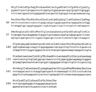
FIG. 1 shows the novel BMY_HDAL1 partial nucleic acid (cDNA)
sequence (SEQ ID N0:1) and the encoded amino acid sequence (SEQ ID
N0:2) of the BMY HDAL1 polypeptide product. The top line in each group of
Fig. 1 presents the BMY_HDAL1 protein sequence (SEQ ID N0:2) in 3-letter
IUPAC form; the middle line presents the nucleotide sequence of the
BMY_HDAL1 coding strand (i.e., SEQ ID N0:1 ); and the bottom line presents
the nucleotide sequence of the reverse strand (SEQ ID N0:3).
FIGS. 2A and 2B show the amino acid sequences of the novel histone
deacetylase-like proteins BMY_HDAL1 (SEQ ID N0:2), BMY HDAL2 (SEQ
ID N0:4) and BMY_HDAL3 (SEQ ID N0:5) aligned with the following known
histone deacetylase proteins: S. cerevisiae HDA1 (SC_HDA1), {SEQ ID
N0:6); human HDAC4 (HDA4), (SEQ ID N0:7); human HDAC5 (HDAS),
(SEQ ID N0:8); human HDAC7 (HDA7), (SEQ ID N0:9) and to a histone
deacetylase-like protein ACUC from Aquifex aeolicus (AQUIFEX HDAL),
(SEQ ID N0:10), (M.S. Finnin et al., 1999, Nature, 401 (6749):188-193).
Residues identical among all proteins are in shown in black text on a gray
background. The sequences were aligned using the ClustalW algorithm as
implemented in the VectorNTl sequence analysis package (1998, 5.5 Ed.,
Informax, Inc.) with a gap opening penalty of 10, a gap extension penalty of
0.1 and no end gap penalties.
FIGS. 3A and 3B show a GenewiseDB comparison of BMY HDAL1
amino acid sequence (SEQ ID N0:2) and human HDAC5 (HDAS) amino acid
sequence (SEQ ID NO:8). Genewise results from HDA5 HUMAN runt
applied to AC002088 nucleic acid (coding) sequence. (SEQ ID N0:11).
FIG. 4 presents the results of sequence motif analysis of motifs within
the BMY_HDAL1 amino acid sequence.
FIG. 5 shows the novel BMY HDAL2 partial nucleic acid (cDNA)
sequence (SEQ ID N0:12) and the encoded amino acid sequence (SEQ ID
N0:4) of the BMY_HDAL2 polypeptide product. The top line in each group of
Fig. 5 presents the BMY_HDAL2 protein sequence (SEQ ID N0:4) in 3-letter
IUPAC form; the middle line presents the nucleotide sequence of the
6
CA 02450129 2003-12-12
WO 02/102323 PCT/US02/19560
BMY_HDAL2 coding strand (i.e., SEQ ID N0:12); and the bottom line
presents the nucleotide sequence of the reverse strand (SEQ ID N0:13).
FIG. 6 presents a GenewiseDB comparison of the BMY_HDAL2 amino
acid sequence (SEQ ID N0:4) and human HDAC5 (HDAS) amino acid
sequence (SEQ ID N0:8). Genewise results from HDAS_HUMAN_run3
applied to AC002410 nucleic acid sequence (SEQ ID N0:14).
FIG. 7 shows PROSITE motifs identified in the predicted amino acid
sequence of the novel BMY_HDAL2 (SEQ ID N0:4). MOTIFS are from:
bmy_hdal2.aa.fasta.
FIGS. 8A and 8B show the sequences of the N- and C-terminal
sequences of BMY_HDAL3 as determined from BAC AC004994 and BAC
AC004744. FIG. 8A presents the most N-terminal region of the BMY_HDAL3
amino acid sequence (SEQ ID N0:15) presented herein as encoded by the
human genomic BAC AC004994 polynucleotide sequence (SEQ ID N0:17).
FIG. 8B presents an additional C-terminal portion of the BMY HDAL3 amino
acid sequence (SEQ ID NO:16) as encoded by human genomic BAC
AC004744 polynucleotide sequence (SEQ ID N0:18).
FIG. 9 shows partial transcripts identified from the AC004994
polynucleotide sequence (SEQ ID N0:17) and from the AC004744
polynucleotide sequence (SEQ ID N0:18) assembled into a single contig,
which was designated BMY_HDAL3 (SEQ ID N0:19) using the VectorNTl
ContigExpress program (Informax, Inc.).
FIG. 10 presents the BMY_HDAL3 partial nucleic acid sequence (SEQ
ID N0:19) and the encoded amino acid sequence (SEQ ID N0:5) based on
the assembled BMY_HDAL3 sequence described in FIG. 9. The top line in
each group of FIG. 10 presents the BMY_HDAL3 protein sequence (SEQ ID
N0:5) in 3-letter IUPAC form; the middle line presents the nucleotide
sequence of the BMY_HDAL3 coding strand (i.e., SEQ ID N0:19); and the
bottom line presents the nucleotide sequence of the reverse strand (SEQ ID
NO:20).
FIG. 11 presents the results of the GCG Motifs program used to
analyze the BMY_HDAL3 partial predicted amino acid sequence for motifs in
7
CA 02450129 2003-12-12
WO 02/102323 PCT/US02/19560
the PROSITE collection (K. Hofmann et al., 1999, Nucleic Acids Res.,
27(1 ):215-219) with no allowed mismatches.
FIG. 12 shows a multiple sequence alignment of the novel human
HDAC, BMY_HDAL3, amino acid sequence (SEQ ID NO:S) with the amino
acid sequence ~ of AAC78618 (SEQ I D N0:21 ) and with the amino acid
sequence of AAD15364 (SEQ ID N0:22). AAC78618 is a histone
deacetylase-like protein predicted by genefinding and conceptual translation
of AC004994 and which was entered in Genbank. AAD15364 is a similar
predicted protein derived from AC004744 and entered in Genbank.
AAC78618, AAD15364 and BMY_HDAL3 were aligned using the ClustalW
algorithm as implemented in the VectorNTl sequence analysis package
(1998, 5.5 Ed., Informax, Inc.) with a gap opening penalty of 10, a gap
extension penalty of 0.1 and no end gap penalties. Residues identical among
all proteins are shown in white text on a black background; conserved
residues are shown in black text on a gray background.
FIG. 13 shows a BLASTN alignment of the AA287983 polynucleotide
sequence (SEQ ID N0;23) and BMY_HDAL3 polynucleotide sequence from
SEQ ID N0:19. Genbank accession AA287983 is a human EST sequence
(GI # 1933807; Incyte template 1080282.1) which was identified by BLASTN
searches against the Incyte LifeSeq database using the NCBI Blast algorithm
(S.F. Altschul et al., 1997, Nucl. Acids Res., 25(17):3389-3402) with default
parameters. The AA287983 human EST was isolated from a germinal B-cell
library. No additional ESTs are included in the Incyte template derived from
this cluster (Incyte gene ID 180282).
FIGS. 14A-14H present other histone deacetylase sequences, as
shown in FIGS. 2A and 2B. FIG. 14A: Aquifex ACUC protein amino acid
sequence (SEQ ID N0:10); FIG. 14B: Saccharomyces cerevisiae histone
deacetylase 1 amino acid sequence (SEQ iD N0:6); FIG. 14C: Homo
sapiens histone deacetylase 4 amino acid sequence (SEQ ID N0:7); FIG.
14D: Homo sapiens histone deacetylase 5 amino acid sequence (SEQ ID
N0:8); FIG. 14E: Homo sapiens histone deacetylase 7 amino acid sequence
(SEQ ID N0:9); FIG. 14F: Human EST AA287983 nucleic acid sequence
8
CA 02450129 2003-12-12
WO 02/102323 PCT/US02/19560
(SEQ ID N0:23); FIG. 14G: Human predicted protein AAD15364 amino acid
sequence(SEQ ID N0:22); and FIG. 14H: Human predicted protein
AAC78618 amino acid sequence (SEQ ID N0:21).
FIGS. 15A-15C depict the nucleotide and amino acid sequence
information for HDAC9c. The polypeptide sequence (SEQ ID N0:87) is
shown using the standard 3-letter abbreviation for amino acids. The DNA
sequence (SEQ ID N0:88) of the coding strand is also shown. FIGS. 15D
15F depict an amino acid sequence alignment of HDAC9c. The predicted
amino acid sequence of HDAC9c (SEQ ID N0:87) was aligned to previously
identified HDACs, including HDAC9 (AY032737; SEQ ID N0:89), HDAC9a
(AY032738; SEQ ID N0:90), and HDAC4 (ALF132608; SEQ ID N0:91 ), using
ClustalW (D.G. Higgins et al., 1996, Methods Enzymol. 266:383-402).
Identical amino acids are shown in white text on a black background;
conserved amino acids are shown in black text on a gray background.
FIGS. 16A-16C depict expression levels of HDAC9 in human cancer
cell lines and normal adult tissue. FIG 16A: Northern blot analysis of HDAC9
expression in normal adult tissue. FIG 16B: Quantitative PCR mRNA
analysis of HDAC9 expression in human tumor cell lines. FIG 16C: Nuclease
protection assay analysis of HDAC9 expression in human tumor cell lines.
FIG. 16D shows the nucleotide sequence of HDAC9c used to derive the
probes used for Northern blotting and nuclease protection analysis (SEQ ID
N0:92). The probes were derived from the HDAC9c nucleotide sequence,
and were predicted to hybridize to HDAC9c and HDAC9 (AY032737), but not
HDAC9a (AY032738).
FIGS. 17A-17C illustrate the increase of HDAC9 gene expression in
human cancer tissues. FIGS. 17A-17B: Summary of HDAC9 expression in
selected tissues, as assayed by in situ hybridization. FIG. 17C:
Photomicrographs of representative cells showing HDAC9 or actin staining.
FIG. 18 shows HDAC9c-mediated induction of morphological
transformation of NIH/3T3 cells. The panels show photomicrographs of soft
agar growth of vector (upper panel), FGF8 (middle panel) and HDAC9c (lower
panel) transfected NIH/3T3 cells. Cells are shown at 10 X magnification.
9
CA 02450129 2003-12-12
WO 02/102323 PCT/US02/19560
FIG. 19 shows HDAC9c induction of actin stress fiber formation in
NIH/3T3 cells. Stable NIH/3T3 cells expressing the indicated constructs were
stained with phalloidin-TRITC and visualized by fluorescent microscopy.
FIGS. 20A-20C depict the nucleotide and amino acid sequence
information for BMY HDACX variant 1, also called BMY HDACX_v1 and
HDACX_v1. BMY_HDACX_v1 represents a partial cDNA sequence obtained
from cells expressing a transcript variant of human HDAC9. The polypeptide
sequence (SEQ ID N0:93) is shown using the standard 3-letter abbreviation
for amino acids. The DNA sequence (SEQ ID N0:94) of the coding strand is
also shown.
FIGS. 21 A-21 B depict the nucleotide and amino acid sequence
information for BMY HDACX variant 2, also called BMY_HDACX v2 and
HDACX_v2. BMY_HDACX_v2 represents a full-length sequence of a novel
transcript variant (i.e., splice product) of HDAC9. The polypeptide sequence
(SEQ ID N0:95) is shown using the standard 3-letter abbreviation for amino
acids. The DNA sequence (SEQ ID N0:96) of the coding strand is also
shown.
FIGS. 22A-221 depict the nucleotide and. amino acid sequence
information for the previously identified HDAC9 transcript variants. FIGS.
22A-22C: HDAC9 variant 1 (HDAC9vl; NCBI Ref. Seq. NM 058176). The
polypeptide sequence (SEQ ID N0:89) is shown using the standard 3-letter
abbreviation for amino acids. The DNA sequence (SEQ ID N0:97) of the
coding strand is also shown. FIGS. 22D-22F: HDAC9 variant 2 (HDAC9v2;
NCBI Ref. Seq. NM 058177). The polypeptide sequence (SEQ ID N0:90) is
shown using the standard 3-letter abbreviation for amino acids. The DNA
sequence (SEQ ID N0:98) of the coding strand is also shown. FIGS. 226-
221: HDAC9 variant 3 (HDAC9v3; NCBI Ref. Seq. NM 014707). The
polypeptide sequence (SEQ ID NO:99) is shown using the standard 3-letter
abbreviation for amino acids. The DNA sequence (SEQ ID N0:100) of the
coding strand is also shown.
FIGS. 23A-23K depict a multiple sequence alignment of nucleotide
sequences representing known and novel HDAC9 splice products. The
CA 02450129 2003-12-12
WO 02/102323 PCT/US02/19560
cDNAs for BMY HDACX v1 (SEQ ID NO:94) and BMY HDACX v2 (SEQ ID
N0:96) nucleotide sequences were aligned to the three reported splice
products of the HDAC9 gene, including HDAC9v1 (NCBI Ref. Seq.
NM 058176; SEQ ID N0:97), HDAC9v2 (NCBI Ref .Seq. NM 058177; SEQ
ID NO:98), and HDAC9v3 (NCBI Ref. Seq. NM 014707; SEQ ID N0:100)
using the sequence alignment program ClustalW (D.G. Higgins et al., 1996,
Methods En~ymol. 266:383-402). The consensus sequence is shown on the
bottom line (SEQ ID N0:106). Identical nucleotides are shown in white text
on a black background, Selected splice junctions are indicated below the
alignment; these junctions were identified by comparison of the cDNA
sequences to the assembled genomic contig NT 00798.1 using the Sim4
algorithm (L. Florea et al., 1998, Genome Res. 8:967-74). It is noted that the
HDAC9 (AY032737) nucleotide and amino acid sequences are identical to the
HDAC9v1 (NM 058176) nucleotide and amino acid sequences, Similarly, the
HDAC9a (AY032738) nucleotide and amino acid sequences are identical to
the HDAC9v2 (NM_058177) nucleotide and amino acid sequences.
FIGS. 24A-24D depict a multiple sequence alignment of amino acid
sequences representing known and novel HDAC polypeptides. The amino
acid sequences encoded by transcript variants BMY_HDACX v1 (SEQ ID
N0:93) and BMY_HDACX v2 (SEQ ID N0:95) were aligned to amino acid
sequences encoded by known splice variants of human histone deacetylase 9
including HDAC9v1 (NCBI Ref. Seq. NM 058176; SEQ ID N0:89), HDAC9v2
(NCBI Ref ,Seq. NM 058177; SEQ ID N0:90), and HDAC9v3 (NCBI Ref.
Seq, NM 014707; SEQ ID N0:99), and to human histone deacetylases 4 and
5 (HDAS, SEQ ID N0:8; HDA4, SEQ ID N0:7) using the multiple sequence
alignment program ClustalW (D.G. Higgins et al., 1996, Methods Enzymol.
266:383-402). The consensus sequence is shown on the bottom line (SEQ ID
N0:107). Residues conserved among all polypeptides are shown in white
text on a black background; residues conserved in a majority of polypeptides
are shown in black text on a gray background.
FIGS. 25A-25C depict a multiple sequence alignment of amino acid
sequences showing novel HDAC polypeptides. The amino acid sequences of
11
CA 02450129 2003-12-12
WO 02/102323 PCT/US02/19560
BMY_HDAL1 (SEQ ID N0:2), BMY HDAL2 (SEQ ID N0:4), BMY_HDAL3
(SEQ JD N0:5), HDAC9c (SEQ ID NO:87), HDACX v1 (SEQ ID N0:93), and
HDACX v2 (SEQ ID N0:95) were aligned using the T-Coffee program (C.
Notredame et al., 2000, J. Mol. Biol. 302:205-217; C. Notredame et al., 1998,
Bioinformatics 14:407-422). Identical residues are shown in black text on a
gray background.
12
CA 02450129 2003-12-12
WO 02/102323 PCT/US02/19560
DESCRIPTION OF THE INVENTION
The present invention discloses several novel HDAC nucleotide
sequences and encoded products. New members of the histone deacetylase
protein family have been identified as having identity to known HDACs. Three
new HDACs are referred to as BMY_HDAL1, BMY_HDAL2, and BMY_HDAL3
herein, wherein HDAL signifies histone deacetylase like proteins in current
nomenclature. These proteins are most similar to the known human histone
deacetylase, HDAC9. Novel HDAC9 splice variants, termed HDACX v1 and
HDACX_v2, have also been identified. In addition, HDAC9c, an HDAC9-
related family member, has been newly identified and cloned. The nucleic
acid sequences encoding the novel HDAC polypeptides are provided together
with the description of the means employed to obtain these novel molecules.
Such HDAC products can serve as protein deacetylases, which are useful for
disease treatment and/or diagnosis of diseases and disorders associated with
cell growth or proliferation, cell differentiation, and cell survival, e.g.,
neoplastic cell growth, cancers, and tumors.
As shown herein, HDAC9 expression is elevated in tumor cell lines, as
determined by quantitative PCR analysis. Elevated expression of HDAC9
was also observed in clinical specimens of human tumor tissue compared to
normal tissue, using in situ hybridization (ISH) and an HDAC9-specific
riboprobe. Further, cell biological assessment of HDAC9c revealed that
overexpression of HDAC9c confers a growth advantage to normal fibroblasts.
These results indicate that HDAC9c can be used as a diagnostic marker for
tumor progression and that selective HDAC9c inhibitors can be used to target
specific cancer or tumor types, such as breast and prostate cancers or
tumors.
Definitions
The following definitions are provided to more fully describe the present
invention in its various aspects. The definitions are intended to be useful
for
guidance and elucidation, and are not intended to limit the disclosed
invention
and its embodiments.
13
CA 02450129 2003-12-12
WO 02/102323 PCT/US02/19560
HDAC polypeptides (or proteins) refer to the amino acid sequence of
isolated, and preferably substantially purified, human histone deacetylase
proteins isolated as described herein. HDACs may also be obtained from any
species, preferably mammalian, including mouse, rat, non-human primates,
and more preferably, human; and from a variety of sources, including natural,
synthetic, semi-synthetic, or recombinant. The probes and oligos described
may be used in obtaining HDACs from mammals other than humans. The
present invention more particularly provides six new human HDAC family
members, namely, BMY HDAL1, BMY HDAL2, BMY_HDAL3, HDACX v1,
HDACX v2, and HDAC9c, their polynucleotide sequences (e.g., SEQ ID
N0:1, SEQ ID N0:12, SEQ ID N0:19, SEQ ID N0:88, SEQ ID N0:94, SEQ
ID N0:96, and sequences complementary thereto), and encoded products
(e.g., SEQ ID N0:2, SEQ ID N0:4, SEQ ID N0:5, SEQ ID N0:87, SEQ ID
N0:93, and SEQ ID NO:95).
An agonist (e.g., activator) refers to a molecule which, when bound to,
or interactive with, an HDAC polypeptide, or a functional fragment thereof,
increases or prolongs the duration of the effect of the HDAC polypeptide.
Agonists may include proteins, nucleic acids, carbohydrates, or any other
molecules that bind to and modulate the effect of an HDAC polypeptide. An
antagonist (e.g., inhibitor, blocker) refers to a molecule which, when bound
to,
or interactive with, an HDAC polypeptide, or a functional fragment thereof,
decreases or eliminates the amount or duration of the biological or
immunological activity of the HDAC polypeptide. Antagonists may include
proteins, nucleic acids, carbohydrates, antibodies, or any other molecules
that
decrease, reduce or eliminate the effect and/or function of an HDAC
polypeptide.
"Nucleic acid sequence", as used herein, refers to an oligonucleotide,
nucleotide, or polynucleotide (e.g., DNA, cDNA, RNA), and fragments or
portions thereof, and to DNA or RNA of genomic or synthetic origin which may
be single- or double-stranded, and represent the sense (coding) or antisense
(non-coding) strand. By way of nonlimiting example, fragments include
nucleic acid sequences that can be about 10 to 60 contiguous nucleotides in
14
CA 02450129 2003-12-12
WO 02/102323 PCT/US02/19560
length, preferably, at least 15-60 contiguous nucleotides in length, and also
preferably include fragments that are at least 70-100 contiguous nucleotides,
or which are at least 1000 contiguous nucleotides or greater in length.
Nucleic acids for use as probes or primers may differ in length as described
herein.
In specific embodiments, HDAC polynucleotides of the present
invention can comprise at least 15, 20, 25, 50, 100, 150, 200, 250, 300, 350,
400, 450, 500, 600, 700, 800, 900, 1000, 1195, 1200, 1500, 2000, 2160,
2250, 2500, 2755, or 2900 contiguous nucleotides of SEQ ID N0:1, SEQ ID
N0:12, SEQ ID N0:19, SEQ ID N0:88, SEQ ID N0:94, SEQ ID N0:96, or a
sequence complementary thereto. Additionally, a polynucleotide of the
invention can comprise a specific region of a HDAC nucleotide sequence,
e.g., a region encoding the C-terminal sequence of the HDAC polypeptide.
Such polynucleotides can comprise, for example, nucleotides 3024-4467 of
HDAC9c (SEQ ID NO:88), nucleotides 2156-3650 of HDACX v1 (SEQ ID
N0:94), nucleotides 1174-3391 of HDACX v2 (SEQ ID N0:96), or portions or
fragments thereof.
As specific examples, polynucleotides of the invention may comprise at
least 183 contiguous nucleotides of SEQ ID N0:88; or at least 17 contiguous
nucleotides of SEQ ID N0:96. As additional examples, the polynucleotides of
the invention may comprise nucleotides 1 to 3207 of SEQ ID N0:88;
nucleotides 1 to 2340 of SEQ ID N0:94; or nucleotides 307 to 1791 of SEQ (D
N0:96. Further, the polynucleotides of the invention may comprise
nucleotides 4 to 3207 of SEQ ID N0:88, wherein said nucleotides encode
amino acids 2 to 1069 of SEQ ID N0:87 lacking the start methionine; or
nucleotides 310 to 1791 of SEQ ID NO:96, wherein said nucleotides encode
amino acids 2 to 495 of SEQ ID N0:95 lacking the start methionine. In
addition, polynucleotides of the invention may comprise nucleotides 3024-
3207 of SEQ ID N0:88; or nucleotides 1174-1791 of SEQ ID N0:96.
"Amino acid sequence" as used herein refers to an oligopeptide,
peptide, polypeptide, or protein sequence, and fragments or portions thereof,
and to naturally occurring or synthetic molecules. Amino acid sequence
CA 02450129 2003-12-12
WO 02/102323 PCT/US02/19560
fragments are typically from about 4 or 5 to about 35, preferably from about 5
to about 15 or 25 amino acids in length and, optimally, retain the biological
activity or function of an HDAC polypeptide. However, it will be understood
that larger amino acid fragments can be used, depending on the purpose
therefor, e.g., fragments of from about 15 to about 50 or 60 amino acids, or
greater.
Where "amino acid sequence" is recited herein to refer to an amino
acid sequence of a naturally occurring protein molecule, "amino acid
sequence" and like terms, such as "polypeptide" or "protein" are not meant to
limit the amino acid sequence to the complete, native amino acid sequence
associated with the recited protein molecule. In addition, the terms HDAC
polypeptide and HDAC protein are frequently used interchangeably herein to
refer to the encoded product of an HDAC nucleic acid sequence of the
present invention.
A variant of an HDAC polypeptide can refer to an amino acid sequence
that is altered by one or more amino acids. The variant may have
"conservative" changes, wherein a substituted amino acid has similar
structural or chemical properties, e.g., replacement of leucine with
isoleucine.
More rarely, a variant may have "nonconservative" changes, e.g.,
replacement of a glycine with a tryptophan. Minor variations may also include
amino acid deletions or insertions, or both. Guidance in determining which
amino acid residues may be substituted, inserted, or deleted without
abolishing functional biological or immunological activity may be found using
computer programs well known in the art, for example, DNASTAR software.
An allele or allelic sequence is an alternative form of an HDAC nucleic
acid sequence. Alleles may result from at least one mutation in the nucleic
acid sequence and may yield altered mRNAs or polypeptides whose structure
or function may or may not be altered. Any given gene, whether natural or
recombinant, may have none, one, or many allelic forms. Common
mutational changes that give rise to alleles are generally ascribed to natural
deletions, additions, or substitutions of nucleotides. Each of these types of
16
CA 02450129 2003-12-12
WO 02/102323 PCT/US02/19560
changes may occur alone, or in combination with the others, one or more
times in a given sequence.
Altered nucleic acid sequences encoding an HDAC polypeptide include
nucleic acid sequences containing deletions, insertions and/or substitutions
of
different nucleotides resulting in a polynucleotide that encodes the same or a
functionally equivalent HDAC polypeptide. Altered nucleic acid sequences
may further include polymorphisms of the polynucleotide encoding an HDAC
polypeptide; such polymorphisms may or may not be readily detectable using
a particular oligonucleotide probe. The encoded protein may also contain
deletions, insertions, or substitutions of amino acid residues, which produce
a
silent change and result in a functionally equivalent HDAC protein of the
present invention. Deliberate amino acid substitutions may be made on the
basis of similarity in polarity, charge, solubility, hydrophobicity,
hydrophilicity,
and/or the amphipathic nature of the residues, as long as the biological
activity or function of the HDAC protein is retained. For example, negatively
charged amino acids may include aspartic acid and glutamic acid; positively
charged amino acids may include lysine and arginine; and amino acids with
uncharged polar head groups having similar hydrophilicity values may include
leucine, isoleucine, and valine; glycine and alanine; asparagine and
glutamine; serine and threonine; and phenylalanine and tyrosine.
"Peptide nucleic acid" (PNA) refers to an antisense molecule or anti-
gene agent which comprises an oligonucleotide ("oligo") linked to a peptide
'backbone of amino acid residues, which terminates in lysine. PNA typically
comprise oligos of at least 5 nucleotides linked to amino acid residues. These
small molecules stop transcript elongation by binding to their complementary
strand of nucleic acid (P.E. Nielsen et al., 1993, Anticancer Drug Des., 8:53-
63). PNA may be pegylated to extend their lifespan in the cell where they
preferentially bind to complementary single stranded DNA and RNA.
Oligonucleotides or oligomers refer to a nucleic acid sequence,
preferably comprising contiguous nucleotides, typically of at least about 6
nucleotides to about 60 nucleotides, preferably at least about 8 to 10
nucleotides in length, more preferably at least about 12 nucleotides in
length,
17
CA 02450129 2003-12-12
WO 02/102323 PCT/US02/19560
e.g., about 15 to 35 nucleotides, or about 15 to 25 nucleotides, or about 20
to
35 nucleotides, which can be typically used, for example, as probes or
primers, in PCR amplification assays, hybridization assays, or in microarrays.
It will be understood that the term oligonucleotide is substantially
equivalent to
the terms primer, probe, or amplimer, as commonly defined in the art. It will
also be appreciated by those skilled in the pertinent art that a longer
oligonucleotide probe, or mixtures of probes, e.g., degenerate probes, can be
used to detect longer, or more complex, nucleic acid sequences, for example,
genomic DNA. In such cases, the probe may comprise at least 20-200
nucleotides, preferably, at least 30-100 nucleotides, more preferably, 50-100
nucleotides.
Amplification refers to the production of additional copies of a nucleic
acid sequence and is generally carried out using polymerase chain reaction
(PCR) technologies, which are well known and practiced in the art (See, D.W.
Dieffenbach and G.S. Dveksler, 1995, PCR Primer, a Laboratory Manual,
Cold Spring Harbor Press, Plainview, NY).
Microarray is an array of distinct polynucleotides or oligonucleotides
synthesized on a substrate, such as paper, nylon, or other type of membrane;
filter; chip; glass slide; or any other type of suitable solid support.
The term antisense refers to nucleotide sequences, and compositions
containing nucleic acid sequences, which are complementary to a specific
DNA or RNA sequence. The term "antisense strand" is used in reference to a
nucleic acid strand that is complementary to the "sense" strand. Antisense
(i.e., complementary) nucleic acid molecules include PNA and may be
produced by any method, including synthesis or transcription. Once
introduced into a cell, the complementary nucleotides combine with natural
sequences produced by the cell to form duplexes that block either
transcription or translation. The designation "negative" is sometimes used in
reference to the antisense strand, and "positive" is sometimes used in
reference to the sense strand.
The term consensus refers to the sequence that reflects the most
common choice of base or amino acid at each position among a series of
18
CA 02450129 2003-12-12
WO 02/102323 PCT/US02/19560
related DNA, RNA, or protein sequences. Areas of particularly good
agreement often represent conserved functional domains.
A deletion refers to a change in either nucleotide or amino acid
sequence and results in the absence of one or more nucleotides or amino
acid residues. By contrast, an insertion (also termed "addition") refers to a
change in a nucleotide or amino acid sequence that results in the addition of
one or more nucleotides or amino acid residues, as compared with the
naturally occurring molecule. A substitution refers to the replacement of one
or more nucleotides or amino acids by different nucleotides or amino acids.
A derivative nucleic acid molecule refers to the chemical modification of
a nucleic acid encoding, or complementary to, an encoded HDAC
polypeptide. Such modifications include, for example, replacement of
hydrogen by an alkyl, acyl, or amino group. A nucleic acid derivative encodes
a polypeptide that retains the essential biological andlor functional
characteristics of the natural molecule. A derivative polypeptide is one that
is
modified by glycosylation, pegylation, or any similar process that retains the
biological and/or functional or immunological activity of the polypeptide from
which it is derived.
The term "biologically active", i.e., functional, refers to a protein or
polypeptide or peptide fragment thereof having structural, regulatory, or
biochemical functions of a naturally occurring molecule. Likewise,
"immunologically active" refers to the capability of the natural, recombinant,
or
synthetic HDAC, or any oligopeptide thereof, to induce a specific immune
response in appropriate animals or cells, for example, to generate antibodies,
and to bind with specific antibodies.
An HDAC-related protein refers to the HDAC and HADL proteins or
polypeptides described herein, as well as other human homologs of these
HDAC or HDAL sequences, in addition to orthologs and paralogs (homologs)
of the HDAC or HADL sequences in other species, ranging from yeast to
other mammals, e.g., homologous histone deacetylase. The term ortholog
refers to genes or proteins that are homologs via speciation, e.g., closely
related and assumed to have common descent based on structural and
19
CA 02450129 2003-12-12
WO 02/102323 PCT/US02/19560
functional considerations. Orthologous proteins function as recognizably the
same activity in different species. The term paralog refers to genes or
proteins that are homologs via gene duplication, e.g., duplicated variants of
a
gene within a genome. (See, W.M. Fritch, 1970, Syst. Zool., 19:99-113.
It will be appreciated that, under certain circumstances, it may be
advantageous to provide homologs of one of the novel HDAC polypeptides
which function in a limited capacity as one of either an HDAC agonist (i.e.,
mimetic), or an HDAC antagonist, in order to promote or inhibit only a subset
of the biological activities of the naturally-occurring form of the protein.
Thus,
specific biological effects can be elicited by treatment with a homolog of
limited function, and with fewer side effects, relative to treatment with
agonists
or antagonists which are directed to all of the biological activities of
naturally-
occurring forms of HDAC proteins.
Homologs (i.e., isoforms or variants) of the novel HDAC polypeptides
can be generated by mutagenesis, such as by discrete point mutation(s), or
by truncation. For example, mutation can yield homologs that retain
substantially the same, or merely a subset of, the biological activity of the
HDAC polypeptide from which it was derived. Alternatively, antagonistic
forms of the protein can be generated which are able to inhibit the function
of
the naturally-occurring form of the protein, such as by competitively binding
to
an HDAC substrate, or HDAC-associated protein. Non-limiting examples of
such situations include competing with wild-type HDAC in the binding of p53
or a histone. Also, agonistic forms of the protein can be generated which are
constitutively active, or have an altered K~at or Km for deacylation
reactions.
Thus, the HDAC protein and homologs thereof may be either positive or
negative regulators of transcription and/or replication.
The term hybridization refers to any process by which a strand of
nucleic acid binds with a complementary strand through base pairing.
The term "hybridization complex" refers to a complex formed between
two nucleic acid sequences by virtue of the formation of hydrogen bonds
between complementary G and C bases and between complementary A and
T bases. The hydrogen bonds may be further stabilized by base stacking
CA 02450129 2003-12-12
WO 02/102323 PCT/US02/19560
interactions. The two complementary nucleic acid sequences hydrogen bond
in an anti-parallel configuration. A hybridization complex may be formed in
solution (e.g., Cot or Rot analysis), or between one nucleic acid sequence
present in solution and another nucleic acid sequence immobilized on a solid
support (e.g., membranes, filters, chips, pins, or glass slides, or any other
appropriate substrate to which cells or their nucleic acids have been
affixed).
The terms stringency or stringent conditions refer to the conditions for
hybridization as defined by nucleic acid composition, salt and temperature.
These conditions are well known in the art and may be altered to identify
and/or detect identical or related polynucleotide sequences in a sample. A
variety of equivalent conditions comprising either low, moderate, or high
stringency depend on factors such as the length and nature of the sequence
(DNA, RNA, base composition), reaction milieu (in solution or immobilized on
a solid substrate), nature of the target nucleic acid (DNA, RNA, base
composition), concentration of salts and the presence or absence of other
reaction components (e.g., formamide, dextran sulfate and/or polyethylene
glycol) and reaction temperature (within a range of from about 5°C
below the
melting temperature of the probe to about 20°C to 25°C below the
melting
temperature). One or more factors may be varied to generate conditions,
either low or high stringency, that are different from but equivalent to the
aforementioned conditions.
As will be understood by those of skill in the art, the stringency of
hybridization may be altered in order to identify or detect identical or
related
polynucleotide sequences. As will be further appreciated by the skilled
practitioner, Tm can be approximated by the formulas as known in the art,
depending on a number of parameters, such as the length of the hybrid or
probe in number of nucleotides, or hybridization buffer ingredients and
conditions (See, for example, T. Maniatis et al., Molecular Cloning: A
Laboratory Manual, Cold Spring Harbor Laboratory, Cold Spring Harbor, NY,
1982 and J. Sambrook et al., Molecular Cloning: A Laboratory Manual, Cold
Spring Harbor Laboratory, Cold Spring Harbor, NY, 1989; Current Protocols in
Molecular Biology, Eds. F.M. Ausubel et al., Vol. 1, "Preparation and Analysis
21
CA 02450129 2003-12-12
WO 02/102323 PCT/US02/19560
of DNA", John Wiley and Sons, Inc., 1994-1995, Suppls. 26, 29, 35 and 42;
pp. 2.10.7- 2.10.16; G.M. Wahl and S. L. Berger (1987; Methods Enzymol.
152:399-407); and A.R. Kimmel, 1987; Methods of En~ymol., 152:507-511).
As a general guide, Tm decreases approximately 1 °-C -1.5°-
C with every 1
decrease in sequence homology. Also, in general, the stability of a hybrid is
a
function of sodium ion concentration and temperature. Typically; the
hybridization reaction is initially performed under conditions of low
stringency,
followed by washes of varying, but higher stringency. Reference to
hybridization stringency, e.g., high, moderate, or low stringency, typically
relates to such washing conditions.
Thus, by way of nonlimiting example, high stringency refers to
conditions that permit hybridization of those nucleic acid sequences that form
stable hybrids in 0.018M NaCI at about 65°-C (i.e., if a hybrid is not
stable in
0.018M NaCI at about 65°C, it will not be stable under high stringency
conditions). High stringency conditions can be provided, for instance, by
hybridization in 50% formamide, 5 X Denhart's solution, 5 X SSPE (saline
sodium phosphate EDTA) (1 X SSPE buffer comprises 0.15 M NaCI, 10 mM
Na2HP0~, 1 mM EDTA), (or 1 X SSC buffer containing 150 mM NaCI, 15 mM
Na3 citrate ~ 2 H20, pH 7.0), 0.2% SDS at about 42°-C, followed by
washing in
1 X SSPE (or saline sodium citrate, SSC) and 0.1 % SDS at a temperature of
at least about 42°C, preferably about 55°C, more preferably
about 65°C.
Moderate stringency refers, by way of nonlimiting example, to
conditions that permit hybridization in 50% formamide, 5 X Denhart's solution,
5 X SSPE (or SSC), 0.2% SDS at 42°-C (to about 50°-C), followed
by washing
in 0.2 X SSPE (or SSC) and 0.2% SDS at a temperature of at least about
42°C, preferably about 55°C, more preferably about 65°C.
Low stringency refers, by way of nonlimiting example, to conditions that
permit hybridization in 10% formamide, 5 X Denhart's solution, 6 X SSPE (or
SSC), 0.2% SDS at 42°-C, followed by washing in 1 X SSPE (or SSC)
and
0.2% SDS at a temperature of about 45°C, preferably about 50°C.
For additional stringency conditions, see T. Maniatis et al., Molecular
Cloning: A Laboratory Manual, Cold Spring Harbor Laboratory, Cold Spring
22
CA 02450129 2003-12-12
WO 02/102323 PCT/US02/19560
Harbor, NY (1982). It is to be understood that the low, moderate and high
stringency hybridization / washing conditions may be varied using a variety of
ingredients, buffers and temperatures well known to and practiced by the
skilled practitioner.
The terms complementary or complementarity refer to the natural
binding of polynucleotides under permissive salt and temperature conditions
by base-pairing. For example, the sequence "A-G-T" binds to the
complementary sequence "T-C-A". Complementarity between two single-
stranded molecules may be "partial", in which only some of the nucleic acids
bind, or it may be complete when total complementarity exists between single
stranded molecules. The degree of complementarity between nucleic acid
strands has significant effects on the efficiency and strength of
hybridization
between nucleic acid strands. This is of particular importance in
amplification
reactions, which depend upon binding between nucleic acids strands, as well
as in the design and use of PNA molecules.
The term homology refers to a degree of complementarity. There may
be partial sequence homology or complete homology, wherein complete
homology is equivalent to identity, e.g., 100% identity. A partially
complementary sequence that at least partially inhibits an identical sequence
from hybridizing to a target nucleic acid is referred to using the functional
term
"substantially homologous." The inhibition of hybridization of the completely
complementary sequence to the target sequence may be examined using a
hybridization assay (e.g., Southern or Northern blot, solution hybridization
and
the like) under conditions of low stringency. A substantially homologous
sequence or probe will compete for and inhibit the binding (i.e., the
hybridization) of a completely homologous sequence or probe to the target
sequence under conditions of low stringency. Nonetheless, conditions of low
stringency do not permit non-specific binding; low stringency conditions
require that the binding of two sequences to one another be a specific (i.e.,
selective) interaction. The absence of non-specific binding may be tested by
the use of a second target sequence which lacks even a partial degree of
complementarity (e.g., less than about 30% identity). In the absence of non-
23
CA 02450129 2003-12-12
WO 02/102323 PCT/US02/19560
specific binding, the probe will not hybridize to the second non-
complementary target sequence.
Those having skill in the art will know how to determine percent identity
between/among sequences using, for example, algorithms such as those
based on the CLUSTALW computer program (J.D. Thompson et al., 1994,
Ncrcieic Acids Research, 2(22):4673-4680), or FASTDB, (Brutlag et al., 1990,
Comp. App. Biosci., 6:237-245), as known in the art. Although the FASTDB
algorithm typically does not consider internal non-matching deletions or
additions in sequences, i.e., gaps, in its calculation, this can be corrected
manually to avoid an overestimation of the % identity. CLUSTALW, however,
does take sequence gaps into account in its identity calculations.
Also available to those having skill in this art are the BLAST and
BLAST 2.0 algorithms (Altschul et al., 1977, Nucl. Acids Res., 25:3389-3402
and Altschul et al., 1990, J. Mol. Biol., 215:403-410). The BLASTN program
for nucleic acid sequences uses as defaults a wordlength (W) of 11, an
expectation (E) of 10, M=5, N=4, and a comparison of both strands. For
amino acid sequences, the BLASTP program uses as defaults a wordlength
(W) of 3, and an expectation (E) of 10. The BLOSUM62 scoring matrix
(Henikoff and Henikoff, 1989, Proc. Natl. Acad. Sci., USA, 89:10915) uses
alignments (B) of 50, expectation (E) of 10, M=5, N=4, and a comparison of
both strands.
An HDAC polynucleotide of the present invention may show at least
27.7%, 35%, 40%, 44.1 %, 48.2%, 50%, 55.4%, 58.6%, 59.8%, 60%, 60.2%,
67.8%, 70%, 80%, 81.5%, 85%, 90%, 91 %, 92%, 93%, 94%, 94.2%, 94.4%,
95%, 96%, 97%, 97.2%, 97.5%, 98%, 99%, 99.1 %, 99.2%, 99.3%, 99.4%,
99.5%, 99.6%, 99.7%, 99.8%, or 99.9% identity to a sequence provided in
SEQ ID N0:1, SEQ ID N0:12, SEQ ID N0:19, SEQ ID N0:88, SEQ ID
N0:94, SEQ ID N0:96, or a sequence complementary thereto. An HDAC
polypeptide of the present invention may show at least 25%, 35%, 40%, 45%,
48.1 %, 55.2%, 55.3%, 60%, 65%, 70%, 72%, 75%, 79%, 80%, 80.6%, 85%,
90%, 91 %, 92%, 93%, 94%, 94.2%, 95%, 96%, 97%, 97.2%, 97.5%, 98%,
99%, 99.1 %, 99.2%, 99.3%, 99.4%, 99.5%, 99.6%, 99.7%, 99.8%, or 99.9%
24
CA 02450129 2003-12-12
WO 02/102323 PCT/US02/19560
identity to a sequence provided in any one of SEQ ID N0:2, SEQ ID N0:4,
SEQ ID NO:S, SEQ ID N0:87, SEQ ID N0:93, or SEQ ID N0:95.
In a preferred aspect of the invention, a HDAC polynucleotide shows at
least 60.2%, 81.5%, or 94.4% identity to the HDAC9c nucleotide sequence
(SEQ ID N0:88 or a sequence complementary thereto); or at least 27.7%,
48.2%, or 55.4% identity to the HDACX v2 nucleotide sequence (SEQ ID
N0:96 or a sequence complementary thereto). A HDAC polypeptide of the
invention preferably shows at least 55.2%, 80.6%, or 94.2% identity to the
HDAC9c amino acid sequence (SEQ ID N0:87); at least 55.3% identity to the
HDACX v2 amino acid sequence (SEQ ID N0:95); at least 72% identity to
the amino acid sequence of BMY_HDAL1 (SEQ ID N0:2); at least 79%
identity to the amino acid sequence of BMY_HDAL2 (SEQ ID N0:4); or at
least 70% identity to the amino acid sequence of BMY_HDAL3 (SEQ ID
NO:S).
A composition comprising a given polynucleotide sequence refers
broadly to any composition containing the given polynucleotide sequence.
The composition may comprise a dry formulation or an aqueous solution.
Compositions comprising the polynucleotide sequences (e.g., SEQ ID N0:1,
SEQ ID N0:12, SEQ ID N0:19, SEQ ID NO:88, SEQ ID N0:94, or SEQ ID
N0:96) encoding the novel HDAC polypeptides of this invention, or fragments
thereof, or complementary sequences thereto, may be employed as
hybridization probes. The probes may be stored in freeze-dried form and may
be in association with a stabilizing agent such as a carbohydrate. In
hybridizations, the probe may be employed in an aqueous solution containing
salts (e.g., NaCI), detergents or surfactants (e.g., SDS) and other components
(e.g., Denhardt's solution, dry milk, salmon sperm DNA, and the like).
The term "substantially purified"- refers to nucleic acid sequences or
amino acid sequences that are removed from their natural environment, i.e.,
isolated or separated by a variety of means, and are at least 60% free,
preferably 75% to 85% free, and most preferably 90% or greater free from
other components with which they are naturally associated.
CA 02450129 2003-12-12
WO 02/102323 PCT/US02/19560
The term sample, or biological sample, is meant to be interpreted in its
broadest sense. A biological sample suspected of containing nucleic acid
encoding an HDAC protein, or fragments thereof, or an HDAC protein itself,
may comprise a body fluid, an extract from cells or tissue, chromosomes
isolated from a cell (e.g., a spread of metaphase chromosomes), organelle, or
membrane isolated from a cell, a cell, nucleic acid such as genomic DNA (in
solution or bound to a solid support such as for Southern analysis), RNA (in
solution or bound to a solid support such as for Northern analysis), cDNA (in
solution or bound to a solid support), a tissue, a tissue print and the like.
Transformation refers to a process by which exogenous DNA enters
and changes a recipient cell. It may occur under natural or artificial
conditions
using various methods well known in the art. Transformation may rely on any
known method for the insertion of foreign nucleic acid sequences into a
prokaryotic or eukaryotic host cell. The method is selected based on the type
of host cell being transformed and may include, but is not limited to, viral
infection, electroporation, heat shock, lipofection, and partial bombardment.
Such "transformed" cells include stably transformed cells in which the
inserted
DNA is capable of replication either as an autonomously replicating plasmid or
as part of the host chromosome. Transformed cells also include those cells
that transiently express the inserted DNA or RNA for limited periods of time.
The term "mimetic" refers to a molecule, the structure of which is
developed from knowledge of the structure of an HDAC protein, or portions
thereof, and as such, is able to effect some or all of the actions of HDAC
proteins.
The term "portion" with regard to a protein (as in "a portion of a given
protein") refers to fragments or segments, for example, peptides, of that
protein. The fragments may range in size from four or five amino acid
residues to the entire amino acid sequence minus one amino acid. Thus, a
protein "comprising at least a portion of the amino acid sequence of the HDAC
molecules presented herein can encompass a full-length human HDAC
polypeptide, and fragments thereof.
26
CA 02450129 2003-12-12
WO 02/102323 PCT/US02/19560
In specific embodiments, HDAC polypeptides of the invention can
comprise at least 5, 10, 20, 30, 50, 70, 100, 200, 300, 400, 500, 600, 700,
720, 750, 800, 920, or 950 contiguous amino acid residues of SEQ ID N0:2,
SEQ ID N0:4, SEQ ID NO:S, SEQ ID N0:87, SEQ ID N0:93, or SEQ ID
N0:95. Additionally, a polypeptide of the invention can comprise a specific
region, e.g., the C-terminal region, of a HDAC amino acid sequence. Such
polypeptides can comprise, for example, amino acids 1009-1069 of HDAC9c
(SEQ ID N0:87), amino acids 720-780 of HDACX v1 (SEQ ID N0:93), or
portions or fragments thereof.
The term antibody refers to intact molecules as well as fragments
thereof, such as Fab, F(ab')2, Fv, which are capable of binding an epitopic or
antigenic determinant. Antibodies that bind to the HDAC polypeptides can be
prepared using intact polypeptides or fragments containing small peptides of
interest or prepared recombinantly for use as the immunizing antigen. The
polypeptide or oligopeptide used to immunize an animal can be derived from
the transition of RNA or synthesized chemically, and can be conjugated to a
carrier protein, if desired. Commonly used carriers that are chemically
coupled to peptides include bovine serum albumin (BSA), keyhole limpet
hemocyanin (KLH), and thyroglobulin. The coupled peptide is then used to
immunize the animal (e.g, a mouse, a rat, or a rabbit).
The term "humanized" antibody refers to antibody molecules in which
amino acids have been replaced in the non-antigen binding regions, e.g., the
complementarity determining regions (CDRs), in order to more closely
resemble a human antibody, while still retaining the original binding
capability,
e.g., as described in U.S. Patent No. 5,585,089 to C.L. Queen et al., which is
a nonlimiting example. Fully humanized antibodies, such as those produced
transgenically or recombinantly, are also encompassed herein.
The term "antigenic determinant" refers to that portion of a molecule
that makes contact with a particular antibody (i.e., an epitope). When a
protein or fragment of a protein is used to immunize a host animal, numerous
regions of the protein may induce the production of antibodies which bind
specifically to a given region or three-dimensional structure on the protein;
27
CA 02450129 2003-12-12
WO 02/102323 PCT/US02/19560
these regions or structures are referred to an antigenic determinants. An
antigenic determinant may compete with the intact antigen (i.e., the
immunogen used to elicit the immune response) for binding to an antibody.
The terms "specific binding" or "specifically binding" refer to the
interaction between a protein or peptide and a binding molecule, such as an
agonist, an antagonist, or an antibody. The interaction is dependent upon the
presence of a particular structure (e.g., an antigenic determinant or epitope,
or
a structural determinant) of the protein that is recognized by the binding
molecule. For example, if an antibody is specific for epitope "A", the
presence
of a protein containing epitope A (or free, unlabeled A) in a reaction
containing
labeled "A" and the antibody will reduce the amount of labeled A bound to the
antibody.
The term "correlates with expression of a polynucleotide" indicates that
the detection of the presence of ribonucleic acid that is similar to one or
more
of the HDAC sequences provided herein by Northern analysis is indicative of
the presence of mRNA encoding an HDAC polypeptide in a sample and
thereby correlates with expression of the transcript from the polynucleotide
encoding the protein.
An alteration in the polynucleotide of an HDAC nucleic acid sequence
comprises any alteration in the sequence of the polynucleotides encoding an
HDAC polypeptide, including deletions, insertions, and point mutations that
may be detected using hybridization assays. Included within this definition is
the detection of alterations to the genomic DNA sequence which encodes an
HDAC polypeptide (e.g., by alterations in the pattern of restriction fragment
length polymorphisms capable of hybridizing to the HDAC nucleic acid
sequences presented herein, (i.e., SEQ ID N0:1, SEQ ID N0:12, SEQ ID
N0:19, SEQ ID N0:88, SEQ ID N0:94, and/or SEQ ID N0:96), the inability of
a selected fragment of a given HDAC sequence to hybridize to a sample of
genomic DNA (e.g., using allele-specific oligonucleotide probes), and
improper or unexpected hybridization, such as hybridization to a locus other
than the normal chromosomal locus for the polynucleotide sequence encoding
28
CA 02450129 2003-12-12
WO 02/102323 PCT/US02/19560
an HDAC polypeptide (e.g., using fluorescent in situ hybridization (FISH) to
metaphase chromosome spreads).
Description of Embodiments of the Present Invention
In one of its embodiments, the present invention is directed to a novel
HDAC termed, BMY HDAL1, which is encoded by the human BAC clones
AC016186, AC00755 and AC002088. The BMY_HDAL1 nucleic acid (cDNA)
sequence is provided as SEQ ID N0:1; the BMY_HDAL1 amino acid
sequence encoded by the BMY_HDAL1 nucleic acid sequence is presented
as SEQ ID N0:2. (FIG. 1).
BMY HDAL1 was identified by HMM analysis using PFAM model
PF00850. (Example 1 ). The PFAM-HMM database is a collection of protein
families and domains and contains multiple protein alignments (A. Bateman et
al., 1999, Nucleic Acids Research, 27:260-262). BMY_HDAL1 is most closely
related to the known human histone deacetylase HDAC5; the two proteins are
71 % identical and 77% similar over 105 amino acids, as determined by the
GCG Gap program with a gap weight of 8 and a length weight of 2. The gene
structure and predicted cDNA and protein sequence of BMY HDAL1 were
determined by comparison to the known human histone deacetylase HDAC5
using the GenewiseDB program to analyze human BAC AC002088 (E. Birney
and R. Durbin, 2000, Genome Res., 10(4):547-548).
Sequence motifs of BMY_HDAL1 were examined using the GCG
Motifs program to ascertain if there were motifs common to other known
proteins in the PROSITE collection (K. Hofmann et al., 1999, Nucleic Acids
Res., 27(1 ):215-219) with no allowed mismatches. Motifs programs typically
search for protein motifs by searching protein sequences for regular-
expression patterns described in the PROSITE Dictionary. FIG. 4 shows
PROSITE motifs identified in the partial predicted amino acid sequence of
BMY HDAL1.
In another embodiment, the present invention is directed to the novel
HDAC termed BMY_HDAL2, a novel human histone deacetylase-like protein
encoded by genomic BACs AC002410. The BMY_HDAL2 nucleic acid
sequence (SEQ ID N0:12) and its encoded polypeptide (SEQ ID NO:4) are
29
CA 02450129 2003-12-12
WO 02/102323 PCT/US02/19560
presented in FIG. 5. BMY_HDAL2 was identified by hidden Markov model
searches using the PFAM HMM PF00850 to search predicted proteins from
human genomic DNA. BMY_HDAL2 is most closely related to the known
human histone deacetylase HDACS; the two proteins are 78% identical and
86% similar over 163 amino acids as determined by the GCG Gap program
with a gap weight of 8 and a length weight of 2. The gene structure and
predicted cDNA and protein sequences of BMY_HDAL2 were determined by
comparison to BMY_HDA5 using the GenewiseDB program (E. Birney and R.
Durbin, 2000, Genome Res., 10(4):547-548).
Sequence motifs of BMY_HDAL2 were examined using the GCG
Motifs program to ascertain if there were motifs in the PROSITE collection (K.
Hofmann et al., 1999, Nucleic Acids Res., 27(1 ):215-219) with no allowed
mismatches. FIG. 7 shows PROSITE motifs identified in the partial predicted
amino acid sequence of BMY_HDAL2.
In addition, the genomic location surrounding BMY_HDAL2 was
investigated. Based on the genomic location of BAC AC002410 as reported
by the NCBI MapViewer, BMY_HDAL2 has been localized to chromosome 7
region q36.
In another embodiment, the present invention further provides a third
HDAC termed BMY_HDAL3. The BMY_HDAL3 nucleic acid sequence (SEQ
ID N0:19) and its encoded polypeptide (SEQ ID N0:5) are presented in FIG.
10. BMY_HDAL3 is encoded by the human genomic BAC clones AC004994
and AC004744. BMY_HDAL3 was identified by HMM analysis using PFAM
model PF00850 to search predicted proteins generated from human genomic
DNA sequences using Genscan. BMY_HDAL3 is most closely related to the
known human histone deacetylase HDAC5; the two proteins are 69% identical
over 1122 amino acids as determined by the GCG Gap program with a gap
weight of 8 and a length weight of 2.
The partial transcripts identified from BAC clones AC004994 (SEQ ID
N0:15) and AC004744 (SEQ ID N0:16) were assembled into a single contig
(designated BMY_HDAL3) using the VectorNTl ContigExpress program
(Informax). (FIG. 9). The gene structure and predicted cDNA and protein
CA 02450129 2003-12-12
WO 02/102323 PCT/US02/19560
sequence of BMY_HDAL3 were determined by comparison to the known
human histone deacetylase HDAC5 using the GenewiseDB program (I~.
Hofmann et al., 1999, Nucleic Acids Res., 27(1):215-219) and are presented
in FIG. 9. The most N-terminal region of the BMY HDAL3 sequence
described herein is encoded by human genomic BAC AC004994. (FIG. 8A).
BMY_HDAL3 has been localized to chromosome 7, region q36 based
on the locations reported for AC004994 and by the NCBI MapViewer.
Sequence motifs of BMY_HDAL3 were examined using the GCG
Motifs program to ascertain if there were motifs in the PROSITE collection
(IC.
Hofmann et al., 1999, Nucleic Acids Res., 27(1 ):215-219) with no allowed
mismatches. FIG. 11 shows PROSITE motifs identified in the partial
predicted amino acid sequence of BMY_HDAL3. FIG. 12 shows a multiple
sequence alignment of the novel human HDAC, BMY_HDAL3, ammo acid
sequence (SEQ ID N0:5) with the amino acid sequence of AAC78618 (SEQ
ID N0:21) and with the amino acid sequence of AAD15364 (SEQ ID NO:22).
AAC78618 is a histone deacetylase-like protein predicted by genefinding and
conceptual translation of AC004994 and which was entered in Genbank.
AAD15364 is a similar predicted protein derived from AC004744 and entered
in Genbank. AAC78618, AAD15364 and BMY_HDAL3 were aligned using the
ClustalW algorithm as implemented in the VectorNTl sequence analysis
package (1998, 5.5 Ed., Informax, Inc.) with a gap opening penalty of 10, a
gap extension penalty of 0.1 and no end gap penalties.
Novel HDAC9 variants, termed HDACX v1 and HDACX v2, have also
been identified. In addition, HDAC9c, an HDAC9-related family member, has
been newly identified and cloned.
HDAC Polynucleotides and Polypeptides
The present invention encompasses novel HDAC nucleic acid
sequences (e.g., SEQ ID N0:1, SEQ ID N0:12, SEQ ID NO:19, SEQ ID
NO:88, SEQ ID N0:94, SEQ ID N0:96, and sequences complementary
thereto) encoding newly discovered histone deacetylase like polypeptides
(e.g., SEQ ID N0:2, SEQ ID N0:4, SEQ ID N0:5, SEQ ID N0:87, SEQ ID
N0:93, and SEQ ID N0:95). These HDAC polynucleotides, polypeptides, or
31
CA 02450129 2003-12-12
WO 02/102323 PCT/US02/19560
compositions thereof, can be used in methods for screening for antagonists or
inhibitors of the activity or function of HDACs.
In another of its embodiments, the present invention encompasses new
HDAC polypeptides comprising the amino acid sequences of, e.g., SEQ ID
N0:2, SEQ 1D N0:4, SEQ ID N0:5, SEQ ID N0:87, SEQ ID NO:93, and SEQ
!D N0:95, and as shown in FIG. 1, FIG. 5, FIG. 10, FIGS. 15A-15C, FIGS.
20A-20C, and FIGS. 21A-21 B.
The HDAC polypeptides as described herein show close similarity to
HDAC proteins, including HDAC5 and HDAC9. FIGS. 2A and 2B portray the
structural similarities among the novel HDAC polypeptides and several other
proteins, namely Aquifex HDAL, Human HDAC4, Human HDACS, Human
HDAC7, and Saceharomyces cerevisiae HDA1. FIGS. 15D-15F show the
amino acid sequence similarity and identity shared by HDAC9c and previously
identified HDAC9 amino acid sequences. FIGS. 23A-23K show the
nucleotide sequence identity shared by HDACX_v1, HDACX v2, and
previously identified HDAC9 nucleotide sequences.
Variants of the disclosed HDAC polynucleotides and polypeptides are
also encompassed by the present invention. In some cases, a HDAC
polynucleotide variant (i.e., variant of SEQ ID N0:1, SEQ ID N0:12, SEQ ID
N0:19, SEQ ID N0:88, SEQ ID NO:94, or SEQ ID N0:96) will encode an
amino acid sequence identical to a HDAC sequence (e.g., SEQ ID N0:2, SEQ
ID N0:4, SEQ ID N0:5, SEQ ID N0:87, SEQ ID N0:93, and SEQ ID NO:95).
This is due to the redundancy (degeneracy) of the genetic code, which allows
for silent mutations. In other cases, a HDAC polynucleotide variant will
encode a HDAC polypeptide variant (i.e., a variant of SEQ ID N0:2, SEQ ID
N0:4, SEQ ID N0:5, SEQ ID N0:87, SEQ ID N0:93, or SEQ ID N0:95).
Preferably, an HDAC polypeptide variant has at least 75 to 80%, more
preferably at least 85 to 90%, and even more preferably at least 90% or
greater amino acid sequence identity to one or more of the HDAC amino acid
sequences (e.g., SEQ ID N0:2, SEQ ID N0:4, SEQ ID N0:5, SEQ ID N0:87,
SEQ ID N0:93, and SEQ ID N0:95) as disclosed herein, and which retains at
least one biological or other functional characteristic or activity of the
HDAC
32
CA 02450129 2003-12-12
WO 02/102323 PCT/US02/19560
polypeptide. Most preferred is a variant having at least 95% amino acid
sequence identity to the amino acid sequences set forth in SEQ ID N0:2,
SEQ ID N0:4, SEQ ID N0:5, SEQ ID NO:87, SEQ ID N0:93, and SEQ ID
N0:95.
An amino acid sequence variant of the HDAC proteins can be
categorized into one or more of three classes: substitutional, insertional, or
deletional variants. Such variants are typically prepared by site-specific
mutagenesis of nucleotides in the DNA encoding the HDAC protein, using
cassette or PCR mutagenesis, or other techniques that are well known and
practiced in the art, to produce DNA encoding the variant. Thereafter, the
DNA is expressed in recombinant cell culture as described herein. Variant
HDAC protein fragments having up to about 100-150 residues may be
prepared by in vitro synthesis using conventional techniques.
Amino acid sequence variants are characterized by the predetermined
nature of the variation, a feature that sets them apart from naturally
occurring
allelic or interspecies variations of an HDAC amino acid sequence. The
variants typically exhibit the same qualitative biological activity as that of
the
naturally occurring analogue, although variants can also be selected having
modified characteristics. While the site or region for introducing an amino
acid sequence variation is predetermined, the mutation per se need not be
predetermined. For example, in order to optimize the performance of a
mutation at a given site, random mutagenesis may be performed at the target
codon or region, and the expressed HDAC variants can be screened for the
optimal combination of desired activity. Techniques for making substitution
mutations at predetermined sites in DNA having a known sequence are well
known, for example, M13 primer mutagenesis and PCR mutagenesis.
Screening of the ,mutants is accomplished using assays of HDAC protein
activity, for example, for binding domain mutations, competitive binding
studies may be carried out.
Amino acid substitutions are typically of single residues; insertions
usually are on the order of from one to twenty amino acids, although
considerably larger insertions may be tolerated. Deletions range from about
33
CA 02450129 2003-12-12
WO 02/102323 PCT/US02/19560
one to about 20 residues, although in some cases, deletions may be much
larger.
Substitutions, deletions, insertions, or any combination thereof, may be
used to arrive at a final HDAC derivative. Generally, these changes affect
only a few amino acids to minimize the alteration of the molecule. However,
larger changes may be tolerated in certain circumstances. When small
alterations in the characteristics of the HDAC protein are desired or
warranted, substitutions are generally made in accordance with the following
table:
Original Conservative Original Conservative
Residue Substitution Residue Substitution
s s
Ala Ser Leu Ile, Val
Ar L s L s Ar , Gln,
Glu
Asn Gln, His Met Leu, Ile
As Glu Phe Met, Leu,
T r
C s Ser Ser Thr
Gln Asn Thr Ser
Glu As Tr T r
GI Pro T r Tr , Phe
His Asn, Gln Val Ile, Leu
Ile - ~ Leu, Val I
I
Substantial changes in function or immunological identity are made by
selecting substitutions that are less conservative than those shown in the
above Table. For example, substitutions may be made which more
significantly affect the structure of the polypeptide backbone in the area of
the
alteration, for example, the alpha-helical, or beta-sheet structure; the
charge
or hydrophobicity of the molecule at the target site; or the bulk of the side
chain. The substitutions which generally are expected to produce the greatest
changes in the polypeptide's properties are those in which (a) a hydrophilic
residue, e.g., seryl or threonyl, is substituted for (or by) a hydrophobic
residue,
e.g., leucyl, isoleucyl, phenylalanyl, valyl, or alanyl; (b) a cysteine or
proline is
substituted for (or by) any other residue; (c) a residue having an
electropositive side chain, e.g., lysyl, arginyl, or histidyl, is substituted
for (or
by) an electronegative residue, e.g., glutamyl or aspartyl; or (d) a residue
34
CA 02450129 2003-12-12
WO 02/102323 PCT/US02/19560
having a bulky side chain, e.g., phenylalanine, is substituted for (or by) a
residue that does not have a side chain, e.g., glycine.
While HDAC variants will ordinarily exhibit the same qualitative
biological activity or function, and elicit the same immune response, as the
naturally occurring analogue, the variants are also selected to modify the
characteristics of HDAC proteins as needed. Alternatively, the variant may be
designed such the that biological activity of the HDAC protein is altered,
e.g.,
improved.
In another embodiment, the present invention
encompasses polynucleotides that encode the novel HDAC polypeptides
disclosed herein. Accordingly, any nucleic acid sequence that encodes the
amino acid sequence of an HDAC polypeptide of the invention can be used to
produce recombinant molecules that express that HDAC protein. In a
particular embodiment, the present invention encompasses the novel human
HDAC polynucleotides comprising the nucleic acid sequences of SEQ ID
NO:1, SEQ ID N0:12, SEQ ID N0:19, SEQ ID N0:88, SEQ ID NO:94, and
SEQ ID N0:96 as shown in FIG. 1, FIG. 5, FIG. 10, FIGS. 15A-15C, FIGS.
20A-20C, and FIGS. 21 A-21 B. More particularly, the present invention
embraces cloned full-length open reading frame human BMY HDAL1,
BMY_HDAL2 and BMY_HDAL3 deposited at the American Type Culture
Collection (ATCC), 10801 University Boulevard, Manassas, VA 20110-2209
on under ATCC Accession No.
according to the terms of the Budapest
Treaty.
As will be appreciated by the skilled practitioner in the art, the
degeneracy of the genetic code results in the production of more than one
appropriate nucleotide sequence encoding the HDAC polypeptides of the
present invention. Some of the sequences bear minimal homology to the
nucleotide sequences of any known and naturally occurring gene.
Accordingly, the present invention contemplates each and every possible
variation of nucleotide sequence that could be made by selecting
combinations based on possible codon choices. These combinations are
CA 02450129 2003-12-12
WO 02/102323 PCT/US02/19560
made in accordance with the standard triplet genetic code as applied to the
nucleotide sequence of a naturally occurring HDAC protein, and all such
variations are to be considered as being embraced herein.
Although nucleotide sequences which encode the HDAC polypeptides
and variants thereof are preferably capable of hybridizing to the nucleotide
sequence of the naturally occurring HDAC polypeptides under appropriately
selected conditions of stringency, it may be advantageous to produce
nucleotide sequences encoding the HDAC polypeptides, or derivatives
thereof, which possess a substantially different codon usage. Codons may be
selected to increase the rate at which expression of the peptide/polypeptide
occurs in a particular prokaryotic or eukaryotic host in accordance with the
frequency with which particular codons are utilized by the host, for example,
in
plant cells or yeast cells or amphibian cells. Other reasons for substantially
altering the nucleotide sequence encoding the HDAC polypeptides, and
derivatives, without altering the encoded amino acid sequences, include the
production of mRNA transcripts having more desirable properties, such as a
greater half-life, than transcripts produced from the naturally occurring
sequence.
The present invention also encompasses production of DNA
sequences, or portions thereof, which encode the HDAC polypeptides, and
derivatives of these polypeptides, entirely by synthetic chemistry. After
production, the synthetic sequence may be inserted into any of the many
available expression vectors and cell systems using reagents that are well
known and practiced by those in the art. Moreover, synthetic chemistry may
be used to introduce mutations into a sequence encoding an HDAC
polypeptide, or any fragment thereof.
Also encompassed by the present invention are polynucleotide
sequences that are capable of hybridizing to the HDAC nucleotide sequences
presented herein, such as those shown in SEQ ID N0:1, SEQ ID N0:12, SEQ
ID N0:19, SEQ ID N0:88, SEQ ID NO:94, and SEQ ID N0:96, or sequences
complementary thereto, under various conditions of stringency. Hybridization
conditions are typically based on the melting temperature (Tm) of the nucleic
36
CA 02450129 2003-12-12
WO 02/102323 PCT/US02/19560
acid binding complex or probe (See, G.M. Wahl and S.L. Berger, 1987;
Methods Enzymol., 152:399-407 and A. R. Kimmel, 1987; Methods of
Enzymol., 152:507-511 ), and may be used at a defined stringency. For
example, included in the present invention are sequences capable of
hybridizing under moderately stringent conditions to the HDAC nucleic acid
sequences of SEQ ID N0:1, SEQ ID N0:12, or SEQ ID N0:19, SEQ ID
NO:88, SEQ ID NO:94, and SEQ ID N0:96, and other sequences which are
degenerate to those which encode the HDAC polypeptides (e.g., as a
nonlimiting example: prewashing solution of 2 X SSC, 0.5% SDS, I.OmM
EDTA, pH 8.0, and hybridization conditions of 50°C, 5 X SSC,
overnight).
In another embodiment of the present invention, polynucleotide
sequences or fragments (peptides) thereof which encode the HDAC
polypeptide may be used in recombinant DNA molecules to direct the
expression of the HDAC polypeptide products, or fragments or functional
equivalents thereof, in appropriate host cells. Because of the inherent
degeneracy of the genetic code, other DNA sequences, which encode
substantially the same or a functionally equivalent amino acid sequences,
may be produced, and these sequences may be used to express recombinant
HDAC polypeptides.
As will be appreciated by those having skill in the art, it may be
advantageous to produce HDAC polypeptide-encoding nucleotide sequences
possessing non-naturally occurring codons. For example, codons preferred
by a particular prokaryotic or eukaryotic host can be selected to increase the
rate of protein expression or to produce a recombinant RNA transcript having
desirable properties, such as a half-life which is longer than that of a
transcript
generated from the naturally occurring sequence.
The nucleotide sequences of the present invention can be engineered
using methods generally known in the art in order to alter HDAC polypeptide-
encoding sequences for a variety of reasons, including, but not limited to,
alterations which modify the cloning, processing, andlor expression of the
gene products. DNA shuffling by random fragmentation and PCR reassembly
of gene fragments and synthetic oligonucleotides may be used to engineer
37
CA 02450129 2003-12-12
WO 02/102323 PCT/US02/19560
the nucleotide sequences. For example, site-directed mutagenesis may be
used to insert new restriction sites, alter glycosylation patterns, change
codon
preference, produce splice variants, or introduce mutations, and the like.
In another embodiment of the present invention, natural, modified, or
recombinant nucleic acid sequences, or a fragment thereof, encoding the
HDAC polypeptides may be ligated to a heterologous sequence to encode a
fusion protein. For example, for screening peptide libraries for inhibitors or
modulators of HDAC activity or binding, it may be useful to encode a chimeric
HDAC protein or peptide that can be recognized by a commercially available
antibody. A fusion protein may also be engineered to contain a cleavage site
located between an HDAC protein-encoding sequence and the heterologous
protein sequence, so that the HDAC protein may be cleaved and purified
away from the heterologous moiety.
In another embodiment, ligand-binding assays are useful to identify
inhibitor or antagonist compounds that interfere with the function of the HDAC
protein, or activator compounds that stimulate the function of the
HDAC protein. Preferred are inhibitor or antagonist compounds. Such
assays are useful even if the function of a protein is not known. These assays
are designed to detect binding of test compounds (i.e., test agents) to
particular target molecules, e.g., proteins or peptides. The detection may
involve direct measurement of binding. Alternatively, indirect indications of
binding may involve stabilization of protein structure, or disruption or
enhancement of a biological function. Non-limiting examples of useful ligand-
binding assays are detailed below.
One useful method for the detection and isolation of binding proteins is
the Biomolecular Interaction Assay (BIAcore) system developed by
Pharmacia Biosensor and described in the manufacturer's protocol (LKB
Pharmacia, Sweden). The BIAcore system uses an affinity purified anti-GST
antibody to immobilize GST-fusion proteins onto a sensor chip. The sensor
utilizes surface plasmon resonance, which is an optical phenomenon that
detects changes in refractive indices. Accordingly, a protein of interest,
e.g.,
an HDAC polypeptide, or fragment thereof, of the present invention, is coated
38
CA 02450129 2003-12-12
WO 02/102323 PCT/US02/19560
onto a chip and test compounds (i.e., test agents) are passed over the chip.
Binding is detected by a change in the refractive index (surFace plasmon
resonance).
A different type of ligand-binding assay involves scintillation proximity
assays
(SPA), as described in U.S. Patent No. 4,568,649. In a modification of this
assay currently undergoing development, chaperonins are used to distinguish
folded and unfolded proteins. A tagged protein is attached to SPA beads, and
test compounds are added. The bead is then subjected to mild denaturing
conditions, such as, for example, heat, exposure to SDS, and the like, and a
purified labeled chaperonin is added. If a test compound (i.e., test agent)
has
bound to a target protein, the labeled chaperonin will not bind; conversely,
if
no test compound has bound, the protein will undergo some degree of
denaturation and the chaperonin will bind. In another type of ligand binding
assay, proteins containing mitochondria) targeting signals are imported into
isolated mitochondria in vitro (Hurt et al., 1985, EMBO J., 4:2061-2068;
Eilers
and Schatz, 1986, Nature, 322:228-231 ).
In a mitochondria) import assay, expression vectors are constructed in which
nucleic acids encoding particular target proteins are inserted downstream of
sequences encoding mitochondria) import signals. The chimeric proteins are
synthesized and tested for their ability to be imported into isolated
mitochondria in the absence and presence of test compounds. A test
compound that binds to the target protein should inhibit its uptake into
isolated
mitochondria in vitro.
Another type of ligand-binding assay suitable for use according to the
present invention is the yeast two-hybrid system (Fields and Song, 1989,
Nature, 340:245-246). The yeast two-hybrid system takes advantage of the
properties of the GAL4 protein of the yeast S, cerevisiae. The GAL4 protein is
a transcriptional activator required for the expression of genes encoding
enzymes involving the utilization of galactose. GAL4 protein consists of two
separable and functionally essential domains: an N-terminal domain, which
binds to specific DNA sequences (UASG); and a C-terminal domain
containing acidic regions, which is necessary to activate transcription. The
39
CA 02450129 2003-12-12
WO 02/102323 PCT/US02/19560
native GAL4 protein, containing both domains, is a potent activator of
transcription when yeast cells are grown on galactose medium. The N-
terminal domain binds to DNA in a sequence-specific manner but is unable to
activate transcription. The C-terminal domain contains the activating regions
but cannot activate transcription because it fails to be localized to UASG. In
the two-hybrid system, a ,system of two hybrid proteins containing parts of
GAL4: (1 ) a GAL4 DNA-binding domain fused to a protein 'X', and (2) a GAL4
activation region fused to a protein 'Y'. If X and Y can form a protein-
protein
complex and reconstitute proximity of the GAL4 domains, transcription of a
gene regulated by UASG occurs. Creation of two hybrid proteins, each
containing one of the interacting proteins X and Y, allows the activation
region
of UASG to be brought to its normal site of action.
The binding assay described in Fodor et al., 1991, Science, 251:767-
773, which involves testing the binding affinity of test compounds for a
plurality of defined polymers synthesized on a solid substrate, may also be
useful. Compounds that bind to an HDAC polypeptide, or portions thereof,
according to this invention are potentially useful as agents for use in
therapeutic compositions.
In another embodiment, sequences encoding an HDAC polypeptide
may be synthesized in whole, or in part, using chemical methods well known
in the art (See, for example, M.H. Caruthers et al., 1980, NucL Acids Res.
Symp. Ser., 215-223 and T. Horn, T et al., 1980, Nuel. Acids Res. Symp. Ser.,
225-232). Alternatively, an HDAC protein or peptide itself may be produced
using chemical methods to synthesize the amino acid sequence of the HDAC
polypeptide or peptide, or a fragment or portion thereof. For example, peptide
synthesis can be performed using various solid-phase techniques (J.Y.
Roberge et al., 1995, Science, 269:202-204) and automated synthesis may
be achieved, for example, using the ABI 431A Peptide Synthesizer (PE
Biosystems).
The newly synthesized peptide can be substantially purified by
preparative high performance liquid chromatography (e.g., T. Creighton, 1983,
Proteins, Structures and Molecular Principles, WH Freeman and Co., New
CA 02450129 2003-12-12
WO 02/102323 PCT/US02/19560
York, N.Y), by reversed-phase high performance liquid chromatography, or
other purification methods as are known in the art. The composition of the
synthetic peptides may be confirmed by amino acid analysis or sequencing
(e.g., the Edman degradation procedure; Creighton, supra). In addition, the
amino acid sequence of an HDAC polypeptide, peptide, or any portion
thereof, may be altered during direct synthesis and/or combined using
chemical methods with sequences from other proteins, or any part thereof, to
produce a variant polypeptide.
Expression of Human HDAC Proteins
To express a biologically active / functional HDAC polypeptide or
peptide, the nucleotide sequences encoding the HDAC polypeptides, or
functional equivalents, may be inserted into an appropriate expression vector,
i.e., a vector which contains the necessary elements for the transcription and
translation of the inserted coding sequence. Methods that are well known to
and practiced by those skilled in the art may be used to construct expression
vectors containing sequences encoding an HDAC polypeptide or peptide and
appropriate transcriptional and translational control elements. These methods
include in vitro recombinant DNA techniques, synthetic techniques, and in
vivo genetic recombination. Such techniques are described in J. Sambrook et
al., 1989, Molecular Toning, A Laboratory Manual, Cold Spring Harbor Press,
Plainview, N.Y. and in F.M. Ausubel et al., 1989, Current Protocols in
Molecular Biology, John Wiley & Sons, New York, N.Y.
A variety of expression vector/host systems may be utilized to contain
and express sequences encoding an HDAC polypeptide or peptide. Such
expression vector/host systems include, but are not limited to,
microorganisms such as bacteria transformed with recombinant
bacteriophage, plasmid, or cosmid DNA expression vectors; yeast or fungi
transformed with yeast or fungal expression vectors; insect cell systems
infected with virus expression vectors (e.g., baculovirus); plant cell systems
transformed with virus expression vectors (e.g., cauliflower mosaic virus
(CaMV) and tobacco mosaic virus (TMV)), or with bacterial expression vectors
41
CA 02450129 2003-12-12
WO 02/102323 PCT/US02/19560
(e.g., Ti or pBR322 plasmids); or animal cell systems. The host cell employed
is not limiting to the present invention.
"Control elements" or "regulatory sequences" are those non-translated
regions of the vector, e.g., enhancers, promoters, 5' and 3' untranslated
regions, which interact with host cellular proteins to carry out transcription
and
translation. Such elements may vary in their strength and specificity.
Depending on the vector system and host utilized, any number of suitable
transcription and translation elements, including constitutive and inducible
promoters, may be used. For example, when cloning in bacterial systems,
inducible promoters such as the hybrid IacZ promoter of the BLUESCRIPT
phagemid (Stratagene, La Jolla, CA) or PSPORT1 plasmid (Life
Technologies), and the like, may be used. The baculovirus polyhedrin
promoter may be used in insect cells. Promoters or enhancers derived from
the genomes of plant cells (e.g., heat shock, RUBISCO; and storage protein
genes), or from plant viruses (e.g., viral promoters or leader sequences), may
be cloned into the vector. In mammalian cell systems, promoters from
mammalian genes or from mammalian viruses are preferred. If it is necessary
to generate a cell line that contains multiple copies of the sequence encoding
an HDAC polypeptide or peptide, vectors based on SV40 or EBV may be
used with an appropriate selectable marker.
In bacterial systems, a number of expression vectors may be selected,
depending upon the use intended for the expressed HDAC product. For
example, when large quantities of expressed protein are needed for the
induction of antibodies, vectors that direct high level expression of fusion
proteins that are readily purified may be used. Such vectors include, but are
not limited to, the multifunctional E, coli cloning and expression vectors
such
as BLUESCRIPT (Stratagene), in which the sequence encoding an HDAC
polypeptide, or peptide, may be ligated into the vector in-frame with
sequences for the amino-terminal Met and the subsequent 7 residues of f3-
galactosidase, so that a hybrid protein is produced; pIN vectors (See, G. Van
Heeke and S.M. Schuster, 1989, J. Biol. Chem., 264:5503-5509); and the like.
pGEX vectors (Promega, Madison, WI) may also be used to express foreign
42
CA 02450129 2003-12-12
WO 02/102323 PCT/US02/19560
polypeptides, as fusion proteins with glutathione S-transferase (GST). In
general, such fusion proteins are soluble and can be easily purified from
lysed
cells by adsorption to glutathione-agarose beads followed by elution in the
presence of free glutathione. Proteins made in such systems may be
designed to include heparin, thrombin, or factor XA protease cleavage sites
so that the cloned polypeptide of interest can be released from the GST
moiety at will.
In the yeast, Saccharomyces cerevisiae, a number of vectors
containing constitutive or inducible promoters such as alpha factor, alcohol
oxidase, and PGH may be used. (For reviews, see F.M. Ausubel et al., supra,
and Grant et al., 1987, Methods Enzymol., 153:516-544).
Should plant expression vectors be desired and used, the expression
of sequences encoding an HDAC polypeptide or peptide may be driven by
any of a number of promoters. For example, viral promoters such as the 35S
and 19S promoters of CaMV may be used alone or in combination with the
omega leader sequence from TMV (N. Takamatsu, 1987, EMBO J., 6:307-
311). Alternatively, plant promoters such as the small subunit of RUBISCO,
or heat shock promoters, may be used (G. Coruzzi et al., 1984, EMBO J.,
3:1671-1680; R. Broglie et al., 1984, Science, 224:838-843; and J. Winter et
al., 1991, Results Probl. Cell Differ. 17:85-105). These constructs can be
introduced into plant cells by direct DNA transformation or pathogen-mediated
transfection. Such techniques are described in a number of generally
available reviews (See, for example, S. Hobbs or L.E. Murry, In: McGraw Hill
Yearbook of Science and Technology (1992) McGraw Hill, New York, N.Y.;
pp.191-196).
An insect system may also be used to express an HDAC polypeptide
or peptide. For example, in one such system, Autographa californica nuclear
polyhedrosis virus (AcNPV) is used as a vector to express foreign genes in
Spodoptera frugiperda cells or in Trichoplusia larvae. The sequences
encoding an HDAC polypeptide or peptide may be cloned into a non-essential
region of the virus such as the polyhedrin gene and placed under control of
the polyhedrin promoter. Successful insertion of the HDAC polypeptide or
43
CA 02450129 2003-12-12
WO 02/102323 PCT/US02/19560
peptide will render the polyhedrin gene inactive and produce recombinant
virus lacking coat protein. The recombinant viruses may then be used to
infect, for example, S. frugiperda cells or Trichoplusia larvae in which the
HDAC polypeptide or peptide product may be expressed (E.K. Engelhard et
al., 1994, Proc. Nat. Acad Sci., 91:3224-3227).
In mammalian host cells, a number of viral-based expression systems
may be utilized. In cases where an adenovirus is used as an expression
vector, sequences encoding an HDAC polypeptide or peptide may be ligated
into an adenovirus transcription/translation complex containing the late
promoter and tripartite leader sequence. Insertion in a non-essential E1 or E3
region of the viral genome may be used to obtain a viable virus which is
capable of expressing the HDAC polypeptide or peptide in infected host cells
(J. Logan and T. Shenk, 1984, Proc. Natl. Acad. Sci., 81:3655-3659). In
addition, transcription enhancers, such as the Rous sarcoma virus (RSV)
enhancer, may be used to increase expression in mammalian host cells.
Specific initiation signals may also be used to achieve more efficient
translation of sequences encoding an HDC polypeptide or peptide. Such
signals include the ATG initiation codon and adjacent sequences. In cases
where sequences encoding an HDAC polypeptide or peptide, its initiation
codon, and upstream sequences are inserted into the appropriate expression
vector, no additional transcriptional or translational control signals may be
needed. However, in cases where only coding sequence, or a fragment
thereof, is inserted, exogenous translational control signals, including the
ATG
initiation codon, should be provided. Furthermore, the initiation codon should
be in the correct reading frame to ensure translation of the entire insert.
Exogenous translational elements and initiation codons may be of various
origins, both natural and synthetic. The efficiency of expression may be
enhanced by the inclusion of enhancers which are appropriate for the
particular cell system that is used, such as those described in the literature
(D.
Scharf et al., 1994, Results Probl. Cell Differ., 20:125-162).
Moreover, a host cell strain may be chosen for its ability to modulate
the expression of the inserted sequences or to process the expressed protein
44
CA 02450129 2003-12-12
WO 02/102323 PCT/US02/19560
in the desired fashion. Such modifications of the polypeptide include, but are
not limited to, acetylation, carboxylation, glycosylation, phosphorylation,
lipidation, and acylation. Post-translational processing which cleaves a
"prepro" form of the protein may also be used to facilitate correct insertion,
folding andlor function. Different host cells having specific cellular
machinery
and characteristic mechanisms for such post-translational activities (e.g.,
COS, CHO, HeLa, MDCK, HEK293, and W138) are available from the
American Type Culture Collection (ATCC), American Type Culture Collection
(ATCC), 10801 University Boulevard, Manassas, VA 20110-2209, and may
be chosen to ensure the correct modification and processing of the foreign
protein.
For long-term, high-yield production of recombinant proteins, stable
expression is preferred. For example, cell lines which stably express an
HDAC protein may be transformed using expression vectors which may
contain viral origins of replication and/or endogenous expression elements
and a selectable marker gene on the same, or on a separate, vector.
Following the introduction of the vector, cells may be allowed to grow for 1-2
days in an enriched cell culture medium before they are switched to selective
medium. The purpose of the selectable marker is to confer resistance to
selection, and its presence allows the growth and recovery of cells that
successfully express the introduced sequences. Resistant clones of stably
transformed cells may be proliferated using 1 tissue culture techniques
appropriate to the cell type.
Any number of selection systems may be used to recover transformed
cell lines. These include, but are not limited to, the Herpes Simplex Virus
thymidine kinase (HSV TK), (M. Wigler et al., 1977, Cell, 11:223-32) and
adenine phosphoribosyltransferase (I. Lowy et al., 1980, Cell, 22:817-23)
genes which can be employed in tk- or aprt- cells, respectively. Also, anti
metabolite, antibiotic or herbicide resistance can be used as the basis for
selection; for example, dhfr, which confers resistance to methotrexate (M.
Wigler et al., 1980, Proc. Natl. Acad Sci., 77:3567-70); npt, which confers
resistance to the aminoglycosides neomycin and G-418 (F. Colbere-Garapin
CA 02450129 2003-12-12
WO 02/102323 PCT/US02/19560
et al., 1981, J. Mol. Biol., 150:1-14); and als or pat, which confer
resistance to
chlorsulfuron and phosphinotricin acetyltransferase, respectively (Murry,
supra). Additional selectable genes have been described, for example, trpB,
which allows cells to utilize indole in place of tryptophan, or hisD, which
allows
cells to utilize histinol in place of histidine (S.C. Hartman and R.C.
Mulligan,
1988, Proc. Natl. Acad. Sci., 85:8047-51 ). Recently, the use of visible
markers has gained popularity with such markers as the anthocyanins, f3-
glucuronidase and its substrate GUS, and luciferase and its substrate
luciferin, which are widely used not only to identify transformants, but also
to
quantify the amount of transient or stable protein expression that is
attributable to a specific vector system (C.A. Rhodes et al., 1995, Methods
Mol. Biol., 55:121-131 ).
Although the presence/absence of marker gene expression suggests
that the gene of interest is also present, the presence and expression of the
desired gene of interest may need to be confirmed. For example, if an HDAC
nucleic acid sequence is inserted within a marker gene sequence,
recombinant cells containing sequences encoding the HDAC polypeptide or
peptide can be identified by the absence of marker gene function.
Alternatively, a marker gene can be placed in tandem with a sequence
encoding an HDAC polypeptide or peptide under the control of a single
promoter. Expression of the marker gene in response to induction or
selection usually indicates co-expression of the tandem gene.
Alternatively, host cells which contain the nucleic acid sequence
encoding an HDAC polypeptide or peptide and which express the HDAC
product may be identified by a variety of procedures known to those having
skill in the art. These procedures include, but are not limited to, DNA-DNA or
DNA-RNA hybridizations and protein bioassay or immunoassay techniques,
including membrane, solution, or chip based technologies, for the detection
and/or quantification of nucleic acid or protein.
Preferably, the HDAC polypeptide or peptide of this invention is
substantially purified after expression. HDAC proteins and peptides can be
isolated or purified in a variety of ways known to and practiced by those
46
CA 02450129 2003-12-12
WO 02/102323 PCT/US02/19560
having skill in the art, depending on what other components may be present in
the sample. Standard purification methods include electrophoretic, molecular,
immunological and chromatographic techniques, including, but not limited to,
ion exchange, hydrophobic affinity and reverse phase HPLC chromatography,
and chromatofocusing. For example, an HDAC protein or peptide can be
purified using a standard anti-HDAC antibody column. Ultrafiltration and
diafiltration techniques, in conjunction with protein concentration, are also
useful. For general guidance in suitable purification techniques, see R.
Scopes, 1982, Protein Purification, Springer-Verlag, NY. As will be
understood by the skilled practitioner, the degree of purification necessary
will
vary depending on the intended use of the HDAC protein or peptide; in some
instances, no purification will be necessary.
In addition to recombinant production, fragments of an HDAC
polypeptide or peptide may be produced by direct peptide synthesis using
solid-phase techniques (J. Merrifield, 1963, J. Am. Chem. Soc., 85:2149-
2154). Protein synthesis may be performed using manual techniques or by
automation. Automated synthesis may be achieved, for example, using ABI
431 A Peptide Synthesizer (PE Biosystems). If desired, various fragments of
an HDAC polypeptide can be chemically synthesized separately and then
combined using chemical methods to produce the full length molecule.
Detection of Human HDAC Polynucleotide
The presence of polynucleotide sequences encoding an HDAC
polypeptide or this invention can be detected by DNA-DNA or DNA-RNA
hybridization, or by amplification using probes or portions or fragments of
polynucleotides encoding the HDAC polypeptide. Nucleic acid amplification
based assays involve the use of oiigonucleotides or oligomers, based on the
sequences encoding a particular HDAC polypeptide or peptide, to detect
transformants containing DNA or RNA encoding an HDAC polypeptide or
peptide.
A wide variety of labels and conjugation techniques are known and
employed by those skilled in the art and may be used in various nucleic acid
and amino acid assays. Means for producing labeled hybridization or PCR
47
CA 02450129 2003-12-12
WO 02/102323 PCT/US02/19560
probes for detecting sequences related to polynucleotides encoding an HDAC
polypeptide or peptide include oligo-labeling, nick translation, end-labeling,
or
PCR amplification using a labeled nucleotide. Alternatively, the sequences
encoding an HDAC polypeptide, or any portions or fragments thereof, may be
cloned into a vector for the production of an mRNA probe. Such vectors are
known in the art, are commercially available, and may be used to synthesize
RNA probes in vitro by addition of an appropriate RNA polymerase, such as
T7, T3, or SP(6) and labeled nucleotides. These procedures may be
conducted using a variety of commercially available kits (e.g., Amersham
Pharmacia Biotech, Promega and U.S. Biochemical Corp.).
Suitable reporter molecules or labels which may be used include
radionucleotides, enzymes, fluorescent, chemiluminescent, or chromogenic
agents, as well as substrates, cofactors, inhibitors, magnetic particles, and
the
like. Non-limiting examples of labels include radioisotopes, such as 3H, 14C,
and 32P, and non-radioactive molecules, such as digoxigenin. In addition,
nucleic acid molecules may be modified using known techniques, for
example, using RNA or DNA analogs, phosphorylation, dephosphorylation,
methylation, or demethylation.
Human HDAC Polypeptides - Production, Detection, Isolation
Host cells transformed with nucleotide sequences encoding an HDAC
protein or peptide, or fragments thereof, may be cultured under conditions
suitable for the expression and recovery of the protein from cell culture. The
protein produced by a recombinant cell may be secreted or contained
intracellularly depending on the sequence and/or the vector used. As will be
understood by those having skill in the art, expression vectors containing
polynucleotides which encode an HDAC protein or peptide may be designed
to contain signal sequences that direct secretion of the HDAC protein or
peptide through a prokaryotic or eukaryotic cell membrane.
Other constructions may be used to join nucleic acid sequences
encoding an HDAC protein or peptide to a nucleotide sequence encoding a
polypeptide domain that will facilitate purification of soluble proteins. Such
purification facilitating domains include, but are not limited to, metal
chelating
4~
CA 02450129 2003-12-12
WO 02/102323 PCT/US02/19560
peptides such as histidine-tryptophan modules that allow purification on
immobilized metals; protein A domains that allow purification on immobilized
immunoglobulin; and the domain utilized in the FLAGS extension/affinity
purification system (Immunex Corp., Seattle, WA). The inclusion of cleavable
linker sequences such as those specific for Factor XA or enterokinase
(Invitrogen, San Diego, CA) between the purification domain and the HDAC
protein or peptide may be used to facilitate purification. One such expression
vector provides for expression of a fusion protein containing HDAC-encoding
sequence and a nucleic acid encoding 6 histidine residues preceding a
thioredoxin or an enterokinase cleavage site. The histidine residues
facilitate
purification on IMAC (immobilized metal ion affinity chromatography) as
described by J. Porath et al., 1992, Prot. Exp. Purif., 3:263-281, while the
enterokinase cleavage site provides a means for purifying from the fusion
protein. For a discussion of suitable vectors for fusion protein production,
see
D.J. Kroll et al., 1993; DNA Cell Biol., 12:441-453.
Human artificial chromosomes (HACs) may be used to deliver larger
fragments of DNA than can be contained and expressed in a plasmid vector.
HACs are linear microchromosomes which may contain DNA sequences of
1 OK to 1 OM in size, and contain all of the elements that are required for
stable
mitotic chromosome segregation and maintenance (See, J.J. Harrington et al.,
1997, Nature Genet., 15:345-355). HACs of 6 to 10M are constructed and
delivered via conventional delivery methods (e.g., liposomes, polycationic
amino polymers, or vesicles) for therapeutic purposes.
A variety of protocols for detecting and measuring the expression of an
HDAC polypeptide using either polyclonal or monoclonal antibodies specific
for the protein are known and practiced in the art. Examples include enzyme-
linked immunosorbent assay (ELISA), radioimmunoassay (RIA), and
fluorescence activated cell sorting (FACS). A two-site, monoclonal-based
immunoassay utilizing monoclonal antibodies reactive with two non-interfering
epitopes on the HDAC polypeptide is preferred, but a competitive binding
assay may also be employed. These and other assays are described in the
art as represented by the publication of R. Hampton et al., 1990; Serological
49
CA 02450129 2003-12-12
WO 02/102323 PCT/US02/19560
Methods, a Laboratory Manual, APS Press, St Paul, MN and D.E. Maddox et
al., 1983; J. Exp. Med., 158:1211-1216).
For use with these assays, amino acid sequences (e.g., polypeptides,
peptides, antibodies, or antibody fragments) may be attached to a label
capable of providing a detectable signal, either directly or indirectly,
including,
but not limited to, radioisotope, fluorescent, and enzyme labels. Fluorescent
labels include, for example, Cy3, CyS, Alexa, BODIPY, fluorescein (e.g.,
FIuorX, DTAF, and FITC), rhodamine (e.g., TRITC), auramine, Texas Red,
AMCA blue, and Lucifer Yellow. Preferred isotope labels include 3H, '4C, 32P,
355 36C1~ SiCr~ 57C~~ 58C0, 5gFe, 90Y, 7251, X371, and i86Re. Preferred enzyme
labels include peroxidase, (3-glucuronidase, ~-D-glucosidase, ~-D-
galactosidase, urease, glucose oxidase plus peroxidase, and alkaline
phosphatase (see, e.g., U.S. Pat. Nos. 3,654,090; 3,850,752 and 4,016,043).
Enzymes can be conjugated by reaction with bridging molecules such as
carbodiimides, diisocyanates, glutaraldehyde, and the like. Enzyme labels
can be detected visually, or measured by calorimetric, spectrophotometric,
fluorospectrophotometric, amperometric, or gasometric techniques. Other
labeling systems, such as avidin/biotin, Tyramide Signal Amplification
(TSAT""), are known in the art, and are commercially available (see, e.g., ABC
kit, Vector Laboratories, Inc., Burlingame, CA; NEN~ Life Science Products,
Inc., Boston, MA).
A compound that interacts with a histone deacetylase according to the
present invention may be one that is a substrate for the enzyme, one that
binds the enzyme at its active site, or one that otherwise acts to alter
enzyme
activity by binding to an alternate site. A substrate may be acetylated
histones, or a labeled acetylated peptide fragment derived therefrom, such as
AcGly-Ala-Lys,(.epsilon.-Ac)-Arg-His-Arg-Lys,(.epsilon.-Ac)-VaINH2, or other
synthetic or naturally occurring substrates. Examples of compounds that bind
to histone deacetylase are known inhibitors such as n-butyrate, trichostatin,
trapoxin and SAHA (S. Swendeman et al., 1999, Cancer Res., 59(17):4392-
4399). The compound that interacts with a histone deacetylase is preferably
CA 02450129 2003-12-12
WO 02/102323 PCT/US02/19560
labeled to allow easy puantification of the level of interaction between the
compound and the enzyme. A preferred radiolabel is tritium.
The test compound (i.e., test agent) may be a synthetic compound, a
purified preparation, crude preparation, or an initial extract of a natural
product
obtained from plant, microorganism or animal sources.
One aspect of the present method is based on test compound- induced
inhibition of histone deacetylase activity. The enzyme inhibition assay
involves adding histone deacetylase or an extract containing histone
deacetylase to mixtures of an enzyme substrate and the test compound, both
of which are present in known concentrations. The amount of the enzyme is
chosen such that approximately 20% of the substrate is consumed during the
assay. The assay is carried out with the test compound at a series of
different
dilution levels. After a period of incubation, the labeled portion of the
substrate released by enzymatic action is separated and counted. The assay
is generally carried out in parallel with a negative control (i.e., no test
compound) and a positive control (i.e., containing a known enzyme inhibitor
instead of a test compound). The concentration of the test compound at
which 50% of the enzyme activity is inhibited (ICSn) is determined using art
recognized method.
Although enzyme inhibition is the most direct measure of the inhibitory
activity of the test compound, results obtained from a competitive binding
assay in which the test compound competes with a known inhibitor for binding
to the enzyme active site correlate well with the results obtained from enzyme
inhibition assay described above. The binding assay represents a more
convenient way to assess enzyme inhibition, because it allows the use of a
crude extract containing histone deacetylase rather than partially purified
enzyme. The use of a crude extract may not always be suitable in the
enzyme inhibition assay because other enzymes present in the extract may
act on the histone deacetylase substrate.
The competition binding assay is carried out by adding a histone
deacetylase, or an extract containing histone deacetylase activity, to a
mixture
of the test compound and a labeled inhibitor, both of which are present in the
51
CA 02450129 2003-12-12
WO 02/102323 PCT/US02/19560
mixture in known concentrations. After incubation, the enzyme-inhibitor
complex is separated from the unbound labeled inhibitors and unlabeled test
compound, and counted. The concentration of the test compound required to
inhibit 50% of the binding of the labeled inhibitor to the histone deacetylase
(ICSO) is calculated.
In one method suitable for this invention, the ICSO of test compounds
against host histone deacetylase is determined using either the enzyme
inhibition assay or the binding assay as described above, to identify those
compounds that have selectivity for a particular type of histone deacetylase
over that of a host.
Anti-Human HDAC Antibodies and Uses Thereof
Antagonists or inhibitors of the HDAC polypeptides of the present
invention may be produced using methods that are generally known in the art.
In particular, purified HDAC polypeptides or peptides, or fragments thereof,
can be used to produce antibodies, or to screen libraries of pharmaceutical
agents or other compounds, particularly, small molecules, to identify those
which specifically bind to the novel HDACs of this invention.
Antibodies specific for an HDAC polypeptide, or immunogenic peptide
fragments thereof, can be generated using methods that have long been
known and conventionally practiced in the art. Such antibodies may include,
but are not limited to, polyclonal, monoclonal, chimeric, single chain, Fab
fragments, and fragments produced by an Fab expression library.
Neutralizing antibodies, (i.e., those which inhibit dimer formation) are
especially preferred for therapeutic use.
For the production of antibodies, various hosts including goats, rabbits,
sheep, rats, mice, humans, and others, can be immunized by injection with
HDAC polypeptide, or'any peptide fragment or oligopeptide thereof,.which has
immunogenic properties. Depending on the host species, various adjuvants
may be used to increase the immunological response. Nonlimiting examples
of suitable adjuvants include Freund's (incomplete), mineral gels such as
aluminum hydroxide or silica, and surface active substances such as
lysolecithin, pluronic polyols, polyanions, peptides, oil emulsions, KLH, and
52
CA 02450129 2003-12-12
WO 02/102323 PCT/US02/19560
dinitrophenol. Adjuvants typically used in humans include BCG (bacilli
Calmette Guerin) and Corynebacterium parvumn.
Preferably, the peptides, fragments, or oligopeptides used to induce
antibodies to HDAC polypeptides (i.e., immunogens) have an amino acid
sequence having at least five amino acids, and more preferably, at least 7-10
amino acids. It is also preferable that the immunogens are identical to a
portion of the amino acid sequence of the natural protein; they may also
contain the entire amino acid sequence of a small, naturally occurring
molecule. The peptides, fragments or oligopeptides may comprise a single
epitope or antigenic determinant or multiple epitopes. Short stretches of
HDAC amino acids may be fused with those of another protein, such as KLH,
and antibodies are produced against the chimeric molecule.
Monoclonal antibodies to HDAC polypeptides, or immunogenic
fragments thereof, may be prepared using any technique which provides for
the production of antibody molecules by continuous cell lines in culture.
These include, but are not limited to, the hybridoma technique, the human B-
cell hybridoma technique, and the EBV-hybridoma technique (G. Kohler et al.,
1975, Nature, 256:495-497; D. Kozbor et al., 1985, J. Immunol. Methods,
81:31-42; R.J. Cote et al., 1983, Proc. NatL Acad. Sci. USA, 80:2026-2030;
and S.P. Cole et al., 1984, Mol. Cell Biol., 62:109-120). The production of
monoclonal antibodies is well known and routinely used in the art.
In addition, techniques developed for the production of "chimeric
antibodies," the splicing of mouse antibody genes to human antibody genes to
obtain a molecule with appropriate antigen specificity and biological activity
can be used (S.L. Morrison et al., 1984, Proc. Natl. Acad. Sci. USA, 81:6851-
6855; M.S. Neuberger et al., 1984, Nature, 312:604-608; and S. Takeda et al.,
1985, Nature, 314:452-454). Alternatively, techniques described for the
production of single chain antibodies may be adapted, using methods known
in the art, to produce HDAC polypeptide- or peptide-specific single chain
antibodies. Antibodies with related specificity, but of distinct idiotypic
composition, may be generated by chain shuffling from random combinatorial
immunoglobulin libraries (D.R. Burton, 1991, Proc. Natl. Acad. Sci. USA,
53
CA 02450129 2003-12-12
WO 02/102323 PCT/US02/19560
88:11120-3). Antibodies may also be produced by inducing in vivo production
in the lymphocyte population or by screening recombinant immunoglobulin
libraries or panels of highly specific binding reagents as disclosed in the
literature (R. Orlandi et al., 1989, Proc. Natl. Acad. Sci. USA, 86:3833-3837
and G. Winter et al., 1991, Nature, 349:293-299).
Antibody fragments that contain specific binding sites for an HDAC
polypeptide or peptide may also be generated. For example, such fragments
include, but are not limited to, F(ab')2 fragments which can be produced by
pepsin digestion of the antibody molecule and Fab fragments which can be
generated by reducing the disulfide bridges of the F(ab')2 fragments.
Alternatively, Fab expression libraries may be constructed to allow rapid and
easy identification of monoclonal Fab fragments with the desired specificity
(W.D. Huse et al., 1989, Science, 254.1275-1281).
Various immunoassays can be used for screening to identify antibodies
having the desired specificity. Numerous protocols for competitive binding or
immunoradiometric assays using either polyclonal or monoclonal antibodies
with established specificities are well known in the art. Such immunoassays
typically involve measuring the formation of complexes between an HDAC
polypeptide and its specific antibody. A two-site, monoclonal-based
immunoassay utilizing monoclonal antibodies reactive with two non-interfering
HDAC epitopes is preferred, but a competitive binding assay may also be
employed (Maddox, supra).
Antibodies which specifically bind HDAC epitopes can also be used in
immunohistochemical staining of tissue samples to evaluate the abundance
and pattern of expression of each of the provided HDAC polypeptides. Anti
HDAC antibodies can be used diagnostically in immuno-precipitation and
immunoblotting techniques to detect and evaluate HDAC protein levels in
tissue as part of a clinical testing procedure. For instance, such
measurements can be useful in predictive evaluations of the onset or
progression of proliferative or differentiation disorders. Similarly, the
ability to
monitor HDAC protein levels in an individual can allow the determination of
the efficacy of a given treatment regimen for an individual afflicted with
such a
54
CA 02450129 2003-12-12
WO 02/102323 PCT/US02/19560
disorder. The level of HDAC polypeptide may be measured from cells in a
bodily fluid, such as in samples of cerebral spinal fluid or amniotic fluid,
or can
be measured in tissue, such as produced by biopsy. Diagnostic assays using
anti-HDAC antibodies can include, for example, immunoassays designed to
aid in early diagnosis of a disorder, particularly ones that are manifest at
birth.
Diagnostic assays using anti-HDAC polypeptide antibodies can also include
immunoassays designed to aid in early diagnosis and phenotyping of
neoplastic or hyperplastic disorders.
Another application of anti-HDAC antibodies according to the present
invention is in the immunological screening of cDNA libraries constructed in
expression vectors such as ~,gt11, ~,gt 18-23, ,ZAP, and ~,ORFB. Messenger
libraries of this type, having coding sequences inserted in the correct
reading
frame and orientation, can produce fusion proteins. For example, ~,gtl1 will
produce fusion proteins whose amino termini contain 13-galactosidase amino
acid sequences and whose carboxy termini contain a foreign polypeptide.
Antigenic epitopes of an HDAC protein, e.g. other orthologs of a particular
HDAC protein or other paralogs from the same species, can then be detected
with antibodies by, for example, reacting nitrocellulose filters lifted from
infected plates with anti-HDAC antibodies. Positive phage detected by this
assay can then be isolated from the infected plate. Thus, the presence of
HDAC homologs can be detected and cloned from other animals, as can
alternative isoforms (including splice variants) from humans.
Therapeutics/Treatments/Methods of Use Involving HDACs
In an embodiment of the present invention, the polynucleotide
encoding an HDAC polypeptide or peptide, or any fragment or complement
thereof, may be used for therapeutic purposes. In one aspect, antisense to
the polynucleotide encoding a novel HDAC polypeptide may be used in
situations in which it would be desirable to block the transcription of HDAC
mRNA. In particular, cells may be transformed or transfected with sequences
complementary to polynucleotides encoding an HDAC polypeptide. Thus,
complementary molecules may be used to modulate human HDAC
polynucleotide and polypeptide activity, or to achieve regulation of gene
CA 02450129 2003-12-12
WO 02/102323 PCT/US02/19560
function. Such technology is now well known in the art, and sense or
antisense oligomers or oligonucleotides, or larger fragments, can be designed
from various locations along the coding or control regions of polynucleotide
sequences encoding the HDAC polypeptides. For antisense therapeutics, the
oligonucleotides in accordance with this invention preferably comprise at
least
3 to 50 nucleotides of a sequence complementary to SEQ ID N0:1, SEQ ID
N0:12, SEQ ID N0:19, SEQ ID N0:88, SEQ ID N0:94, or SEQ ID N0:96. It
is more preferred that such oligonucleotides and analogs comprise at feast 8
to 25 nucleotides, and still more preferred to comprise at least 12 to 20
nucleotides of this sequence.
Expression vectors derived from retroviruses, adenovirus, herpes or
vaccinia viruses, or from various bacterial plasmids may be used for delivery
of nucleotide sequences to the targeted organ, tissue or cell population.
Methods which are well known to those skilled in the art can be used to
construct recombinant vectors which will express nucleic acid sequences that
are complementary to the nucleic acid sequences encoding the novel HDAC
polypeptides and peptides of the present invention. These techniques are
described both in J. Sambrook et al., supra and in F.M. Ausubel et al., supra.
A preferred approach for in vivo introduction of nucleic acid into a cell is
by use of a viral vector containing nucleic acid, e.g. a cDNA encoding the
particular HDAC polypeptide desired. Infection of cells with a viral vector
has
the advantage that a large proportion of the targeted cells can receive the
nucleic acid. In addition, molecules encoded within the viral vector, e.g., by
a
cDNA contained in the viral vector, are expressed efficiently in cells that
have
taken up viral vector nucleic acid. As mentioned, retrovirus vectors,
adenovirus vectors and adeno-associated virus vectors are exemplary
recombinant gene delivery system for the transfer of exogenous genes in
vivo, particularly into humans. These vectors provide efficient delivery of
genes into cells, and the transferred nucleic acids are stably integrated into
the chromosomal DNA of the host.
In addition to the above-illustrated viral transfer methods, non-viral
methods can also be employed to yield expression of an HDAC polypeptide in
56
CA 02450129 2003-12-12
WO 02/102323 PCT/US02/19560
the cells and/or tissue of an animal. Most non-viral methods of gene transfer
rely on normal mechanisms used by mammalian cells for the uptake and
intracellular transport of macromolecules. In preferred embodiments, non-
viral gene delivery systems rely on endocytic pathways for the uptake of the
novel HDAC polypeptide-encoding gene by the targeted cell. Exemplary gene
delivery systems of this type include liposomal derived systems, poly-lysine
conjugates, and artificial viral envelopes.
In clinical settings, the gene delivery systems for a therapeutic HDAC
gene can be introduced into a patient by any of a number of methods, each of
which is familiar in the art. For instance, a pharmaceutical preparation of
the
gene delivery system can be introduced systematically, e.g., by intravenous
injection, and specific transduction of the protein in the target cells occurs
predominantly from the specificity of transfection provided by the gene
delivery vehicle, cell-type or tissue-type expression due to the
transcriptional
regulatory sequences controlling expression of the receptor gene, or a
combination thereof.
In other aspects. the initial delivery of a recombinant HDAC gene is
more limited, for example, with introduction into an animal being quite
localized. For instance, the gene delivery vehicle can be introduced by
catheter (see, U.S. Patent No. 5,328,470) or by stereotactic injection (e.g.,
Chen et al., 1994, Proc. Natl. Acad. Sci. USA, 91:3054-3057). An HDAC
nucleic acid sequence (gene), e.g., sequences represented by SEQ ID N0:1,
SEQ ID N0:12, SEQ ID NO:19, SEQ ID N0:88, SEQ ID N0:94, and/or SEQ
ID NO:96, or a fragment thereof, can be delivered in a gene therapy construct
by electroporation using techniques described, for example, by Dev et al.
(1994, Cancer Treat. Rev., 20:105-115).
The gene encoding an HDAC polypeptide can be turned off by
transforming a cell or tissue with an expression vector that expresses high
levels of an HDAC polypeptide-encoding polynucleotide, or a fragment
thereof. Such constructs may be used to introduce untranslatable sense or
antisense sequences into a cell. Even in the absence of integration into the
DNA, such vectors may continue to transcribe RNA molecules until they are
57
CA 02450129 2003-12-12
WO 02/102323 PCT/US02/19560
disabled by endogenous nucleases. Transient expression may last for a
month or more with a non-replicating vector, and even longer if appropriate
replication elements are designed to be part of the vector system.
Modifications of gene expression can be obtained by designing
antisense molecules or complementary nucleic acid sequences (DNA, RNA,
or PNA), to the control, 5', or regulatory regions of the genes encoding the
novel HDAC polypeptides, (e.g., signal sequence, promoters, enhancers, and
introns). Oligonucleotides derived from the transcription initiation site,
e.g.,
between positions -10 and +10 from the start site, are preferable. Similarly,
inhibition can be achieved using "triple helix" base-pairing methodology.
Triple helix pairing is useful because it causes inhibition of the ability of
the
double helix to open sufficiently for the binding of polymerases,
transcription
factors, or regulatory molecules. Recent therapeutic advances using triplex
DNA have been described (See, for example, J.E. Gee et al., 1994, In: B.E.
Huber and B.f. Carr, Molecular and Immunologic Approaches, Futura
Publishing Co., Mt. Kisco, NY). The antisense molecule or complementary
sequence may also be designed to block translation of mRNA by preventing
the transcript from binding to ribosomes.
Ribozymes, i.e., enzymatic RNA molecules, may also be used to
catalyze the specific cleavage of RNA. The mechanism of ribozyme action
involves sequence-specific hybridization of the ribozyme molecule to
complementary target RNA, followed by endonucleolytic cleavage. Suitable
examples include engineered hammerhead motif ribozyme molecules that can
specifically and efficiently catalyze endonucleolytic cleavage of sequences
encoding the HDAC polypeptides.
Specific ribozyme cleavage sites within any potential RNA target are
initially identified by scanning the target molecule for ribozyme cleavage
sites
which include the following sequences: GUA, GUU, and GUC. Once
identified, short RNA sequences of between 15 and 20 ribonucleotides
corresponding to the region of the target gene containing the cleavage site
may be evaluated for secondary structural features which may render the
oligonucleotide inoperable. The suitability of candidate targets may also be
58
CA 02450129 2003-12-12
WO 02/102323 PCT/US02/19560
evaluated by testing accessibility to hybridization with complementary
oligonucleotides using ribonuclease protection assays.
Complementary ribonucleic acid molecules and ribozymes according to
the invention may be prepared by any method known in the art for the
synthesis of nucleic acid molecules. Such methods include techniques for
chemically synthesizing oligonucleotides, for example, solid phase
phosphoramidite chemical synthesis. Alternatively, RNA molecules may be
generated by in vitro and in vivo transcription of DNA sequences encoding the
human HDACs of the present invention. Such DNA sequences may be
incorporated into a wide variety of vectors with suitable RNA polymerase
promoters such as T7 or SP. Alternatively, the cDNA constructs that
constitutively or inducibly synthesize complementary HDAC RNA can be
introduced into cell lines, cells, or tissues.
RNA molecules may be modified to increase intracellular stability and
half-life, Possible modifications include, but are not limited to, the
addition of
flanking sequences at the 5' andlor 3' ends of the molecule, or the use of
phosphorothioate or 2' O-methyl (rather than phosphodiesterase linkages)
within the backbone of the molecule. This concept is inherent in the
production of PNAs and can be extended in all of these molecules by the
inclusion of nontraditional bases such as inosine, queosine, and wybutosine,
as well as acetyl-, methyl-, thio-, and similarly modified forms of adenine,
cytidine, guanine, thymine, and uridine which are not as easily recognized by
endogenous endonucleases.
Many methods for introducing vectors into cells or tissues are available
and are equally suitable for use in vivo, in vitro, and ex vivo. For ex vivo
therapy, vectors may be introduced into stem cells taken from the patient and
clonally propagated for autologous transplant back into that same patient.
Delivery by transfection and by liposome injections may be achieved using
methods that are well known in the art.
In another embodiment of the present invention, an expression vector
containing the complement of the polynucleotide encoding an HDAC
polypeptide, or an antisense HDAC oligonucleotide, may be administered to
59
CA 02450129 2003-12-12
WO 02/102323 PCT/US02/19560
an individual to treat or prevent a disease or disorder associated with
uncontrolled or neoplastic cell growth, hyperactivity or stimulation, for
example. A variety of specialized oligonucleotide delivery techniques may be
employed, for example, encapsulation in unilamellar liposomes and
reconstituted Sendai virus envelopes for RNA and DNA delivery (Arad et al.,
1986, Biochem. Biophys. Acta., 859:88-94).
In another embodiment, the proteins, antagonists, antibodies, agonists,
complementary sequences, or vectors of the present invention can be
administered in combination with other appropriate therapeutic agents.
Selection of the appropriate agents for use in combination therapy may be
made by one of ordinary skill in the art, according to conventional
pharmaceutical principles. The combination of therapeutic agents may act
synergistically to effect the treatment or prevention of the various disorders
described above. Using this approach, one may be able to achieve
therapeutic efficacy with lower dosages of each agent, thus reducing the
potential for adverse side effects.
Any of the therapeutic methods described above may be applied to any
individual in need of such therapy, including, for example, mammals such as
dogs, cats, cows, horses, rabbits, monkeys, and most preferably, humans.
Another aspect of the present invention involves a method for
modulating one or more of growth, differentiation, or survival of a mammalian
cell by modulating HDAC bioactivity, e.g., by inhibiting the deacetylase
activity
of HDAC proteins, or disrupting certain protein-protein interactions. In
general, whether carried out in vivo, in vitro, ex vivo, or in situ, the
method
comprises treating a cell with an effective amount of an HDAC therapeutic so
as to alter, relative to an effect in the absence of treatment, one or more of
(i)
rate of growth or proliferation, (ii) differentiation, or (iii) survival of
the cell.
Accordingly, the method can be carried out with HDAC therapeutics, such as
peptide and peptidomimetics, or other molecules identified in the drug
screening methods as described herein which antagonize the effects of a
naturally-occurring HDAC protein on a cell.
CA 02450129 2003-12-12
WO 02/102323 PCT/US02/19560
Other HDAC therapeutics include antisense constructs for inhibiting
expression of HDAC proteins, and dominant negative mutants of HDAC
proteins which competitively inhibit protein-substrate and/or protein- protein
interactions upstream and downstream of the wild-type HDAC protein. In an
exemplary embodiment, an antisense method is used to treat tumor cells by
antagonizing HDAC activity and blocking cell cycle progression. The method
includes, but is not limited to, the treatment of testicular cells, so as
modulate
spermatogenesis; the modulation of osteogenesis or chondrogenesis,
comprising the treatment of osteogenic cells or chondrogenic cell,
respectively, with an HDAC polypeptide. In addition, HDAC polypeptides can
be used to modulate the differentiation of progenitor cells, e.g., the method
can be used to cause differentiation of hematopoietic cells, neuronal cells,
or
other stem/progenitor cell populations, to maintain a cell in a differentiated
state, and/or to enhance the survival of a differentiated cell, e.g., to
prevent
apoptosis or other forms of cell death.
The present method is applicable, for example, to cell culture
techniques, such as in the culturing of hematopoietic cells and other cells
whose survival or differentiation state is dependent on HDAC function.
Moreover, HDAC agonists and antagonists can be used for therapeutic
intervention, such as to enhance survival and maintenance of cells, as well as
to influence organogenic pathways, such as tissue patterning and other
differentiation processes. As an example, such a method is practiced for
modulating, in an animal, cell growth, cell differentiation or cell survival,
and
comprises administering a therapeutically effective amount of an HDAC
polypeptide to alter, relative the absence of HDAC treatment, one or more of
(i) rate of cell growth or proliferation, (ii) cell differentiation, and/or
(iii) cell
survival of one or more cell types in an animal.
In another of its aspects the present invention provides a method of
determining if a subject, e.g., a human patient, is at risk for a disorder
characterized by unwanted cell proliferation or aberrant control of
differentiation. The method includes detecting, in a tissue of the subject,
the
presence or the absence of a genetic lesion characterized by at least one of
61
CA 02450129 2003-12-12
WO 02/102323 PCT/US02/19560
(i) a mutation of a gene encoding an HDAC protein, e.g. represented in one of
SEQ ID N0:1, SEQ ID N0:12, SEQ ID N0:19, SEQ ID NO:88, SEQ ID
N0:94, or SEQ ID N0:96, or a homolog thereof, or (ii) the mis-expression of
an HDAC gene. More specifically, detecting the genetic lesion includes
ascertaining the existence of at least one of a deletion of one or more
nucleotides from an HDAC gene; an addition of one or more nucleotides to
the gene, a substitution of one or more nucleotides of the gene, a gross
chromosomal rearrangement of the gene; an alteration in the level of a
messenger RNA transcript of the gene; the presence of a non-wild type
splicing pattern of an mRNA transcript of the gene; or a non-wild type level
of
the protein.
For example, detecting a genetic lesion can include (i) providing a
probe/primer including an oligonucleotide containing a region of nucleotide
sequence which hybridizes to a sense or antisense sequence of an HDAC
gene, e.g., a nucleic acid represented in one of SEQ ID N0:1, SEQ ID N0:12,
SEQ ID N0:19, SEQ ID N0:88, SEQ ID N0:94, or SEQ ID N0:96, or naturally
occurring mutants thereof, or 5' or 3' flanking sequences naturally associated
with the HDAC gene; (ii) exposing the probe/primer to nucleic acid of the
tissue; and (iii) detecting, by hybridization of the probe/primer to the
nucleic
acid, the presence or absence of the genetic lesion; e.g., wherein detecting
the lesion comprises utilizing the probe/primer to determine the nucleotide
sequence of the HDAC gene and, optionally, of the flanking nucleic acid
sequences. For instance, the probe/primer can be employed in a polymerase
chain reaction (PCR) or in a ligation chain reaction (LCR). In alternative
embodiments, the level of an HDAC protein is detected in an immunoassay
using an antibody that is specifically immunoreactive with the HDAC protein.
Methods And Therapeutic Uses Related To Cell Modulation
Another aspect of the present invention relates to a method of inducing
and/or maintaining a differentiated state, enhancing survival, and/or
inhibiting
(or alternatively, potentiating) the proliferation of a cell, by contacting
cells with
an agent that modulates HDAC-dependent transcription. In view of the
apparently broad involvement of HDAC proteins in the control of chromatin
62
CA 02450129 2003-12-12
WO 02/102323 PCT/US02/19560
structure and, in turn, transcription and replication, the present invention
contemplates a method for generating and/or maintaining an array of different
tissue both in vitro and in vivo. An "HDAC therapeutic," whether inhibitory or
potentiating with respect to modulating histone deacetylation, can be, as
appropriate, any of the preparations described herein, including isolated
polypeptides, gene therapy constructs, antisense molecules, peptidomimetics,
or agents identified in the drug and bioactive screening assays and methods
described herein.
As an aspect of the present invention, the HDAC modulatory (i.e.,
inhibitory or stimulatory) compounds are likely to play an important role in
effecting cellular proliferation. There are a wide variety of pathological
cell
proliferative conditions for which HDAC therapeutic agents of the present
invention may be used in treatment. For instance, such agents can provide
therapeutic benefits in the inhibition of an anomalous cell proliferation.
Nonlimiting examples of diseases and conditions that may benefit from such
methods include various cancers and leukemias, psoriasis, bone diseases,
fibroproliferative disorders, e.g., those involving connective tissues,
atherosclerosis and other smooth muscle proliferative disorders, as well as
chronic inflammation.
Non-limiting cancer types include carcinoma (e.g., adenocarcinoma),
sarcoma, myeloma, leukemia, and lymphoma, and mixed types of cancers,
such as adenosquamous carcinoma, mixed mesodermal tumor,
carcinosarcoma, and teratocarcinoma. Representative cancers include, but
are not limited to, bladder cancer, lung cancer, breast cancer, colon cancer,
rectal cancer, endometrial cancer, ovarian cancer, head and neck cancer,
prostate cancer, and melanoma. Specifically included are AIDS-related
cancers (e.g., Kaposi's Sarcoma, AIDS-related lymphoma), bone cancers
(e.g., osteosarcoma, malignant fibrous histiocytoma of bone, Ewing's
Sarcoma, and related cancers), and hematologic/blood cancers (e.g., adult
acute lymphoblastic leukemia, childhood acute lymphoblastic leukemia, adult
acute myeloid leukemia, childhood acute myeloid leukemia, chronic
lymphocytic leukemia, chronic myelogenous leukemia, hairy cell leukemia,
63
CA 02450129 2003-12-12
WO 02/102323 PCT/US02/19560
cutaneous T-cell lymphoma, adult Hodgkin's disease, childhood Hodgkin's
disease, Hodgkin's disease during pregnancy, mycosis fungoides, adult non-
Hodgkin's lymphoma, childhood non-Hodgkin's lymphoma, non-Hodgkin's
lymphoma during pregnancy, primary central nervous system lymphoma,
Sezary syndrome, cutaneous T-cell lymphoma, Waldenstrom's
macroglobulinemia, multiple myeloma/plasma cell neoplasm, myelodysplastic
syndrome, and myeloproliferative disorders).
Also included are brain cancers (e.g., adult brain tumor, childhood
brain stem glioma, childhood cerebellar astrocytoma, childhood cerebral
astrocytoma, childhood ependymoma, childhood medulloblastoma,
supratentorial primitive neuroectodermal and pineal, and childhood visual
pathway and hypothalamic glioma), digestivelgastrointestinal cancers (e.g.,
anal cancer, extrahepatic bile duct cancer, gastrointestinal carcinoid tumor,
colon cancer, esophageal cancer, gallbladder cancer, adult primary liver
cancer, childhood liver cancer, pancreatic cancer, rectal cancer, small
intestine cancer, and gastric cancer), musculoskeletal cancers (e.g.,
childhood rhabdomyosarcoma, adult soft tissue sarcoma, childhood soft
tissue sarcoma, and uterine sarcoma), and endocrine cancers (e.g.,
adrenocortical carcinoma, gastrointestinal carcinoid tumor, islet cell
carcinoma
(endocrine pancreas), parathyroid cancer, pheochromocytoma, pituitary
tumor, and thyroid cancer).
Further included are neurologic cancers (e.g., neuroblastoma, pituitary
tumor, and primary central nervous system lymphoma), eye cancers (e.g.,
intraocular melanoma and retinoblastoma), genitourinary cancers (e.g.,
bladder cancer, kidney (renal cell) cancer, penile cancer, transitional cell
renal
pelvis and ureter cancer, testicular cancer, urethral cancer, Wilms' tumor and
other childhood kidney tumors), respiratory/thoracic cancers (e.g., non-small
cell lung cancer, small cell lung cancer, malignant mesothelioma, and
malignant thymoma), germ cell cancers (e.g., childhood extracranial germ cell
tumor and extragonadal germ cell tumor), skin cancers (e.g., melanoma, and
merkel cell carcinoma), gynecologic cancers (e.g., cervical cancer,
endometrial cancer, gestational trophoblastic tumor, ovarian epithelial
cancer,
64
CA 02450129 2003-12-12
WO 02/102323 PCT/US02/19560
ovarian germ cell tumor, ovarian low malignant potential tumor, uterine
sarcoma, vaginal cancer, and vulvar cancer), and unknown primary cancers.
In certain aspects of the inventions, the disclosed HDAC inhibitors,
antisense molecules, anti-HDAC antibodies, or antibody fragments can be
used as treatments for breast or prostate cancers. In particular, HDAC9c
inhibitors,. HDAC9c antisense molecules, anti-HDAC9c antibodies, or
fragments thereof, can be used. Specific breast cancers include, but are not
limited to, non-invasive cancers, such as ductal carcinoma in situ (DCIS),
intraductal carcinoma lobular carcinoma in situ (LCIS), papillary carcinoma,
and comedocarcinoma, or invasive cancers, such as adenocarcinomas, or
carcinomas, e.g., infiltrating ductal carcinoma, infiltrating lobular
carcinoma,
infiltrating ductal and lobular carcinoma, medullary carcinoma, mucinous
(colloid) carcinoma, comedocarcinoma, Paget's Disease, papillary carcinoma,
tubular carcinoma, and inflammatory carcinoma. Specific prostate cancers
may include adenocarcinomas and sarcomas, or pre-cancerous conditions,
such as prostate intraepithelial neoplasia (PIN).
In addition to proliferative disorders, the present invention envisions the
use of HDAC therapeutics for the treatment of differentiation disorders
resulting from, for example, de-differentiation of tissue which may
(optionally)
be accompanied by abortive reentry into mitosis, e.g. apoptosis. Such
degenerative disorders include chronic neurodegenerative diseases of the
nervous system, including Alzheimer's disease, Parkinson's disease,
Huntington's chorea, amylotrophic, lateral sclerosis (ALS) and the like, as
well
as spinocerebellar degenerations. Other differentiation disorders include, for
example, disorders associated with connective tissue, such as can occur due
to de-differentiation of chondrocytes or osteocytes, as well as vascular
disorders which involve de-differentiation of endothelial tissue and smooth
muscle cells, gastric ulcers characterized by degenerative changes in
glandular cells, and renal conditions marked by failure to differentiate, e.g.
Wilm's tumors.
It will also be recognized that, by transient use of modulators of HDAC
activities, in vivo reformation of tissue can be accomplished, for example, in
CA 02450129 2003-12-12
WO 02/102323 PCT/US02/19560
the development and maintenance of organs. By controlling the proliferative
and differentiation potential for different cell types, HDAC therapeutics can
be
used to re-form injured tissue, or to improve grafting and morphology of
transplanted tissue. As an example, HDAC antagonists and agonists can be
employed in a differential manner to regulate different stages of organ repair
after physical, chemical or pathological insult or injury. Such regimens can
be
utilized, for example, in the repair of cartilage, increasing bone density,
liver
repair subsequent to a partial hepatectomy, or to promote regeneration of
lung tissue in the treatment of emphysema.
The present method is also applicable to cell culture techniques.
More specifically, HDAC therapeutics can be used to induce
differentiation of uncommitted progenitor cells, thus giving rise to a
committed
progenitor cell, or causing further restriction of the developmental fate of a
committed progenitor cell toward becoming a terminally differentiated cell. As
an example, methods involving HDAC therapeutics can be used in vitro, ex
vivo, or in vivo to induce and/or to maintain the differentiation of
hematopoietic
cells into erythrocytes and other cells of the hematopoietic cell lineage.
Illustratively, the effect of erythropoietin (EPO) on the growth of EPO-
responsive erythroid precursor cells is increased to influence their
differentiation into red blood cells. Also, as an example, the amount of EPO,
or other differentiating agent, that is required for growth and/or
differentiation
is reduced based on the administration of an inhibitor of histone
deacetylation.
(PCT/US92/07737).
Accordingly, HDAC therapeutics as described, particularly those that
antagonize HDAC deacetylase activity, can be administered alone or in
conjunction with EPO, for example, in a suitable carrier, to vertebrates to
promote erythropoiesis. Alternatively, ex vivo cell treatments are suitable.
Similar types of treatments can be used for a variety of disease states,
including use in individuals who require bone marrow transplants (e.g.,
patients with aplastic anemia, acute leukemias, recurrent lymphomas, or solid
tumors). As an example, prior to receiving a bone marrow transplant, a
recipient is prepared by ablating or removing endogenous hematopoietic stem
66
CA 02450129 2003-12-12
WO 02/102323 PCT/US02/19560
cells. Such treatment is typically performed by total body irradiation, or by
delivery of a high dose of an alkylating agent or other chemotherapeutic
cytotoxic agent (Anklesaria et al., 1987, Proc. NatL Acad. Sci. USA), 84:7681-
7685). Following the preparation of the recipient, donor bone marrow cells
are injected intravenously. Optionally, HDAC therapeutics could be contacted
with the cells ex vivo or administered to the subject with the re-implanted
cells.
In addition, there may be cell-type specific HDAC proteins, and/or
some cell types may be more sensitive to the modulation of HDAC
deacetylase activities. Even within a cell type, the stage of differentiation
or
position in the cell cycle could influence a cell's response to a modulatory
HDAC therapeutic agent. Accordingly, the present invention contemplates the
use of agents that modulate histone deacetylase activity to specifically
inhibit
or activate certain cell types. As an illustrative example, T cell
proliferation
could be preferentially inhibited so as to induce tolerance by a procedure
similar to that used to induce tolerance using sodium butyrate (see, for
example, PCT/US93/03045). Accordingly, HDAC therapeutics may be used
to induce antigen specific tolerance in any situation in which it is desirable
to
induce tolerance, such as autoimmune diseases, in allogeneic or xenogeneic
transplant recipients, or in graft versus host (GVH) reactions. Tolerance is
typically induced by presenting the tolerizing compound (e.g., an HDAC
inhibitor compound) substantially concurrently with the antigen, i.e., within
a
time period that is reasonably close to that in which the antigen is
administered. Preferably, the HDAC therapeutic is administered after
presentation of the antigen, so that the cumulative effect will occur after
the
particular repertoire of TH cells begins to undergo clonal expansion.
Additionally, the present invention contemplates the application of HDAC
therapeutics for modulating morphogenic signals involved in organogenic
pathways. Thus, it is apparent that compositions comprising HDAC
therapeutics can be employed for both cell culture and therapeutic methods
involving the generation and maintenance of tissue.
67
CA 02450129 2003-12-12
WO 02/102323 PCT/US02/19560
In a further aspect, HDAC therapeutics are useful in increasing the
amount of protein produced by a cell, including a recombinant cell. Suitable
cells may comprise any primary cell isolated from any animal, cultured cells,
immortalized cells, transfected or transformed cells, and established cell
lines.
Animal cells preferably will include cells which intrinsically have an ability
to
produce a desired protein; cells which are induced to have an ability to
produce a desired protein, for example, by stimulation with a cytokine such as
an interferon or an interleukin; genetically engineered cells into which a
gene
encoding a desired protein is introduced. The protein produced by the
process can include peptides or proteins, including peptide-hormone or
proteinaceous hormones such as any useful hormone, cytokine, interleukin, or
protein which it may be desirable to be produced in purified form and/or in
large quantity.
In specific aspects, the HDAC inhibitors, antisense molecules, anti
HDAC antibodies, or antibody fragments of the invention can be used in
combination with other HDAC inhibitory agents, e.g., trichostatin A (D.M.
Vigushin et al., 2001, Clin. Cancer Res. 7(4):971-6); trapoxin A (Itazaki et
al.,
1990, J. Antibiot. 43:1524-1532), MS-275 (T. Suzuki et al., 1999, J. Med.
Chem. 42(15):3001-3), CHAPs (Y. Komatsu et al., 2001, Cancer Res.
61(11):4459-66), CI-994 (see, e.g., P.M. LoRusso et al., 1996, New Drugs
14(4):349-56), SAHA (V.M. Richon et al., 2001, Blood Cells Mol. Dis.
27(1 ):260-4), depsipeptide (FR901228; FK228; V. Sandor et al., 2002, Clin.
Cancer Res. 8(3):718-28), CBHA (D.C. Coffey et al., 2001, Cancer Res.
61(9):3591-4), pyroxamide, (L.M. Butler et al, 2001, Clin. Cancer Res.
7(4):962-70), CHAP31 (Y. Komatsu et al., 2001, Cancer Res. 61 (11 ):4459-
66), HC-toxin (Liesch et al., 1982, Tetrahedron 38:45-48), chlamydocin
(Closse et al., 1974, Helv. Chim. Acta 57:533-545), Cly-2 (Hirota et al.,
1973,
Agri. Biol. Chem. 37:955-56), WF-3161 (Umehana et al., 1983, J. Antibiot. 36,
478-483; M. Kawai et al., 1986, J. Med. Chem. 29(11 ):2409-11 ), Tan-1746
(Japanese Patent No. 7196686 to Takeda Yakuhin Kogyo KK), apicidin (S.H.
Kwon et al., 2002, J. Biol. Chem. 277(3):2073-80), and analogs thereof.
68
CA 02450129 2003-12-12
WO 02/102323 PCT/US02/19560
Screening Methods
The novel HDAC proteins, peptides and nucleic acids can be used in
screening assays to identify candidate bioactive agents or drugs that
modulate HDAC bioactivity, preferably HDAC inhibitors, for potential use to
treat neoplastic disorders, for example, to kill cancer cells and tumor cells
exhibiting uncontrolled cell growth for numerous reasons, e.g., the lack of a
suppressor molecule such as p53. In addition, HDAC proteins and encoding
nucleic acids, as well as the bioactive agents that modulate HDAC activity or
function, can be used as effectors in methods to regulate cell growth, e.g.,
to
kill neoplastic cells.
The HDAC polynucleotides and polypeptides can also be modulated by
interactive molecules. By "modulate" herein is meant that the bioactivity of
HDAC is altered, i.e., either increased or decreased. In a preferred
embodiment, HDAC function is inhibited. The HDACs can be used as targets
to screen for inhibitors of HDAC, e.g., naturally-occurring HDAC, function,
bioactivity, or expression in neoplastic cells and/or uncontrolled cell
growth.
Examples of HDAC biological activity include the ability to modulate the
proliferation of cells. For example, inhibiting histone deacetylation causes
cells to arrest in the G1 and G2 phases of the cell cycle. The biochemical
activity associated with the novel HDAC proteins of the present invention are
also characterized in terms of binding to and (optionally) catalyzing the
deacetylation of an acetylated histone. Another biochemical property of
certain HDAC proteins involves binding to other cellular proteins, such as
RbAp48 (Qian et al., 1993, Nature, 364:648), or Sin3A. (see, e.g., WO
97/35990)
Generally, in performing screening methods, HDAC polypeptide or
peptide can be non-diffusably bound to an insoluble support having isolated
sample receiving areas (e.g. a microtiter plate, an array, etc.), The criteria
for
suitable insoluble supports are that they can be made of any composition to
which polypeptides can be bound; they are readily separated from soluble
material; and they are otherwise compatible with the overall method of
screening. The surface of such supports may be solid or porous and of any
69
CA 02450129 2003-12-12
WO 02/102323 PCT/US02/19560
convenient size or shape. Examples of suitable insoluble supports include
microtiter plates, arrays, membranes and beads. These are typically made of
glass, plastic (e.g., polystyrene), polysaccharides, nylon or nitrocellulose.
Microtiter plates and arrays are especially convenient, because a large
number of assays can be carried out simultaneously, using small amounts of
reagents and samples. The particular manner of binding the polypeptide is
not crucial, so long as it is compatible with the reagents and overall methods
of the invention, maintains the activity of the peptide and is nondiffusable.
Preferred methods of binding include the use of antibodies (which
should not hinder the binding of HDACs to associated proteins), direct binding
to "sticky" or ionic supports, chemical crosslinking, etc. Following binding
of
the polypeptide, excess unbound material is removed by washing. The
sample receiving areas may then be blocked as needed through incubation
with bovine serum albumin (BSA), casein or other innocuouslnonreactive
protein.
A candidate bioactive agent is added to the assay. Novel binding
agents include specific antibodies, non-natural binding agents identified in
screens of chemical libraries, peptide analogs, etc. Of particular interest
are
screening assays for agents that have a low toxicity for human cells. A wide
variety of assays may be used for this purpose, including labeled in vitro
protein-protein binding assays, electrophoretic mobility shift assays,
immunoassays for protein binding, and the like. The term "agent" as used
herein describes any molecule, e.g., protein, oligopeptide, small organic
' molecule, polysaccharide, polynucleotide, etc., having the capability of
directly
or indirectly altering the activity or function of HDAC polypeptides.
Generally
a plurality of assay mixtures are run in parallel with different agent
concentrations to obtain a differential response to the various
concentrations.
Typically, one of these concentrations serves as a negative control, i.e., at
zero concentration, or below the level of detection.
Candidate agents encompass numerous chemical classes, though
typically they are organic molecules, preferably small organic compounds
having a molecular weight of more than 100 and less than about 10,000
CA 02450129 2003-12-12
WO 02/102323 PCT/US02/19560
daltons, preferably, less than about 2000 to 5000 daltons, as a nonlimiting
example. Candidate agents comprise functional groups necessary for
structural interaction with proteins, particularly hydrogen bonding, and
typically include at least an amine, carbonyl, hydroxyl or carboxyl group,
preferably at least two of the functional chemical groups. The candidate
agents often comprise cyclical carbon or heterocyclic structures andlor
aromatic or polyaromatic structures substituted with one or more of the above
functional groups. Candidate agents are also found among biomolecules
including peptides, saccharides, fatty acids, steroids, purines, pyrimidines,
derivatives, structural analogs or combinations thereof.
Candidate agents are obtained from a wide variety of sources including
libraries of synthetic or natural compounds. For example, numerous means
are available for random and directed synthesis of a wide variety of organic
compounds and biomolecules, including expression of randomized
oligonucleotides. Alternatively, libraries of natural compounds in the form of
bacterial, fungal, plant and animal extracts are available or readily
produced.
In addition, natural or synthetically produced libraries and compounds are
readily modified through conventional chemical, physical and biochemical
means. Known pharmacological agents may be subjected to directed or
random chemical modifications, such as acylation, alkylation, esterification,
amidification to produce structural analogs.
The determination of the binding of the candidate biomolecule or agent
to an HDAC polypeptide may be accomplished in a number of ways practiced
in the art. In one aspect, the candidate bioactive agent is labeled, and
binding
is determined directly. Where the screening assay is a binding assay, one or
more of the molecules may be joined to a label, where the label can directly
or
indirectly provide a detectable signal. Various labels include radioisotopes,
enzymes, fluorescent and chemiluminescent compounds, specific binding
molecules, particles, e.g. magnetic particles, and the like. Specific binding
molecules include pairs, such as biotin and streptavidin, digoxin and
antidigoxin etc. For the specific binding members, the complementary
member would normally be labeled with a molecule which allows detection, in
71
CA 02450129 2003-12-12
WO 02/102323 PCT/US02/19560
accordance with known procedures. In some embodiments, only one of the
components is labeled. Alternatively, more than one component may be
labeled with different labels; for example, the HDAC polypeptide may be
labeled with one fluorophor and the candidate agent labeled with another
In one embodiment, the candidate bioactive agent is labeled. Labeled
candidate bioactive agents are incubated with an HDAC polypeptide for a time
sufficient to allow binding, if present. Incubations may be performed at any
temperature which facilitates optimal activity, typically between 4°C
and 40°-C.
Incubation periods are selected for optimum activity, but may also be
optimized to facilitate rapid high throughput screening. Typically between 0.1
and 1 hour is sufficient. Excess reagent is generally removed or washed
away. The presence or absence of the labeled component is detected to
determine and indicate binding.
A variety of other reagents may be included in the screening assay.
Such reagents include, but are not limited to, salts, neutral proteins, e.g.
albumin, detergents, etc., which may be used to facilitate optimal protein-
protein binding and/or to reduce non-specific or background interactions. In
addition, reagents that otherwise improve the efficiency of the assay, such as
protease inhibitors, nuclease inhibitors, anti-microbial agents, etc. may be
used. Further, the mixture of components in the method may be added in any
order that provides for the requisite binding.
Kits are included as an embodiment of the present invention which
comprise containers with reagents necessary to screen test compounds.
Depending on the design of the test and the types of compounds to be
screened, such kits include human HDAC polynucleotide, polypeptide, or
peptide and instructions for performing the assay.
Inhibitors of the enzymatic activity of each of the novel HDAC
polypeptides can be identified using assays which measure the ability of an
agent to inhibit catalytic conversion of a substrate by the HDAC proteins
provided by the present invention. For example, the ability of the novel HDAC
proteins to deacetylate a histone substrate, such as histone H4, in the
72
CA 02450129 2003-12-12
WO 02/102323 PCT/US02/19560
presence and absence of a candidate inhibitor, can be determined using
standard enzymatic assays.
A number of methods have been employed in the art for assaying
histone deacetylase activity, and can be incorporated in the drug screening
assays of the present, invention. Preferably, the assay method will employ a
labeled acetyl group linked to appropriate histone lysine residues as
substrates. In other embodiments, a histone substrate peptide can be labeled
with a group whose signal is dependent on the simultaneous presence or
absence of an acetyl group, e.g., the label can be a fluorogenic group whose
fluorescence is modulated (either quenched or potentiated) by the presence
of the acetyl moiety.
Using standard enzymatic analysis, the ability of a test agent (i.e., test
compound) to cause a statistically significant change in substrate conversion
by a histone deacetylase can be measured, and as desirable, inhibition
constants, e.g., K; values, can be calculated. The histone substrate can be
provided as a purified or semi-purified polypeptide or as part of a cell
lysate.
Likewise, the histone deacetylase can be provided to a reaction mixture as a
purified or semi-purified polypeptide, or as a cell lysate. Accordingly, the
reaction mixtures can range from reconstituted protein mixtures derived with
purified preparations of histories and deacetylases, to mixtures of cell
lysates,
e.g., by admixing baculovirus lysates containing recombinant histories and
deacetylases.
As an example, the histone substrate for assays described herein can
be provided by isolation of radiolabeled histories from metabolically labeled
cells. Cells such as HeLa cells can be labeled in culture by the addition of
[3H]acetate (New England Nuclear) to the culture media. (Hay et al., 1983, J.
Biol. Chem., 258:3726-3734). The addition of an HDAC inhibitor, such as
butyrate, trapoxin and the like, can be used to increase the abundance of
acetylated histories in the cells. Radiolabeled histories can be isolated from
the cells by extraction with H2S04 (Marushige et al., 1966, J. Mol. Biol.,
15:160-174). Briefly, cells are homogenized in buffer, centrifuged to isolate
a
nuclear pellet, and the subsequently homogenized nuclear pellet is
73
CA 02450129 2003-12-12
WO 02/102323 PCT/US02/19560
centrifuged through sucrose. The resulting chromatin pellet extracted by
addition of H2S04 to yield [3H]acetyl-labeled histones. Alternatively,
nucleosome preparations containing [3H]acetyl-labeled histones can be
isolated from metabolically labeled cells. As known in the art, nucleosomes
can be isolated from cell preparations by sucrose gradient centrifugation
(e.g.,
Hay et al., 1983, J. Biol. Chem., 258:3726-3734 and Noll, 1967, Nature,
215:360-363), and polynucleosomes can be prepared by NaCI precipitation
from micrococcal nuclease digested cells (Hay et al., supra).
Similar procedures for isolating labeled histones from other cells types,
including yeast, have been described. (See for example, Alonso et al., 1986,
Biochem Biophys Acta, 866:161-169 and Kreiger et al, 1974, J. Biol. Chem.,
249:332 334). Also, histones are generated by recombinant gene expression,
and include an exogenous tag (e.g., an HA epitope, a poly(his) sequence, and
the like) which facilitates purification from cell extracts. Further, whole
nuclei
can be isolated from metabolically labeled cells by micrococcal nuclease
digestion (Hay et al., supra).
The deacetylase substrate can also be provided as an acetylated
peptide including a sequence corresponding to the sequence around the
specific lysyl residues acetylated on histones, e.g., peptidyl portions of the
core histones H2A, H2B, H3, or H4. Such fragments can be produced by
cleavage of acetylated histones derived from metabolically labeled cells,
e.g.,
by treatment with proteolytic enzymes or cyanogen bromide (Kreiger et al.,
supra). The acetylated peptide can also be provided by standard solid phase
synthesis using acetylated lysine residues (Id.).
The activity of a histone deacetylase in assay detection methods
involving use of [3H]acetyl-labeled histones is detected by measuring the
release of [3H]acetate by standard scintillation techniques. As an
illustrative
example, a reaction mixture is provided which contains a recombinant HDAC
protein suspended in buffer, along with a sample of [3H]acetyl-labeled
histones and (optionally) a test compound. The reaction mixture is
maintained at a desired temperature and pK such as 22°C at pH 7.8, for
several hours, and the reaction is terminated by boiling, or another form of
74
CA 02450129 2003-12-12
WO 02/102323 PCT/US02/19560
denaturation. Released [3H]acetate is extracted and counted. For example,
the quenched reaction mixture can be acidified with concentrated HCI and
used to create a biphasic mixture with ethyl acetate. The resulting two-phase
system is thoroughly mixed, centrifuged, and the ethyl acetate phase
collected and counted by standard scintillation methods. Other methods for
detecting acetate release will be easily recognized by those having skill in
the
art.
In yet another aspect, the drug screening assay is designed to include
a reagent cell recombinantly expressing one or more of a target protein or
HDAC protein. The ability of a test agent to alter the activity of the HDAC
protein can be detected by analysis of the recombinant cell. For instance,
agonists and antagonists of the HDAC biological activity can by detected by
scoring for alterations in growth or differentiation (phenotype) of the cell.
General techniques for detecting these characteristics are well known, and
will vary with respect to the source of the particular reagent cell utilized
in any
given assay. For example, quantification of cell proliferation in the presence
and absence of a candidate agent can be measured by using a number of
techniques well known in the art, including simple measurement of population
growth curves.
Where an assay involves proliferation in a liquid medium, turbidimetric
techniques (i.e. absorbance/transmittance of light of a given wavelength
through the sample) can be utilized. For example, in a case in which the
reagent cell is a yeast cell, measurement of absorbance of light at a
wavelength at between 540 and 600 nm can provide a conveniently fast
measure of cell growth. Moreover, the ability of yeast cells to form colonies
in
solid medium (e.g. agar) can be used to readily score for proliferation. In
other embodiments, an HDAC substrate protein, such as a histone, can be
provided as a fusion protein which permits the substrate to be isolated from
cell lysates and the degree of acetylation detected. Each of these techniques
is suitable for high throughput analysis necessary for rapid screening of
large
numbers of candidate HDAC modulatory agents.
CA 02450129 2003-12-12
WO 02/102323 PCT/US02/19560
In addition, in assays in which the ability of an agent to cause or
reverse a transformed phenotype is being determined, cell growth in solid or
semi-solid medium, such as agar, can further aid in establishing whether a
mammalian cell is transformed. Visual inspection of the morphology of the
reagent cell can also be used to determine whether the biological activity of
the targeted HDAC protein has been affected by the added agent. By
illustration, the ability of an agent to influence an apoptotic phenotype
which is
mediated in some way by a recombinant HDAC protein can be assessed by
visual microscopy. Similarly, the formation of certain cellular structures as
part of normal cell differentiation, such as the formation of neuritic
processes,
can be visualized under a light microscope.
The nature of the effect of a test agent on a reagent cell can be
assessed by measuring levels of expression of specific genes, e.g., by
reverse transcription PCR. Another method of scoring for an effect on HDAC
activity is by detecting cell-type specific marker expression through
immunofluorescent staining. Many such markers are known in the art for
which antibodies are readily available. For example, the presence of
chondroitin sulfate proteoglycans, as well as type-II collagen, is correlated
with cartilage production in chondrocytes, and each can be detected by
immunastaining. Similarly, the human kidney differentiation antigen gp160,
human aminopeptidase A, is a marker of kidney induction, and the
cytoskeletal protein troponin I is a marker of heart induction.
Also, the alteration of expression of a reporter gene construct provided
in the reagent cell provides a means of detecting an effect on HDAC activity.
For example, reporter gene constructs designed using transcriptional
regulatory sequences, e.g. the promoters, for developmentally regulated
genes can be used to drive the expression of a detectable marker, such as a
luciferase gene. For example, the construct can be prepared using the
promoter sequence from a gene expressed in a particular differentiation
phenotype.
76
CA 02450129 2003-12-12
WO 02/102323 PCT/US02/19560
Pharmaceutical Compositions
A further embodiment of the present invention embraces the
administration of a pharmaceutical composition, in conjunction with a
pharmaceutically acceptable carrier, diluent, or excipient, for any of the
above-described therapeutic uses and effects. Such pharmaceutical
compositions may comprise HDAC nucleic acid, polypeptide, or peptides,
antibodies to HDAC polypeptides or peptides, or fragments thereof, mimetics,
agonists (e.g., activators), antagonists (e.g., inhibitors, blockers) of the
HDAC
polypeptide, peptide, or polynucleotide. The compositions may be
administered alone or in combination with at least one other agent, such as a
stabilizing compound, which may be administered in any sterile,
biocompatible pharmaceutical (or physiologically compatible) carrier,
including, but not limited to, saline, buffered saline, dextrose, and water.
The
compositions may be administered to a patient alone, or in combination with
other agents, drugs, hormones, or biological response modifiers. Preferred
are compositions comprising one or more HDAC inhibitors.
The pharmaceutical compositions for use in the present invention can
be administered by any number of routes including, but not limited to,
parenteral oral, intravenous, intramuscular, intra-arterial, intramedullary,
intrathecal, intraventricular, transdermal, subcutaneous, intraperitoneal,
intranasal, ophthalmic, enteral, topical, sublingual, vaginal, or rectal
means.
Transdermal patches have the added advantage of providing controlled
delivery of a compound of the present invention to the body. Such dosage
forms can be made by dissolving or dispersing a deacetylase inhibitor in the
proper medium. Absorption enhancers can also be used to increase the flux
of the deacetylase inhibitor across the skin. The rate of such flux can be
controlled by either providing a rate controlling membrane or dispersing the
deacetylase inhibitor in a polymer matrix or gel.
Ophthalmic formulations, eye ointments, powders, solutions and the
like, are also contemplated as being within the scope of this invention.
In addition to the active ingredients (i.e., an HDAC antagonist
compound), the pharmaceutical compositions may contain suitable
77
CA 02450129 2003-12-12
WO 02/102323 PCT/US02/19560
pharmaceutically acceptable carriers or excipients comprising auxiliaries
which facilitate processing of the active compounds into preparations that can
be used pharmaceutically. Further details on techniques for formulation and
administration are provided in the latest edition of Remington's
Pharmaceutical Sciences (Maack Publishing Co., Easton, Pa.).
Pharmaceutical compositions for oral administration can be formulated
using pharmaceutically acceptable carriers well known in the art in dosages
suitable for oral administration. Such carriers enable the pharmaceutical
compositions to be formulated as tablets, pills, dragees, capsules, liquids,
gels, syrups, slurries, suspensions, and the like, for ingestion by the
patient.
Pharmaceutical preparations for oral use can be obtained by the
combination of active compounds with solid excipient, optionally grinding a
resulting mixture, and processing the mixture of granules, after adding
suitable auxiliaries, if desired, to obtain tablets or dragee cores. Suitable
excipients are carbohydrate or protein fillers, such as sugars, including
lactose, sucrose, mannitol, or sorbitol; starch from corn, wheat, rice,
potato, or
other plants; cellulose, such as methyl cellulose, hydroxypropyl-
methylcellulose, or sodium carboxymethylcellulose; gums, including arabic
and tragacanth, and proteins such as gelatin and collagen. If desired,
disintegrating or solubilizing agents may be added, such as cross-linked
polyvinyl pyrrolidone, agar, alginic acid, or a physiologically acceptable
salt
thereof, such as sodium alginate.
Dragee cores may be used in conjunction with physiologically suitable
coatings, such as concentrated sugar solutions, which may also contain gum
arabic, talc, polyvinylpyrrolidone, carbopol gel, polyethylene glycol, and/or
titanium dioxide, lacquer solutions, and suitable organic solvents or solvent
mixtures. Dyestuffs or pigments may be added to the tablets or dragee
coatings for product identification, or to characterize the quantity of active
compound, i.e., dosage.
Pharmaceutical preparations which can be used orally include push-fit
capsules made of gelatin, as well as soft, scaled capsules made of gelatin
and a coating, such as glycerol or sorbitol. Push-fit capsules can contain
7~
CA 02450129 2003-12-12
WO 02/102323 PCT/US02/19560
active ingredients mixed with a filler or binders, such as lactose or
starches,
lubricants, such as talc or magnesium stearate, and, optionally, stabilizers.
In
soft capsules, the active compounds may be dissolved or suspended in
suitable liquids, such as fatty oils, liquid, or liquid polyethylene glycol
with or
without stabilizers.
Pharmaceutical formulations suitable for parenteral administration may
be formulated in aqueous solutions, preferably in physiologically compatible
buffers such as Hanks' solution, Ringer's solution, or physiologically
buffered
saline. Aqueous injection suspensions may contain substances which
increase the viscosity of the suspension, such as sodium carboxymethyl
cellulose, sorbitol, or dextran. In addition, suspensions of the active
compounds may be prepared as appropriate oily injection suspensions.
Suitable lipophilic solvents or vehicles include fatty oils such as sesame
oil, or
synthetic fatty acid esters, such as ethyloleate or triglycerides, or
liposomes.
Optionally, the suspension may also contain suitable stabilizers or agents
which increase the solubility of the compounds to allow for the preparation of
highly concentrated solutions.
For topical or nasal administration, penetrants or permeation agents
that are appropriate to the particular barrier to be permeated are used in the
formulation. Such penetrants and permeation enhancers are generally known
in the art.
The pharmaceutical compositions of the present invention may be
manufactured in a manner that is known in the art, e.g., by means of
conventional mixing, dissolving, granulating, dragee-making, levigating,
emulsifying, encapsulating, entrapping, or lyophilizing processes.
The pharmaceutical composition may be provided as a salt and can be
formed with many acids, including but not limited to, hydrochloric, sulfuric,
acetic, lactic, tartaric, malic, succinic, and the like. Salts tend to be more
soluble in aqueous solvents, or other protonic solvents, than are the
corresponding free base forms. In other cases, the preferred preparation may
be a lyophilized powder which may contain any or all of the following: 1-50
mM histidine, 0.1 %-2% sucrose, and 2-7% mannitol, at a pH range of 4.5 to
79
CA 02450129 2003-12-12
WO 02/102323 PCT/US02/19560
5.5, combined with a buffer prior to use. After the pharmaceutical
compositions have been prepared, they can be placed in an appropriate
container and labeled for treatment of an indicated condition. For
administration of an HDAC inhibitor compound, such labeling would include
amount, frequency, and method of administration.
Pharmaceutical compositions suitable for use in the present invention
include compositions wherein the active ingredients are contained in an
effective amount to achieve the intended purpose. The determination of an
effective dose or amount is well within the capability of those skilled in the
art.
For any compound, the therapeutically effective dose can be estimated
initially either in cell culture assays, e.g., using neoplastic cells, or in
animal
models, usually mice, rabbits, dogs, or pigs. The animal model may also be
used to determine the appropriate concentration range and route of
administration. Such information can then be used and extrapolated to
determine useful doses and routes for administration in humans.
A therapeutically effective dose refers to that amount of active
ingredient, for example, an HDAC inhibitor or antagonist compound,
antibodies to an HDAC polypeptide or peptide, agonists of HDAC
polypeptides, which ameliorates, reduces, or eliminates the symptoms or the
condition. Therapeutic efficacy and toxicity may be determined by standard
pharmaceutical procedures in cell cultures or experimental animals, e.g., EDSo
(the dose therapeutically effective in 50% of the population) and LDSO (the
dose lethal to 50% of the population). The dose ratio of toxic to therapeutic
effects is the therapeutic index, which can be expressed as the ratio,
LDSO/EDSO. Pharmaceutical compositions which exhibit large therapeutic
indices are preferred. The data obtained from cell culture assays and animal
studies are used in determining a range of dosages for human use. Preferred
dosage contained in a pharmaceutical composition is within a range of
circulating concentrations that include the EDSO with little or no toxicity.
The
dosage varies within this range depending upon the dosage form employed,
sensitivity of the patient, and the route of administration.
CA 02450129 2003-12-12
WO 02/102323 PCT/US02/19560
The exact dosage will be determined by the practitioner, who will
consider the factors related to the individual requiring treatment. Dosage and
administration are adjusted to provide sufficient levels of the active moiety
or
to maintain the desired effect. Factors which may be taken into account
include the severity of the individual's disease state, general health of the
patient, age, weight, and gender of the patient, diet, time and frequency of
administration, drug combination(s), reaction sensitivities, and
tolerance/response to therapy. As a general guide, long-acting
pharmaceutical compositions may be administered every 3 to 4 days, every
week, or once every two weeks, depending on half-life and clearance rate of
the particular formulation.
Normal dosage amounts may vary from 0.1 to 100,000 micrograms
(~.g), up to a total dose of about 1 gram (g), depending upon the route of
administration. Guidance as to particular dosages and methods of delivery is
provided in the literature and is generally available to practitioners in the
art.
Those skilled in the art will employ different formulations for nucleotides
than
for proteins or their inhibitors. Similarly, delivery of polynucleotides or
polypeptides will be specific to particular cells, conditions, locations, and
the
like.
Assa~is and Diagnostics
In another embodiment of the present invention, antibodies which
specifically bind to the HDAC polypeptides or peptides of the present
invention may be used for the diagnosis of conditions or diseases
characterized by expression (or overexpression) of an HDAC polynucleotide
or polypeptide, or in assays to monitor patients being treated modulatory
compounds of HDAC polypeptides, or, for example, HDAC antagonists or
inhibitors. The antibodies useful for diagnostic purposes may be prepared in
the same manner as those described above for use in therapeutic methods.
Diagnostic assays for the HDAC polypeptides include methods which utilize
the antibody and a label to detect the protein in human body fluids or
extracts
of cells or tissues. The antibodies may be used with or without modification,
and may be labeled by joining them, either covalently or non-covalently, with
a
81
CA 02450129 2003-12-12
WO 02/102323 PCT/US02/19560
reporter molecule. A wide variety of reporter molecules which are known in
the art may be used, several of which are described above.
Several assay protocols including ELISA, RIA, and FAGS for
measuring an HDAC polypeptide or peptide are known in the art and provide
a basis for diagnosing altered or abnormal levels of HDAC polypeptide
expression. Normal or standard values for HDAC polypeptide expression are
established by combining body fluids or cell extracts taken from normal
mammalian subjects, preferably human, with antibody to HDAC polypeptide
or peptide under conditions suitable for complex formation. The amount of
standard complex formation may be quantified by various methods;
photometric means are preferred. Quantities of HDAC polypeptide or peptide
expressed in subject sample, control sample, and disease samples from
biopsied tissues are compared with the standard values. Deviation between
standard and subject values establishes the parameters for diagnosing
disease.
In one embodiment of the present invention, anti-HDAC antibodies
(e.g., anti-HDAC9c antibodies) can be used in accordance with established
methods to detect the presence of specific cancers or tumors, such as breast
or prostate cancers or tumors. Representative cancers and cancer types are
listed above.
According to another embodiment of the present invention, the
polynucleotides encoding the novel HDAC polypeptides may be used for
diagnostic purposes. The polynucleotides which may be used include
oligonucleotide sequences, complementary RNA and DNA molecules, and
PNAs. The polynucleotides may be used to detect and quantify HDAC-
encoding nucleic acid expression in biopsied tissues in which expression (or
under- or overexpression) of HDAC polynucleotide may be correlated with
disease. The diagnostic assay may be used to distinguish between the
absence, presence, and excess expression of HDAC, and to monitor
regulation of HDAC polynucleotide levels during therapeutic treatment or
intervention.
82
CA 02450129 2003-12-12
WO 02/102323 PCT/US02/19560
In a related aspect, hybridization with PCR probes which are capable
of detecting polynucleotide sequences, including genomic sequences,
encoding an HDAC polypeptide, or closely related molecules, may be used to
identify nucleic acid sequences which encode an HDAC polypeptide. The
specificity of the probe, whether it is made from a highly specific region,
e.g.,
about 8 to 1,0 or 12 or 15 contiguous nucleotides in the 5' regulatory region,
or
a less specific region, e.g., especially in the 3' coding region, and the
stringency of the hybridization or amplification (maximal, high, intermediate,
or
low) will determine whether the probe identifies only naturally occurring
sequences encoding the HDAC polypeptide, alleles thereof, or related
sequences.
Probes may also be used for the detection of related sequences, and
should preferably contain at least 50%, preferably at least 80%, of the
nucleotides encoding an HDAC polypeptide. The hybridization probes of this
invention may be DNA or RNA and may be derived from the nucleotide
sequence of SEQ ID N0:1, SEQ ID N0:12, SEQ ID N0:19, SEQ ID N0:88,
SEQ ID N0:94, or SEQ ID N0:96, or from genomic sequence including
promoter, enhancer elements, and introns of the naturally occurring HDAC
protein.
The nucleotide sequences of the novel HDAC genes presented herein
will further allow for the generation of probes and primers designed for use
in
identifying and/or cloning HDAC homologs in other cell types, e.g. from other
tissues, as well as HDAC homologs from other organisms. For example, the
present invention also provides a probe/primer comprising a substantially
purified oligonucleotide, which oligonucleotide comprises a region of
nucleotide sequence that hybridizes under stringent conditions to at least 10
consecutive nucleotides of sense or anti-sense sequence selected from the
group consisting of HDAC SEQ ID N0:1, SEQ ID N0:12, SEQ ID N0:19,
SEQ ID N0:88, SEQ ID N0:94, or SEQ ID NO:96, or naturally occurring
mutants thereof. Primers based on the nucleic acid represented in SEQ ID
N0:1, SEQ ID NO:12, SEQ ID N0:19, SEQ ID N0:88, SEQ ID N0:94, or
SEQ ID N0:96, or as presented in the tables herein, can be used in PCR
83
CA 02450129 2003-12-12
WO 02/102323 PCT/US02/19560
reactions to clone HDAC homologs. Likewise, probes based on the HDAC
sequences provided herein can be used to detect transcripts or genomic
sequences encoding the same or homologous proteins. The probe preferably
comprises a label moiety attached thereto and is able to be detected, e.g.,
the
label moiety is selected from radioisotopes, fluorescent compounds,
chemiluminescent compounds, enzymes, enzyme co-factors, and the like.
Such probes can also be used as a part of a diagnostic test kit for
identifying cells or tissue which mis-express an HDAC protein, such as by
measuring a level of an HDAC encoding nucleic acid in a sample of cells from
a patient; e.g., detecting HDAC mRNA levels, or determining whether a
genomic HDAC gene has been mutated or deleted. To this end, nucleotide
probes can be generated from the HDAC sequences herein which facilitate
histological screening of intact tissue and tissue samples for the presence
(or
absence) of HDAC-encoding transcripts. Similar to the diagnostic uses of
anti-HDAC antibodies, the use of probes directed to HDAC messages, or to
genomic HDAC sequences, can be used for both predictive and therapeutic
evaluation of allelic mutations which might be manifest in, for example,
neoplastic or hyperplastic disorders (e.g. unwanted cell growth), or the
abnormal differentiation of tissue. Used in conjunction with immunoassays as
described herein, the oligonucleotide probes can help facilitate the
determination of the molecular basis for a developmental disorder which may
involve some abnormality associated with expression (or lack thereof) of an
HDAC protein. For instance, variation in polypeptide synthesis can be
differentiated from a mutation in a coding sequence.
Accordingly, the present invention provides a method for determining if
a subject is at risk for a disorder characterized by aberrant cell
proliferation
and/or differentiation. Such a method can be generally characterized as
comprising detecting, in a sample of cells from a subject, the presence or
absence of a genetic lesion characterized by at least one of (i) an alteration
affecting the integrity of a gene or nucleic acid sequence encoding an HDAC
polypeptide, or (ii) the mis-expression of an HDAC gene. To illustrate, such
genetic lesions can be detected by ascertaining the existence of at least one
84
CA 02450129 2003-12-12
WO 02/102323 PCT/US02/19560
of (i) a deletion of one or more nucleotides from an HDAC gene, (ii) an
addition of one or more nucleotides to an HDAC gene, (iii) a substitution of
one or more nucleotides of an HDAC gene, (iv) a gross chromosomal
rearrangement of an HDAC gene, (v) a gross alteration in the level of a
messenger RNA transcript of an HDAC gene, (vii) aberrant modification of an
HDAC gene, such as of the methylation pattern of the genomic DNA, (vii) the
presence of a non-wild type splicing pattern of a messenger RNA transcript of
an HDAC gene, (viii) a non-wild type level of an HDAC polypeptide, and (ix)
inappropriate post-translational modification of an HDAC polypeptide.
Accordingly, the present invention provides a large number of assay
techniques for detecting lesions in an HDAC gene, and importantly, provides
the ability to distinguish between different molecular causes underlying
HDAC-dependent aberrant cell growth, proliferation andlor differentiation.
Methods for producing specific hybridization probes for DNA encoding
the HDAC polypeptides include the cloning of nucleic acid sequence that
encodes the HDAC polypeptides, or HDAC derivatives, into vectors for the
production of mRNA probes. Such vectors are known in the art, commercially
available, and may be used to synthesize RNA probes in vifro by means of
the addition of the appropriate RNA polymerases and the appropriate labeled
nucleotides. ~ Hybridization probes may be labeled by a variety of
detectorlreporter groups, e.g., radionuclides such as 32P or 35S, or enzymatic
labels, such as alkaline phosphatase coupled to the probe via avidin! biotin
coupling systems, and the like.
The polynucleotide sequences encoding the HDAC polypeptides may
be used in Southern or Northern analysis, dot blot, or other membrane-based
technologies; in PCR technologies; or in dip stick, pin, ELISA or chip assays
utilizing fluids or tissues from patient biopsies to detect the status of,
e.g.,
levels or overexpression of HDAC, or to detect altered HDAC expression.
Such qualitative or quantitative methods are well known in the art.
In a particular aspect, the nucleotide sequences encoding the HDAC
polypeptides may be useful in assays that detect activation or induction of
various tumors, neoplasms or cancers. The nucleotide sequences encoding
CA 02450129 2003-12-12
WO 02/102323 PCT/US02/19560
the HDAC polypeptides may be labeled by standard methods, and added to a
fluid or tissue sample from a patient under conditions suitable for the
formation of hybridization complexes. After a suitable incubation period, the
sample is washed and the signal is quantified and compared with a standard
value. If the amount of signal in the biopsied or extracted sample is
significantly altered from that of a comparable control sample, the nucleotide
sequence has hybridized with nucleotide sequence present in the sample,
and the presence of altered levels of nucleotide sequence encoding the
HDAC polypeptides in the sample indicates the presence of the associated
disease. Such assays may also be used to evaluate the efficacy of a
particular therapeutic treatment regimen in animal studies, in clinical
trials, or
in monitoring the treatment of an individual patient.
In one embodiment of the present invention, HDAC (e.g., HDAC9c)
nucleic acids can be used in accordance with established methods to detect
the presence of specific cancers or tumors, such as breast or prostate
cancers or tumors. Representative cancers and cancer types are listed
herein above.
To provide a basis for the diagnosis of disease associated with HDAC
expression, a normal or standard profile for expression is established. This
may be accomplished by combining body fluids or cell extracts taken from
normal subjects, either animal or human, with a sequence, or a fragment
thereof, which encodes an HDAC polypeptide, under conditions suitable for
hybridization or amplification. Standard hybridization may be quantified by
comparing the values obtained from normal subjects with those from an
experiment where a known amount of a substantially purified polynucleotide is
used. Standard values obtained from normal samples may be compared with
values obtained from samples from patients who are symptomatic for disease.
Deviation between standard and subject (patient) values is used to establish
the presence of disease.
Once disease is established and a treatment protocol is initiated,
hybridization assays may be repeated on a regular basis to evaluate whether
the level of expression in the patient begins to approximate that which is
~6
CA 02450129 2003-12-12
WO 02/102323 PCT/US02/19560
observed in a normal individual. The results obtained from successive assays
may be used to show the efficacy of treatment over a period ranging from
several days to months.
With respect to cancer, the presence of an abnormal amount of
transcript in biopsied tissue from an individual may indicate a predisposition
for the development of the disease, or may provide a means for detecting the
disease prior to the appearance of actual clinical symptoms. A more definitive
diagnosis of this type may allow health professionals to employ preventative
measures or aggressive treatment earlier, thereby preventing the
development or further progression of the cancer.
Additional diagnostic uses for oligonucleotides designed from the
nucleic acid sequences encoding the novel HDAC polypeptides may involve
the use of PCR. Such oligomers may be chemically synthesized, generated
enzymatically, or produced from a recombinant source. Oligomers will
preferably comprise two nucleotide sequences, one with sense orientation
(5'--~3') and another with antisense (3'-~5'), employed under optimized
conditions for identification of a specific gene or condition. The same two
oligomers, nested sets of oligomers, or even a degenerate pool of oligomers
may be employed under less stringent conditions for detection and/or
quantification of closely related DNA or RNA.sequences.
Methods suitable for quantifying the expression of HDAC include
radiolabeling or biotinylating nucleotides, co-amplification of a control
nucleic
acid, and standard curves onto which the experimental results are
interpolated (P.C. Melby et al., 1993, J. Immunol. Methods, 159:235-244; and
C. Duplaa et al., 1993, Anal. Biochem., 229-236). The speed of quantifying
multiple samples may be accelerated by running the assay in an ELISA
format where the oligomer of interest is presented in various dilutions and a
spectrophotometric or colorimetric response gives rapid quantification.
In another embodiment of the present invention, oligonucleotides, or
longer fragments derived from the HDAC polynucleotide sequences described
herein, may be used as targets in a microarray. The microarray can be used
to monitor the expression level of large numbers of genes simultaneously (to
87
CA 02450129 2003-12-12
WO 02/102323 PCT/US02/19560
produce a transcript image), and to identify genetic variants, mutations and
polymorphisms. This information may be used to determine gene function, to
understand the genetic basis of a disease, to diagnose disease, and to
develop and monitor the activities of therapeutic agents. In a particular
aspect, the microarray is prepared and used according to the methods
described in WO 95/11995 (Chee et al.); D.J. Lockhart et al., 1996, Nature
Biotechnology, 14:1675-1680; and M. Schena et al., 1996, Proc. Natl. Acad.
Sci. USA, 93:10614-10619). Microarrays are further described in U.S. Patent
No. 6,015,702 to P. Lal et al.
In another embodiment of this invention, a nucleic acid sequence which
encodes one or more of the novel HDAC polypeptides may also be used to
generate hybridization probes which are useful for mapping the naturally
occurring genomic sequence. The sequences may be mapped to a particular
chromosome, to a specific region of a chromosome, or to artificial
chromosome constructions (HACs), yeast artificial chromosomes (YACs),
bacterial artificial chromosomes (BACs), bacterial PI constructions, or single
chromosome cDNA libraries, as reviewed by C.M. Price, 1993, Blood Rev.,
7:127-134 and by B.J. Trask, 1991, Trends Genet., 7:149-154.
In another embodiment of the present invention, an HDAC polypeptide,
its catalytic or immunogenic fragments or oligopeptides thereof, can be used
for screening libraries of compounds in any of a variety of drug screening
techniques. The fragment employed in such screening may be free in
solution, affixed to a solid support, borne on a cell surface, or located
intracellularly. The formation of binding complexes, between an HDAC
polypeptide, or portion thereof, and the agent being tested, may be measured
utilizing techniques commonly practiced in the art and as described above.
Another technique for drug screening which may be used provides for
high throughput screening of compounds having suitable binding affinity to the
protein of interest as described in WO 84/03564. In this method, as applied to
HDAC protein, large numbers of different small test compounds are
synthesized on a solid substrate, such as plastic pins or some other surface.
The test compounds are reacted with an HDAC polypeptide, or fragments
88
CA 02450129 2003-12-12
WO 02/102323 PCT/US02/19560
thereof, and washed. Bound HDAC polypeptide is then detected by methods
well known in the art. Purified HDAC polypeptide can also be coated directly
onto plates for use in the aforementioned drug screening techniques.
Alternatively, non-neutralizing antibodies can be used to capture the peptide
and immobilize it on a solid support.
Other screening and small molecule (e.g., drug) detection assays
which involve the detection or identification of small molecules that can bind
to
a given protein, i.e., an HDAC protein, are encompassed by the present
invention. Particularly preferred are assays suitable for high throughput
screening methodologies. In such binding-based screening or detection
assays, a functional assay is not typically required. All that is needed is a
target protein, preferably substantially purified, and a library or panel of
compounds (e.g., ligands, drugs, small molecules) to be screened or assayed
for binding to the protein target. Preferably, most small molecules that bind
to
the target protein will modulate activity in some manner, due to preferential,
higher affinity binding to functional areas or sites on the protein.
An example of such an assay is the fluorescence based thermal shift
assay (3-Dimensional Pharmaceuticals, Inc., 3DP, Exton, PA) as described in
U.S. Patent Nos. 6,020,141 and 6,036,920 to Pantoliano et al.; see also, J.
Zimmerman, 2000, Gen. Eng. News 20(8)). The assay allows the detection of
small molecules (e.g., drugs, ligands) that bind to expressed, and preferably
purified, HDAC polypeptide based on affinity of binding determinations by
analyzing thermal unfolding curves of protein-drug or ligand complexes. The
drugs or binding molecules determined by this technique can be further
assayed, if desired, by methods, such as those described herein, to determine
if the molecules affect or modulate function or activity of the target
protein.
In a further embodiment of this invention, competitive drug screening
assays can be used in which neutralizing antibodies capable of binding an
HDAC polypeptide specifically compete with a test compound for binding to
HDAC polypeptide. In this manner, the antibodies can be used to detect the
presence of any peptide which shares one or more antigenic determinants
with an HDAC polypeptide.
89
CA 02450129 2003-12-12
WO 02/102323 PCT/US02/19560
In yet another of its aspects, the present invention provides the
identification of compounds with optimum therapeutic indices, or drugs or
compounds which have therapeutic indices more favorable than known HDAC
inhibitors, such as trapoxin, tichostatin, sodium butyrate, and the like. The
identification of such compounds can be made by the use of differential
screening assays which detect and compare drug mediated inhibition of
deacetylase activity between two or more different HDAC-like enzymes, or
which compare drug mediated inhibition of formation of complexes involving
two or more different types of HDAC-like proteins.
For example, an assay can be designed for side-by side comparison of
the effect of a test compound on the deacetylase activity or protein
interactions of tissue-type specific HDAC proteins. Given the apparent
diversity of HDAC proteins, it is probable that different functional HDAC
activities, or HDAC complexes, exist and in certain instances, are localized
to
particular tissue or cell types. Thus, test compounds can be screened to
identify agents that are able to inhibit the tissue-specific formation of only
a
subset of the possible repertoire of HDAC/regulatory protein complexes, or
which preferentially inhibit certain HDAC enzymes. For instance, an
"interaction trap assay" can be derived using two or more different human
HDAC "bait" proteins, while the "fish" protein is constant in each, e.g., a
human RbAp48 construct. Running the interaction trap side- by-side permits
the detection of agents which have a greater effect (e.g., statistically
significant) on the formation of one of the HDAC/RbAp48 complexes than on
the formation of the other HDAC complexes. (See, e.g., WO 97!35990).
Similarly, differential screening assays can be used to exploit the
difference in protein interactions and/or catalytic mechanisms of mammalian
HDAC proteins and yeast RPD3 proteins, for example, in order to identify
agents which display a statistically significant increase in specificity for
inhibiting the yeast enzyme relative to the mammalian enzyme. Thus, lead
compounds which act specifically on pathogens, such as fungus involved in
mycotic infections, can be developed. By way of illustration, assays can be
used to screen for agents which may ultimately be useful for inhibiting at
least
CA 02450129 2003-12-12
WO 02/102323 PCT/US02/19560
one fungus implicated in pathologies such as candidiasis, aspergillosis,
mucomycosis, blastomycosis, - geotrichosis, cryptococcosis,
chromoblastomycosis, coccidiomycosis, conidiosporosis, histoplasmosis,
maduromycosis, rhinosporidosis, nocaidiosis, para actinomycosis,
penicilliosis, monoliasis, or sporotrichosis.
As an example, if the mycotic infection to which treatment is desired is
candidiasis, the described assay can involve comparing the relative
effectiveness of a test compound on inhibiting the deacetylase activity of a
mammalian HDAC protein with its effectiveness in inhibiting the deacetylase
activity of an RPD3 homolog that has been cloned from yeast selected from
the group consisting of Candida albicans, Candida stellatoidea, Candida
tropicalis, Candida parapsilosis, Candida krusei, Candida pseudotropicalis,
Candida eluillermondii, or Candida rugosa. Such an assay can also be used to
identify anti-fungal agents which may have therapeutic value in the treatment
of aspergillosis by selectively targeting RPD3 homologs cloned from yeast
such as Aspergillus fumigatus, Aspergillus flavus, Aspergillus niger,
Aspergillus nidulans, or Aspergillus terreus. Where the mycotic infection is
muco-mycosis, the RPD3 deacetylase can be derived from yeast such as
Rhizopus arrhizus, Rhizopus oryzae, Absidja corymbiera, Absidia ramosa, or
Mucor pusillus.
Sources of other RPD3 activities for comparison with a mammalian HDAC
activity include the pathogen Pneumocystis carinii.
In addition to such HDAC therapeutic uses, anti-fungal agents
developed from such differential screening assays can be used, for example,
as preservatives in foodstuff, feed supplement for promoting weight gain in
livestock, or in disinfectant formulations for treatment of non-living matter,
e.g., for decontaminating hospital equipment and rooms. In a similar fashion,
side by side comparison of the inhibition of a mammalian HDAC protein and
an insect HDAC-related protein, will permit selection of HDAC inhibitors which
are capable of discriminating between the human/mammalian and insect
enzymes. Accordingly, the present invention envisions the use and
91
CA 02450129 2003-12-12
WO 02/102323 PCT/US02/19560
formulations of HDAC therapeutics in insecticides, such as for use in
management of insects like the fruit fly.
In yet another embodiment, certain of the subject HDAC inhibitors can
be selected on the basis of inhibitory specificity for plant HDAC-related
activities relative to the mammalian enzyme. For example, a plant HDAC-
related protein can be disposed in a differential screen with one or more of
the
human enzymes to select those compounds of greatest selectivity for
inhibiting the plant enzyme. Thus, the present invention specifically
contemplates formulations of HDAC inhibitors for agricultural applications,
such as in the form of a defoliant or the like.
In many drug screening programs that test libraries of compounds and
natural extracts, high throughput assays are desirable in order to maximize
the number of compounds surveyed in a given period of time. Assays
performed in cell-free systems, such as may be derived with purified or semi-
purified proteins, are often preferred as "primary" screens in that they can
be
rapidly generated to permit the quick development and relatively easy
detection of an alteration in a molecular target which is mediated by a test
compound. In addition, the effects of cellular toxicity and/or bioavailability
of
the test compound can be generally ignored in an in vitro system, since the
assay is focused primarily on the effect of the drug on the molecular target
which may be manifest in an alteration of binding affinity with upstream or
downstream elements.
Accordingly, in an exemplary screening assay, a reaction mixture is
generated to include an HDAC polypeptide, compounds) of interest, and a
"target polypeptide", e.g., a protein, which interacts with the HDAC
polypeptide, whether as a substrate or by some other protein-protein
interaction. Exemplary target polypeptides include histones, RbAp48
polypeptides, p53 polypeptides, and/or combinations thereof, or with other
transcriptional regulatory proteins (such as myc, max, etc.). Detection and
quantification of complexes containing the HDAC protein provide a means for
determining a compound's efficacy at inhibiting (or potentiating) complex
formation between the HDAC and the target polypeptide. The efficacy of the
92
CA 02450129 2003-12-12
WO 02/102323 PCT/US02/19560
compound can be assessed by generating dose response curves from data
obtained using various concentrations of the test compound. Moreover, a
control assay can also be performed to provide a baseline for comparison. In
the control assay, isolated and purified HDAC polypeptide is added to a
composition containing the target polypeptide and the formation of a complex
is quantified in the absence of the test compound.
Complex formation between an HDAC polypeptide and the target
polypeptide may be detected by a variety of techniques. Modulation of the
formation of complexes can be quantified using, for example, detectably
labeled proteins such as radiolabeled, fluorescently labeled, or enzymatically
labeled HDAC polypeptides, by immunoassay, by chromatography, or by
detecting the intrinsic activity of the acetylase.
Transctenics and Knock Outs
The present invention further encompasses transgenic non-human
mammals, preferably mice, that comprise a recombinant expression vector
harboring a nucleic acid sequence that encodes a human HDAC (e.g., SEQ
ID N0:2, SEQ ID NO:4, SEQ ID N0:5, SEQ ID N0:87, SEQ tD N0:93, or
SEQ ID N0:95).
Transgenic non-human mammals useful to produce recombinant
proteins are well known to the skilled practitioner, as are the expression
vectors necessary and the techniques for generating transgenic animals.
Generally, the transgenic animal comprises a recombinant expression vector
in which the nucleotide sequence that encodes a human HDAC is operably
linked to a tissue specific promoter whereby the coding sequence is only
expressed in that specific tissue. For example, the tissue specific promoter
can be a mammary cell specific promoter and the recombinant protein so
expressed is recovered from the animal's milk.
The transgenic animals, particularly transgenic mice, containing a
nucleic acid molecule which encodes a novel human HDAC may be used as
animal models for studying in vivv the overexpression of HDAC and for use in
drug evaluation and discovery efforts to find compounds effective to inhibit
or
modulate the activity of HDAC, such as for example compounds for treating
93
CA 02450129 2003-12-12
WO 02/102323 PCT/US02/19560
disorders, diseases, or conditions related to cell proliferation and
neoplastic
cell growth, for example. One having ordinary skill in the art using standard
techniques, such as those taught in U.S. Patent No. 4,873,191, issued Oct.
10, 1989 to Wagner and in U.S. Patent No. 4,736,866, issued April 12, 1988
to Leder, can produce transgenic animals which produce human HDAC, and
use the animals in drug evaluation and discovery projects.
The transgenic non-human animals according to this aspect of the
present invention can express a heteroiogous HDAC-encoding gene, or which
have had one or more genomic HDAC genes disrupted in at least one of the
tissue or cell types of the animal. Accordingly, the invention features an
animal model for developmental diseases, which animal has one or more
HDAC alleles which are improperly expressed. For example, a mouse can be
bred which has one or more HDAC alleles deleted or otherwise rendered
inactive. Such a mouse model can then be used to study disorders arising
from improperly expressed HDAC genes, as well as for evaluating potential
therapies for similar disorders.
Another aspect of transgenic animals are those animals which contain
cells harboring an HDAC transgene according to the present invention and
which preferably express an exogenous HDAC protein in one or more cells in
the animal. An HDAC transgene can encode the wild-type form of the protein,
or can encode homologs thereof, including both agonists and antagonists, as
well as antisense constructs. Preferably, the expression of the transgene is
restricted to specific subsets of cells, tissues or developmental stages
utilizing, for example, cis-acting sequences that control expression in the
desired pattern. According to the invention, such mosaic expression of an
HDAC protein can be essential for many forms of lineage analysis and can
also provide a means to assess the effects of, for example, lack of HDAC
expression which might grossly alter development in small portions of tissue
within an otherwise normal embryo. Toward this end, tissue specific
regulatory sequences and conditional regulatory sequences can be used to
control the expression of the transgene in certain spatial patterns. Moreover,
94
CA 02450129 2003-12-12
WO 02/102323 PCT/US02/19560
temporal patterns of expression can be provided by, for example, conditional
recombination systems or prokaryotic transcriptional regulatory sequences.
Genetic techniques which allow for the expression of transgenes can
be regulated via site-specific genetic manipulation in vivo are known to those
skilled in the art. For instance, genetic systems are available which permit
the
regulated expression of a recombinase that catalyzes the genetic
recombination of a target sequence. The phrase "target sequence" in this
instance refers to a nucleotide sequence that is genetically recombined by a
recombinase. The target sequence is flanked by recombinase recognition
sequences and is generally either excised or inverted in cells expressing
recombinase activity. Recombinase catalyzed recombination events can be
designed such that recombination of the target sequence results in either the
activation or repression of expression of one of the present HDAC proteins.
For example, excision of a target sequence which interferes with the
expression of a recombinant HDAC gene, such as one which encodes an
antagonistic homolog or an antisense transcript, can be designed to activate
the expression of that gene. This interference with expression of an encoded
product can result from a variety of mechanisms, such as spatial separation of
the HDAC gene from the promoter element, or an internal stop codon.
Moreover, the transgene can be made so that the coding sequence of the
gene is flanked by recombinase recognition sequences and is initially
transfected into cells in a 3' to 5' orientation with respect to the promoter
element. In this case, inversion of the target sequence will reorient the
subject gene by placing the 5' end of the coding sequence in an orientation
with respect to the promoter element which allows for promoter driven
transcriptionaf activation.
Illustratively, transgenic non-human animals are produced by
introducing transgenes into the germline of the non-human animal. Embryonic
target cells at various developmental stages can be used to introduce
transgenes. Different methods are used depending on the stage of
development of the embryonic target cell. The zygote is a preferred target for
micro-injection.
CA 02450129 2003-12-12
WO 02/102323 PCT/US02/19560
In the mouse, the male pronucleus reaches the size of approximately
20 micrometers in diameter which allows reproducible injection of 1-2pl of
DNA solution. The use of zygotes as a target for gene transfer has a major
advantage in that in most cases the injected DNA will be incorporated into the
host gene before the first cleavage (e.g., Brinster et al., 1985, Proc. Natl.
Acad. Sci. USA, 82:4438-4442). As a consequence, all cells of the transgenic
non-human animal will carry the incorporated transgene. This will generally
also be reflected in the efficient transmission of the transgene to offspring
of
the founder mice since 50% of the germ cells will harbor the transgene.
Microinjection of zygotes is the preferred method for incorporating HDAC
transgenes.
In addition, retroviral infection can also be used to introduce HDAC
transgenes into a non human animal. The developing non-human embryo
can be cultured in vitro to the blastocyst stage. During this time, the
blastomeres are targets for retrovira! infection (R. Jaenisch, 1976, Proc.
Natl.
Acad. Sci. USA., 73:1260-1264). Efficient infection of the blastomeres is
obtained by enzymatic treatment to remove the zona pellucida (Manipulating
the Mouse Embryo, Hogan eds. (Cold Spring Harbor Laboratory Press, Cold
Spring Harbor, 1986). The viral vector system used to introduce the
transgene is typically a replication-defective retrovirus carrying the
transgene
(Jahner et al., 1985, Proc. Natl. Acad. Sci. USA., 82:6927 6931; Van der
Putten et al., 1985, Proc. Natl. Acad. Sci. USA., 82:6148-6152). Transfection
is easily and efficiently obtained by culturing the blastomeres on a monolayer
of virus-producing cells (Stewart et al., 1987, EMBO J., 6:383-388).
Alternatively, infection can be performed at a later developmental
stage. For example, virus or virus-producing cells can be injected into the
blastocoele (e.g., Jahner et al., 1982, Nature, 298:623-628). Most of the
founder animals win be mosaic for the transgene, because incorporation
occurs only in the subset of cells which formed the transgenic non-human
animal. Further, the founders may contain various retroviral insertions of the
transgene at different positions in the genome which generally will segregate
in the offspring. It is also possible to introduce transgenes into the
germline
96
CA 02450129 2003-12-12
WO 02/102323 PCT/US02/19560
by intrauterine retroviral infection of the midgestation embryo (Jahner et
al.,
1982, supra).
A third type of target cell for transgene introduction is the embryonic
stem cell (ES). ES cells are obtained from pre-implantation embryos that are
. cultured in vitro and fused with embryos (Evans et al., 1981, Nafure,
292:154
156; Bradley et al., 1984, Nature, 309:255-258; Gossler et al., 1986, Proc.
Natl. Acad. Sci. USA., 83:9065-9069; and Robertson et al., 1986, Nature,
322:445-448). Cultured ES cell lines are available. Transgenes can be
efficiently introduced into the ES cells by DNA transfection or by retrovirus-
mediated transduction. Transformed ES cells can thereafter be combined
with blastocysts from a non-human animal. The ES cells then colonize the
embryo and contribute to the germ line of the resulting chimeric animal. See,
e.g., R. Jaenisch., 1988, Science, 240:1468-1474.
Methods for making HDAC knock-out animals, or disruption transgenic
animals are also generally known. See, for example, Manipulating the Mouse
Embryo, (Cold Spring Harbor Laboratory Press, Cold Spring Harbor, N.Y.,
1986). Recombinase dependent knockouts can also be generated, e.g. by
homologous recombination, to insert recombinase target sequences flanking
portions of an endogenous HDAC gene, such that tissue specific andlor
temporal control of inactivation of an HDAC gene sequence or allele can be
controlled as above.
In knock-outs, transgenic mice may be generated which are
homozygous for a mutated, non-functional HDAC gene which is introduced
into the animals using well known techniques. Surviving knock-out mice
produce no functional HDAC and thus are useful to study the function of
HDAC. Furthermore, the mice may be used in assays to study the effects of
test compounds in HDAC deficient animals. For instance, HDAC-deficient
mice can be used to determine if, how and to what extent HDAC inhibitors will
effect the animal and thus address concerns associated with inhibiting the
activity of the molecule.
More specifically, methods of generating genetically deficient knock-out
mice are well known and are disclosed in M.R. Capecchi, 1989, Science,
97
CA 02450129 2003-12-12
WO 02/102323 PCT/US02/19560
244:1288-1292 and P. Li et al., 1995, Cell, 80:401-411. Far example, a
human HDAC cDNA clone can be used to isolate a murine HDAC genomic
clone. The genomic clone can be used to prepare an HDAC targeting
construct which can disrupt the HDAC gene in the mouse by homologous
recombination. The targeting construct contains a non-functioning portion of
an HDAC gene which inserts in place of the functioning portion of the native
mouse gene. The non-functioning insert generally contains an insertion in the
exon that encodes the active region of the HDAC polypeptide. The targeting
construct can contain markers for both positive and negative selection. The
positive selection marker allows for the selective elimination of cells which
do
not carry the marker, while the negative selection marker allows for the
elimination of cells that carry the marker.
For example, a first selectable marker is a positive marker that will
allow for the survival of cells carrying it. In some instances, the first
selectable
marker is an antibiotic resistance gene, such as the neomycin resistance
gene, which can be placed within the coding sequence of a novel HDAC gene
to render it non-functional, while at the same time rendering the construct
selectable. The antibiotic resistance gene is within the homologous region
which can recombine with native sequences. Thus, upon homologous
recombination, the non-functional and antibiotic resistance selectable gene
sequences will be taken up. Knock-out mice may be used as models for
studying inflammation-related disorders and screening compounds for treating
these disorders.
The targeting construct also contains a second selectable marker
which is a negative selectable marker. Cells with the negative selectable
marker will be eliminated. The second selectable marker is outside the
recombination region. Thus, if the entire construct is present in the cell,
both
markers will be present. If the construct has recombined with native
sequences, the first selectable marker will be incorporated into the genome
and the second will be lost. The herpes simplex virus thymidine kinase (HSV
tk) gene is an example of a negative selectable marker which can be used as
98
CA 02450129 2003-12-12
WO 02/102323 PCT/US02/19560
a second marker to eliminate cells that carry it. Cells with the HSV tk gene
are selectively killed in the presence of gangcyclovir.
Cells are transfected with targeting constructs and then selected for the
presence of the first selection marker and the absence of the second.
Constructs l DNA are then injected into the blastocyst stage and implanted
into pseudopregnant females. Chimeric offspring which are capable of
transferring the recombinant genes in their germline are selected, mated and
their offspring examined for heterozygous carriers of the recombined genes.
Mating of the heterozygous offspring can then be used to generate fully
homozygous offspring which constitute HDAC-deficient knock-out mice.
Embodiments of the Invention
~ An isolated polynucleotide encoding a histone deacetylase polypeptide
comprising an amino acid sequence selected from the group consisting of
SEQ ID N0:2, SEQ ID N0:4, SEQ ID N0:5, SEQ ID N0:87, SEQ ID
N0:93, and SEQ ID NO:95.
~ An isolated polynucleotide encoding an amino acid sequence selected
from the group consisting of:
a. an amino acid sequence comprising residues 1009-1069
of SEQ ID N0:87; and
b. an amino acid sequence comprising residues 720-780 of SEQ
ID N0:93.
~ An isolated polynucleotide comprising a nucleotide sequence selected
from the group consisting of SEQ ID N0:1, SEQ ID N0:12, SEQ ID
N0:19, SEQ ID N0:88, SEQ ID N0:94, and SEQ ID NO:96.
~ An isolated polynucleotide comprising a nucleotide sequence selected
from the group consisting of:
a. a nucleotide sequence which is at least 60% identical to
SEQ ID N0:1;
b. a nucleotide sequence which is at least 60% identical to
SEQ ID N0:12;
c. a nucleotide sequence which is at least 60% identical to
SEQ ID N0:19;
99
CA 02450129 2003-12-12
WO 02/102323 PCT/US02/19560
d. a nucleotide sequence which is at least 67.8% identical to
SEQ ID N0:88;
e. a nucleotide sequence which is at least 70% identical to SEQ ID
N0:94;
f. a nucleotide sequence which is at least 59.8% identical to SEQ
1D N0:96; g.
a nucleotide sequence which is at least 94.4% identical to nucleotides
1 to 3207 of SEQ ID N0:88; h.
a nucleotide sequence which is at least 55.4% identical to nucleotides
307 to 1791 of SEQ ID N0:96. i.
a nucleotide sequence comprising nucleotides 1 to 3207 of SEQ ID
N0:88; j. a
nucleotide sequence comprising nucleotides 1 to 2340 of SEQ ID NO:94;
k. a
nucleotide sequence comprising nucleotides 307 to 1791 of SEQ ID
I.
N0:96;
a nucleotide sequence comprising nucleotides 4 to 3207 of SEQ lD
N0:88 wherein said nucleotides encode amino acids 2 to 1069 of SEQ ID
N0:87 lacking the start methionine; and m. a
nucleotide sequence comprising nucleotides 310 to 1791 of SEQ ID
N0:96 wherein said nucleotides encode amino acids 2 to 495 of SEQ ID
N0:95 lacking the start methionine.
~ An isolated polynucleotide comprising a nucleotide sequence selected
from the group consisting of:
a. a nucleotide sequence comprising at least 25 contiguous
nucleotides of SEQ ID N01; b.
a nucleotide sequence comprising at least 25 contiguous nucleotides of
SEQ ID N0:12; c. a
nucleotide sequence comprising at least 25 contiguous nucleotides of
SEQ ID NO:19; d. a
nucleotide sequence comprising at least 2755 contiguous nucleotides of
SEQ ID N0:88; e, a
100
CA 02450129 2003-12-12
WO 02/102323 PCT/US02/19560
nucleotide sequence comprising at least 2160 contiguous nucleotides of
SEQ ID N0:94; f. a
nucleotide sequence comprising at least 1195 contiguous nucleotides of
SEQ ID N0:96; g. a
nucleotide sequence comprising at least 183 contiguous nucleotides of
SEQ ID N0:88; and h. a
nucleotide sequence comprising at least 17 contiguous nucleotides of
SEQ ID N0:96.
~ An isolated polynucleotide comprising a nucleotide sequence selected
from the group consisting of:
a. a nucleotide sequence comprising nucleotides 3024-4467
of SEQ ID N0:88;
b. a nucleotide sequence comprising nucleotides 2156-3650
of SEQ ID N0:94;
c. a nucleotide sequence comprising nucleotides 1174-3391
of SEQ ID N0:96;
d. a nucleotide sequence comprising nucleotides 3024-3207
of SEQ ID N0:88; and
e. a nucleotide sequence comprising nucleotides 1174-1791 of
SEQ ID N0:96.
~ An primer comprising a nucleotide sequence selected from the group
consisting of SEQ ID N0:24-27, SEQ ID N0:28-35, SEQ ID N0:39-46,
SEQ ID N0:47-62, SEQ 1D N0:65-66, SEQ ID N0:67-74, SEQ ID N0:75-
82, and SEQ ID NO:104-105.
~ A probe comprising a nucleotide sequence selected from the group
consisting of SEQ ID N0:36, SEQ ID N0:63-64, SEQ ID N0:83-86, SEQ
ID N092, and SEQ ID N0:101-103.
~ A cell line comprising the isolated polynucleotide according to any one of
the preceding embodiments.
~ A gene delivery vector comprising the isolated polynucleotide according to
any one of the preceding embodiments.
101
CA 02450129 2003-12-12
WO 02/102323 PCT/US02/19560
~ An expression vector comprising the isolated polynucleotide according to
any one of the preceding embodiments.
~ A host cell comprising the expression vector according to any one of the
preceding embodiments, wherein the host cell is selected from the group
consisting of bacterial, yeast, insect, mammalian, and human cells.
~ An isolated polypeptide comprising an amino acid sequence selected from
the group consisting of SEQ ID N0:2, SEQ ID N0:4, SEQ ID N0:5, SEQ
ID N0:87, SEQ ID N0:93, and SEQ ID N0:95.
~ An isolated pofypeptide comprising an amino acid sequence selected from
the group consisting of:
a. an amino acid sequence which is at least 72% identical to SEQ
ID N0:2;
b. an amino acid sequence which is at least 79% identical to SEQ
ID N0:4;
c. an amino acid sequence which is at least 70% identical to SEQ
ID N0:5;
d. an amino acid sequence which is at least 94.2% identical to
SEQ ID N0:87; e.
an amino acid sequence which is at least 95% identical to SEQ ID
N0:93; and f.
an amino acid sequence which is at least 55.3% identical to SEQ ID
N0:95.
~ An isolated polypeptide comprising an amino acid sequence selected from
the group consisting of:
a, an amino acid sequence comprising at least 8 contiguous
amino acids of SEQ ID NO:2; b.
an amino acid sequence comprising at least 8 contiguous amino acids
of SEQ ID N0:4; c. an amino
acid sequence comprising at least 8 contiguous amino acids of SEQ ID
N0:5; d. an amino acid
sequence comprising at least 920 contiguous amino acids of SEQ ID
N0:87; e. an amino acid
102
CA 02450129 2003-12-12
WO 02/102323 PCT/US02/19560
sequence comprising at least 720 contiguous amino acids of SEQ ID
N0:93; and f, an amino acid
sequence comprising at least 400 contiguous amino acids of SEQ ID
N0:95.
~ An isolated polypeptide comprising an amino acid sequence selected from
the group consisting of:
a. an amino acid sequence comprising residues 1009-1069
of SEQ ID N0:87; and
b. an amino acid sequence comprising residues 720-780 of SEQ
ID N0:93.
~ An isolated fusion protein comprising the isolated polypeptide according to
any one of the preceding embodiments.
~ An antibody which binds specifically to the isolated polypeptide according
to any one of the preceding embodiments, wherein the antibody is
selected from the group consisting of polyclonal and monoclonal
antibodies.
~ An antibody which binds specifically to the isolated fusion protein
according to any one of the preceding embodiments.
~ An antisense polynucleotide comprising a nucleotide sequence that is
complementary to at least 20 contiguous nucleotides of the isolated
polynucleotide accoraing to any one of the preceding embodiments.
~ An antisense polynucleotide comprising a nucleotide sequence selected
from the group consisting of SEQ ID N0:36, SEQ ID N0:63-64, and SEQ
ID N0:83-86.
~ An expression vector comprising the antisense polynucleotide according to
any one of the preceding embodiments.
~ A pharmaceutical composition comprising the monoclonal antibody
according to any one of the preceding embodiments, and a physiologically
acceptable carrier, diluent, or excipient.
~ A pharmaceutical composition comprising the antisense polynucleotide
according to any one of the preceding embodiments and a physiologically
acceptable carrier, diluent, or excipient.
103
CA 02450129 2003-12-12
WO 02/102323 PCT/US02/19560
~ A pharmaceutical composition comprising the expression vector according
to any one of the preceding embodiments, and a physiologically
acceptable carrier, diluent, or excipient.
~ A pharmaceutical composition comprising the gene delivery vector
according to any one of the preceding embodiments, and a physiologically
acceptable carrier, diluent, or excipient.
~ A pharmaceutical composition comprising the host cell according to any
one of the preceding embodiments, and a physiologically acceptable
carrier, diluent, or excipient.
~ A pharmaceutical composition comprising the modulating agent according
to any one of the following embodiments, and a physiologically acceptable
carrier, diluent, or excipient.
~ A method of treating cancer comprising administering the pharmaceutical
composition according to any one of the preceding embodiments in an
amount effective for treating the cancer.
In various aspects, the cancer is selected from the group
consisting of bladder cancer, lung cancer, breast cancer, colon cancer,
rectal cancer, endometrial cancer, ovarian cancer, head and neck cancer,
prostate cancer, and melanoma.
In other aspects, the breast cancer is selected from the group
consisting of ductal carcinoma in situ, intraductal carcinoma lobular
carcinoma in situ, papillary carcinoma, and comedocarcinoma,
adenocarcinomas, and carcinomas, such as infiltrating ductal carcinoma,
infiltrating lobular carcinoma, infiltrating ductal and lobular carcinoma,
medullary carcinoma, mucinous carcinoma, comedocarcinoma, Paget's
Disease, papillary carcinoma, tubular carcinoma, and inflammatory
carcinoma.
In further aspects, the prostate cancer is selected from the
group consisting of adenocarcinomas and sarcomas, and pre-cancerous
conditions, such as prostate intraepithelial neoplasia.
~ A method of diagnosing a cancer comprising:
a. incubating the isolated polynucleotide according to any
104
CA 02450129 2003-12-12
WO 02/102323 PCT/US02/19560
one of the preceding embodiments with a biological sample under
conditions to allow the isolated polynucleotide to amplify a polynucleotide
in the sample to produce a amplification product; and
b. measuring levels of amplification product formed in (a),
wherein an alteration in these levels compared to standard levels indicates
diagnosis of the cancer.
In various aspects, the cancer is selected from the group consisting of
bladder cancer, lung cancer, breast cancer, colon cancer, rectal cancer,
endometrial cancer, ovarian cancer, head and neck cancer, prostate
cancer, and melanoma. In
other aspects, the breast cancer is selected from the group consisting of
ductal carcinoma in situ, intraductal carcinoma lobular carcinoma in situ,
papillary carcinoma, and comedocarcinoma, adenocarcinomas, and
carcinomas, such as infiltrating ductal carcinoma, infiltrating lobular
carcinoma, infiltrating ductal and lobular carcinoma, medullary carcinoma,
mucinous carcinoma, comedocarcinoma, Paget's Disease, papillary
carcinoma, tubular carcinoma, and inflammatory carcinoma.
In further
aspects, the prostate cancer is selected from the group consisting of
adenocarcinomas and sarcomas, and pre-cancerous conditions, such as
prostate intraepithelial neoplasia.
~ A method of diagnosing cancer comprising:
a. contacting the antibody according to any one of the
preceding embodiments with a biological sample under conditions to allow
the antibody to associate with a polypeptide in the sample to form a
complex; and
b. measuring levels of complex formed in (a), wherein an
alteration in these levels compared to standard levels indicates diagnosis
of the cancer.
In various aspects, the cancer is selected from the group
consisting of bladder cancer, lung cancer, breast cancer, colon cancer,
rectal cancer, endometrial cancer, ovarian cancer, head and neck cancer,
105
CA 02450129 2003-12-12
WO 02/102323 PCT/US02/19560
prostate cancer, and melanoma.
In other aspects, the breast cancer is selected from the group
consisting of ductal carcinoma in situ, intraductal carcinoma lobular
carcinoma in situ, papillary carcinoma, and comedocarcinoma,
adenocarcinomas, and carcinomas, such as infiltrating ductal carcinoma,
infiltrating lobular carcinoma, infiltrating ductal and lobular carcinoma,
medullary carcinoma, mucinous carcinoma, comedocarcinoma, Paget's
Disease, papillary carcinoma, tubular carcinoma, and inflammatory
carcinoma.
In further aspects, the prostate cancer is selected from the
group consisting of adenocarcinomas and sarcomas, and pre-cancerous
conditions, such as prostate intraepithelial neoplasia.
~ A method of detecting a histone deacetylase polynucleotide comprising:
a. incubating the isolated polynucleotide according to any
one of the preceding embodiments with a biological sample under
conditions to allow the polynucleotide to hybridize with a polynucieotide in
the sample to form a complex; and
b. identifying the complex formed in (a), wherein identification of
the complex indicates detection of a histone deacetylase polynucleotide.
~ A method of detecting a histone deacetylase polypeptide comprising:
a. incubating the antibody according to any one of the
preceding embodiments with a biological sample under conditions to allow
the antibody to associate with a polypeptide in the sample to form a
complex; and
b. identifying the complex formed in (a), wherein
identification of the complex indicates detection of a histone deacetylase
polypeptide.
~ A method of screening test agents to identify modulating agents capable of
altering deacetylase activity of a histone deacetylase polypeptide
comprising:
a. contacting the isolated polypeptide according to any one
of the preceding embodiments with test agents under conditions to allow
106
CA 02450129 2003-12-12
WO 02/102323 PCT/US02/19560
the polypeptide to associate with one or more test agents; and
b. selecting test agents that alter the deacetylase activity of the
polypeptide, whereby this alteration indicates identification of modulating
agents. In
various aspects, the modulating agents are selected from the group
consisting of antagonists and inhibitors of histone deacetylase activity.
In
other aspects, the modulating agents are selected from the group
consisting of agonists or activators of histone deacetylase activity.
~ A method for screening test agents to identify modulating agents which
inhibit or antagonize deacetylation activity of a histone deacetylase,
comprising:
a. combining an isolated polypeptide according any one of
the preceding embodiments having a histone deacetylase activity with a
histone deacetylase substrate and a test agent in a reaction mixture; and
b. determining the conversion of the substrate to product;
wherein a statistically significant decrease in the conversion of the
substrate in the presence of the test agent indicates identification of a
modulating agent which inhibits or antagonizes the deacetylation activity of
histone deacetylase.
~ A method for screening test agents to identify modulating agents that
inhibit or antagonize interaction of histone deacetylase with a histone
deacetylase binding protein, comprising:
a. combining the isolated polypeptide according any one of
the preceding embodiments having a histone deacetylase activity with the
histone deacetylase binding protein and a test agent in a reaction mixture;
and
b. detecting the interaction of the polypeptide with the histone
deacetylase binding protein to form a complex; wherein a statistically
significant decrease in the interaction of the polypeptide and protein in the
presence of the test agent indicates identification of a modulating agent
which inhibits or antagonizes interaction of the histone deacetylase
107
CA 02450129 2003-12-12
WO 02/102323 PCT/US02/19560
polypeptide with the histone deacetylase binding protein.
In various aspects, one or both of the histone deacetylase polypeptide
and the histone deacetylase binding protein is a fusion protein.
In other
aspects, at least one of the histone deacetylase polypeptide and the
histone deacetylase binding protein comprises a detectable label for
detecting the formation of the complex. In a
further aspect, the interaction of the histone deacetylase polypeptide and
the histone deacetylase binding protein is detected in a two-hybrid assay
system.
~ A method of screening a library of molecules or compounds to identify at
least one molecule or compound therein which specifically binds to a
histone deacetylase polynucleotide, comprising:
a. combining the isolated polynucleotide according to any
one of the preceding embodiments with a library of molecules or
compounds under conditions to allow specific binding of the polynucleotide
to at least one of the molecules or compounds; and b.
detecting the specific binding in (a), thereby identifying a molecule or
compound which specifically binds to the histone deacetylase
polynucleotide. In various aspects, the library comprises molecules
selected from the group consisting of selected from the group consisting of
DNA molecules, RNA molecules, artificial chromosomes, PNAs, peptides,
and polypeptides. In one aspect,
the detecting is performed by the use of high throughput screening.
~ A method of treating a disease or disorder associated with abnormal cell
growth or proliferation in a mammal comprising administrating the
antagonist or inhibitor of histone deacetylase polypeptide according to any
one of the preceding embodiments in an amount effective to treat the
disease or disorder.
In various aspects, the disease or disorder is selected from neoplasms,
tumors and cancers.
108
CA 02450129 2003-12-12
WO 02/102323 PCT/US02/19560
~ A method of treating a disease or disorder associated with abnormal cell
growth or proliferation in a mammal comprising administrating the
antisense polynucleotide according to any one of the preceding
embodiments in an amount effective to treat the disease or disorder.
In various aspects, the disease or disorder is selected from
neoplasms, tumors and cancers.
~ A method of modulating one or more of cell growth or proliferation, cell
differentiation, or cell survival of a eukaryotic cell, comprising combining
the cell with an effective amount of a modulating agent that alters the
deacetylase activity of a histone deacetylase polypeptide comprising an
amino acid sequence selected from the group consisting of SEQ ID N0:2,
SEQ ID N0:4, SEQ ID N0:5, SEQ ID N0:87, SEQ ID N0:93, and SEQ ID
N0:95, and thereby modulating the rate of one or more of cell growth or
proliferation, cell differentiation, or cell survival of the eukaryotic cell,
relative to the effect on the eukaryotic cells in the absence of the
modulating agent.
EXAMPLES
The Examples below are provided to illustrate the subject invention and
are not intended to limit the invention in any way.
EXAMPLE 1: IDENTIFICATION OF NOVEL HDAC GENE FRAGMENTS
Gene fragments encoding the novel HDAC (HDAL) polypeptides of this
invention were identified by a combination of the following methods.
Homology-based searches using the TBLASTN program (S.F. Altschul et al.,
1997, Nucl. Acids Res., 25(17):3389-3402) were performed to compare
known histone deacetylases with human genomic (gDNA) and EST
sequences. EST or gDNA sequences having significant homology to one or
more of phosphatases (expect score less than or equal to 1 x10⁻³) were
retained for further analysis.
Hidden Markov Model (HMM) searches using PFAM motifs (listed in
Table 2) (A. Bateman et al., 1999, Nucleic Acids Research, 27:260-262 and
E.L. Sonnhammer et al., 1997, Proteins, 28(3):405-420) to search human
genomic sequence using the Genewise program. EST or gDNA sequences
109
CA 02450129 2003-12-12
WO 02/102323 PCT/US02/19560
having a significant score (greater than or equal to 10) with any of the
following motifs were retained for further analysis.
HMM searches using PFAM motifs (listed in Table 1) to search
predicted protein sequences identified by GENSCAN analysis of human
genomic sequence (C. Burge and S. Karlin, 1997, J. Mol. Biol., 268(1 ):78-94).
gDNA sequences having a significant score (greater than or equal to 10) with
any of the following motifs were retained for further analysis.
Table 1: PFAM motifs used to identify histone deacetylases
Motif Name PFAM Accession Descri tion
#
Hist deacety! PF00850 Histone deacetylase family
len th 342
Once a bacterial artificial chromosome (BAC) encoding a novel histone
deacetylase-like protein was identified by any of the methods listed above,
its
predicted protein sequence was used to identify the most closely related
known histone deacetylase using the BLASTP program(NCBI). This known
protein was used as the query for a GenewiseDB search of the original BAC
and all nearby BACs (identified by the Golden Path tiling map, UCSC). The
results were used to identify additional potential exons, intron/exon
boundaries, partial transcript cDNA sequence and partial predicted protein
sequence for the novel HDAC gene. The Primer3 program (S. Rozen et al.,
1998, 0.6 Ed., Whitehead Institute Center for Genomic Research, Cambridge,
MA) was used to design PCR primers within single exons and between
adjacent exons and to design antisense 80mer probes for use in isolating
cDNA clones.
EXAMPLE 2: ANALYSIS OF HDACs
Enz~rmatic ActivitLr,Measurements
Constructs representing the open reading frames of the identified novel
sequences are engineered in frame with c-MYC or FLAG epitopes using
commercially available mammalian expression vectors. These plasmids are
transfected into HEK293 or COS7 cells and novel HDAC protein expression
are analyzed by Western blot analysis of protein lysates from the
transfectants using anti-MYC epitope or anti-FLAG epitope antibodies.
110
CA 02450129 2003-12-12
WO 02/102323 PCT/US02/19560
MYC or FLAG tagged-HDAC proteins are immunoprecipitated from the
lysates and incubated with (3H) acetate- or fluorescent-labeled acetylated
proteins. Release of {3H} acetate or decrease in fluorescent signal intensity
is
used to establish the activity of the putative HDACs. The effects of pan-
s HDAC chemical inhibitors on the enzymatic activity of the novel HDACs is
also assessed and compared with the activity of known HDAC proteins and
their inhibition with these chemical agents.
Transcriptional Assays
HDAC proteins have been shown to positively or negative regulate
transcriptional pathways. The ability of the novel HDAC proteins to repress or
activate the constitutive or regulated activity of transcriptional reporter
plasmids is assessed. These assays are performed using transient
transfections of mammalian expression constructs encoding the novel HDAC
proteins with reporter plasmid constructs of containing response elements of
specific transcriptional pathways (e.g., p53, AP1, androgen receptor,
LEF1/TCF4), a minimal promoter and a reporter gene product (e.g., alkaline
phosphatase, luciferase, green fluorescent protein).
Alternatively, the novel HDACs are transfected into cell lines
engineered to stably express these transcript(ona( reporter p(asmids.
Because the consequence of HDAC expression could be inhibitory or
stimulatory, the effects of the novel HDAC proteins on these transcript(onal
responses are monitored in the presence and absence of activators of the
pathway. Similar to enzymatic activity measurements, pan-inhibitors of the
known HDACs are also examined to establish the enzymatic activity of the
novel HDAC gene products as protein deacetylases.
Expression Analysis
Initial insights into the role of the novel HDACs in normal physiology
and disease states is assessed by a variety of expression analyses.
Quantitative reverse transcriptase polymerase chain reaction (RT-PCR) using
primers specific to the novel sequences is implemented to evaluate the
expression of novel HDAC mRNA in a variety of normal cell lines and tissue
as well as a spectrum of human tumor cell lines. Expression profiles of novel
111
CA 02450129 2003-12-12
WO 02/102323 PCT/US02/19560
HDACs are confirmed using Northern blot analysis or ribonuclease protection
assays.
In addition, tissue arrays containing a variety of patient organ samples
and arrays of malignant tissue are evaluated by in situ hybridization to gain
further insights into the association of the novel HDAC proteins with
particular
physiological responses and in neoplasia.
Subcellular Localization
The subcellular localization of MYC- or FLAG-tagged novel HDAC
proteins is determined upon ectopic expression in mammalian cells. Cells are
fixed, permeabilized and incubated with anti-MYC or anti-FLAG antibodies to
detect expressed protein. The localization of tagged proteins is then detected
using CY3 or FITC-conjugated secondary antibodies and visualized by
fluorescent microscopy. These studies can determine if the assayed HDACs
deacetylate nuclear or cytoplasmic protein substrates.
EXAMPLE 3: OLIGONUCLEOTIDES FOR THE ISOLATION OF HDACs
BMY HDAL1
Based on the predicted gene structure of BMY_HDAL1, the Primer3
program designed the following PCR primers and probe oligos for isolation of
cDNAs. Table 2 presents single exon primers and probes for BMY_HDAL1
cDNA isolation. Table 3 presents multiple exon primers for BMY_HDAL1
cDNA isolation. Table 4 presents BMY HDAL1 capture oligonucleotides. As
shown below in Table 5, a separately designed primer set was used to test for
BMY_HDAL1 expression using a cDNA pool from human placenta and the
following human tumor cell lines including Caco-2, LS174-T, MIP, HCT-116,
A2780, OVCAR-3, HL60, A431, Jurkat, A549, PC3 and LnCAP cells.
BMY HDAL2
Based on the predicted gene structure of BMY_HDAL2, the Primer3
program designed the following PCR primers and probe oligonucleotides for
isolation of cDNAs. BMY_HDAL2 single exon primers and probes are shown
in Table 6. Multiple exon primers for BMY-HDAL2 cDNA isolation are shown
in Table 7. BMY HDAL2 capture oligonucleotides are shown in Table 8. As
shown in Table 9, a separately designed primer set was used to test for
112
CA 02450129 2003-12-12
WO 02/102323 PCT/US02/19560
BMY_HDAL2 expression using a cDNA pool from human placenta and the
following human tumor cell lines: Caco-2, LS174-T, MIP, HGT-116, A2780,
OVCAR-3, HL60, A431, Jurkat, A549, PC3 and LnCAP cells.
BMY HDAL3
Based on the predicted gene structure of BMY_HDAL3, the Primer3
program designed the following PCR primers and probe oligonucleotides for
isolation of cDNAs. For BMY HDAL3, the following primer sets were
designed from the AC002410 sequence using Primer3. Single exon primers
for the novel BMY-HDAL3 isolation are shown in Table 10. Multiple exon
primers for BMY_HDAL3 isolation are presented in Table 11. BMY HDAL3
capture oligonucleotides are shown in Table 12.
113
CA 02450129 2003-12-12
WO 02/102323 PCT/US02/19560
E
~n ~n ~.c~~r
a~ ~ ~,
.,..... ~ U
L ~ ~ ~ ~
c~
M
E 7 ~ ~ ~ :~ c~
N M M M ttt
a ' ~z ~z uz ~z
U
0 U0 ~0 U
~a ~a ~a ~a
~J
Z
N N N N N
vt Cf'O M 3U
M M OD 00 a
N N r r
7 ~
.. ca
Q U
(Q +U.
U
N M ~ ~ '~ ~ d"
d
U
V ~ U U U
t~
U
~
~~N
~aO~O ~N ~~h
c~ co p~~~~ ~Z
p Q
J fn~Z ~Z i0ZiaZ UD_
~n a10~o ~ ~
Q C~
UJ Cn Cn Cn
f~ V .FU..V
~
_
C N
J
N N N N
N
N_
L
V d
.Q
r r r r ~T
itrt'
s. N ~- N
E C C C C '~~C
a ~ m m ~ ~ _m
'
a~ n.
E
o 0 0 0 ~o
E
r
~ l I I
f~ CO m m
r- r N N
114
CA 02450129 2003-12-12
WO 02/102323 PCT/US02/19560
'a
a~
r In N d:
O ~ CO ~ f~
", ~.(~~I7 tn tn
v
O tn
a >,
U
U) U
L
(S$U
p, V U ~
~ cf 'ctd:
~O ~O ca0UO
Wz p~z p~z~
z
O p ~ ~
~ D D
~ ~
U
a a a a
~ ~ U ~ ~
~ ~ ~ (I!
j (a U p) (if
~.(
t
C1
C
47
J
O O c0 N
r r N N
iC M r N r
O O r r
,u r r r r
U
U
a (LS
N U
~ i'~~ H
00
'
L U
O ~Y d r r
'o
~
w
_~ _~
3
U U ~'d:~
C~ d; d;
0
JlO z z ~z ~z
~ ~ (LSca
U U
a a
~~ ~
~
M
7
.,
t
r
rr
n N N
O O O
J
O O
p-~ N CV r
V
G7~ '
~ N
j
_~ a
.
~L~ ~
~ ~
N O Ch N
Q U G. O O O O
z
U r r r r
0
. ~
o ~a nd
aw
c
L~ Q
N r N z r N
E
~L
N ad N N N N N
a a Q Q a
a
d = r I,n
r
0 0 ~ , , y y I
~ ~ ~ ~ ~
o o o o o
m m m
= z m~ ma a a a
115
CA 02450129 2003-12-12
WO 02/102323 PCT/US02/19560
E
r
N c0
N CO 'chd'O O r O
N M ~ M
O ~
~ ~
N V (a V ~ ~ p~ U
E C ~ U ~ U
p~ ~ (fl00 c~ ~
~S?N d'~ ~ O N
~ ,~~,Ln In U (0
Ln Ln CO CO
~O ~O ~0 0 ~O ~O ~O UO
~
(n ~Z (~Z+.Z,~UZ ~Z ,..Z~Z
,
o~Z
~0 C~00QS (~p~~ ~0 UD faD
~
_ _ _ _ _ _
~a Ua Va Ua(Cfa(~a~a Va
~ V U V ~ ca O ~
LL!LIJLIJLilLIJLlJLLJLJJ
(n Cn CO U)~ Cn U) ~
(O CO
V ~ ~ ~ ~
t
rr
C
N
J
N N N N N N N
~ 'd N N ~ ~ ~ O
r r r r r r r N
r ~ r
r
~t O O O ~ O O
a
w
~ ~ U ~ ~
C U c
a d ~ O 6
o' a'~ ~ ca
N U4~U0)~r ~M(~LnU[wU~
(n 'd:d: ~ LOU ~ Ln
LO Ln
~ ~ ~ ~ ~
~
ctSZcaZ~ UZ
~~ ~ ~~ ~a~0 0
~a ~ ~a ~a~a ~ ~a ~
~a ~a
o
cu c0 ~ ~ U ~ U
LIJLIJLIJL1JLtlLLlL1JU
~ ~ ~ ~ ~ U
~ ~ ~
c N ~ ~ Z
C
(n N N N N N N N N
d
c~7CV tn
N N O O N N d- c4
U
3
N
N
N O O O O r
r r r r C P7 r r
YJ r
r
a~d
fnCn
r N r N r N r N
L
N N N N N N N N
p, ~ ~ ~ 0 0 ~ 0 ~
N N CrJM ~ et ~ ~
r N N M M d' d'
d = =r = = = = = =
F- I~ I~ I~ INI~ I~ I~ I~
~- >- ~ )-~- ~- >- 'r
C C C C C C C C
~
m CO m m CO m m CO
N ~ N ~ N ~ N
116
CA 02450129 2003-12-12
WO 02/102323 PCT/US02/19560
U
U O
U
V O
(LS O
U CC!
~
V U
O) U
~'C U U
U ~ ~ ~ ~ U ~
OD O N 'd'
o~ o~ ~ co r ~a U
. ~ rs
i~ ' La ~ ~ ~O ~
a ~ O ; O
O
c o ~' ,..'v~ ~z ~z ~z ~z
n
Z c~ o~ 'C ~ ~ ~ U
D O 0 D
~'a~'a~a ~a
~ n v v
~c ~ ~ ~
n
,
o~
J
CL
O O O O
V ~ ~ N r N N
is ~ r r N N
U r r r r
(~
U U
U
~ ~ ~
~
.Q O H
C U
C ~
c U
..
CO CO Lf7tn
U
~ O~ U U f0 f0
O~
L cG (~
O
~~ C ~
O ~
' '
tn ~c a,0a,0~ O
O c
~
~,a~ cuzasz~z ~sz
O o~ .
U U
O
a a
N r ~Cn~Cn~(n~Cn
L
3 J
Z
pp '~ N N O O
N N
00 o "
o
r ~. d~
Z
r N
v
7
O
O
N
N
0 0 0 _o>
r r
r 'd- d- N
C C C
N N -
r s N
N N N
J J J a4: J J J J
a a a . a a a a
~
0 0 o a o 0 0 0
r r N N
~ ~-I~- ~
C C C C
N N N ~
m m m m
117
CA 02450129 2003-12-12
WO 02/102323 PCT/US02/19560
E
U V U U
(~
(~
U U U U
_p~U
(i5
U ~ U U
U (~ (~
C U ~ ~ ~ O ~ ~ U
U U U
L c
d o L ~ ~
a o eo o N U
~ (L3~ ~
I~ 00 00
O O ~ ~
U U
cUCU N c~
Z O Z Z U U
"' ~ ~ i~ ~ ~ ~ ~ ca N
W ~ ~ f15(~
U U
V ~
~ ~
N ft7~ ( f
U
~ f15(LS
s a U
' y
r
~ U U
J ~C V ~ U U
'C o o o o '~ ~ ~ ' '
N N N N L~ V ,~ U U
~
O d' d' U)~ V (~ is
N N N N
T U ~ ~ O
H
O O O O
'.. ~ ~f7Lf~tI7~ ~
~ (C (13
U U
~ O
~ ~
O
a~ a~ ~ ~ ~ ~ ~O ~O
O O
~ o, a, ~ a, ~ a, z z
z z
L _ ca~l~r~~~ _ ~~ ~D ~ ~
caao ~~ o~
s a ~o ~o ~o ~'o ~a c ~a ~a
~a
~z ;~z~z ~z y.u~u.lULUoLu
o a a' ~ ~ ~ ~
- cn o go asoD ~ .~ ~ o
~a ~o ~ ~
a a
~ ~ , ~~
C~17~ ~C~
s
,..
a~ ~o
J N ~ I~ f~
c9 r N N
'
C O O O
N N N N ~,
,
N
~
O O N ~
r N r N
v
N
N_
~
I~ C4 O r
a d- d- co ao
r r r r
N N ~w.~. r N N
N
r N r N p,O O O O
J J J J ~ J J J J
C. D D D D O D D O
N N M M
= = = =
r r N N
I I I I ~ ~ ~I ~
u~ v~ v~ en I I
7- ~ ~- >-
C C G C
X ~ ~ ~
X X X
m CO m m m CO m CC
N U N U
11~
CA 02450129 2003-12-12
WO 02/102323 PCT/US02/19560
EXAMPLE 4: COMPLEMENTARY POLYNUCLEOTIDES
Antisense molecules or nucleic acid sequence complementary to an
HDAC protein-encoding sequence, or any part thereof, can be used to
decrease or to inhibit the expression of naturally occurring HDAC. Although
the use of antisense or complementary oligonucleotides comprising about 15
to 35 base-pairs is described, essentially the same procedure is used with
smaller or larger nucleic acid sequence fragments. An oligonucleotide based
on the coding sequence of an HDAC polypeptide or peptide, for example, as
shown in FIG. 1, FIG. 5, FIG. 10, FIGS. 15A-15C, FIGS. 20A-20C, and FIGS.
21A-21 B, and as depicted in SEQ !D N0:1, SEQ ID N0:12, SEQ ID N0:19,
SEQ ID N0:88, SEQ ID N0:94, or SEQ ID N0:96, for example, is used to
inhibit expression of naturally occurring HDAC. The complementary
oligonucleotide is typically designed from the most unique 5' sequence and is
used either to inhibit transcription by preventing promoter binding to the
coding sequence, or to inhibit translation by preventing the ribosome from
binding to an HDAC protein-encoding transcript..
Using a portion SEQ ID N0:1, SEQ ID N0:12, SEQ ID N0:19, SEQ ID
N0:88, SEQ ID N0:94, or SEQ ID N0:96, for example, an effective antisense
oligonucleotide includes any of about 15-35 nucleotides spanning the region
which translates into the signal or 5' coding sequence of the HDAC
polypeptide. Appropriate oligonucleotides are designed using OLIGO 4.06
software and the HDAC coding sequence (e.g., SEQ ID N0:1, SEQ ID N0:12,
SEQ ID N0:19, SEQ ID N0:88, SEQ ID N0:94, or SEQ ID N0:96).
EXAMPLE 5: NORTHERN BLOT ANALYSIS FOR HDACs
Northern Blot analysis is used to detect the presence of a transcript of
a gene and involves the hybridization of a labeled nucleotide sequence to a
membrane on which RNA from a particular cell or tissue type has been bound
(See, J. Sambrook et al., supra). Analogous computer techniques using
BLAST (S.F. Altschul, 1993, J. Mol. Evol., 36:290-300 and S.F. Altschul et
al.,
1990, J. Mol. EvoL, 215:403-410) are used to search for identical or related
molecules in nucleotide databases, such as GenBank or the LIFESEQ
database (Incyte Pharmaceuticals). This analysis is much more rapid and
119
CA 02450129 2003-12-12
WO 02/102323 PCT/US02/19560
less labor-intensive than performing multiple, membrane-based
hybridizations. In addition, the sensitivity of the computer search can be
modified to determine whether any particular match is categorized as being
exact (identical) or homologous.
The basis of the search is the product score, which is defined as
follows: (% sequence identity x maximum BLAST score) / 100. The prodUCt SCOre
takes into account both the degree of similarity between two sequences and
the length of the sequence match. For example, with a product score of 40,
the match will be exact within a 1-2% error; at 70, the match will be exact.
Homologous molecules are usually identified by selecting those which show
product scores between 15 and 40, although lower scores may identify related
molecules. The results of Northern analysis are reported as a list of
libraries
in which the transcript encoding HDAC polypeptides occurs. Abundance and
percent abundance are also reported. Abundance directly reflects the number
of times that a particular transcript is represented in a cDNA library, and
percent abundance is abundance divided by the total number of sequences
that are examined in the cDNA library.
EXAMPLE 6: MICROARRAYS FOR ANALYSIS OF HDACs
For the production of oligonucleotides for a microarray, an HDAC
sequence, e.g., a novel HDAC having SEQ ID NO:1, SEQ ID N0:12, SEQ ID
N0:19, SEQ ID N0:88, SEQ ID N0:94, or SEQ ID N0:96, for example, is
examined using a computer algorithm which starts at the 3' end of the
nucleotide sequence. The algorithm identifies oligomers of defined length that
are unique to the gene, have a GC content within a range that is suitable for
hybridization and lack predicted secondary structure that would interfere with
hybridization. The algorithm identifies specific oligonucleotides of 20
nucleotides in length, i.e., 20-mers. A matched set of oligonucleotides is
created in which one nucleotide in the center of each sequence is altered.
This process is repeated for each gene in the microarray, and double sets of
20-mers are synthesized in the presence of fluorescent or radioactive
nucleotides and arranged on the surface of a substrate. When the substrate
120
CA 02450129 2003-12-12
WO 02/102323 PCT/US02/19560
is a silicon chip, a light-directed chemical process is used for deposition
(WO
95/11995, M. Chee et al.).
Alternatively, a chemical coupling procedure and an ink jet device is
used to synthesize oligomers on the surface of a substrate. (WO 95/25116,
J.D. Baldeschweiler et al.). As another alternative, a "gridded" array that is
analogous to a dot (or slot) blot is used to arrange and link cDNA fragments
or
oligonucleotides to the surface of a substrate using, for example, a vacuum
system, or thermal, UV, mechanical, or chemical bonding techniques. A
typical array may be produced by hand, or by using available materials and
equipment, and may contain grids of 8 dots, 24 dots, 96 dots, 384 dots, 1536
dots, or 6144 dots. After hybridization, the microarray is washed to remove
any non-hybridized probe, and a detection device is used to determine the
levels and patterns of radioactivity or fluorescence. The detection device may
be as simple as X-ray film, or as complicated as a light scanning apparatus.
Scanned fluorescent images are examined to determine degree of
complementarity and the relative abundance/expression level of each
oligonucleotide sequence in the microarray.
EXAMPLE 7: PURIFICATION OF HDAC POLYPEPTIDES
Naturally occurring or recombinant HDAC polypeptide is substantially
purified by immunoaffinity chromatography using antibodies specific for an
HDAC polypeptide, or a peptide derived therefrom. An immunoaffinity column
is constructed by covalently coupling anti-HDAC polypeptide antibody to an
activated chromatographic resin, such as CNBr-activated SEPHAROSE
(Amersham Pharmacia Biotech). After the coupling, the resin is blocked and
washed according to the manufacturer's instructions.
Medium containing HDAC polypeptide is passed over the
immunoaffinity column, and the column is washed under conditions that allow
the preferential absorbance of the HDAC polypeptide (e.g., high ionic strength
buffers in the presence of detergent). The column is eluted under conditions
that disrupt antibody/HDAC polypeptide binding (e.g., a buffer of pH 2-3, or a
high concentration of a chaotrope, such as urea or thiocyanate ion), and
HDAC polypeptide is collected.
121
CA 02450129 2003-12-12
WO 02/102323 PCT/US02/19560
EXAMPLE 8: IDENTIFICATION OF MOLECULES THAT INTERACT WITH
HDAC POLYPEPTIDES
HDAC polypeptides, or biologically active fragments thereof, are
labeled with '251 Bolton-Hunter reagent (Bolton et al., 1973, Biochem. J.,
133:529). Candidate molecules previously arrayed in wells of a multi-welled
plate are incubated with the labeled HDAC polypeptide, washed, and any
wells having labeled HDAC polypeptide-candidate molecule complexes are
assayed. Data obtained using different concentrations of HDAC polypeptide
are used to calculate values for the number, affinity and association of an
HDAC polypeptide with the candidate molecules.
Another method suitable for identifying proteins, peptides or other
molecules that interact with an HDAC polypeptide include ligand binding
assays such as the yeast-two hybrid system as described hereinabove.
EXAMPLE 9: IDENTIFICATION AND CLONING OF HDAC9c
Bioinformatic searches of the assembled human genome sequence
were performed using a conserved consensus sequence derived from the
catalytic domain of class I and class II HDACs. Three gene fragments
(HDAL1, HDAL2, HDAL3) were identified from the assembled sequence of
human chromosome 7q36 that encoded amino acids sequence with homology
to class II HDACs. Biotinylated single stranded oligonucleotides representing
unique sequences from these predicted gene fragments of the following
sequence were prepared:
HDAL1, 5-gtttcttgcagtcgtgaccagatactctgattcgtccagcatgctcagggt
gggtgggtggaattgccacaaacgca (SEQ ID N0:101);
HDAL2, 5'-tgccagggaaaaagt tcccttcatcatagcgatggagtgaaatgtaca
ggatgctggggtcagcataaaaggcctgctgg (SEQ ID N0:102); and
HDAL3, 5' tgatccagacatggtcttagtatctgctggatttgatgcattggaaggcca
cacccctcctctaggagggtacaaagtga (SEQ ID N0:103).
The biotinylated oligonucleotides were hybridized to fractions of cDNA
prepared from human placenta, and positive sequences were identified by
PCR. Three of the clones identified (HDACX1 A, HDACX2A, and HDACX3A)
contained overlapping cDNAs that showed sequence identity to the predicted
122
CA 02450129 2003-12-12
WO 02/102323 PCT/US02/19560
gene fragments. These cDNAs encoded a novel sequence, designated
HDAC9c (FIGS. 15A-15C), that shared homology to class II HDACs. A full
length HDAC9c construct was prepared by combining a 1.3 kb BamHl-Pstl
fragment from the HDACX2A clone with a 3.5 kb Psti-Nofl fragment from the
HDACX3A. These fragments were ligated into mammalian expression
vectors pcDNA3.1 and pcDNA4Ø The resulting constructs were evaluated
by DNA sequencing to confirm the identity of the inserts. The HDAC9c
pcDNA3.1 construct was deposited at the American Type Culture Collection
(ATCC), 10801 University Boulevard, Manassas, VA 20110-2209 on June 12,
2002 under ATCC Accession No. according to the
terms of the Budapest Treaty.
Three fragments that encoded homology to class 1l HDACs were
identified from the assembled sequence of human chromosome 7q36.
Subsequent cDNA cloning bioinformatics analysis revealed that these gene
fragments encoded a single class II HDAC, comprising a protein of 1147
amino acids. This sequence was provisionally designated as HDAC-9, and
later renamed HDAC9c. During the course of this work, similar sequences
were reported by Zhou et al. (2001, Proc. Natl. Acad. Sci. USA 98:10572-7),
including two isoforms related to class II HDAC proteins. Sequence
alignments revealed the HDAC-9 sequence was closely related to the
previously identified HDAC9 sequences (GenBank Accession Nos. AY032737
and AY032738). However, the published sequences lacked a large portion of
the C-terminal domain common to known class HDAC proteins (FIGS. 15D-
15F).
One of the HDAC9 isoforms (HDAC9a, (GenBank Accession No.
AY032737) lacked ~ 185 C-terminal amino acids compared to other HDAC
family members. Another isoform of HDAC9 (HDAC9, (GenBank Accession
No. AY032738) lacked approximately 65 C-terminal amino acids compared to
other HDAC family members. In contrast to these sequences, the HDAC9c
sequence, also designated as HDAC-X, contained more than 50 additional
amino acids at its C-terminus (FIGS. 15D-15F). The HDAC9c sequence was
deemed to represent the full-length version of HDAC9. Notably, HDAC9c
123
CA 02450129 2003-12-12
WO 02/102323 PCT/US02/19560
contained an LQQ sequence motif at positions 123-125. This motif was
missing in the HDAC9 C-terminal truncated isoforms, but was conserved in
other HDAC family members. Thus, the LQQ sequence motif may be
important for the function of the HDAC9c protein. No, other motifs were
identified by PFAM analysis (A. Bateman et al., 2002, Nucl. Acids Res.
30:276-80).
EXAMPLE 10: EXPRESSION PROFILING FOR HDAC9
To determine the distribution of HDAC9 in adult normal tissues, the
expression profile of HDAC9 was examined by Northern blot analysis.
Northern blotting was performed as described (Sambrook et al., Molecular
Cloning: A Laboratory Manual, 2"d Edition). Tissue samples were obtained
from CLONTECH (Palo Alto, CA). The probe for Northern blotting was
derived from nucleotides 2917-3211 of HDAC9c (FIG. 16D; SEQ ID N0:92).
Two > 8.0 kb HDAC9 transcripts were detected at low levels in brain, skeletal
muscle, stomach, and trachea tissue (FIG. 16A). Upon longer exposure,
HDAC9 mRNA was also detected in mammary gland and prostate tissue
(FIG. 16A).
Given the low level of expression in normal tissues, experiments were
performed to determine the expression of HDAC9 in human tumor cell lines.
HDAC9 mRNA expression levels were evaluated by quantitative PCR
analysis on first-strand cDNA prepared from a variety of human tumor cell
lines (ATCC, Rockville, MD). HDAC9 levels were normalized to GAPDH
mRNA levels within the samples, and RNA levels were quantified using the
fluorophore SYBR green. For amplification, HDAC9 primers were used:
forward primer 5'-gtgacaccatttggaatgagctac (SEQ ID N0:104); and reverse
primer 5'ttggaagccagctcgatgac (SEQ ID N0:105). HDAC9 expression was
found to be elevated in ovarian, breast, and certain lung cancer cell lines
(FIG. 16B). In contrast, HDAC9 was poorly expressed in tumor cell lines
derived from colon tumor specimens (FIG. 16B).
To confirm these results, nuclease protection experiments were
performed on RNAs isolated from select tumor cell displaying a range of
HDAC9 expression. Nuclease protection was performed using 35S-labeled
124
CA 02450129 2003-12-12
WO 02/102323 PCT/US02/19560
UTP as a radioactive precursor for a in accordance with published methods
(Sambrook et al., Molecular Cloning: A Laboratory Manual, 2"d Edition). The
riboprobe sequence was derived from nucleotides 2917-3211 in HDAC9c
(FIG. 16D; SEQ ID NO:92). Brain tissue was included as a control to show
normal tissue expression levels. The profile of HDAC9 expression observed
by quantitative RT-PCR was confirmed by nuclease protection (i.e., A2780 >
MDA-MB453 > MCF7; FIG. 16C). The pervasive expression of HDAC9 in
tumor cell lines of diverse origin, and the low level expression of HDAC9 in
normal adult tissue, suggested that the expression of this gene was regulated
in tumor progression.
EXAMPLE 11: IN SITU HYBRIDIZATION TO ANALYZE HDAC9
EXPRESSION
To further analyze the upregulation of HDAC9 in tumor cells, a variety
of human tumor and normal tissue specimens were subjected to in situ
hybridization using an HDAC9 antisense riboprobe and tissue microarrays. A
s5S-labeled cRNA riboprobe was prepared from a 295 by cDNA fragment from
the HDAC9 coding region (FIG. 16D; SEQ ID N0:92). This fragment encoded
the most divergent region of the HDAC9 protein. The riboprobe was
hybridized to paraffin-embedded clinical tissue specimens derived from
normal or cancerous tissues, and processed by standard procedures (Lorenzi
et al., 1999, Onc~gene 18:4742-4755). Hybridized sections were incubated
for 3 to 6 weeks, and the level and localization of HDAC9 staining was
evaluated by microscopy. Staining levels were quantified by a board-certified
pathologist.
HDAC9 mRNA levels were generally below the limit of detection
(staining level = 0) in normal tissues, including breast, kidney, testis, and
liver
tissues. Low to moderate levels of HDAC9 mRNA (staining level = 1-2) were
detected in lymph node, brain, adrenal gland, pancreas, bladder, lung, and
gastric tissues (data not shown). Normal breast and prostate tissue showed
average staining levels of 0 and 1, respectively (FIGS. 17A-17C). A dramatic
increase in HDAC9 mRNA expression was detected in breast tumor (average
staining level = 2-3) and prostate tumor (average staining level = 2) tissues
125
CA 02450129 2003-12-12
WO 02/102323 PCT/US02/19560
(FIGS. 17A-17C). Preliminary data also showed increased expression of
HDAC9 in endometrial and ovarian tumors. Thus, HDAC9 was expressed at
very low levels in normal adult peripheral tissues, but was overexpressed in a
variety of tumors, including breast and prostate adenocarcinomas. This
suggested that HDAC9 expression correlated with the progression of breast
and prostate tumors.
EXAMPLE 12: EFFECT OF HDAC9c ON CELLULAR TRANSFORMATION
Results of the experiments, above, indicated that elevated HDAC9c
expression was associated with certain tumor cells. To further investigate its
involvement in tumorogenesis, HDAC9c was evaluated for its ability to
morphologically transform mouse fibroblasts. HDAC9c in pcDNA3.1 was
introduced by calcium phosphate transfection into 1.5 x 10⁵ NIH/3T3 cells
(ATCC, Rockville, MD) in duplicate at 1.0 p.g/10 cm plate. One set of cultures
received growth medium (DMEM containing 5% calf serum) while the parallel
culture received growth medium containing 750 p.g/ml of 6418 to develop
stable clonal populations.
After 10-14 days in culture, unselected plates were stained with
Geimsa (Sigma-Aldrich, St. Louis, MO), and morphologically transformed foci
were visualized. Selected clones were examined for growth in soft agar at
105, 104, or 103 cells/15 mm well following standard protocols. After 2-3
weeks in culture, colonies were visualized by microscopy and tetrazolium
violet staining. HDAC9c transfectants produced some foci in monolayer
culture (data not shown). However, the response was not robust, suggesting
that higher levels HDAC9c expression levels were required to transform
NIH/3T3 cells.
HDAC9c transfectants were also evaluated for anchorage-independent
growth. NIH/3T3 cells stably transfected with HDAC9c or FGF8 constructs, or
vector alone, were suspended in soft agar containing growth medium and
cultured for 2-3 weeks. FGF8 is a cDNA that potently transforms NIH/3T3
through autocrine stimulation of endogenous FGF receptors (Lorenzi et al.,
1995, Oncogene 10:2051-2055). In vector transfectants, very few colonies
greater than 50 ~,m in diameter were observed after three weeks in culture
126
CA 02450129 2003-12-12
WO 02/102323 PCT/US02/19560
(FIG. 18). In contrast, FGFS transfectants produced several colonies greater
than 50 p,~m after three weeks (FIG. 18). HDAC9c transfectants also
produced significant colony growth compared to vector transfectants, but less
than that observed for FGF8 transfectants (FIG. 18). These results suggested
that overexpression of HDAC9c induced an oncogenic phenotype in mouse
fibroblasts.
EXAMPLE 13: EFFECT OF HDAC9c ON THE ACTIN CYTOSKELETON
Changes in the actin cytoskeleton often accompany the transformed
phenotype of cells expressing oncogenes such as Ras, Rho, or src. In
general, gene products that affect cell adhesion or motility are associated
with
changes in the actin cytoskeleton. To investigate whether the transformation
induced by HDAC9c was associated with changes in the cytoskeletal
architecture, NIH/3T3 transfectants expressing HDAC9c were subjected to
fluorescent staining with TRITC-conjugated phalloidin to visualize filamentous
actin (F-actin).
In these experiments, a HDAC4 construct was used as a control. For
the control construct, full-length HDAC4 cDNA was amplified by RT-PCR from
first-strand cDNA based on the sequence reported by Grozinger et al. (Proc.
Natl. Acad. Sci. USA 96:4868-4873), and cloned into pcDNA3.1. Mass-
selected stable NIH/3T3 clones of HDAC9c (in pcDNA3.1), Ras, HDAC4, or
vector alone, were plated in 8 well chamber slides in duplicate and allowed to
adhere overnight in growth medium (DMEM high glucose containing 10% calf
serum). Cells were subsequently serum-starved for 18 hours and one set
was stimulated with 10% calf serum for 15 minutes. The cultures were fixed
for 30 minutes in 4% paraformaldehyde, permeabilized in 0.02% Triton-X100,
and incubated with TRITC or FITC conjugated phalloidin (Sigma, St. Louis,
MO) for 2 hours. Filamentous actin was visualized by fluorescence
microscopy, and images were captured with a digital camera.
In parental NIH/3T3 cells (data not shown) or vector transfectants, low
levels of F-actin stress fiber formation were observed following serum
starvation for 18 hours (FIG. 19). Stimulation of these cells for 15 minutes
with serum promoted an extensive stress fiber network (FIG. 19), indicating
127
CA 02450129 2003-12-12
WO 02/102323 PCT/US02/19560
that the extracellular signals regulating these pathways were intact in these
cells. A dramatic increase in stress fiber content and organization was
observed in serum starved HDAC9c-expressing cells (FIG. 19), indicating that
that expression of HDAC9c was sufficient to induce reorganization of the actin
cytoskeleton. In contrast, no stress fiber formation was observed in serum
starved NIH/3T3 cells expressing the HDAC4 protein (FIG. 19). These results
suggested that induction of actin stress fiber formation underlay the
transformed~phenotype associated with expression of HDAC9c.
Conclusion
Inhibitors of HDAC activity are involved in the regulation of cellular
proliferation, apoptosis, and differentiation of a variety of cell types.
However,
little is known about the role of individual HDACs in tumor cells or in their
genesis. In accordance with the present invention, a unique HDAC isoform,
HDAC9c, has been identified and characterized. HDAC9 shows restricted
expression in normal adult tissues, but is overexpressed in several primary
human tumors, including those derived from breast and prostate cancers.
The overexpression of HDAC9c in in vitro models promoted the oncogenic
transformation of fibroblasts and this transformed phenotype was associated
with the induction of actin cytoskeletal stress fiber formation. These results
suggest a functional consequence of HDAC9c overexpression is the
promotion and/or maintenance of the transformation state of certain tumor
cells.
Members of the HDAC protein family have been shown to possess
potent ability to repress transcription. For instance, tumor suppressor genes
p21 and gelsolin are expressed upon HDAC inhibition (Sowa et al., 1999,
Cancer Res. 59(17):4266-70; Saito et al., 1999, Proc. Natl. Acad. Sci. USA
96:4592-4597). It is interesting to note that gelsolin negatively regulates
the
formation of the actin cytoskeleton (Sun et al., 1999, J. Biol. Chem.
274:33179-33182). In contrast, actin cytoskeleton formation is positively
regulated by HDAC9c expression (FIG. 19). Thus, HDAC9c inhibition or
overexpression may regulate gelsolin levels, and this regulation may underlie
the cytoskeletal changes mediated by HDAC9c.
128
CA 02450129 2003-12-12
WO 02/102323 PCT/US02/19560
HDAC9 was overexpressed greater than 90% of the breast and
prostate tumor specimens examined compared to corresponding tissue from
normal patients (FIGS. 17A-17B). By comparison, the epidermal growth
factor (EGF) receptor, erbB2, has been estimated to be overexpressed in
roughly 30% of certain tumor types (King et al., 1985, Science 229:974-976).
These observations strongly suggest that HDAC9c can be used as a
diagnostic marker for breast or prostate tumorigenesis. Hormonal signaling is
critical to the progression and treatment of breast cancers, and HDAC9 has
been implicated in transcription (Zhou et al., Proc. Natl. Acad. Sci. USA
98:10572-10577). Without wishing to be bound by theory, it is possible that
HDAC9 regulates estrogen or androgen responsive promoters in these tumor
cells. As shown herein, HDAC9 expression is increased in primary cancers,
and restricted in normal tissue expression. Further, HDAC9c expression
induces oncogenic transformation. The sum of these observations indicates
that HDAC9c can be used as a diagnostic and/or therapeutic target for certain
tumors or cancers, in particular, breast and prostate tumors or cancers.
EXAMPLE 14: HDAC9 SPLICE VARIANTS
Using the methods described herein, HDAC9 splice variants were
identified, including BMY_HDACX variant 1 (FIGS. 20A-20C; SEQ ID N0:94;
also called BMY_HDACX v1 and HDACX_v1 ) and BMY_HDACX variant 2
(FIGS. 21A-21B; SEQ ID N0:96; also called BMY_HDACX v2 and
HDACX_v2). The cDNA sequences for BMY_HDACX v1 (SEQ ID NO:94)
and BMY_HDACX v2 (SEQ ID N0:96) were aligned to the nucleotide
sequences of three reported splice products of the HDAC9 gene, including
HDAC9v1 (NCBI Ref. Seq. NM 058176; FIGS. 22A-22C; SEQ ID NO:97),
HDAC9v2 (NCBI Ref. Seq. NM 058177; FIGS. 22D-22F; SEQ ID N0:98),
and HDAC9v3 (NCBI Ref. Seq. NM 014707; FIGS. 22G-221; SEQ ID
N0:100). The sequence alignment produced by ClustalW (D.G. Higgins et
al., 1996, Methods Enzymol. 266:383-402) is shown in FIGS. 23A-23K.
ClustalW sequence alignments indicated that the HDAC9c amino acid
sequence showed 80.5% identity to the HDAC9a (AY032738) amino acid
sequence, 94.1 % identity to the HDAC9 (AY032737) amino acid sequence,
129
CA 02450129 2003-12-12
WO 02/102323 PCT/US02/19560
and 55.1 % identity to the HDAC5 (AF132608) amino acid sequence. The
HDAC9e nucleotide sequence showed 81.4% identity to the HDAC9a
(AY032738) nucleotide sequence, 94.3% identity to the HDAC9 (AY032737)
nucleotide sequence, and 60.1 % identity to the HDAC5 (AF132608)
nucleotide sequence. In addition, the HDACX v2 amino acid sequence
showed 55.2% identity to the most closely related amino acid sequence, and
the HDACX v2 nucleotide sequence showed 55.3% identity to the HDAC9a
(AY032738) nucleotide sequence, 48.1 % identity to the HDAC9 (AY032737)
nucleotide sequence, and 27.6% identity to the HDAC5 (AF132608)
nucleotide sequence.
Additional amino acid sequence alignments are shown in FIGS. 24A-
24D and FIGS. 25A-25C. For reference, the SEQ ID NOs of the sequences
of the present invention are listed in the table shown below. HDACX v1 and
HDACX v2 constructs were deposited at the American Type Culture
Collection (ATCC), 10801 University Boulevard, Manassas, VA 20110-2209
on under ATCC Accession No.
according to the terms of the Budapest Treaty.
Description SEQ ID NO:
BMY_HDAL1 nucleic acid sequence SEQ ID N0:1
BMY_HDAL1 amino acid sequence SEQ ID N0:2
BMY_HDAL1 reverse nucleic acid sequence SEQ ID N0:3
BMY_HDAL2 amino acid sequence SEQ ID NO:4
BMY_HDAL3 amino acid sequence SEQ ID N0:5
SC HDA1 amino acid sequence SEQ ID N0:6
Human HDAC4 amino acid sequence SEQ ID N0:7
Human HDAC5 amino acid sequence SEQ ID N0:8
Human HDAC7 amino acid sequence SEQ ID N0:9
AquifexACUC HDAL amino acid sequence SEQ ID N0:10
AC002088 nucleic acid sequence SEQ ID N0:11
BMY_HDAL2 nucleic acid sequence SEQ ID N0:12
BMY_HDAL2 reverse nucleic acid sequence SEQ ID N0:13
AC002410 nucleic acid sequence SEQ ID N0:14
130
CA 02450129 2003-12-12
WO 02/102323 PCT/US02/19560
Description SEQ ID NO:
N-terminus SEQ ID N0:15
of
BMY
HDAL3
C-terminus SEQ ID N0:16
of
BMY_HDAL3
BAC SEQ ID NO:17
AC004994
nucleic
acid
sequence
BAC SEQ ID N0:18
AC004744
nucleic
acid
sequence
BMY _HDAL3 nucleic acid sequence SEQ ID N0:19
BMY _HDAL3 reverse strand nucleic SEQ ID N0:20
acid sequence
AAC78618 SEQ ID N0:21
amino
acid
sequence
AAD15364 SEQ ID N0:22
amino
acid
sequence
AA287983 SEQ 1D N0:23
nucleic
acid
sequence
BMY _HDAL1 single exon primer SEQ ID NO:24
BMY _HDAL1 single exon primer SEQ ID N0:25
BMY HDAL1 single exon primer SEQ ID N0:26
BMY _HDAL1 single exon primer SEQ ID N0:27
BMY _HDAL1 multiple exon primer SEQ ID N0:28
BMY _HDAL1 multiple exon primer SEQ ID N0:29
BMY _HDAL1 multiple exon primer SEQ ID N0:30
BMY _HDAL1 multiple exon primer SEQ ID N0:31
BMY HDAL1 multiple exon primer SEQ ID N0:32
BMY _HDAL1 multiple exon primer SEQ ID N0:33
BMY _HDAL1 multiple exon primer SEQ ID N0:34
BMY _HDAL1 multiple exon primer SEQ ID N0:35
BMY_ HDAL1 capture oligonucleotide SEQ ID N0:36
BMY_ HDAL1 5' oligo primer SEQ ID N0;37
BMY HDAL1 3' oligo primer SEQ ID N0:38
BMY_ HDAL2 single exon primer SEQ ID N0:39
BMY_ HDAL2 single exon primer SEQ ID N0:40
BMY_ HDAL2 single exon primer SEQ ID N0:41
BMY_ HDAL2 single exon primer SEQ ID N0:42
BMY HDAL2 single exon primer SEQ ID N0:43
BMY_ HDAL2 single exon primer SEQ ID N0:44
BMY_ HDAL2 single exon primer SEQ ID N0:45
BMY HDAL2 single exon primer SEQ ID N0:46
BMY_ HDAL2 multiple exon primer SEQ ID N0:47
131
CA 02450129 2003-12-12
WO 02/102323 PCT/US02/19560
Descriation SEQ ID NO:
BMY_ HDAL2 multiple axon primer SEQ ID N0:48
BMY_ HDAL2 multiple axon primer SEQ ID N0:49
BMY_ HDAL2 multiple axon primer SEQ ID N0:50
BMY_ HDAL2 multiple axon primer SEQ ID N0:51
BMY_ HDAL2 multiple axon primer SEQ ID NO:52
BMY_ HDAL2 multiple axon primer SEQ ID N0:53
BMY HDAL2 multiple axon primer SEQ ID N0:54
BMY_ HDAL2 multiple axon primer SEQ ID N0:55
BMY_ HDAL2 multiple axon primer SEQ ID N0:56
BMY_ HDAL2 multiple axon primer SEQ ID N0:57
BMY_ HDAL2 multiple axon primer SEQ ID N0:58
BMY_ HDAL2 multiple axon primer SEQ ID N0:59
BMY_ HDAL2 multiple axon primer SEQ ID N0:60
BMY_ HDAL2 multiple axon primer SEQ ID N0:61
BMY_ HDAL2 multiple axon primer SEQ ID N0:62
BMY_ HDAL2 capture oligonucleotide SEQ ID N0:63
BMY_ HDAL2 capture oligonucleotide SEQ ID N0:64
BMY_ HDAL2 5' oligo primer SEQ ID N0:65
BMY_ HDAL2 3' oligo primer SEQ ID N0:66
BMY_ HDAL3 single axon primer SEQ ID N0:67
BMY_ HDAL3 single axon primer SEQ ID N0:68
BMY_ HDAL3 single axon primer SEQ ID N0:69
BMY_ HDAL3 single axon primer SEQ ID N0:70
BMY_ HDAL3 single axon primer SEQ ID N0:71
BMY_ HDAL3 single axon primer SEQ ID N0:72
BMY_ HDAL3 single axon primer SEQ ID N0:73
BMY_ HDAL3 single axon primer SEQ ID N0:74
BMY_ HDAL3 multiple axon primer SEQ ID N0:75
BMY_ HDAL3 multiple axon primer SEQ ID N0:76
BMY_ HDAL3 multiple axon primer SEQ 1D N0:77
BMY_ HDAL3 multiple axon primer SEQ ID N0:78
BMY_ HDAL3 multiple axon primer SEQ ID N0:79
BMY_ HDAL3 multiple axon primer SEQ ID N0:80
132
CA 02450129 2003-12-12
WO 02/102323 PCT/US02/19560
Description SEQ 1D NO:
BMY HDAL3 multiple exon primer SEQ ID N0:81
BMY_HDAL3 multiple exon primer SEQ ID N0:82
BMY_HDAL3 capture oligo SEQ ID N0:83
BMY_HDAL3 capture oligo SEQ ID N0:84
BMY_HDAL3 capture oligo SEQ ID NO:85
BMY_HDAL3 capture oligo SEQ ID N0:86
HDAC9c amino acid sequence SEQ ID N0:87
HDAC9c nucleotide sequence SEQ ID N0:88
HDAC9 (AY032737) amino acid sequence SEQ ID NO:89
HDAC9a~ (AY032738) amino acid sequence SEQ ID N0:90
HDAC4 (ALF132608) amino acid sequence SEQ ID N0:91
HDAC9 probe SEQ ID N0:92
BMY_HDACX v1 amino acid sequence SEQ ID N0:93
BMY_HDACX v1 nucleotide sequence SEQ ID NO:94
BMY_HDACX v2 amino acid sequence SEQ ID NO:95
BMY_HDACX_v2 nucleotide sequence SEQ ID N0:96
HDAC9v1 (NM_058176) amino acid sequenceSEQ ID N0:89
HDAC9v1 (NM_058176) nucleotide sequenceSEQ ID N0:97
HDAC9v2 (NM_058177) amino acid sequenceSEQ ID NO:90
HDAC9v2 (NM_058177) nucleotide sequenceSEQ ID N0:98
HDAC9v3 (NM_014707) amino acid sequenceSEQ ID N0:99
HDAC9v3 (NM_014707) nucleotide sequenceSEQ ID N0:100
HDAL1 primer SEQ ID N0:101
HDAL2 primer SEQ ID N0:102
HDAL3 primer SEQ !D N0:103
HDAC9 forward primer SEQ ID N0:104
HDAC9 reverse primer SEQ ID N0:105
HDAG consensus nucleotide sequence SEQ ID N0:106
HDAC consensus amino acid sequence SEQ ID N0:107
The contents of all patents, patent applications, published PCT
applications and articles, books, references, reference manuals and abstracts
133
CA 02450129 2003-12-12
WO 02/102323 PCT/US02/19560
cited herein are hereby incorporated by reference in their entirety to more
fully
describe the state of the art to which the invention pertains.
As various changes can be made in the above-described subject
matter without departing from the scope and spirit of the present invention,
it
is intended that all subject matter contained in the above description, or
defined in the appended claims, be interpreted as descriptive and illustrative
of the present invention. Many modifications and variations of the present
invention are possible in light of the above teachings.
134