Note: Descriptions are shown in the official language in which they were submitted.
CA 02450650 2005-01-31
TITLE
ENCAPSULATED CELL THERAPY
FIELD OF THE INVENTION
This invention relates to cell-based therapy in a mammalian patient using
encapsulated cells.
BACKGROUND OF THE INVENTION
Cell therapy involves the administration of cells which have been selected,
multiplied
and pharmacologically treated or altered (i.e. genetically modified) outside
of the
body (Bordignon et al, 1999). The aim of cell therapy is to replace, repair or
enhance
the biological function of damaged tissues or organs (Bordignon et al, 1999).
The
use of transplanted cells has been investigated for the treatment of numerous
endocrine disorders such as anemia and dwarfism, hematological disorders,
kidney
and liver failure, pituitary and CNS deficiencies and diabetes mellitus
(Uludag et al,
2000).
Transplanted cells may function by releasing naturally occurring bioactive
compounds such as growth factors, hormones, or neurotransmitters which are
absent or produced in insufficient quantities in an effected system. Examples
include
the implantation of pancreatic islet cells for the treatment of insulin-
dependent
diabetes mellitus (Miyamoto, 2001) and the implantation of dopamine producing
neurons for the treatment of Parkinson's disease (Lindvall and Hagell, 2001).
Therapeutic applications for cell therapy have also been suggested in the
areas of
diabetes and neural degenerative diseases such as Alzheimer's Disease, and
epilepsy. Additionally, cells have also been shown to have great therapeutic
potential
for the removal of detrimental substances from the body. For example,
hepatocytes
have been implanted for the treatment of high cholesterol levels as shown by
Wang
CA 02450650 2005-01-31
2
et al., Transplantation Proceedings, 23:894-895 (1991). Cell therapy provides
several
advantage over the use of more conventional pharmacological treatments
including:
localized delivery of the therapeutic, continuous delivery and the ability to
adjust
production in response to natural feedback mechanisms (Uludag et al, 2000).
Another use for cell therapy is the enhancement of immune responses through
the
administration of different types of lymphocytes. Adoptive immunotherapy has
been
shown to be useful for the treatment of certain cancers such as leukemia where
infused cells secrete lymphokines which activate tumour specific cytotoxic
responses
(Bordignon et al, 1999). Immunotherapy involving virus specific T-cells may
also be
useful for the treatment of persistent viral diseases such as Epstein-Barr
virus
(Bordignon et al, 1999).
In comparison to whole organ transplants, cell therapies are more easily
available.
However, rejection of the transplanted cells by the recipient's immune system
is still
an issue especially where long term use is desired such as in the case of
islet
implants for diabetic patients (Morris, 1996). As an alternative to
immunosuppression, encapsulation methods have been developed whereby the
transplanted cells are physically protected from the recipient's immune system
by a
membrane barrier (Morris, 1996). The use of encapsulated cells is preferable
since
the systemic administration of immunosuppressant drugs is associated with
deleterious side effects and complications due to non-specific suppression of
the
immune system (Morris, 1996).
Encapsulation methods are generally classified into two categories:
(1) microencapsulation, typically involving small spherical vesicles ranging
in size
from 0.3 to 1.5 mm in diameter containing individual cells or small cell
masses and
(2) macroencapsulation, which involve the larger cell masses in tubular or
disc
shaped hollow devices (Uludag et al, 2000).
It is believed that, ideally, the membrane will protect the encapsulated cells
from
immune responses while at the same time be sufficiently permeable to allow for
the
influx of molecules necessary for cell survival and the secretion of the
desired
bioactive compounds and waste products. Numerous materials have been employed
for cell encapsulation with the polysaccharide alginate being the most common
(Rowley et al, 1999). Membranes are typically composed of oppositely charged
natural or synthetic polymers which form gelled complexes; with the
combination of
CA 02450650 2003-12-24
3
polyanionic alginate and polycationic poly(L-lysine) being widely used (Uludag
et al,
2000). By varying the concentration of the respective polymers and their
contact
time; porosity of the resultant hydrogel membrane can be modulated (Uludage et
al,
2000). Other commonly used materials include (meth)acrylmates which tend to be
more toxic and agarose, a neutral polymer (Uludag et al, 2000).
Cells or cell masses may be encapsulated by conformal coating techniques
whereby
the membrane is in direct contact with the cells (Uludag et al, 2000).
Alternatively,
the membrane may be formed around a core containing the cell mass. The core
may
be engineered to include components which promote cell survival or cell
function
such as the inclusion of nutrients and trophic factors.
Membranes or cores may also be engineered to function as a synthetic
extracellular
matrix (ECM). The addition of ECM components may assist cells in the
expression
of differentiated functions and the organization of the cell mass within the
capsule
(Uludag et al, 2000). The use of synthetic ECM has been investigated in
relation to
adherent cells since the hydrophilic nature of most alginate membranes
generally
excludes the cell attachment and spreading (Rowley et al, 1999).
Alginate hydrogel sheets covalently modified with RGD-containing ligand have
been
shown to support the growth of myoblasts (Rowley et al, 1999). Cell
interaction with
modified alginate hydrogels have only been achieved where the cells are grown
on
flat sheets, as opposed to enclosed capsules (Rowley et al, 1999).
Thus, in vitro, the prior art has focused on the use of encapsulation
techniques
increasing the durability of cells and stabilizing the cell environment for
increased cell
survival. In vivo, the prior,art has focused on encapsulation as a means to
reduce
the recipient's immune response in order to promote cell survival. The prior
art is
deficient in encapsulation methods which allow for the interaction between
encapsulated cells and their capsule. Further, the prior art is deficient in
encapsulation methods which allow the encapsulated cells to interact with
specific
molecules exterior to their capsules. The prior art is also deficient in
encapsulation
methods which allow encapsulated cells to selectively shed their capsule.
SUMMARY OF THE INVENTION
CA 02450650 2003-12-24
4
It is an object of the present invention to provide novel procedures of cell
therapy
using encapsulated mammalian cells.
It is a further and more specific object of the invention to provide an
encapsulation
medium containing biological factors capable of interacting with the
encapsulated
cells which improve cell survival in vivo or which control a desired
differentiation
state.
It is a further and more specific object of the invention to provide novel
procedures of
cell-based therapy whereby encapsulated cells can interact with specific
molecules
exterior to the capsule through biological factors contained in the
encapsulation
medium, which factors promote specific cell contact and adhesion.
It is a further and more specific object of the invention to provide novel
encapsulation
medium capable of promoting or improving the transfer of genes, proteins, or
factors
into the encapsulated cells.
The present invention teaches a method of preparing a cell comprising
encapsulating
the cell in a cell encapsulation medium in vitro to form an encapsulation
product for
use in cell therapy in vivo wherein the encapsulation product includes an
integrin
binding partner.
In various embodiments, the integrin binding partner is selected from the
group
consisting of collagen, Fibronectin, Fibrinogen, laminin, thrombospondin,
vitronectin,
factor X, C3bi, Ig-like cell adhesion molecule (ICAM-1,2,3), type 1 collagen,
vascular
cell adhesion molecule (VCAM-1), mucosal addressin cell adhesion molecule-1
(MAdCAM-1), vitronectin, collagens, laminin, LFA, Mac-1, tenascin, von
Willebrand
factor, thrombospondin, factor X, FXIII, FXllla, Arg-Gly-Asp, Leu-Asp-VaI, His-
His-
Leu-Giy-Gly-Ala-Lys-Gln-Ala-Gly-Asp-Val, an integrin binding partner
containing the
sequence Arg-Gly-Asp, Leu-Asp-Val, and an integrin binding partner containing
the
sequence His-His-Leu-Gly-Gly-Ala-Lys-Gln-Ala-Gly-Asp-Val. In a further
embodiment, the encapsulation product include FXllla, a transglutaminase cross
linking agent.
In another embodiment, the encapsulation product may have factors which bind
with
or otherwise interact with or effect a particular tissue or cell in the host.
Examples of
such factors include DCAM, ICAM and VCAM.
CA 02450650 2003-12-24
The integrin binding partner may be bound to the cell. The integrin binding
partner
may be bound to the cell prior to encapsulation.
5 In another embodiment, the integrin binding partner is not bound to the
cell.
In another embodiment, the integrin binding partner is in the cell
encapsulation
medium.
In another embodiment, the integrin binding partner is at the surface of the
cell
encapsulation medium.
In an embodiment, the cell encapsulation medium is alginate, agarose, a
natural
polymer compatible with the survival and function of the cell, or a synthetic
polymer
compatible with the survival and function of the cell.
In another embodiment, the encapsulation product contains one cell.
The invention also teaches a method of preparing a cell for use in vivo
comprising
encapsulating the cell in a cell encapsulation medium in vitro to form an
encapsulation product, wherein the encapsulation product includes an integrin
binding partner, and wherein the encapsulation product contains one cell.
The invention also teaches a method of preparing a cell for storage or
transportation
for later use in vivo comprising encapsulating the cell in a cell
encapsulation medium
in vitro to form an encapsulation product, wherein the encapsulation product
includes
an integrin binding partner.
In an embodiment, the cell encapsulation medium contains a transgene. In
another
embodiment, the cell contains a transgene. The transgene may be incorporated
into
the cell by including the transgene in the encapsulation medium.
The invention further comprises the use of a prepared cell of the invention
for cell
therapy by administration to a patient in need thereof. The administration may
be
intercardiac.
The invention also teaches kits for carrying out the methods of the invention.
CA 02450650 2003-12-24
6
BRIEF DESCRIPTION OF THE DRAWINGS
Figure 1 is a pictograph illustrating cell viability by flow cytometry.
Figure 2 is a graph showing the percentage of viable cells with and without
encapsulation grown on untreated plates and polyHEMA coated plates.
Figure 3 is a pictograph showing forward and side scatter of light
encapsulated rat
fibroblasts on a flow cytometer.
Figure 4 is a graph showing ELISA VEGF protein secretion results for
transfected
cells.
Figure 5 is a graph showing VEGF secretion from encapsulated and non-
encapsulated cells.
Figure 6 is a graph showing VEGF secretion from encapsulated and non-
encapsulated cells.
Figure 7 is a photograph showing the morphology of encapsulated rat
fibroblasts.
Figure 8 is a graph showing the viability of rat fibroblasts with and without
encapsulation.
Figure 9 is a graph showing the viability of encapsulated cells when the
encapsulation medium is with or without various integrin binding partners.
Figure 10 is a photograph shows the morphology of encapsulated cells with or
without various integrin binding partners. The top row shows encapsulated
cells 30
minutes after plating at lOx magnification (top left image) and at 20x
magnification
(top right image). The bottom row shows cells encapsulated with agarose and
fibronectin, fibrinogen, and vitronectin 30 minutes after plating at lOx
magnification
(bottom left image) and at 20x magnification (bottom right image).
CA 02450650 2003-12-24
7
Figure 11 is a graph showing the percentage of adherent cells when the
encapsulation medium is with or without various integrin binding partners.
Figure 12 is a graph showing WST-1 assay results for cells where the
encapsulation
medium is with or without various integrin binding partners.
Figure 13 is a graph showing the percentage of non-viable cells, with and
without
encapsulation and with various supplements in the encapsulation medium.
Figure 14 is a graph showing the number of cells coming out of encapsulation
with
and without various supplements.
Figure 15 is a graph showing the percentage of apoptotic and necrotic cells
when
various levels of integrin binding partners are added to the culture medium.
Figure 16 is a graph showing the percentage of viable cells without
encapsulation,
and with different levels of integrin binding partners added to the culture
medium.
Figure 17 is a graph showing the effect of FXIII on cell survival.
Figure 18 is a graph showing the effect of FXIII on cell proliferation.
Figure 19 is a graph showing the percentage of viable bone marrow stromal
cells
when the encapsulation medium is with or without various integrins.
Figure 20 is a confocal image of CMTMR labelled bone marrow stromal cells
(left
image) and encapsulate bone marrow stromal cells (right image) injected into
the
lung microvasculature at lOx magnification.
Figure 21 is a confocal image of 200 m lung sections showing engraftment of
CMTMR labeled bone marrow stromal cells. The top panel shoes the engraftment
of
encapsulated bone marrow stromal cells at 1 day after cell injection at 20x
magnification (top left image) and 40x magnification (top right image). The
bottom
panel shows the engraftment of encapsulated bone marrow stromal cells at 7
days
after cell injection at 60x magnification.
CA 02450650 2003-12-24
8
Figure 22 is a graph showing the percentage of viable human fibroblast cells
when
the encapsulation medium is with or without various integrin binding partners.
Figure 23 is a graph showing mean adhesion data from rat fibroblasts grown in
various mediums.
DESCRIPTION OF THE PREFERRED EMBODIMENTS
Advances in techniques involving adult stem cells and advances in autologous
cell
therapies have resulted in immune rejection issues becoming less important in
cell
therapies. Despite this, in vivo cell survival and in vivo cell engraftment
remains
poor. As shown in the examples below, upon placement in the recipient, cells,
whether encapsulated or not, (a) generally don't remain where they are meant
to be;
(b) stem cells or precursor cells tend to differentiate into cells they were
not meant to
differentiate into; and (c) cell apoptosis occurs or the cells otherwise do
not survive.
These problems have led the present inventor to invent further advances in the
art.
The present invention provides herein a variety of methods and therapies which
use
cell encapsulation to increase the efficiency of cell therapies.
Firstly, the present invention teaches the use of various factors, such as
integrins and
matrix molecules, in a cell capsule to interact with the cell and enhance cell
survival
and selective control of cell differentiation.
Secondly, the present invention teaches the use of various factors, such as
integrins
and matrix molecules, in a cell capsule to interact with environment outside
of the cell
and enhance the binding of the encapsulated cell to a selected tissue or organ
in the
recipient of the cell therapy.
Thirdly, the present invention teaches the use of the cell capsule to retain
various
factors, such as proteins, drugs, genetic material, for uptake into the
encapsulated
cell to assist in cell-based therapy. Such uptake may occur through
phagocytosis of
a portion of the cell encapsulation medium or through other methods known in
the
art, such as electroporation or viral methods of gene transfer, or, for small
molecules;
it may occur via passive diffusion through the cell membrane.
CA 02450650 2003-12-24
9
Fourthly, the present invention teaches the use of various factors, such as
integrins
and matrix molecules, in a cell capsule to interact with the cell to control
the
differentiation of an encapsulated precursor or stem cell before or upon
arrival at a
selected tissue or organ in the recipient of the cell therapy. The loss of
regenerative
cells used in cell therapy is largely attributed to 'anoikis' - programmed
cell death in
adherence dependent cells due to the loss of integrin-matrix contacts and Cell
survival can be enhanced and maintained by using microencapsulation in agarose
to
promote specific cell matrix interactions in transplanted cells.
Fifthly, the present invention teaches the selection of a cell capsule to
enhance the
shedding of the encapsulation material from the encapsulated cell upon arrival
at a
selected tissue or organ in the recipient of the cell therapy.
Sixthly, the present invention teaches the selection of a cell capsule to
provide
mechanical advantages which enhance cell survival in vivo and which enhance
cell
migration. For example, by selection of the diameter of the encapsulated cell,
the
present invention improves the ability of the cell to lodge at the appropriate
organ or
tissue (e.g. lodging into the pulmonary microvessels or the kidney tubules).
For
example, encapsulation will reduce cell shearing for a cell which is to be
injected into
the body.
The use of genetically modified cells as delivery vehicles in gene therapy is
becoming increasingly more significant. And as clinical studies using in vitro
engineered cells are approaching, their survival and functionality becomes a
central
issue in the applicability of these cells. To ensure that the modified cells
are viable
and functioning to express the therapeutic gene, optimum survival conditions
are
necessary. In this, micro-encapsulation has the potential in providing optimum
survival conditions for the therapeutic cells. The present inventors explored
the
potential of the agarose micro-capsules in providing a temporary home for
individually transfected cells maintaining their viability and functionality.
These
microcapsules were also applied in transplantation and delivery. The
microcapsuies
are both biodegradable and biocompatible and the agarose matrix can be used
for
selective targeting in vivo.
The present inventors have developed methods for using micro-encapsulation to
investigate the transient gene expression profiles on a single cell level.
This provides
CA 02450650 2003-12-24
significant information with regards to transfection efficiency (defined here,
as the
number of cells expressing the gene of interest), plasmid number per cell and
duration of transgene expression.
5 The present inventors have also employed the encapsulation technique to
provide
optimum survival conditions for genetically modified cells for use in cell
therapy. Thus
the present inventors have found methods to obtain longer more stable gene
expression as well as to manipulate and engineer better survival conditions
for
genetically modified cells. The overall goal is to optimize cell based gene
therapy.
An enabling technology for the transportation and cell survival of genetically
modified
cells is thus provided. Both gene expression and cell survival are important
in cell
based gene therapy. Both of these go towards improving cell based therapy for
diseases such as chronic ischemia (heart or limbs), heart failure, and Primary
Pulmonary Hypertension and other lung diseases in which cell-based gene
therapy
offers great potential, such as acute respiratory distress syndrome
(disruption of the
alveolar-capillary membrane), oxidative lung injury, radiation-induced lung
injury and
lung inflammation. Common to all these lung diseases is that pulmonary injury
may
lead to transient or permanent alterations in the structure and function of
the
epithelium and other lung cell types. A treatment, which would include
differentiation
into these cell types and regeneration of damaged lung tissue is in most cases
be the
ideal therapy. The ability to deliver cells with the potential to
differentiate into lung cell
lineages and if necessary, regenerate damaged tissue is a powerful tool. A
component in the development of a cell-based therapeutic system is the
selection of
the cell type as the vehicle for delivery. The low-pressure system and natural
filtering
function of the pulmonary microvasculature offers a critical advantage in that
cells
delivered via the pulmonary circulation will easily migrate and lodge into the
lung. In
fact the present inventors have previously shown prevention and reversal of
primary
pulmonary hypertension in the rat model using transiently transfected somatic
cells
expressing nitric oxide synthase and vascular endothelial growth factor.
Although rat
fibroblasts show a therapeutic effect, the homing and differentiation
potential of stem
and progenitor cells remains a crucial factor lacking from current cell types
used for
treatment. Regulating transgene expression and enhancing the survival of the
cell
used as the delivery vehicle gives tools to obtain longer more uniform gene
expression in cell based gene therapy treatments.
CA 02450650 2005-01-31
11
Cell encapsulation ensures the retention of transplanted cells and, since
these cells
must commit to a migratory phenotype to exit the capsule, this in turn ensures
efficient penetration and engraftment of the organ. Thus, the enhanced
retention and
engraftment of encapsulated cells represents a unique advantage of the present
invention. This applies not only to the delivery of regenerative cells through
the
vasculature, for example injection into the pulmonary or coronary arteries,
but also to
the direct injection of cells into the target organ, for example into the
myocardium of
the heart.
Lim, U.S. Pat. Nos. 4,409,331 and 4,352,883, discloses the use of
microencapsulation methods to produce biological materials generated by cells
in
vitro, wherein the capsules have varying permeabilities depending upon the
biological materials of interest being produced. Wu et al, Int. J.
Pancreatology, 3:91-
100 (1988), disclose the transplantation of insulin-producing,
microencapsulated
pancreatic islets into diabetic rats. Aebischer et al., Biomaterials, 12:50-55
(1991),
disclose the macroencapsulation of dopamine-secreting cells.
The term "cell therapy" refers to a therapy comprising injecting,
transplanting or
otherwise placing cells into a mammalian body for therapy. The cells may be
autologous, the cells may produce a protein, the cells may be regenerative,
the cells
may be modified, the cells may be genetically modified, the cells may be
somatic
cells, precursor cells or stem cells.
The term "alginate" refers to any compound consisting of (1-4) linked beta-D-
manuronic acid monomers and x-L-guluronic acid monomers.
The term "encapsulating" refers to the process of coating the exterior of
individual
cells or groups of cells with an artificial membrane.
The term "encapsulating medium" refers to any compound capable of forming an
artificial membrane surrounding individual cells or groups of cells.
Representative examples of microencapsulation devices include, but are not
limited
to, U.S. Pat. Nos. 5,182,111, 5,283,187, and 5,389,535, all issued to
Aebischer et al.,
U.S. Pat. Nos. 4,487,758, 4,673,566, 4,689,293, 4,806,355, and 4,897,758, each
issued to Goosen et al., U.S. Pat. No. 4,803,168, issued to Jarvis, Jr., U.S.
Pat. Nos.
CA 02450650 2003-12-24
12
4,352,883 and 4,391,909, both issued to Lim, U.S. Pat. No. 4,298,002, issued
to
Ronel et al., and U.S. Pat. No. 4,353,888, issued to Sefton.
In a macroencapsulation device, larger numbers of cells are enclosed in a
chamber
of some type. These devices have at least one semi-permeable membrane to allow
the necessary flow of fluids while safely retaining the cells. Representative
examples
of macroencapsulation devices include, but are not limited to, U.S. Pat. No.
5,262,055, issued to Bae et al., U.S. Pat. No. 4,911,717, issued to Gaskill,
III, U.S.
Pat. No. 4,298,002, issued to Ronel et al., U.S. Pat. No. 5,387,237, issued to
Fournier et al., PCT/AU90/00281, filed by Baxter International, Inc., U.S.
Pat. No.
5,413,471, issued to Brauker et al., U.S. Pat. No. 5,344,454, issued to Clarke
et al.,
U.S. Pat. No. 5,002,661, issued to Chick et al., and PCT/US94/07190, filed by
W.L.
Gore & Associates, Inc.
The term "encapsulating product" refers to the end result of the encapsulating
process: an individual cell or a group of cells coated with an artificial
membrane.
The term "integrin" refers to a polypeptide belonging to the integin family of
cell
surface receptors.
Integrins in general and their subunits are described in detail in Ruoslahti
and
Pierschbacher, Science 238:491 (1987), which is incorporated herein by
reference.
A11 terminology used herein is intended to conform to the definitions and
descriptions
provided by this reference. These integrins comprise a family of related cell
surface
heterodimeric glycoproteins that are involved in mediating cell adhesive
interactions.
The integrins include, but are not limited to, receptors for Fibronectin,
vitronectin,
collagens, laminin, tenascin, and the cell surface protein Ilb/Illa that
recognizes
Fibronectin, Fibrinogen, von Willebrand factor and thrombospondin. The
leukocyte
adhesion receptors LFA-1, Mac-1 and gp 150/95 are also members of the integrin
family of receptors. Examples of such integrins include av R1 (Fibronectin
receptor),
a,03 (vitronectin receptor) and a3 (33 (type I collagen receptor).
Integrins are heterodimeric cell surface receptors that are composed of
noncovalently
associated a and R subunits. Using molecular biology and protein chemistry, a
number of a and (3 subunits have been identified. The integrin family can be
subdivided into classes based on the (3 subunits, which can be associated with
one
or more a subunits. The most widely distributed integrins belong to the (31
class, also
CA 02450650 2003-12-24
13
known as the very late antigens (VLA). The second class of integrins are
leukocyte-
specific receptors and consist of one of three a subunits (aL, aM, or aX)
complexed
with the P2 protein. The cytoadhesins allbP3 and av(33, constitute a third
class of
integrins. A fourth class of integrins includes a4(37.
A wide variety of proteins serve as ligands for integrin receptors. In
general, the
proteins recognised by integrins fall into one of three classes: extracellular
matrix
proteins, plasma proteins, and cell surface molecules. Extracellular matrix
proteins
such as collagen, Fibronectin, Fibrinogen, laminin, thrombospondin, and
vitronectin
bind to a number of integrins. Many of these adhesive proteins also circulate
in
plasma and bind to activated blood cells. Additional components in plasma that
are
ligands for integrins include Fibrinogen and factor X. Cell-bound complement
C3bi
and several transmembrane proteins, such as Ig-like cell adhesion molecule
(ICAM-
1,2,3) and vascular cell adhesion molecule (VCAM-1), which are members of the
Ig
superfamily, also serve as cell-surface ligands for some integrins. Mucosal
addressin
cell adhesion molecule-1 (MAdCAM-1) is another member of the Ig superfamily
and
is bound by the integrin a4(37.
The target amino acid sequences for many integrins have been identified. For
example, the target sequence in a5(31, a11P3, and av(33, is the Arg-Gly-Asp
tripeptide
found in proteins such as Fibronectin, Fibrinogen, thrombospondin, type 1
collagen,
vitronectin and vWF. However, the Arg-Gly-Asp sequence is not the only
integrin
recognition motif used by adhesive ligands. Another integrin a4R1 binds the
variable
region (CS1) of Fibronectin via the sequence Leu-Asp-Val and the platelet
integrin
allb(33 also recognises the sequence His-His-Leu-Gly-Gly-Ala-Lys-Gln-Ala-Gly-
Asp-
Val at the carboxy-terminus of the gamma chain of Fibrinogen.
The term "integrin binding partner" refers to any polypeptide which interacts
with any
member of the integrin family of cell surface receptors with a high degree of
specificity.
The term "non-immobilized biological factors" refers to any polypeptide which
causes
or enhances cell to cell interaction, but which is not naturally found
immobilized to a
cell surface.
A wide variety of encapsulation mediums can be used in the processes and
products
of the present invention. Examples include: agarose with fibrin, agrarose with
CA 02450650 2003-12-24
14
Fibronectin, or a combination of Fibronectin and Fibrinogen. Suitable
naturally-
derived mediums include plant-derived gums, such as the alkali metal alginates
and
agarose, and other plant-derived substances, such as cellulose and its
derivatives
(e.g., methylcellulose). Animal tissue-derived mediums such as gelatin and
chitosan
are also useful. Alternatively, the core matrix can be made of extracellular
matrix
(ECM) components, as described by Kleinman et al., U.S. Pat. No. 4,829,000.
Suitable synthetic hydrogels include polyvinyl alcohol, block copolymer of
ethylene-
vinylalcohol, sodium polystyrene sulfonate, vinyl-methyl-tribenzyl ammonium
chloride
and polyphosphazene (Cohen, S. et al. J. Anal. Chem. Soc., 112, pp. 7832-7833
(1990)).
Cells can be encapsulated in hollow fibers or in microcapsules that are
several
hundred microns in size. The former has the advantage of higher mechanical
stability
and retrievability. Microcapsules on the other hand have a higher surface to
volume
ratio for growth of anchorage-dependent cells and lower mass transfer
resistance for
nutrients supply and product secretion. To combine the strength of the two
approaches, microencapsulated cells can further be macroencapsulated, for
instance, in hollow fibers; choice of highly permeable hollow fibers would add
little to
the overall mass transfer resistance.
Microcapsule formulation is a known technology used by the pharmaceutical
industry
to manufacture sustained release products. In the area of cell encapsulation,
gelation of alginates is the most extensively studied system. Alginate is a
glycuranan
extracted from brown seaweed algae. Calcium or other multivalent counterions
chelates contiguous blocks of alpha-l,4-L-guluronan residues present in the
polysaccharide. Cell encapsulation is achieved when alginate solution
containing
suspended living cells is dropped or extruded into a solution containing
calcium ions.
The microcapsules formed can further be coated by adsorption of polyions such
as
polylysine, which can be coated by alginate again. Many cell types, including
islets,
hepatocytes, PC 112 cells, chondrocytes, and fibroblasts, have been
encapsulated by
this method.
A wide variety of non-immobilized biological factors can be used in the
processes
and products of the present invention. Examples include: steroids such as
testosterone, androgen, gonadotrophins, oestradiol, and progesterone, and NO
releasing molecules such as NO-donor compounds.
CA 02450650 2003-12-24
A wide variety of trans-genes encoding therapeutic factors can be used in the
processes and products of the present invention. Therapeutic factors expressed
by
the trans-genes and delivered by the circulation of other body organs
downstream of
the lungs are within the scope of this invention.
5
The genetic material that is delivered to the target cell using the method of
the
present invention may be genes, for example, those that encode a variety of
proteins
including anticancer and antiviral agents. Such genes include those encoding
various
hormones, growth factors, enzymes, cytokines, receptors, MHC molecules, eNOS
10 (endothelial nitric oxide synthase), iNOS (inducible nitric oxide
synthase), nNOS
(neuronal nitric oxide synthase), and the like.
The term "genes" includes nucleic acid sequences both exogenous and endogenous
to cells into which the virus vector, for example, a pox virus such as swine
pox
15 containing the human TNF gene may be introduced. Of particular interest for
use as
genes for delivery are those genes encoding polypeptides either absent,
produced in
diminished quantities, or produced in mutant form in individuals suffering
from a
genetic disease, such as a tumor suppressor gene product such as the
retinoblastoma gene product, Wilm's Tumor gene product, adenosine deaminase
(ADA) or immunoglobulin. Additionally, it is of interest to use genes encoding
polypeptides for secretion from the target cell so as to provide for a
systemic effect
by the protein encoded by the gene. Specific genes of interest include those
encoding TNF, TGF-alpha, TGF-beta, hemoglobin, interleukin-1, interieukin-2,
interleukin-3, interieukin-4, interleukin-5, interleukin-6, interleukin-7,
interleukin-8,
interieukin-9, interieukin-10, interleukin-1 1, interieukin-12 etc., GM-CSF, G-
CSF, M-
CSF, SDF-1, human growth factor, co-stimulatory factor B7, insulin, factor
VIII, factor
IX, PDGF, EGF, NGF, IL-ira, EPO, beta-globin and the like, as well as
biologically
active muteins of these proteins. Genes for insertion into the viral vectors
may be
from a variety of species; however, preferred species sources for genes of
interest
are those species into which the viral vector containing the gene of interest
is to be
inserted. The gene may further encode a product that regulates expression of
another gene product or blocks one or more steps in a biological pathway, such
as
the sepsis pathway. In addition, the gene may encode a toxin fused to a
polypeptide,
e.g., a receptor ligand, or an antibody that directs the toxin to a target,
such as a
tumor cell or a virus. Similarly, the gene may encode a therapeutic protein
fused to a
targeting polypeptide, to deliver a therapeutic effect to a diseased tissue or
organ.
CA 02450650 2005-01-31
16
The gene may also encode a marker, such as beta-galactosidase, ds-RED,
fluorescent proteins such as GFP, CAT, neomycin or methotrexate resistance,
whereby the target cells may be selected or detected. The use of such a marker
allows the skilled artisan to screen various viral vectors for those that are
non-lytic or
non-cytopathic in a particular target host cell. For example, the gene
encoding beta-
galactosidase (lacZ) can be inserted into a viral vector, the modified virus
vector is
then introduced into the target host cell and the production of beta-
galactosidase is
measured. Expression of beta-gal provides an indication of viral infectivity
and gene
expression.
Other examples include those set out in United States Patent No. 6,482,406,
filed
September 24, 1999, by the present inventor. Other examples include:
- trans-genes expressing hormones, for example growth hormone for treatment of
hypopituitary dysfunction, insulin, (thyroid stimulating hormone (TSH) for
treatment
hypothyroidism following pituitary failure, and other hormones;
- trans-genes expressing beneficial lipoproteins such as Apo 1 and other
proteins/enzymes participating in lipid metabolism such as lipoprotein lipase;
- trans-genes expressing prostacyclin and other vasoactive substances;
- trans-genes expressing anti-oxidants and free radical scavengers;
- trans-genes expressing soluble cytokine receptors to neutralize actions of
damaging levels of immune mediators, for example soluble TNF receptor, or
cytokine
receptor antagonists, for example IL1 ra;
- trans-genes expressing soluble adhesion molecules, for example ICAM-1, to
interrupt pathological cell adhesion processes such as those which occur in
inflammatory diseases;
- trans-genes expressing soluble receptors for viruses to inhibit infection of
cells, e.g.
CD4, CXCR4, CCR5 for HIV;
- trans-genes expressing cytokines, for example IL-2, to activate immune
responses
for combating infections;
- the cystic fibrosis gene, as a trans-gene.
Other examples of trans-genes for use in the cell based therapy of the
invention
include trans-genes encoding for:
- elastase inhibitors for use in treating pulmonary vascular disease such as
pulmonary hypertension or systemic vascular disease;
CA 02450650 2003-12-24
17
bone morphogenetic proteins (BMP) and BMP receptors 1 and 2, endoglin and
genes coding for serotonin receptors or uptake mechanisms for the treatment of
genetically based pulmonary arterial hypertension
- tissue inhibiting metaloproteinases for use in treating atherosclerosis or
arterial
aneurysms
- potassium channels or potassium channel modulators for use in treating
pulmonary
hypertension
- anti-oxidants such as superoxide dismutase for use in treating pulmonary
hypertension, ARDS and pulmonary fibrosis
- anti-inflammatory factors such as cytokines, IL-10 and IL-4 for use in
treating
inflammatory vascular disease such as atherosclerosis or arterial aneurysms
Specific examples of useful angiogenic factors for delivery by way of trans-
genes in
cells, or by way of other routes of the additional aspect of this invention
include
vascular endothelial growth factor (VEGF) in all of its various known forms,
i.e.
VEGF165 which is the commonest and is preferred for use herein, VEGF205,
VEGF189,VEGF121,VEGFB and VEGFC(collectively referred to herein as VEGF);
fibroblast growth factor(FGF, acid and basic), angiopoietin-1 and other
angiopoietins, transforming growth factor -(TGF-), and hepatic growth factor
(scatter
factor) and hypoxia inducible factor (HIF). VEGF is the preferred angiogenic
factor,
on account of the greater experience with this factor and its level of
effective
expression in practice. Specific examples of useful vasoactive factors for
delivery by
way of trans-genes in cells, or by way of other routes of the additional
aspect of this
invention include nitric oxide synthase (NOS), PGIS, and hemoxygenase. DNA
sequences constituting the genes for these factors are known, and they can be
prepared by the standard methods of recombinant DNA technologies (for example
enzymatic cleavage and recombination of DNA), and introduced into mammalian
cells, in expressible form, by standard genetic engineering techniques such as
those
mentioned above (viral transfection, electroporation, lipofection, use of
polycationic
proteins, etc).
In one embodiment, cells of the invention can be used for introduction into
the
patients pulmonary system. In preparing cells for transfection and subsequent
introduction into a patient's pulmonary system, it is preferred to start with
somatic
mammalian cells obtained from the eventual recipient of the cell-based gene
transfer
treatment of then present invention. A wide variety of different cell types
may be
used, including fibroblasts, endothelial cells, smooth muscle cells,
progenitor cells
CA 02450650 2003-12-24
18
(e.g. from bone marrow, adipose, or peripheral blood), dermal fibroblasts, EPC
(endothelial progenitor cells) or other mesenchymal stem cells, marrow stromal
cells
(MSC), and epithelial cells, and others. Dermal fibroblasts are simply and
readily
obtained from the patient's exterior skin layers, readied for in vitro
culturing by
standard techniques. Endothelial cells are harvested from the eventual
recipient, e.g.
by removal of a saphenous vein and culture of the endothelial cells.
Progenitor cells
can be obtained from bone marrow biopsies or isolated from the circulating
blood,
and cultured in vitro. The culture methods are standard culture techniques
with
special precautions for culturing of human cells with the intent of re-
implantation.
In accordance with an embodiment of the present invention, circulating
endothelial
progenitor cells (EPCs) or dermal fibroblasts from the patient may be used as
the
cells for gene transfer. Given the fact that the logical choice of cell types
for one
skilled in the art to make would be a cell type naturally found in the
patient's
pulmonary system, such as smooth muscle cells, the use of fibroblasts is
counter-
intuitive. Surprisingly, it has been found that fibroblasts are eminently
suitable for this
work, exhibiting significant and unexpected advantages over cells such as
smooth
muscle cells. They turn out to be easier to grow in culture, and easier to
transfect
with a trans-gene, given the appropriate selection of technique. They yield a
higher
proportion of transfectants, and a higher degree of expression of the
angiogenic
factors in vivo, after introduction into the patient's pulmonary system. They
contribute
very favourably to the repair of the microvasculature. The anticipated greater
risk
with fibroblasts of possibly causing fibrosis in the pulmonary system, as
compared
with smooth muscle cells, has not materialized. Circulating EPCs have been
found
to be particularly suitable for repair of the microvasculature.
The somatic gene transfer in vitro to the recipient cells, i.e. the genetic
engineering,
is performed by standard and commercially available approaches to achieve gene
transfer, as outlined above. Preferably, the methods include electroporation,
the use
of poly cationic proteins (e.g. SUPERFECT*) or lipofection (e.g. by use of
GENEFECTOR), agents available commercially and which enhance gene transfer.
In particular, electroporation provides a high degree of transfection and does
not
require the use of any foreign material. However, other methods besides
electroporation, lipofection and polycationic protein use, such as viral
methods of
gene transfer including adeno and retro viruses, may be employed. These
methods
and techniques are well known to those skilled in the art, and are readily
adapted for
use in the process of the present invention. Electroporation is the most
preferred
CA 02450650 2003-12-24
19
technique, for use with dermal fibroblast host cells and EPCs, providing a
higher
transfection rate without requiring the use of any foreign material.
Polycationic
proteins is useful for use with smooth muscle cells. In another embodiment,
the
present inventors have found good transfection may be achieved by
incorporating the
gene or a plasmid containing the gene of interest into the capsule. As the
capsule
degrades, the cell uptakes the gene, likely by endocytosis.
The encapsulated cells can be administered directly to the patient, e.g. by
direct
infusion of the encapsulated cell suspension, into the vasculature
intravenously, or by
injection directly into the target tissue, for example heart muscle, by
percutaneous or
surgical administration. They can also be administered to the lungs of a
patient by
processes of inhalation.
The re-introduction of the genetically engineered cells into the pulmonary
circulation
can be accomplished by infusion of the cells either into a peripheral vein or
a central
vein, from where they move with the circulation to the pulmonary system as
previously described, and become lodged in the smallest arterioles of the
vascular
bed of the lungs. Direct injection into the pulmonary circulation can also be
adopted,
for example through a Swan Ganz catheter. Injection into the right ventricle
or right
atrium may be carried out using the pacing port of a Swan Ganz catheter. The
infusion can be done either in a bolus form i.e. injection of all the cells
during a short
period of time, or it may be accomplished by a continuous infusion of small
numbers
of cells over a long period of time, or alternatively by administration of
limited size
boluses on several occasions over a period of time.
While the transfected cells themselves are largely or completely retained in
the
pulmonary circulation, and especially in the arterioles of the patient's
lungs, the
expression products of the trans-genes thereof are not restricted in this
manner.
They can be expressed and secreted from the transfected cells, and travel
through
the normal circulation of the patient to other, downstream body organs where
they
can exert a therapeutic effect. Thus, while a preferred use of the process of
the
invention is in the treatment of pulmonary disorders, since the expression
products
initially contact the patient's pulmonary system, it is not limited to such
treatments.
The transfectants can contain trans-genes expressing products designed for
treatment of other body organs of the patient. Such products expressed in the
pulmonary system will target the other, predetermined organs and be delivered
thereto by the natural circulation system of the patient.
CA 02450650 2003-12-24
An amount effective to treat the disorders hereinbefore described depends on
the
usual factors such as the nature and severity of the disorders being treated
and the
weight of the mammal. However, a unit dose will normally contain for example
5 500,000 to 500 million cells, depending on the size of the recipient and the
nature of
the therapy (i.e. intravenous versus injection directly into the tissue). The
amount of
therapeutic material incorporated into the encapsulation medium will vary
depending
on the potency of the particular agent, from 10-9 to 10 mg, of the
encapsulated cell
therapeutic, or a pharmaceutically acceptable salt thereof. Unit doses of the
10 encapsulated therapeutic may be administered once only, or with repeated
applications, for example, weekly, monthly, or possibly more than once a day,
depending on the half life and purpose, for example 2, 3, or 4 times a day,
more
usually 1 to 3 times a day, such that the total daily dose is normally in the
range of
0.0001 to 1 mg/kg; thus a suitable total daily dose for a 70 kg adult is 0.01
to 50 mg,
15 for example 0.01 to 10 mg or more usually 0.1 to 10 mg.
As is common practice, the compositions will usually be accompanied by written
or
printed directions for use in the medical treatment concerned.
20 In other embodiments, the cells of the invention may not contain
transgenes. For
example, stem cells, precursor cells, progenitor cells from skin, fat, muscle,
bone
marrow, blood, or liver, endothelial progenitor cells, embryonic stem cells,
islet cells,
endocrine celis, neural cells, including neurons and glia, epithelial cells,
lung cells,
cardiac muscle cells; adult cells or cell cultured from such cells can be used
for
regenerative or replacement therapies.
The cells of the invention can be delivered to the patient by various methods,
including those known in the art, and those described in U.S. patent nos:
6,004,295
and 6,482,406 to the present inventors. For example, the cells can be
delivered by
injection into the arteriole or venous vascular system, for travel and
delivery by
lodging in any vascular bed, such as the lungs, kidney, or liver. The
appropriate site
of injection may be the lung or may be an inter-cardiac injection.
Alternatively, the
cells can be delivered by direct injection into the tissue or by insertion
into the tissue
by percutaneous or surgical means.
CA 02450650 2003-12-24
21
The invention is further described for illustrative purposes, in the following
specific,
non-limiting Examples.
EXAMPLE 1- BEHAVIOUR AND VIABILITY OF ENCAPSULATED CELLS
Rat fibroblasts were individually encapsulated with agarose. Empty capsules
were
prepared in varying percent compositions and kept at cell culture conditions
for up to
21 days. The capsules remained intact for the most part. Clumping (two or
three
capsules sticking together) occurred beginning at day 5 and consistently
increased.
In some cases "blobs" of agarose were forming.
The cell micro-encapsulation technique was optimized for rat fibroblasts and
their
behaviour in capsules was studied.
Briefly, methodology developed by Weaver et al. was adapted for the selection
of
antibody producing hybridoma cells. In gel microdrop assays, specific proteins
secreted from single cells are captured and quantified. Biotinylated protein
specific
capture antibodies are bound to the biotin-derivatized gel matrix through a
steptavidin. Proteins secreted by the encapsulated cell bind to the capture
antibody
sites and are subsequently quantified with a fluorescently labeled reporter
antibody.
Cells are suspended in a Hank's Buffered Salt Solution and added to 4%
agarose,
which may be biotinylated. The mixture is then added dropwise to an inert oil,
dimethylpolysiloxane. This is then immediately vortexed at the highest speed
for 1
minute and immediately placed in an ice bath for 10 minutes. The mixture is
then
centrifuged at 1800 RPM for 10 minutes and the oil phase and aqueous phase are
subsequently removed to give a final phase containing encapsulated cells. The
encapsulated cells are then washed with Hank's Buffered Salt Solution and
filtered
through a 70 micron cell strainer.
Encapsulated cells are processed in bulk culture and individually analyzed
using
FACS and sorted by methods known in the art.
The fluorescence signal from the specific reporter antibody can be quantified,
allowing subsequent isolation and recovery of a subpopulation of cells. As the
capture antibodies are linked with the biotin-conjugated agarose through an
avidin
bridge, any secreted molecule for which there is an appropriate antibody pair
can be
captured and quantified.
CA 02450650 2003-12-24
22
It was found that the encapsulation technique did not have any adverse effects
on
the cells. Most of the cells remain in the capsules but there is a small
percentage that
breaks through the agarose gel and adheres to the bottom of the flask. This
mechanism could be observed under microscope. Cells break free of the gels by
first
attaching to the bottom of the flask and then shedding the agarose coating. As
has
been observed under microscope, cells migrate to the edge of the gel, break it
open
and leave the agarose shell behind. It is hypothesized that the viable,
healthy cells
are the ones breaking free of the gel. As the rat fibroblast cells are
adherent, they
don't remain viable in the capsules. In fact propidium iodide staining showed
that a
significant percentage of the cells are not viable after 4 days in culture.
Similar
observations were made using trypan blue staining and by simply observing the
cells
under the microscope.
Figure 1 shows an analysis of cell viability by flow cytometry, using Annexin
V
intensity (x-axis) and Propidium iodide intensity (y-axis) immediately
following
encapsulation, and 24 hours later. Rat fibroblasts were encapsulated and
stained (0
hours or 24 hours) with Annexin V (green fluorescent) which binds to
phosphotidylserine on cell surface, an early event in apoptosis. In addition,
the cells
were also stained with -Propidium Iodide (Red fluorescent), which is a nuclear
stain.
The double stain distinguishes apoptotic cells from those that are necrotic.
Figure 1
illustrates that the encapsulation procedure does not have any immediate (B)
detrimental effects when compared to non-encapsulated cells (A). However,
after 24
hours (C) in the capsules a considerable percentage of the encapsulated
population
were apoptotic or dead as a result of apoptosis.
Continued viability of the encapsulated cells was also examined by propidium
iodide
staining and flow cytometery. The majority of encapsulated cells were found to
be
viable after 3 days in culture.
Rat fibroblasts are adherent cells. The viability of encapsulated cells was
compared
to cells grown in adherent culture and cells grown on PoIyHEMA. PoIyHEMA is a
thin synthetic polymer coating which inhibits the adherence of cells to a
tissue culture
flask. As shown in Figure 2, that there is no significant difference between
the
number of viable cells grown on PoIyHEMA coated wells and cells
microencapsulated in 2.5% agarose.
CA 02450650 2003-12-24
23
To increase the survival of encapsulated cells, Fibrinogen, an ECM component,
was
incorporated into the agarose gels at concentration of Fibrinogen (0.02
ng/gel) and
Fibronectin (0.09 pg/gel). Suitable ranges for integrin concentrations are
between
0.01 pg/gel and 0.1 ng/gel, or between 0.05 pg/gel and 0.05 ng/gel, or between
0.09
pg/gel and 0.01 ng/gel. Note that concentrations depend on the potency of the
integrin binding partner. For example, Fibrinogen has a more potent effect
than
Fibronectin or vitronectin on the adhesion and integrin binding properties of
the cells.
For example, suggested ranges for Fibrinogen are 1.5 to 5 mg per 400 mL of
agarose sample. Suggested ranges for Fibronectin are 25 to 200 mg per 400 mL
of
agarose sample. Suggested ranges for vitronectin are 1.5 to 5 mg per 400 mL of
agarose sample. Briefly, to quantify the amount of Fibrinogen remaining.on the
capsules following the encapsulation process, a GeminiTM spectra max plate
reader
was used. A standard curve was prepared using the serial dilution to obtain a
relationship between the fluorescence intensity of the Oregon green and the
concentration of Fibrinogen in the sample. For example, the addition of 1.5 mg
of
Fibrinogen to the 400 mi (1.87 million microcapsuies) sample of agarose
resulted in
the incorporation of 0.0055 ng/microcapsule.
To investigate the mechanism behind this improvement in viability, studies
were
begun to determine the role of adhesion molecules and integrin binding sites.
The
effect of fibronectin and fibrinogen in solution were investigated. Results
illustrated
that the addition of fibronectin and fibrinogen to the culture media of the
encapsulated cells (encapsulated in agarose with no supplement), had a
significantly
detrimental effect on the viability of the cells (Figure 13). This result is
in agreement
with anoikis literature in which immobilized ligands induce cell-matrix
adhesion
through integrins where as soluble ligands have the opposite effect and
inhibit the
cell matrix adhesions. This further implicates integrin binding as a major
factor in the
viability of the encapsulated rat fibroblasts. To further assess a role for
integrins,
adhesion studies comparing the effects of Fibronectin, Fibrinogen and
Vitronectin
and combinations of the three adhesion molecules have been done.
Quantification of
the adhesion study implicated a role for of integrins a5R1 and avP3 (figure
13)
(receptors for the above termed ligands) in promoting the adhesion to the
supplemented agarose matrix. Figure 23 shows mean adhesion data from rat
fibroblasts grown on a thin coating of 2.5% agarose coated (Ag) or
supplemented
with fibronectin (Fn), Fibronectin and Fibrinogen (Fn/Fb), Fibronectin &
Fibrinogen &
Vitronectin (Fn/FbNn) or without any coating. Numbers were obtained by
trypsinization and trypan blue staining using a hemocytometer
CA 02450650 2003-12-24
24
Cell viability was examined by light microscopy and cells encapsulated in
Fibrinogen
capsules appeared healthier as compared to cells in capsules without
Fibrinogen.
The addition of Fibrinogen was also found to increase the percentage of cells
which
broke free of the agarose gel and adhered to the flask.
EXAMPLE 2- FIBRINOGEN IMPROVES CELL VIABILITY AND RELEASE
To increase the survival of rat fibroblasts in the capsules, Fibrinogen was
incorporated into the agarose gels. The overall concentration per gel was
found to be
0.0055 ng of Fibrinogen. The addition of Fibrinogen resuited in better
survivai of the
cells. The cells appeared much healthier. Also, the addition of Fibrinogen, it
was
found that a greater percentage of the cells were breaking out of the capsule
and
adhering to the flask.
The population characteristics for both the cells and the encapsulated cells
were
determined using a flow cytometer. The flow cytometer looks at the forward,
and
side scatter of light. The forward scatter provides information on the size of
the cells
and the side scatter provides information on the granularity of the cells. The
resulting
figures illustrate the population profile of the rat fibroblast population and
the profile of
the encapsulated cell population.
Figure 3 shows foward and side scatter (representing size and granularity
respectively) of light by encapsulated rat fibroblasts as seen on Beckton-
Dickinson
flow cytometer. Cells were encapsulated in 4% agarose and analyzed by flow
cytometry. Selected regions were sorted and analyzed by light microscopy to
confirm
the profile of the sub-population. The figure shows that 82% of the population
is
composed of empty agarose beads, while 8.4% of the population are singly
encapsulated cells and 6.0% of the population are unusually large agarose
beads or
multiply encapsulated cells.
EXAMPLE 3- GENE EXPRESSION OCCURS IN ENCAPSULATED CELLS
Although a significant therapeutic effect is observed with the delivery of
transfected
cells, appropriate characterization of cell based expression needs to be
performed if
CA 02450650 2003-12-24
this therapy is to be optimized. Transient (plasmid based) transfection of
cell
populations in vitro may result in a wide range of therapeutic protein
synthesis when
measured in individual cells, and this may likely be the result of varying
plasmid copy
numbers being introduced per cell. Transfection efficiency, simply measured as
the
5 number of cells expressing any detectable level of the transgene, is a
primary
endpoint measure in all gene therapy experiments. Ideally all cells would
contain
equal transgene expression, however in practice in vitro transfection
efficiencies can
be low (even as low as 10-20%, although the present inventors are now
achieving
95% transfection efficiency) and the level of gene expression in transfected
cells is
10 variable. An understanding of the gene expression profiles on a single cell
basis and
analysis of expressed protein is important for developing improved cell based
therapies.
There are numerous barriers to gene expression each with its own respective
15 regulatory mechanisms. One of the main barriers to gene expression may be
the
plasmid copy number introduced in each cell during the transfection procedure.
It
may be that there is an optimum number of plasmid copies that results in gene
expression. This example investigates this relationship. Gene expression was
investigated in a primary cell line of rat fibroblasts. The cells were
transfected by
20 using a plasmid based non-viral technique. This alleviates safety issues
surrounding
viral transfection methods. The selected gene in this example is VEGF. The
transfection results in the secretion of the protein, which will be the end-
point
measure in the assays. The present inventors developed a cytokine secretion
assay
for the VEGF transgene expression. This involves using the micro-encapsulation
25 along with an antibody capturing system, which will result in the
quantification of the
secreted VEGF (on a single cell level) from a transiently transfected rat
fibroblast cell
line.
In another example, the transfection with eNOS results in the intracellular
expression
of eNOS protein and synthesis of Nitric Oxide.
Results: VEGF Protein Secretion
To examine the expression levels and duration of expression, the present
inventors
carried out several experiments to investigate VEGF expression globally (on a
population basis) using ELISA's. Figure 4 shows ELISA VEGF protein secretion
CA 02450650 2003-12-24
26
results for transfected cells. The secretion of VEGF from encapsulated cells
has
also been quantified using the ELISA assay.
Figure 5 shows vascular endothelial growth factor (VEGF) secretion from an
encapsulated (in 4% Agarose) transfected population of rat fibroblasts, as
measured
by enzyme linked immuno sorbent assays (ELISA). Rat fibroblasts were
transfected
with plasmid encoding the VEGF gene. One half of the population was
encapsulated
and both subpopulations were incubated for 24 hours to ensure detectable
amounts
of VEGF. Supernatant was removed from both groups and cells were incubated in
fresh media for 3 hours after which samples were taken and analyzed by
commercially avalaible ELISA kit. Non-transfected cells were used as a
negative
control. Figure 6 illustrates that although considerably less then the non-
encapsulated cells (VEGF2-3h), the encapsulated population (VEGF1e3h) is
nevertheless secreting detectable levels of VEGF protein.
Capture Antibody to Protein Ratio.
The appropriate capture antibody to protein ratio was determined to be at
least 8:1.
Ideally it would be better to use a higher ratio to ensure that the high
secreting cells
are being captured. The fluorescence intensity was correlated with the amount
of
VEGF secreted by the encapsulated cells.
EXAMPLE 4- FIBRONECTIN AND FIBRINOGEN FACTORS PROMOTE
SURVIVAL OF ENCAPSULATED CELLS
The present inventors investigated the viability of rat fibroblasts in the
capsules and
the effect of Fibrinogen & Fibronectin on the survival of the encapsulated
cells.
Results: Encapsulated Cell Morphology
Upon encapsulation cells appear very round and remain that way for varying
time
periods. Although they are an adherent population, they do not spread and
adhere to
the surrounding agarose matrix (Figure 7). Over time, they appear apoptotic
and
membrane integrity is lost in many of the cells. As shown in Figure 7, the
Black
arrows show the rounded morphology of the cells in the capsule. The white
arrow
shows the cells that are apoptotic and/or have lost their membrane integrity.
CA 02450650 2003-12-24
27
Encapsulated Cell Viability
Using a dual stain for apoptosis and necrosis, the present inventors
determined that
a small percentage of the cells are apoptotic as a result of the encapsulation
process.
This number increases to approximately 28% of the cells after 24 hours in the
capsuies (Figure 4).
The inventors also found that incorporation of Fibronectin and Fibrinogen into
the
agarose matrix appears to improve the viability of the cells.
Effect of Fibrinogen & Fibronectin on Encapsulated Cells
Results indicate that Fibronectin and a combination of Fibronectin and
Fibrinogen in
the encapsulation medium increase the viability of the rat fibroblasts from
65% to
approximately 85% (Figure 9).
EXAMPLE 5- FIBRONECTIN FIBRINOGEN AND VITRONECTIN FACTORS
PROMOTE CELL ADHESION
The present inventors investigated the effect of Fibronectin, Fibrinogen,
Vitronectin,
and combinations of the three adhesion molecules in promoting cell adhesion to
a
supplemented agarose matrix.
Rat fibroblast were grown on a thin coating of 2.5% agarose (Ag) coated or
supplemented with Fibronectin (Fn), Fibronectin and Fibrinogen (Fn/Fb),
Fibronectin
and Fibrinogen and Vitronectin (Fn/FbNn) or without any coating. Cells were
plated
at a density of 15000 cells per well in a 24 well plate and allowed to adhere
for 1
hour.
The top panel of Figure 10 shows the morphology of cells encapsulated in
agarose
minutes after plating. The bottom panel of Figure 10 shows the morphology of
30 cells encapsulated in agarose supplemented with Fibronectin and Fibrinogen
and
Vitanectin 30 minutes after plating.
Figure 11 shows the percentage of adhesive cells as determined by
trypsinization
and trypan blue staining using a hemocytometer. Results illustrate that the
percentage of adherent cells trended upwards with Fibronectin and Fibronectin
and
CA 02450650 2003-12-24
28
Fibrinogen and was significantly increased by the addition of Fibrinogen and
Fibronectin and vitronectin into the agarose matrix.
EXAMPLE 6- FIBRONECTIN AND FIBRINOGEN FACTORS PROMOTE
METABOLIC ACTIVITY
The present inventors further investigated the viability of encapsulated rat
fibroblasts
by examining metabolic activity.
The viability of cells was assessed using the WST-1 assay which assesses the
cells
ability to convert tertazolium salt (WST-1) to formazon through the enzymatic
action
of the mitochondrial enzymes. Adherent cells, cells grown on polyHEMA coated
wells, cells encapsulated in 2.5% agarose and cells encapsulated in agarose
supplemented with Fibronectin and Fibrinogen.
Results indicate that cells encapsulated in supplemented agarose showed
increased
metabolic activity (Figure 12).
EXAMPLE 7 - FIBRONECTIN AND FIBRINOGEN FACTORS IN THE
ENCAPSULATION MEDIUM PROMOTE SURVIVAL AND RELEASE OF
ENCAPSULATED CELLS
The present inventors determined the appropriate combination of growth factors
and
adhesion molecules to improve the viability of the cells in the capsules and
to
encourage breaking out of the capsules. Other methods of incorporating the
growth
factors were also investigated. To ensure that the modified cells are viable
and
functioning to express the therapeutic gene, optimizing survival conditions
are
important.
Results:
More data was collected on the effect of Fibronectin and Fibrinogen on the
viability of
the encapsulated rat fibroblasts. Figure 13 shows mean fluorescence intensity
as a
result of different supplements in added to the agarose and the effect of
agarose
supplements on the percentage of apoptotic and necrotic Cells. For each
experiment,
1 million cells were encapsulated. The percentage of apoptotic cells as
detected by
Annexin V and necrotic cells detected by Propidium Iodide staining. Cells were
CA 02450650 2003-12-24
29
encapsulated in 4% agarose with no supplement or in 4% agarose gels
supplemented with Fibronectin or Fibrinogen & Fibronectin. Results illustrate
that the
percentage of apoptotic cells trended downwards with Fibronectin and was
significantly reduced by the addition of Fibrinogen and Fibronectin into the
agarose
matrix. Results confirm the initial data showing an improvement in the
viability of the
cells. Viability results from experiments were statistically significant.
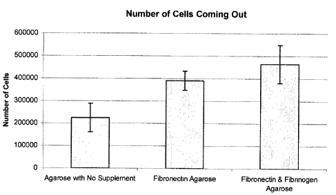
The number of cells coming out of the gels was also significantly improved
with the
addition of Fibronectin and Fibrinogen (Figure 14). Figure 14 compares the
number
of cells breaking out of the capsule in the no supplement 4% agarose matrix to
the
4% agarose matrix supplemented with Fibrinogen and fibronection. The addition
of
Fibronectin & Fibrinogen to the 4% agarose matrix significantly increased the
number
of cells breaking out of the capsule and adhering to the bottom of the tissue
culture
flask (24 hours) as compared to the cells encapsulated with agarose only. This
effect
was also confirmed by visual (microscopic) observation.
To investigate the mechanism behind this improvement in viability, studies
were
begun to determine the role of adhesion molecules and integrin binding sites.
The
present inventors developed a system in which Nitric Oxide production and eNOS
protein production can be detected and quantified. Specifically, the effect of
Fibronectin and Fibrinogen in the cell culture medium, as opposed to the
encapsulation medium was investigated. Results illustrated that the addition
of
Fibronectin and Fibrinogen to the culture media of the encapsulated cells
(encapsulated in agarose with no supplement), had a significantly detrimental
effect
on the viability of the cells (Figures 15 and 16). This further implicates
integrin
binding via use of an integrin in encapsulation as a major factor in the
viability of the
encapsulated rat fibroblasts. Studies were also carried out with human
fibroblasts.
Increased viability is observed in the human fibi-oblasts encapsulated in the
supplemented agarose.
Figure 15 thus shows the effect of Fibronectin and Fibrinogen added to cell
culture
media of encapsulated cells. Note that all cells are encapsulated in agarose
with no
supplement. NOsl, NOs2 and NOs3 represent different concentrations of
Fibronectin
and Fibrinogen added to cell culture media (0.5 g Fibronectin + 25 g of
Fibrinogen,
2 g of Fibronectin + 100 g Fibrinogen, 5 g of Fibronectin + 250 g of
Fibrinogen,
respectively). In each case 300 000 cells were encapsulated.
CA 02450650 2003-12-24
3O
Figure 16 shows the effect of Fibronectin and Fibrinogen added to cell culture
media
of encapsulated cells and the role of integrin-extracellular matrix protein
interactions.
In this experiment cells were encapsulated in 4% agarose. Cells were divided
into 4
groups and cultured for 24 hours under different culture conditions to
investigate the
effect of the addition of extracellular proteins to the cells' culture media.
The viability
results were compared to the controlled group of non-encapsulated cells. The
first
group contained encapsulated cells incubated for 24 hours in the regular rat
fibroblast culture conditions of DMEM + 10% FBS +2% P/S (4%Ags). The second,
third and fourth groups were encapsulated cells incubated in DMEM +10% FBS +
2%
P/S supplemented with varying concentrations of soluble Fibronectin and
Fibrinogen.
Group 4%AgsL* represents the encapsulated cells cultured in 0.5 g/mL
Fibronectin
and 25 g/mL of Fibrinogen. Group 4%AgsM* represents encapsulated cells
cultured
in 2 g/mL of Fibronectin and 100 g/mL of Fibrinogen. And finally, group
4%AgsH*
represents encapsulated cells cultured in 5 g/mL of Fibronectin and 250 g/mL
of
Fibrinogen. The addition of Fibronectin and Fibrinogen in solution (i.e. in
the cell
culture medium, as opposed to the encapsulation medium) appeared to have a
detrimental effect on the viability of the cells, although the addition of
Fibronectin and
Fibrinogen at the chosen concentrations did not have a dose dependent effect.
Results were obtained by Annexin V & Propidium Iodide staining. The y-axis
represents the percentage of Annexin V and PI positive encapsulated cells.
EXAMPLE 8- FXIII FACTOR REGULATES THE PROLIFERATION OF CELLS
The present inventors investigated the effect of FXIII on the survival of the
encapsulated cells.
Figure 17 and 18 show the effect of FXIII cross-linking on Human Umbilical
Vein
Endothelial Cell (HUVEC) phenotype was examined on thick fibrin gels.
Escalating
concentrations of FXIII had a significant effect on cell survival at both 24
(solid bars)
and 48 (hatched) hours after seeding. FXIII also had a dramatic inhibitory
effect on
cell proliferation measured over a 24 hour time period. This illustrated that
the effect
of FXIII was to keep the cells in statis and to prevent cell division in the
capules. Cell
division is undesirable in the encapsulated cells, as it will encourage the
cells to
break out of the capsule as an uncontrolled (premature) event.
CA 02450650 2003-12-24
31
EXAMPLE 9 - PREPARATION OF TRANSGENE ENCAPSULATED CELLS
Briefly, 40 micrograms of Beta-Galactosidase plasmid was added to 200
microlitres
of 4% agarose and empty capsules were prepared. Capsules were then stained
with
ethidium bromide and observed under UV light. Incorporation of the plasmid was
observed, showing incorporation of the DNA in the agarose microcapsules.
The present inventors have found good transfection achieved by incorporating
the
gene or a plasmid containing the gene of interest into the capsuie. As the
capsule
degrades, the cell uptakes the gene, likely by endocytosis.
EXAMPLE 10 - ENCAPSULATED BONE MARROW STROMAL CELLS
The present inventors investigated the viability of encapsulated rat bone
marrow
stromal cells (BMSC). The plasticity, pluripotentiality, ease of isolation,
and high in
vitro expansion potential make bone marrow stromal cells (BMSC) an attractive
tool
for cell and gene therapy. Such cells must home to the diseased tissue,
properly
engraft into the extracellular matrix, and once engrafted differentiate down
appropriate cell lineages. Non-hematopoietic cells referred to as mesenchymal
stem
cells or bone marrow stromal cells, have been investigated for their
multipotential
character (Bianco et al. 2001 , Theise et al. 2002 , Cognet et al. 2000 ,
Jiang et al.
2002 Krause et al. 2001). These cells have been found to be multipotential
(have the
ability to differentiate into various cell types) and serve as long-lasting
precursors for
bone marrow, bone, cartilage, and connective tissues (Cognet et al. 1999).
Encapsulated cell viability was assessed using a dual stain for apoptosis and
necrosis. The viability of adherent BMSCs was compared with BMSCs grown on
polyHEMA coated wells, BMSCs encapsulated in agarose, BMSCs encapsulated in
agarose supplemented with Fibronectin, BMSCs encapsulated in agarose
supplemented with Fibronectin and Fibrinogen.
As shown in Figure 19, the viability of the encapsulated BMSCs was increased
significantly by the incorporation of Fibronectin (82.6%) and Fibronectin and
Fibrinogen (84.1 %).
CA 02450650 2003-12-24
32
EXAMPLE 11 - MICROENCAPSULATION ENHANCES ENGRAFTMENT OF
BONE MARROW STROMAL CELLS IN LUNG MICROVASCULATURE
The present inventors investigated the role of encapsulation in improving the
engraftment potential of bone marrow stromal cells in the lung
microvasculature.
CMTMR labeled cells were delivered to normal Fisher 344 rats by injection into
the
jugular vein. Animals were sacrificed at selected time points (15 minutes, 1d,
3d, &
7d) and the lungs were excised and examined. Tissues from the lung were fixed
and
analyzed by confocal microscopy. Results illustrate that there is significant
engraftment of the non-encapsulated bone marrow stromal cells in the lung at
15
minutes. As shown in Figure 20, there is no evidence of any CMTMR labeled non-
encapsulated cells at 1 day and 3days after injection. However, the
encapsulation of
the bone marrow stromal cells in enriched agarose resulted in engraftment of
the
fluorescently labeled cells in the lung microvasculature 3 days after
delivery. . The top
panel of images in Figure 21 illustrates the encapsulated cells one day after
delivery
at higher magnification. Encapsulated cells and clusters of cells were also
observed
at 7 days (lower panel) after delivery indicating long-term engraftment.
EXAMPLE 12 - VIABILITY OF ENCAPSULATED HUMAN FIBROBLASTS
In the above examples, the present inventors examined the effect of
encapsulation
on rat fibroblast cells and rat bone marrow stromal cells. The present
inventors also
investigated the effect of encapsulation on human fibroblast cells.
Encapsulated cell viability was assessed using a dual stain for apoptosis and
necrosis. The viability of non-encapsulated cells was compared with cells
encapsulated in agarose and cells encapsulated in agarose supplemented with
Fibronectin and Fibrinogen.
As shown in Figure 22, the viability of the encapsulated human fibroblasts was
significantly increased by the incorporation of Fibronectin and Fibrinogen
into the
agarose matrix,
It is to be understood that only the preferred embodiments have been shown,
and
that modifications thereof would be readily apparent to one skilled in the
art.
Therefore, the true scope and spirit of the invention resides in the appended
claims
and their legal equivalents, rather than by the given examples.
CA 02450650 2003-12-24
33
REFERENCES
Bordignon, C. et al, Cell Therapy: Achievements and Perspectives (1999),
Haematologica, 84, pp. 1110-1149.
Lindvall, O. and Hagell, P., Cell Therapy and Transplantation in Parkinson's
Disease
(2001), Clinical Chemistry and Laboratory Medicine, 39, pp. 356-361.
Miyamoto, M, Current Progress and Perspectives in Cell Therapy for Diabetes
Mellitus (2001), Human Cell, 14, pp. 293-300.
Morris, P.J., Immunoprotection of Therapeutic Cell Transplants by
Encapsulation
(1996), Trends in Biotechnology, 14, pp. 163-167,
Rowely, J.A. et al, Alginate Hydrogels as Synthetic Extracellular Matrix
Materials
(1999), Biomaterials, 20, pp. 45-53.
Ruoslahti, E., RGD and Other Recognition Sequences for Integrins (1996),
Annual
Review in Cell and Developmental Biology, 12, pp. 697-715.
Uludag et al., Technology of Mammalian Cell Encapsulation (2000), Advanced
Drug
Delivery Reviews, 42, pp. 29-64.
Bianco P, Riminucci M, Gronthos S, Robey P G. "Bone marrow stromal stem cells:
nature, biology and potential applications". Stem Cells (2001) 19: 180-192.
Brown M D, Schatzlein A G, Uchegbu I F. "Gene delivery with synthetic (non
viral)
carriers". lnternatlonal journal of pharmaceutics (2001) 229: 1-21.
Bally M B, Harvie P, Wong F, Kong S, Wasan E K, Reimer D L. "Bilogical
barriers to
cellular delivery of lipid-based DNA carriers.". Advanced Drug Delivery
Reviews
(1999) 38: 291-315.
Campbell A, Kuliszewski M A., Stewart D J. "Cell-based gene transfer to the
pulmonary vasculature: endothelial nitric oxide synthase overexpression
inhibits
monocrotaline-induced pulmonary hypertension" American Journal of Respiratory
Cell and Mol. Biol. (1999) 21:567-575.
CA 02450650 2003-12-24
34
Colter D C, Sekiya I, Prockop D J. "Identification of a subpopulation of
rapidly self-
renewing and multipotential adult stem cells in colonies of human marrow
stromal
cells". PNAS (2001) 98: 7841-7845.
Cognet P, Minguell J . "Adenoviral-mediated gene transfer into ex vivo
expanded
human bone marrow mesenchymal progenitor cells". Experimental Hematology
(2000) 28: 382-390.
Conget P A., Minguell J."Phenotypical and Functional Properties of Human Bone
Marrow Mesenchymal Progenitor Cells". Journal of Cellular Physiology (1999)
181:67-73.
Ding L, Lu S, Batch R B, Sailors R L, Mushy N C. "Bone marrow stromal cells as
a
vehicle for gene transfer" Gene Therapy (1999) 6:1611-1616.
Eliceiri B P, Cheresh D A. "The role of alpha v integrins during angiogenesis:
insights
into potential mechanisms of action and clinical development". The Joumal of
Clinical
Investigation (1999) 103: 1227-1230.
Fukai F, Mashimmo M, Akiyama K, Goto T, Tanuma S, Katayama T. "Mudulation of
apoptotic cell death by extracellular matrix proteins and a fibronectin-
derived
antiadhesive peptide". Experimental Cell Research (1998) 242: 92-99.
Gailit J, Clarke C, Newman D, Tonnesen M G, Mosessen M W, Clark R A F. "Human
fibroblasts bind directiy to fibrinogen at RGD sites through integrin alpha V
beta 3".
Experimental Celi Research (1997) 232: 118-126.
Giaid A, Yanagisawa M, Langleben D, Michel R P, Levy R, Shennib H, Kimura S,
Masaki T, Duguid W P., Stewart D J. "Expression of endothelin-1 in the lungs
of
patients with pulmonary hypertension" The New England Journal of Medicine
(1993)
328:1732-9.
Gelband C H., Katovich M J, Raizada M K. "Current perspectives on the use of
gene
therapy for hypertension" Circulation Research (2000) 87:1118-1122.
CA 02450650 2003-12-24
Giaid A, Saleh D. "Reduced expression of endothelial nitric oxide synthase in
the
lungs of patients with pulmonary hypertension" N. Eng, J. of Med. (1995)
333:214-
221.
Giancotti F G. "Integrin signaling: specificity and control of cell survival
and cell cycle
progression". Current Opinion in Cell Biology (1997) 9: 691-700.
Gomez-Navarro J, Contreras J L, Arafat W, Jiang X L, Krisky D, Oligino T;
Marconi
P, Hubbard B, Glorioso J C, Curiel D T, Thomas J M. "Genetically modified
CD34+
cells as cellular vehicles for gene delivery into areas of angiogenesis in a
rhesus
model". Gene Therapy (2000) 7:43-52.
Gravanis M B. Cardiovascular Pathophysiology. McGraw Hill Book Company. New
York: 1987.
Gronthos S, Simmons P J, Graves S E, Robey P G. "Integrin-mediated
interactions
between human bone marrow strmal precurson cells and the extracellular
matrix".
Bone (2001) 28: 174-181.
Jiang Y, Jahagirdar B M, Reinhardt R L, Schwartz R E, Keene C D, Ortiz-
Gonzalez X
R, Reyes M, Lenvik T, Lund T, Blackstad M, Du J, Aldrich S, Lisberg A, Low W,
Largaespada D A, Verfaillie C M. "Pluripotency of senchymal stem cells derived
from
adults marrow". Nature advance online publication (2002) 1-9.
Krause D S, Thiese N D, Collector M I, Henegariu 0, Hwang S, Gardner R,
Neutzel
S, Sharkis S J. "Multi-organ, multi-lineage engraftment by a single bone
marrow-
derived stem cell". Cell (2001) 105:369-377.
Krause DS. "Plasticity of marrow-derived stem cells". Gene Therapy (2002) 9:
754-
758.
Kamiya H., Tsuchiya H., Yamazaki J., Harashima H. "Intracellular trafficking
and
transgene expression of viral and non-viral gene vectors.". Advanced Drug
Delivery
Reviews. 2001. 52: 153-164.
CA 02450650 2003-12-24
36
Kotton D N, Ma B Y, Cardonso W V, Sanderson E A, Summer R S, Williams M C,
Fine A. "Bone marrow-derived cells as progenitors of lung alveolar
epithelium".
Development (2001) 128: 5181-5188.
Minguel J J., Erices A, Cognet P. "Mesenchymal Stem Cells". Exp. Biol. Med.
(2001)
226: 507-520..
Pasqualini R, Bodorova J, Ye S, Hemler M. "A study of the structure, function
and
distribution of beta 5 integrins using novel anti beta 5 monoclonal
antibodies". Journal
of Cell Science (1993) 105: 101-111.
Pereira R F, Halford K W, O'Hara M D, Leeper D B, Sokolov B P, Pollard M D,
Bagasra 0, Prockop D J. "Cultured adherent cells from marrow can serve as long-
lasting precursor cells for bone, cartilage, and lung in irradiated mice".
Proc. Natl.
Acad. Sci. USA. (1995) 92:4857-4861.
Plath T, Detjen K, Welzel M, Marschall Z, Murphy D, Schirner M, Wiedenmann B,
Rosewicz S. "A novel function for the tumor suppressor p161NK4a. Induction of
anoikis via upregulaation of the alpha 5 beta 1 fibronectin receptor". The
Journal of
Cell Biology. (2000) 150: 1467-1477.
Powell K T, Weaver J C. Gel microdroplets and flow cytometry: rapid
determination
of antibody secretion by individual cells within a cell population.
Biotechnology (1990)
4: 333-7.
Reinecke H, Murry C E. "Taking the death toll after cardiomyocyte grafting: a
reminder of the importance of quantitative biology". J Mol Cell Cardiol (2002)
34: 251-
253.
Reinecke H, Zhang M, Bartosek T, Murry C E. "Survival, Integration, &
Differentiation
of Cardiomyocyte Grafts". Circulation (1999) 100: 193-202.
Rubanyi, G.M. "The future of human gene therapy.". Molecular Aspects of
Medicine
(2001) 22: 113-142.
CA 02450650 2003-12-24
37
Scott G, Cassidy L, Busacco A. "Fibronectin suppresses apoptosis in normal
human
melanocytes through an integrin-dependent mechanism". The Journal of
Investigative Dermatology (1997) 108: 147-153.
Stupack D G, Puente X S, Boutsaboualoy S, Storgard C M, Cheresh D A.
"Apoptosis
of adherent cells by recruitment of caspase-8 to unligated integrins". The
Joumal of
Cell Biology (2001) 155: 459-470.
Theise N D, Krause D S. "Toward a new paradigm of cell plasticity". Leukemia
(2002)
16: 542-548.
Thierry B, Molano D R., Pileggi A, Cattan P, Li H, Ricorde C, Inverardi L.
"Patterns of
engraftment in different strains of immunodificient mice reconstituted with
human
peripheral blood lymphocytes". Transplantation (2001) 72:133-140.
Thurmond F A, Cronin E M, Willams S R. "A deadly game of musical chairs:
survival
of cells transplanted for myocardial repair". J Mol Cell Caro'iol. (2001) 33:
883-885.
Turcanu V. and Williams N. "Cell identification and isolation on the basis of
cyokine
secretion: A novel tool for investigating immune responses.". Nature Medicine
(2001)
7: 373-376.
Wang E, Marcotte R, Petroulakis E. "Signalling pathway for apoptosis: a
racetrack for
life and death.". Journal of Cellular Biochemistry Supplements (1999) 32: 95-
102.
Weaver J C, Bliss J G, Powell K T, Harrison G T, Williams G B. "Rapid clonal
growth
measurements at the single-cell level: gel microdroplets and flow cytometry".
Biotechnology (1991) 9: 873-7.
Weaver J C, McGrath P, Adams S, "Gel microdrop technology for rapid isolation
of
rare and high producer cells". Nature Medicine (1997) 5: 583-5.
Young J L and Dean D A. "Nonviral gene transfer strategies for the
vasculature".
Microcirculation (2002) 9: 35-50.
Zhang W W. "Development and application of adenoviral vectors for gene therapy
of
cancer". Cancer Gene Therapy (1999) 6:113-38.
CA 02450650 2003-12-24
38
Zhang M, Methot D, Poppa V, Fuio Y, Walsh K, Murry C E. "Cardiomyocyte
grafting
for cardiac repair: graft cell death and anti-death strategies". Journal of
Molecular
Cell Cardiology (2001) 33: 907-921.
Zhang Z, Vuori K, Reed J C, Rouslahti E. "The alpha 5 beta I integrin supports
survival of cells on fibronectin an up-regulates Bcl-2 expression". PNAS
(1995) 92:
6161-6165.
CA 02450650 2004-02-16
SEQUENCE LISTING
(1) GENERAL INFORMATION:
(i) APPLICANT:
(A) NAME: An-Go-Gen Inc.
(B) STREET: 438 University Ave, Suite 300
(C) CITY: Toronto
(D) STATE: Ontario
(E) COUNTRY: Canada
(F) POSTAL CODE (ZIP): M5P 2P9
(G) TELEPHONE: 416 864 1482
(H) TELEFAX: 4163620823
(ii) TITLE OF INVENTION: Encapsulated Cell Therapy
(iii) NUMBER OF SEQUENCES: 3
(iv) COMPUTER READABLE FORM:
(A) MEDIUM TYPE: Floppy disk
(B) COMPUTER: IBM PC compatible
(C) OPERATING SYSTEM: PC-DOS/MS-DOS
(D) SOFTWARE: PatentIn Release #1.0, Version #1.25 (EPO)
(v) CURRENT APPLICATION DATA:
APPLICATION NUMBER: CA 2450650
(vi) PRIOR APPLICATION DATA:
(A) APPLICATION NUMBER: US 60/435,858
(B) FILING DATE: 24-DEC-2002
(2) INFORMATION FOR SEQ ID NO: 1:
(i) SEQUENCE CHARACTERISTICS:
(A) LENGTH: 3 amino acids
(B) TYPE: amino acid
(C) STRANDEDNESS: single
(D) TOPOLOGY: unknown
(ii) MOLECULE TYPE: peptide
(iii) HYPOTHETICAL: NO
(iii) ANTI-SENSE: NO
(v) FRAGMENT TYPE: N-terminal
(vi) ORIGINAL SOURCE:
(A) ORGANISM: artificial
(xi) SEQUENCE DESCRIPTION: SEQ ID NO: 1:
Arg Gly Asp
1
(2) INFORMATION FOR SEQ ID NO: 2:
(i) SEQUENCE CHARACTERISTICS:
1/2
CA 02450650 2004-02-16
(A) LENGTH: 3 amino acids
(B) TYPE: amino acid
(C) STRANDEDNESS: single
(D) TOPOLOGY: unknown
(ii) MOLECULE TYPE: peptide
(iii) HYPOTHETICAL: NO
(iii) ANTI-SENSE: NO
(vi) ORIGINAL SOURCE:
(A) ORGANISM: artificial
(xi) SEQUENCE DESCRIPTION: SEQ ID NO: 2:
Leu Asp Val
1
(2) INFORMATION FOR SEQ ID NO: 3:
(i) SEQUENCE CHARACTERISTICS:
(A) LENGTH: 12 amino acids
(B) TYPE: amino acid
(C) STRANDEDNESS: single
(D) TOPOLOGY: unknown
(ii) MOLE?CULE TYPE: peptide
(iii) HYPOTHETICAL: NO
(iii) ANTI-SENSE: NO
(vi) ORIGINAL SOURCE:
(A) ORGANISM: artificial
(xi) SEQUENCE DESCRIPTION: SEQ ID NO: 3:
His His Leu Gly Gly Ala Lys Gln Ala Gly Asp Val
1 5 10
2/2