Note: Descriptions are shown in the official language in which they were submitted.
CA 02450730 2003-12-12
WO 02/103029 PCT/US02/18823
-1-
METHODS FOR STERILIZING PREPARATIONS OF MONOCLONAL
IMMUNOGLOBULINS
Field of the Invention
The present invention relates to methods for
sterilizing preparations of monoclonal immunoglobulins to
reduce the level of active biological contaminants therein,
such as viruses, bacteria, yeasts, molds, mycoplasmas, prions
and/or parasites.
Background of the Invention
Antibodies are produced by organisms in response to
exposure to foreign substances that the body perceives as a
threat. Antibodies, or as they are collectively known,
immunoglobulins (2g), are proteins secreted by cells of the
immune system known as B-cells or plasma cells. The structure
of immunoglobulins is complex, but is well characterized. In
brief, each immunoglobulin consists of a complex of protein
chains known as the heavy and light chains. Each heavy chain
is linked to a single light chain via disulfide bonds. The
resulting complex is in turn linked by additional disulfide
bonds to an identical heavy-light chain complex. This basic
unit can be assembled by the cell into several specialized
forms by varying the structure and number of heavy chains.
Different heavy chain structures produce differing molecules,
known as "classes" of immunoglobulins. These classes may also
have different numbers of the basic units described above.
The production of these various physical forms of the
immunoglobulin molecule occurs in a sequential manner. During
this process, the specificity of the molecule for a single
molecule or antigen remains unchanged. This is because the
CA 02450730 2003-12-12
WO 02/103029 PCT/US02/18823
-2-
changes described above all occur to the portion of the
immunoglobulin molecule that is not involved in determining the
specificity of the particular immunoglobulin molecule. This
"hypervariable region" is subject to an unusually high degree
of recombination events during B-cell maturation. These
recombination events cease prior to the production of the first
immunoglobulin molecule by the cell. The.result is that from a
relatively small number of variable region genes, the body
generates a large number of potential immunoglobulin molecules
of differing specificities.
Once a B-cell encounters a molecule to which its own
immunoglobulin molecule binds (an "antigen"), and upon
receiving signals from other cells in the immune system, the B-
cell first multiplies into a large number of identical cells,
(collectively referred to as a clone) and then differentiates
into an immunoglobulin-secreting plasma cell. In this way, the
extremely large number of potential immunoglobulin molecules
that might be manufactured is limited to only those molecules
that recognize antigens to which the body must respond.
The vast array of immunoglobulin specificities that
are produced results in an ongoing protection for the body
against infection from those organisms that the body has made
immunoglobulins against in the past. Taken in sum, the result
is that the immunoglobulins contained in the plasma from a
single donor may have millions of useful immunoglobulin
specificities. A preparation of immunoglobulins from plasma is
thus referred to as a polyclonal immunoglobulin preparation,
since it contains the immunoglobulin molecules produced by all
of the plasma cell clones in the body.
Polyclonal immunoglobulins are particularly useful
for treating human disease in which the ability to produce Ig
is absent or impaired. Since all plasma cell clones are
CA 02450730 2003-12-12
WO 02/103029 PCT/US02/18823
-3-
affected, a mixture of all the immunoglobulin specificities
found in the plasma is needed to correct the deficiency. In
contrast, when an extreme degree of specificity is required, or
when a single defined therapeutic goal is sought, polyclonal
immunoglobulins are not the best solution. Instead, an
immunoglobulin preparation consisting of the immunoglobulin
molecules produced by a single clone with the desired
specificity is the most precise and predictable solution. Such
a preparation is known as a monoclonal immunoglobulin.
Monoclonal immunoglobulin have many differences as
compared to polyclonal immunoglobulins. Their monospecificity
makes them very precise when used as detection reagents. As
therapeutics, they are free of confounding or dangerous side
effects that arise from polyclonal immunoglobulin preparations,
such as the introduction of immunoglobulins of unwanted
specificities being introduced into the patient. Their
physical characteristics may also be different. Since each
monoclonal immunoglobulin has a unique and unvarying structure,
its potential for stability, degradation, aggregation,
temperature sensitivity and other~characteristics are unique
and unchanging. Once a suitable monoclonal immunoglobulin has
been chosen for production, its characteristics will not
change, and it thus can be manufactured with great consistency
and assurance of its performance and storage characteristics.
The ability to tailor production volumes to product
requirements also makes monoclonal immunoglobulin a highly
desirable alternative to polyclonal immunoglobulins.
Monoclonal immunoglobulin preparations are made in
one of three general fashions. The first involves production
in a cell culture system, the second uses an animal as a
temporary bioreactor for monoclonal immunoglobulin production,
and the third involves inserting the gene for a desired
monoclonal immunoglobulin into an animal in such a manner as to
CA 02450730 2003-12-12
WO 02/103029 PCT/US02/18823
-4-
induce continuous production of the monoclonal immunoglobulin
into a fluid or tissue of the animal so that it can be
continuously harvested (transgenic production).
Each of these methods may result in contamination of
the product by pathogens. In the first method, the cells
producing the monoclonal immunoglobulin may harbour undetected
viruses that can be produced in the culture system.
Contamination of the culture system by bacteria, yeast or mold
may also occur.
Both of the remaining methods involve the use of an
animal to either serve as a host for the monoclonal
immunoglobulin-producing cells or as a bioreactor to
manufacture the monoclonal immunoglobulin product itself.
Obviously, these products face the risk of contamination by
pathogens infecting or harboured by the host animal. Such
pathogens include, viruses, bacteria, yeasts, molds,
mycoplasmas, and parasites, among others.
Consequently, it is of utmost importance that any
biologically active contaminant in the monoclonal
immunoglobulin product be inactivated before the product is
used. This is especially critical when the product is to be
administered directly to a patient. This is also critical for
various monoclonal immunoglobulin products which are prepared
in media which contain various types of plasma and which may be
subject to mycoplasma or other viral contaminants.
Previously, most procedures have involved methods
that screen or test products for a particular contaminant
rather than removal or inactivation of the contaminant from the
product. Products that test positive for a contaminant are
merely not used. Examples of screening procedures include the
testing for a particular virus in human blood from blood
donors. Such procedures, however, are not always reliable and
CA 02450730 2003-12-12
WO 02/103029 PCT/US02/18823
-5-
are not able to detect the presence of viruses in very low
numbers. This reduces the value or certainty of the test in
view of the consequences associated with a false negative
result. False negative results can be life threatening in
certain cases, for example in the case of Acquired Immune
Deficiency Syndrome (AIDS). Furthermore, in some instances it
can take weeks, if not months, to determine whether or not the
product is contaminated.
In conducting experiments to determine the ability of
l0 technologies to inactivate viruses, the actual viruses of
concern are seldom utilized. This is a result of safety
concerns for the workers conducting the tests, and the
difficulty and expense associated with the containment
facilities and waste disposal. In their place, model viruses
of the same family and class are used.
In general, it is acknowledged that the most
difficult viruses to inactivate are those with an outer shell
made up of proteins, and that among these, the most difficult
to inactivate are those of the smallest size. This has been
shown to be true for gamma irradiation and most other forms of
radiation as these viruses diminutive size is a consequence of
their small genome. The magnitude of direct effects of
radiation upon a molecule are directly proportional to the size
of the molecule, that is the larger the target molecule, the
greater the effect. As a corollary, it has been shown for
gamma-irradiation that the smaller the viral genome, the higher
the radiation dose required to inactive it.
Among the viruses of concern for~both human and
animal-derived biologics, the smallest viruses of concern
belong to the family of Parvoviruses and the slightly larger
protein-coated Hepatitis virus. In humans, the Parvovirus B19,
and Hepatitis A are the agents of concern. In porcine-derived
CA 02450730 2003-12-12
WO 02/103029 PCT/US02/18823
-6-
products and tissues, the smallest corresponding virus is
Porcine Parvovirus. Since this virus is harmless to humans, it
is frequently chosen as a model virus for the human B19
Parvovirus and Hepatitis A. The demonstration of inactivation
of this model parvovirus is considered adequate proof that the
method employed will kill human B19 virus and Hepatitis A, and
by extension, that it will also kill the larger and less hardy
viruses such as HIV, CMV, Hepatitis B and C and others.
More recent efforts have focused on methods to remove
or inactivate contaminants in the products. Such methods
include heat treating, filtration and the addition of chemical
inactivants or sensitizers to the product. Heat treatment
requires that the product be heated to approximately 60°C for
about 70 hours which can be damaging to sensitive products.
Heat inactivation can destroy 50% or more of the biological
activity of the product. Filtration involves filtering the
product in order to physically remove contaminants.
Unfortunately this method may also remove products that have a
high molecular weight. Further, in certain cases small viruses
may not be removed by the filter because of the larger
molecular structure of the product. The procedure of chemical
sensitization involves the addition of noxious agents which
bind to the DNA/RNA of the virus and which are activated either
by UV or radiation to produce reactive intermediates and/or
free radicals which bind to the DNA/RNA or break the chemical
bonds in the backbone of the DNA/RNA of the virus or crosslink
or complex it in such a way that the virus can no longer
replicate. This procedure requires that unbound sensitizer is
washed from products since the sensitizers are toxic, if not
mutagenic or carcinogenic, and can not be administered to a
patient.
Irradiating a product with gamma radiation is another
method of sterilizing a product. Gamma radiation is effective
CA 02450730 2003-12-12
WO 02/103029 PCT/US02/18823
in destroying viruses and bacteria when given in high total
doses (Keathly et al., "Is There Life After Irradiation? Part
2," BioPharm July-August, 1993, and Leitman, Use of Blood Cell
Irradiation in the Prevention of Post Transfusion Graft-vs-Host
Disease," Transfusion Science 10:219-239 (1989)). The
published literature in this area, however, teaches that gamma
radiation can be damaging to radiation sensitive products, such
as blood, blood products, protein and protein-containing
products. In particular, it has been shown that high radiation
doses are injurious to red cells, platelets and granulocytes
(Leitman). U.S. Patent No. 4,620,908 discloses that protein
products must be frozen prior to irradiation in order to
maintain the viability of the protein product. This patent
concludes that "[i]f the gamma irradiation were applied while
the protein material was at, for example, ambient temperature,
the material would be also completely destroyed, that is the
activity of the material would be rendered so low as to be
virtually ineffective". This would apply as well to monoclonal
immunoglobulins which are, of course, proteins. Unfortunately,
many sensitive biologicals, such as monoclonal antibodies
(Mab), would lose viability and activity if subjected to
freezing for irradiation purposes and then thawing prior to
administration to a patient.
In view of the difficulties discussed above, there
remains a need for methods of sterilizing monoclonal
immunoglobulins that are effective for reducing the level of
active biological contaminants without an adverse effect on the
monoclonal immunoglobulins.
Summary of the Invention
Accordingly, it is an object of the present invention
to provide methods of sterilizing preparations of monoclonal
immunoglobulins by reducing the level of active biological
CA 02450730 2003-12-12
WO 02/103029 PCT/US02/18823
_g_
contaminants without adversely affecting the monoclonal
immunoglobulins. Other objects, features and advantages of the
present invention will be set forth in the detailed description
of preferred embodiments that follows, and in part will be
apparent from the description or may be learned by practice of
the invention. These objects and advantages of the invention
will be realized. and attained by the compositions and methods
particularly pointed out in the written description and claims
hereof .
l0 In accordance with these and other objects, a first
embodiment of the present invention is directed to a method for
sterilizing a preparation of monoclonal immunoglobulins that is
sensitive to radiation comprising: (i) reducing the residual
solvent content of a preparation of monoclonal immunoglobulins
to a level effective to protect the preparation of monoclonal
immunoglobulins from radiation; and (ii) irradiating the
preparation of monoclonal immunoglobulins with radiation at an
effective rate for a time effective to sterilize the
preparation of monoclonal immunoglobulins.
A second embodiment of the present invention is
directed to a method for sterilizing a preparation of
monoclonal immunoglobulins that is sensitive to radiation
comprising: (i) adding to a preparation of monoclonal
immunoglobulins at least one stabilizer in an amount effective
to protect the preparation of monoclonal. immunoglobulins from
radiation; and (ii) irradiating the preparation of monoclonal
immunoglobulins with radiation at an effective rate for a time
effective to sterilize the preparation of monoclonal
immunoglobulins.
A third embodiment of the present invention is
directed to a method for sterilizing a preparation of
monoclonal immunoglobulins that is sensitive to radiation
CA 02450730 2003-12-12
WO 02/103029 PCT/US02/18823
_g_
comprising: (i) reducing the residual solvent content of a
preparation of monoclonal immunoglobulins to a level effective
to protect the preparation of monoclonal immunoglobulins from
radiation; (ii) adding to the preparation of monoclonal
immunoglobulins at least one stabilizer in an amount effective
to protect the preparation of monoclonal immunoglobulins from
radiation; and (iii) irradiating the preparation of monoclonal
immunoglobulins with radiation at an effective rate for a time
effective to sterilize the preparation of monoclonal
immunoglobulins. According to this embodiment, steps (i) and
(ii) may be reversed.
The invention also provides a composition comprising
at least one monoclonal immunoglobulin and a least one
stabilizer selected from the group consisting of: ascorbic acid
or a salt or ester thereof; glutathione; 6-hydroxy-2,5,7,8-
tetramethylchroman-2-carboxylic acid; uric acid or a salt or
ester thereof; methionine; histidine; N-acetyl cysteine; the
dipeptide glycine-glycine; diosmin; silymarin; a mixture of
ascorbic acid, or a salt or ester thereof, and uric acid, or a
salt or ester thereof; a mixture bf ascorbic acid, or a salt or
ester thereof, and 6-hydroxy-2,5,7,8-tetramethylchroman-2-
carboxylic acid; a mixture of ascorbic acid, or a salt or ester
thereof, uric acid, or a salt or ester thereof, and 6-hydroxy-
2,5,7,8-tetramethylchroman-2-carboxylic acid; and a mixture of
uric acid, or a salt or ester thereof and 6-hydroxy-2,5,7,8-
tetramethylchroman-2-carboxylic acid, said at least one
stabilizer being present in an amount effective to preserve
said monoclonal immunoglobulin for its intended use following
sterilization of the composition with radiation.
Brief Description of the Drawings
Figures 1 and 2 are graphs showing the protective
effects of certain stabilizers on lyophilized anti-insulin
CA 02450730 2003-12-12
WO 02/103029 PCT/US02/18823
-10-
monoclonal immunoglobulin exposed to 45 kGy of low dose gamma
irradiation.
Figures 3A-3C are graphs showing t-he protective
effects of certain stabilizers on lyophilized anti-insulin
monoclonal immunoglobulin exposed to 45 kGy of low dose gamma
irradiation.
Figure 4 & 5 are graphs showing the protective
effects of primary lyophilizing and secondary lyophilizing on'
the sensitivity of a monoclonal immunoglobulin.
l0 Figures 6-11 are graphs showing the protective effect
of certain stabilizers on the activity of lyophilized anti
insulin monoclonal immunoglobulin.
Figure 12 is a graph showing the protective effect of
stabilizers on the activity of lyophilized anti-insulin
monoclonal immunoglobulin when the sample was irradiated at a
high dose rate (30 kGy/hr).
Figure 13 is a graph showing the effect of a
stabilizer on IgM activity after irradiation with gamma
radiation.
Figure 14 and 15 are graphs showing the effect of
stabilizers on immobilized anti-insulin monoclonal
immunoglobulin after irradiation with gamma radiation.
Figures 16A, 16B, 17A, and 17B are graphs showing the
effect of ascorbate at varying concentrations on immobilized
anti-insulin monoclonal immunoglobulin after irradiation with
gamma radiation.
Figures 18A-18H are graphs showing the effect of
stabilizers on lyophilized anti-insulin monoclonal
CA 02450730 2003-12-12
WO 02/103029 PCT/US02/18823
-11-
immunoglobulin supplemented with human serum albumin or
sucrose, after irradiation with gamma radiation.
Figures 19A-19F are graphs showing the effect of
stabilizers on lyophilized anti-insulin monoclonal
immunoglobulin supplemented with bovine serum albumin, after
irradiation with gamma radiation.
Figures 20A and 20B are graphs showing the effect of
various doses of gamma radiation on anti-insulin monoclonal
immunoglobulin, with and without ascorbate.
Figures 21A and 21B are graphs showing the effect of
low pH (4.5) on the stabilizing effect of L-ascorbic acid on
monoclonal immunoglobulin irradiated to 45 kGy with gamma
radiation.
Figure 22 is a graph showing the level of viral
inactivation by irradiation with gamma radiation of an anti-
insulin monoclonal immunoglobulin contaminated with porcine
parvovirus (PPV).
Figures 23A and 23B are~graphs showing the effect of
the presence or absence of sodium ascorbate on the level of
activity retention achieved when irradiating monoclonal
immunoglobulins in both liquid and lyophilized forms with
e-beam radiation.
Detailed Description of the Preferred Embodiments
A. Defini tioas
Unless defined otherwise, all technical and
scientific terms used herein are intended to have the same
meaning as is commonly understood by one of ordinary skill ~in
the relevant art. All patents and publications mentioned
herein are expressly incorporated by reference.
CA 02450730 2003-12-12
WO 02/103029 PCT/US02/18823
-12-
As used herein, the singular forms "a," "an," and
"the" include the plural reference unless the context clearly
dictates otherwise.
As used herein, the term "immunoglobulin" is used
synonymously with the term "antibody", and encompasses all
classes of immunoglobulins including, without limitation, IgG,
IgM, IgA, IgD and IgE and all subclasses of immunoglobulins
such as the IgG subclasses IgGl, IgG2, IgG3, and IgG4 found in
or produced by cells or animals including humans. The team
"immunoglobulin" encompasses both membrane immunoglobulins and
secreted immunoglobulins. Membrane immunoglobulins are
transmembrane proteins of B cells, and act as the B cells'
antigen receptor. Secreted immunoglobulins are structurally
identical to their membrane counterparts except that they lack
the traps-membrane region of amino acids at the C-terminus of
membrane immunoglobulins. Secreted immunoglobulins are present
in extracellular fluids and secretions.
The term "immunoglobulin" also encompasses fragments
of immunoglobulins including, without limitation, fragments
F(ab')2, Fab', Fab, Fc, Facb, pFc', and Fd, as well as
immunoglobulin derivatives and metabolites as are known in the
art. Metabolites of immunoglobulin are products resulting from
the metabolism of immunoglobulins by a living organism. A wide
variety of derivatives of immunoglobulins as are known in the
art may be prepared by known methods, which typically involve
breaking peptide or disulfide bonds in the immunoglobulin.
Immunoglobulins may also be derivatized to include modified or
synthetic or unnatural amino acids. Derivatives of
immunoglobulins also include immunoglobulins conjugated to a
moiety such as a toxin (e.g. diphtheria toxin, ricin), a
labelling molecule (e. g. fluorescin, Texas Red), a radioactive
atom or molecule (e. g. last) for therapeutic or diagnostic use,
an enzyme (e. g. avidin, horseradish peroxidase, alkaline
CA 02450730 2003-12-12
WO 02/103029 PCT/US02/18823
-13-
phosphatase), et cetera. Immunoglobulins may include post-
translational modifications such as phosphorylation,
glyocsylation, myristilation, prenylation, ADP-ribosylation,
methylation, acetylation, hydroxylation, carboxylation, and
oxidation-reduction, or may be cationized or anionized to alter
the overall charge of the immunoglobulin.
The term "monoclonal immunoglobulins" refers to
homogeneous immunoglobulins produced by a single clone, and are
to be contrasted with polyclonal immunoglobulins. Monoclonal
immunoglobulins are usually made from hybridomas, which are
prepared by fusing immunized mouse or rat spleen cells with a
non-secretor myeloma using polyethylene glycol (PEG). The
fusion mixture is plated out in HAT medium, containing
hypoxanthine, aminopterin and thymidine. Aminopterin blocks a.
metabolic pathway which can be bypassed if hypoxanthine and
thymidine are present. The myeloma cells lack this bypass and
consequently die in the HAT medium. Spleen cells die naturally
in culture after one or two weeks, but fused cells survive
because they have the immortality of the myeloma and the
metabolic bypass of the spleen cells. Some of the fused cells
secrete antibody, and the supernatants are tested in a specific
assay. Wells which produce the desired antibody are then
cloned. Monoclonal immunoglobulins may also be produced by
various techniques familiar to one skilled in the art of
genetic engineering that cause the inducible or constitutive
expression of a single set of endogenous genes that code for a
single immunoglobulin, or that involve the addition of such a
set or portion of a set, of genes into a cell or organism in
such a manner as to result in either the inducible or
3o constitutive expression of the resulting single set of genes
that code for a single immunoglobulin.
Monoclonal immunoglobulins may be produced using the
above-described techniques in combination with in vitro cell
CA 02450730 2003-12-12
WO 02/103029 PCT/US02/18823
-14-
culture, in vivo cell culture (for example as an ascites
culture) or by the techniques of transgenesis. Transgenesis
involves the insertion of one or more genes into a recipient
organism. The recipient organism then produces the protein
product of these genes, either constitutively or following
induction, and either incorporates them or secretes the into a
tissue (including eggs) or fluid (including blood, sweat, milk
or urine). The resulting tissue or fluid is then harvested and
the desired monoclonal immunoglobulin is purified from it.
As used herein, the term "preparation of monoclonal
immunoglobulins" encompasses, without limitation: (1)
compositions consisting solely of monoclonal immunoglobulins
(such as monoclonal immunoglobulins of a single specificity or
combinations of monoclonal immunoglobulins of different
specificities and/or different classes) and which may contain
impurities (including the naturally-occuring components of a
tissue or fluid into which the monoclonal antibody was produced
by transgenic means); (2) compositions comprising monoclonal
immunoglobulins, pharmaceutically acceptable diluents,
carriers, adjuvants, liposomes and other therapeutic agents, et
cetera, as are known in the art; (3) partially-purified in-
process intermediate preparations of monoclonal
immunoglobulins; and (4) articles containing monoclonal
immunoglobulins or having monoclonal immunoglobulins
immobilized upon them or otherwise disposed thereon. Preferred
"preparations of monoclonal immunoglobulins" are discussed
below.
In therapeutic applications, monoclonal
immunoglobulins are typically combined with buffer or salt
solutions as are known in the art. The monoclonal
immunoglobulin may also be combined with another therapeutic
agent. For instance, anti-platelet monoclonal immunoglobulins
which prevent platelets from aggregating are being evaluated
CA 02450730 2003-12-12
WO 02/103029 PCT/US02/18823
-15-
for long-term therapy for the prevention of formation of
thrombi. In this application, the anti-platelet monoclonal
immunoglobulin is administered together with aspirin. The
monoclonal immunoglobulin and the other therapeutic agent may
be co-packaged for administration by, for example, intravenous
injection. This may take the form of simple inclusion into a
single external package, or they may be provided in a single
container. An example of a more advanced form of packaging
would be the use of monoclonal immunoglobulins in liposome
preparations. Typically, the immunoglobulin is embedded a.n at
least the outer layer of the liposome where it can act as a
targeting agent by binding to structures on or in the desired
cells or tissues. The drug, which is contained within the
liposome, is then released at this specific site, providing a
more concentrated drug therapy with a larger therapeutic index
than achievable by ordinary systemic therapy.
For therapeutic use, the monoclonal immunoglobulins
are typically provided in liquid, frozen, or freeze-dried
(lyophilized) form packaged under nitrogen or vacuum. The
monoclonal immunoglobulins are usually then reconstituted with
sterile water (if required, or thawed as needed) and
administered by an appropriate route, such as an intramuscular
injection or intravenous injection either directly or following
placement in an IV bag. In accordance with the invention, the
preparation of monoclonal immunoglobulins may be irradiated at
any stage, including but not limited to as a raw material, a
purified or partially-purified in-process intermediate, in bulk
or in individual or multi-dose packaging, before or after
packaging, after dilution for administration, or in the IV bag
or other delivery vehicle itself. The irradiation may be
carried out at any convenient temperature that does not have a
deleterious effect upon the preparation, and which may be above
or below the freezing or eutectic point of the preparation.
CA 02450730 2003-12-12
WO 02/103029 PCT/US02/18823
-16-
Various preparations of monoclonal immunoglobulins are
available for therapeutic use, as set forth in Table 1.
Table
1: Therapeutic
Monoclonal
Immunoglobulin
Products
Physical
Name mAb Name TargetIndication Company Status State*
Metastatic LicensedFD
Anti- Breast
HerceptinTrastuzumabHER2 Cancer Genentech,
Anti- B Cell non- L
Rituxan Rituximab CD20 Hodgkin's Genentech, I
Anti- ClinicalFD
Xolair OmalizumabIgE asthma Genetech, Trials
Relapsed
Metastatic
Anti- Breast Clinical
Anti-'iIEGFVEGF Cancer Genetech, Trials
Anti- Severe ClinicalFD
Xanelim Anti-CDllaCDlla Psoriasis Genetech, Trials
Anti- ClinicalFD
LDP-02 Anti-a4b7 a4b7 IBD Genetech, Trials
Anti-HER2 r Clinical
2C4 Solid TumorsGenetech, Trials
I
Crohn's ClinicalFD
Remicade InfliximabcA2 Disease Centacor Trials
Gleevec CML Novartis C
Berlex/
I~ Campath~ Alemtuzu~ ~ CLL ~ Millennium
Source for above Table: A practical guide to ELISA, D.M.
Kemeny, Pergamon Press
*; FD - Freeze-Dried (Lyophylized), L - Liquid, I - Immobilized
on a surface, C - Capsule (Oral Dosing) containing dried
material
CA 02450730 2003-12-12
WO 02/103029 PCT/US02/18823
_17_
Preparations of monoclonal immunoglobulins also find
application as research tools and a vast number are used for
diagnostic purposes, particularly in blood work. Typical
research tools and diagnostic tests involving monoclonal
immunoglobulins include enzyme-linked imunnosorbent assays
(ELISA), radioimmunoassays (RIA), magnetic-bead-based assays
and separation kits, and fluorescent activated cell analysis
and/or sorting, among others. It is desirable to sterilize
such diagnostic tools, both to preserve them, as well as for
safety, as they are handled by lab technicians. Preparations
of monoclonal immunoglobulins used for diagnostic purposes
often comprise a solid support, such as a dipstick or plastic
plate, having the monoclonal immunoglobulins immobilized on or
in it by covalent chemistry, drying, or other known means. In
accordance with the invention, an entire commercial package or
kit, containing the solid support with monoclonal
immunoglobulins affixed thereto, instructions, containers of
reagents (for instance a reference sample for generating a
standard curve), et cetera, may be sterilized in accordance
with the invention. Table 2 sets forth a number of commercial
diagnostic monoclonal immunoglobulin products.
CA 02450730 2003-12-12
WO 02/103029 PCT/US02/18823
-18-
Table 2:
Diagnostic
Monoclonal
Immunoglobulin
Products
Physical
Name mAb NameTarget IndicationCompany Status State*
i
CMV IgG ELISA Sigma FD ,
IgM ELISA Sigma FD
HSV Type FD/L
1
and 2
(IgG) ELISA Sigma
Rubella FD/L
IgG
(Indirect ELISA Sigma
Rubella FD
IgG
(Capture) ELISA Sigma
Lyme FD
Disease
(Indirect ELISA Sigma
Mumps FD
(Indirect) ELISA ~ Sigma
Pregnancy
Test Kits J&J
FAGS FD/L
analysis
(diagnosti BD.
c imaging) Coulter
Sources for above Table: A practical guide to ELISA, D.M.
Kemeny, Pergamon Press
*: FD - Freeze-Dried (Lyophylized), L - Liquid, I - Immobilized
on a surface, C - Capsule (Oral. Dosing) containing dried
material
As used herein, the term "sterilize" is intended to
mean a reduction in the level of at least one active biological
CA 02450730 2003-12-12
WO 02/103029 PCT/US02/18823
9_
contaminant found in the preparation of monoclonal
immunoglobulins being treated according to the present
invention.
As used herein, the term "biological contaminant" is
intended to mean a contaminant that, upon direct or indirect
contact with a preparation of monoclonal immunoglobulins, may
have a deleterious effect on a preparation of monoclonal
immunoglobulins or upon a recipient thereof. Such biological
contaminants include the various viruses, bacteria and
parasites known to those of skill in the art to generally be
found in or infect preparation of monoclonal immunoglobulins.
Examples of biological contaminants include, but are not
limited to, the following: viruses, such as human immuno-
deficiency viruses and other retroviruses, herpes viruses,
parvoviruses, filoviruses, circoviruses, paramyxoviruses,
cytomegaloviruses, hepatitis viruses (including hepatitis B and
hepatitis C), pox viruses, toga viruses, Epstein-Barr virus and
parvoviruses; bacteria, such as Escherichia, Bacillus,
Campylobacter, Streptococcus and Staphylococcus; parasites,
such as Trypanosoma and malarial parasites, including
Plasmodium species; yeasts; molds; mycoplasmas; and prions. As
used herein, the term "active biological contaminant" is
intended to mean a biological contaminant that is capable of
causing the deleterious effect.
2S As used herein, the term "a biologically compatible
solution" is intended to mean a solution to which monoclonal
immunoglobulins may be exposed, such as by being suspended or
dissolved therein, and remain viable, i.e., retain their
essential biological and physiological characteristics.
As used herein, the term "a biologically compatible
buffered solution" is intended to mean a biologically
compatible solution having a pH and osmotic properties (e. g,
CA 02450730 2003-12-12
WO 02/103029 PCT/US02/18823
-20-
tonicity, osmolality and/or oncotic pressure) suitable for
maintaining the integrity of monoclonal immunoglobulins.
Suitable biologically compatible buffered solutions typically
have a pH between 4 and 8.5 and are isotonic or only moderately
hypotonic or hypertonic. Biologically compatible buffered
solutions are known and readily available to those of skill in
the art.
As used herein, the term "stabilizer" is intended to
mean a compound or material that reduces any damage to the
preparation of monoclonal immunoglobulins being irradiated to a
level that is insufficient to preclude the~safe and effective
use of the preparation of monoclonal immunoglobulins.
Illustrative examples of stabilizers include, but are not
limited to, the following: antioxidants, such as ascorbic acid.
and tocopherol; and free radical scavengers, such as ethanol.
Preferred examples of stabilizers include, but are not limited
to, the following: fatty acids, including 6,8-dimercapto-
octanoic acid (lipoic acid) and its derivatives and analogues
(alpha, beta, dihydro, bisno and tetranor lipoic acid),
2o thioctic acid, 6,8-dimercapto-oct'anoic acid, dihydrolopoate
(DL-6,8-dithioloctanoic acid methyl ester), lipoamide, bisonor
methyl ester and tatranor-dihydrolipoic acid, furan fatty
acids, oleic and linoleic and palmitic acids and their salts
and derivatives; flavonoids, phenylpropaniods, and flavenols,
such as quercetin, rutin and its derivatives, apigenin,
aminoflavone, catechin, hesperidin and, naringin; carotenes,
including beta-carotene; Co-Q10; xanthophylls; polyhydric
alcohols, such as glycerol, mannitol; sugars, such as xylose,
glucose, ribose, mannose, fructose and trehalose; amino acids,
such as histidine, N-acetylcysteine (NAC), glutamic acid,
trypt'ophan, sodium carpryl N-acetyl tryptophan and methionine;
azides, such as sodium azide; enzymes, such as Superoxide
Dismutase (SOD) and Catalase; uric acid and its derivatives,
CA 02450730 2003-12-12
WO 02/103029 PCT/US02/18823
-21-
such as 1,3-dimethyluric acid and dimethylthiourea;
allopurinol; thiols, such as glutathione and reduced
glutathione and cysteine; trace elements, such as selenium;
vitamins, such as vitamin A, vitamin C (including its
derivatives and salts such as sodium ascorbate and palmitoyl
ascorbic acid) and vitamin E (and its derivatives and salts
such as tocopherol acetate and alpha-tocotrienol); chromanol-
alpha-C6; 6-hydroxy-2,5,7,8-tetramethylchroma-2 carboxylic acid
(Trolox) and derivatives; extraneous proteins, such as gelatin
and albumin; tris-3-methyl-1-phenyl-2-pyrazolin-5-one (MCI-
186); citiolone; puercetin; chrysin; dimethyl sulfoxide (DMSO);
piperazine diethanesulfonic acid (PIPES); imidazole;
methoxypsoralen (MOPS); 1,2-dithiane-4,5-diol; reducing
substances, such as butylated hydroxyanisole (BHA) and
butylated hydroxytoluene (BHT); cholesterol; probucol; indole
derivatives; thimerosal; lazaroid and tirilazad mesylate;
proanthenols; proanthocyanidins; ammonium sulfate; Pegorgotein
(PEG-SOD); N-text-butyl-alpha-phenylnitrone (PBN); 4-nydroxy-
2,2,6,6-Tetramethylpiperidin-1-oxyl (Tempol); mixtures of
ascorbate, urate and Trolox C (Asc/urate/Trolox C); proteins,
peptides and dipeptides; the Glycine homodipeptide glycine-
glycine (Gly-Gly); reduced glutathione; diosmin; pupurogalin;
gallic acid and its derivatives including but not limited to
propyl gallat;, sodium formaldehyde sulfoxylate and silymarin.
As used herein, the term ~~residual solvent content°
is intended to mean the amount or proportion of freely-
available liquid in the preparation of monoclonal
immunoglobulins. Freely-available liquid means that liquid,
such as water or an organic solvent (e. g. ethanol, isopropanol,
polyethylene glycol, etc.), present in the preparation of
monoclonal immunoglobulins that is not bound to or complexed
with one or more of the non-liquid components of the
preparation of monoclonal immunoglobulins. Freely-available
CA 02450730 2003-12-12
WO 02/103029 PCT/US02/18823
-22-
liquid includes intracellular water. The residual solvent
contents related as water referenced herein refer to levels
determined by the FDA approved, modified Karl Fischer method
(Meyer and Boyd, Analytical Chem., 31, 215-219, 1959; May, et
al., J. Biol. Standardization, 10, 249-259, 1982; Centers for
Biologics Evaluation and Research, FDA, Docket No. 89D-0140,
.83-93; 1990). Quantitation of the residual levels of other
solvents may be determined by means well known in the art,
depending upon which solvent is employed. The proportion of
residual solvent to solute may also be considered to be a
reflection of the concentration of the solute within the
solvent. When so expressed, the greater the concentration of
the solute, the lower the amount of residual solvent.
As used herein, the term "sensitizer" is intended to
mean a substance that selectively targets viral, bacterial,
prion and/or parasitic contaminants, rendering them more
sensitive to inactivation by radiation, therefore permitting
the use of a lower rate or dose of radiation and/or a shorter
time of irradiation than in the absence of the sensitizer.
Illustrative examples of suitable~sensitizers include, but are
not limited to, the following: psoralen and its derivatives and
analogs (including 3-carboethoxy psoralens); angelicins,
khellins and coumarins which contain a halogen substituent and
a water solubilization moiety, such as quaternary ammonium ion
or phosphonium ion; nucleic acid binding compounds; brominated
hematoporphyrin; phthalocyanines; purpurins; porphorins;
halogenated or metal atom-substituted derivatives of
dihematoporphyrin esters, hematoporphyrin derivatives,
benzoporphyrin derivatives, hydrodibenzoporphyrin dimaleimade,
3o hydrodibenzoporphyrin, dicyano disulfone, tetracarbethoxy
hydrodibenzoporphyrin, and tetracarbethoxy hydrodibenzo-
porphyrin dipropionamide; doxorubicin and daunomyci.n, which may
be modified with halogens or metal atoms; netropsin; BD
CA 02450730 2003-12-12
WO 02/103029 PCT/US02/18823
-23-
peptide, S2 peptide; S-303 (ALE compound); dyes, such as
hypericin, methylene blue, eosin, fluoresceins (and their
derivatives), flavins, merocyanine 540; photoactive compounds,
such as bergapten; and SE peptide.
As used herein, the term "radiation" is intended to
mean radiation of sufficient energy to sterilize at least some
component of the irradiated monoclonal immunoglobulins. Types
of radiation include, but are not limited to, the following:
(i) corpuscular (streams of subatomic particles such as
neutrons, electrons, and/or protons); and (ii) electromagnetic
(originating in a varying electromagnetic field, such as radio
waves, visible (both mono and polychromatic) and invisible
Light, ultraviolet radiation, x-radiation, and gamma rays and
mixtures thereof). Such radiation is often described as either
25 ionizing (capable of producing ions in irradiated materials)
radiation such as gamma rays and non-ionizing radiation such as
visible light. The sources of such radiation may vary, however
in general the specific source is of little material difference
as long as sufficient radiation is given in an appropriate time
and at an appropriate rate to effect sterilization. In
practice, gamma radiation is usually produced by isotopes of
Cobalt or Cesium, while X-rays are produced by machines that
emit X-radiation, and electrons are often used to sterilize
materials in a method known as "e-beam" irradiation that
involves their production via a machine.
B. Particularly Preferred Embodiments
A first preferred embodiment of the present invention
is directed to a method for sterilizing a preparation of
monoclonal immunoglobulins that is sensitive to radiation
comprising: (i) reducing the residual solvent content of the
preparation of monoclonal immunoglobulins to a level effective
to protect the preparation of monoclonal immunoglobulins from
CA 02450730 2003-12-12
WO 02/103029 PCT/US02/18823
-24-
radiation; and (ii) irradiating the preparation of monoclonal
immunoglobulins with radiation at an effective rate for a time
effective to sterilize the preparation of monoclonal
immunoglobulins.
~ A second embodiment of the present invention is
directed to a method for sterilizing a preparation of
monoclonal immunoglobulins that .is sensitive to radiation
comprising: (i) adding to a preparation of monoclonal
immunoglobulins at least one stabilizer in an amount effective
to protect the preparation of monoclonal immunoglobulins from
radiation; and (ii) irradiating the preparation of monoclonal
immunoglobulins with radiation at an effective rate for a time
effective to sterilize the preparation of monoclonal
immunoglobulins.
A third embodiment of the present invention is
directed to a method for sterilizing a preparation of
monoclonal immunoglobulins that is sensitive to radiation
comprising: (i) reducing the residual solvent content of a
preparation of monoclonal immunoglobulins to a level effective
to protect the preparation of monoclonal immunoglobulins from
radiation; (ii) adding to the preparation of monoclonal
immunoglobulins at least one stabilizer in an amount effective
to protect the preparation of monoclonal immunoglobulins from
radiation; and (iii) irradiating the preparation of monoclonal
immunoglobulins with radiation at an effective rate for a time
effective to sterilize the preparation of monoclonal
immunoglobulins. The order of steps (i) and (ii) may, of
course, be reversed as desired.
According to the methods of the present invention,
the residual solvent content of the preparation of monoclonal
immunoglobulins may be reduced prior to irradiation of the
preparation of monoclonal immunoglobulins with radiation. The
CA 02450730 2003-12-12
WO 02/103029 PCT/US02/18823
-25-
residual solvent content is reduced to a level that is
effective to protect the preparation of monoclonal immuno-
globulins from the radiation. Suitable levels of residual
solvent content may vary depending upon the nature and
characteristics of the particular preparation of monoclonal
immunoglobulins being irradiated and can be determined
empirically by one skilled in the art. Preferably, when the
solvent is water, the residual solvent content is less than
about 10%, more preferably less than about 2.0%, more
preferably less than about 1.0%, even more preferably less than
about 0.5% and most preferably less than about 0.2%.
According to the methods of the present invention,
the monoclonal antibody to be sterilized may be immobilized
upon a solid surf ace by a means familiar to one skilled in the
art.
According to the methods of the present invention,
the rate of irradiation may be optimized to produce the most
advantageous combination of product recovery and time required
to complete the operation. Both low {<3 kGy/hour) and high {>3
kGy/hour) rates may be achieved by the appropriate application
of the methods described herein.
While not wishing to be bound by any theory of
operability, it is believed that the reduction in residual
solvent content reduces the degrees of freedom of the
preparation of monoclonal immunoglobulins and thereby protects
it from the effects of the radiation. Similar results might
therefore be achieved by lowering the temperature of the
preparation of monoclonal immunoglobulins below its eutectic
point or below its freezing point, or by vitrification to
likewise reduce the degrees of freedom of the preparation of
monoclonal immunoglobulins. These results may permit the use
of a higher rate of irradation than might otherwise be
CA 02450730 2003-12-12
WO 02/103029 PCT/US02/18823
-26-
acceptable. Thus the inventions described herein may be
carried out at any temperature that does not result in damage
to the monoclonal immunoglobulin.
The residual solvent content of the preparation of
monoclonal immunoglobulins may be reduced by any of the methods
and techniques known to those skilled in the art for reducing
solvent from a preparation of monoclonal immunoglobulins. Such
methods may include, but are not limited to, evaporation,
concentration, centrifugal concentration, vitrification and
spray-drying. A particularly preferred method for reducing the
residual solvent content of a preparation of monoclonal
immunoglobulins is lyophilization. According to a particularly
. preferred embodiment of the present invention, a preparation of
monoclonal immunoglobulins which has been lyophilized is stored
under vacuum or an inert atmosphere (preferably a noble gas,
such as helium or argon, more preferably a higher molecular
weight noble gas, and most preferably argon) prior to
irradation.
The radiation employed in the present invention may
be any radiation effective for the inactivation of one or more
biological contaminants of the preparation of monoclonal
immunoglobulins being treated. The radiation may be
corpuscular, including e-beam radiation. Preferably the
radiation is electromagnetic radiation, including visible
light, W light and mixtures of various wavelengths of
electromagnetic radiation and a particularly preferred form of
radiation is gamma radiation.
According to the methods of the present invention,
the preparation of monoclonal immunoglobulins is irradiated
with the radiation at a rate effective for the inactivation of
one or more biological contaminants of the preparation of
monoclonal immunoglobulins. Suitable rates of irradiation may
CA 02450730 2003-12-12
WO 02/103029 PCT/US02/18823
-27-
vary depending upon the particular form of radiation and the
nature and characteristics of the particular preparation of
monoclonal immunoglobulins being irradiated and the particular
biological contaminants being inactivated. Suitable rates of
irradiation can be determined empirically by one skilled in the
art. Preferably, the rate of irradiation is constant for the
duration of the sterilization procedure. When this is
impractical, a variable or discontinuous irradiation may be
utilized.
according to a particularly preferred embodiment of
the present invention, the rate of irradiation is not more than
about 3.0 kGy/hour, more preferably between about 0.1 kGy/hr.
and 3.0 kGy/hr, even more preferably between about 0.25 kGy/hr
and 2.0 kGy/hour, still even more preferably between about 0.5
kGy/hr and 1.5 kGy/hr and most preferably between about 0.5
kGy/hr and 1.0 kGy/hr.
According to another particularly preferred
embodiment of the present invention, the rate of irradiation is
at least about 3.0 kGy/hr., more preferably at least about 6
kGy/hr., even more preferably at least about 16 kGy/hr., and
even more preferably at least about 30 kGy/hr and most
preferably at least about 45 kGy/hr or greater.
The preparation of monoclonal immunoglobulins is
irradiated with the radiation for a time effective for the
inactivation of one or more biological contaminants of the
preparation of monoclonal immunoglobulins. Combined with
irradiation rate, the appropriate irradiation time results in
the appropriate dose of irradiation being applied to the
monoclonal immunoglobilin. Suitable ionization times may vary
depending upon the particular form and rate of radiation and
the nature and characteristics of the particular preparation of
monoclonal immunoglobulins being irradiated and the particular
CA 02450730 2003-12-12
WO 02/103029 PCT/US02/18823
-28-
biological contaminants being inactivated. Suitable
irradiation times can be determined empirically by one skilled
in the art.
Optionally, an effective amount of at least one
sensitizing compound may be added to the monoclonal
immunoglobulin prior to irradiation to enhance the anti-
microbial effect of the irradiation, while employing the
methods described herein to minimize the deleterious effects of
irradiation upon the monoclonal immunoglobulin. Suitable
sensitizers are known to those skilled in the art.
According to methods of the present invention, the
irradiation of the preparation of monoclonal immunoglobulins
may occur at any temperature which is not deleterious to the
preparation of monoclonal immunoglobulins being treated.
According to a preferred embodiment, the preparation of
monoclonal immunoglobulins is irradiated at ambient
temperature. According to an alternate preferred embodiment,
the preparation of monoclonal immunoglobulins is irradiated at
reduced temperature, preferably at or below the freezing or
eutectic point of the preparation of monoclonal
immunoglobulins.
In order to avoid aggregation of the monoclonal
immunoglobulins, the preparation of monoclonal immunoglobulins
may have a pH of less than 7, preferably less than 6, more
preferably less than 5, even more preferably less than 4, and
most preferably less than 3.
It will be appreciated that combination of the
several methods described herein may be employed to further
minimize undesirable effects upon the monoclonal immunoglobulin
caused by irradiation, while maintaining adequate effectiveness
of the anti-microbial properties of the irradiation process.
CA 02450730 2003-12-12
WO 02/103029 PCT/US02/18823
-29-
Examples
The following examples are illustrative, but not
limiting, of the present invention. Other suitable
modifications and adaptations are of the variety normally
encountered by those skilled in the art and are fully within
the spirit and scope of the present invention. Unless
otherwise noted, all irradiation was accomplished using~a 6°Co
source.
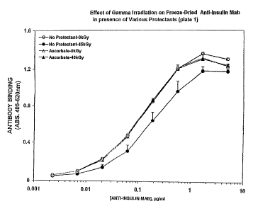
Example 1
In this experiment the protective effects of certain
stabilizers were evaluated using lyophilized anti-insulin
monoclonal immunoglobulin exposed to 45 kGy of low dose gamma
irradiation. The stabilizers tested were: sodium ascorbate,
methionine, and lipoic acid.
Method
In 2 ml glass vials, a 0.5 ml total volume was
lyophilized containing 50~.g anti-insulin monoclonal
immunoglobulin, 5 mg bovine serum~albumin (1%) and either no
stabilizer or 50mM of the stabilizer of interest. The samples
were stoppered under vacuum. Samples were irradiated with
gamma radiation (45 kGy total dose, dose rate 1.83 kGy/hr,
temperature 4°C) and then reconstituted with water.
Immunoglobulin binding activity of independent
duplicate samples was determined by a standard ELISA protocol:
96-well microtitre plates were coated overnight with 2.5ug/ml
insulin antigen. Three-fold serial dilutions of anti-insulin
monoclonal antibody samples starting at 5ug/ml were used. Goat
anti-mouse Ig conjungated to phosphatase used at 50 ng/ml.
Sigma 104 alkaline phosphatase substrate was used at 1 mg/ml in
CA 02450730 2003-12-12
WO 02/103029 PCT/US02/18823
-30-
DEA buffer. Binding activity was determined by absorbance at
405-620nm.
Relative protection was determined by estimating the
shift in the titration curve (i.e. concentration of immuno-
globulin needed to observe the same amount of binding) of the
irradiated sample compared to an unirradiated sample at
approximately 50% of the maximum absorbance signal for the
unirradiated sample.
Results
Lyophilized samples containing no stabilizer retained
50% of immunoglobulin avidity following irradiation with 45 kGy
gamma irradiation. This is in contrast to previous results in
which 45 kGy of gamma radiation destroyed essentially all the
activity of immunoglubulin when it was irrradiated in solution.
Thus, it is apparent that the reduction in. residual water
content by lyophilizing afforded significant protection on its
own to the monoclonal immunoglobulin.
The addition of sodium ,ascorbate provided full
recovery of activity after irradiation of the sample. Both
methionine and lipoic acid provided significant recovery of
activity (76-83%) of activity after irradiation as compared to
the unirradiated sample. The results are shown in Figures 1
and 2. Similar results (65% recovery of activity) were also
seen for pupurogalin (data not shown).
Example 2
In this experiment, the protective effects of certain
stabilizers were evaluated using lyophilized anti-insulin
monoclonal immunoglobulin exposed to 45 kGy of low dose gamma
irradiation. The stabilizers tested were: sodium ascorbate,
CA 02450730 2003-12-12
WO 02/103029 PCT/US02/18823
-31-
N-acetyl cysteine, glutathione and mixtures of urate/trolox and
ascorbate/urate/trolox.
Me thod
Tn 3 ml glass vials, a 1.0 ml total volume was
lyophilized containing 100ug anti-insulin monoclonal
immunoglobulin, 10 mg~bovine serum albumin (1%) and either no
stabilizer or the stabilizer of interest. The samples were
stoppered under vacuum, Samples were irradiated with gamma
radiation (45 kGy total dose, dose rate 1.83 kGy/hr,
temperature 4°C) and then reconstituted with 1.0 ml water.
Immunoglobulin binding activity of independent
duplicate samples was determined by a standard ELISA protocol:
Maxisorb plates were coated overnight with 2.5ug/ml insulin
antigen. Three-fold serial dilutions of anti-insulin
monoclonal immunoglobulin samples starting at 5ug/ml were used.
Goat anti-mouse Ig conjugated to phosphatase was used at 50
ng/ml. Binding activity was determined by absorbance at 405-
620nm.
Relative protection was determined using a parallel
line analysis software package (PLA 1.2 from Stegmann
Systemberatung).
Resu1 is
Lyophilized samples containing no stabilizer retained
70°s of immunoglobulin avidity following irradiation with 45 kGy
gamma irradiation. This is in contrast to previous results in
which 45 kGy of gamma radiation destroyed essentially all the
activity of immunoglubulin when it was irrradiated in solution.
Thus, it is apparent that the reduction in residual water
content by lyophilizing afforded significant protection on its
own protein.
CA 02450730 2003-12-12
WO 02/103029 PCT/US02/18823
-32-
The presence of sodium ascorbate increased recovery
by 20%, i.e. such that there is 90% avidity recovered after
irradiation. The remaining stabilizers resulted in recovery of
77-84% of avidity. The results are shown in Figures 3A-3C.
Example 3
In this experiment, the protective effects of primary
lyophilizing (which leaves a relatively "high moisture" content
in the product) and the combination of both primary and
secondary lyophilizing (which results in a product with
relatively "low moisture") on the radiation sensitivity of a
monoclonal immunoglobulin were determined.
Methods
~In 3 ml glass vials, 1.0 ml total volume was
lyophilized (using either only primary or a combination of both
Z5 primary and secondary drying) containing 100ug anti-insulin
monoclonal immunoglobulin, 10 mg bovine serum albumin (1%) and
either no stabilizer or 100 mM of sodium ascorbate. The samples
were stoppered under vacuum. Samples were irradiated with
gamma radiation (45 kGy total dose, dose rate between 2.03 and
2.13 kGy/hr, temperature 4°C) and then reconstituted with 1.0
ml water.
Immunoglobulin binding activity of independent
duplicate samples was determined by a standard ELISA protocol:
Maxisorb plates were coated overnight with 2.5ug/ml insulin
antigen. Three-fold serial dilutions of anti-insulin mAb
samples starting at 5ug/ml were used. Goat anti-mouse Ig
conjugated to phosphatase was used at 50 ng/ml. Binding
activity was determined by absorbance at 405-620nm.
CA 02450730 2003-12-12
WO 02/103029 PCT/US02/18823
-33-
lZesul is
In the absence of a stabilizer, there was better
recovery of the anti-insulin immunoglobulin after irradiation
from the samples that had undergone the secondary "low
moisture" drying cycle, i.e. a lower total moisture content in
the absence of a stabilizer improved recovery.
In the presence of the stabilizer, however, there was
very good recovery of antibody activity after 45 kGy
irradiation, irrespective of whether the sample had undergone
only the primary "high moisture" drying cycle or had also
undergone the secondary "low moisture" drying cycle.
The results of this experiment are shown in Figures 4
and S.
Example 4
In this experiment, the protective effect of certain
stabilizers on the activity of lyophilized anti-insulin
monoclonal immunoglobulin was determined. The stabilizers
tested were; sodium ascorbate; trolo.x/urate/ ascorbate
mixtures; and N-acetyl cysteine.
2 0 Me thods
Anti-insulin monoclonal immunoglobulin supplemented
with 1% of human serum albumin (and, optionally, 5% sucrose)
was lyophilized, stoppered under vacuum, and irradiated (total
dose 45 kGy; dose rate between 1.83 and 1.88 kGy/hr).
Immunoglobulin binding activity was determined using the
standard EZISA protocol described above.
CA 02450730 2003-12-12
WO 02/103029 PCT/US02/18823
-34-
Resu1 is
Irradiation of lyophilized anti-insulin immuno-
globulin supplemented with 1% HSA to a dose of 45 kGy resulted
in an average loss of avidity of about 33%. The addition of
the following stabilizers significantly improved recovery: 2omM
sodium ascorbate (100% recovery); 200~ZM trolox/l.5mM uratei20
mM ascorbate (87%) recovery); 20 mM N-acetyl cysteine (82%
recovery The addition of 5% sucrose to the lyophilized
immunoglobulin containing 1% HSA resulted in an average loss of
avidity of about 30% when irradiated to a dose of 45 kGy. The
addition of the following stabilizers significantly improved
recovery: 20mM sodium ascorbate (88% recovery); 200uM
trolox/l.5mM urate/20 mM ascorbate (84%) recovery); 20 mM N-
acetyl cysteine (72% recovery).
The results of these experiments are shown in Figures
6-11.
Example 5
In this experiment, the protective effect of
stabilizers (ascorbate) on the activity of lyophilized anti-
.insulin monoclonal immunoglobulin was determined when the
sample was irradiated at a high dose rate (30 kGy/hr).
Methods
Anti-insulin monoclonal immunoglobulin was
lyophilized and irradiated at a rate of 30 kGy/hr (total dose
45 kGy). Immunoglobulin binding activity was determined using
the standard EhISA protocol described above.
CA 02450730 2003-12-12
WO 02/103029 PCT/US02/18823
-35-
Resu1 is
Irradiation of lyophilized anti-insulin
immunoglobulin to a dose of 45 kGy resulted in an average loss
of activity of about 32%, The addition of 20mM sodium
ascorbate provided 85% recovery of avidity compared to an
unirradiated sample. The results are shown in Figure 12.
Examgle 6
In this experiment, an IgM monoclonal immunoglobulin
specific for marine IgG3 was irradiated at a low dose rate in
l0 the presence or absence of a stabilizer.
Method
Liquid rat anti-marine IgG3 monoclonal IgM (in a PBS
buffer with 10 mM sodium azide; concentration of antibody was
666 ng/~1) was irradiated at a rate of 1.8 kGy/hr to a total
dose of either 10 kGy or 45 kGy, Samples either contained no
stabilizer or a stabilizer mixture containing 20 mM citrate,
300 uM urate and 200 mM ascorbate.
Immunoglobulin activity was analyzed by standard
LISA protocol using marine IgG3 as the coating antigen and a
phosphatase-conjugated anti-rat IgM detection antibody.
Resu3 is
Liquid samples containing no stabilizer lost all
functional immunoglobulin activity following irradiation with
either lOkGy or 45 kGy gamma irradiation. The presence of a
stabilizer mixture, however, provided full recovery of activity
following irradiation with 10 kGy gamma radiation and 88%
recovery of activity following irradiation with 45 kGy gamma
radiation. The results of this experiment are shown
graphically in Figure 13.
CA 02450730 2003-12-12
WO 02/103029 PCT/US02/18823
-36-
Example 7
In this experiment, the protective effects of certain
stabilizers were evaluated using immobilized anti-human insulin
monoclonal immunoglobulin exposed to 45 kGy of low dose-rate
gamma irradiation. The stabilizers tested were: sodium
ascorbate, reduced glutathione, sodium formaldehyde
sulfoxylate, and polypropylene glycol.
Me thod
Two plates were coated with 100 ul/well of freshly
prepared 2 ug/ml anti-insulin immunoglobulin in coating buffer
overnight at 4°C. The plates were washed briefly three times
with PBS. A two-fold dilution series of each stabilizer in PBS
was prepared. 100 u1 of a selected stabiliser solution was
added to each well. The plates were covered tightly with a cap
mat. One plate was irradiated at 1,92 kGy/hr for a total of 45
kGy at 4°C. The control plate received 0 kGy and was stored at
4°C.
Immunoglobulin binding activity was determined by a
standard ELISA protocol. The plate wells were emptied and were
washed four times with a full volume of PBS . A full volume of
blocking buffer (approximately 380 u1) was added to all wells
and incubated for two hours at 37°C. All wells were washed four
times with TBST (TBS pH 7.4 with 0.05% TWEEN 20). One hundred
u1 of 50 ng/ml biotin-labelled insulin in binding buffer was
added to each well. The plates were covered with a plate
sealer and incubated at 37°C while shaking (LabLine titer plate
shaker set at 3) for 1.5 hours. The plates were then washed
four times with TEST. One hundred u1 of 0.5 ug/ml phosphatase
labelled Streptavidin (stock diluted 1:1000 in binding buffer)
was added to each well. The plates were covered with a plate
sealer and incubated at 37°C for one hour with shaking. The
plates were then washed four times with TBST. One hundred u1
CA 02450730 2003-12-12
WO 02/103029 PCT/US02/18823
-37-
of 1 mg/ml Sigma 104 phosphatase substrate in DEA buffer was
added to each well. The plates were then incubated at 37°C with
shaking. Absorbance was determined at 405nm-628nm at 5 minute
intervals.
Resu1 is
As shown in Figures 14 and 15, sodium ascorbate
exhibited a dose-dependent protective effect. Samples
containing between 31-250 mM of sodium ascorbate exhibited 73~
81% greater retained activity.
l0 Samples containing glutathione exhibited
approximately 25% greater retention of monoclonal
immunoglobulin activity, that was dose dependent up to a
glutathione concentration of about 31 mM.
Samples treated with sodium formaldehyde sulfoxylate
exhib7.ted approximately 50% greater retained activity than
control samples at a stabilizer concentration of 31 mM.
All three forms of polypropylene glycol (i.e.
polypropylene P400 (Fluka 81350);~polypropylene P1200 (Fluka
81370); and polypropylene P2000 (Fluka 81380)) exhibited a
protective effect. Samples treated with polypropylene glycol
exhibited approximately 50-60% increased retention of activity
relative to control samples.
Example 8
In this experiment, the optimal concentration of
sodium ascorbate to protect immobilized anti-insulin monoclonal
immunoglobulins from 45 kGy of gamma irradiation was
determined. Tt was also determined whether the presence of 1.5
mM uric acid has any effect on the stabilizing nature of
ascorbate of immobilized monoclonal immunoglobulin exposed to
45 kGy gamma irradiation.
CA 02450730 2003-12-12
WO 02/103029 PCT/US02/18823
-38-
Method
Two plates were coated overnight at 4°C with 100 u1 of
2.5 ug/ml anti-insulin monoclonal immunoglobulin in coating
buffer. The coating solution was discarded and the wells
washed two times with PBS. Twenty-five u1 of 4X ascorbate
solution was added to appropriate wells. Seventy-five ~1 of
water was added to the orate-free wells (rows a-d). Twenty
five ~.1 of water was added to the orate containing wells (rows
e-h). Fifteen u1 of 3 mM orate was added to the orate
containing wells (rows e-h). The plates were covered with a
96-well cap mat. One plate was irradiated with gamma radiation
at 1.9 kGy/hr for a total of 45 kGy at 4°C. The other plate was
stored at 4°C as a travel control.
Immunoglobulin binding activity was determined by a
standard ELISA protocol as follows. The well contents were
removed; and the wells washed twice with a full volume of. PBS.
Non-specific binding sites were blocked by adding a full volume
of blocking buffer (approximately 380 u1) to all wells and
incubated for two hours at 37°C. All wells were washed three
times with TBST. One hundred u1 of 10 ng/ml insulin-biotin in
binding buffer was added to each well (stock diluted 1:100,000
in binding buffer). The plates were covered with a plate
sealer and incubated at 37°C with shaking (LabLine titer plate
shaker set at three) for one hour. The plates were washed with
TBST for four sets of two washes each set, usually leaving five
minutes between each set. One hundred u1 of 25 ng/ml
phosphatase-labelled Streptavidin (stock diluted 1:20,000 in
binding buffer) was added to each well. Plates were covered
with a plate sealer and incubated at 37°C for one hour with
shaking. Each plate was washed with TBST for four sets of two
washes each set, usually leaving approximately five minutes
between each set. One hundred u1 of 1 ng/ml Sigma 104
phosphatase substrate in DEA buffer was added to each well.
CA 02450730 2003-12-12
WO 02/103029 PCT/US02/18823
-39-
The plates were incubated at ambient temperature with nutation.
Absorbance was determined at 405nM-620nM.
Resu1 is
It was determined that the optimal concentration of
sodium ascorbate necessary to provide maximal protection of
immobilized anti-insulin monoclonal immunoglobulins in an
aqueous environment (in the absence of uric acid) is
approximately 150 mM. Approximately 50% recovery of the anti~-
insulin binding activity was achieved at a concentration of
approximately 150 mM ascorbate. The addition of 1.5 mM uric
acid resulted in a slight left shift in the ascorbate dose
curve (~5 mM) and appeared to cause maximal recovery of
activity to be achieved at a lower concentration of ascorbate
(~3o mM). Figure 16A shows the complete data set, and Figure
16B is an expansion of the critical region of the data used to
determine these values.
Example 9
In this experiment, the~optimal concentration of
sodium ascorbate to protect immobilized monoclonal
immunoglobulin from 45 kGy gamma irradiation was determined.
The experiment also determined whether the presence of 2.25 mM
of uric acid effects the stabilizing effect of ascorbate.
Method
Two plates were coated overnight at 4°C with 1.00 uz of
2.5 ug/ml anti-insulin monoclonal immunoglobulin in coating
buff er. The coating solution was discarded and the wells
washed twice with PBS. Twenty-five u1 of 4X ascorbate solution
was added to appropriate wells. Seventy-five u1 of water was
added to the urate-free wells (rows a-d). Seventy-five u1 of 3
mM urate stock was added to the urate-containing wells (rows e-
CA 02450730 2003-12-12
WO 02/103029 PCT/US02/18823
-40-
h)(f.c. - 2.25 mM). The plates were covered with a 96-well cap
mat. One plate was irradiated with gamma radiation at 1.9
kGy/hr for a total of 45 kGy at 4°C. The other plate was stored
at 4°C as a travel control.
Monoclonal immunoglobulin binding activity was
determined as in Example 8.
Results
As illustrated in Figures 17A and 17B, the optimal
concentration of sodium ascorbate necessary to provide maximum
protection of immobilized anti-insulin monoclonal
immunoglobulin in an aqueous environment (in the absence of
uric acid) was determined to be approximately 70 mM. This
contrasted with Example 8, which showed the optimal
concentration of ascorbate to be approximately 150 mM.
Approximately 100% recovery of anti-insulin binding activity
was achieved in this example as opposed to approximately 50%
recovery in Example 8. The addition of uric acid (2.25 mM)
again resulted in a slight left shift of the ascorbate dose
curve (~5 mM) and appeared to cause maximum recovery of
activity to be achieved at a lower concentration of ascorbate
~(~25 mM). It was found that there is a biphasic nature to the
irradiated samples without uric acid. Recovery improved
significantly between 0-20 mM ascorbate, levelled off from 20
50 mM ascorbate, and then went up again until maximum recovery
was observed at approximately 70 mM ascorbate.
Example 10
In this experiment, the protective effect of various
stabilizers on gamma irradiated freeze-dried anti-insulin
monoclonal immunoglobulin supplemented with 1% human serum
albumin (HSA) and 5% sucrose was evaluated. The stabilizers
tested were: ascorbate (20mM); a mixture of trolox(200mM),
CA 02450730 2003-12-12
WO 02/103029 PCT/US02/18823
-41-
urate (l.SuM) , and ascorbate (20mM) ; n-acetyl-1-cysteine (20mM) ;
reduced glutathione(2omM); and the dipeptide, Gly-Gly(2omM).
Me thod
Samples were freeze-dried for approximately 64 hours
and stoppered under vacuum and sealed with an aluminum, crimped
seal. Samples were irradiated at a dose rate of 1.83-1.88
kGy/hr to a total dose of 45.1-46.2 kGy at 4°C.
Monoclonal immunoglobulin activity was determined by
a standard EhISA protocol. Maxisorp plates were coated with
human recombinant insulin at 2.5 ug/ml overnight at 4°C. The
plate was blocked with 200 ~1 of blocking buffer (PBS, pH 7.4,
2% BSA) for two hours at 37°C and then washed six times with
wash buffer (TBS, pH 7, 0.05% TWEEN 20). Samples were
re-suspended in 500 u1 of high purity water (100 ng/ul),
diluted to 5 ug/ml in a 300 u1 U-bottomed plate coated for
either overnight or two hours with blocking buffer. Serial 3-
fold dilutions were performed, with a final concentration of
0.0022~ug1ml. Plates were incubated for one hour at 37°C with
agitation and then washed six times with a wash buffer.
Phosphatase-labelled goat anti-mouse IgG (H+L) was diluted to
50 ng/ml in binding buffer and 100 u1 was added to each well.
The plate was incubated for one hour at 37°C with agitation and
washed six times with wash buffers. One hundred u1 of Sigma-
104 substrate (1 mg/ml in DEA buffer) was added to each well
and reacted at room temperature. The plate was read on a
Multiskan MCC/340 at 40SnM with the 620nM absorbance
subtracted.
CA 02450730 2003-12-12
WO 02/103029 PCT/US02/18823
-42-
Results
As shown in Figures 18A-18H, freeze-dried anti-
insulin monoclonal immunoglobulin, supplemented with 1% HSA,
gamma irradiated to 45 kGy resulted in an average loss in
. activity of 1.5 fold (average loss in avidity of 33%) .
Samples irradiated to 45 kGy in the presence of
stabilizers gave varying results:
20 mM ascorbate = 100% recovery
200uM trolox, 1.5 mM urate, 20 mM ascorbate = ~87% recovery
l0 20 mM, n-acetyl-1-cysteine = ~82% recovery
20 mM reduced glutathione = ~76% recovery
20 mM Gly-Gly = 100% recovery
Adding 5% sucrose to freeze-dried anti-insulin
monoclonal immur~oglobulin containing 1% HSA resulted in an
average recovery of 70% of the activity in the sample
irradiated to 45 kGy (average loss in activity of approximately
1.5 fold or approximately 30% loss ir_ avidity) .
The samples that radiated to 45 kGy in the presence
of the aforementioned stabilizers had reduced activities upon
addition of 5% sucrose:
2o mM ascorbate = ~88% recovery
200 uM trolox, 1.5 mM urate, 20 mM ascorbate = ~84% recovery
20 mM n-acetyl-1-cysteine = ~72% recovery
20 mM reduced glutathione = ~69% recovery
20 mM gly-gly = ~79% recovery
CA 02450730 2003-12-12
WO 02/103029 PCT/US02/18823
-43-
Similar results have been obtained upon the addition
of 2omM ascorbate, 20mM Gly-Gly or the addition of 20mM of both
ascorbate and Gly-Gly to another monoclonal IgG preparation of
different specificity (anti-Ig Lambda Light Chain).
Example 11
In this experiment, the protective effect of
ascorbate(Asc,20mM), ascorbate(20mM)/urate(l.5mM)/trolox(200uM)
cocktail(AUT), n-acetyl-cysteine(neutral form: NAC-n, acidic
form: NAC-a, both at 20mM), Gly-Gly(20mM), reduced
glutathione (GSH, 20mM) , diosmin (39. 3 uM) and silymarin (246 uM)
on lyophilized anti-insulin monoclonal irnmunoglobulin was
evaluated.
Me thod
In 3 ml glass vials, 1.0 ml total volume containing
100 ~,~g anti-insulin monoclonal immunoglobulin; with 10 mg BSA
(1%) and either no stabilizer or the stabilizer of interest was
lyophilized. Samples were irradiated with gamma radiation (45
kGy total dose, dose rate 1.83 kGy/hr, temperature 4°C) and~then
reconstituted with 1 ml of water. Karl Fischer moisture
analysis was performed on the quadruplicate samples that did
not contain immunoglobulin.
Immunoglobulin binding activity of independent
duplicate samples was determined by a standard ELISA protocol:
Maxisorp plates were coated overnight with 2.5 ug/ml insulin
antigen. Three-fold serial dilutions of anti-insulin
monoclonal immunoglobulin samples starting at 5 ug/ml were
used. Goat anti-mouse phosphatase conjugate was used at 50
mg/ml. Relative potency values of irradiated samples compared
to their corresponding unirradiated sample were calculated
using the parallel line analysis software package (PLA 1.2 from
CA 02450730 2003-12-12
WO 02/103029 PCT/US02/18823
-44-
Stegmann Systemberatung). Mass spectroscopy analysis was
performed by M-scan, Inc. of WestchesterePennsylvania.
Results
As illustrated in Figures 19A-19F, irradiation of
lyophilized anti-insulin monoclonal immunoglobulin in the
presence of 1% bovine serum albumin resulted in the loss of
approximately 30% avidity (relative to unirradiated samples) of
the immunoglobulin for its immobilized antigen. The addition
of ascorbate alone improved the recovery by 20%, such that
there was approximately 90% avidity recovered after
irradiation. The addition of ascorbate/urate/trolox cocktail,
the dipeptide Gly-Gly, neutral n-acetyl-cysteine, reduced
glutathione, or silymarin resulted in recovery of 77-84%
avidity.
Similar results have been obtained upan the addition
of 200mM ascorbate, 200mM Gly-Gly or the addition of 200mM of
both ascorbate and Gly-Gly to two other monoclonal IgG
preparations of different specificity (anti-Ig Lambda Light
Chain and anti-IgGl).
2 0 Exzmple 12
In this experiment, the stability of anti-insulin
monoclonal immunoglobulin irradiated in the liquid form in the
presence or absence of ascorbate was evaluated.
Me thod
Anti-insulin monoclonal immunoglobulin was diluted to
1 mg/ml and irradiated at 4°C in the presence or absence of 200
mM ascorbate to a total dose of 0, 15, or 45 kGy of gamma
radiation.
CA 02450730 2003-12-12
WO 02/103029 PCT/US02/18823
-45-
Immunoglobulin binding activity of independent
duplicate samples was determined by a standard direct EZISA
protocol generally as described in the previous example.
Results
As shown in Figures 20A and 20B, the addition of 200
mM ascorbate resulted in recovery of 100% of immunoglobulin
binding activity of samples irradiated with 15 kGy of radiation
and recovery of 71.7% and 80.4% of the activity of samples
radiated with 45 kGy of radiation compared to the in-house
dilution control and the 0 kGy plus ascorbate control,
respectively. As determined by polyacrylamide gel electro-
phoresis, irradiation of the anti-insulin immunoglobulins and
the absence of the stabilizer resulted in protein aggregation
as evidenced by high molecular weight'bands on polyacrylamide
gels. Additionally, a significant loss of material was
apparent. The addition of 200 mM ascorbate had a protective
effect on the immunoglobulins irradiated at 15 kGy and 45 kGy,
as demonstrated by the recovery of an intact IgGl band and as
well as heavy and light chain bands.
When duplicates of the 15 kGy plus 200 mM ascorbate
samples were averaged, the antigen binding activity was not
significantly different from that of the dilution control. In
contrast, irradiating the samples containing ascorbate to 45
kGy resulted in an average 2- and 2,5- fold decrease in avidity
when compared to the in-house dilution control and stock
control, respectively. The SDS-PAGE analysis indicated that in
the absence of ascorbate, irradiating the anti-insulin
monoclonal immunoglobulins resulted in significant loss of
material and a generation of high molecular weight aggregate.
The addition of 200 mM ascorbate prevented aggregate formation
and resulted in recovery of approximately 80% and approximately
CA 02450730 2003-12-12
WO 02/103029 PCT/US02/18823
-46-
50% of the immunoglobulins following irradiation to 15 kGy and
45 kGy, respectively.
Example 13
This experiment was conducted to determine whether
low pH (4.5) diminishes the stabilizing effect of L-ascorbic
acid on monoclonal immunoglobulin irradiated to 45 kGy with
gamma radiation.
Method
An anti-human insulin monoclonal Ig (Anti-Human
Insulin Monoclonal Immunoglobulin, Purified Clone
#7F8;BioDesign International #E86102M, lot 7125000) was
irradiated as a liquid at a rate of 1.774 kGy/hr (6°Co) in the
presence and absence of 200 mM L-Ascorbic acid to a total dose
of 45kGy, at pH 6.8 and 4.5. Following irradiation, the
samples were assayed for their antigen-specif is binding
capability in an ELISA assay using insulin-coated plates as
targets. Structural analysis of the Ig was done via standard
SDS-PAGE electrophoresis under both reduced and non-reduced
conditions.
Results
As illustrated in Figures 21A and 21B, the ELISA
functional assay results clearly show that recovery of the
monoclonal immunoglobulin in the presence of ascorbate is not
dependent on pH. The graphs for pH 6.8 and 4.5 are virtually
superimposable. A slight loss of activity is seen at both pH
values upon the addition of ascorbic acid and again following
irradiation, however the magnitude of this reduction is small
in comparison to the complete loss of activity seen when
irradiation takes place in the absence of ascorbate.
CA 02450730 2003-12-12
WO 02/103029 PCT/US02/18823
-47-
SDS-PAGE electrophoresis gels showed a complete
destruction of the immunoglobulin at 45 kGy in the absence of
ascorbic acid at both pH 3.8 and pH 4.5. The addition of 200
mM ascorbic acid maintained the same apparent structure upon
irradiation. A pH of 4.5 may have inhibited aggregation.
These results indicate that, in the presence of 200
mM ascorbic acid, monoclonal Ig can be irradiated to at least
45 kGy while retaining structure and activity at both pH 6.7
and 4 . 5 .
Example 14
In this experiment the level of viral inactivation
and monoclonal immunoglobulin activity retention in anti-
insulin monoclonal immunoglobulin infected with porcine
parvovirus (PPV) irradiated with 6°Co gamma radiation at an
approximate rate of 1.8 kGy/hour at 4°C was evaluated.
Method
PPV was utilized as a model virus for Human
Parvovirus B19, a non-enveloped virus that is considered the
most difficult virus of concern in human-sourced biologics and
a close analog of the other members of the Parvovirus family
that are also considered the most difficult viruses of concern
in animal-sourced biologics.
A high titre PPV stock was spiked into a preparation
of a monoclonal immunoglobulin directed against insulin. A
protectant, (sodium ascorbate) was added to some samples at a
final concentration of 200 mM.
The samples to be irradiated were exposed to 6°Co
radiation at an approximate rate of 1.8 kGy/hour at 4°C.
CA 02450730 2003-12-12
WO 02/103029 PCT/US02/18823
_g8_
After irradiation of some of the samples, aliquots of
the spiked samples were taken and used to titre the amount of
remaining infective virus particles. Briefly, the samples were
assayed in a viral detection bioassay known as a Cytopathic
Effect test (CPE). A cell line capable of being infected by
the PPV virus and lysed by it (Porcine Kidney cells, also known
as PK-13 cells) were added to 96-well assay plates to form a
monolayer of approximately 70% confluence. Quadruplicate
aliquots of the samples were added to the wells in a limiting-
dilution series (5-fold dilutions). The plates were then
incubated for 7-8 days and then examined to determine if viable
cells remained in the wells. The resulting data was analysed
using a Limiting-Dilution method as described by Karber to
determine the viral titer which is shown in the accompanying
Figure as LogloTCIDSO Titer per 0.1m1.
Resu1 is
As shown in Table 4 below and in Figure 22, the
application of gamma radiation effectively inactivated the
virus in a dose-dependent manner. The addition of 200mM sodium
ascorbate to the monoclonal immunoglobulin resulted in a
significant reduction in the viral inactivation at lower doses,
but at higher doses this effect was much smaller. The
application of 45 kGy of gamma radiation to samples containing
ascorbate resulted in greater than 4 logs of viral
inactivation.
CA 02450730 2003-12-12
WO 02/103029 PCT/US02/18823
-~9-
Table 4:
'y Radiation Ascorbate TCTDso Loglo Reduction
Dose Titre/0.1m1 of Viral Titre
0 kGy - 7.075
" + 7.95
kGy - 5.4125 1.66
" + 3.575 4.3 7S
30 kGy - 3.4 3.675
" + 2.35 ~ 5.6
45 kGY - 2.525 4.55
" + 2.35 5.6
Example 15
This experiment was conducted to evaluate the level
5 of activity retention achieved when irradiating monoclonal
immunoglobulins in both liquid and lyophilized forms with
e-beam radiation in the presence or absence of sodium
ascorbate.
Me thod
10 Anti-Insulin IgG1 was tested in liquid and after
having been lyophilized. Samples were prepared both with and
CA 02450730 2003-12-12
WO 02/103029 PCT/US02/18823
-50-
without 200 and 20mM sodium ascorbate in the liquid and
lyophilized state respectively.
The samples to be irradiated (both with and without ascorbate)
were exposed to e-beam irradiation at an approximate rate of 45
kGy/hour at 77-88 °F. The e-beam energy was 7 MeV and a total
dose of approximately 45 kGy was given. Control samples
consisted of unirradiated samples with and without ascorbate
that traveled to and from the irradiation site, and a reference
control sample that did not travel to the irradiation site.
During transport the samples were kept at 4 °C. The samples are
shown in Table 5 below:
CA 02450730 2003-12-12
WO 02/103029 PCT/US02/18823
-51-
Table 5: Samples
Sample Name Physical Ascorbate Travel to e-Beam
State Irradiator Dose
(kGy)
Fresh Ig Liquid 0
.
L-I/0 Liquid ~ 0
L-I/45 " ~ 45~
L-I/A/ o " ~I ~I 0
L-I/A/45 " ~ ~ 45
Fresh Ig Lyophilized 0
fd-I/0 Lyophilized ~ 0
fd -I/45 " ~ 45
fd -I/A/0 " ~ '~ 0
fd -I/A/45 " ~ ~ 45
After irradiation the lyophilized samples were
reconstituted with distilled water. All samples were then
tested in an anti-human insulin ELISA assay as described in
Example 10. Approximate measures of the recovery of antigen-
binding activity were performed by hand as the concentration of
Ig that produced approximately 50% of the maximum OD.
CA 02450730 2003-12-12
WO 02/103029 PCT/US02/18823
-52-
Results
As seen Table 6 below, when in the liquid state the
application of E-beam radiation completely inactivated the Ig.
In the presence of ascorbate, there was a clear recovery of
activity, but the magnitude was limited.
The lyophilization of the Ig prior to irradiation had a greater
effect upon the recovery of activity than the addition of
ascorbate alone to the liquid. Approximately 50% of the
antigen-binding activity was retained when ascorbate-free Ig
was irradiated. The addition of 20mM ascorbate prior to
lyophilization resulted in complete recovery of activity. The
results of the ELISA are shown in Figures 23A and 23B.
CA 02450730 2003-12-12
WO 02/103029 PCT/US02/18823
-53 -
Table 6: Approximate Antigen-Binding Activity
Sample Name Physical e-Beam X50% of ~ % Recovery
State Dose Max
of Activity
~~GY~ OD4os-szo
Fresh Ig Liquid 0 0.065
L-I/0 Liquid 0 0.12 100
L-I/45 " 45 co 0
L-I/A/0 " 0 0.18 100
L-I/A/45 " 45 1.8 10
Fresh Ig Lyophilized 0 0.17
'fd-I/0 Lyophilized 0 0.21 100
fd -I/45 " 45 0.45 50
fd -I/A/0 " 0 0.22 100
fd -I/A/45 " 45 0.21 100
Having now fully described this invention, it will be
understood to those of ordinary skill in the art that the
methods of the present invention can be carried out with a wide
and equivalent range of conditions, formulations, and other
parameters without departing from the scope of the invention or
any embodiments thereof. All patents and publications cited
herein are hereby fully incorporated by reference in their
entirety. The citation of any publication is for its
CA 02450730 2003-12-12
WO 02/103029 PCT/US02/18823
-54-
disclosure prior to the filing date and should not be construed
as an admission that the present invention is not entitled to
antedate such publication by virtue of prior invention.