Note: Descriptions are shown in the official language in which they were submitted.
CA 02450831 2003-12-16
WO 02/103051 PCT/IB02/01972
NUCLEIC ACID MULTTPLEX FORMATION
SPECIFICATION
BACKGROUND OF THE INVENTION
1. FIELD OF INVENTION
The invention relates to nucleic acid multiplexes, and more
particularly to methods of creating them as triplexes and
quadruplexes, and furthermore employing them in assays to detect
specific nucleic acids.
2. DESCRIPTION OF RELATED ART
The ability of two single-stranded nucleic acid molecules
of complementary base sequence to bind specifically to each
other has provided the basis for both,powerful research and
powerful diagnostic tools. Less fully explored than such
"conventional hybridization" has been the ability of single
stranded molecules to bind to double-stranded targets and the
ability of double-stranded molecules to bind to double-stranded
targets. The ability to bind to double-stranded taz~gets
potentially has advantages over conventional hybridilzation.
These could stem in part from the fact that the double-stranded
target would not be denatured, allowing "milder" hybridization
conditions and providing a target less prone to becoming a
totally random coil. They could also stem in part from the fact
that the base-pairing mechanisms would be at least partially
different than in conventional hybridization, allowing the
{:
possibility for more favorable kinetics and a reduction in the
amount of probe needed in the hybridization reaction mixture.
Prior work on creating multiplexes have included:
1) The formation of triplexes as part of the homologous
recombination process, a process mediated by the bacterial
protein RecA and proteins, of similar function in other
organisms;
2) The creation of 3-stranded structures during in situ
hybridization (e. g., U.S. patent 5,707,801 of Bresser et al.);
and
-1-
CA 02450831 2003-12-16
WO 02/103051 PCT/IB02/01972
3)3-stranded or 4-stranded complexes that rely on
Hoogstein-type bonding.
This prior work does not fully exploit the potential for
forming multiplexes. The RecA-mediated process requires a
protein. The in situ hybridization processes are based on the
principle that the double-stranded intracellular target will
locally open its double-stranded structures, providing a single-
stranded target that will hybridize according to conventional
hybridization principles. Complexes reported to rely on
Hoogsteir..-type polymers are limited to structures that are not
true heteropolymers. Rather they require that a given strand be
a polypurine or polypyrimidine or very close thereto. See,
e.g., Floris et al., "Effect of rations on
purine-purine-pyrimidine triple helix formation in mixed-valence
salt solutions," 260 Eur. J. Biochem. 801-809 (1999).
As was the case with triplex nucleic acids, the
conventional wisdom regarding quadruplex nucleic acids has been
that such peculiar structures only exist under relatively
extreme conditions for a relatively narrow class of nucleic
acids. In particular, Sen et al. (Nature 334:364-366 (1988))
disclosed that guanine-rich oligonucleotides can spontar_eously
self-assemble into four-stranded helices in vitro. Sen et al.
(Biochemistry 31:65-70 (1992)) disclosed that these
four-stranded complexes can further associate into
superstructures composed of 8, 12, or 16 oligomers.
Marsh et 'al. (Biochemistry 33:10718-10724 (1994), and
Nucleic Acids Research 23:696-700 (1995)) disclosed that some
guanine-rich oligonucleotides can also assemble in an offset,
parallel alignment, forming long "G-wires". These higher-order
structures are stabilized by G-quartets that consist of four
guanosine residues arranged in a plane and held together through
Hoogsteen base pairings. According to Sen et al. (Biochemistry
31:65-70 (1992)), at least three contiguous guanines within the
oligomer are critical for the formation of these higher order
structures.
-2 _
CA 02450831 2003-12-16
WO 02/103051 PCT/IB02/01972
It has been suggested that four-stranded DNAs play a role
in a variety of biological processes, such as inhibition of
HIV-1 integrase (Mazumder et al., Biochemistry 35:13762-13771
(1996)), formation of synapsis during meiosis (Sen et al.,
Nature 334 :364-366 (1988) ) , and telomere maintenance (Williamson
et al., Cell 59:871-880 (1989)); Baran et al., Nucleic Acids
Research 25:297-303 (1997)).
It has been further suggested that controlling the
production of guanine-rich quadruplexes might be the key to
controlling such biological processes. For example, U.S. Patent
No. 6, 017, 709 to Hardin et al . suggests that telomerase activity
might be controlled through drugs that inhibit the formation of
guanine quartets.
U.S. Patent No. 5,888,739 to Pitner et al. discloses that
G-quartet based quadruplexes can be employed in an assay for
detecting nucleic acids. Upon hybridization to a complementary
oligonucleotide, the G-quartet structure unfolds or linearizes,
thereby increasing the distance between a donor and an acceptor
on different parts of the G-quartet structure, resulting in a
decrease in their interaction and a detectable change in a
signal (e. g., fluorescence) emitted from the structure.
U.S.~Patent No. 5,912,332 to Agrawal et al. discloses a
method for the purification of synthetic oligonucleotides,
wherein the synthetic oligonucleotides hybridize specifically
with a desired, full-length oligonucleotide and concomitantly
form a multimer aggregate, such as quadruplex DNA. The multimer
aggregate containing the oligonucleotide to be purified is then
isolated using size-exclusion techniques.
Despite the foregoing developments, the full potential of
quadruplex nucleic acid has neither been fully appreciated nor
fully exploited.
Related to the problem of performing hybridization-type
experiments with double-stranded targets is the mes.ns of
detecting them. Fluorescent dyes have been used to detect and
quantitate nucleic acids for decades . In their most basic form,
-3-
CA 02450831 2003-12-16
WO 02/103051 PCT/IB02/01972
fluorescent intensity-based assays have typically comprised
contacting a target with a fluorophore-containing. probe,
removing any unbound probe from bound probe, and detecting
fluorescence in the washed sample. Homogeneous assays improve
upon such basic assays, in that the former do not require a
washing step or the provision of a non-liquid phase support.
Fox example, U.S. Patents Nos. 5,538,848 to Livak et al.
and 4,220,450 to Maggio disclose homogeneous fluorescence-based
assays of nucleotide sequences using oligonucleotide probes in
solution. However, these patents require the use of a quenching
agent in combination with a reporting agent, so as to
distinguish between the signals generated by hybridized probes
and unhybridized probes. Livak et al. also requires the use of
enzymes in its disclosed method. Quenching agents and enzymes
add complexity and expense to the methods.
U.S. Patent No. 5,332,659 to I~idwell discloses a method for
detecting nucleotide sequences in solution using probes
comprising at least two fluorophore moieties. The fluorophores
must be selected to electronically interact with each other when
close enough to vary the wavelength dependence of their spectra.
Unhybridized probes are much more flexible than probes
hybridized to the target sequence, and consequently the two
fluorophore moieties on each probe are more likely to be close
to each other when the probe is unhybridized than when the probe
is hybridized. Thus, a change in emission wavelength correlated
with free probe can be monitored as an indication of the amount
of free probe in the sample.
U.S. Patent No. 5,846,729 to Wu et al. also discloses
homogeneous fluorescence-based assays for detecting nucleic
acid.
In addition to the aforementioned developments which detect
fluorescent intensity, some have touted the advantages of
fluorescent polarization assays. However, there are significant
drawbacks to polarization-based assays. The degree of change in
polarization as a function of binding can be unpredictable, and
-4-
CA 02450831 2003-12-16
WO 02/103051 PCT/IB02/01972
interpretation of data to conform inconsistent data to
theoretical expectations can require more effort than is
desirable in an analytical method, particularly when the method
is to be automated. There are as well constraints arising from
the molecular weight of the molecules whose motion is being
evaluated in a fluorescent polarization assay.
The present inventions will be seen, in various important
embodiments to take advantage of the properties of fluorescent
molecules for purposes of detecting triplexes and quadruplexes.
All references cited herein are incorporated herein by
reference in their entireties.
BRIEF SUMMARY OF THE INVENTION
Methods of creating multiplexes
In one general aspect, the invention is a method of
creating a nucleic acid multiplex, said method comprising the
steps of
1) creating a mixture comprising water, a Watson-Crick
duplex, a sufficient number of single-stranded mixed base
sequence molecules to form a multiplex that includes the Watson-
Crick duplex, and an accelerator agent that increases a rate or
amount of multiplex formation, said multiplex being a triplex or
quadruplex, wherein said single-stranded molecule or molecules
are selected so that, if in a multiplex, they would each be
related to all other strands of the multiplex by adherence to
base pairing rules, said rules being either Watson-Crick base-
pairing rules or homologous binding base-pairing rules; and
2) incubating said mixture to allow the multiplex to form,
each strand of said multiplex related to all other strands of
the multiplex by adherence to base-pairing rules;
provided that, within the multiplex, the Watson-Crick
duplex added in step (1) is heteropolymeric with a G-C content
between loo and 900.
In one particular aspect of the method, the multiplex
created is a triplex, in step (1) the sufficient number of
single-stranded molecules is 1, and in step (2) the triplex is
-5-
CA 02450831 2003-12-16
WO 02/103051 PCT/IB02/01972
formed. In a particular embodiment of the method, in the
triplex, the single-stranded molecule is related to one strand
of the duplex by Watson-Crick base-pairing rules and to the
second strand of the duplex by homologous binding base-pairing
rules. In a further particular embodiment, the duplex
substantially retains its double-helical structure and the
single-stranded molecule resides in a groove of that double-
helical structure. All such triplexes are also aspects of the
invention.
In another particular aspect of the method, the multiplex
created is a quadruplex, in step (1) the Watson-Crick duplex is
a first Watson-Crick duplex, and in step (1) the sufficient
number of single-stranded molecules is 2, those single-stranded
molecules are in a second Watson-Crick duplex, and in step (2)
the quadruplex is formed from said first and second duplexes.
Preferably step (1) is done with the two single-stranded
molecules already in the second Watson-Crick duplex.
Methods of detecting a triplex
The method of creating a triplex can be adapted to be a.
method for detecting a triplex by adding an additional step (3)
in which the triplex is detected.
Methods of detecting a quadruplex
The method of creating a quadruplex can be adapted to be a
method for detecting a quadruplex by adding an additional step
(3) in w~.ich the quadruplex is detected.
Triplex
In another general aspect, the invention is a triplex ( a
triplex complex) comprising a single-stranded probe bound to a
double-stranded nucleic acid target, wherein said probe
comprises a heteropolymeric nucleic acid or a heteropolymeric
nucleic acid analog, and all base triplets of said triplex are
members selected from the group consisting of A-T-A, T-A-T, U-A-
T, T-A-U, A-U-A, U-A-U, G-C-G and C-G-C.
-6-
CA 02450831 2003-12-16
WO 02/103051 PCT/IB02/01972
Quadruplex:
In another aspect, the invention is a multiplex structure
that is a quadruplex, the quadruplex comprising:
a first strand containing a first sequence of nucleobases;
a second strand containing a second sequence of
nucleobases, wherein said second strand is associated
with said first strand by Watson-Crick bonding;
a third strand containing a third sequence of nucleobases;
and
a fourth strand containing a fourth sequence of
nucleobases, wherein said fourth strand is associated
with said second strand and said third strand by
Watson-Crick bonding.
BRIEF DESCRIPTION OF THE DRAWINGS
The invention will be described in conjunction with the
following drawings in which like reference numerals designate
like elements and wherein:
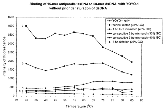
Figs. 1,2A and 2B show the intensity of fluorescence as a
function of temperature, GC content, and extent of base pair
matching.
Figs. 3, 4, 5A, 5B, 5C and 5D show the intensity of
fluorescence as a function of wavelength, extent of base pair
matching and ration.
Figs. 6A,6B, and 6C show intensity of fluorescence as a
function of lasing protocol, and ration.
Figs. 7A, 7B, 7C, 8A, 8B, 9A, 9B, 9C, and 10 show, as
regards quadruplexes, the intensity of fluorescence as a
function of the extent of base pair matching and ration.
Fig. 11 shows the intensity of fluorescence as a function
of the extent of base pair matching in a solution containing
ethanol.
Figs. 12A, 12B, 12C and 12D show the intensity of
fluorescence as a function of wavelength for perfect and
imperfect base pair matches in dsDNA:ssDNA complexes when the
cationic DNA intercalator YOYO-1 is present.
CA 02450831 2003-12-16
WO 02/103051 PCT/IB02/01972
DETAILED DESCRIPTION OF THE INVENTION
GLOSSARY AND DEFINITIONS
The one-letter codes for the bases that form part of their
respective nucleotides are: A: adenine; T: thymine; G: guanine;
C: cytosine; U: uracil. These letters are also used to represent
their respective nucleotides.
The G-C content of a duplex is the 100 times the number of
G-C base pairs divided by the sum of the number of G-C base pairs
plus the number of A-T (or A-U) base pairs and is expressed as
a percentage (e.g., 20 %).
An accelerator agent is understood here as one that
"increases a rate or amount of said triplex or quadruplex
formation." A rate can be obtained with as few as two
measurements, each at different time points. Amounts refer to the
number of triplexes or quadruplexes formed.
The term "accelerator agent" is used interchangeably with
the terms "promoter" and "promoter agent" except where the
promoter specifically refers to a gene promoter.
The terms nucleic acid, triplex, quadruplex, and the like
are intended to refer to molecules that comprise DNA, RNA, and
analogues thereof capable of forming similar structures such as
Watson-Crick duplexes, and the triplexes and quadruplexes formed
herein.
"Nucleobase" refers to the bases A, U, G, C, T, and those
analogs that can conform to Watson-Crick base-pairing rules.
Analogues of A,U,G,C, and T are those analogues that can
conform to Watson-Crick base pairing rules.
A Watson-Crick duplex is a 2-stranded molecule or molecular
segment in which the two strands are anti-parallel, their 5'->3'
directions being opposite. The overall structure of the duplex
is that of a double helix. The strands are held together by
hydrogen bonds and hydrophobic interactions. There is base pair
complementarity, A is paired with. T by two hydrogen. bonds (or,
in the case of RNA, A is paired with U) and G is paired with C
_g_
CA 02450831 2003-12-16
WO 02/103051 PCT/IB02/01972
by three hydrogen bonds . As a result, the base-pairing rules are
A is paired with T, A is paired with U, and G is paired with C.
A 3-stranded nucleic acid molecule is not necessarily a
triplex and it is not necessary that any segment of a 3-stranded
molecule be a triplex. Tt is possible that, at no region within
any strand, is that strand bonded to more than one other strand.
A simple example would be a Y-shaped molecule where Strands 1 and
2 form the stem, Strands 1 and 3 form the left upper branch, and
Strand 2 forms a single-stranded right upper branch.
An example of a triplex is a 3-stranded nucleic acid
molecule or molecular segment in which one strand (arbitrarily
named Strand 1) follows the Watson-Crick base-pairing rules (A-
T, A-U, and G-C) with both Strand 2 and Strand 3. Strands 1 and
2 form a structure that is, or is close to, that of a Watson-
Crick duplex. Strand 3 resides in the major groove of that
duplex. An example of such a triplex is the one elaborated by
V.B. Zhurkin et al, J. Mol. Biol., (1994) vol. 239, 181-200 (See
especially Figure 2 of that reference), which article is
incorporated herein by reference. As part of the stabilization
of such a triplex, Strand 3 is bonded to the other two strands
by base pairing rules as follows:
An A on Strand 3 is paired with both. an A on Strand 1 and
a T on Strand 2;
a G on Strand 3 is paired with both a G on Strand 1 and a
C on Strand 2;
a C on Strand 3 is paired with both a C on Strand 1 and a
G on strand 2; and
a T on strand 3 is paired with both a T on Strand 1 and an
A on strand 2
These base pairing rules axe satisfied regardless of which
variant (C or C+ or C', T or T') of the Zhurkin model is
considered.
The term ~~base-pairing rules" are those that define the
specificity between one nucleic acid molecule and another nucleic
acid molecule when the two bind to each other with specificity.
_g_
CA 02450831 2003-12-16
WO 02/103051 PCT/IB02/01972
Examples are Watson-Crick base pairing rules (G-C, and A-T or A-
U) and homologous binding base-pairing rules (A-A, T-T, G-G, C-C,
U-U) .
"Decondensation" of a duplex is defined as an increase in
the overall helical repeat length of the duplex. For example, the
B conformation of the duplex has an overall helical repeat length
of 10 base pairs; a decondensation of 106° results in that repeat
length being 13 base pairs.
PNA stands for polyamide analogs of DNA and RNA (see e.g.,
U.S. patent No. 5,539,082 to Nielsen et al.)
Specific embodiments and alternative formulations of the
inventions
The inventions as described in the Summary of The Tnvention
section have specific embodiments and preferred embodiments of
interest. These embodiments are described throughout this
application. However, for convenience, many of them are
summarized in this section, Similarly, the inventions as
summarized in the Summary of Invention section can be phrased in
alternative fashions expressing some variation of the invention
but retaining substantial overlap as to the essential invention.
Such alternative formulations of the invention are also included
in this section.
(A) Methods of making the multiplexes
The methods of forming the triplex or quadruplex,
described generally in the Summary of the Invention section, can
optionally be performed by incorporating one or more of the
following into the method:
within the multiplex, the Watson-Crick duplex added in step
(1) is heteropolymeric with a G-C content between 25% and 75%;
within the multiplex, the Watson-Crick duplex added in step
(1) is heteropolymeric with a G-C content between 10% and 900,
and furthermore the combined frequencies therein of purine-
pyrimidine dimers and pyrimidine-purine dimers exceeds 25%
(dimers are identified starting at the 5' end of the sequence and
progressing one base at a time until the 3' end is reached; for
-10-
CA 02450831 2003-12-16
WO 02/103051 PCT/IB02/01972
example, the sequence 5' -AA.A.GGGT has one purine-pyrimidine dimer
(GT) and no pyrimidine-purine dimers - their combined frequencies
equal 1/6);
performing steps (1) and/or (2) with the nucleic acid
strands and/or duplexes not in a cell (and not in a virus);
performing step (2) without the assistance of a protein
(such as recA or protein of similar function); i
in step (1), adding the water so that it accounts, on a
volume basis, for at least 50 percent of the final volume of the
mixture (more preferably for at least 80% of the final volume,
most preferably in step (1) water is the only liquid added to the
mixture);
in step (1), not adding any protein;
performing step (2) at a temperature or temperatures above
the freezing temperature of the aqueous solution and at not more
than 85 °C (more preferably between 5 °C andF 30 °C, most
preferably between 15 °C and 25 °C)
in step (1), adding an anion or, more preferably,~a ration
as the accelerator agent (monovalent, divalent or multivalent;
for example, a metallic canon or a cationic peptide);
wherein said ration is at least one member selected from the
group consisting of alkali metal rations, alkaline earth metal
rations, transition metal rations, Co(NH3)6~3, trivalent
spermidine and tetravalent spermine;
wherein said,cation is Na'~ provided at a concentration of
50mM to 125mM;
wherein said ration is selected from the group consisting
of Mn+~ provided at a concentration of lOmM to 45mM, Mg+~
provided at a concentration of lOmM to 45mM, and Ni~z provided at
a concentration of 20mM;
in step (1) adding an intercalator as an accelerator agent
{especially a fluorescent intercalator; preferably a bis-
intercalator);
in step {1) said accelerator agent is an intercalating
fluorophore selected from the group consisting of YOYO-1, TOTO-1,
-11-
CA 02450831 2003-12-16
WO 02/103051 PCT/IB02/01972
YOYO-3, TOTO-3, POPO-1, BOBO-1, POPO-3, BOBO-3, LOLO-1, JOJO-l,
cyanine dimers, YO-PRO-1, TO-PRO-1, YO-PRO-3, TO-PRO-3, TO-PRO-5,
PO-PRO-1, BO-PRO-1, PO-PRO-3, BO-PRO-3, LO-PRO-l, JO-PRO-1,
cyanine monomers, ethidium bromide, ethidium homodimer-1,
ethidium homodimer-2, ethidium derivatives, acridine, acridine
orange, acridine derivatives, ethidium-acridine heterodimer,
ethidium monoazide, propidium iodide, SYTO dyes, SYBR Green l,
SYBR dyew~, Pico Green, SYTOX dyes, and 7-aminoactinomycin D.
in step (1) said accelerator agent is a non-intercalating
fluorophore (especially one selected from the group consisting
of biotin, rhodamine, Alexa dyes, BODIPY dyes, biotin conjugates,
thiol-reactive probes, fluorescein and derivatives (including the
"caged" probes), Oregon Green, Rhodamine Green, QSY dyes)) and
said intensity is inversely correlated with formation of the
triplex or quadruplex;
in step (1 ) said accelerator agent is tethered to at least
one of said first strand, said second strand, said third strand
and said fourth strand;
in step (1) adding an accelerator agent that is an
intercalator that binds to the minor groove of the Watson-Crick
duplex (or at least one of the two Watson-Crick duplexes);
in step (1) adding an accelerator agent (especially an
organic liquid soluble in water, such as dimethyl formamide,
ethanol, and glycerol) that at 25 °C is a liquid;
in step (1) adding more than one accelerator agent;
in step (1) adding an accelerator agent that is a
condensation or decondensation agent as regards the Watson-Crick
duplex;
in step (1) adding an accelerator agent that is an analog
of A, T, U, C, or G;
in step (1) adding an accelerator agent selected from the
group consisting of lectins and polysaccharides;
in steps (1) and (2 ), buffering the mixture with a pH of
about 5 to about 9;
-12-
CA 02450831 2003-12-16
WO 02/103051 PCT/IB02/01972
one cytosine in at least one C-G-C or G-C-G base triplet
is positively charged;
one cytosine in each ~C-G-C and G-C-G base triplet is
positively charged;
in step (2) the incubation time is not more than about two
hours (more preferably not more than 1 hour; and within either
time frame, prefererably at least 25%, more preferably at least
50%, of the possible multiplexes have been formed);
saia at least one accelerator agent is a minor groove
nucleic acid binding molecule, which binds in a non
intercalating manner and binds with an association constant of
at least 103 M-1;
wherein the multiplex is part of an electrical circuit.
(Alternatively, the invention is an electrical circuit comprising
the multiplex structure.)
It will be apparent to someone of ordinary skill in the art
that the foregoing specific conditions also apply to the
following method for making a quadruplex.
In an alternatively phrased version, the method of forming
the quadruplex comprises:
(1) providing a hybridization medium comprising a first
strand, a second strand, a third strand, a fourth
strand, water, a buffer and at least one accelerator
agent; and
(2) incubating said hybridization medium for an incubation
time effective to hybridize said second strand to said
fourth strand to provide said multiplex structure
wherein said multiplex structure comprises:
a first strand containing a first sequence of nucleobases;
a second strand containing a second sequence of
nucleobases, wherein said second strand is associated
with said first strand by Watson-Crick bonding;
a third strand containing a third sequence of nucleobases;
and
-13-
CA 02450831 2003-12-16
WO 02/103051 PCT/IB02/01972
a fourth strand containing a fourth sequence of
nucleobases, wherein said fourth strand is associated
with said second strand and said third strand by
Watson-Crick bonding.
Tn particular embodiments of the methods of forming a
quadruplex, the following apply alone or in combination (the
descriptions that follow utilize the terminology of the method
of making a quadruplex that specifies 4 separate strands but they
are also applicable to the method that specifies a Watson-Crick
duplex as one or two of the starting materials):
at least one of said first strand and said second strand
further comprises a pharmaceutical agent, and hybridization of
said second strand to said fourth strand places said
pharmaceutical agent an effective distance from a target on said
third strand, said fourth strand or on another molecule
associated with at least one of said third strand and said fourth
strand;
said pharmaceutical agent is a member selected from the
group consisting of nucleic acids designed to bind gene promoter
sequences of clinically relevant genes, nucleic acids designed to
bind clinically relevant genes, and nucleic acids designed to
bind origin-of- replication sites of pathogens;
said third strand and said fourth strand are provided in
said hybridization medium before said first strand and said
second strand, and said first strand and said second strand are
provided in dehydrated form prior to rehydration by contact with
said hybridization medium;
at least one of said first strand and said second strand is
covalently labeled with a non-intercalating fluorophore and said
intensity is inversely correlated with said binding affinity;
at least one accelerator agent is an intercalating
fluorophore, and a fluorescent intensity of a test medium
containing said multiplex structure is directly correlated with
a binding affinity of said second strand for said fourth strand;
-14-
CA 02450831 2003-12-16
WO 02/103051 PCT/IB02/01972
hybridization of said second strand to said fourth strand is
detected as a change in a fluorescent, chemiluminescent,
electrochemiluminescent or electrical signal;
an intensity of said signal is correlated with a binding
affinity between said second strand and said fourth strand;
hybridization of said second strand to said fourth strand
inactivates an activity associated with at least one of said
third strand and said fourth strand.
Methods of detecting triplexes
The methods of detecting a triplex, described generally in
the Summary of the Invention section, can optionally be
performed by incorporating one or more of the following into the
method:
carrying out the method as a h.omogenous_ assay such that,
during or prior to step (3), single-stranded molecules that are
not part of the triplex are not placed in a vessel or container
separate from that containing the triplex;
using the detection method to discriminate between a perfect
base-pairing-rules match, a one-base mismatch (or deletion), and
a 2-base mismatch (or deletion), between the duplex and the
single-stranded molecule in the triplex (the method preferably
comprisir~g calibrating the method with molecules comprising
known mismatches);
using the extent of binding of an intercalator (e.g., as
indicated by increased fluorescence) as an indication of the
formation of the triplex;
such that a wavelength at which said intercalating
fluorophore fluoresces shifts to a second wavelength upon
intercalation, a difference between said wavelength and said
second wavelength indicating whether a complex between said probe
and said target is a duplex or a triplex and whether said target
is DNA or RNA;
the probe is covalently labeled with a non-intercalating
fluorophore and said intensity is inversely correlated with said
binding affinity (especially wherein said non-intercalating
-15-
CA 02450831 2003-12-16
WO 02/103051 PCT/IB02/01972
fluorophore is a member selected from the group consisting of
biotin, rhodamine and fluorescein);
using a fluorophore-labeled single stranded molecule as the
single-stranded molecule;
the method is a homogeneous assay conducted without
providing a signal quenching agent on said target sequence (i.e.,
in the duplex) or on said probe (i.e., the single-stranded
molecule) and/or without prior denaturation of said target
sequence and/or without PCR amplification of said target
sequence;
said method is a homogeneous assay conducted without
providing a signal quenching agent on said target sequence or on
said probe;
the probe has a partially charged or uncharged backbone;
~ the probe comprises a PNA sequence and/or is ssPNA prepared
by parallel synthesis;
the probe and said target sequence are the same length;
the probe is 5 to 30 nucleotides long;
the fluorescence-exciting radiation is emitted from an argon
ion laser at a wavelength from about 200 nm to about 1000 nm;
the test sample has a volume of about 20 microliters
containing about 10 femtomoles of target sequence and about 10
femtomoles of probe;
the concentration of the target sequence in said sample is
not more than 5 x 10⁻¹° M;
the concentration of the probe in the sample is not more
than 5 x 10⁻¹° M;
the method is conducted on a biochip;
the intercalating fluorophore is added to the medium in a
form free of said probe and free of said target sequence;
the intercalating fluorophore is a member selected from the
group consisting of YOYO-1, TOTO-1, ethidium bromide, ethidium
homodimer-1, ethidium homodimer-2 and acridine.
-16-
CA 02450831 2003-12-16
WO 02/103051 PCT/IB02/01972
In an alteratively phrased aspect, the invention is a
detection method comprising:
providing a target double-stranded nucleic acid or nucleic
acid analogue comprising a target sequence, wherein
said target sequence contains at least one purine base
and at least one pyrimidine base;
providing a probe comprising a nucleic acid sequence or a
nucleic acid analog sequence;
providing an accelerator agent;
adding said probe, said target sequence and said accelerator
agent to a medium to provide a test sample containing
a triplex complex comprising said probe bound to said
target sequence, wherein all base triplets of said
complex are members selected from the group consisting
of A-T-A, T-A-T, U-A-T, T-A-U, A-U-A, U-A-U, G-C-G and
C-G_C:
irradiating said test sample with exciting radiation to
cause the test sample to emit fluorescent radiation;
detecting an intensity of said fluorescent radiation,
wherein said intensity is correlated with a banding
affinity between said probe and said target sequence;
and
determining from said intensity an extent of matching
between said probe and said target sequence.
In another alternatively phrased related aspect, the method
comprises:
providing a target nucleic acid or nucleic acid analogue
having a target sequence, wherein said target sequence
contains at least one purine base and at least one
pyrimidine base;
providing a double-stranded probe comprising a nucleic acid
sequence or a nucleic acid analog sequence;
providing a hybridization accelerator agent;
adding said probe, said target and said hybridization
accelerator agent to a medium to provide a test sample
-17-
CA 02450831 2003-12-16
WO 02/103051 PCT/IB02/01972
containing a Watson-Crick triplex comprising said probe
bound to said target sequence;
irradiating said test sample with exciting radiation to
cause test sample to emit fluorescent radiation;
detecting an intensity of said fluorescent radiation,
wherein said intensity is correlated with a binding
affinity between said probe and said target sequence;
and
determining from said intensity an extent of matching
between said probe and said target sequence
wherein said method is a homogeneous assay conducted without
providing a signal quenching agent on said target sequence or on
said probe;
(C) Methods of detecting quadruplexes
The methods of detecting a quadruplex, described generally
in the Summary of the Invention section, can optionally be
performed by incorporating one or more of the following into the
method:
carrying out the method as a homogenous assay, such that
during or prior to step {3) nucleic acid molecules that are not
part of the quadruplex are not placed in a vessel or container
separate from that containing the quadruplex;
the method is a homogeneous assay conducted without
providing a signal quenching agent on said target sequence or on
said probe (i.e., the second Watson-Crick duplex) and/or without
prior denaturation of said target sequence and/or without PCR
amplification of said target sequence;
using the detection method to discriminate between a perfect
base-pairing-rules match, a one-base mismatch (or deletion), and
a 2-base mismatch (or deletion), between the first and second
Watson-Crick duplexes (preferably by calibrating the method with
molecules comprising known mismatches);
using the extent of binding of an intercalator as an
indication of the formation of the quadruplex (especially by
-18-
CA 02450831 2003-12-16
WO 02/103051 PCT/IB02/01972
using a fluorescent intercalator and using increased fluorescence
as an indicator);
the intercalating fluorophore is added to the medium in a
form free of said probe and free of said target sequence;
the intercalating fluorophore is a member selected from the
group consisting of YOYO-1, TOTO-1, ethidium bromide, ethidium
homodimer-l, ethidium homodimer-2 and acridine;
a wavelength at which said intercalating fluorophore
fluoresces shifts to a second wavelength upon intercalation, a
difference between said wavelength and said second wavelength
indicating whether a~complex between said probe and said target
is a duplex or a triplex and whether said target is DNA or RNA;
the probe is covalently labeled with a non-intercalating
fluorophore anal said intensity is inversely correlated with said
binding affinity (especially wherein said non-intercalating
fluorophore is a member selected from the group consisting of
biotin, rhodamine and fluorescein);
using a fluorophore-labeled single stranded molecule for
part of the second Watson-Crick duplex;
the method further comprises quantifying the binding
affinity;
the probe has a partially charged or uncharged backbone;
the probe comprises a PNA sequence and/or is ssPNA prepared
by parallel synthesis;
the probe and said target sequence are the same length;
the probe is 5 to 30 nucleotides (or base pairs) long;
the fluorescence-exciting radiation is emitted from an argon
ion laser at a wavelength from about 200 nm to about 1000 nm;
the test sample has a volume of about 20 microliters
containing about 10 femtomoles of target sequence and about 10
femtomoles of probe;
the concentration of the target sequence in said sample is
not more than 5 x 10⁻¹° M;
the concentration of the probe in the sample is not more
than 5 x 10⁻¹° M;
-19-
CA 02450831 2003-12-16
WO 02/103051 PCT/IB02/01972
a ratio of said first strand and said second strand to said
third strand and said fourth strand is about 10:1;
concentrations of each of said first strand, said second
strand, said third strand and said fourth strand are not more
than 5 x 10⁻¹° M;
the method is conducted on a biochip.
In an alternatively phrased related aspect, the method
comprises:
providing a target nucleic acid or nucleic acid analogue
having a target sequence, wherein said target sequence
contains at least one purine base and at,_least one
pyrimidine base;
providing a double-stranded probe comprising a nucleic acid
sequence or a nucleic acid analog sequence;
providing a hybridization accelerator agent;
adding said probe, said target and said hybridization
accelerator agent to a medium to provide a test sample
containing a Watson-Crick quadruplex comprising said
probe bound to said target sequence;
irradiating said test sample with exciting radiation to
cause test sample to emit fluorescent radiation;
detecting an intensity of said fluorescent radiation,
wherein said intensity is correlated with a binding
affinity between said probe and said target sequence;
and
determining from said intensity an extent of matching
between said probe and said target sequence
wherein said method is a homogeneous assay conducted without
providing a signal quenching agent on said target sequence or on
said probe.
~,D? The triplexes
The triplex described generally in the Summary of the
Invention section, can optionally have one or more of the
following features:
-20-
CA 02450831 2003-12-16
WO 02/103051 PCT/IB02/01972
each strand is heteropolymeric with a G-C content between
2 5 o and 7 5 0 ;
each strand is heteropolymeric with a G-C content between
10o and 90%, and furthermore the combined frequencies therein of
purine-pyrimidine dimers and pyrimidine-purine dimers exceeds
25%;
it is not in a cell (and not in a virus);
it is stable at pH greater than 7.6 (but less than pH 9);
it is in a medium at a pH greater than 7.6 (and preferably
less than pH 9);
the single-stranded nucleic acid or nucleic acid analog is
5 to 30 bases long and the double-stranded nucleic acid target
is 8 to 3.3 X 10⁹ base pairs long;
the target sequence is heteropolymeric and contains 25o to
75o purine bases and 75% to 25a pyrimidine bases in any order
(preferably wherein the frequency of purine-pyrimidine dimers
plus the frequency of pyrimidine-purine dimers exceeds 25%);
the probe (i.e, the single-stranded molecule) is covalently
bound to a double-stranded nucleic acid cleaving agent;
the probe is covalently bound to a chemotherapeutic agent;
the probe is covalently bound to a label (for example, a
mufti-molecule signaling complex, a redox pair, a
chemiluminescent agent, an electrochemiluminescent agent, or in
a preferred embodiment, a fluorophore, especially such that the
fluorescent intensity of the complex is correlated with a binding
affinity between the probe and the target sequence);
the base pairing rules for the single-stranded nucleic acid
molecule are, as regards one strand of the duplex, the Watson-
Crick base-pairing rules, G-C and either A- T or A-U, and, as
regards the other strand of the duplex are A-A and either T-T or
U-U;
the duplex substantially retains its Watson-Crick double
helical structure, and the single-stranded molecule resides in a
groove of the double helix;
-21-
CA 02450831 2003-12-16
WO 02/103051 PCT/IB02/01972
the accelerator agent forms a bond between part of the
duplex and part of the single-stranded nucleic acid molecule;
the accelerator agent is covalently linked to the single-
stranded nucleic acid molecule;
both strands of the Watson-Crick duplex are DNA (especially
where all strands of the triplex are DNA);
the accelerator reagent binds to a base in the Watson-Crick
duplex, said base being one to which a base in the single-
stranded nucleic acid molecule binds;
the accelerator reagent binds to a base in the Watson-Crick
duplex, said base not being one in the triplex;
the accelerator agent binds to the phosphate backbone of
the Watson-Crick duplex;
the accelerator agent binds to more than one site on the
Watson-Crick duplex, each site either on a base or a place on a
phosphate backbone of said duplex;
the accelerator agent binds to one site on the Watson-Crick
duplex, said site either on a base or on a phosphate backbone of
said duplex;
the accelerator agent binds to a base in the Watson-Crick
duplex and to a base in the single-stranded nucleic acid
molecule;
the accelerator agent binds to a base in the single-stranded
nucleic acid molecule.
2 5 ~,E ) The Ouadrup 1 exe s
The quadruplex described generally in the Summary of the
Invention section, can optionally have one or more of the
following features:
each of said four strands is heteropolymeric with a G-C
content between 10% and 900;
the second and fourth. strands are aligned in a parallel 3'
to 5' direction and binding between those 2 strands is according
to homologous base-pairing rules;
-22-
CA 02450831 2003-12-16
WO 02/103051 PCT/IB02/01972
the first and third strands are aligned in a parallel 5' to
3' direction and binding between those 2 strands is according to
homologous base-pairing rules;
the second and fourth strands are aligned in a parallel 3'
to 5' direction and binding between said second and fourth
strands is according to homologous base-pairing rues and
furthermore the first and third strands are aligned in a parallel
5' to 3' direction and binding between said first and third
strands is according to homologous base-pairing rules;
the second and fourth strands are aligned in a parallel 3'
to 5' direction and binding between those 2 strands is according
to Watson-Crick base-pairing rules;
the first and third strands are aligned in a parallel 5' to
3' direction and binding between those 2 strands is according to
Watson-Crick base-pairing rules;
the second and fourth strands are aligned in a parallel 3'
to 5' direction and binding between said second and fourth
strands is according to Watson-Crick base-pairing rules and
furthermore the first and third strands are aligned in a parallel
5' to 3' direction and binding between said first and third
strands is according to Watson-Crick base-pairing rules;
the first and fourth strands are aligned in anti-parallel 5'
to 3' and 3' to 5' directions, respectively, and binding between
the 2 strands is according to Watson-Crick base-pairing rules;
the second and third strands are aligned in anti-parallel 3'
to 5' and 5' to 3' directions, respectively, and binding between
those 2 strands is according to Watson-Crick base-pairing rules;
the first and fourth strands are aligned in anti-parallel 5'
to 3' and 3' to 5' directions, respectively, and binding between
said first and fourth strands is according to Watson-Crick base
pairing rules and furthermore the second and third strands are
aligned in anti-parallel 3' to 5' and 5' to 3' directions,
respectively, and binding between said second and third strands
is according to Watson-Crick base-pairing rules;
-23-
CA 02450831 2003-12-16
WO 02/103051 PCT/IB02/01972
the first and fourth strands are aligned in anti-parallel 5'
to 3' and 3' to 5' directions, respectively, and binding between
those 2 strands is according to homologous base-pairing rules;
the second and third strands are aligned in anti-parallel 3'
to 5' and 5' to 3' directions, respectively, and binding between
those 2 strands is according to homologous base-pairing rules;
the first and fourth strands are aligned in anti-parallel 5'
to 3' and 3' to 5' directions, respectively, and binding between
said first and fourth strands is according to homologous base
pairing rules and furthermore the second and third strands are
aligned in anti-parallel 3' to 5' and 5' to 3' directions,
respectively, and binding between said second and third strands
is according to homologus base-pairing rules;
each interacting base of the said first strand interacts
specifically with both the adj acent base on the said third strand
and with the base on the said fourth strand, the base to which
the said third strand base is bound;
each interacting base of the said second strand interacts
specifically with both the adjacent base on the said fourth
strand and the base on the said third strand, the base to which
the said fourth strand base is bound;
it is an isolated, purified, artificial or synthetic
quadruplex;
each strand is heteropolymeric with a G-C content between
25o and 750;
each strand is heteropolymeric with a G-C content between
10% and 900, and furthermore the combined frequencies therein of
purine-pyrimidine diners and pyrimidine-purine diners exceeds
25%;
it is not in a cell (and not in a virus);
each said strand independently comprises a heteropolymeric
nucleic acid or a heteropolymeric nucleic acid analogue;
each said strand independently comprises DNA or RNA;
-24-
CA 02450831 2003-12-16
WO 02/103051 PCT/IB02/01972
each said strand independently comprises a heteropolymeric
nucleic acid analogue containing an uncharged or partially
charged backbone;
one of said second strand or said fourth strand comprises
DNA and the other of said second strand or said fourth strand
comprises RNA, mRNA, hnRNA, rRNA, tRNA or cDNA;
the second strand and said fourth strand are parallel
homologous to each other;
a major groove of said first strand and said second strand
is placed in a minor groove of said third strand and said fourth
strand;
the second strand and said fourth strand are parallel
complementary to each other;
a major groove of said first strand and said second strand
is placed in a minor groove of said third strand and said fourth
strand;
each nucleobase binds to no more than two other nucleobases;
no strand is contiguous with another strand;
the multiplex structure is substantially free of Hoogsteen
bonding;
the multiplex structure is substantially free of G-G
quartets;
the first strand and said second strand are 5 to 50 base
pairs long;
the third strand and said fourth strand are genomic DNA;
the third strand and said fourth strand include a haplotype
in genomic DNA;
the third strand and fourth strand are PCR amplified
products;
wherein said multiplex structure is free of solid support;
the multiplex structure is bound to a solid support (where
the solid support is either electrically conductive or is not
electrically conductive);
wherein the multiplex structure further comprises a
therapeutic, prophylactic or diagnostic agent bound to at least
-25-
CA 02450831 2003-12-16
WO 02/103051 PCT/IB02/01972
one of said first strand, said second strand, said third strand
and said fourth strand;
wherein the first strand and said second strand are each 5
to 30 bases long and said third strand and said fourth strand are
each 8 to 3.3 X 10⁹ base pairs long;
wherein the fourth sequence contains 25% to 75% purine bases
and 75% to 25% pyrimidine bases in any order (preferably wherein
the frequency of purine-pyrimidine dimers plus the frequency of
pyrimidine-purine dimers exceeds 250);
the first and second Watson-Crick duplexes (see the method
of making a quadruplex in the Summary of th.e Invention section)
each have a G-C content between 30 and 70%;
both strands of the first Watson-Crick duplex are DNA
(especially where all strands of the quadruplex are DNA);
the accelerator reagent binds to a base in the first Watson-
Crick duplex, said base being one to which a base in the second
duplex binds;
the accelerator reagent binds to a base in the first or
second Watson-Crick duplexes, said base not being part of the
quadruplex;
the accelerator reagent binds to a phosphate backbone of
the first or second Watson-Crick duplex;
the accelerator reagent binds to more than one site on one
of the first or second Watson-Crick duplexes, each site either on
a base or on a phosphate backbone;
the accelerator agent binds to one site on the Watson-Crick
duplex, said site either on a base or on a phosphate backbone;
the accelerator agent binds to a base in the first Watson-
Crick duplex and to a base on the second Watson-Crick duplex;
the accelerator agent binds to the minor groove of the first
and/or second Watson-Crick duplex;
the accelerator agent forms a bond between part of the first
Watson-Crick duplex and part of the second Watson-Crick duplex.
-26-
CA 02450831 2003-12-16
WO 02/103051 PCT/IB02/01972
Additional aspects of the invention
The invention provides triplex complexes comprising a
single-stranded probe bound to a double-stranded nucleic acid
target, wherein the probe comprises a heteropolymeric nucleic
acid or a heteropolymeric nucleic acid analog, and all base
triplets of the complex are members selected from the group
consisting of A-T-A, T-A-T, U-A-T, T-A-U, A-U-A, U-A-U, G-C-G and
C-G-C.
Unlike certain Hoogsteen triplexes disclosed by the prior
art, the triplexes of the invention are stable at pH values
greater than 7.6. Moreover, the inventive triplexes do not
require the presence of homopyrimidine sequences or homopurine
sequences, as in certain prior art triplexes. For example, the
target sequence can contain 25o to 75o purine bases and 75% to
25% pyrimidine bases in any order.
Preferably the single-stranded nucleic acid or nucleic acid
analog of the triplex is 5 to 30 bases long anc3 the
double-stranded nucleic acid target is 8 to 3.3 X 10⁹ base pairs
long.
Triplex formation according to the invention is suitable for
a variety of uses. For example, probes covalently bound to a
double-stranded nucleic acid cleaving agent can be used to
specifically cleave target sequences of double-stranded nucleic
acids. Probes covalently bound to a chemotherapeutic agent can
be used to specifically treat target sequences of double-stranded
nucleic acids.
In preferred embodiments, the invention provides a rapid,
sensitive, environmentally friendly, and safe method for assaying
binding between a double-stranded target and a single-stranded
probe, wherein the target comprises a nucleic acid sequence or a
nucleic acid analog sequence and the probe comprises a nucleic
acid sequence or a nucleic acid analog sequence.
Unlike certain prior art assays, the invention not only
detects the presence of specific probe-target binding, but also
provides qualitative and quantitative information regarding the
_ _27_
CA 02450831 2003-12-16
WO 02/103051 PCT/IB02/01972
nature of interaction between a probe and target. Thus, the
.invention enables the practitioner to distinguish among a perfect
match, a one base pair mismatch, a two base pair mismatch, a
three base pair mismatch, a one base pair deletion, a two base
pair deletion and a three base pair deletion arising between a
base sequence in the probe and in a strand of the double-stranded
target. .
Embodiments of the invention comprise calibrating the
measured signal (e. g., fluorescent intensity) for a first probe
target mixture against the same type of signal exhibited by other
probes combined with the same target, wherein each of the other
probes differs from the first probe by at least one base.
A calibration curve can be generated, wherein the magnitude
of the measured signal (e.g., fluorescent intensity) is a
function of the binding affinity between the target and probe.
As the binding affinity between the target and a plurality of
different probes varies with the number of mismatched bases, the
nature of the mismatches) (A-G vs. A-C vs. T-G vs. T-C, etc.),
the location of the mismatches) within the triplex, etc., the
assay of the invention can be used to sequence the target.
In embodiments, the signal measured can be the fluorescent
intensity of a fluorophore included in the test sample. In such.
embodiments, the binding affinity between the probe and target
can be directly or inversely correlated with the intensity,
depending on whether the fluorophore signals hybridization
through signal quenching or signal amplification. Under selected
conditions, the fluorescent intensity generated by intercalating
agents can be directly correlated with probe-target binding
affinity, whereas the intensity of preferred embodiments
employing a non-intercalating fluorophore covalently bound to the
probe can be inversely correlated with probe-target binding
affinity. The fluorescent intensity decreases for non-
intercalating fluorophores as the extent of matching between the
probe and target increases, preferably over a range inclusive of
-28-
CA 02450831 2003-12-16
WO 02/103051 PCT/IB02/01972
0-2 mismatches and/or deletions, more preferably over a range
inclusive of 0-3 mismatches and/or deletions.
The invention enables quantifying the binding affinity
between probe and target. Such information can be valuable for
a variety of uses, including designing antisense drugs with
optimized binding characteristics.
Unlike prior art methods, the assay of the invention is
preferably homogeneous. The assay can be conducted without
separating the probe-target complex from the free probe and
target prior to detecting the magnitude of the measured signal.
The assay does not require a gel separation step, thereby
allowing a great increase in testing throughput. Quantitative
analyses are simple and accurate. Consequently the binding assay
saves a lot of time and expense, and can be easily automated.
Furthermore, it enables binding variables such as buffer, pH,
ionic concentration, temperature, incubation time, relative
concentrations of probe and target sequences, intercalator
concentration, length of target sequences, length of probe
sequences, and possible cofactor requirements to be rapidly
determined.
The assay can be conducted in, e.g., a solution within a
well, on an impermeable surface or on a biochip.
Moreover, the inventive assay is preferably conducted
without providing a signal quenching agent on the target or on
the probe.
Although the inventors have previously disclosed the
advantages of fluorescent intensity assays for hybridization
(see, e.g., U.S. Patent Application No. 09/224,505, filed
December 31, 1998), assays according to the present invention
specifically detect triplexes of the probe and the
double-stranded target, thus obviating the need to denature the
target . While nucleic acid (and nucleic acid analog) probes have
been known to form triplexes with certain limited classes of
targets (see, e.g., Floris et al., supra, Dervan et al., supra,
Egholm et al., 365 Nature 566 (2993), and Tomac et al., 118
-29-
CA 02450831 2003-12-16
WO 02/103051 PCT/IB02/01972
J.Am.Chem.Soc. 5544 (1996)), it is surprising that the inventors
have been able to specifically assay triplexes formed between
single-stranded nucleic acid (e.g., ssDNA and RNA) probes and
double-stranded nucleic acid (e.g., dsDNA) targets, wherein the
interaction between the probes and targets is based on Watson-
Crick base pairing (at least in the sense that A binds to T (or
U, in the case of RNA) and G binds to C), rather than the very
limited Hoogsteen model of triplex hybridization of, e:g., Dervan
et al. The term "Watson-Crick triplex," which is employed
herein, is intended to crystallize these differences by limiting
the nature of base pairing between. the single-stranded probe and
the double-stranded target to A-T-A, T-A-T, U-A-T, T-A-U, A-U-A,
U-A-U, G-C-G and/or C-G-C (including C+-G-C, and/or any other
ionized species of base). These three-member groups are
hereinafter denoted Watson-Crick base triplets and the resulting
structures denoted Watson-Crick triplexes.
Suitable probes for use in the inventive assay include,
e.g., ssDNA, RNA, PNA and other nucleic acid analogs having
uncharged or partially-charged backbones. Probe sequences having
any length from 8 to 20 bases are preferred since this is the
range within which the smallest unique DNA sequences of
prokaryotes and eukaryotes are found. Probes of 12 to 18 bases
are particularly preferred since this is the length of the
smallest unique sequences in the human genome. In embodiments,
probes of 5 to 30 bases are most preferred. However, a plurality
of shorter probes can be used to detect a nucleotide sequence
having a plurality of non-unique target sequences therein, which
combine to uniquely identify the nucleotide sequence. The length
of the probe can be selected to match the length of the target.
The inventors have discovered the surprising development
that they were able to specifically assay a wide-variety of
triplexes formed in a Watson-Crick base-pair dependent manner
between single-stranded nucleic acid (e.g., ssDNA, RNA, ssPNA and
other analogs of DNA or RNA) probes and double-stranded nucleic
-30-
CA 02450831 2003-12-16
WO 02/103051 PCT/IB02/01972
acid (e. g., dsDNA) targets. The inventors have discovered that
triplex formation and/or stabilization is enhanced by the
presence of an intercalating agent in the sample being tested.
The inventors have discovered that Watson-Crick triplex
formation and/or stabilization is enhanced by the presence of
rations in the sample being tested. Suitable rations include,
e.g., monovalent rations, such as Na+ (preferably at a
concentration of 50mM to 125mM), I~+, and other alkali metal ions;
divalent rations, such as alkaline earth metal ions (e.g., Mg+z
and Ca~a) and divalent transition metal ions (e . g . , Mn~2, Ni+z
Cd~2, Co+2 and Zn+2) ; and rations having a positive charge of at
least three, such as Co (NH3) 6+3, trivalent spermidine and
tetravalent spermine. Mn+2 is preferably provided at a
concentration of lOmM to 30mM. Mg+2 is preferably provided at a
concentration of l5mM to 20mM. Ni+~ is preferably provided at a
concentration of about 20mM. In embodiments, Mg+~ and Mn+2 are
provided in combination at a concentration of lOmM each, l5mM
each or 20mM each (i.e., 10-20 mM each).
The amount of ration added to the medium in which the
triplex forms depends on a number of factors, including the
nature of the ration, the concentration of probe, the
concentration of target, the presence of additional rations and
the base content of the probe and target. The preferred ration
concentrations and mixtures can routinely be discovered
experimentally.
The instant invention does not require the use of
radioactive probes, which are hazardous, tedious and
time-consuming to use, and need to be constantly regenerated.
Probes of the invention are preferably safe to use and stable for
years. Accordingly, probes can be made or ordered in large
quantities and stored.
In embodiments, the probe is labeled with a multi-molecule
signaling complex or a redox pair, or with a label that elicits
chemiluminescent or electrochemiluminescent properties.
-31-
CA 02450831 2003-12-16
WO 02/103051 PCT/IB02/01972
It is preferred that the probe or target (preferably the
probe) have a fluorescent label covalently bound thereto. The
label is preferably a non-intercalating fluorophore. In such
embodiments, the fluorophore is preferably bound to the probe at
either end. Preferred fluorescent markers include biotin,
rhodamine and fluorescein, and other markers that fluoresce when
irradiated with exciting energy.
The excitation wavelength is selected (by routine
experimentation and/or conventional knowledge) to correspond to
this excitation maximum for the fluorophore being used, and is
preferably 200 to 1000 nm. Fluorophores are preferably selected
to have an emission wavelength of 200 to 1000 nm. In preferred
embodiments, an argon ion laser is used to irradiate the
fluorophore with light having a wavelength in a range of 400 to
540 nm, and fluorescent emission is detected in a range of 500 to
750 nm.
The assay of the invention can be performed over a wide
variety of temperatures, such as, e.g., from 5 to 85°C. Certain
prior art assays require elevated temperatures, adding cost and
delay to the assay. On the other hand, the invention can be
conducted at room temperature or below (e. g., at a temperature
below 25°C).
The reliability of the invention is independent of guanine
and cytosine content in said target. Since G-C base pairs form
three hydrogen bonds, while A-T base pairs form only two hydrogen
bonds, target and probe sequences with a higher G or C content
are more stable, possessing higher melting temperatures.
Consequently, base pair mismatches that increase the GC content
of the hybridized probe and target region above that present in
perfectly matched hybrids may offset the binding weakness
associated with a mismatched probe. Triplexes containing every
possible base pair mismatch between the probe and the target
proved to be more unstable than perfectly matched triplexes,
always resulting in lower fluorescent intensities than did
-32-
CA 02450831 2003-12-16
WO 02/103051 PCT/IB02/01972
perfectly complementary hybrids, when an intercalating
fluorophore was used.
The inventive assay is extremely sensitive, thereby
obviating the need to conduct PCR amplification of the target .
For example, it is possible to assay a test sample having a
volume of about 20 microliters, which contains about 10
femtomoles of target and about 10 femtomoles of probe.
Embodiments of the invention are sensitive enough to assay
targets at a concentration of 5 X 10'9 M, preferably at a
concentration of not more than 5 x 10⁻¹° M. Embodiments of the
invention are sensitive enough to employ probes at a
concentration of 5 X 10⁻⁹M, preferably at a concentration of not
more than 5 x 10⁻¹° M, It should go without saying that the
foregoing values are not intended to suggest that the method
cannot detect higher concentrations.
The medium in which triplexes form can be any conventional
medium known to be suitable for preserving nucleotides. See,
e.g., Sambrook et al., "Molecular Cloning: A Lab Manual," Vol. 2
(1989). For example, the liquid medium can comprise nucleotides,
water, buffers and standard salt concentrations. When divalent
rations are used exclusively to promote triplex formation,
chelators such as EDTA or EGTA should not be included in the
reaction mixtures.
Specific binding between complementary bases occurs under a
wide variety of conditions having variations in temperature, salt
concentration, electrostatic strength, and buffer composition.
Examples of these conditions and methods for applying them are
known in the art.
Unlike many Hoogsteen-type triplexes, which are unstable or
non-existent at pH levels above about 7.6, the Watson-Crick
triplexes of the invention are stable over a wide range of pH
levels, preferably from about pH 5 to about pH 9.
It is preferred that triplexes be formed at a temperature of
about 5°C to about 25°C for about one hour or less. Longer
reaction times are not required, but incubation for up to 24
-33-
CA 02450831 2003-12-16
WO 02/103051 PCT/IB02/01972
hours in most cases did not adversely affect the triplexes. The
fast binding times of Watson-Crick triplexes of the invention
contrast with the much longer binding times for Hoogsteen
triplex-based assays.
Although not required, it is possible to facilitate triplex
formation in solution by using certain reagents in addition to
rations. Preferred examples of these reagents include single
stranded binding proteins such as Rec A protein, T4 gene 32
protein, E. coli single stranded binding protein, major or minor
nucleic acid groove binding proteins, viologen and intercalating
substances such as ethidium bromide, actinomycin D, psoralen, and
angelicin. Such facilitating reagents may prove useful in
extreme operating conditions, far example, under abnormal pH
levels or extremely high temperatures.
The inventive assay can be used to, e.g., Identify
accessible regions in folded nucleotide sequences, to determine
the number of mismatched base pairs in a hybridization complex,
and to map genomes.
The inventors may sometimes herein suggest that Watson-Crick
triplexes result from hybridization of the probe to duplex
target. While fluorophores tethered to the probe produced
quenched fluorescent emissions upon being exposed to duplex
targets containing a strand of Watson-Crick complementary bases,
which indicates the occurrence of some kind of binding event, the
inventors are not sure that what occurs in the Watson-Crick
triplex is best described as hybridization in the sense
traditionally associated with Watson-Crick duplex formation.
While the formation of a Watson-Crick triplex may sometimes be
referred to as a hybridization event herein, that is merely for
convenience and is not intended to limit the scope of the
invention with. respect to how the formation of a Watson-Crick
triplex can be best characterized.
Unlike the quadruplexes discussed in the Background Section
above, the preferred multiplex structures of the invention
-34-
CA 02450831 2003-12-16
WO 02/103051 PCT/IB02/01972
contain at least four strands of nucleic acid bonded together
according to traditional Watson-Crick bonding rules.
As used herein, the term "Watson-Crick bonding" is intended
to define specific association between opposing pairs of nucleic
acid (and/or nucleic acid analogue) strands via matched, opposing
bases. While the formation of a Watson-Crick quadruplex may
sometimes be referred to as a hybridization event herein, that is
merely for convenience and is not intended to limit the'scope of
the invention with respect to how the formation of a Watson-Crick
quadruplex can be best characterized.
The multiplex structures of the invention are preferably
quadruplexes. Each strand of the multiplex independently
comprises a nucleic acid or a nucleic acid analogue. Suitable
nucleic acids include, e.g., DNA or RNA. Preferred nucleic acid
analogues contain an uncharged or partially charged backbone
(i.e., a backbone having a charge that is not as negative as a
native DNA backbone).
In certain embodiments, one of the second and fourth strands
of the four-stranded quadruplex comprises DNA and the other of
the second and fourth strands comprises RNA, mRNA, hnRNA, rRNA,
tR.NA or cDNA.
In certain embodiments, the second strand and the fourth
strand are parallel homologous to each other. In these
embodiments, a major groove of the first and second strands is
placed in a minor groove of the third and fourth strands.
In other embodiments, the second and fourth strands are
parallel complementary to each other. In these embodiments,
which possess "nested complementarity," a major groove of the
first and second strands is placed in a minor groove of the third
and fourth strands.
Tn certain embodiments, each nucleobase binds to no more
than two other nucleobases. In some of these embodiments, the
bases of the second strand specifically bond (via Watson-Crick
rules) to the matching bases of the first strand and to the
matching bases of the fourth strand, and the bases of the fourth
-35-
CA 02450831 2003-12-16
WO 02/103051 PCT/IB02/01972
strand specifically bond (via Watson-Crick rules) to the matching
bases of the third strand and to the matching bases of the second
strand, wherein the bases of the first and third strands bind to
no more than one other base each. Thus, in addition to the
traditional Watson-Crick base pairs, such embodiments include the
following Watson-Crick base triplets: A-T-A, T-A-T, U-A-T,
T-A-U, A-U-A, U-A-U, G-C-G and/or C-G-C (including C+-G-C, and/or
any other ionized species of base).
In certain embodiments, it is believed that opposing bases
of the first and third strands also bind to each other, in
addition to: (a) the binding between opposing bases of the first
and second strands; (b) the binding between opposing bases of the
third and fourth. strands; and (c) the binding between opposing
bases of the second and fourth strands.
In certain embodiments of the multiplex structure of the
invention, no strand is contiguous with another strand. That is,
there are at least four separate strands. Although folded
conformations and the like (e. g., hairpin turns, etc.) are within
the scope of the invention, folded portions of a single strand do
not make the strand count more than once toward the minimum of
four separate strands.
Multiplex structures of the invention preferably do not rely
on Hoogsteen bonding or G-G quartets for maintenance of the
multiplex structure, although insignificant amounts of Hoogsteen
bonding and/or G-G quartets may be present. That is, multiplex
structures of the invention are preferably substantially free of
Hoogsteen bonding, and substantially free of G-G quartets.
Tn certain embodiments, the first and second strands of the
multiplex are 5 to 50 bases long (more preferably 5 to 30 bases
long) and the third and fourth strands are 8 to 3.3 X 10⁹ base
pairs long. For example, the first and second strands can
constitute a double-stranded probe and the third and fourth
strands can constitute a double-stranded target, such as genomic
DNA, which can contain a haplotype.
-36-
CA 02450831 2003-12-16
WO 02/103051 PCT/IB02/01972
In embodiments, the third strand and the fourth strand are
PCR amplified products.
The multiplexes of the invention can be present in solution,
on a solid support, in vitro or in vivo. The solid support can
be electrically conductive (e.g., an electrode) or
non-conductive.
Quadruplex formation according to the invention is suitable
for a variety of uses. For example, double-stranded probes
covalently bound to a double-stranded nucleic acid cleaving agent
can be used to specifically cleave target sequences of double-
stranded nucleic acids. Double-stranded probes covalently bound
to a chemotherapeutic agent can be used to specifically treat
target sequences of double-stranded nucleic acids. Thus, the
invention encompasses multiplex structures further comprising a
therapeutic, prophylactic or diagnostic agent bound to at least
one of the first, second, third and fourth strands.
In~addition, multiplexes of the invention are suitable for
use in nanoengineering, such as to provide electrical circuitry
on a molecular (i.e., nanoscale) level. Further details
regarding nanoengineering with nucleic acids can be found in U.S.
Patent No. 5,948,897 to Sen et al. and the references cited
therein.
Multiplex structures of the invention can be provided by a
method comprising: providing a hybridization medium comprising
the first strand, the second strand, the third strand, the fourth
strand, water, a buffer and a promoter; and incubating the
hybridization medium for an incubation time effective to
hybridize the second strand to the fourth strand.
The hybridization medium can include any conventional medium
known to be suitable for preserving nucleotides. See, e.g.,
Sambrook et al., "Molecular Cloning: A Lab Manual," Vol. 2
(1989). For example, the medium can comprise nucleotides, water,
buffers and standard salt concentrations. When divalent rations
are used exclusively to promote quadruplex formation, chelators
-37-
CA 02450831 2003-12-16
WO 02/103051 PCT/IB02/01972
such as EDTA or EGTA should not be included in the reaction
mixtures.
Specific binding between complementary bases occurs under a
wide variety of conditions having variations in temperature, salt
concentration, electrostatic strength, and buffer composition.
Examples of these conditions and methods for applying them are
known in the art.
Unlike many Hoogsteen-type multiplexes, which are unstable
or non-existent at pH levels above about 7.6, the Watson-Crick
multiplexes of the invention are stable over a wide range of pH
levels, preferably from about pH 5 to about pH 9.
Moreover, the inventive multiplexes do not require the
presence of homopyrimidine sequences or homopurine sequences, as
in certain prior art quadruplexes. For example, the target
sequence can contain 25% to 75% purine bases and 75% to 25%
pyrimidine bases in any order.
It is preferred that multiplexes be formed at a temperature
of about 5°C to about 25°C for about two hours or less. The
incubation time is preferably less than five minutes, even at
room temperature. Longer reaction times are not required, but
incubation for up to 24 hours in most cases did not adversely
affect the quadruplexes. The fast binding times of Watson-Crick
quadruplexes of the invention contrast with the much longer
binding times for Hoogsteen quadruplexes.
The promoter in the hybridization medium is preferably an
intercalating agent or a ration. The intercalating agent can be,
e.g., a fluorophore, such as a member selected from the group
consisting of YOYO-1, TOTO-1, ethidium bromide, ethidium
homodimer-Z, ethidium homodimer-2 and acridine.
Suitable rations include, e.g., monovalent rations, such as
Na+ (preferably at a concentration of 50mM to 125mM) , K+, and
other alkali metal ions; divalent rations, such as alkaline earth
metal ions (e.g. , Mg+2 and Ca+2) and divalent transition metal ions
( a . g . , Mn+2 , Ni+2 , Cd+~ , Co+Z and Zn+~ ) ; and rat ions having a
positive charge of at least three, such as Co (NH3) 6+3, trivalent
-38-
CA 02450831 2003-12-16
WO 02/103051 PCT/IB02/01972
spermidine and tetravalent spermine. Mn+z is preferably provided
at a concentration of lOmM to 45mM. Mg+z is preferably provided
at a concentration of lOmM to 45mM. Ni+2 is preferably provided
at a concentration of about 20mM. In embodiments, Mg+z anal Mn+z
are provided in combination at a concentration of lOmM each, l5mM
each, 20mM each, 25mM each, 30mM each, 35 mM each, or 40 mM each
(i.e., 10-40 mM each).
The amount of ration added to the medium in which the
multiplex forms depends on a number of factors, including the
nature of the ration, the concentration of probe, the
concentration of target, the presence of additional rations and
the base content of the probe and target. The preferred ration
concentrations and mixtures can routinely be discovered
experimentally.
Although not required, other promoters include, e.g., single
stranded binding proteins such as Rer A protein, T4 gene 32
protein, E. coli single stranded binding protein, major or minor
nucleic acid groove binding proteins, viologen and additional
intercalating substances such as actinomycin D, psoralen, and
angelicin. Such facilitating reagents may prove useful in
extreme operating conditions, for example, under abnormal pH
levels or extremely high temperatures.
The invention also enables a method in which hybridisation
of the second strand to the fourth strand inactivates an activity
associated with at least one of the third strand and the fourth
strand. Thus, at least one of the first strand and the second
strand further comprises a pharmaceutical agent, wherein
hybridization of the second strand to the fourth strand places
the pharmaceutical agent an effective distance from a target on
the third strand, the fourth strand or on another molecule
associated with at least one of the third strand and the fourth
strand. The pharmaceutical agent is preferably a member selected
from the group consisting of nucleic acids designed to bind
promoter sequences of clinically relevant genes, nucleic acids
-39-
CA 02450831 2003-12-16
WO 02/103051 PCT/IB02/01972
designed to bind clinically relevant genes, or nucleic acids
designed to bind origin of replication sites of pathogens.
In preferred embodiments, the invention provides a rapid,
sensitive, environmentally friendly, and safe method for assaying
binding between a single-stranded or double-stranded target and
a double-stranded probe, wherein the target comprises a nucleic
acid sequence or a nucleic acid analogue sequence and the probe
comprises a nucleic acid sequence or a nucleic acid analogue
sequence.
The inventive assay can be used to, e.g., identify
accessible regions in folded nucleotide sequences, to determine
the number of mismatched base pairs in a hybridization complex,
and to map genomes.
The invention not only detects the presence of specific
probe-target binding, but also provides qualitative and
quantitative information regarding the nature of interaction
between a probe and target. Thus, the invention enables the
practitioner to distinguish among a perfect match, a one base
pair mismatch, a two base pair mismatch, a three base pair
mismatch, a one base pair deletion, a two base pair deletion and
a three base pair deletion arising between a sequence in the
double-stranded probe and in a sequence in the double-stranded
target.
Embodiments of the invention comprise calibrating the
measured signal (e. g., fluorescence, chemiluminescence,
electrochemiluminescence or electrical properties) for a first
probe-target mixture against the same type of signal exhibited by
other probes combined with the same target, wherein each of the
other probes differs from the first probe by at least one base.
A calibration curve can be generated, wherein the magnitude
of the measured signal (e.g., fluorescent intensity) is a
function of the binding affinity between the target and probe.
As the binding affinity between the target and a plurality of
different probes varies with the number of mismatched bases, the
nature of the mismatches) (A-G vs. A-C vs. T-G vs. T-C, etc.),
-40-
CA 02450831 2003-12-16
WO 02/103051 PCT/IB02/01972
the location of the mismatch (es) within the quadruplex, etc . , the
assay of the invention can be used to sequence the target.
In embodiments, the signal measured can be the fluorescent
emission intensity of a fluorophore included in the test sample.
In such embodiments, the binding affinity between the probe and
target can be directly or inversely correlated with the emission
intensity, depending on whether the fluorophore signals
hybridization through signal quenching or signal amplification.
Under selected conditions, the fluorescent emission intensity
generated by intercalating agents can be directly correlated with
probe-target binding affinity, whereas the emission intensity of
preferred embodiments employing a non-intercalating fluorophore
covalently bound to the probe can be inversely correlated with
probe-target binding affinity. The fluorescent emission
intensity decreases for non-intercalating fluorophores as the
extent of matching between the probe and target increases,
preferably over a range inclusive of 0-2 mismatches and/or
deletions, more preferably over a range inclusive of 0-3
mismatches and/or deletions.
The invention enables quantifying the binding affinity
between probe and target. Such information can be valuable for
a variety of uses, including designing antisense drugs with
optimized binding characteristics.
The assay of the invention is preferably homogeneous. The
assay can be conducted without separating the probe-target
complex from the free probe and free target prior to detecting
the magnitude of the measured signal. The assay does not require
a gel separation step, thereby allowing a great increase in
testing throughput. Quantitative analyses are simple and
accurate. Consequently the binding assay saves a lot of time and
expense, and can be easily automated. Furthermore, it enables
binding variables such as buffer, pH, ionic concentration,
temperature, incubation time, relative concentrations of probe
and target sequences, intercalator concentration, length of
-41-
CA 02450831 2003-12-16
WO 02/103051 PCT/IB02/01972
target sequences, length of probe sequences, and possible
cofactor (i.e., promoter) requirements to be rapidly determined.
The assay can be conducted in, a . g . , a solution within a
well or microchannel, on an impermeable surface or on a biochip.
In certain embodiments, the third and fourth strands are provided
in the hybridization medium before the first and second strands,
and the first and second strands are provided in dehydrated form
prior to rehydration by contact with the hybridization medium.
Moreover, the inventive assay is preferably conducted
without providing a signal quenching agent on the target or on
the probe.
Although the inventors have previously disclosed the
advantages of fluorescent intensity assays for hybridization
(see, e.g., U.S. Patent Application No. 09/224,505, filed
December 31, 1998), certain embodiments of the inventive assay
specifically detect quadruplexes of the probe and the
double-stranded target, thus obviating the need to denature the
target. It is surprising that the inventors have been able to
specifically assay quadruplexes formed between double-stranded
probes and double-stranded targets, wherein the interaction
between the probes and targets is based on Watson-Crick base
pairing (at least in the sense that A binds to T (or U, in the
case of RNA) and G binds to C), rather than the very limited
Hoogsteen model of quadruplex hybridization of, e.g., Pitner et
al., supra.
Suitable probes for use in the inventive assay include,
e.g., dsDNA, dsRNA, DNA:RNA hybrids, dsPNA, PNA:DNA hybrids and
other double-stranded nucleic acid analogues having uncharged or
partially-charged backbones. Probe sequences having any length
from 8 to 20 bases are preferred since this is the range within
which the smallest unique DNA sequences of prokaryotes and
eukaryotes are found. Probes of 12 to 18 bases are particularly
preferred since this is the length of the smallest unique
sequences in the human genome. In embodiments, probes of 5 to 30
bases are most preferred. However, a plurality of shorter probes
-42-
CA 02450831 2003-12-16
WO 02/103051 PCT/IB02/01972
can be used to detect a nucleotide sequence having a plurality of
non-unique target sequences therein, which combine to uniquely
identify the nucleotide sequence. The length of the probe can be
selected to match the length of the target.
The instant invention does not require the use of
radioactive probes, which are hazardous, tedious and
time-consuming to use, and need to be constantly regenerated.
Probes of the invention are preferably safe to use and stable for
years. Accordingly, probes can be made or ordered in large
quantities and stored.
In embodiments, the probe is labeled with a multi-rlolecule
signaling complex or a redox pair, or with a label that elicits
chemiluminescent or electrochemiluminescent properties.
When a fluorescent intercalator is not present in the
hybridization medium, it is preferred that the probe or target
(preferably the probe) have a fluorescent label covalently bound
thereto. The label is preferably a non-intercalating fluorophore
or an intercalating fluorophore. In such embodiments, the
fluorophore is preferably bound to the probe at either end.
Preferred fluorescent markers include biotin, rhodamine, acridine
and fluorescein, and other markers that fluoresce when irradiated
with exciting energy.
The excitation wavelength is selected (by routine
experimentation and/or conventional knowledge) to correspond to
this excitation maximum for the fluorophore being used, and is
preferably 200 to 1000 nm. Fluorophores are preferably selected
to have an emission wavelength of 200 to 1000 nm. In preferred
embodiments, an argon ion laser is used to irradiate the
fluorophore with light having a wavelength in a range of 400 to
540 nm, and fluorescent emission is detected in a range of 500 to
750 nm.
The assay of the invention can be performed over a wide
variety of temperatures, such as, e.g., from 5 to 85°C. Certain
prior art assays require elevated temperatures, adding cost and
delay to the assay. On the other hand, the invention can be
-43-
CA 02450831 2003-12-16
WO 02/103051 PCT/IB02/01972
conducted at room temperature or below (e. g., at a temperature
below 25°C) .
The reliability of the invention is independent of guanine
and cytosine content in said target. Since G-C base pairs form
three hydrogen bonds, while A-T base pairs form only two hydrogen
bonds, target and probe sequences with a higher G or C content
are more stable, possessing higher melting temperatures.
Consequently, base pair mismatches that increase the GC content
of the hybridized probe and target region above that present in
perfectly matched hybrids may offset the binding weakness
associated with a mismatched probe.
The inventive assay is extremely sensitive, thereby
obviating the need to conduct PCR amplification of the target.
For example, it is possible to assay a test sample having a
volume of about 20 microliters, which contains about 10
femtomoles of target and about 10 femtomoles of probe.
Embodiments of the invention are sensitive enough to assay
targets at a concentration of 5x10⁻⁹ M, preferably at a
concentration of not more than 5x10⁻¹° M. Embodiments of the
invention are sensitive enough to employ probes at a
concentration of 5x10⁻⁹ M, preferably at a concentration of not
more than 5x10⁻¹° M. It should go without saying that the
foregoing values are not intended to suggest that the method
cannot detect higher concentrations.
The ratio of probe (e.g., first and second strands) to
target (e. g., third and fourth strands) is 30:1 to 1:1,
preferably about 10:1.
The Examples will show that YOYO-1, a known minor groove
inhabiting intercalator of duplex DNA, can facilitate and signal
triplex association or binding of a DNA oligo with a duplex
target, which is indicative of the degree of complementarity,
ascertained on the basis of Watson-Crick base pair recognition
between the bases on the oligo and the bases of the complementary
sequence in the duplex target.
-44-
CA 02450831 2003-12-16
WO 02/103051 PCT/IB02/01972
The triplex binding observed can not be a version of the
homopyrimidine triplex motif, well characterized in the
literature, due to that complexes' requirement for high acid
conditions. Similarly, if our typical wild type 33o GC content
15-mer oligo was evaluated as a binding partner to the "purine
rich strand" in the duplex target, as is the requirement for the
homopurine triplex motif, our oligo would be mismatched by 9
bases in either the oligo sequence or the putative binding site
on the purine rich strand of the target duplex.
The YOYO-1 intercalator has been reported by Johansen and
Jacobsen (1998) as inhabiting the minor groove of duplex DNA
based upon NMR spectroscopic studies. It is also reported that
YOYO-1 acted to locally decondense the B conformation of the
duplex DNA by 106° resulting in an overall helical repeat of 13
25 base pairs rather than the 10 base pairs usually found in a B
conformation helical repeat.
As a fluorophore, YOYO-1's emission. was greatly enhanced by
the addition of complementary oligo to the medium bearing the
duplex target. The enhanced emission associated with the
interaction of the oligo and the duplex can be credited either to
the YOYO-1 being present in the duplex minor groove and emitting
light more energetically upon the contacting of the oligo with
the duplex or to the YOYO-1 finding a second hospitable place to
locate itself in the groove created by the oligo and the
complementary strand in the duplex when the respective bases
thereof contact one another. It is also possible that both
events are occurring. Further studies, such as X-ray
crystallography and NMR spectroscopy will establish the possible
locations and interactions of YOYO-1 with the Natural Triplex.
Johansen and Jacobsen (1998) concluded that the
decondensation of the duplex DNA by 106° is caused by the bis-
intercalation of the YOYO-1 Chromophore while the polypropylene
amine linker chain remains in the minor groove of the duplex.
This may be part of the mechanism whereby the target is
de.condensed 106°, a relaxation which will be expressed out from
-45-
CA 02450831 2003-12-16
WO 02/103051 PCT/IB02/01972
the site of the YOYO-1 interaction in both directions. It may
also be possible that the +4 cationic nature of YOYO-1 located in
the minor groove between the backbone of the duplex results in
relaxation of the repulsion between the phosphate anionic charges
in the immediate vicinity of the YOYO-1 binding site, causing the
backbone strands to move closer to one another in the vicinity of
the YOYO-1. Should a second YOYO-1 locate itself near a YOYO-1
present in the duplex minor groove, by inhabiting the groove
created by the oligo and the complementary strand when contacting
one another in the minor groove, that YOYO-1 would also act in
reducing the anionic repulsion between the oligo backbone and the
complementary sequence's region of the backbone.
We also observed that reagents which condense duplex DNA
have the ability to facilitate triplex formation. In addition to
the well known ability of monovalent, divalent, and multivalent
rations to condense duplex DNA, our results suggest that rations
form bridges which make possible binding between bases in the
oligo and bases in the complementary strand of the duplex. We
showed this by first forming triplexes facilitated by rations and
then destroying the bridging structures. The result of continued
lasing of the medium in which the rations and triplex were
present was the rapid disappearance of all triplex structures.
Our experience with triplexes facilitated by YOYO-1 is that
they are readily formed at room temperature and persist for many
hours when formed. Over time, mismatched oligos continue the
same lower level of fluorophore emission indicating the same
level of triplex formation. Cations on the other hand appear to
result in transitory conditions which are conducive to triplex or
quadruplex formations. The rations included interralators such
as YOYO-1, metal center rations such as MgCl2, or cationic
peptides such as spermidine, all of which art on the
conformation by interacting with anionic charges to result in the
modification of the conformation of the duplex DNA target. The
results varied with ration species, concentration or the presence
of several species at varying concentrations.
-46-
CA 02450831 2003-12-16
WO 02/103051 PCT/IB02/01972
We have shown that appropriate concentrations of organic
solvents allow modification of the target duplex DNA, which
allows triplexes to occur.
The transitory nature of much of triplex and quadruplex
formation is congruent with the organism's requirement that many
processes involving the accessing of sequence information from
duplex DNA be easily reversible, that is activated and
terminated. Nevertheless, such formations are sufficiently stable
so that diagnostic and other assays can be based upon them.
We have therefore discovered that it is condensation or
decondensation of the duplex DNA by whatever means which makes
possible recognition between the oligo and the complementary
sequence sufficient to achieve a contacting of at least one base
on the oligo with a base in the complementary strand of the
duplex target. There are probably a number of departures from
the position usually enj oyed by adj acent base pairs in a sequence
when the size of the helical repeat is modified by condensation
or decondensation. This realignment of stacked base pairs in the
modified helix most likely allows for a sufficient change in
electron orbits from that present within the base stack of an
unmodified double helix, to allow binding of the third strand to
the complementary strand to commence.
Duplex modification by either condensation or decondensation
may occur in a rather homogeneous fashion with all anionic
charges in the dsDNA and vicinity affected at generally the same
time or the modification may be localized. In the latter case,
a series of greatly varying modifications will occur in the DNA
base pairs and backbones progressively distant from the locus of
modification. In either case conditions can be created to allow
3 0 a binding event between a base on the of igo and a base in the
complementary strand of the duplex to generate triplex pairing.
Once such a binding has been initiated it can be understood how
the two flanking sequences of bases on the oligo might be swung
into position to bind to bases in the flanking sequences of the
complementary strand of the duplex.
-47-
CA 02450831 2003-12-16
WO 02/103051 PCT/IB02/01972
While our experiments show great stability in the triplexes
facilitated by YOYO-1, many other reagents have effects on the
conformation of the duplex DNA which result in Natural Triplexes
being formed transiently. These triple helix structures continue
to progress into conformations which are less favorable to
triplex maintenance, resulting in loss of the triplex structure.
Any agent capable of acting to modify the conformation of
target dsDNA must be monitored as to its effects over time to
establish concentrations and incubation times suitable in the
conditions of temperature, pH, etc. selected. This application
teaches many methods of evaluating such reagents which can be
used or modified by those skilled in the art so as to practice
what is herein taught.
The invention will be illustrated in more detail with
reference to the Examples that follow, but it should be
understood that the present invention is not deemed to be limited
thereto.
EXAMPLES
The invention will be illustrated in more detail with
reference to the following Examples, but it should be understood
that the present invention is not deemed to be limited thereto.
Example 1
Example 1 demonstrates that the assay of the invention can
discriminate between perfectly complementary dsDNA:ssDNA
complexes and dsDNA:ssDNA complexes containing 1 bp, 2 by and 3
by mismatches or deletions when a cationic DNA intercalator,
YOYO-1 (Molecular Probes, Eugene, OR, USA), is present. NMR
spectroscopic analyses of the mechanism of interaction between
YOYO-1 and dsDNA have shown that the intercalation of YOYO-1
results in a localized decondensation of the dsDNA helix by 106°
generating an overall helical repeat of 13 base pairs as opposed
to the normal 10 base pair helical repeat in non-condensed B
conformation dsDNA [J. Biomolec. Struct. and Dynamics 16, 205-222
(1998) ] .
-48-
CA 02450831 2003-12-16
WO 02/103051 PCT/IB02/01972
Complementary sense and antisense 50-mer ssDNA target
sequences, derived from exon 10 of the human cystic fibrosis gene
[Nature 380, 207 (1996)] were synthesized on a DNA synthesizer
(Expedite 8909, PerSeptive Biosystems) and purified by HPLC.
Equimolar amounts of the complementary oligonucleotides were
heated at 95°C for 10 min and allowed to anneal gradually as the
temperature cooled to 21°C over 1.5 hours. DsDNA
oligonucleotides were dissolved in ddH20 at a concentration of 1
pmole/~,l .
20 Sequence for the sense strand of the wild-type target DNA
( SEQ ID NO : 1 ) was
5'-TGG CAC CAT TAA AGA AAA TAT CAT CTT TGG TGT TTC
CTA TGA TGA ATA TA-3'
Sequence for the antisense strand of the wild-type target
DNA (SEQ ID NO : 1) was
5'-TAT ATT CAT CAT AGG AAA CAC CAA AGA TGA TAT TTT
CTT TAA TGG TGC CA-3'
SEQ ID N0:2 was a 50-mer mutant dsDNA target sequence
identical to wild-type target DNA (SEQ ID N0:1) except for a one
base pair mutation (underlined) at amino acid position 507 at
which the wild-type sense strand sequence CAT was changed to CGT.
Sequence for the sense strand of SEQ ID N0:2 was:
5'-TGG CAC CAT TAA AGA AAA TAT CGT CTT TGG TGT TTC
CTA TGA TGA ATA TA-3'
Sequence for the antisense strand of SEQ ID N0:2 was:
5'-TAT ATT CAT CAT AGG AAA CAC CAA AGA CGA TAT TTT
CTT TAA TGG TGC CA-3'
SEQ ID N0:3 was ~a 50-mer 'mutant dsDNA target sequence
identical to wild-type target DNA (SEQ ID N0:1) except for a
consecutive two base pair mutation (underlined) at amino acid
positions 506 and 507 at which. the wild-type sense strand
sequence CAT was changed to ACT.
Sequence for the sense strand of SEQ ID N0:3 was:
5'-TGG CAC CAT TAA AGA AAA TAT ACT CTT TGG TGT TTC
CTA TGA TGA ATA TA-3'
-49-
CA 02450831 2003-12-16
WO 02/103051 PCT/IB02/01972
Sequence~for the antisense strand of SEQ ID N0:3 was:
5'-TAT ATT CAT CAT AGG AAA CAC CAA AGA GTA TAT TTT
CTT TAA TGG TGC CA-3'
SEQ ID N0:4 was a 50-mer mutant dsDNA target sequence
identical to wild-type target DNA (SEQ ID N0:1) except for a
consecutive three base pair mutation (underlined) at amino acid
positions 506 and 507 at which the wild-type sense strand
sequence CAT was changed to ACG.
Sequence for the sense strand of SEQ ID N0:4 was:
5'-TGG CAC CAT TAA AGA AAA TAT ACG CTT TGG TGT TTC
CTA TGA TGA ATA TA-3'
Sequence for the antisense strand of SEQ ID N0:4 was:
5'-TAT ATT CAT CAT AGG AAA CAC CAA AGC GTA TAT TTT
CTT TAA TGG TGC CA-3'
SEQ ID N0:5 was a 47-mer mutant dsDNA target sequence
identical to wild-type target DNA (SEQ ID N0:1) except for a
consecutive three base pair deletion (indicated by three dots) at
amino acid positions 507 and 508 at which the wild-type sense
strand sequence CTT is deleted.
Sequence for the sense strand of SEQ ID N0:5 was:
5'-TGG CAC CAT TAA AGA AAA TAT CAT . . . TGG TGT TTC
CTA TGA TGA ATA TA-3'
Sequence for the antisense strand of SEQ ID N0:5 was:
5'-TAT ATT CAT CAT AGG AAA CAC CA . . . A TGA TAT TTT
CTT TAA TGG TGC CA-3'
Probe No. 1 was a 15-mer ssDNA probe designed to be
completely complementary to a 15 nucleotide segment of the sense
strand of the 50-mer wild-type target DNA (SEQ ID N0:1),
overlapping amino acid positions 505 to 510 [Nature 380, 207
(1996)]. The chirality of the probe was opposite or antiparallel
to that of the sense strand in the target. Probe No. 1 was
synthesized on a DNA synthesizer, purified by HPLC, and dissolved
in ddH~O at a concentration of 1 pmole/~.1.
Sequence for Probe No. 1 was:
5'-CAC CAA AGA TGA TAT-3'
-50-
CA 02450831 2003-12-16
WO 02/103051 PCT/IB02/01972
The hybridization reaction mixture (120 ~.1) contained the
following: 6 pmoles of target dsDNA, 6 pmoles of ssDNA probe,
0.5 x TBE and 500 nM of the DNA intercalator YOYO-1 (Molecular
Probes, Eugene, OR, USA). The reaction mixtures were incubated
at room temperature (21°C) for 5 minutes, placed into a quartz
cuvette, irradiated with an argon ion laser beam having a
wavelength of 488 nm and monitored repeatedly for fluorescent
emission as the temperature increased with time, in a heated
chamber. Concurrent temperature measurements of the samples were
achieved by a software-controlled temperature probe placed
directly into each sample. Maximum fluorescent intensities were
plotted as a function of temperature for each sample analyzed.
Figure 1 illustrates that the highest fluorescent
intensities were achieved when the wild-type 50-mer non-denatured
dsDNA target sequence (SEQ ID NO:1) was reacted with the 15-mer
ssDNA Probe No. 1 from 30°C to 85°C. At temperatures below
65°C,
the Tm of the 50-mer wild-type dsDNA, dsDNA:ssDNA complexes were
formed, enhanced by the DNA intercalator YOYO-1. As the
temperature increased above 65°C, the dsDNA:ssDNA complexes
converted to ssDNA:ssDNA complexes. Clearly YOYO-1 was able to
intercalate and fluoresce efficiently in both types of complexes.
In contrast, incompletely complementary probe and target
combinations generating a l by mismatch (SEQ ID N0:2 + Probe No.
1), a consecutive 2 by mismatch (SEQ ID N0:3 + Probe No. 1), a
consecutive 3 by mismatch (SEQ ID N0:4 + Probe No. 1) and a 3 by
deletion (SEQ ID NO:5 + Probe No. 1) resulted in fluorescent
intensities that were 570, 94%, 97o and 98% lower at 30°C, and
470, 790, 92o and 91% lower at 65°C, respectively, than that
observed with the perfectly matched sequences (Fig. 1). Control
samples comprising 50-mer dsDNA targets plus 500 nM YOYO-1
exhibited levels of fluorescence which were at or below the level
of fluorescence observed with 3 by mismatched complexes at 30°C
(data not shown). The level of fluorescence emitted by the ssDNA
Probe No. 1 plus 500 nM YOYO-1 sample was identical to that
emitted by YOYO-1 alone (data not shown). As the temperature
-51-
CA 02450831 2003-12-16
WO 02/103051 PCT/IB02/01972
increased above 65°C, the degree of discrimination between
perfect match and the base pair mismatches decreased, indicative
of the gradual breakdown of the dsDNA:ssDNA structure. By 85°C,
the fluorescent intensities achieved by a 1 by mismatch, a 2 by
mismatch, a 3 by mismatch and a 3 by deletion were 400, 630, 92%
and 830 lower than that obtained by the perfect match (Fig. 1).
At any given temperature, the characteristic level of
fluorescence emitted by each complex was monitored over time and
was stable between 5 minutes and 24 hours.
The presence of the DNA decondensing agent, YOYO-1, allowed
a ssDNA probe to be used to differentiate between perfectly
complementary dsDNA:ssDNA complexes and those containing 1 bp, 2
by .or 3 by mismatches or deletions, without the requirement for
prior denaturation of dsDNA targets.
Example 2
To ensure that the fluorescent intensity assay using a DNA
decondensing agent, ssDNA probes and non-denatured dsDNA targets,
would apply to probe and target DNAs possessing dramatically
different percent GC contents (and potentially different
annealing temperatures), new 15-mer ssDNA probes and 50-mer dsDNA
target sequences were synthesized, purified and annealed as
above. Both ssDNA probes and dsDNA targets were dissolved in
ddH20 at a concentration of 1 pmole/~.1.
SEQ ID N0:6 was a 50-mer dsDNA target sequence modified from
SEQ ID N0:1, wherein the percent GC content was changed from 300
to 52%.
Sequence for the sense strand of the wild-type target DNA
(SEQ ID NO:6) was:
5'-GAG CAC CAT GAC AGA CAC TGT CAT CTC TGG TGT GTC
CTA CGA TGA CTC TG-3'
Sequence for the antisense strand of the wild-type target
DNA (SEQ ID N0:6) was:
5'-CAG AGT CAT CGT AGG ACA CAC CAG AGA TGA CAG TGT
CTG TCA TGG TGC TC-3'
-52-
CA 02450831 2003-12-16
WO 02/103051 PCT/IB02/01972
SEQ TD N0:7 was a 50-mer mutant dsDNA target sequence
identical to SEQ ID N0:6, except for a one base pair mutation
(underlined), at which the sense strand sequence CTC was changed
to CTT.
Sequence for the sense strand mutant SEQ ID :7 was:
of N0
5'-GAG CAC CAT GAC AGA CAC TGT CAT CTT TGG TGT GTC
CTA CGA TGA CTC TG-3'
Sequence for the antisense strandof mutant SEQ N0 : 7
ID was
5'-CAG AGT CAT CGT AGG ACA CAC CAA AGA TGA CAG TGT
CTG TCA TGG TGC TC-3'
SEQ ID N0:8 was a 50-mer mutant sequence
dsDNA target
identical to SEQ ID N0:6, except for a one base pair mutation
(underlined), at which the sense strand sequence CAT
was changed
to CGT.
Sequence for the sense strand mutant SEQ ID :8 was:
of N0
5~'-GAG CAC CAT GAC AGA CAC TGT CGT CTC TGG TGi GTC
CTA CGA TGA CTC TG-3'
Sequence for the antisense strandof mutant SEQ NO: 8
ID was
5'-CAG AGT CAT CGT AGG ACA CAC CAG AGA CGA CAG TGT
CTG TCA TGG TGC TC-3'
SEQ ID N0:9 was a 50-mer mutant sequence
dsDNA target
identical to SEQ ID N0:6, except for a one base pair mutation
(underlined), at which the sense strand
sequence CAT was changed
to CTT.
Sequence for the sense strand mutant SEQ ID :9 was:
of N0
5-GAG CAC CAT GAC AGA CAC TGT CTT CTC TGG TGT GTC
CTA CGA TGA CTC TG-3'
Sequence for the antisense strandof mutant SEQ NO : 9
ID was
5'-CAG AGT CAT CGT AGG ACA CAC CAG AGA AGA CAG TGT
CTG TCA TGG TGC TC-3'
SEQ ID N0:10 sequence
was a 50-mer
mutant dsDNA
target
identical to SEQ ID N0:6, except for a one base pair mutation
(underlined), at which the sense strand sequence CTC
was changed
to CCC.
Sequence for the sense strand mutant SEQ ID :10 was:
of N0
-53-
CA 02450831 2003-12-16
WO 02/103051 PCT/IB02/01972
5'-GAG CAC CAT GAC AGA CAC TGT CAT CCC TGG TGT GTC
CTA CGA TGA CTC TG-3'
Sequence for the antisense strand of mutant SEQ ID N0:10
was:
5'-CAG AGT CAT CGT AGG ACA CAC CAG GGA TGA CAG TGT
CTG TCA TGG TGC TC-3'
SEQ ID N0:11 was a 50-mer mutant dsDNA target sequence
identical to SEQ TD N0:6, except for a one base pair mutation
(underlir~ed), at which the sense strand sequence CTC was changed
to CGC.
Sequence for the sense strand of mutant SEQ ID NO:11 was:
5'-GAG CAC CAT GAC AGA CAC TGT CAT CGC TGG TGT GTC
CTA CGA TGA CTC TG-3'
Sequence for the antisense strand of mutant SEQ ID N0:11
was:
5'-CAG AGT CAT CGT AGG ACA CAC CAG CGA TGA CAG TGT
CTG TCA TGG TGC TC-3'
SEQ ID N0:12 was a 50-mer mutant dsDNA target sequence
identical to SEQ ID N0:6, except for a consecutive two base pair
mutation (underlined) , at which the sense strand sequence CAT was
changed to ACT.
Sequence for the sense strand of mutant SEQ ID N0:12 was:
5'-GAG CAC CAT GAC AGA CAC TGT ACT CTC TGG TGT GTC
CTA CGA TGA CTC TG-3'
Sequence for the antisense strand of mutant SEQ ID N0:12
was:
5'-CAG AGT CAT CGT AGG ACA CAC CAG AGA GTA CAG TGT
CTG TCA TGG TGC TC-3'
SEQ ID N0:13 was a 50-mer dsDNA target sequence modified
from SEQ ID N0:1, wherein the percent GC content was changed from
30% to 72%.
Sequence for the sense strand of the wild-type target DNA
(SEQ ID N0:13) was:
5'-GAG CAC CCT CCC AGG CAC GGT CGT CCC TGG TGC GAC
CTC CGA CGA GCG TG-3'
-54-
CA 02450831 2003-12-16
WO 02/103051 PCT/IB02/01972
Sequence for the antisense strand of the wild-type target
DNA (SEQ ID N0:13) was:
5'-CAC GCT CGT CGG AGG TCG CAC CAG GGA CGA CCG TGC
CTG GGA GGG TGC TC-3'
SEQ ID N0:14 was a 50-mer mutant dsDNA target sequence
identical to SEQ ID N0:13, except for a one base pair mutation
(underlined), at which the sense strand sequence CGT was changed
to CAT.
Sequence for the sense strand of mutant SEQ ID N0:14 was:
5"-GAG CAC CCT CCC AGG CAC GGT CAT CCC TGG TGC GAC
CTC CGA CGA GCG TG-3'
Sequence for the antisense strand of mutant SEQ ID N0:14
was:
5'-CAC GCT CGT CGG AGG TCG CAC CAG GGA TGA CCG TGC
CTG GGA GGG TGC TC-3'
SEQ ID N0:15 was a 50-mer'mutant dsDNA target sequence
identical to SEQ ID N0:13, except for a consecutive two base pair
mutation (underlined), at which the sense strand sequence CGT was
changed to ATT.
Sequence for the sense strand of mutant SEQ ID N0:15 was:
5'-GAG CAC CCT CCC AGG CAC GGT ATT CCC TGG TGC GAC
CTC CGA CGA GCG TG-3'
Sequence for the antisense strand of mutant SEQ ID N0:15
was:
5'-CAC GCT CGT CGG AGG TCG CAC CAG GGA ATA CCG TGC
CTG GGA GGG TGC TC-3'
Probe No. 2 was a 15-mer ssDNA probe designed to be
completely complementary to a 15 nucleotide segment of the sense
strand of the 50-mer wild-type target DNA (SEQ ID N0:6). The
chirality of the probe was opposite or antiparallel to that of
the sense strand in the target.
Sequence for Probe No. 2 was:
5'-CAC CAG AGA TGA CAG-3'
Probe No. 3 was a 15-mer ssDNA probe designed to be
completely complementary to a 15 nucleotide segment of the sense
-55-
CA 02450831 2003-12-16
WO 02/103051 PCT/IB02/01972
strand of the 50-mer wild-type target DNA (SEQ ID N0:13). The
chirality of the probe was opposite or antiparallel to that of
the sense strand in the target.
Sequence for Probe No. 3 was:
5'-CAC CAG GGA CGA CCG-3'
The hybridization assay conditions were identical to that
described in Example 1.
When the ssDNA Probe No. 2 (with a 53% GC content) was
reacted with the 50-mer wild-type dsDNA target (SEQ ID N0:6) and
mutant dsDNA targets (SEQ ID N0:8 and SEQ ID N0:12), dsDNA:ssDNA
complexes were formed at low temperatures under non-denaturing
conditions (Fig. 2A). While perfectly matched DNA complexes
achieved the highest fluorescent intensities, incompletely
complementary complexes with a 1 by mismatch (SEQ ID N0:8 + Probe
No. 2) and a consecutive 2 by mismatch (SEQ ID N0:12 + Probe No.
2) produced fluorescent intensities that were 63% and 950 lower,
respectively, than that observed with the perfectly matched
sequences at 30°C (Fig. 2A). As the temperature increased, the
gradual breakdown of the dsDNA:ssDNA complex occurred, resulting
in diminished fluorescent intensities and less discrimination
between perfect match and the base pair mismatches. By 85°C,
very little difference in fluorescence was seen between perfectly
matched sequences and those containing base pair mismatches (Fig.
2A) .
Similarly, in the presence of YOYO-1, dsDNA:ssDNA complexes
were formed when the ssDNA Probe No. 3 (possessing a 73o GC
content) was reacted with. the corresponding 50-mer wild-type
dsDNA target (SEQ ID N0:13) and mutant dsDNA targets (SEQ ID
N0:14 and SEQ ID N0:15). The fluorescent intensities for a 1 by
mismatched DNA complex (SEQ ID N0:14 + Probe No. 3) and a
consecutive 2 by mismatched DNA complex (SEQ ID N0:15 + Probe No.
3) were 48% and 64% lower, respectively, than that obtained by
the perfectly matched sequences at 30°C (Fig. 2B). Fluorescence
of all samples decreased as the temperature increased from 30°C
-56-
CA 02450831 2003-12-16
WO 02/103051 PCT/IB02/01972
to 85°C, indicative of diminished YOYO-1 binding and dsDNA:ssDNA
complex breakdown.
Regardless of the percent GC content of the ssDNA probes and
dsDNA targets, YOYO-1 was able to facilitate dsDNA:ssDNA complex
formation under non-denaturing conditions, to allow accurate
discrimination between perfectly complementary sequences and
those containing 1 or 2 by mutations.
Example 3
The next examples will demonstrate the specificity of the
assay utilizing different DNA condensing agents to promote and
stabilize complex formation with non-denatured dsDNA targets and
ssDNA-F probes.
Probe No. 4 was a 15-mer antiparallel ssDNA probe identical
to Probe No. 1 except it had an attached fluorescein moiety at
the 5' position. Probe No. 4 was synthesized on a DNA
synthesizer, purified by HPLC, and dissolved in ddH20 at a
concentration of 1 pmole/~,1.
Sequence for Probe No. 4 was:
5'-Flu-CAC CAA AGA TGA TAT-3'
The hybridization reaction mixture (40 ~,1) contained the
following: 0.4 pmoles of target dsDNA, 4 pmoles of 5'-
fluorescein labeled ssDNA Probe No. 4, 10 mM Tris-HC1, pH 7.5,
and 10 mM to 125 mM NaCl. The reaction mixtures were incubated
at room temperature (21°C) for 1 hour, without prior denaturation
of dsDNA targets. Samples were placed into a quartz cuvette,
irradiated with an argon ion laser beam having a wavelength of
488 nm and monitored for fluorescent emission. The maximum
fluorescent intensities occurred at a wavelength of 525 nm, the
emission wavelength for fluorescein. The intensity of
fluorescent emission was plotted as a function of wavelength for
each sample analyzed.
In the absence of NaCl or presence of 10 mM or 25 mM NaCl,
no binding between the dsDNA targets and the antiparallel ssDNA-F
probe was detected (data not shown).
-57-
CA 02450831 2003-12-16
WO 02/103051 PCT/IB02/01972
After a 1 hour incubation in the presence of 50 mM NaCl,
dsDNA:ssDNA-F complexes consisting of perfectly complementary
sequences (SEQ ID N0:1 + Probe No. 4) formed readily, resulting
in a 49o decrease in fluorescent emission intensity compared to
that emitted by the control Probe No. 4 (labeled ssDNA-F) (Fig.
3). By contrast, incompletely complementary dsDNA:ssDNA-F
complexes containing a 1 by G-T mismatch (SEQ ID N0:2 + Probe No.
4) yielded a 11o decrease in fluorescent emission intensity
compared to that exhibited by the Probe No. 4 control sample.
The presence of 75 mM, 100 mM and 125 mM NaCl in the
reaction mixture also resulted in fluorescent emission quenching
consistera with significant amounts of complex formation between
the perfectly matched SEQ ID N0:1 target and antiparallel Probe
No. 4, and significantly less quenching when the 1 by G-T
mismatched SEQ ID N0,:,2 target and Probe No. 4 were present,
producing similar fluorescent intensities to that observed in the
presence of 50 mM NaCl (data not shown).
Use of monovalent rations, which are known DNA condensing
agents, facilitated DNA complex formation between non-denatured
dsDNA targets and fluorescently labeled antiparallel ssDNA
probes, to allow reliable differentiation between perfectly
complementary DNA sequences and those containing a single 1 by
mismatch. The reaction occurred at room temperature within 1
hour of incubation at a ratio of probe to target of 10 to 1. The
dsDNA targets and ssDNA probe used in this example contained a
33o GC content, and did not contain homopurine or homopyrimidine
stretches of DNA. Despite the presence of 6 pyrimidine bases
interspersed within the 15 nucleotide ssDNA probe, dsDNA:ssDNA
complexes formed readily in a sequence specific manner.
Example 4
To ensure that the fluorescent intensity assay, which used
5'-fluorescein labeled ssDNA probes and non-denatured dsDNA
targets in the presence of DNA condensing agents such as rations,
would apply to probe and target DNAs possessing dramatically
different percent GC contents (and potentially different
-58-
CA 02450831 2003-12-16
WO 02/103051 PCT/IB02/01972
annealing preferences), 15-mer ssDNA-F probes and 50-mer dsDNA
target sequences (with varying percent GC contents) were
synthesized, purified and annealed as above. Both ssDNA-F probes
and dsDNA targets were dissolved in ddHaO at a concentration of
1 pmole/~,1.
Probe No. 5 was a 15-mer antiparallel ssDNA probe identical
to Probe No. 2 except it had an attached fluorescein moiety at
the 5' position.
Sequence for Probe No. 5 was:
5'-Flu-CAC CAG AGA TGA CAG-3'
Probe No. 6 was a 15-mer antiparallel ssDNA probe identical
to Probe No. 3 except it had an attached fluorescein moiety at
the 5' position.
Sequence for Probe No. 6 was:
5'-Flu-CAC CAG GGA CGA CCG-3'
The assays performed in Example 3 were facilitated by the
addition of monovalent rations in the reaction mixtures. The
specificity of the assay was further examined utilizing divalent
rations (instead of monovalent rations) to promote complex
formation with dsDNA targets and ssDNA-F probes possessing
various percent GC contents.
The hybridization reaction mixture (40 ~,1) contained the
following: 0.4 pmoles of target dsDNA, 4 pmoles of 5'-
fluorescEin labeled ssDNA probe, 10 mM Tris-HCl, pH 7.5, and 5 mM
2 5 to 3 0 mM Mn.Cl2 or 5 mM to 3 0 mM MgCl2 or 5 mM to 3 0 mM NiCl~ . The
reaction mixtures were incubated at room temperature (21°C) for
1 hour, without prior denaturation of dsDNA targets. Samples
were placed into a quartz cuvette, irradiated with an argon ion
laser beam having a wavelength of 488 nm and monitored for
fluorescent emission. The maximum fluorescent intensities
occurred at a wavelength of 525 nm, the emission wavelength for
fluorescein. The intensity of fluorescent emission was plotted
as a function of wavelength for each sample analyzed.
When the ssDNA-F Probe No, 5 (with a 53 o GC content) was
incubated. with the 50-mer wild-type dsDNA target (SEQ ID N0:6)
-59-
CA 02450831 2003-12-16
WO 02/103051 PCT/IB02/01972
and mutant dsDNA targets (SEQ ID N0:7 to SEQ ID N0:12) in the
presence of 10 mM MnCl~, dsDNA:ssDNA-F complexes were formed at
room temperature under non-denaturing conditions. While
perfectly matched DNA complexes yielded the maximum decrease in
fluorescent intensity (a 43 o decrease after a 1 hour incubation) ,
the less stable dsDNA:ssDNA-F complexes containing a 1 by T-G
mismatch (SEQ ID N0:7 + Probe No. 5) produced a fluorescent
intensity that was 20% lower than that observed with Probe No. 5
alone after a 1 hour incubation (Fig. 4). dsDNA:ssDNA-F
complexes that resulted in a 1 by G-T mismatch (SEQ ID N0:8 +
Probe No. 5), a 1 by T-T mismatch (SEQ ID N0:9 + Probe No. 5), a
1 by C-A mismatch (SEQ ID N0:10 + Probe No. 5) and a consecutive
2 by A-G and C-T mismatch (SEQ ID N0:12 + Probe No. 5) were all
less stable than the perfectly matched dsDNA: ssDNA-F complex (SEQ
TD N0:6 + Probe No. 5) yielding fluorescent intensities in
between that observed for Probe No . 5 alone and that observed for
the perfectly matched DNA complex (data not shown). Except for
the 1 by T-T mismatched dSDNA:SSDNA-F complex, which was the
least stable (resulting in only a 5% decrease in fluorescent
intensity after 1 hour), all of the other mismatched DNA
complexes generated very similar fluorescent intensities. Only
the dSDNA:SSDNA-F complex that contained a 1 by G-A mismatch (SEQ
ID N0:11 + Probe No. 5) yielded a fluorescent intensity lower
than that produced by the perfectly matched dsDNA: ssDNA-F complex
(data not shown).
The inclusion of 20 mM MgCl2 or 20 mM MnCl2 or 20 mM NiCl2
also facilitated dsDNA:ssDNA-F complex formation when the ssDNA-F
Probe No. 6 (possessing a 73% GC content) was reacted with the
corresponding 50-mer wild-type dsDNA target (SEQ ID N0:13) and
mutant dsDNA target (SEQ ID N0:14) for 1 hour (data not shown).
As expected, the perfectly matched dsDNA:ssDNA-F complexes
generated the maximum decreases in fluorescent intensity, while
1
the less stable 1 by A-C mismatched dsDNA:ssDNA-F complexes (SEQ
ID NO: 14 + Probe No. 6) produced intermediate levels of
fluorescence (data not shown).
-60-
CA 02450831 2003-12-16
WO 02/103051 PCT/IB02/01972
Perfectly matched dsDNA:ssDNA-F complexes (possessing a 33%
GC content) (SEQ ID N0:1 + Probe No. 4) formed readily within 1
hour in the presence of 10 mM MnCl~, resulting in a 57% decrease
in fluorescent intensity compared to that emitted by Probe No. 4
alone (data not shown). These reaction conditions were highly
unfavorable for dsDNA: ssDNA-F complexes that contained a 1 by G-T
mismatch (SEQ ID N0:2 + Probe No. 4), resulting in an increased
fluorescence compared to that observed by Probe No. 4 alone (data
not shown).
Regardless of the percent GC content of the dsDNA targets
and ssDNA probes, the addition of divalent rations such as Mn+z~
Mg+2 or Ni+2 promoted dsDNA:ssDNA complex formation under non
denaturing conditions, to allow accurate discrimination between
perfectly complementary sequences and those containing 1 by
mutations.
Example 5
The dsDNA:ssDNA complex formation assays in Examples 3 and
4 were performed in the presence of one type of monovalent or
divalent canon. The next Examples will demonstrate the
reliability of the assay of the invention to differentiate
between perfect matches and 1 by mismatches in dsDNA:ssDNA
complexes when combinations of divalent rations were used as
promoting agents for complex formation.
The hybridization reaction mixture (40 ~,1) contained the
following: 0.4 pmoles of target dsDNA, 4 pmoles of 5'
fluorescein labeled ssDNA probe, 10 mM Tris-HC1, pH 7.5, and 5 mM
to 20 mM each of MgCl~ and MnClz. The reaction mixtures were
incubated at room temperature (21°C) for 1 hour, without prior
denaturation of dsDNA targets. Samples were placed into a quartz
cuvette, irradiated with an argon ion laser beam having a
wavelength of 488 nm and monitored for fluorescent emission. The
intensity of fluorescence was plotted as a function of wavelength
for each sample analyzed.
When the antiparallel ssDNA-F Probe No. 6 (with a 73% GC
content) was incubated for 1 hour with the 50-mer wild-type dsDNA
-61-
CA 02450831 2003-12-16
WO 02/103051 PCT/IB02/01972
target (SEQ ID N0:13) in the presence of 20 mM MgClz and 20 mM
MnClz, perfectly complementary dsDNA:ssDNA-F complexes were formed
efficiently, generating a 46% decrease in fluorescence compared
to that Emitted by Probe No. 6 alone (Fig. 5A). These reaction
conditions were highly unfavorable for dsDNA:ssDNA-F complexes
that contained a 1 by A-C mismatch (SEQ ID N0:14 + Probe No. 6),
resulting in a 3o reduction in fluorescence compared to that
observed with Probe No. 6 alone (Fig. 5A). Very similar results
were obtained when the same samples were incubated for 1 hour in
the presence of 10 mM MgCl2 and 10 mM Mn.Cla, or 15 mM MgCl2 and 15
mM MnCl2 (data not shown) . The, addition of 5 mM MgCl2 and 5 mM
MnCl~ was insufficient to allow complex formation between the
antiparallel ssDNA-F Probe No. 6 and all dsDNA targets tested
following a 1 hour incubation (data not shown).
When the antiparallel ssDNA-F Probe No. 4 (with a 33o GC
content) was incubated with the wild-type dsDNA target (SEQ ID
N0:1) or mutant dsDNA targets (SEQ ID N0:2 and SEQ ID N0:3), in
the presence of 10 mM MgCl2 and 10 mM MnCl2, minimal DNA complex
formation was observed (data not shown). However, incubation in
the presence of 15 mM MgCl2 and 15 mM MnCl2 for 1 hour facilitated
perfectly matched dsDNA:ssDNA-F (SEQ ID N0:1 + Probe No. 4)
complex formation, as evidenced by the 49o decrease in
fluorescent intensity observed, compared to that obtair~ed by
Probe No. 4 (Fig. 5B). dsDNA:ssDNA-F complexes that resulted in
a 1 by G-T mismatch (SEQ ID N0:2 + Probe No. 4) or a 3 by
deletion (SEQ ID NO:3 + Probe No. 4) were very unstablE in the
presence of 15 mM MgCl2 and 15 mM MnClz, yielding a 2o decrease in
fluorescence and a 5% increase in fluorescence, respectively,
compared to that emitted by Probe No. 4 alone (Fig. 5B).
Treatment with 20 mM MgCl2 and 20 mM MnClz for 1 hour, resulted in
a 68 0, a 48% and a 6 o reduction in fluorescence for perfectly
matched dsDNA:ssDNA-F complexes, and for dsDNA:ssDNA-F complexes
containing a 1 by G-T mismatch or a 3 by deletion, respectively,
compared to that observed with Probe No. 4 alone (data not
shown) .
-62-
CA 02450831 2003-12-16
WO 02/103051 PCT/IB02/01972
As illustrated in Fig. 5C, in the presence of 10 mM MgCl2 and
mM MnCl2, the dsDNA:ssDNA-F complexes possessing a 53o GC
content and containing perfectly complementary sequences (SEQ ID
N0;6 + Probe No. 5) or a 1 by T-G mismatch (SEQ ID N0:7 + Probe
5 No, 5) generated fluorescent intensities after an 1 hour
incubation that were 68% and 200 lower, respectively, than that
emitted by the antiparallel Probe No. 5 alone.
When the antiparallel ssDNA-F Probe No. 5 (with a 53o GC
content) was incubated for 1 hour with the 50-mer wild-type dsDNA
10 target (SEQ ID N0:6) in the presence of 15 mM MgCl2 and 15 mM
MnClz, perfectly complementary dsDNA: ssDNA-F complexes were formed
very efficiently, generating a 74% decrease in fluorescence
compared to that achieved by Probe No. 5 alone (Fig. 5D). By
contrast, dsDNA:ssDNA-F complexes that contained a 1 by T-G
mismatch (SEQ ID N0:7 + Probe No. 5) were much less stable in the
presence of 15 mM MgCl2 and 15 mM MnCl2, yielding a 15% decrease
in fluorescence compared to that emitted by Probe No. 5 alone
after a 1 hour incubation (Fig. 5D). Similarly, dsDNA:ssDNA-F
complexes that resulted in a 1 by G-T mismatch (SEQ ID NO:8 +
Probe No. 5), a 1 by C-A mismatch (SEQ ID N0:10 + Probe No. 5),
a 1 by G-A mismatch (SEQ ID NO:11 + Probe No. 5) and a
consecutive 2 by A-G and C-T mismatch (SEQ ID N0:12 + Probe No.
5) were all much less stable than the perfectly matched DNA
complex (data not shown). When Probe No. 5 (containing a 53o GC
content) was reacted with the dsDNA target SEQ ID N0:3
(containing a 33% GC content), a 3% increase in fluorescence was
observed compared to that obtained by Probe No. 5 alone (Fig.
5D), indicative of no DNA complex formation. This result was
expected considering this probe and target combination would
result in a 5 by mismatch.
Collectively, Examples 3, 4 and 5 demonstrated that the
addition of condensing agents such as monovalent rations or
divalent rations (on their own or in combination), promoted DNA
complex formation between dsDNA targets and fluorescently-labeled
ssDNA probes, possessing dramatically different percent GC
-63-
CA 02450831 2003-12-16
WO 02/103051 PCT/IB02/01972
contents, to allow accurate and reliable discrimination between
perfectly complementary sequences and those containing various 1
by mutations.
Example 6
DsDNA: ssDNA complexes facilitated by YOYO-1 readily form at
room temperature within 5 minutes of incubation and generate
fluorescent emissions at the same level of intensity for hours.
Complexes containing base pair mismatches similarly emit
fluorescent signals which persist, indicating the same level of
complex formation over time. To examine the rate of formation,
stability and rate of disassociation of dsDNA:ssDNA complexes
formed in the presence of condensing agents such as rations, time
course experiments were performed.
The hybridization reaction mixture (40 ~,1) contained the
following: 0.4 pmoles of non-denatured target dsDNA, 4 pmoles
of 5' -fli.iorescein labeled ssDNA probe, 10 mM Tris-HC1, pH 7. 5,
and 10 mM MgCl2 and 10 mM MnCl2. The reaction mixtures were
incubated at room temperature (21°C) for various periods ranging
from l minute to 2 hours. Following incubation, samples were
placed into a quartz cuvette, irradiated once with an argon ion
laser beam having a wavelength of 488 nm and monitored for
fluorescent emission. Further fluorescent measurements were
taken of the same samples after subsequent multiple laser
irradiation, at the indicated times (Fig. 6). The intensity of
fluorescence was plotted as a function of time for each sample
analyzed.
. The fluorescence emitted by control samples comprising 4
pmoles of Probe No. 5 plus 10 mM MgCl2 and 10 mM MnCl2, in the
absence of target dsDNA, dramatically decreased 3-fold within
just 5 minutes of incubation (data not shown), and then steadily
declined at a much slower rate within the next few hours (Fig.
6A). This effect we refer to as "Cationic Quench". This
inhibition of fluorescence, associated with increased incubation
periods of ssDNA-F probes with specific rations, occurred
-64-
CA 02450831 2003-12-16
WO 02/103051 PCT/IB02/01972
routinely in the presence of divalent rations, but not in the
presence of monovalent rations (data not shown). This
observation makes evident the importance of incubating the
control sample in an experiment under exactly the same conditions
that the test samples of an experiment are reacted. Multiple
lacing of each ssDNA-F control sample after varying periods of
incubation inhibited further quenching of the fluorophore,
resulting in a steady level of fluorescence thereafter (Fig. 6A).
This result was entirely unanticipated.
When the antiparallel ssDNA-F Probe No. 5 was incubated with
the 50-mer wild-type dsDNA target (SEQ ID N0:6) in the presence
of 10 mM MgCl2 and 10 mM MnCl2, dsDNA:ssDNA-F complex formation
was evident after 15 minutes of incubation resulting in a
decrease in fluorescence, which was 6o greater than the
progressive cationic quench of the control Probe No. 5 (Fig. 6B).
Complex formation was greatly indicated after 30 and 60 minutes
of incubation of SEQ ID N0:6 with Probe No. 5 in the presence of
10 mM MgCl2 and 10 mM MnCl2, generating a 76% and 61% decrease in
fluorescence, respectively, compared to that achieved by the
cationically quenched Probe No. 5 alone (Fig. 6B). After 90 and
120 minutes of incubation in the presence of 10 mM MgCl2 and 10
mM MnCl2, no complex formation was being signaled (Fig. 6B). The
level of fluorescent emission seen at 90 and 120 minutes was
wholly attributable to the cationic quench effect (compare Figs.
6A and 6B).
By contrast, dsDNA:ssDNA-F complexes that contained a 1 by
T-G mismatch (SEQ ID N0:7 + Probe No. 5) formed at a slower rate
and were much. less stable once formed in the presence c~f 10 mM
MgCla and 10 mM MnCl2. The 1 by T-G mismatched complex was first
observed after 30 minutes of incubation, and appeared to have
been eliminated after 60 minutes of incubation (Fig. 6C). Once
again, the probe was antiparallel to the complementary strand in
the duplex (Fig. 6C) .
Multiple laser irradiation of perfectly complementary
dsDNA:ssDNA-F complexes (SEQ ID N0:6 + Probe No. 5) formed after
-65-
CA 02450831 2003-12-16
WO 02/103051 PCT/IB02/01972
30 minutes or 60 minutes of incubation in the presence of 10 mM
MgCl~ and 10 mM MnCl2 resulted in fluorescent emissions consistent
with the destruction of these complexes at a rate characteristic
for DNA complexes containing an antiparallel ssDNA probe (Fig.
6B). When a subsequent measurement was made at 45 minutes after
lacing of the perfectly complementary complex at 30 minutes, the
emission intensity level was 1869, testimony to the rapidity with
which the complex was destroyed (data not shown). The level of
fluorescent emission, after multiple lacing, returned to the
canonically quenched values observed by the uncomplexed Probe
No. 5 alone control (compare Figs. 6A and 6B). The only
exception was the perfectly matched complexes formed after 15
minutes of incubation and repeated irradiated thereafter (Fig.
6B). In this case the fluorescent emission was not consistent
with the destruction of the complexes (Fig. 6B), even though
further cationic quench of Probe No. 5, when multiply irradiated
after a 15 minute incubation, was totally inhibited (Fig. 6A).
DsDNA:ssDNA-F complexes containing a 1 by T-G mismatch (SEQ ID
N0:7 + Probe No. 5) were similarly apparently destroyed by
multiple lacing (Fig. 6C).
An experiment was performed to determine the basis for the
effect of multiple lacing on the complexes. It was found that
when fresh rations were added to the reaction mixture which had
been laced twice, the inhibition of cationic quench in
fluorescence emitted by the ssDNA-F probe could not be reversed
and further cationic quench did not occur upon further
incubation, strongly suggesting that the ssDNA-F probe was
inactivated by multiple irradiation, by a yet unknown mechanism
(data not shown). Similarly, when fresh ssDNA-F probes were
added to the reaction mixture which had been laced twice, after
normalizing for the increased fluorescent emission of the fresh
probe, no subsequent progressive cationic quenching was observed
upon further incubation of the reaction mixture, strongly
suggesting that the laced rations were somehow disabled (data not
3 5 shown) .
-66-
CA 02450831 2003-12-16
WO 02/103051 PCT/IB02/01972
Example 7
Examples 1-6 demonstrated dsDNA:ssDNA complex formation in
a sequence specific manner between dsDNA targets and ssDNA probes
facilitated by either DNA decondensing agents such as YOYO-1 or
by DNA condensing agents such as monovalent or divalent rations.
The next examples will examine the rate of formation, stability
and rate of disassociation of dsDNA:dsDNA complexes formed in the
presence of various rations. These examples will show how the
species of ration, the concentration of each ration, and the
combination of different rations at different concentrations
influence the rate of formation, stability and rate of
disassociation of dsDNA:dsDNA complexes formed.
Complementary sense and antisense 15-mer ssDNA probe
sequences were synthesized on a DNA synthesizer and purified by
HPLC. Equimolar amounts of the complementary oligonuclec~tides
were heated at 95°C for 10 min and allowed to anneal gradually as
the temperature cooled to 21°C over 1.5 hours. DsDNA
oligonucleotides were dissolved in ddH20 at a concentrat~.on of 1
pmole/~,1.
Probe No. 7 was a 15 by dsDNA probe with an attached
fluorescein moiety at each 5' position, designed to be completely
homologous to a l5 by segment of the 50-mer wild-type target DNA
(SEQ ID N0:6). The antisense strand of Probe No. 7 was identical
to Probe No. 5.
Sequence for the sense strand of Probe No. 7 was:
5'-Flu-CTG TCA TCT CTG GTG-3'
Sequence for the antisense strand of Probe No. 7 was:
5'-Flu-CAC CAG AGA TGA CAG-3'
The hybridization reaction mixture (40 ~,l) contained the
following: 0.4 pmoles of ,non-denatured target dsDNA, 4 pmoles
of 5'-fluorescein labeled dsDNA probe, 10 mM Tris-HC1, pH 7.5, 70
mM to 90 mM KCl and 0 mM to 20 mM NaCl. The reaction mixtures
were incubated at room temperature (21°C) for various periods
ranging from 1 minute to 2 hours. Following incubation, samples
-67-
CA 02450831 2003-12-16
WO 02/103051 PCT/IB02/01972
were placed into a quartz cuvette, irradiated once with an argon
ion laser beam having a wavelength of 488 nm and monitored for
fluorescent emission. The intensity of fluorescence was plotted
as a function of time for each sample analyzed.
The fluorescence emitted by control samples, comprising 4
pmoles of Probe No. 7 plus 90 mM KCl in the absence of target
dsDNA, remained relatively constant throughout the 120 minute
incubation, indicating that no cationic quenching of the dsDNA-F
probe was occurring in the presence of the monovalent KCl (Fig.
7A). Tnlhen the dsDNA-F Probe No. 7 was incubated with the 50-mer
wild-type dsDNA target (SEQ ID N0:6) and mutant dsDNA target (SEQ
ID N0:7) in the presence of 90 mM KCl, dsDNA:dsDNA-F complexes
were formed at room temperature under non-denaturing conditions
within just 15 minutes of incubation and persisted for at least
120 minutes (Fig. 7A). Maximum discrimination between perfectly
matched and 1 by mismatched dsDNA:dsDNA-F complexes was observed
after 30 and 45 minutes of incubation in the presence of 90 mM
KCl, generating fluorescent intensities that were 27o and 3%
lower, respectively after 30 minutes, and 34o and 150 lower,
respectively after 45 minutes, than that emitted by Probe No. 7
alone (Fig. 7A) .
In the presence of 70 mM KCl and 20 mM NaCl, the
dsDNA:dsDNA-F complexes containing perfectly matched sequences
( SEQ ID NO : 6 + Probe No . 7 ) or a 1 by mismatch ( SEQ TD NO : 7 +
Probe No. 7) produced fluorescent intensities that were 47% and
190 lower, respectively after 30 minutes, and 35a and 140 lower,
respectively after 45 minutes, than that achieved by Probe No. 7
alone (Fig. 7B). A small amount of cationic quench of the dsDNA-
F Probe No. 7 control sample was observed over the 120 minute
incubation in the presence of 70 mM KC1 and 20 mM NaCl (Fig. 7B).
This minimal cationic quench was caused by the inclusion of NaCl,
which when present alone causes a similar progressive
fluorescence quench of Probe No. 7 (data not shown).
The presence of 80 mM KCl and 10 mM NaCl, preferentially
promoted perfectly matched dsDNA:dsDNA-F complex formation
-68-
CA 02450831 2003-12-16
WO 02/103051 PCT/IB02/01972
between the wild-type dsDNA target (SEQ ID N0:6) and dsDNA-F
Probe No. 7, resulting in a decrease in fluorescent emission of
180, 48% and 34o after a 30 minute, 45 minute and 60 minute
incubation, respectively, compared to the fluorescence emitted by
the control Probe No. 7 sample at these times (Fig. 7C). By
contrast, formation of the 1 by mismatched dsDNA:dsDNA-F
complexes (SEQ ID N0:7 + Probe No. 7) was very inefficient
throughout the entire 120 minute incubation in the presence of 80
mM KCl and 10 mM NaCl, as evidenced by the low 1% to 5% reduction
in fluorescence observed with these mismatched complexes compared
to that exhibited by Probe No. 7 (Fig. 7C).
Therefore the inclusion of monovalent rations such as KCl
and NaCl at close to physiological concentrations promoted DNA
complex formation between non-denatured dsDNA targets and
fluorescently-labeled dsDNA probes. Complex formation occurred
on the basis of homologous base pair recognition, with a
measurable and significantly greater amount of complex formation
between fully matched homologous duplex strands. The reaction
occurred at room temperature within relatively short incubation
periods of 15 minutes to 60 minutes at a ratio of probe to target
of 10 to 1. The dsDNA targets and dsDNA probe used in this
example were homologous, contained 53o GC content, and did not
contain homopurine or homopyrimidine stretches on any DNA strand.
The assay of the invention was able to identify perfectly matched
dsDNA sequences and those containing a pair of mismatched bases,
using dsDNA probes.
Example 8
The assays performed in Example 7 were facilitated by the
addition of monovalent rations in the reaction mixtures. This
example will demonstrate the rate of formation, stability and
rate of dissassociation of dsDNA:dsDNA complexes formed in the
presence of divalent rations. The reaction conditions were
identical to that described in Example 7, except that KCl and
NaCl was replaced with 30 mM to 40 mM each of MgCl2 and MnCl2.
-69-
CA 02450831 2003-12-16
WO 02/103051 PCT/IB02/01972
The control dsDNA-F Probe No. 7 sample exhibited a
progressive reduction in fluorescence with increased time of
incubation in the presence of 30 mM MgCla and 30 mM MnCl~ (Fig.
8A) or 40 mM MgCl2 and 40 mM MnCl2 (Fig. 8B) . This cationic quench
was similar to that observed with ssDNA-F Probe No. 5 in the
presence of 10 mM MgCl2 and 10 mM MnCl2 (Fig. 6A).
When the dsDNA-F Probe No. 7 was incubated with the 50-mer
wild-type dsDNA target (SEQ ID N0:6) in the presence of 30 mM
MgCl2 and 30 mM MnCl2, dsDNA:dsDNA-F complex formation was evident
after 60 minutes of incubation resulting in a decrease in
fluorescence, which was 13% greater than the progressive cationic
quench of the control Probe No. 7 (Fig. 8A). Complex formation
was greatly indicated after 75 minutes of incubation of SEQ ID
N0:6 with Probe No. 7 in the presence of 30 mM MgClz and 30 mM
MnCl~, generating an 81% decrease in fluorescence, compared to
that achieved by the cationically quenched Probe No. 7 alone
(Fig. 8A). DsDNA:dsDNA-F complexes that contained a 1 by
mismatch (SEQ ID N0:7 + Probe No. 7) also formed after 60 minutes
and 75 minutes of incubation in the presence of 30 mM MgCla and
30 mM MnCl2, generating a 16% and 30% decrease in fluorescence,
respectively, compared to that achieved by the cationically
quenched Probe No. 7 alone (Fig. 8A). After 90 and 120 minutes
of incubation in the presence of 30 mM MgCl2 and 30 mM MnCl2, no
complex formation was being signaled (Fig. 8A). After 90
minutes, the level of fluorescence observed was wholly
attributable to the cationic quench effect (Fig. 8A).
In the presence of 40 mM MgCl2 and 40 mM MnCl2, the
dsDNA:dsDNA-F complexes containing perfectly matched sequences
(SEQ ID N0:6 + Probe No. 7) or a 1 by mismatch (SEQ ID N0:7 +
Probe No. 7) produced fluorescent intensities that were 17% lower
and 4o higher, respectively after 60 minutes, 36% and 22% lower,
respectively after 75 minutes, and 57% lower and 0.2 % higher,
respectively after 90 minutes, than that emitted by the
cationically quenched Probe No. 7 alone (Fig. 8B). After 120
-70-
CA 02450831 2003-12-16
WO 02/103051 PCT/IB02/01972
minutes of incubation in the presence of 40 mM MgCl~ and 40 mM
MnCl2, no complex formation was being signaled (Fig. 8B).
The addition of divalent rations, such as MgCl~ and MnCl2,
facilitated DNA complex formation between non-denatured dsDNA
targets and fluorescently-labeled dsDNA probes, to allow accurate
discrimination between perfectly matched homologous sequences and
those containing 1 by mutations. Approximately double the
concentration of both MgCl2 and MnCl2 was required for the
formation of dsDNA:dsDNA complexes compared to that required for
the formation of dsDNA:ssDNA complexes. The rate of formation of
the dsDNA:dsDNA complexes was slower in the presence of divalent
rations than in the presence of monovalent rations. Once formed
the divalent ration induced dsDNA:dsDNA complexes seemed to be
stable over a shorter time period.
l5 Example 9
The rate of formation, stability and rate of dissassociation
of dsDNA:dsDNA complexes formed in the presence of monovalent and
' divalent rations was examined next. The hybridization reaction
mixture (40.1) contained the following: 0.4 pmoles of non
denatured target dsDNA, 4 pmoles of 5'-fluorescein labeled dsDNA
probe, 10 mM Tris-HCl, pH 7.5, 60 mM to 80 mM KC1, 10 mM to 20 mM
NaCI, 30 mM MgClz and 30 mM MnCl2. The reaction mixtures were
incubated at room temperature (21°C) for various periods ranging
from 1 minute to 150 minutes. Following incubation, samples were
plated into a quartz cuvette, irradiated once with an argon ion
laser beam having a wavelength of 488 nm and monitored for
fluorescent emission. The intensity of fluorescence was plotted
as a function of time for each sample analyzed.
The presence of various KCl and NaCl concentrations together
with 30 mM MgCl2 and 30 mM MnCl2 resulted in a cationic quench in
fluorescence of control dsDNA-F Probe No. 7 (Fig. 9) that was
very similar to that observed in the presence of only the
divalent rations (Fig. 8). It seems that the inclusion of the
monovalent rations did not affect the cationic quench of the
dsDNA-F probe by the divalent rations.
-71-
CA 02450831 2003-12-16
WO 02/103051 PCT/IB02/01972
When the dsDNA-F Probe No. 7 was incubated with the 50-mer
wild-type dsDNA target (SEQ ID N0:6) and mutant dsDNA target (SEQ
ID NO : 7 ) in the presence of 6 0 mM KCl , 2 0 mM NaCl , 3 0 mM MgCl2 and
30 mM MnClz, dsDNA:dsDNA-F complexes were formed at room
temperature under non-denaturing conditions within just 15
minutes of incubation and persisted for at least 150 minutes
(Fig. 9A). Maximum discrimination between perfectly matched and
1 by mismatched dsDNA:dsDNA-F complexes was observed after 30, 45
and 150 minutes of incubation, generating fluorescent intensities
l0 that were 43% and 16% lower, respectively after 30 minutes, 38%
and 16% lower, respectively after 45 minutes, and 65% and 1%
lower, respectively after 150 minutes, than that emitted by the
cationically quenched Probe No. 7 alone (Fig. 9A).
In the presence of 70 mM KC1, 20 mM NaCl, 30 mM MgClz and 30
mM MnCl2, the dsDNA:dsDNA-F complexes containing perfectly matched
sequences (SEQ ID N0:6 + Probe No. 7) or a 1 by mismatch (SEQ TD
N0:7 + Probe No. 7) produced fluorescent intensities that were
17% and 1% lower, respectively after 60 minutes, 48% and 110
lower, respectively after 75 minutes, 22% and 3% lower,
respectively after 90 minutes, and 34% and 20 lower, respectively
after 120 minutes, than that achieved by the cationically
quenched Probe No. 7 alone (Fig. 9B).
The presence of 80 mM KCl, 10 mM NaCl, 30 mM MgCl2 and 30 mM
Mn.Cl2, preferentially promoted perfectly matched dsDNA:dsDNA-F
complex formation between the wild-type dsDNA target (SEQ TD
N0:6) and dsDNA-F Probe No. 7, after 30 minutes to 120 minutes of
incubation. The perfectly matched dsDNA:dsDNA-F complexes
generated a decrease in fluorescent emission of 270, 43%, 29% and
52% after a 60 minute, 75 minute, 90 minute and 120 minute
incubation, respectively, compared to the fluorescence emitted by
the cationically quenched Probe No. 7 sample at these times (Fig.
9C) . By contrast, formation of the 1 by mismatched dsDNA:dsDNA-F
complexes (SEQ ID N0:7 + Probe No. 7) was very inefficient
following a 45 minute to 90 minute incubation in the presence of
80 mM KCl, 10 mM NaCI, 30 mM MgCl~ and 30 mM MnCl2, as evidenced
-72-
CA 02450831 2003-12-16
WO 02/103051 PCT/IB02/01972
by the low 2 o to 3% reduction in fluorescence observed with these
mismatched complexes compared to that exhibited by the
cationically quenched Probe No. 7 (Fig. 9C). Minimal 1 by
mismatched dsDNA:dsDNA-F complex formation occurred after 120
minutes of incubation, resulting in a 14% decrease in
fluorescence compared to that emitted by the cationically
quenched Probe No. 7.
The presence of both monovalent and divalent rations
promoted dsDNA:dsDNA complex formation at a faster rate than did
divalent rations alone. Once formed the complexes facilitated by
both types of rations persisted for a longer time than did the
complexes promoted by either monovalent rations or divalent
rations separately. Maximum discrimination between perfectly
matched and 1 by mismatched dsDNA:dsDNA-F complexes was observed
for a longer time interval in the presence of both monovalent and
divalent rations. Therefore physiological concentrations of KCl,
NaCl and MgCl2 preferentially facilitated complex formation
between fully matched homologous duplex strands, on the basis of
homologous base pair recognition.
Example 10
Examples 3-6 demonstrated dsDNA:ssDNA complex formation in
a sequence specific manner between dsDNA targets and ssDNA probes
facilitated by DNA condensing agents such as monovalent or
divalent rations. The next example will examine the rate of
formation, stability and rate of disassociation of dsDNA:ssDNA
complexes formed in the presence of the multivalent ration,
spermidine (possessing a charge of +3), which is also capable of
condensing DNA.
The hybridization reaction mixture (40 ~.1) contained the
following: 0.2 pmoles of non-denatured target dsDNA, 2 pmoles
of 5'-fluorescein labeled ssDNA probe, 10 mM Tris-HCl, pH 7.5,
and 1 mM spermidine. The reaction mixtures were incubated at
room temperature (21°C) for various periods ranging from 1 minute
to 2 hours. Following incubation, samples were placed into a
-73-
CA 02450831 2003-12-16
WO 02/103051 PCT/IB02/01972
quartz cuvette, irradiated once with an argon ion laser beam
having a wavelength of 488 nm and monitored for fluorescent
emission. The intensity of fluorescence was plotted as a
function of time for each sample analyzed.
The control ssDNA-F Probe No. 5 exhibited a progressive
reduction in fluorescence with increased incubation time in the
presence of 1 mM spermidine (Fig. 10). This cationic quench was
not as pronounced as that observed when the same probe was
incubated in the presence of 10 mM MgCl2 and 10 mM MnCl2,
especially within the first 5 minutes of incubation (Fig. 6A).
Perfectly complementary dsDNA:ssDNA-F complex formation
between the dsDNA target (SEQ ID N0:6) and the ssDNA-F Probe No.
5 was indicated after just 15 and 30 minutes of incubation in the
presence of 1 mM spermidine, generating a 11% and 20% decrease in
fluorescence, respectively, compared to that achieved by the
cationically quenched Probe No. 5 alone (Fig. 10). After 45
minutes of incubation in the presence of 1 mM spermidine, minimal
complex formation was being signaled (Fig. 10).
The inclusion of 1 mM spermidine was highly unfavourable for
dsDNA:ssDNA-F complex formation that contained a 1 by T-G
mismatch (SEQ TD N0:7 + Probe No. 5), as evidenced by an
increased fluorescence of 11 o and 7% compared to that observed by
the cationically quenched Probe No. 5 after 15 minutes and 30
minutes of incubation, respectively (Fig. 10). Minimal complex
formation involving incompletely complementary sequences appeared
only after 75 minutes of incubation in the presence of Z mM
spermidine and disappeared by l20 minutes.
The addition of the DNA condensing agent, spermidine,
facilitated rapid dsDNA: ssDNA complex formation between perfectly
matched sequences under non-denaturing conditions, to allow
differentiation between perfectly complementary sequences and
those containing a 1 by mutation.
Example 11
_74-
CA 02450831 2003-12-16
WO 02/103051 PCT/IB02/01972
Many different agents besides rations are capable of
condensing dsDNA. Example 11 demonstrates the rate of formation,
stability and rate of disassociation of dsDNA:ssDNA complexes
formed in the presence of ethanol, another type of DNA condensing
agent.
The hybridization reaction mixture (40 ~.1) contained the
following: 0.2 pmoles of non-denatured target dsDNA, 2 pmoles
of 5'-fluorescein labeled ssDNA probe, 10 mM Tris-HCl, pH 7.5,
and 10o ethanol, The reaction mixtures were incubated at room
temperature (21°C) for various periods ranging from 1 minute to
90 minutes. Following incubation, samples were placed into a
quartz cuvette, irradiated once with an argon ion laser beam
having a wavelength of 488 nm and monitored for fluorescent
emission. The intensity of fluorescence was plotted as a
function of time for each sample analyzed.
Unlike divalent or multivalent rations, theladdition of 100
ethanol did not result in a quench in fluorescence emitted by the
control ssDNA-F Probe No. 5, but actually caused a slight
increase in fluorescence of the probe over time (Fig. 11).
The presence of loo ethanol preferentially promoted
perfectly complementary dsDNA:ssDNA-F complex formation between
the wild-type dsDNA target (SEQ ID N0:6) and ssDNA-F Probe No. 5
following a l5 minute to 60 minute incubation (Fig. 11). By
contrast, formation of the 1 by T-G mismatched dsDNA:ssDNA-F,
complex (SEQ ID N0:7 + Probe No. 5) was very inefficient during
this incubation time in the presence of 10% ethanol. The
fluorescent intensities produced by the perfectly matched
complexes and 1 by mismatched complexes were 11o and 2% lower,
respectively after 15 minutes, 12% and 1% lower, respectively
after 30 minutes, 25o and 2% lower, respectively after 45
minutes, and 13o and 7% lower, respectively after 60 minutes,
than that emitted by the Probe No. 5 control (Fig. 11). No
significant dsDNA:ssDNA-F complex formation was observed after a
75 minute incubation in the presence of 10% ethanol.
-75-
CA 02450831 2003-12-16
WO 02/103051 PCT/IB02/01972
The addition of low concentrations of ethanol, facilitated
DNA complex formation between non-denatured dsDNA targets and
fluorescently-labeled ssDNA probes, to allow discrimination
between perfectly complementary sequences and those containing a
1 by mutation.
Example 12
Example 12 demonstrates that the assay of the invention can
discriminate between perfectly complementary dsDNA:ssDNA
complexes and dsDNA:ssDNA complexes containing every type of 1 by
mismatch possible, when the cationic DNA intercalator YOYO-1 is
present.
Complementary sense and antisense 15-mer ssDNA probes were
synthesized on a DNA synthesizer, purified by HPLC, and dissolved
in ddH20 s.t a concentration of 1 pmole/~,1. Probe No. 1 was a 15-
mer ssDNA probe designed to be completely complementary to a 15
nucleotide segment of the sense strand of the 50-mer wild-type
target DNA (SEQ ID N0:1), overlapping amino acid positions 505 to
510 [Nature 380, 207 (1996)]. The chirality of the probe was
opposite or antiparallel to that of the sense strand in the
target. Mutant probes (Probe No. 8 to Probe No. 16) were
identical in sequence to Probe No. 1 except for a 1 base mutation
(underlined) .
Sequence for Probe No. 1 was:
5'-CAC CAA AGA TGA TAT-3'
f5 Sequence for Probe No. 8 was:
5'-CAC CAA AGA AGA TAT-3'
Sequence for Probe No. 9 was:
5'-CAC GAA AGA TGA TAT-3'
Sequence for Probe No. 10 was:
5'-CAC CAA ACA TGA TAT-3'
Sequence for Probe No. 11 was:
5'-CAC CAT AGA TGA TAT-3'
Sequence for Probe No. 12 was:
5'-CAC CAG AGA TGA TAT-3'
-76-
CA 02450831 2003-12-16
WO 02/103051 PCT/IB02/01972
Sequence for Probe No. was:
13
5'-CAC CAC AGA TGA TAT-3'
Sequence for Probe No. was;
14
5'-CAC CAA AGA CGA TAT-3'
Sequence for Probe No. was:
15
5'-CAC CAA AAA TGA TAT-3'
Sequence for Probe No. was:
16
5'-CAC AAA AGA TGA TAT-3'
Probe No. 17 was
a 15-mer ssDNA probe
designed to be
completely comple mentary to
a 15 nucleotide
segment of
the
antisense strand of the 50-mer
wild-type
target DNA
(SEQ ID
NO: l), overlapping
amino acid positions
505 to 510 [Nature
380,
207 (1996)]. The of the probe was opposite or
chirality
antiparallel to that antisense strand in the target.
of the
Mutant probes (Probe
No. 18 to Probe
No. 26) were identical
in
sequence to Probe except for a 1 base mutation
No. 17
(underlined) .
Sequence for Probe No. was:
17
5'-ATA TCA TCT TTG GTG-3'
Sequence for Probe No. was:
18
5'-ATA TCT TCT TTG GTG-3'
Sequence for Probe No. was:
19
5'-ATA TCA TCT TTC GTG-3'
Sequence for Probe No. was:
20
5'-ATA TCA TGT TTG GTG-3'
Sequence for Probe No. was:
21
5'-ATA TCA TCT ATG GTG-3'
Sequence for Probe No. was:
22
5'-ATA TCA TCT CTG GTG-3'
Sequence for Probe No. was:
23
5'-ATA TCA TCT GTG GTG-3'
Sequence for Probe No. was:
24
5'-ATA TCG TCT TTG GTG-3'
Sequence for Probe No. was:
25
5'-ATA TCA TTT TTG GTG-3'
-77-
CA 02450831 2003-12-16
WO 02/103051 PCT/IB02/01972
Sequence for Probe No. 26 was:
5'-ATA TCA TCT TTT GTG-3'
The hybridization reaction mixture (40 ~tl) Contained the
following: 2 pmoles of target dsDNA, 2 pmoles of ssDNA probe,
0.5 x TBE and 500 nM of the DNA intercalator YOYO-i. The
reaction mixtures were incubated at room temperature (21°C) for
5 minutes, without prior denaturation of dsDNA targets. Samples
were placed into a quartz cuvette, irradiated with an argon ion
laser beam having a wavelength of 488 nm and monitored for
fluorescent emission. The intensity of fluorescent emission was
plotted as a function of wavelength for each sample analyzed.
The 50-mer wild-type non-denatured dsDNA target (SEQ ID
NO: l) was reacted with the 15-mer wild-type antisense ssDNA Probe
No. 1 and various 15-mer 1 base mutated antisense ssDNA probes
(Probe No. 8 to Probe No. 16), that would generate every type of
1 by mismatch possible. As expected, the highest fluorescent
emission intensities were generated by the dsDNA:ssDNA complexes
consisting of perfectly complementary sequences (SEQ ID N0:1 +
Probe No. 1) (Figures 12A and 12B). DsDNA:ssDNA complexes that
resulted in a 1 by A-A mismatch (SEQ ID N0:1 + Probe No. 8), a 1
by G-G mismatch (SEQ ID N0:1 + Probe No. 9), a 1 by C-C mismatch
(SEQ ID N0:1 + Probe No. 10), a 1 by T-T mismatch (SEQ ID N0:1 +
Probe No. 11), a 1 by T-G mismatch (SEQ ID N0:1 + Probe No. 12),
a 1 by T-C mismatch (SEQ ID N0:1 + Probe No. 13), a 1 by A-C
mismatch (SEQ ID N0:1 + Probe No. 14), a 1 by C-A mismatch (SEQ
ID NO:l + Probe No. 15), and a 1 by G-A mismatch (SEQ ID N0:1 +
Probe No. 16) produced fluorescent emission intensities that were
630, 66%, 50%, 470, 49%, 79%, 570, 51% and 71% lower,
respectively, than that emitted by the perfectly matched
dsDNA:ssDNA complexes (Figures 12A and 12B).
The triple strand assay was also evaluated by reacting the
50-mer wild-type non-denatured dsDNA target (SEQ ID N0:1) with
the 15-mer wild-type sense ssDNA Probe No. 17 and various 15-mer
1 base mutated sense ssDNA probes (Probe No. 18 to Probe No. 26),
that would generate every type of 1 by mismatch possible. The
_78-
CA 02450831 2003-12-16
WO 02/103051 PCT/IB02/01972
perfectly complementary dsDNA:ssDNA triple strand complexes (SEQ
ID N0:1 + Probe No. 17) containing a sense strand probe (Figures
12C and 12D) formed with similar efficacy as the perfectly
complementary triple strand complexes (SEQ TD N0:1 + Probe No. 1)
containing an antisense strand probe (Figures 12A and 12B).
DsDNA:ssDNA complexes that resulted in a 1 by T-T mismatch (SEQ
ID N0:1 + Probe No. 18), a 1 by C-C mismatch (SEQ ID N0:1 + Probe
No. 19), a Z by G-G mismatch (SEQ ID N0:1 + Probe No. 20), a 1 by
A-A mismatch (SEQ ID N0:1 + Probe No. 21), a 1 by C-A mismatch
(SEQ ID NO:l + Probe No. 22), a 1 by G-A mismatch (SEQ ID N0:1 +
Probe No. 23), a Z by G-T mismatch (SEQ ID N0:1 + Probe No. 24),
a 1 by T-G mismatch (SEQ ID N0:1 + Probe No. 25), and a 1 by T-C
mismatch (SEQ ID N0:1 + Probe No. 26) produced fluorescent
emission intensities that were 63%, 67%, 76a, 59%, 54%, 57a, 59o,
56o and 820 lower, respectively, than that emitted by the
perfectly matched dsDNA:ssDNA complexes (Figures 12C and 12D).
The variability in the fluorescent emission intensities
observed between the various 1 by mismatches depended more on the
particular base pair mismatch than the change in percent GC
content of the mutant triple strand sequences (Fig. 12). The
results of Figure 12 confirmed the reliability of the triple
strand assay to identify all possible 1 by mismatches with great
accuracy when antisense or sense ssDNA probes were reacted with
non-denatured dsDNA targets in the presence of the DNA
decondensing agent YOYO-1.
While the invention has been described in detail and with
reference to specific examples thereof, it will be apparent to
one skilled in the art that various changes and modifications can
be made therein without departing from the spirit and scope
thereof.
-79-
CA 02450831 2003-12-16
WO 02/103051 PCT/IB02/01972
SEQUENCE LISTING
<110> Erikson, Glen
Daksis, Jasmine
Kandic, Ivana
Picard, Pierre
<120> NUCLEIC ACID MULTIPLEX FORMATION
<230> E1047/20056
<140> 09/885,731
<141> 2001-06-20
<150> 09/664,827
<151> 2000-09-19
<150> 09/613,263
<151> 2000-07-10
<150> 09/468,679
<151> 1999-12-21
<160> 15
<170> PatentIn version 3.0
<210> 1
<211> 50
<212> DNA
<213> Artificial
<220>
<223> Description of Artificial Sequence: derived from exon 10 of the
h
uman cystic fibrosis gene
<400> 1
tggcaccatt aaagaaaata tcatctttgg tgtttcctat gatgaatata
<210> 2
<211> 50
<212> DNA
<213> Artificial
<220>
1
CA 02450831 2003-12-16
WO 02/103051 PCT/IB02/01972
<223> Description of Artificial Sequence: variant of sequence derived
f
rom axon 10 of the human cystic fibrosis gene
<400> 2
tggcaccatt aaagaaaata tcgtctttgg tgtttcctat gatgaatata
<210> 3
<211> 50
<212> DNA
<213> Artificial
<220>
<223> Description of Artificial Sequence: variant of sequence derived
f
rom axon 20 of the human cystic fibrosis gene
<400> 3
tggcaccatt aaagaaaata tactctttgg tgtttcctat gatgaatata
<210> 4
<211> 50
<212> DNA
<213> Artificial
<220>
<223> Description of Artificial Sequence: variant of sequence derived
f
rom axon 10 of the human cystic fibrosis gene
<400> 4
tggcaccatt aaagaaaata tacgctttgg tgtttcctat gatgaatata
<210> 5
<211> 47
<212> DNA
<213> Artificial
<220>
<223> Description of Artificial Sequence: variant of sequence derived
f
rom axon 10 of the human cystic fibrosis gene
2
CA 02450831 2003-12-16
WO 02/103051 PCT/IB02/01972
<400> 5
tggcaccatt aaagaaaata tcattggtgt ttcctatgat gaatata
47
<210> 6
<211> 50
<212> DNA
<223> Artificial
<220>
<223> Description of Artificial Sequence: variant of sequence derived
f
rom exon 10 of the human cystic fibrosis gene
<400> 6
gagcaccatg acagacactg tcatctctgg tgtgtcctac gatgactctg
<210> 7
<211> 50
<212> DNA
<213> Artificial
<220>
<223> Description of Artificial Sequence: variant of sequence derived
f
rom exon 10 of the human cystic fibrosis gene
<400> 7
gagcaccatg acagacactg tcatctttgg tgtgtcctac gatgactctg
<210> 8
<211> 50
<212> DNA
<213> Artificial
<220>
<223> Description of Artificial Sequence: variant of sequence derived
f
rom exon 10 of the human cystic fibrosis gene
<400> 8
gagcaccatg acagacactg tcgtctctgg tgtgtcctac gatgactctg
3
CA 02450831 2003-12-16
WO 02/103051 PCT/IB02/01972
<210> 9
<211> 50
<212> DNA
<213> Artificial
<220>
<223> Description of Artificial Sequence: variant of sequence derived
f
rom exon 10 of the human cystic fibrosis gene
<400> 9
gagcaccatg acagacactg tcttctctgg tgtgtcctac gatgactctg
<210> 10
<211> 50
<212> DNA
<213> Artificial
<220>
<223> Description of Artificial Sequence: variant of sequence derived
f
rom exon 10 of the human cystic fibrosis gene
<400> 10
gagcaccatg acagacactg tcatccctgg tgtgtcctac gatgactctg
<210> 11
<211> 50
<212> DNA
<213> Artificial
<220>
<223> Description of Artificial Sequence: variant of sequence derived
f
rom exon 10 of the human cystic fibrosis gene
<400> 11
gagcaccatg acagacactg tcatcgctgg tgtgtcctac gatgactctg
4
CA 02450831 2003-12-16
WO 02/103051 PCT/IB02/01972
<210> 12
<211> 50
<212> DNA
<213> Artificial
<220>
<223> Description of Artificial Sequence: variant of sequence derived
f
rom exon 10 of the human cystic fibrosis gene
<400> 12
gagcaccatg acagacactg tactctctgg tgtgtcctac gatgactctg
<210> 13
<211> 50
<212> DNA
<213> Artificial
<220>
<223> Description of Artificial Sequence: variant of sequence derived
f
rom exon 10 of the human cystic fibrosis gene
<400> 13
gagcaccctc ccaggcacgg tcgtccctgg tgcgacctcc gacgagcgtg
<210> 14
<211> 50
<212> DNA
<213> Artificial
<220>
<223> Description of Artificial Sequence: variant of sequence derived
f ,
rom exon 10 of the human cystic fibrosis gene
<400> 14
gagcaccctc ccaggcacgg tcatccctgg tgcgacctcc gacgagcgtg
<210> 15
<211> 50
<212> DNA
5
CA 02450831 2003-12-16
WO 02/103051 PCT/IB02/01972
<213> Artificial
<220>
<223> Description of Artificial Sequence: variant of sequence derived
f
rom axon 10 of the human cystic fibrosis gene
<400> 15
gagCaCCCtC CCaggCr'lCgg tattCCCtgg tgCgaCC'tCC gacgagcgtg
6