Note: Descriptions are shown in the official language in which they were submitted.
CA 02451603 2003-12-19
1
DESCRIPTION
PROCESS FOR FACILITATING NUCLEIC ACID TRANSFER
FILED OF THE INVENTION
The present invention belongs to medical field, specifically to
gene therapy and genetic fundamental research. More specifically, the
invention relates to preparations for facilitating the transfer of a desired
nucleic acid into a target cell, and processes therefor.
BACKGROUND ART
Recently, gene therapy has been actively studied, and applied
practically to clinical therapy of various cancers and genetic diseases.
Gene therapy is an approach to treat a disease by repairing or
correcting a defective gene, and comprises transferring a gene encoding
an intended enzyme, cytokine, or the like into a cell of a patient, and
allowing to produce the intended substance from the gene in the body,
thereby treating the disease. Gene therapy is a medication that
controls a basis of life, and has a potential to treat various diseases
such as AIDS, rheumatoid arthritis, lifestyle-related diseases, in
addition to cancers and genetic diseases.
In gene therapy, transfer efficiency of gene into a target cell is
an important factor in the increased efficacy of the therapy. Gene
therapy for cancers includes therapies by virus such as adenovirus
(Cardiovascular Research, 28, 445 (1994); Science, 256, 808 (1992);
Gastroenterology, 106, 1076 (1994); TIBTECH, 11, 182 (1993); J. Biol.
CA 02451603 2003-12-19
2
Chem, 266, 3361 (1991); Nature Medicine, 1, 583 (1995) and the cited
references therein) and those by liposome formulations (Biochem
Biophys Acta, 1097, 1 (1991); Human Gene Therapy, 3, 399 (1992);
Proc. Natl. Acad. Sci. USA, 89, 11277 (1992)). Transfer efficiency of
genes is generally higher in therapy using virus vectors than therapy
using liposome formulations. However, therapy using virus vectors
suffers from a problem that multiple administrations are hardly
conducted due to immunological responses to viruses (J. Biol. Chem.,
269, 13695(1994), Am. J. Respir. Cell Mol. Biol., 10, 369 (1994)).
On the other hand, since the analysis on the whole human
genetic information (human genome) was almost completed, the focus
has been shifted to post-genome strategies how to utilize the
accumulated human genetic information in the fields of medication and
industry. Specifically, examinations on human gene functions, as well
as the structures and functions of the proteins encoded by the gene
using the analyzed genetic information have been emphasized. Such a
post-genome examinations require the expression and the production
of proteins, which necessarily involve the transfer of intended genes
into cells. Genes to be transferred into host cells by adenovirus
vectors and liposome vectors or plasmid DNA vectors are not integrated
into the genome of the cells, and are transiently expressed. Such
vectors can not accomplish the constitutive expression of the genes,
which is important in gene therapy and analysis on gene functions.
DISCLOSURE OF THE INVENTION
Thus, an approach to efficiently transfer a nucleic acid
J
CA 02451603 2003-12-19
3
representing a gene into a desired cell, and to express the gene during
a long period of time without integration of the gene into chromosome
of host cells is expected to provide a great utility.
The inventors of the present application found that collagens
have an unexpected action, and created an approach to efficiently
transfer a nucleic acid into a desired cell. Specifically, we found that
the contact of a collagen and a nucleic acid such as plasmid DNA
surprisingly results in the formation of a complex, and the formation of
a complex facilitates the transfer of a nucleic acid into a cell and
expresses the gene during a long period of time. Although Japanese
Patent Publication (kokai) No. 71542/1997 describes formulations
containing a gene wherein the gene is comprised in a carrier of a
biocompatible material such as a collagen, the formulations are
sustained release formulations that gradually releases the gene in a
living body.
The invention is based on the newly founded use of a collagen
or a collagen derivative.
More specifically, the invention relates to:
(1) A preparation for facilitating the transfer of a nucleic acid
into a target cell, which comprises a collagen or a collagen derivative;
(2) A preparation for facilitating the transfer of a nucleic acid
into a target cell, which comprises a collagen or a collagen derivative
complexed with a desired nucleic acid, preferably a preparation for
facilitating the transfer of a nucleic acid, wherein the complex is in a
form of particle, more preferably a preparation for facilitating the
transfer of a nucleic acid, wherein the major axis of the particle is 300
1
CA 02451603 2003-12-19
4
nm to 300 pm, preferably 300 nm to 100 pm, more preferably 300 nm
to 50 pm, even more preferably 300 nm to 30 pm; Specifically, a
preparation for facilitating the transfer of a nucleic acid wherein the
desired nucleic acid is a plasmid DNA, and wherein the ratio of the
number of a collagen molecule or a collagen derivative molecule to the
number of a nucleotide monomer of the plasmid DNA in the complex is
1 : 20 to 1 : the number of a nucleotide monomer of the plasmid DNA,
preferably 1 : 50 to 1 : the number of a nucleotide monomer of the
plasmid DNA, more preferably 1 : 50 to 1 : 4000, still more preferably
1 : 50 to 1 : 2000, and still more preferably 1 : 50 to 1 : 1000, or a
preparation for facilitating the transfer of a nucleic acid wherein the
nucleic acid is an oligonucleotide, and which the ratio of the number of
a collagen molecule or a collagen derivative molecule to the number of a
nucleotide monomer of the oligonucleotide in the complex is 1 : 1 to 1
200, preferably 1 : 3 to 1 : 150, more preferably 1 : 20 to 1 : 120, and
still more preferably 1 : 50 to 1 : 120;
(3) A particle of the complex comprising a collagen or a collagen
derivative and a desired nucleic acid;
(4) A process for preparing a particle of the complex according
to the present invention, which comprises mixing a collagen or a
collagen derivative and a desired nucleic acid in a solution comprising
an agent that inhibits the formation of collagen association body;
(5) A medical instrument, of which the surface is coated with a
particle of the complex according to above (3) or a cell culture
instrument, of which the surface is coated with the particle of the
complex;
CA 02451603 2011-07-25
(6) A process for transferring a desired nucleic acid into a target
cell or a process for improving the expression level of a desired nucleic
acid in a target cell, which comprises using a particle of the complex
according to above (3);
5 (7) A process for examining the function of a gene or a protein
in a target cell, which comprises coating a solid surface with a particle
of the complex according to above (3) that comprises the gene, a gene
encoding the protein, or a nucleic acid inhibiting the expression of the
gene or the protein in a cell; culturing the target cell on the solid
surface; and examining the expression level of the nucleic acid or the
expression level of the gene or the protein in the target cell, or the
proliferation ratio or the phenotype of the cell; and
(8) A process for screening for a nucleic acid that treats a
disease, which comprises coating a solid surface with a particle of the
complex according to above (3) that comprises a nucleic acid candidate
that inhibits the expression of a gene associated with the disease in a
cell; culturing the cell presenting the condition of the disease on the
solid surface; and examining the expression level of the gene to be
inhibited with each of the nucleic acid candidate, or the proliferation.
ratio or the phenotype of the cell.
CA 02451603 2011-07-25
5a
In one particular embodiment there is provided a
pharmaceutical composition in a form for administration to a living
body comprising an aqueous solution of an electrostatic complex
consisting essentially of an oligonucleotide and a water-soluble
atelocollagen, wherein said electrostatic complex facilitates the
transfer of said oligonucleotide to a target tissue or a target organ, and
said electrostatic complex exhibits a major axis of 100 pm or less in
length.
In another particular embodiment there is provided a
pharmaceutical composition in a form for administration to a living
body comprising an aqueous solution of an electrostatic complex
consisting essentially of an oligonucleotide and, a water-soluble
atelocollagen, wherein said electrostatic complex facilitates the
transfer of said oligonucleotide to a target tissue or a target organ and
said electrostatic complex exhibits a major axis of 10 pm or less in
length.
The working examples hereinafter illustrate that the preparations
for facilitating the transfer of a nucleic acid according to the present
invention improved the transfer efficiency of gene into a target cell as
shown to express the nucleic acid in a cell culture system in vitro
where the gene expression is not observed by mere plasmid DNA.
Further, those examples illustrate that the preparations for
CA 02451603 2003-12-19
6
facilitating the transfer of a nucleic acid according to the present
invention increased the stability of a nucleic acid within a cell as shown
to sustain the expression of the nucleic acid during a longer period of
time than liposome formulations.
According to the invention, it has been found that a collagen is
interacted electrostatically and/or physically with a nucleic acid to
form a complex. Thus, it is believed that the sustained expression of a
nucleic acid as observed in the working examples would result from the
complex formation leading to the increased stability of nucleic acids
within cells. This is quite different from the mechanism of the
sustained release of gene by collagens that was conventionally
understood that a gene encapsulated in collagen matrix is gradually
released according to the biological degradation of collagen.
BRIEF DESCRIPTION OF DRAWINGS
Figure 1 is a photograph substitute for drawing which depicts
an agarose-gel electrophoresis showing the electrostatic interaction
between the defined concentration of plasmid DNA and the various
concentrations of atelocollagen.
Figure 2 is a photograph substitute for drawing which depicts
an agarose-gel electrophoresis showing the effect of sodium chloride on
the electrostatic interaction between plasmid DNA and atelocollagen.
Figure 3 is a photograph substitute for drawing which depicts
an agarose-gel electrophoresis showing the effect of heparan sulfate on
the electrostatic interaction between plasmid DNA and atelocollagen.
Figure 4 is a micrograph showing a form of the complexes
CA 02451603 2003-12-19
7
between plasmid DNA and atelocollagen in various concentrations.
Figure 5 is a micrograph showing a form of the complexes
between plasmid DNA and atelocollagen in various concentrations that
were stored for a week.
Figure 6 is a graph showing a comparison in the duration time
of gene expression among the atelocollagen gel formulation, the cationic
liposome formulation, and the plasmid DNA in a PBS solution.
Figure 7 is a graph showing the relationship between the
complexes comprising a collagen in various concentrations and the
transfer efficiency of plasmid DNA seven days after the transfection by
dropwise addition.
Figure 8 is a graph showing a fluorescence intensity
representing the transfer efficiency of plasmid DNA in the complexes
comprising a collagen in various concentrations seven days after the
transfection.
Figure 9 is a graph showing the relationship between the
complexes comprising a collagen in various concentrations and the
transfer efficiency of plasmid DNA seven days after the transfection by
solid coating.
Figure 10 is a graph showing the relationship between the
complexes comprising a plasmid DNA in various concentrations and
the transfer efficiency of plasmid DNA seven days after the transfection
by dropwise addition.
Figure 11 is a graph showing the relationship between the
complexes comprising a plasmid DNA in various concentrations and
the transfer efficiency of plasmid DNA seven days after the transfection
CA 02451603 2003-12-19
8
by solid coating.
Figure 12 is a graph showing a fluorescence intensity
representing the transfer efficiency of plasmid DNA seven days after the
transfection by dropwise addition, and a micrograph showing the
fluorescence.
Figure 13 is a graph showing inhibitory effects of the present
invention on the cell proliferation.
Figure 14 is a graph showing that adenovirus was transferred
by solid coating in a dose-dependent manner, and a micrograph
showing the fluorescence.
Figure 15 is a graph showing the relationship between the
number of collagen bound to one molecule of plasmid DNA and the
average major axis of the complexes.
Figure 16 is a graph showing the relationship between the
number of nucleotide monomer of desired nucleic acids per collagen
molecule and the average major axis of the complexes.
Figure 17 is a graph showing the relationship between the
molecular number ratio of oligonucleotide to collagen at the time of the
mixture and the number of oligonucleotide bound to one molecule of
collagen in the complex.
Figure 18 is a fluorescence micrograph obtained by observing
the complexes comprising the oligonucleotides released from the
surface of the cell culture instrument according to the present
invention with fluorescence microscopy.
Figure 19 is a fluorescence micrograph obtained by observing
the complexes comprising the plasmid DNA released from the surface of
CA 02451603 2003-12-19
9
the cell culture instrument according to the present invention with
fluorescence microscopy.
BEST MODE FOR CARRYING OUT THE INVENTION
1) A preparation for facilitating the transfer of a nucleic acid
As the first embodiment, the invention provides a preparation
for facilitating the transfer of a nucleic acid into a target cell, which
comprises a collagen or a collagen derivative. The embodiment is
based on the effect of a collagen or a collagen derivative on the
facilitation of the transfer of a nucleic acid into a target cell, which has
been found for the first time. In other words, the present embodiment
of the invention provides a new use of a collagen or a collagen
derivative to facilitate the transfer of a nucleic acid into a target cell.
As used herein, "a collagen or a collagen derivative" generally
means any kind of collages or collagen derivatives as used in medical,
cosmetic, industrial, and food fields. A soluble collagen or a
solubilized collagen is preferably utilized. Soluble collagens are
soluble in an acidic or neutral water or a water containing a salt,
whereas solubilized collagens include an enzymatically solubilized
collagen which may be solubilized with an enzyme, an alkali-solubilized
collagen which may be solubilized with an alkali, both collagens being
preferably capable of penetrating through a membrane filter having a
pore size of 1 micrometer. Solubility of collagen varies depending on
the crosslinking degree of the collagen, and higher is the crosslinking
degree, more difficult the collagen is solubilized. Accordingly, the
crosslinking degree of a collagen as used in the present invention is, for
CA 02451603 2003-12-19
example, not more than trimer, more preferably not more than dimer.
Preferable molecular weight of the collagen is, for example, from about
300,000 to about 900,000, and more preferably from about 300,000 to
about 600,000. Collagens as used herein include those extracted from
5 any animal species, and it is desired that preferable collagens are
extracted from vertebrates, more preferable collagens are extracted
from a mammal, a bird, or a fish, and still more preferable collagens
are extracted from a mammal or a bird having a high denaturation
temperature. Any type of collagen may be used, and, because of the
10 type existing in animal bodies, type I - V collagens are preferable. For
example, such collagens include a type I collagen obtained by acid
extraction from a mammal dermis, and, more preferably, they include,
for example, a type I collagen obtained by acid extraction from calf
dermis, a type I collagen produced by genetic engineering, and the
like. Collagens derived from tendon, which are also type I collagens,
are not suitable because they have a high degree of crosslinking and
are insoluble. Further, an atelocollagen that is obtained by removing
enzymatically a telopeptide having a high antigenicity or an
atelocollagen produced by genetic engineering is preferable for the sake
of safety, and an atelocollagen having three or less tyrosine residues
per 1000 residues is more preferable. Alternatively, collagens having a
modified side chain, crosslinked collagens or the like may be utilized if
desired. Collagens having a modified side chain includes, for example,
succinylated collagens and methylated collagens, whereas crosslinked
collagens include, for example, collagens treated with glutaraldehyde,
hexamethylene diisocyanat, a polyepoxy compound or the like
CA 02451603 2003-12-19
11
(Fragrance Journal 1989-12, 104-109, Japanese Patent
Publication(kokai) No. 59522/1995). Preferred collagen derivatives are
a gelatin or a gelatin-crosslinking complex, or a crosslinking complex
thereof with a collagen.
Collagens or collagen derivatives may be used in admixture
with another biocompatible material. Biocompatible materials include,
for example, gelatin, fibrin, albumin, hyaluronic acid, heparin,
chondroitin sulfate, chitin, chitosan, alginic acid, pectin, agarose,
hydroxyapatite, polypropylenes, polyethylenes, polydimethylsiloxane,
and a polymer of glycolic acid, lactic acid or amino acid, and a
copolymer thereof, and a mixture containing two or more of those
biocompatible materials.
2) A preparation for facilitating the transfer of a nucleic acid, which
comprises a complex
As the second embodiment, the present invention provides a
preparation for facilitating the transfer of a nucleic acid into a target
cell, which comprises a collagen or a collagen derivative and a desired
nucleic acid, preferably comprises particles of a complex as an essential
component.
Nucleic acids are hardly transferred into cells when
administered to the cells in vitro solely in the presence of blood
serum. Nucleic acids can be efficiently transferred into cells when
formed with a collagen or a collagen derivative into a complex.
As used herein, "a nucleic acid" may be any polynucleotide or
any oligonucleotide, and may be any DNA or RNA molecule. DNA
CA 02451603 2003-12-19
12
molecules include a plasmid DNA, cDNA, a genomic DNA or a
synthesized DNA. Both DNA and RNA may be double-stranded or
single-stranded. Single-stranded ones include a coding strand and a
non-cording strand. As used herein, "a nucleic acid" includes a DNA
derivative and an RNA derivative, which derivative means a nucleic acid
having a phosphorothioate bond, or a nucleic acid containing an
internucleotide having a phosphate, sugar or base moiety chemically
modified to avoid enzymatic degradations. As used herein, "a nucleic
acid" also includes viruses such as adenovirus and retrovirus.
Preferably, "a nucleic acid" is an oligonucleotide or a ribozyme,
and more preferably an oligonucleotide or a ribozyme that is from 5 to
100 mer, more preferably from 5 to 30 mer. It is preferred to utilize a
plasmid DNA encoding a protein exhibiting a physiological activity to
treat or ameliorate pathological conditions or a plasmid DNA encoding
a protein inducing an immunological response to treat or ameliorate
pathological conditions.
When the nucleic acid is a vector as used in gene therapy such
as a plasmid DNA or a virus, it is preferably a system as constructed to
express the encoded genetic information in cells, such as a vector that
comprises an element such as a promoter necessary to express an
intended gene, or an element capable of integrating into
chromosomes. Size of plasmid DNAs as a nucleic acid used herein is
not limited, and may be selected appropriately from the sizes that allow
the encoded genetic information to be efficiently prepared via genetic
engineering, and efficiently expressed in cells to be transferred.
The preparation for facilitating the transfer of a nucleic acid
CA 02451603 2003-12-19
13
according to the invention may contain a few kinds of separate vectors
incorporated with different desired nucleic acids. Further, a vector
may comprise many genetic information. Amounts of the vector
comprised in the preparation for facilitating the transfer are not
limited.
Nucleic acids encoding a protein necessary to be expressed in
gene therapy includes any gene capable to be used in the treatment of
a genetic disease, which is exemplified by, but is not limited to, a gene
encoding an enzyme such as adenosine deaminase, thymidine kinase; a
cytokine such as GM-CSF, IL-2; or fibroblast growth factor HST- 1
(FGF4). Nucleic acids encoding other proteins necessary to be
expressed in gene therapy includes, but is not limited to, a gene aimed
at the treatment or the prevention for an infection or a tumor, which
encodes a protein or a peptide serving as an antigen to induce immune
response, i.e., the gene encoding the protein or the peptide capable of
serving as an antigen such as mentioned above, for example, a gene
encoding the surface protein HA or NA, or the nuclear protein NP of
influenza virus, type C hepatitis virus E2 or NS 1 protein, type B
hepatitis virus HBs antigen protein, type A hepatitis virus capsid
protein VP1 or VP3 or capsidoid protein, dengue virus Egp protein, RS
virus F or G protein, G or N protein of the rabies virus structural
protein, herpes virus gD protein, Japanese encephalitis virus El or pre-
M protein, rotavirus coat protein VP7 or coat protein VP4, human
immunodeficiency virus gp 120 or gp 160 protein, Leishmania major
surface antigen protein, malaria circum sporozoite major surface
antigen protein, Toxoplasma 54-kd or CS protein, cell surface protein
CA 02451603 2003-12-19
14
PAc of caries-causing Streptococcus mutans; a gene encoding tumor
regression antigens such as MAGE-1, MAGE-3, and BAGE, tissue-
specific antigens such as tyrosinase, Mart-1, gplOO and gp75, p15,
Mucl, CEA, HPV, E6, E7, HPR2/neu, etc.; and the genes which are
described in "Immunization with DNA"; Journal of Immunological
Methods, vol. 176, 1994, pages 145-152.
In the case that nucleic acids are oligonucleotides, they
includes a base sequence, of which at least the portion binds
complementarily under physiological condition to a sense or antisense
strand of a gene encoding a protein that has a physiological effect to
disrupt the homeostasis of a living body, a gene specific to pathogenic
viruses, bacteria or the like, and, more specifically, they include a base
sequence that binds complimentarily to the messenger RNA of a gene
specific to a pathogenic virus, a bacterium or the like, or a gene
encoding a protein having a physiological effect to disrupt the
homeostasis of a living body.
More specifically, oligonucleotides as used in the present
invention includes a sequence complementary to a region containing an
initiation codon of a messenger RNA, or to a splicing site of a precursor
messenger RNA. Examples of the oligonucleotides as used in the
present invention include, for example, those used for treatment or
prevention of a cancer, such as an oligonucleotide having a sequence of
5'-CTCGTAGGCGTTGTAGTTGT-3' (SEQ ID NO: 1) which specifically
inhibits the expression of hst-1; ISIS3521 which specifically inhibits
the expression of protein kinase Ca to effectively treat progressive
cancers such as non-small cell lung carcinoma and colon cancer, and
CA 02451603 2003-12-19
which has been applied in the trials to the treatment of prostate cancer,
breast cancer, ovary cancer, pancreas cancer, large intestinal cancer,
small cell lung carcinoma; ISIS5132/CGP69846A which specifically
inhibits the expression of C-raf kinase and has been applied in the
5 trials to the treatment of prostate cancer, breast cancer, ovary cancer,
cephalophyma, pancreas cancer, large intestinal cancer, small cell lung
carcinoma; ISIS 2503 which specifically inhibits the expression of Ha-
ras and has been applied in the trials to the treatment of large
intestinal cancer, breast cancer, cephalophyma, pancreas cancer, and
10 small cell cancer; GEM231 which specifically inhibits the expression of
protein kinase A type I; MG98 which specifically inhibits the expression
of DNA methyl transferase; INXC-6295 which inhibits the expression of
c-myc; INX-3001 which inhibits the expression of c-myb and has been
considered to be applied to the treatment of leukemia; G-3139
15 (Genasense) which inhibits expression of bcl-2 and has been
considered to be applied to the treatment of non-Hodgkin's lymphoma,
large intestinal cancer, small cell lung carcinoma, chronic lymphatic
leukemia, acute myeloid leukemia, breast cancer, lymphoma,
melanoma, myeloma, non-small cell lung carcinoma, prostate cancer;
an oligonucleotide which inhibits the expression of MDM2 protein; an
oligonucleotide which inhibits the expression of VEGF and the like.
Further, examples of the oligonucleotides used for treatment or
prevention of infectious diseases include GEM92 and GPI-2A which
inhibits the growth of HIV. ISIS2922 (fomivirsen), Vitravene,
ISIS 13312 and GEM 132 which inhibits the growth of cytomegalovirus,
ISIS 14803 which inhibits the growth of hepatitis C virus, and the
CA 02451603 2003-12-19
16
like. Examples of the oligonucleotides used for treatment of
inflammation include ISIS2302 which specifically inhibits the
expression of ICAM- 1, and which has been applied in the trials to the
treatment of Crohn's disease, ulcerative colitis, kidney transplantation
rejection inhibition, psoriasis, asthma; EPI-2000 which inhibits the
expression of adenosine Al receptor and has been applied in the trials
to the treatment of asthma; and oligonucleotides which inhibit the
expression of TNF- a, CD49d (VLA-4), VCAM-1, PECAM-1 and the
like. Further, examples of the oligonucleotides that prevent the
restenosis after percutaneous transluminal coronary angiogenesis
include Resten-NG that inhibits the expression of c-myc.
Genes encoding a protein that exhibits a physiological effect to
disrupt the homeostasis include, for example, a series of genes, so-
called cancer genes. Specifically, they include genes for growth factors,
receptor type tyrosine kinases, non-receptor type tyrosine kinases,
GTP-binding proteins, serine-threonine kinases, transcription factors
and the like. More specifically, they include genes coding for hst-1 or
ornithine decarboxylase and the like.
The preparation for facilitating the transfer of a nucleic acid
according to the present invention may further comprise a
pharmaceutically acceptable additive as appropriate in addition to the
complex of the present invention. Pharmaceutically acceptable
additives include an agent making isotonic, a pH modifier, and a
soothing agent in case of the use of the complex as injection, and an
excipient, a disintegrator, and a coating agent in case of the use of the
complex as solid, as well as those described in Japanese Handbook of
CA 02451603 2003-12-19
17
Pharmaceutical Excipients (Japan Pharmaceutical Excipients
Council). Specific examples include salts and saccharides, which are
used to keep the pH 6-8, or to make isotonic with cells.
The preparation for facilitating the transfer of a nucleic acid
according to the present invention may be in a form of solid or
solution. The preparation in a form of solid is loaded to a desired cell
as it is, or after making a solution with a purified water, a physiological
solution, a buffer isotonic with living bodies, or the like. Such
preparation in a solution form also constitutes a part of the present
invention.
The administration of the preparation for facilitating the
transfer of a nucleic acid according to the present invention may be
selected from oral route, injection, eye drop, nasal drop,
transpulmonary route, transdermal absorption, and oral route and
injection are preferred. The preparation may be administered to
various sites depending on diseases, and may be placed on the
necessary site at the time of operation.
In the present embodiment, the invention provides:
(1) A preparation for facilitating the transfer of a nucleic acid into
a target cell, which comprises as an essential component a complex,
preferably particles of a complex comprising a desired nucleic acid and
a collagen or a collagen derivative, wherein the major axis of the
particle is preferably 300 nm to 300 um, more preferably 300 nm to
100 um, even more preferably 300 nm to 50 pm, still more preferably
300 nm to 30 pm;
(2) The preparation for facilitating the transfer of a nucleic acid
CA 02451603 2003-12-19
18
according to (1) wherein the target cell is an animal cell;
(3) The preparation for facilitating the transfer of a nucleic acid
according to (2) wherein the target cell is an organ or a tissue which
requires to be treated, or its cell around;
(4) The preparation for facilitating the transfer of a nucleic acid
according to any one of (1) to (3) wherein the collagen is atelocollagen:
(5) The preparation for facilitating the transfer of a nucleic acid
according to any one of (1) to (4) wherein the molecular weight of the
collagen is from about 300,000 to about 900,000;
(6) The preparation for facilitating the transfer of a nucleic acid
according to any one of (1) to (5) wherein the collagen derivative is a
gelatin or a gelatin-crosslinking complex;
(7) The preparation for facilitating the transfer of a nucleic acid
according to any one of (1) to (5) wherein the collagen derivative is a
collagen-crosslinking complex;
(8) The preparation for facilitating the transfer of a nucleic acid
according to any one of (1) to (7) wherein the nucleic acid is an
oligonucleotide;
(9) The preparation for facilitating the transfer of a nucleic acid
according to (8) wherein the oligonucleotide is from 5 to 30 mer in
length;
(10) The preparation for facilitating the transfer of a nucleic acid
according to (8) or (9) wherein the oligonucleotide is a DNA or a DNA
derivative;
(11) The preparation for facilitating the transfer of a nucleic acid
according to (8) or (9) wherein the oligonucleotide is an RNA or an RNA
CA 02451603 2003-12-19
19
derivative;
(12) The preparation for facilitating the transfer of a nucleic acid
according to (10) or (11) wherein the DNA derivative or the RNA
derivative has at least one phosphorothioate bond;
(13) The preparation for facilitating the transfer of a nucleic acid
according to any one of (1) to (7) wherein the nucleic acid is a ribozyme
or an oligonucleotide;
(14) The preparation for facilitating the transfer of a nucleic acid
according to any one of (1) to (7) wherein the nucleic acid is a plasmid
DNA;
(15) The preparation for facilitating the transfer of a nucleic acid
according to (13) wherein the plasmid DNA encodes a protein that
exhibits a physiological activity to treat or ameliorate pathological
conditions;
(16) The preparation for facilitating the transfer of a nucleic acid
according to (13) wherein the plasmid DNA encodes a protein that
induces an immunological response to treat or ameliorate pathological
conditions;
(17) The preparation for facilitating the transfer of a nucleic acid
according to (1) to (16) wherein the preparation is in a form of solution,
and the complex comprises 10 mg/ml or less of a nucleic acid;
(18) The preparation for facilitating the transfer of a nucleic acid
according to (17) wherein the complex comprises 1 mg/ml or less of a
nucleic acid;
(19) The preparation for facilitating the transfer of a nucleic acid
according to (18) wherein the complex comprises 500 ug/ml or less of a
CA 02451603 2003-12-19
nucleic acid;
(20) The preparation for facilitating the transfer of a nucleic acid
according to (1) to (19) wherein the preparation has pH5 to pH9,
preferably, pH6 to pH8;
5 (21) The preparation for facilitating the transfer of a nucleic acid
according to (1) to (20) further comprising phosphoric acid in a
concentration from 0.001 M to 0.1 M;
(22) The preparation for facilitating the transfer of a nucleic acid
according to (21) further comprising phosphoric acid in a concentration
10 from 0.0 1M to 0.1 M;
(23) The preparation for facilitating the transfer of a nucleic acid
according to (1) to (22) further comprising an agent that inhibits the
formation of collagen association body, for example sucrose or arginine;
and
15 (24) The preparation for facilitating the transfer of a nucleic acid
according to (1) to (24) further comprising an appropriate amount of a
pharmaceutically acceptable additive.
3) Complexes
20 As the third embodiment, the present invention provides a
complex comprising a collagen or a collagen derivative and a desired
nucleic acid. The complexes include electrostatic complexes that are
bound and formed by the electrostatic force, and physical complexes
that are bound and formed by the physical force such as hydrophobic
binding. Both binding systems may coexist, and this embodiment is
also fallen in the scope of the present invention.
CA 02451603 2003-12-19
21
It has been found for the first time that a collagen is
electrostatically interacted with a nucleic acid to form a complex.
As used herein, "electrostatic complexes" means polyionic
complexes between a collagen or a collagen derivative having many
electric charges in the molecule and a nucleic acid, and specifically
means binding bodies wherein a collagen or a collagen positively
charged electrically attract to a nucleic acid negatively charged. In the
polyionic complexes, many counter ions are released from the molecule
on the complex formation, and therefore very large increase in entropy
is produced. In view of the formation of such electrostatic complexes,
it is understood that the sustained expression of a nucleic acid as
observed in the working examples would result from the complex
formation leading to the increased stability of nucleic acids within
cells.
Minimum unit of the complex of the present invention is a
complex formed by one molecule of collagen and one molecule of
nucleic acid. We found that a collagen forms a complex with a nucleic
acid, and stimulatingly associates with another collagen to form an
association body. The association body is formed in a manner that a
collagen molecule having a cylindrical shape wherein the major axis is
about 300 nm and the diameter is 1.5 nm is predominantly associated
parallel to the longitudinal axis of the molecule. Accordingly, the
complexes are formed by the noncovalent binding between many
association bodies and many nucleic acid molecules, and include
filamentous complexes having a longitudinal axis of 1 nm or more as a
result of the largely developed extension of the bodies, filamentous
CA 02451603 2003-12-19
22
complexes wherein fine bodies are each bound via nucleic acids, and
particulate complexes formed by more fine complexes and nucleic
acids.
The complexes of the present invention can be various in
shape.
The complexes of the present invention are preferably in a form
of particle. As used herein "particle" means a shape that a collagen or
a collagen derivative could assume, and does not necessarily mean a
spherical shape. Minimum size of the complexes of the present
invention is 300 nm that corresponds to the major axis of a complex
formed by one molecule of collagen. The particles have a major axis of
300nm to l mm, and in view of transfer efficiency of nucleic acids, they
preferably have a major axis of 300nm to 30011m, more preferably
300nm to 10011m, even more preferably 300nm to 5011m, still even
more preferably 300nm to 30pm=
We found that the ratio of a collagen or a collagen derivative to
a nucleic acid composed in a complex is responsible for the fact that
the complex can assume various forms or shapes, and that the form of
the complex is dependent exclusively on the development of the
association (fibrosis) of the collagen or collagen derivative promoted by
the formation of the complex with the nucleic acid. We also found that
the excess formation of the association bodies is not suitable for the
transfer of the nucleic acid, and found that the form or the shape of the
complex could be controlled by adjusting the concentration of the
collagen or collagen derivative and the desired nucleic acid to be mixed,
as well as environmental factors such as salt concentration,
CA 02451603 2003-12-19
23
temperature, pH, and glucose concentration. Further, we observed
that a plasmid DNA having 1000 bp or more is extremely promoted in
the association, whereas a plasmid DNA having a 100 bp or less is
weakly promoted in the association, and found that the association of
collagen or collagen derivative involved in the complex formation is
affected by the length of the nucleic acid, concluding that there is a
composition of a collagen or collagen derivative and a nucleic acid
optimal for the association depending on the length of the nucleic
acid.
The form of the shape of the complex may affect the transfer
efficiency of nucleic acids into cells. The finding that collagens are
complexed with nucleic acids, and the shape of the complex affects the
transfer efficiency and the expression efficiency of nucleic acids in cells
provides a strategy for optimizing the transfer efficiency of nucleic
acids. Liposomes as used in laboratory to transfer nucleic acids into
target cells are difficult to be widely utilized from practical view points,
since they tend to aggregate together immediately when combined with
nucleic acids, are varied in transfer efficiency on use, and are
necessarily prepared just before use. However, the complexes of the
present invention are stable in the form or the shape when stored in
cold space. The most important issue in gene transfer technique is
that widely used methods are quite few. The complexes of the present
invention are stable in the form or the shape and could be used
practically.
In the present embodiment, the invention provides:
(1) A particle of the complex comprising a desired nucleic acid and
= CA 02451603 2003-12-19
24
a collagen or a collagen derivative;
(2) The particle of the complex according to (1), wherein the major
axis is 300 nm to 300 pm;
(3) The particle of the complex according to (2), wherein the major
axis is 300 nm to 100 pm;
(4) The particle of the complex according to (3), wherein the major
axis is 300 nm to 50 pm, preferably 300 nm to 30 pm;
(5) The particle of the complex according to (1) to (4), wherein the
nucleic acid is an plasmid DNA;
(6) The particle of the complex according to (5), wherein the ratio
of the number of a collagen molecule or a collagen derivative molecule
to the number of a nucleotide monomer of the plasmid DNA is 1 : 20 to
1 : the number of a nucleotide monomer of the plasmid DNA, preferably
1 : 50 to 1 : the number of a nucleotide monomer of the plasmid DNA,
more preferably 1 : 50 to 1 : 4000, even more preferably 1 : 50 to 1 :
2000, still even more preferably 1 : 50 to 1 : 1000;
(7) The particle of the complex according to (6), wherein the ratio is
1 : 96 to 1 : the number of a nucleotide monomer of the plasmid DNA;
(8) The particle of the complex according to (7), wherein the ratio is
1:96 to 1 : 1122;
(9) The particle of the complex according to (8), wherein the ratio is
1 : 96 to 1 : 701;
(10) The particle of the complex according to (1) to (4), wherein the
nucleic acid is an oligonucleotide;
(11) The particle of the complex according to (10), wherein the ratio
of the number of a collagen or a collagen derivative to the number of a
CA 02451603 2003-12-19
nucleotide monomer of the oligonucleotide in the complex is 1 : 1 to 1 :
200, preferably 1 : 3 to 1 : 150, more preferably 1 : 3 to 1 : 120;
(12) The particle of the complex according to (11), wherein the ratio
is 1 : 20 to 1 : 120;
5 (13) The particle of the complex according to (12), wherein the ratio
is 1 : 50 to 1 : 120;
(14) The particle of the complex according to (1) to (13), which is
comprised in a solution of pH 5 to pH 9, preferably pH 6 to pH 8;
(15) A process for preparing a particle of the complex according to
10 (1) to (13), which comprises mixing a collagen or a collagen derivative
and a desired nucleic acid in a solution cooled to 10 C or less;
(16) A process for preparing a particle of the complex according to
(1) to (13), which comprises mixing a collagen or a collagen derivative
and a desired nucleic acid in a solution comprising phosphoric acid in
15 a concentration from 0.001 M to 0.1 M;
(17) A process for preparing a particle of the complex according to
(1) to (13), which comprises mixing a collagen or a collagen derivative
and a desired nucleic acid in a solution comprising phosphoric acid in
a concentration from 0.01 M to 0.1 M;
20 (18) A process for preparing a particle of the complex according to
(1) to (13), which comprises mixing a collagen or a collagen derivative
and a desired nucleic acid in a solution comprising an agent that
inhibits the formation of collagen association body, for example sucrose
or arginine;
25 (19) A process for preparing a particle of the complex according to
(1) to (13), which comprises mixing a collagen or a collagen derivative
= CA 02451603 2003-12-19
26
and a desired nucleic acid in a solution comprising an appropriate
amount of a pharmaceutically acceptable additive wherein the
pharmaceutically acceptable additive is as defined above;
,(20) A process for preparing a particle of the complex according to
(1) to (13), which comprises combining two or more of the processes
according to (14) to (19);
(21) A process for preparing a particle of the complex according to
(1) to (13), which further comprises penetrating the complex through a
filter having a pore size of 100 pm or less for size selection.
(22) The process according to (21), which comprises penetrating the
complex through a filter having a pore size of 10 pm or less for size
selection;
(23) A process for preparing a particle of the complex according to
(1) to (13), which further comprises centrifuging the complex at 10,000
rpm or more for concentration and isolation;
(24) The process according to (23), which comprises centrifuging the
complex at 50,000 rpm or more.
As used herein, "the number of a nucleotide monomer" means
the number of a nucleotide monomer unit composed of a desired
nucleic acid. As used herein, "the ratio of the number of a collagen
molecule or a collagen derivative molecule to the number of a
nucleotide monomer" means the number of a nucleotide monomer in a
desired nucleic acid relative to one molecule of a collagen or collagen
derivative comprised in a electrostatically or physically bound
complex. The number of a nucleotide monomer is almost the same as
"negative charge of a desired nucleic acid". As used herein, "negative
CA 02451603 2003-12-19
27
charge of a desired nucleic acid" means the number of phosphate
groups intervened between nucleotides composed of a nucleic acid.
The numeral values of the negative charge of nucleic acid is equal to
the number of phosphate groups of a nucleic acid, phosphate groups
intervened between nucleotides, and phosphorus-containing groups
intervened between nucleotides (phosphate groups and
phosphorothioate groups)
As used herein, "an agent that inhibits the formation of
collagen association body" includes an agent that inhibits electrostatic
interactions that would cause the formation of collagen association
body via charges of basic and acidic amino acids comprised mainly in a
collagen molecule, and an agent that inhibits the ordered arrangement
of collagen molecules. Examples of the former include salts amino
acids, and urea, and examples of the latter include saccharides such as
sucrose, and arginine.
The particle of the complex according to the invention may be
comprised in the preparation for facilitating the transfer of a nucleic
acid according to the invention.
4) Medical instruments and cell culture instruments
As the fourth embodiment, the invention provides a medical
instrument or a cell culture instrument, of which the surface is coated
with a particle of the complex according to the present invention.
It has been found that the transfer efficiency of a nucleic acid
is improved when a particle of the complex according to the present
invention is coated onto a solid surface, and target cells are contacted
= CA 02451603 2003-12-19
28
thereto, compared to dropwise addition of a particle of the complex
according to the present invention to the target cells, in other words,
solid coating of the complex particles is superior to dropwise addition in
terms of transfer -efficiency (as shown in Example 6).
Specifically, medical instruments and cell culture instruments
according to the present embodiment include artificial vascular grafts,
medical stents, and artificial hearts. Artificial vascular grafts are
required to allow vascular endothelial cells to proliferate and spread in
their inner side in order to inhibit fibrin development and suppress the
complement activation within the vessels. Thus, when coated onto
artificial vascular grafts, a particle of the complex according to the
present invention wherein nucleic acids encoding endothelial growth
factors such as a vascular endothelial growth factor (VEGF) are bound
to a collagen is expected to readily and rapidly proliferate vascular
endothelial cells.
Cell culture instruments, of which the surface is coated with a
particle of the complex comprising a desired nucleic acid and a collagen
or a collagen derivative include plates, flasks, 96-well microplates as
usually used in cell culture experiments. Example 6 hereinafter
demonstrated that amounts of the complex particles coated onto the
solid surface per unit area drastically affect transfer efficiency of
nucleic acids into cells. Accordingly, the amounts of the complex
particles coated onto the solid surface per unit area constitute a part of
the present invention.
In the present embodiment, the invention specifically provides:
(1) A medical instrument or a cell culture instrument that is
CA 02451603 2003-12-19
29
coated with a particle of the complex comprising a desired nucleic acid
and a collagen or a collagen derivative;
(2) The medical instrument or the cell culture instrument
according to (1), wherein the particle of the complex is coated so that
an amount from 0.1 pg to 50 pg of a nucleic acid is comprised in 1
square centimeter;
(3) The medical instrument or the cell culture instrument
according to (1), wherein the particle of the complex is coated so that
an amount from 0.1 pg to 50 pg of a nucleic acid is comprised in 1
square centimeter;
(4) The medical instrument or the cell culture instrument
according to (3), wherein the particle of the complex is coated so that
an amount from 1 pg to 10 Vg of a nucleic acid is comprised in 1
square centimeter;
(5) The medical instrument or the cell culture instrument
according to (1), wherein the particle of the complex having a major
axis of 300 nm to 300pm is released from the solid surface when
exposed in a solution isotonic with a living body; and
(6) The medical instrument or the cell culture instrument
according to (5), wherein the solution isotonic with a living body is a
phosphate buffer comprising sodium chloride.
Feasible distribution of the invention on the market is
important to carry out the present embodiment. As described above,
liposomes are required to be prepared just before use, meaning
extremely poor distribution. In general, it is believed difficult to
distribute adenovirus as coated onto the solid surface according to the
= CA 02451603 2003-12-19
present embodiment. However, it has been found unexpectedly that
adenovirus as coated and dried on solid surface together with a
collagen according to the present invention retain the infectivity at
room temperature for 7 days. This shows that the medical instrument
5 and the cell culture instrument according to the invention could be
feasibly distributed on the market.
5) A process for transferring a desired nucleic acid into a target cell or a
process for improving the expression level of a desired nucleic acid in a
10 target cell
As described above, a particle of the complex according to the
present invention can be used to facilitate the transfer of a desired
nucleic acid into a target cell. Thus, as the fifth embodiment, the
present invention provides a process for transferring a desired nucleic
15 acid into a target cell or a process for improving the expression level of
a desired nucleic acid in a target cell, which comprises using a particle
of the complex according to the present invention. As used herein,
"improving the expression level" means the increasing of the expression
level of a desired nucleic acid or the extension of the duration time of
20 the expression.
Thus, in the present embodiment, the invention provides:
(1) A process for transferring a desired nucleic acid into a target
cell, which comprises using a particle of the complex as defined above
comprising a desired nucleic acid and a collagen or a collagen
25 derivative;
(2) A process for improving the expression level of a desired
CA 02451603 2003-12-19
31
nucleic acid in a target cell, which comprises using a particle of the
complex as defined above comprising a desired nucleic acid and a
collagen or a collagen derivative;
(3) The process according to (2), wherein- the process is to improve
the expression level, or to extend the duration time of the expression;
(4) The process according to any one of (1) to (3), wherein the
particle of the complex as defined above is coated onto the solid surface,
and the target cell is cultured on the solid surface.
6) A process for examining the function of a gene or a protein in a
target cell
According to the present invention wherein the transfer of a
nucleic acid into a cell is facilitated, it is possible to readily examine the
function of a gene or a protein in a target cell. For example, human
genome project identify a large number of genes, and showed the base
sequences thereof. It is necessary to clarify the functions of identified
genes in order to utilize those information practically in the field of
medication or food industry. However, it has been clear that the
conventional approach to examine the function by producing and
purifying proteins from genes one by one is time-consuming and not
practical. Accordingly, a process for examining the function of a gene
which comprises transferring and expressing a plasmid DNA
incorporated with a gene to be examined into a cell, or a process for
examining the function of a gene which comprises transferring an
antisense oligonucleotide that inhibits the expression of a gene to be
examined into a cell and inhibiting the gene expression should be
CA 02451603 2003-12-19
32
useful. In case of the examination of the gene function by phenotype
of the cell as shown above, it is necessary to conduct the process for
transferring a plasmid DNA or an antisense DNA into a cell without
adverse affection to the cell as much as possible. Thus, a liposome,
which is high in cytotoxicity, would affect the information as obtained
due to its cytotoxicity. On the other hand, a collagen as used herein
hardly affect the cells since a collagen originally exist in a living body
and contacts with the cells, and therefore the invention enables the
measurement of gene functions without noise. Particular
measurements comprise mixing a plasmid DNA or an adenovirus
expressing a gene to be examined, or an antisense oligonucleotide
inhibiting the expression of a gene to be examined, allowing the
formation of particles of the complex, then coating and arranging the
same on the solid surface of the culture plate. Solid plates as used
herein include 96-well multiwellplates and microplates. After the
coated complex particles are dried and immobilized on the solid, cells
are seeded and cultured on the plate for several days. The coated
complex particles are transferred efficiently into cells attached to the
coated part, and allow the expression of a gene to be examined or the
inhibition of the same for a long period of time. After a few days, the
functions of target genes can be clarified by examining the morphology
of the cells, the level of the gene expression in the cells, or the kinds or
the amounts of the proteins produced by the cells. The features of the
present invention also include selective and efficient transfer of the
complex particles coated onto the solid into the cells attached to the
coated part. In other words, it is possible to examine the functions of
CA 02451603 2003-12-19
33
a large number of genes on microplates at a time without comparting of
the cells by wells.
In the present embodiment, the invention provides:
(1) A process for examining the function of a gene or a protein in a
target cell, which comprises coating a solid surface with a particle of
the complex according to above (3) of the present invention that
comprises the gene, a gene encoding the protein, or a nucleic acid
inhibiting the expression of the gene or the protein in a cell; culturing
the target cell on the solid surface; and examining the expression level
of the nucleic acid or the expression level of the gene or the protein in
the target cell, or the proliferation ratio or the phenotype of the cell;
(2) The process according to (1), wherein the gene or a nucleic acid
encoding the protein is a plasmid DNA, and the expression level of the
nucleic acid is examined; and
(3) The process according to (1), wherein the gene or a nucleic acid
that inhibits the expression of the protein in a cell is an antisense
oligonucleotide or a ribozyme, and the expression level of the gene or
the protein is examined.
7) A process for screening for a nucleic acid that treats a disease
According to the present invention wherein the transfer of a
nucleic acid into a cell is facilitated, it is possible to screening for a
nucleic acid that is capable to compensate normal genes, repair or
correct defective genes so as to treat various diseases such as genetic
diseases, cancers, AIDS, rheumatoid arthritis, lifestyle-related
diseases. For example, after the complex particles are formed with a
CA 02451603 2003-12-19
34
nucleic acid that is examined for a therapeutic effect on diseases and a
collagen, and are coated, dried and immobilized on the solid as
described in 6) above, cells presenting pathological condition are
cultured on the plate, transferring efficiently the nucleic acid into the
cell presenting pathological condition. Effects of the nucleic acid may
be examined on the basis of change in the morphology of the cells, cell
death, cell proliferation, pattern of the gene expression in the cells, or
the kinds or the amounts of the proteins produced by the cells. It is
evident that, in the nucleic acid transfer in the embodiment, affection
by vectors to be transferred should be minimized, and a collagen as
used herein makes it possible to examine the effect on pathological
condition without noise. The features of the present invention also
include coincidental examinations of a large number of genes, as
shown by the fact that nucleic acids that is to be examined for function
can be immobilized and arranged on a cell culture solid carrier having
a tiny area.
In the present embodiment, the invention provides:
(1) A process for screening for a nucleic acid that treats a disease,
which comprises coating a solid surface with a particle of the complex
according to the present invention that comprises a nucleic acid
candidate that inhibits the expression of a gene associated with the
disease in a cell; culturing the cell presenting the condition of the
disease on the solid surface; and examining the expression level of the
gene to be inhibited with each of the nucleic acid candidate, or the
proliferation ratio or the phenotype of the cell;
(2) A process for examining a therapeutic effect of a nucleic acid to
CA 02451603 2003-12-19
be expected to inhibit the expression of a gene associated with a
disease, which comprises coating a solid surface with a particle of the
complex according to the present invention that comprises the nucleic
acid; culturing the cell presenting the condition of the disease on the
5 solid surface; and examining the expression level of the gene, or the
proliferation ratio or the phenotype of the cell;
(3) The process according to (1) or (2), wherein the nucleic acid is
to inhibit the expression of the gene associated with the disease in the
cell, or to have a function that inhibits the expression of the gene
10 associated with the disease in the cell; and
(4) The process according to (3), wherein the nucleic acid that
inhibits the expression of the gene associated with the disease in the
cell is a plasmid DNA encoding the gene, or the nucleic acid that have a
function that inhibits the expression of the gene associated with the
15 disease in the cell is an antisense oligonucleotide or a ribozyme.
8) Other embodiments
In the present embodiments, the invention provides:
(1) A cell culture instrument, of which the surface is coated with a
20 desired nucleic acid together with a collagen or a collagen derivative;
preferably the cell culture instrument wherein the desired nucleic acid
is complexed with the collagen or the collagen derivative to form a
particle of the complex;
(2) A cell culture instrument, of which the surface is coated with a
25 film comprising a collagen or a collagen derivative containing a desired
nucleic acid; preferably the cell culture instrument wherein the desired
CA 02451603 2003-12-19
36
nucleic acid is complexed with the collagen or the collagen derivative to
form a particle of the complex;
(3) A cell culture instrument, on which the surface is coated with a
film comprising a collagen or a collagen derivative, and a desired
nucleic acid;
(4) The cell culture instrument according to (1) to (3), wherein the
nucleic acid constitutes a library;
(5) The cell culture instrument according to (4), wherein the
nucleic acid is a cDNA or oligonucleotide that constitutes a library;
(6) The cell culture instrument according to (4) or (5), wherein the
nucleic acid that constitutes a library is comparted each other at a
distance;
(7) A film comprising a collagen or a collagen derivative and a
desired nucleic acid wherein the nucleic acid that constitutes a library
is comparted each other at a distance; preferably the film wherein the
nucleic acid is complexed with the collagen or the collagen derivative to
form a particle of the complex;
(8) The film according to (7), wherein the nucleic acid is a cDNA or
oligonucleotide that constitutes a library;
(9) The cell culture instrument according to (2) or (3), on which the
surface is coated with the film according to (7) or (8);
(10) The cell culture instrument according to (9), that is used for
the expression of proteins;
(11) The cell culture instrument according to (9), that is used for
the inhibition of gene expression;
(12) A process for examining the function of a gene in a cell, which
CA 02451603 2003-12-19
37
comprises culturing the cell on the cell culture instrument according to
(1) to (6) on which a nucleic acid containing a cDNA of the gene is
immobilized; and examining the proliferation ratio or the phenotype of
the cell, or the production level of certain proteins;
(13) A process for examining the function of a gene in a cell, which
comprises culturing the cell on the cell culture instrument according to
(1) to (6) on which an oligonucleotide containing a base sequence
complementary to a messenger RNA of the gene is immobilized; and
examining the proliferation ratio or the phenotype of the cell, or the
production level of certain proteins; and
(14) The cell culture instrument according to (1) to (6), wherein the
surface of the cell culture instrument outside the part immobilized by
the nucleic acid is hydrophilic or hydrophobic as high as the cell is not
attached;
In order to transfer a nucleic acid into a target cell on solid
phase, the cell may be cultured on a cell culture instrument, on which
a desired nucleic acid together with a collagen or a collagen derivative,
or a desired nucleic acid complexed with the collagen or the collagen
derivative to form a particle of the complex is directly immobilized.
Alternatively, target cells may be cultured on a cell culture instrument,
of which the surface is covered with a film comprising a collagen or a
collagen derivative containing a desired nucleic acid, or a film
comprising a particle of the complex, and a film made of agarose and
albumin comprising a collagen or a collagen derivative containing a
desired nucleic acid, or a particle of the complex may be used.
The present invention comprises making a library that is
CA 02451603 2003-12-19
38
constituted with various nucleic acids complexed with a collagen or a
collagen derivative independently on the solid phase, and makes it
possible to examine the functions of the gene in cells at the same time
and in the same condition. Further, the invention provide a library for
gene expression or a library for inhibition of gene expression wherein a
nucleic acid or an oligonucleotide comprising the libraryed cDNA is
arranged on the solid phase, allowing the examination of the gene
functions at a stage of cell. Nucleic acids comprising the cDNA that
constitutes a library are not limited to a specific species, and include
Gene Storm pcDNA3.1 vector (In Vitrogen, Inc.).
When a complex comprising a nucleic acid is arranged on the
solid phase, and cells are cultured thereon to examine the functions of
the transferred gene, it is necessary to avoid the contamination of the
cells, each of which is transferred with respective nucleic acid
independently arranged.
The features of the present invention also include selective and efficient
transfer of the complex particles coated onto the solid into the cells
attached to the coated part. In other words, it is possible to examine
the functions of a large number of genes on microplates at a time
without comparting of the cells by wells. To do so, wells may be used
to compart each part arranged by the nucleic acids, and 6 to 384 wells
may compact one plate. In addition to the wells, alternatively, surface
on the solid phase that is hydrophilic or hydrophobic as high as the cell
is not attached may be used to prevent the cells from moving across the
parts arranged by the nucleic acids. As used herein, the surface that
is highly hydrophilic means surface having a water-contact angle of 40
CA 02451603 2003-12-19
39
degree or less, and the surface that is highly hydrophobic means
surface having a water-contact angle of 110 degree or more. Distance
between the arranged nucleic acids should be kept over the length of
the most extension of the seeded cells. Thus, when the surface of the
cell culture instrument outside the part immobilized by the nucleic acid
or the part coated with a film comprising a nucleic acid, it is not
necessary to carry out culture by comparting the cells with wells, and it
is possible to examine the functions of a large number of the genes on a
microplate.
Examples
The present invention is further illustrated by the following
Examples and Experiments, but is not restricted by these Examples
and Experiments in any way.
Example 1
Formation of complexes between a plasmid DNA and a collagen
Both equal amounts of an aqueous solution containing a
plasmid DNA (pCAHST-1: 7.9 Kbp) incorporated with a gene of
fibroblast growth factor HST- 1 (FGF4) gene (Proc. Natl. Acad. Sci. USA,
84, 2890-2984 (1987)) at 10 pg/ml, and of a neutral aqueous solution
containing 200, 60, 20, 6, 2, 0.6, and 0 pg of an atelocollagen (KOKEN
CO., LTD.) were mixed together, and the mixtures were analyzed on
agarose gel electrophoresis. Agarose gel electrophoresis was carried
out with 0.8 % agarose gel in TAE (Tris-acetate) buffer at Horizontal
Electrophoresis Unit (Mupid, Advance CO.). After electrophoresis, the
CA 02451603 2003-12-19
gel was stained with ethidium bromide, and photographed with a
transilluminator. The results are shown in Figure 1. pCAHST-1 in
the presence of the collagen at 10 pg/mi or more did not electrophorese,
and retained on the well. This means that pCAHST-1 at 5 pg/mi was
5 complexed with the collagen at 10 pg/mi.
Similarly, both equal amounts of lopg/ml pCAHST-1 and
l00pg/ml collagen were mixed together in 10 mM Tris hydrochloride
buffer (pH7.5) containing 1.0 M sodium chloride, and the mixture was
electrophoresed, which results are shown in Figure 2. When 1.0 M
10 sodium chloride coexisted, pCAHST- 1 irrespective of the presence of the
collagen at 100 pg/mi did electrophorese. This means that a complex
formed by pCAHST- 1 and the collagen should be an electrostatic one.
Further, the formation of a complex between 10 pg/mi
pCAHST- 1 and the collagen in 10 mM Tris hydrochloride buffer (pH7.5)
15 containing 1.0 M sodium chloride was compared between the presence
and the absence of heparan sulfate. The results are shown in Figure
3. In the absence of heparan sulfate, all of 5 pg/ mi pCAHST-1 were
complexed with 100 pg/mi collagen, whereas in the presence of
heparan sulfate, it found that there were some pCAHST- 1 that were not
20 complexed with collagen even at 300 pg/mi. This means that heparan
sulfate having a negative charge similar to pCAHST-1 cause the
competition in complex formation with the collagen between pCAHST- 1
and heparan sulfate, suggesting that pCAHST- 1 and a collagen form a
electrostatic complex.
Example 2
CA 02451603 2003-12-19
41
Microscopy of complexes
Both equal amounts of an aqueous solution containing
pCAHST-1 at 200 pg/mi, and an aqueous solution containing an
atelocollagen at 0, 20, 200, and 1000 pg/ml were mixed, and the
mixture was added with PicoGreen dsDNA Quantitation Reagent
(Molecular Probes) to stain pCAHST-1 and observed by microscopy.
The results are shown in Figure 4. In case that the collagen
concentration after the mixing is 500 pg/ml (0.05% collagen in the
figure), the filamentous complexes having a major axis of more than 1
mm were predominantly observed, whereas the particulate complexes
having a major axis of 10 to 100 pm were predominantly observed in
case that the collagen concentration is 100 pg/ml (0.01% collagen in
the figure), and the particulate complexes having a major axis of 10 um
or less were predominantly observed in case that the collagen
concentration is 10 ug/ml (0.001% collagen in the figure). This means
that the formation of the complexes can be controlled by the
concentrations of plasmid DNAs and collagens at the time of mixing.
These complexes maintained stably their shape over a week or more
when stored at 5 C (Figure 5). This means that the complexes can be
stored for a long period of time, and be distributed on the market at
they are, not requiring the preparation just before use.
Example 3
Extension effect of complexes on the expression duration time
Both equal amounts of an aqueous solution containing a
plasmid DNA encoding a fluorescent protein (EGFP) (pCMV-EGFP/
= CA 02451603 2003-12-19
42
pEGFP-N 1, Clontech Co.) at 200 pg/ml, and an aqueous solution
containing an atelocollagen at 0.02 %(w/w) were mixed together to
prepare a formulation in a gel form.
To human embryonic kidney cells, 293 cells cultured on a dish
(diameter: 6 cm), 100 pi of the gel formulation (containing 10 ug of
pCMV-EGFP) was added in the presence of 2 ml of serum-free
medium. Then, after cultured at 37 C overnight, the cells were
washed with PBS to remove the serum-free medium and the
formulation, and cultured in a medium containing 10% calf serum.
The cells were observed by fluorescence microscopy with time course to
check the EGFP expression. As a control, a PBS solution containing
an equal amount of pCMV-EGFP, and a complex of a cationic liposome
formulation and pCMV-EGFP were each added to the 293 cells.
The results are shown in Figure 6. In case of the gel formation
of pCMV-EGFP, the expression of EGFP was observed, and the
expression was found to maintain during a long period of time up to 52
days after the addition. On the other hand, in case of the PBS solution
containing pCMV-EGFP, no expression of EGFP was observed, and in
case of the cationic liposome formulation, the expression of EGFP was
found to merely maintain during a short period of time for 2 days.
These results shows that the formulation of a plasmid DNA with a
collagen promotes the expression efficiency of a plasmid DNA, and
maintains the expression during a longer period of time.
This means that a complex as formed with a plasmid DNA and
a collagen promotes the transfer of a plasmid DNA into a cell, and
enhances the stability of the plasmid DNA inside and outside the cell,
CA 02451603 2003-12-19
43
resulting in extension of the gene expression in the cell. Without being
limited to a particular theory of operation, it is believed that the fact
that the expression was extended for a longer period of time in the
absence of any complex in the system shows that the complexes
interact with the cells as strongly as that the complexes are not
removed by the washing, or that the complexes are incorporated into
the cells.
Example 4
Effect of the composition of complexes on transfer efficiency of plasmid
DNAs
Using pCMV-EGFP as a plasmid DNA, complexes having the
composition as shown in Table 1 were prepared.
Table 1
complex atelocollagen (%) pDNA ml
EGFG-1 0 100
EGFG-2 0.001 100
EGFG-3 0.01 100
EGFG-4 0.05 100
EGFG-5 0.1 100
The results show that their transfer efficiencies are promoted in
order of EGFG-3 complex > EGFG-2 complex > EGFG-4 complex =
EGFG-5 complex. No transfer was found in EGFG- 1. This shows that
the formation of a plasmid DNA with a atelocollagen promotes the
expression efficiency of a plasmid DNA, and that the expression
efficiency in the complexes containing the equal amount of plasmid
DNA varies depending on the content of atelocollagen.
CA 02451603 2003-12-19
44
Example 5
Effects of the amounts of plasmid DNA on the transfer efficiency of DNA
plasmid.
Zero, 10, 50, 100, 250, and 500 pl of EGFG-3 complex
(0.01% atelocollagen, l00pg/ml pCMV-EGFP), which was found in
Example 4 that the transfer efficiency is the highest, was added to the
293 cells cultured on a 6-well dish in the presence of 1 ml of serum-
free medium. Then, after cultured at 37 C overnight, the cells were
washed with PBS to remove the serum-free medium and the complex,
and the medium was replaced for a 10% FBS (a medium containing
10% calf serum). The cells were observed by fluorescence microscopy
with time course over 6 days to check the EGFP expression.
When the amount of plasmid DNA is increased, then the
transfer efficiency was also improved. Particular data are shown in the
followings. In the table, expression efficiency was estimated by
counting the number of the cells emitting fluorescence caused by EGFP
expression among the counted number of all cells existing in the
compartment defined by the microscopic range.
Table 2
Transfection efficiency
EGFG-3 (pl) 100 250 500
EGFP expressing cells (cells/wel) 262 1057 1081
efficiency 0.006 0.03 0.03
Example 6
Comparison in extension effects of complexes on duration time of
CA 02451603 2003-12-19
expression between the dropwise addition and solid coating of the
complexes
First, the composition of a collagen and a plasmid DNA was
-optimized on the basis of the composition of EGFG-3 complex
5 (0.01% atelocollagen, 100 pg/ml pCMV-EGFP), which was found that
the transfer efficiency is the highest, and then, the duration times of
expression were compared between the dropwise addition and the solid
coating.
(1) Using pCMV-EGFP as a plasmid DNA, complexes having the
10 various compositions of atelocollagen concentrations as shown in Table
3 were prepared.
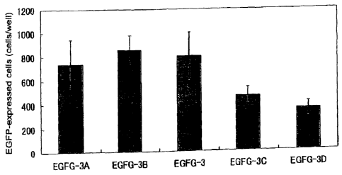
Table 3
Experiment 6:Gene-transfection efficiency relative to atelocollagen
complex atelocollagen pDNA(ug/ml)
EGFG-3A 0.005 100
EGFG-3B 0.008 100
EGFG-3 0.01 100
EGFG-3C 0.015 100
EGFG-3D 0.02 100
EGFG-3E 0.03 100
pDNA solely 0 100
The complexes prepared were added dropwise to the 293 cells
cultured on a 6-well dish in the presence of serum-free medium. Then,
after cultured at 37 C overnight, the cells were washed with PBS to
= CA 02451603 2003-12-19
46
remove the serum-free medium and the complex, and the medium was
replaced for a medium containing 10% calf serum. Seven days later,
the cells were observed by fluorescence microscopy, and the cells
expressing EGFP were counted. The results are shown in Figure 7.
For EGFG-3B, 3D, and 3E, fluorescence intensities of the expressed
EGFP were measured (Array Scan II System, Cellomics, Co.). Relative
fluorescence intensities are shown in Figure 8.
The complexes prepared were coated onto a 6-well dish at 250
pi/well, and the dish was air-dried for 30 minutes. Then, the 293 cells
(4-5x10⁵ cells/well) in a medium containing 10 % FBS were added
thereto, and cultured at 37 C overnight. Seven days later, the cells
were observed by fluorescence microscopy, and the cells expressing
EGFP were counted. The results are shown in Figure 9.
Figure 7 showing the results of dropwise addition and Figure 9
showing the results of solid coating indicate that the concentration of
collagen affects the transfer efficiency, and the solid coating is superior
to the dropwise addition in transfer efficiency.
(2) Complexes having the various compositions of plasmid DNA
concentrations as shown in Table 4 were prepared, and transfer
efficiencies were compared between dropwise addition and solid
coating.
Table 4
complex atelocollagen (%) DNA ml
EGFG-3B 0.008 100
EGFG-3B2 0.008 60
EGFG-3B3 0.008 40
CA 02451603 2003-12-19
47
EGFG-3B4 0.008 20
The results of dropwise addition are shown in Figure 10, and
the results of solid coating are shown in Figure 11. Relative
fluorescence intensities of EGFP in each sample obtained in solid
coating are also shown in Figure 12. These results show that the
concentration of plasmid DNA affects the transfer efficiency, specifically
the expression level of EGFP and the concentration of plasmid or DNA
are in a positive correlation, and also that the solid coating is superior
to the dropwise addition in transfer efficiency.
Example 7
Application of solid coating to screening method
Ten pM of a phosphorothioate antisense oligonucleotide (5'-
CTCGTAGGCGTTGTAGTTGT-3'; Molecular weight, about 6500; SEQ ID
NO: 2) (5) (Sawaday) having a sequence complementary to a sequence
from 4196 bp to 4216 bp of fibroblast growth factor HST-1 (FGF4) gene
(described in Proc. Natl. Acad. Sci. USA, 84, 2890-2984 (1987)) was
mixted with a 0.05 % solution of atelocollagen to form complexes. The
complexes were coated onto the bottom of a 96-well plate for cell
culture, and air-dried to obtain a cell culture instrument wherein the
bottoms were coated with the complex. Similarly, a phosphorothioate
sense oligonucleotide (5'- GAGCATCCGCAACATCAACA -3'; SEQ ID NO:
3) (1) having the same sequence as the HST- 1 gene, as well as
phosphorothioate antisense oligonucleotides having a sequence (5'-
AGTCGCATGCACACAACACA-3'; SEQ ID NO: 4) (2) as obtained by
scrambling the antisense oligonucleotide sequence, and the three
CA 02451603 2003-12-19
48
random sequences (5'-GACCATCGTCGATTCCAGT-3'; SEQ ID NO: 5) (3),
(5'-CATGAACATCCTGAGCATCC-3'; SEQ ID NO: 6) (4), and (5'-
GTTCACGAAGAAAGAAGGCT-3'; SEQ ID NO: 7) (6)) were each
complexed with a collagen to form complexes, which complexes were
coated onto the bottom, and dried. Onto the plate coated with those
oligonucleotides, NEC8 cells in which the proliferation was promoted by
overexpression of HST- 1, and HepG2 cells in which the proliferation
was promoted by the gene other than HST- 1 were seeded at 0.5 X 10⁵
cells/well, and the cells were cultured for 4 days. After the culture,
the inhibition of cell proliferation was assayed using TetraColor ONE
Cell Proliferation Assay Reagent. Specifically, the sample solution
stained was determined spectrometrically at 650 nm, using the
absorbance at 450 nm as control (Figure 13).
As a result, the inhibitory effect of NEC 8 cells proliferation was
observed only in the case of coating of the complex formed between an
antisense oligonucleotide against HST-1 and a collagen (Figure 13-5).
On the other hand, no inhibitory effect was observed in the case of
coating of the complex formed between a sense oligonucleotide against
HST- 1 and a collagen onto the bottom of the plate (Figure 13-1). Also,
no inhibitory effect was observed in the case of coating of the complex
formed between the phosphorothioate antisense oligonucleotides
having the sequence as scrambling the antisense oligonucleotide
sequence (Figure 13-2) and the random sequences (Figure 13-3, 4, 6)
and a collagen onto the bottom of the plate. These results show that it
is possible to screen for a gene responsible for the cell proliferation
without reagents for gene transfer such as cationic liposomes and
CA 02451603 2003-12-19
49
cationic lipids, which do not usually exist in living bodies, when an
antisense oligonucleotide complexed with an atelocollagen is coated
onto the solid phase, and cells are cultured thereon. Further, these
results show that the present process enables to examine sensitively
the gene functions even using a small amount of the cells and the
oligonucleotides without noise due to damage to cells from gene
transfer reagents.
Example 8
Storage of adenovirus vector under air drying using atelocollagen
Preparation by solid coating
Ten pi of a solution of an adenovirus vector, AdCMVEGFP (K.
Aoki, et al., Mol. Ther. (2000) 1 (6): 555-565) at 1x10$ pfu/ml in a
DMEM solution was mixed with 5 pl of a 2% solution of an
atelocollagen in PBS (-). Fifty 111 of the mixture was added to a 24-well
culture plate (non-coated) (Corning Inc.), and the plate was air-dried
with a dryer set on the cool mood at room temperature for 15 minutes
to carry out the solid coating.
Test of viruses for stability during storage
While the resultant culture plate was left at it was at room
temperature, a human hepatoma cell line, the HepG2 cells were added
thereto at 2 x 10⁵ cells/well 1 day, 7 days and 14 days after the leaving,
and each 2 days later, the expression level of GFP was observed by
fluorescence microscopy. As control, a similar experiment was carried
CA 02451603 2003-12-19
out using a culture plate coated with only adenovirus, and with only an
atelocollagen.
The results are shown in Table 5.
5 Table 5
GFP-positive cells ratio 2 days after the seeding (%)
atelo/adeno only adeno only atelo
left at r.t. 1 day 64 72 0
left at r.t. 7 days 42 10 or less 0
left at r.t. 14 days
r.t. means room temperature.
10 Table 5 shows that the adenovirus vector survived during
storage for even 14 days, suggesting that the solid coating of the
present invention provides a plate for screening that comprises
adenovirus vectors, which could be conveniently distributed on the
market.
Example 9
Gene transfer effects of the solid coating in a dose-dependent manner
of adenovirus
Both equal amounts of a solution of an adenovirus vector
AdCMVEGFP (K. Aoki, et al. Mol. Ther. (2000) 1 (6): 555-565) in DMEM
at 4 X 10⁴, 4 X 10⁵, 4X 10⁶ and 4 X 10⁷ pfu/ml, and a 160 1g/ml
solution of an atelocollagen were mixed together. The mixture could
be stored at 4 C for a long period of time without lowered activity.
Fifty p1 of the mixture was added to a 96-well culture plate (non-coated;
Corning Inc.), and the plate was air-dried to carry out the solid
CA 02451603 2003-12-19
51
coating. The plate can be stored at least for 2 weeks to 4 weeks
without lowered activity. Embryonic stem cells (ES cells) were seeded
on the resultant culture plate, and three days later, the expression of
GFP was checked with fluorescence microscopy, simultaneously with
determining the fluorescence intensity. As a result, it was found that
the fluorescence intensity of GFP is positively correlated with the dose
of adenovirus (Figure 14). The results shows that the present
invention should provide a useful tool for high-throughput screening
for ES cells using adenovirus, and a plate for screening, which is
coated with complexes formed by adenovirus and collagen.
Example 10
Relationship between the composition of complexes comprising plasmid
DNAs and the average major axis
Both equal amounts of an aqueous solution, a 0.1 M
phosphate buffer (PB) or a 0.01 M phosphate buffer (PBS) containing
sodium chloride, each of which contained a plasmid DNA encoding a
fluorescent protein (EGFP) (pCMV-EGFP/ pEGFP-N1, 4.7 kbp, Clontech
Co.) at 200 pg/ml and was cooled up to 10 C or less, and an aqueous
solution containing an atelocollagen (KOKEN CO., LTD.) at 0.002 to
0.02 %(w/w) cooled up to 10 C or less were mixed together, and the
mixtures were left as they were overnight at 10 C or less so as to
prepare a formulation in a gel form. The gel formulation was
penetrated through a filter having a pore size of 70 pm or 10 pm to
prepare a gel formulation having an ordered size.
To the formulation, PicoGreen dsDNA Quantitation Reagent
CA 02451603 2003-12-19
52
(Molecular Probes) was added to stain pCMV-EGFP, and the major axis
of the complexes were determined by fluorescent microscopy.
Similarly, a plasmid DNA (pCMV-HST-1-IL-2, 6.2kbp) encoding the
secretory signal peptide of HST-1 and interleukin-2, and an , _
atelocollagen were mixed together to prepare a formulation in a gel form,
and the major axis of the complexes was determined. Further, the gel
formulation was electrophoresed on agarose gel (Tris-acetate buffer,
0.8% agarose gel) to separate plasmid DNAs formed into complexes
from those not formed into complexes. The band of the plasmid DNAs
that were not formed into complexes was stained with ethidium
bromide, and the fluorescence intensity was determined with Fluor-S-
Multi Imager (BIO-RAD). Amount of the plasmid DNAs that were not
formed into complexes were determined on the basis of the standard
curve obtained from the fluorescence intensity of the band of the
plasmid DNA with the known concentration as electrophoresed, and
the ratio of the negative charge of the DNA contained in the complex to
the molecular weight of the collagen. The results are shown in Table
6.
Table 6
C.C. Number of Average major axis of complexes
Sample Plasmid DNA Solv. (%) N.M per
C.M After After 70 After 10
mixing m filter filter
10-1 CMV-EGFP Water 0.001 2406 9.1 8.9 < 3.4
10-2 pCMV-EGFP Water 0.005 742 37 26
10-3 pCMV-EGFP Water 0.008 699 6.6
10-4 pCMV-EGFP Water 0.01 299 53 44 35
10-5 CMV-EGFP Water 0.02 356 17
10-6 pCMV-EGFP Water 0.05 76 122 105
10-7 pCMV-EGFP Water 0.1 66 303 174
CA 02451603 2003-12-19
53
10-8 pCMV-EGFP PB 0.001 448 < 3.4 < 3.4
10-9 pCMV-EGFP PB 0.01 152 7.7
10-10 CMV-EGFP PB 0.1 97 22 14
10-11 pCMV-EGFP PBS 0.01 300 13 9.2
10-12 pCMV-EGFP PBS 0.1 97 22 15
10-13 pCMV-HST-1- Water 0.001 4228 6 6.1 < 3.4
IL-2
10-14 pCMV-HST-1- Water 0.005 1122 20 9.8
IL-2
10-15 pCMV-HST-1- Water 0.008 426 6.1
IL-2
10-16 pCMV-HST-1- Water 0.01 324 20 13 27
IL-2
10-17 pCMV-HST-1- Water 0.02 360 16
IL-2
10-18 pCMV-HST-1- Water 0.05 95 151 201
IL-2
10-19 pCMV-HST-1- Water 0.1 66 256 201
IL-2
10-20 pCMV-HST-1- PB 0.001 701 6.1 < 3.4
IL-2
10-21 pCMV-HST-1- PB 0.01 262 20
IL-2
10-22 pCMV-HST-1- PB 0.1 97 25 9.4
IL-2
10-23 pCMV-HST-1- PBS 0.01 315 9.8
IL-2
10-24 pCMV-HST-1- PBS 0.1 97 29
IL-2
In the table, "Solv." means solvent. "C.C. (%)" means collagen
concentration (%). "Number of N.M per C.M" means number of
nucleotide monomer per collagen molecule. "After 70 11m filter" means
after penetrating through a filter having a pore size of 70 pm. "After
10 um filter" means after penetrating through a filter having a pore size
of 10 pm.
In both cases of pCMV-EGFP and pCMV-HST-I-IL-2, a water
without salts was used as a solvent to obtain complexes wherein the
average major axis is 122 11m or more, when collagen concentration
was 0.05 % or more. When collagen concentration was 0.005 % to
0.02 %, complexes having an average major axis of 6.1 um to 53 um
were obtained, and when collagen concentration was 0.001 %,
CA 02451603 2003-12-19
54
complexes having an average major axis of 10 pm or less were
obtained. The results show that the form or the shape of a complex
can be controlled by adjusting the concentration of a plasmid DNA and
a collagen to be mixed, when a water without salts is used as a
solvent.
The results are consistent with those as obtained in Example 2
wherein pCAHST-1 was used to prepare complexes. The plasmid
DNAs as used therein were different each other in size as pCAHST- 1 is
7.9kbp, pCMV-EGFP is 4.7kbp, and pCMV-HST-1-IL-2 is 6.2kbp,
showing that sizes of plasmid DNAs never affect the form or the shape
of complexes to be formed.
On the other hand, 0.1 M phosphate buffer and 0.01 M
phosphate buffer containing sodium chloride were used as a solvent to
obtain complexes having an average major axis of 3.4 pm to 29 pm,
and no complexes having a 100 pm or more in size, when collagen
concentration was 0.001 % to 0.1 %. It was believed that this was
caused by the salts, which inhibited the formation of collagen
association bodies, showing that when complexes are prepared in the
presence of an agent that inhibits the formation of collagen association
body, then complexes having an average major axis of 100 pm or less
can be prepared irrespective of collagen concentration.
Further, when the gel formulation as obtained by mixing a
plasmid DNA and a collagen was penetrated through a filter having a
pore size of 70 pm or 10 pm, complexes having a smaller average major
axis and having an ordered size could be prepared.
Figure 15 shows the relationship between the number of
CA 02451603 2003-12-19
collagen bound to one molecule of plasmid DNA and the average major
axis of the complexes. In both cases of pCMV-EGFP and pCMV-HST-
1-IL-2 as used in a water without salts, it was found that the average
major axis of complexes trends to extend as increasing in the number
5 of collagen bound to plasmid DNA. On the other hand, when 0.1 M
phosphate buffer and 0.01 M phosphate buffer containing sodium
chloride were used as a solvent to prepare complexes, such trend as
above was not found, and even in the condition that collagen molecules
having an average major axis of 100 pm or more was complexed with a
10 plasmid DNA in case of the use of a water as a solvent, the average
major axis did not exceed 50 um. It is understood that this is caused
by extension reaction along the major axis of collagen association body,
of which formation is developed by the formation of complexes of a
plasmid DNA and a collagen as observed in the case that a water is a
15 solvent as mentioned above. On the other hand, in case of the
presence of salts, it is understood that collagen association body
formed by attaching a collagen molecule with a plasmid DNA does not
extend along the major axis well, and therefore a lot of fine association
bodies attach to a plasmid DNA to form a structure. Collagens have
20 been known to form fine association bodies at appropriate salt
concentrations. Thus, it can be considered that a partial amount of
collagens has already formed with a plasmid DNA into fine bodies
before mixing with a plasmid DNA, and the fine collagen association
bodies are attached to a plasmid DNA to form complexes. This means
25 that complexes having an average major axis of 50 um or less can be
prepared by mixing a plasmid DNA and a collagen that has been
= CA 02451603 2003-12-19
56
formed into fine association bodies.
For the general analysis of those results regardless of size of
plasmid DNAs, the number of nucleotide monomer of a plasmid DNA
per one collagen molecule in complexes was counted. Figure 16 shows
the relationship between the number of nucleotide monomer of plasmid
DNAs per one collagen molecule, and the average major axis of the
complexes. It was found that the average major axis of complexes
trends to extend as decreasing in the number of nucleotide monomer of
plasmid DNAs.
Taken together Figure 16 and Table 6, the followings are
concluded. When the number of nucleotide monomer is 95 or less, the
complexes having an average major axis of 120 um or more were
obtained. This means that the decrease in the number of nucleotide
monomer per one collagen molecule represents the increase in the
number of collagen molecules relative to the plasmid DNA in the
complexes, and thus complexes having an average major axis of 120
pm or less can be prepared by keeping the number of nucleotide
monomer of plasmid DNAs per one collagen molecule to be 96 or
more. On the other hand, minimum unit of the complexes is a
complex formed by one molecule of collagen and one molecule of
plasmid DNA. One molecule of collagen and many plasmid DNA
hardly form complexes since plasmid DNAs repel each other by their
negative charge and undergo steric hindrance. Accordingly, the
maximum number of nucleotide monomer per one collagen molecule is
obtained by forming a complex with each one molecule of collagen and
plasmid DNA, and is defined by the number of nucleotide monomer
= CA 02451603 2003-12-19
57
comprised in a plasmid DNA.
Example 11
Relationship between the composition and the average major axis of the
complex comprising an oligonucleotide
An equal amount of an aqueous solution, or a 0.1 M phosphate
buffer (PB), each of which contained a phosphorothioate antisense
oligonucleotide (5'-CTCGTAGGCGTTGTAGTTGT-3'; Molecular weight,
about 6500; SEQ ID NO: 8) (Espec Inc.) having a sequence
complementary to a sequence from 4196 bp to 4216 bp of fibroblast
growth factor HST- 1 (FGF4) gene (described in Proc. Natl. Acad. Sci.
USA, 84, 2890-2984 (1987)) in a concentration of 2.0 uM to 40.0 PM,
and was cooled up to 10 C or less, and an aqueous solution containing
an atelocollagen (KOKEN CO., LTD.) at 0.002 to 3.0 %(w/w) cooled up
to 10 C or less, 0.1 M phosphate buffer were each mixed together at 10
C or less, and the mixtures were left as they were overnight at 10 C or
less so as to prepare formulations in a gel form. Oligonucleotide
amounts that were complexed with a collagen in the gel formulations
were determined as follows. The gel formulations were centrifuged at
150,000 rotations per hour at 5 C to precipitate the complexes, and
the amounts of the oligonucleotides released in the supernatant were
determined by HPLC (column: Puresil C18 5 (Waters), mobile phase: a
gradient of 24.5% to 35% acetonitrile over 35 minute in 0.1 M
ammonium acetate containing 5mM tetrabutyl ammonium and 1mM
EDTA, flow rate: 1 ml/min, detection wavelength: 260 nm). Single-
Stranded nucleic acid fluorescence staining regent YOYO (Molecular
CA 02451603 2003-12-19
58
Probes) was added to the resultant formulations to stain the
oligonucleotides, which were observed by fluorescent microscopy to
measure the major axis of the complexes. The results are shown in
Table 7.
Sample Solv. O.C.(iM) C.C(%) Number of Number of Average major
O.M per C.M N.M per C.M axis of complexes
11-1 water 1.0 0.05 0.6 12 32
11-2 water 5.0 0.05 3 60 13
11-3 water 10.0 0.05 4.4 88 12
11-4 water 20.0 0.05 5.2 104 17
11-5 water 10.0 0.01 6.3 126 32
11-6 water 10.0 0.1 2.9 58 20
11-7 PB 1.0 0.05 0.5 10 25
11-8 PB 5.0 0.05 1.2 24 29
11-9 PB 10.0 0.05 1.4 28 35
11-10 PB 20.0 0.05 1.3 26 36
11-11 PB 10.0 0.01 3.3 66 23
11-12 PB 10.0 0.1 1.3 26 33
11-13 PB 10.0 0.5 0.5 10 46
11-14 PB 10.0 1.0 0.3 6 49
11-15 PB 10.0 1.5 0.2 4 54
In the table, "Solv." means solvent. "O.C.(pM) means oligonucleotide
concentration (pM). " "C.C. (%)" means collagen concentration (%).
"Number of O.M per C.M" means number of oligonucleotide molecule
per one collagen molecule. "Number of N.M per C.M" means number
of nucleotide monomer per collagen molecule.
Similarly to Example 10 wherein plasmid DNAs were used,
complexes were obtained by mixing oligonucleotides and collagens. In
both cases of use of a water without salts and 0.1 M phosphate buffer
as a solvent, there were no significant change in major axis of the
complexes between the differences in the concentrations of collagen
and oligonucleotide, which was different from the case of plasmid
DNAs. This is believed to be caused by the fact that oligonucleotides
CA 02451603 2003-12-19
59
are smaller than plasmid DNAs in molecular size, and thus the
formation of collagen association bodies is inhibited.
Figure 17 shows the relationship between the molecular
number ratio of oligonucleotide to collagen at the time of the mixture
and the number of oligonucleotide bound to one molecule of collagen in
the complex. It was found that the number of oligonucleotides bound
to one collagen molecule in the formed complexes trends to increase as
increasing in the number of oligonucleotides relative to the number of
collagen molecule at the time of mixing in both cases that a water and
the phosphate buffer were used as a solvent. The maximum number
of oligonucleotide molecule bound to one collagen molecule varies
depending on kinds of solvent, and it is 6 molecules in case of water,
whereas it is 3 molecules in case of the phosphate buffer.
Since molecular weight of a collagen (300,000) is even larger
than that of an oligonucleotide (about 6,500), and the form or the
shape of complexes depends solely on the number of collagen molecule
forming into complexes, the numbers of collagen molecule in complexes
that have the same major axis are approximately the same.
Accordingly, even for the complexes having the same major axis, the
number of oligonucleotide molecule comprised in complexes varies
depending on the number of oligonucleotide bound to one collagen
molecule. Because individual complex contacts with a cell to allow
oligonucleotides to be transferred into the cell, oligonucleotide transfer
is more efficient when a complex that contains a high content of
oligonucleotide molecules is used. Accordingly, a complex wherein the
number of oligonucleotide molecule per one collagen molecule is
CA 02451603 2003-12-19
increased is desired, and generally speaking, a complex wherein the
number of nucleotide monomer bound to one collagen molecule is
increased is preferred.
5 Example 12
Cell culture instrument coated with a complex comprising an
oligonucleotide
Fifty pi of the gel formulation as prepared in Example 11 (11-3,
the composition of the formulation of Example 7) was added dropwise
10 to the bottom of a 96-well microplate, and dried by directly spraying a
cool air (room temperature, 2 hours) or by placing in a desicator with
silica gel (room temperature, 2 days) so as to prepare a cell culture
instrument, of which the cell culture surface is coated with a
complex. One hundred pl of 0.01 M phosphate buffer (PBS) that had
15 been adjusted to be isotonic with living bodies by supplementing
sodium chloride was added to the wells of the instrument, and the
pipetting was slightly conducted immediately for sampling. Further,
100 p1 of 0.01 M phosphate buffer (PBS) that had been adjusted to be
isotonic with living bodies by supplementing sodium chloride was
20 added to the wells of the instrument, and the instrument was left
overnight at 37 C, followed by slightly pipetting for sampling. To 10
III of the resultant sample, 2 pi of Single-Stranded nucleic acid
fluorescence staining regent YOYO (Molecular Probes) was added to
stain the oligonucleotides, and the complexes that released form the
25 surface of the instrument were observed by fluorescent microscopy.
The fluorescence micrograph is shown in Figure 18. The complexes
CA 02451603 2003-12-19
61
having a major axis of 50 um or less were observed in all of the
samples. This means that exposure on PBS allows to release
complexes from the surface of a cell culture instrument. The form or
the shape of the released complexes was not changed between before
and after the coating onto the surface of the instrument irrespective of
drying method, and condition of PBS exposure. This shows that the
complexes are retained on the surface of a cell culture instrument with
keeping their form or shape irrespective of drying method, and that the
form or the shape of the complexes maintains in a temperature
condition of 37 C that is usually used in cell culture.
Example 13
Cell culture instrument coated with a complex comprising a plasmid
DNA
Fifty pi of the gel formulation as prepared in Example 10 (10-3,
having the composition that was found efficient in transfer efficiency in
Example 6) was added dropwise to the bottom of a 96-well microplate,
and dried by directly spraying a cool air (room temperature, 2 hours) or
by placing in a desicator with silica gel (room temperature, 2 days) so
as to prepare a cell culture instrument, of which the cell culture
surface is coated with a complex. One hundred pl of 0.01 M
phosphate buffer (PBS) that had been adjusted to be isotonic with living
bodies by supplementing sodium chloride was added to the wells of the
instrument, and the pipetting was slightly conducted immediately for
sampling. Further, 100 Ill of 0.01 M phosphate buffer (PBS) that had
been adjusted to be isotonic with living bodies by supplementing
CA 02451603 2003-12-19
62
sodium chloride was added to the wells of the instrument, and the
instrument was left overnight at 37 C, followed by slightly pipetting for
sampling. To 10 pi of the resultant sample, 2 p1 of Single-Stranded
nucleic acid fluorescence staining regent YOYO (Molecular Probes) was
added to stain the oligonucleotides, and the complexes that released
form the surface of the instrument were observed by fluorescent
microscopy. The fluorescence micrograph is shown in Figure 19. The
complexes having a major axis of 100 um or less were observed in all of
the samples. This means that exposure on PBS allows to release
complexes from the surface of a cell culture instrument. The form or
the shape of the released complexes was not changed between before
and after the coating onto the surface of the instrument irrespective of
drying method, and condition of PBS exposure. This shows that the
complexes are retained on the surface of a cell culture instrument with
keeping their form or shape irrespective of drying method, and that the
form or the shape of the complexes maintains in a temperature
condition of 37 C that is usually used in cell culture.
Example 14
Gene transfer with a cell culture instrument coated with a complex
Three hundreds p1 of the gel formulations as prepared in
Example 10, 10-1, 10-4, and 10-7, comprising pCMV-EGFP were added
to a 6-well cell culture microplate, and dried by spraying a cool air so
as to prepare a cell culture instrument onto which a complex
comprising pCMV-EGFP was coated. As control, 300 u1 of an aqueous
solution comprising pCMV-EGFP at 100pg/ml was added to a plate,
CA 02451603 2003-12-19
63
and dried similarly. Into each well, 7.5 X 10⁴ cells of 239 cells were
seeded, and a DMEM medium containing 10 % FBS was added thereto,
followed by culturing at 37 C. From the start of the culture, the
medium was replaced with a fresh medium every 4 or 5 days. eleven
days after the seeding, the cells were observed by fluorescent
microscopy, and the number of the cells expressing GFP was counted
to estimate transfer efficiency.
Similarly, 300 pl or 500 pl of the gel formulations as prepared
in Example 10, 10-13 to 22, and 24, comprising pCMV-HST-1-IL-2
were added to a 6-well cell culture microplate, and dried by spraying a
cool air so as to prepare a cell culture instrument onto which a complex
comprising pCMV-HST-1-IL-2 was coated. As control, 300 pl or 500 ul
of an aqueous solution comprising pCMV-HST-l-IL-2 at l00pg/ml, PB
solution or PBS solution were added to a plate, and dried similarly.
1) Transfer of plasmid DNA into 239 cells
Into each well wherein 300 pi of the gel formulation was coated,
7.5 X 10⁴ cells of 239 cells were seeded, and a DMEM medium
containing 10 % FBS was added thereto, followed by culturing at 37 C
for 10-13 to -19. Eight days after the cell seeding, the medium was
replaced, and IL-2 concentration in the medium sampled 11 days later
was determined by ELISA (Quamtikine human IL-2 (R&D Systems)).
For 10-20 to -22, a DMEM medium was added to the plate, and
cultured overnight at 37 C, after which on the following day the
medium was replaced with a DMEM medium containing 10% FBS, and
then cultured at 37 C. Eight days and 11 days after the cell seeding,
the medium was replaced, and IL-2 concentration in the medium
CA 02451603 2003-12-19
64
sampled 15 days later was determined by ELISA. Further, for 10-20,
21, and 24, a DMEM medium containing 10% FBS was added to the
plate, and the plate was cultured at 37 C. IL-2 concentration in the
medium sampled- 8 days after the cell seeding was determined by
ELISA.
2) Transfer of plasmid DNA into NIH3T3 cells
Into the well wherein 500 pi of 10-14 to -17 was coated, 5 X
104 cells of NIH3T3 cells were seeded, and a DMEM medium containing
10 % FBS was added thereto, followed by culturing at 37 C. Eight
days after the cell seeding, the medium was sampled, and IL-2
concentration therein was determined by ELISA.
The results as obtained in above 1) and 29 are summarized in
Tables 8 and 9. When complexed with a collagen and coated onto a
cell culture instrument, both pCMV-EGFP and pCMV-HST-1-IL-2 could
be efficiently transferred into 293 cells and NIH3T3 cells. The result
shows that the effect of the complexes of the present invention on
facilitation of gene transfer is not affected by the kinds of plasmid DNA,
species of the cells, and the presence or the absence of serum in the
culture. Further, in case of plasmid DNAs, it has been found that
coating of the complex having 1112 or less of the nucleotide monomer
per one collagen molecule, and having an average major axis of 151 um
or less, provided superior transfer efficiency and gene expression.
Table 8
Sample Number of nucleotide I Average major axis transfer efficiency
CA 02451603 2003-12-19
monomer per collagen of complexes (pm)
molecule
Control 0.0028
10-1 2406 9.1 0.0760
10-4 299 53 0.0729
10-7 66 303 0.0374
Table 9
Sample Cell Solvent Presence Number of Average major IL-2
species or nucleotide axis of concentration
Absence monomer complexes in media
of FBS per collagen (pmt (pg/ml)
molecule
Control 293 water Presence 5.3
10-13 293 water Presence 4228 6 7.3
10-14 293 water Presence 1122 20 14.8
10-15 293 water Presence 426 6.1 19.3
10-16 293 water Presence 324 20 6.3
10-17 293 water Presence 360 16 18.2
10-18 293 water Presence 95 151 8.4
10-19 293 water Presence 66 256 5.3
Control 293 PB Absence 23.55
10-20 293 PB Absence 701 6.1 72.93
10-21 293 PB Absence 262 20 264.61
10-22 293 PB Absence 97 25 86.22
Control 293 PB Presence 3.35
10-20 293 PB Presence 701 6.1 21.01
10-21 293 PB Presence 262 20 86.68
Control 293 PBS Presence 1.07
10-23 293 PBS Presence 315 9.8 15.26
Control NIH3T3 water Presence 0
10-14 NIH3T3 water Presence 1122 20 0.79
10-15 NIH3T3 water Presence 426 6.1 6.06
10-16 NIH3T3 water Presence 324 20 4.05
10-17 NIH3T3 water Presence 360 16 2.78
5
Example 15
Gene transfer into cells by addition of complexes
Into a 6-well plate, 5 X 10⁴ cells of 239 cells were seeded, and
cultured in the presence of a DMEM medium containing 10 % FBS at
10 37 C. Three days after the seeding, 300 pi of the gel formulations as
CA 02451603 2003-12-19
66
prepared in Example 10, 10-1, 10-4, 10-7, 10-8, 10-9, and 10-10,
comprising pCMV-EGFP, and a water or a PB solution comprising
pCMV-EGFP at 100 pg/ml as control were each added thereto, and the
cells were cultured in the presence of a DMEM medium containing
10 % FBS at 37 C. On the following day, the media were replaced
with a fresh medium, and 13 days later for 10-1, 10-4, and 10-7, or 5
days later for 10-8, 10-9, and 10-10, the cells were observed by
fluorescent microscopy, followed by counting the number of the cells
expressing GFP.
Similarly, 7.5 X 10⁴ cells of 239 cells were seeded into a 6-well
plate, and cultured in the presence of a DMEM medium containing
10 % FBS at 37 C. Two days after the seeding, 300 pl of the gel
formulations as prepared in Example 10, 10-20 and 10-2 1, comprising
pCMV-HST-1-IL-2, and a PB solution comprising pCMV-HST-1-IL-2 at
100 ug/ml as control were each added thereto, and the cells were
cultured in the presence of a DMEM medium containing 10 % FBS at
37 C. On the following day, the media were replaced with a fresh
medium, and 5 days later, the media were sampled, and IL-2
concentration therein were determined by ELISA (Quamtikine human
IL-2 (R&D Systems)).
The results are summarized in Tables 10, 11 and 12. When
complexed with a collagen and added to cells, both pCMV-EGFP and
pCMV-HST-1-IL-2 could be efficiently transferred into the cells. It has
been found that in case of the complexes having an average major axis
of 53 pm or less as shown in the present Example, the addition of the
complex having 2406 or less, and further 701 or less of the nucleotide
= CA 02451603 2003-12-19
67
monomer per one collagen molecule, provided superior transfer
efficiency and gene expression.
Table 10
Sample Solvent Number of nucleotide Average major axis transfer efficiency
monomer per collagen of complexes
molecule
Control water 0.0012
10-1 water 2406 9.1 0.0231
10-4 water 299 53 0.0247
10-7 water 66 303 0.0041
Table 11
Sample Solvent Number of nucleotide Average major axis Number of EGFP-
monomer per collagen of complexes expressing cells
molecule m
Control PB 100
10-8 PB 448 < 3.4 128
10-9 PB 152 7.7 1200
10-10 PB 97 22 12000
Table 12
Sample Solvent Number of nucleotide Average major axis IL-2 concentration
monomer per collagen of complexes in media (pg/ml)
molecule m
Control PB 0
10-20 PB 701 6.1 8.98
10-21 PB 262 20 76.81
10-22 PB 97 25 120.74
INDUSTRIAL APPLICABILITY
According to the complexes of the present invention, it is
possible to preserve and stabilize desired nucleic acids by complexing
with collagens or collagen derivatives, 2) to transfer efficiently desired
CA 02451603 2003-12-19
68
nucleic acids into cells when administered to living bodies, and 3) to
express and inhibit desired nucleic acids without reagents for gene
transfer. The complexes of the present invention can be applied to
various aspects since the complexes of the invention-have advantages
1) that DNAs, antisense oligonucleotides and virus vectors can be
preserved and stabilized, 2) that the complexes can be stored during a
long period of time on plates dried where the complexes are coated, 3)
that genes can be expressed without reagents for gene transfer when
cells are seeded onto the plates, 4) that kinds of subjects of nucleic
acids and methods for immobilization are not limited to specific ones,
and 5) that the duration time of the expression in case of plasmid DNAs
is excellently extended.
According to the present invention, it is possible to readily
examine the functions of genes or proteins, of which the expressions
are inhibited by antisense oligonucleotides by determining the
proliferation or the morphology of the cells, or the expression levels of
cytokines or receptors. Not only antisense oligonucleotides, but also
other materials can be complexed with collagens to form complexes,
which in turn can be coated onto plates; in case of ribozymes, it is
possible to carry out a screening similar to that of antisense
oligonucleotides; and further in case of plasmid DNAs or adenoviruses,
it is possible to examine the functions of genes or proteins by
determining the changes of cells by the expression of certain genes.
Collagens or collagen derivatives comprised in the complexes of the
present invention have been known to affect no cells, and the screening
using the complexes of the present invention makes it possible to carry
CA 02451603 2003-12-19
69
out the examinations of gene functions that seldom avoid the
unspecific affections to cells, which would be induced by conventional
reagents for gene transfer.
CA 02451603 2004-06-21
SEQUENCE LISTING
<110> Sumitomo Pharmaceuticals Company, Limited; Koken Co. Ltd.
<120> Method of promoting nucleic acid transfer
<130> 57182-NP
<140> CA2451603
<141> 2002-06-20
<150> JP 2001-186320
<151> 2001-06-20
<150> JP 2001-278293
<151> 2001-09-13
<160> 8
<210> 1
<211> 20
<212> DNA
<213> Artificial Sequence
<220>
<223> Description of Artificial Sequence: antisense oligomer
<400> 1
ctcgtaggcg ttgtagttgt 20
<210> 2
<211> 20
<212> DNA
<213> Artificial Sequence
<220>
<223> Description of Artificial Sequence: phosphorothioate antisense oligomer
CA 02451603 2004-06-21
71
<400> 2
ctcgtaggcg ttgtagttgt 20
<210> 3
<211> 20
<212> DNA
<213> Artificial Sequence
<220>
<223> Description of Artificial Sequence: phosphorothioate sense oligomer
<400> 3
gagcatccgc aacatcaaca 20
<210> 4
<211> 20
<212> DNA
<213> Artificial Sequence
<220>
<223> Description of Artificial Sequence: phosphorothioate scramble oligomer
<400> 4
agtcgcatgc acacaacaca 20
<210> 5
<211> 19
<212> DNA
<213> Artificial Sequence
<220>
<223> Description of Artificial Sequence: phosphorothioate random oligomer
<400> 5
gaccatcgtc gattccagt 19
<210> 6
<211> 20
CA 02451603 2004-06-21
72
<212> DNA
<213> Artificial Sequence
<220>
<223> Description of Artificial Sequence: phosphorothioate random oligomer
<400> 6
catgaacatc ctgagcatcc 20
<210> 7
<211> 20
<212> DNA
<213> Artificial Sequence
<220>
<223> Description of Artificial Sequence: phosphorothioate random oligomer
<400> 7
gttcacgaag aaagaaggct 20
<210> 8
<211> 20
<212> DNA
<213> Artificial Sequence
<220>
<223> Description of Artificial Sequence: phosphorothioate antisense oligomer
<400> 8
ctcgtaggcg ttgtagttgt 20