Note: Descriptions are shown in the official language in which they were submitted.
CA 02451669 2003-12-19
WO 02/103409 PCT/US02/19314
-1-
OPTICAL GUIDANCE SYSTEM FOR INVASIVE CATHETER PLACEMENT
GOVERNMENT SUPPORT
The present invention was supported by The U. S. National Institutes of Health
under
Grant No. NS-31465. The government may have certain rights in the invention.
CROSS-REFERENCE TO RELATED APPLICATIONS
The present application claims priority to U.S. Provisional Patent Application
No.
60/299,299, filed June 19, 2001.
FIELD OF THE INVENTION
The present invention relates to an optical guidance system and a method for
insertion of endotracheal tubing, nasogastric tubing, feeding tubing, epidural
catheters,
central venous catheters, peripherally inserted central venous catheters,
chest tubes plural
catheters, and similar invasive catheters and tubes.
DESCRIPTION OF THE PRIOR ART
Determining the location of the end of a catheter inserted into patients for
the
purpose of providing nutrients or medications to specific locations within the
body has been
difficult. Currently, catheter placement is either done without visual
guidance or, if the
placement is particularly critical, it is done by x-ray, which can accurately
determine the
location of radio-opaque plastic materials used in making the tubing. However,
multiple x-
rays are often necessary. The necessity for multiple x-rays in order to locate
the end of the
inserted tubing is undesirable. An optical system that is convenient and easy
to use and yet
CA 02451669 2003-12-19
WO 02/103409 PCT/US02/19314
-2-
allows the end of the tubing to be quite accurately located without the use of
x-rays is
desired. Preferably, the position of the catheter tip may be directly observed
during the
insertion process and the position of the tip checked at any time thereafter.
Prior art catheter light delivery devices are known (e.g., Woodward et al;
5,947,958) that provide illumination of internal organs of a patient after
insertion
through, for example, the peritoneal wall. This illumination is to provide
light for either
imaging of the tissue surface or for delivering the light used in photodynamic
therapy.
Such devices are not used for catheter placement.
Other light guides, such as Fontenot; 5,423,321, have multiple light guiding
fibers of different lengths that are inserted into internal organs or vessels
during surgery.
In the case of balloon catheters, such light guides are used to place the
balloon catheter
in positions where inflation of the balloon will occlude the vessel if that
should become
necessary. The light guide is an independent entity and observation is through
the vessel
wall such that visible light is sufficient, although near infra red light is
indicated as
decreasing the intensity of light that is required. A detection system is also
described for
determining when the surgical cutting tool approaches the vessel.
Vander Salm et al; 5,906,579 and Duhaylongsod et al; 6,113,588 similarly
describe methods for visualizing balloon catheters through the vessel wall
under surgical
conditions. In these devices, the optical fiber is an independent entity and
is preferably
inserted through one lumen of a multilumen catheter. The disclosed devices are
specifically disclosed for use in cardiothoracic surgery.
Such prior art light guides do not use a single fiber that is built into the
structure
of catheters with multiple different functions, are not directed primarily to
localizing the
tip of an inserted catheter during non-surgical procedures for endotracheal
tubing,
nasogastric tubing, feeding tubing, epidural catheterization, central venous
catheterization, peripherally inserted central venous catheterizations, chest
tubes plural
CA 02451669 2003-12-19
WO 02/103409 PCT/US02/19314
-3-
catheterization, or with similar invasive catheters and tubes, and such prior
art devices do
not use only near infrared light since the vessels are not surgically exposed
and visible
light (blue through orange) provides insufficient penetration of the tissue.
Moreover,
such prior art devices are relatively expensive and the optical components may
require
difficult FDA scrutiny since they may contact the patient. The present
invention
addresses these limitations in the prior art.
UMMARY OF THE INVENTION
Light from a small laser diode is passed through an optical fiber that is
either
included in the lumen or incorporated into the wall of an invasive catheter
tube during
manufacture. The light is selected to be of a wavelength that is minimally
absorbed by
tissue, preferably in the range from about 620 nm to 1100 nm. In a preferred
embodiment,
780 nm is used as this is where the tissue absorption is near a minimum. The
light passes
out the end of the fiber (at the distal end of the catheter) and through the
tissue to the outside
where it is measured. The light pattern is observed by night vision goggles
that filter out
light in other frequency ranges. The detected light allows location of the end
of the fiber, the
positional accuracy depending on the thickness of tissue between the fiber tip
and the
exterior of the body. The method is highly accurate for small children and for
catheters near
the skin surface of adults but may not be applicable to catheters placed
within the body
cavity of some large adults.
BRIEF DESCRIPTION OF THE DRAWINGS
An optical guidance system and method for insertion of endotracheal tubing,
nasogastric tubing, feeding tubing, epidural catheters, central venous
catheters, peripherally
inserted central venous catheters, chest tubes plural catheters, and similar
invasive catheters
and tubes in accordance with the invention is further described below with
reference to the
accompanying drawings, in which:
CA 02451669 2003-12-19
WO 02/103409 PCT/US02/19314
-4-
Figure 1 illustrates a cross-section of a catheter with an integral optical
fiber that
is used in accordance with the invention to locate the tip of the inserted
catheter.
Figure 2 illustrates a side view of the catheter of Figure 1.
Figure 3 illustrates the catheter of Figure 1 inserted into the body of a
patient and
the detection of the light from the tip of the catheter at the nearest spot of
the patient's
skin in accordance with the method of the invention.
DETAILED DESCRIPTION OF THE INVENTION
An optical guidance system in accordance with the invention includes a laser
diode
having a wavelength in the range of 620 nm to 1100 nm, preferably a 780 nm
wavelength
with an emission less than 2 nm wide and less than 5 mW in power that! is
carried through a
150 micron (or less) core glass optical fiber to an "ST" optical connector at
a distal end. As
shown in Figure 1, the glass optical fiber 10 is embedded in (i.e., partially
or completely
surrounded by) the wall 20 of a catheter 30 having a catheter lumen 40. The
optical fiber 10
runs the entire length of the catheter 30, and the unterminated end of the
optical fiber 10 at
the distal end 50 of the catheter 30 is adapted to be inserted into the
patient as shown in
Figure 2. The proximal end 60 is terminated with an ST optical connector (not
shown)
appropriate for connecting the optical fiber 10 with the laser diode (not
shown) .
Conversely, the optical fiber 10 may be inserted into lumen 40 of the catheter
30 at its
proximal end 60 and fed to the distal tip 50 of the catheter 30 and held in
place so that light
escapes from the distal end 50 once the catheter 30 is inserted into the
patient.
The operator uses a detection system such as near infrared "night vision"
goggles 70
watch the progress of the catheter 30 from the site of entry to the chosen
location. The distal
end 50 of the catheter 30 is treated as a single light source and the diffuse
rays from this light
source are detected. A narrow pass (<10 nm at half height is preferred,
although wider
bandpass filters could be used) interference filter 80 with a center
wavelength of 780 nm (for
CA 02451669 2003-12-19
WO 02/103409 PCT/US02/19314
-5-
a light source of 780 nm) is used to cover the detector surface of the goggles
70. In general,
contribution of other ambient lighting increases with increasing width of the
optical filter
bandpass. The value of less than 10 nm is selected to allow some variation in
the laser diode
wavelength and yet to minimize the amount of light other than that from the
laser diode that
passes through to the detector of the goggles 70. Of course, if other
wavelength light were
used, an appropriate interference filter centered about the other wavelength
would be used.
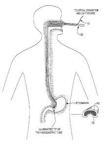
Figure 3 illustrates the catheter 30 of Figures 1 and 2 inserted into the body
of a
patient vie a nasogastric catheter 30 and the detection of the light from the
tip 50 of the
catheter 30 at the nearest spot of the patient's skin in accordance with the
method of the
invention. In the example illustrated in Figure 3, night vision goggles 70
with an
appropriate interference filter 80 thereon allow the operator to see the
infrared light through
the skin outside of the patient's stomach.
Those skilled in the art will appreciate that other designs of the optical
guidance
system for catheters in accordance with the invention could be constructed
using different
light sources and light detectors. While 780 nm light is suitable since tissue
absorption is
near a minimum at that wavelength, it would be possible, for example, to use
an LED as a
light source as long as the light provided was of appropriate wavelength and
energy. In this
case, a wider bandpass filter may be required on the detector (an LED light
output is broader
than that of the laser diode). Similarly, different detectors could be used,
including
photodiodes, photomultipliers, avalanche photodiodes, and microchannel plates.
When
photodiodes or other single site detectors are used they could be moved over
the surface of
the tissue to detect the maximum in the specific light emitted from the
optical fiber. The
sensitivity of the measurement could be maximized by modulating the light at a
specific
frequency (such as 1000 Hz) and detecting only the photosignal of that
frequency.
Another modification that would allow the operator to detect those cases in
which the
catheter had "doubled back" inappropriately would be to incorporate two
optical fibers, one
CA 02451669 2003-12-19
WO 02/103409 PCT/US02/19314
-6-
terminated about 5 centimeters before the tip and the other at the tip. The
two could be
distinguished by differences in modulation frequency and/or wavelengths of
light.
In one variation of the detection system, the night vision goggle 70 could
include a
sensitive microchannel plate imager in a mini-display directly in front of one
eye of the
operator. This would allow the operator to look at either the patient or at
the display as
desired.
Although exemplary implementations of the invention have been described in
detail above, those skilled in the art will readily appreciate that many
additional
modifications are possible in the exemplary embodiments without materially
departing
from the novel teachings and advantages of the invention. Any such
modifications are
intended to be included within the scope of this invention as defined by the
following
exemplary claims.