Note: Descriptions are shown in the official language in which they were submitted.
CA 02451820 2010-11-23
A SYSTEM AND METHOD FOR ASSESSING
URINARY FUNCTION
Field of the Invention
The present invention relates generally to a system and a method for assessing
urinary function. More particularly, the system and method is used for testing
the
integrity of the urinary system for diagnostic purposes and for use with
therapies to
correct urinary incontinence.
Background of the Invention
Women account for more than 11 million of incontinence cases. Moreover, a
majority of women with incontinence suffer from stress urinary incontinence
(SUI).
Women with SUE involuntarily lose urine during normal daily activities and
movements, such as laughing, coughing, sneezing and regular exercise,
SUE may be caused by a functional defect of the tissue or ligaments connecting
the vaginal wall with the pelvic muscles and pubic bone, Common causes include
repetitive straining of the pelvic muscles, childbirth, loss of pelvic muscle
tone and
estrogen loss. Such a defect results in an improperly functioning urethra.
Unlike other
types of incontinence, SUI is not a problem of the bladder.
Normally, the urethra, when properly supported by strong pelvic. floor muscles
and healthy connective tissue, maintains a tight seal to prevent involuntary
loss of
urine. When a woman suffers from the most common form of SUI, however,
weakened
muscle and pelvic tissues are unable to adequately support the urethra in its
correct
position. As a result, during normal movements when pressure is exerted on the
bladder from the diaphragm, the urethra cannot retain its seal, permitting
urine to
escape. Because SUI is both embarrassing and unpredictable, many women with
SUI
avoid an active lifestyle, shying away from social situations.
SUE is categorized into three types. Type I and Type U are directed to
urethral
hypermobility. Type III is directed to intrinsic sphincter deficiency (ISD).
Diagnosis
of ISD requires uodynamic evaluation. Urodynamic evaluation involves complex
and
I
CA 02451820 2003-12-23
WO 03/001977 PCT/US02/20389
invasive equipment and often requires referral to a specialist trained in
urodynamic
evaluation.
Existing diagnostic systems all require a catheter be passed trans-urethraly
to
measure pressure, such as Leak Point Pressure (LPP) or Urethral Pressure
Profile
(UPP). An exemplary system is disclosed in publication (WO 0023127). Detection
of
LPP requires that a pressure sensor and catheter be passed trans-urethrally.
The bladder
is filled, and pressure is recorded. Fluid leakage from the urethral opening
(meatus)
corresponds to the maximum pressure the urethral sphincter can resist, or LPP.
During
the UPP measurement procedure a pressure sensor tipped catheter is placed
trans-
urethral into the bladder and then withdrawn at a constant velocity. The
pressure
profile along the urethra, from bladder neck to meatus is recorded.
Other parameters may also be measured, such as abdominal pressure and
urinary flow. A cystometrogram (CMG) is a pressure study that simultaneously
measures intra-abdominal, total bladder, and true detrusor pressures.
Uroflometry
measures urine flow rate visually, electronically, or via a disposable system.
Video
Urodynamic Systems also exist that simultaneously measure parameters, as
described
above, with radiographic visualization of the lower urinary -tract.
Existing urodynamic evaluation systems are complex, expensive, and require
extensive training. Furthermore, existing urodynamic systems often require at
least 30
minutes to complete a test. This exceeds the time available for most standard
physician
office visits and results in referral to a specialist. No urodynamic system
exists that can
quickly and inexpensively record useful urodynamic measures, without passing a
catheter or instrument trans-urethraly.
There remains a need for an improved system and method for assessing urinary
fiuction.
Summary of the Invention
A system is provided for assessing urinary function including a processor, a
tubing assembly forming a first fluid conduit between a first fluid inlet and
a first fluid
outlet, and an insert member having a channel therethrough and coupled to the
first
fluid outlet so that fluid flowing through the first fluid conduit may flow
through the
insert member. The insert member is also dimensioned for at least partial
insertion into
a patient's urethral canal distal of the patient's urethral sphincter, and
dimensioned to
substantially block fluid flow into or out of the urethral canal other than
through the
insert member channel. The system also includes a fluid delivery device
electrically
coupled to and controlled by the processor, and coupled to the first fluid
conduit for
2
CA 02451820 2003-12-23
WO 03/001977 PCT/US02/20389
pumping fluid therethrough, and a detection system for detecting resistance of
the
urethral sphincter as fluid is infused therein. According to one embodiment,
the
detection system is a pressure detection system for detecting pressure within
the
urethral canal distal of the urethral sphincter as fluid is infused therein,
and providing
information correlating to the detected pressure to the processor.
According to an alternate embodiment, the pressure detection system is in
fluid
communication with the first fluid conduit at a location such that the
pressure within
the first fluid conduit substantially correlates to pressure within the
urethral canal distal
of the urethral sphincter, and in yet another embodiment, the pressure
detection system
detects the Urethral Resistance Pressure.
In yet another embodiment, the system further includes a display device
electrically coupled to the processor and capable of displaying data, and in
yet another
embodiment, the pressure detection system detects Urethral Resistance Pressure
and the
Urethral Resistance Pressure is displayed on the display device.
Also provided is a system for assessing urinary function including a control
device including a processor and pump device electrically coupled to and
controlled by
the processor, a test module removably coupled to the control device and
including a
tubing assembly including a first fluid conduit between a first fluid inlet
and a first fluid
outlet, and an insert member coupled to the first fluid outlet and having a
channel
therethrough in communication with the first fluid conduit such that fluid
flowing
through the first fluid conduit may flow through the insert member. The insert
member
is dimensioned for at least partial insertion into a patient's urethral canal
at a location
distal of the patient's urethral sphincter, and is dimensioned to
substantially block fluid
flow into and out of the urethral canal other than through the insert member
channel.
The system further includes a detection system for detecting resistance of the
urethral
sphincter as fluid is infused therein. In one embodiment, the detection system
is a
pressure detection system capable of detecting pressure within the urethral
canal distal
of the urethral sphincter as fluid is pumped therein. The pump device is
coupled with
the first fluid conduit and capable of pumping fluid through the first fluid
conduit and
insert member channel and into the urethral canal distal of the urethral
sphincter, and
the pressure detection system is capable of detecting pressure in the urethral
canal distal
of the urethral sphincter as fluid is pumped therein.
A method is also provided for assessing urinary function including the step of
coupling a test module to a control device, the test module including a tubing
assembly
having a first fluid conduit between a first fluid inlet and a first fluid
outlet, and an
insert member coupled to the first fluid outlet and having a channel
therethrough in
communication with the first fluid conduit so that fluid flowing through the
first fluid
conduit can flow through the insert member. The method also includes the steps
of
3
CA 02451820 2003-12-23
WO 03/001977 PCT/US02/20389
coupling the first fluid inlet to a fluid source; inserting the insert member
at least
partially into a patient's urethral canal at a location distal of the urethral
sphincter so as
to substantially block fluid flow into or out of the urethral canal other than
through the
insert member channel; infusing fluid from the fluid source into the urethral
canal
through the first fluid conduit and insert member until the urethral sphincter
opens; and
measuring resistance of the urethral sphincter at a location distal of the
urethral
sphincter as fluid is being infused therein.
In another embodiment, the measuring step includes measuring Urethral
Resistance Pressure, and in yet another embodiment, further includes the step
of
displaying the Urethral Resistance Pressure information on a display device.
Also provided is a method for assessing urinary function including the steps
of
inserting an insert member having a channel therethrough at least partially
into a
patient's urethral canal at a location distal of the urethral sphincter, the
insert member
being dimensioned to substantially block fluid flow into and out of the
urethral canal
other than through the insert member channel; infusing fluid from a fluid
source
through the insert member channel and into the urethral canal at a location
distal of the
patient's urethral sphincter until the urethral sphincter opens; measuring
pressure within
the urethral canal at a location distal of the urethral sphincter as fluid is
being infused
therein; and providing data correlating to the measured pressure to a
processor.
A test module is also provided for coupling with a control device. The test
module includes a test module housing including a plurality of tabs projecting
therefrom, the tabs being configured for engagement with the control device,
and a
tubing assembly located at least partially within the test module housing and
including
a first fluid conduit between a first fluid inlet and a first fluid outlet.
The test module
also includes an insert member dimensioned for insertion into a patient's
urethral canal
distal of the patient's urethral sphincter. The insertion member is coupled to
the first
fluid outlet and has a channel therethrough in communication with the first
fluid outlet
so that fluid flowing through the first fluid conduit may flow through the
insert
member. The insertion member is further dimensioned to substantially block
fluid flow
into or out of the urethral canal other than through the insert member
channel.
Further, a test module is provided for coupling with a control device having a
pressure sensor. The test module includes a tubing assembly defining a first
fluid
conduit between a first fluid inlet capable of being coupled with a fluid
source and a
first fluid outlet and an insert member coupled to the first fluid outlet and
having a
channel therethrough in communication with the first fluid conduit such that
fluid
flowing through the first fluid conduit may flow through the insert member.
The insert
member is dimensioned for at least partial insertion into a patient's urethral
canal distal
of the patient's urethral sphincter, and dimensioned to substantially block
fluid flow
4
CA 02451820 2003-12-23
WO 03/001977 PCT/US02/20389
into or out of the urethral canal other than through the insert member
channel. The test
module further includes a pressure interface in fluid communication with the
first fluid
conduit, wherein when the test module is coupled to the control device, the
pressure
interface provides pressure information to the control device pressure sensor.
These and other features and advantages of the present invention will become
apparent from the following more detailed description, when taken in
conjunction with
the accompanying drawings which illustrate, by way of example, the principles
of the
invention.
Brief Description Of The Drawings
Figure 1 is a perspective view of a one embodiment of a portable medical
system according to the present invention;
Figure 2 is a front perspective view of a control device according to the
present
invention;
Figure 3 is a rear perspective view of the control device of Figure 2;
Figure 4 is a front elevational view of a control device in accordance with
the
present invention attached to a pole;
Figure 4a is an exploded perspective view of one embodiment of a pole
attachment mechanism;
Figure 4b is a rear perspective view of the pole attachment mechanism of
Figure
4a;
Figure 5 is an exploded perspective view illustrating interaction of a control
device identification mechanism and module identification components;
Figure 5a is a schematic cross-sectional view taken across line 5a-5a of
Figure 5
prior to engagement of the control device with the test module;
Figure 5b is a schematic cross-sectional view similar to Figure 5a showing
engagement of the control device with the test module;
Figure 6 is a front perspective view of a module according to the present
invention;
Figure 7 is a schematic illustration of one embodiment of control device
electronics assembly;
Figures 8a-8i are flow diagrams illustrating operation of control device
software
and graphical user interface components;
Figure 9 is an alternate embodiment of a medical system according to the
present disclosure;
Figure 10 is a schematic representation of a portable medical system including
an SUI module;
5
CA 02451820 2003-12-23
WO 03/001977 PCT/US02/20389
Figure 10a is a partial cross-sectional view of one embodiment of a portable
medical system including an SUI module;
Figure 1la is a side elevational view and partial cross-section of one
embodiment of a hand actuator in an assembled configuration;
Figure 1 lb is a side elevational view and partial cross-section of the hand
actuator of Figure 11 a in an unassembled configuration;
Figure 11c is a side elevational view and partial cross-section of the hand
actuator of Figure 11 a in an operational mode;
Figure 11 d is an alternative embodiment of a hand actuator according to the
present invention;
Figure 12 is an enlarged perspective view of one embodiment of a meatus plug
device;
Figure 13 is a schematic view of illustrating one embodiment of a urodynamic
system in relation to a female urinary/reproductive system;
Figure 14 is a schematic view illustrating internal components of one
embodiment of a system including a SCMG module;
Figures 15-16 are schematic views of the system of Figure 14 in relation to a
female urinary/reproductive system;
Figure 17 is a schematic view illustrating one internal components of one
embodiment of a system including a CCMG module;
Figures 18-19 are schematic views of the system of Figure 17 in relation to a
female urinary/reproductive system;
Figure 20 is a flow diagram illustrating steps for using the system of Figure
10;
Figure 21 is a flow diagram illustrating steps for using the system of Figure
14;
Figure 22 is a flow diagram illustrating steps for using the system of Figure
17;
Figure 23 is a perspective view of one embodiment of an input pendant
according to the present invention;
Figure 24 is a schematic view illustrating internal components of one
embodiment of a system including a Uroflowmetry module;
Figure 25 is a schematic view illustrating use of the system of Figure 24;
Figure 26 is a perspective view of one embodiment of a vaginal speculum
assembly in accordance the present invention;
Figure 27 is an exploded perspective view of the vaginal speculum assembly of
Figure 26;
Figure 28 is a schematic view of one embodiment of a urodynamic system and
speculum assembly in relation to the female urinary/reproductive system; and
Figure 29 is an exploded perspective view of a battery charger module that can
be used in conjunction with the control device.
6
CA 02451820 2003-12-23
WO 03/001977 PCT/US02/20389
Detailed Description Of The Embodiments
Figures 1 through 26 illustrate generally various systems and methods for
assessing urinary function and/or components of such systems and methods.
Although
the systems and methods disclosed herein are described in detail in relation
to the
female urinary system, it is to be understood that the present invention can
readily be
adapted for use in assessing male urinary function as well. Further, those
skilled in the
art will recognize that inventive principles, apparatus and methods disclosed
herein may
also have application to assessing function in other areas, such as coronary
function or
pulmonary function. The present invention is to be limited only by the claims
set forth
herein.
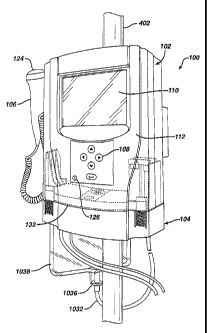
Referring now to Figures 1 and 2, one embodiment of a portable medical system
100 is illustrated having particular application for assessing urinary
function. The
system 100 includes a control device 102 that controls operation of the
system, at least
one module 104 that can be removably coupled to the control device, at least
one input
device, such as the illustrated input pendant 106 and/or keypad 108, and at
least one
output device, such as the illustrated display screen 110. As will be
described in more
detail below, the control device 102 is designed to be removably coupled to
any one of
a plurality of testing modules 104 at any given time. As each module is
uniquely suited
to support a different type of diagnostic test or medical procedure, the
resulting
diagnostic system is not only readily portable, but is also extremely
versatile in that the
single control device, in conjunction with a plurality of small test modules,
is capable
of performing an array of diagnostic tests or other procedures. The system has
particular application useful for assessing urinary function in that it
provides a portable,
modular system in contrast to the non-portable, expensive, and cumbersome
equipment
that is currently used for assessing urinary function. In addition, as will
also be
described in greater detail below, the present invention can perform tests
quicker, and
in a manner that is less uncomfortable and less invasive for a patient.
The control device 102 includes a housing 112 for housing various components,
including one or more batteries 114, an electronics assembly 116, a pump
device 118
including a motor, and various other circuitry. Batteries supply power to the
control
device 102, and are contained within a battery compartment 120 that is
accessible by
removing the battery cover 122 that forms part of the housing 112. The control
device
further includes an input keypad 108 for allowing a user to input data (such
as patient
name or other identifier, numeric identifiers, patient history, date etc.) and
an input
pendant 106 including one or more switches 124 that allow user input of
additional
information (i.e., event input based on patient feedback), and an activation
switch 126
for turning the device on and off. The pump device 118 and at least one
pressure
7
CA 02451820 2003-12-23
WO 03/001977 PCT/US02/20389
transducer 128 are also contained within the housing. The pump device is
electrically
coupled to the battery and the electronics assembly, and the pressure
transducer is
electrically coupled to the electronics assembly. The control device 102 may
also
include a pole mounting mechanism 400 for mounting the control device on a
pole such
as the pole of an IV solution caddy 402 including a hook 404. One embodiment
of a
pole mounting mechanism is illustrated in Figures 4a and 4b. The device may
also
include an interface 130 including appropriate electrical pinouts to enable
the control
device to communicate for purposes of battery recharging or printing of
patient test
data.
As indicated above, any one of a plurality of modules 104, such as diagnostic
test modules, can be removably coupled to the control device 102, and the
control
device is designed to uniquely identify the attached module, and perform
routines
specific to that module. Thus, the control device includes a module detection
mechanism 500 capable of identifying the attached module that is electrically
coupled
to the electronics assembly (see Fig. 5). This module detection mechanism
includes
one or more identification probes 502 that project from the interface side 132
of the
control device and are electrically coupled to the electronics assembly. The
modules
104 may include one or more apertures in the module housing 506 that are
designed to
receive therein the identification probes when the module is removably coupled
to the
control device. When so coupled, the identification probes will bridge one or
more
module identification elements or components 504, such as resistors,
capacitors, fuses
or other suitable electronic components, present within the module. The
identification
probes are electrically coupled to the electronics assembly 116 (described
more fully
below), which determines a value, such as resistance, associated with the
module
identification element(s) that they bridge. Each module is designed to have a
value so
that identification of this value by the electronics control assembly enables
the control
device to uniquely identify the attached module. In a preferred embodiment,
the
control device may include one or more sets of identification probes 502 at
different
locations, and different modules have a module identification components 504
at
different locations. The location, as detected by the control device,
identifies the
attached module. In yet another embodiment, the module identification
component(s)
may be coupled to an exterior side of the module housing so that apertures in
the
module housing are not required.
The module further includes at least one coupling element 600 for removably
coupling the module to the control unit (see Figure 6). In the illustrated
embodiment,
the module includes four coupling elements placed toward the ends of each of
the front
and rear faces 602, 604 of the test module. Each coupling element contains a
tab
element 606 that engages a corresponding ridge 607 (best seen in Fig. 5) on an
interior
8
CA 02451820 2003-12-23
WO 03/001977 PCT/US02/20389
surface of the control device when the module is removably coupled to the
control
device. To couple the module to the control unit, the coupling elements are
depressed
slightly in the direction indicated by the arrow in Figure 6. The module is
then aligned
with the control device as shown in Figure 1, and the coupling elements
released to
allow engagement with the corresponding ridges described above. The module can
subsequently be removed from the control unit by once again depressing the
coupling
elements and removing the module from the control device.
Finally, the module housing 506 includes first 608 and possibly second 610
ports therein as shown in Figure 6. Each of the first and second ports are
configured so
as to define a recess capable of receiving a control device pressure sensor,
such as a
pressure transducer, therein when the module is coupled to the control device.
For
example, a first control device pressure transducer 128 is received within the
first port
recess 608 and comes in physical contact with a pressure interface 1024 (see
Fig. 10) so
that pressure changes at the pressure interface can be transmitted to and
detected by
pressure transducer 128 and converted to electrical signals that are sent to
the
electronics assembly for interpretation. Similarly, the second port 610 also
defines a
recess capable of receiving therein a second control device pressure
transducer 1030.
The first and second ports are further configured to form an airtight seal
with the
control device when coupled thereto, preferably by incorporating sealing
elements such
as gaskets or the like. Individual modules and their operation in conjunction
with the
control device will be described in greater detail below.
As indicated above, contained within the housing 112 of the control device 102
is an electronics assembly 116 (see Figure 7) that is designed to control
operation of the
pump device 118, to acquire and format data from the pressure transducer(s),
to drive a
display 110 and/or other output device, and to accept and interpret input
data, such as
from switches 108, 126, and/or 124. The electronics assembly 116 consists of
an
integrated circuit board 702, hardware interfaces to the pump device 708,
pressure
transducer 706, 707, display 709 and switches 703, 704 and 705; and a
microprocessor
710. The microprocessor 710 serves as the main controller for the diagnostic
system
and is supported by the custom integrated circuit 702 and powered by the
batteries.
Also included are interface connection elements including an electronic module
identification connection 712 to the electronic detection mechanism 500, and
electronic
connections 714 that enable downloading of data to a printer or other external
device.
The microprocessor 710 is programmed with a custom program file. In the
illustrated embodiment, this software has multiple functions. First is the
acquisition of
input from the operator. This input data is captured from the input keypad
108, and/or
switches 124, 126, pressure transducer(s) or other input device, depending
upon which
test module is in use. The software also controls operation of the pump device
118.
9
CA 02451820 2003-12-23
WO 03/001977 PCT/US02/20389
Input data is interpreted and appropriate signals are sent to the pump device
motor via
the integrated circuit board 702. Yet another function is to acquire and
condition data
from the pressure transducer(s). This data is then sent in the appropriate
format to the
display 110, along with applicable pump device data in the form of volume or
time
information. Finally, as indicated above, the software receives input from the
module
detection mechanism 500 and interprets this input to determine which test
module is
coupled to the control device.
Figure 8a-8i are flow diagrams illustrating operation of the diagnostic system
software and features of the system graphical use interface for a preferred
embodiment
of the invention. When the system is powered on, the user is first presented
with a
welcome screen. While this screen is being displayed the system is undergoing
a self-
test routine 802 to test the integrity of system hardware and software
components.
Upon completion of this routine, the user is provided with information
relating to the
amount of available system memory 804. Following the pressing of any key 806
on
input device 108 by the user, the system identifies the attached module 808 as
described
above, and following such identification, the processor executes a software
subroutine
specific to the identified module. For each software subroutine, however, 'a
main menu
is displayed next, such as that indicated by reference numeral 810. In the
illustrated
embodiment, the main menu includes six possible selections. "Utilities"
enables the
user to access various system features, such as setting the date, time etc, or
adjusting the
brightness or contrast of the screen; "Quit" terminates the session;
"Patients" enables
the user to access any previously stored data relating to other patients and
tests already
performed; "Prime" initiates the pump priming process; "Patient ID" enables
the user to
enter a patient identification number; and "Test" initiates a software
subroutine specific
to the attached module to carry out the desired test procedure. In the
presently described
embodiment, the software and user interface associated with the "Prime,"
"Utilities,"
"Quit," and "Patient ID" selections are substantially the same for each
software
subroutine. The "Test" and "Patients" selections, however, are different for
each test
module. Each of these selections will be described in greater detail below.
As is illustrated in Fig. 8a, the first time the main menu is displayed both
"Test"
and "Prime" appear in a different color or shade from the other options,
indicating that
they are not currently available. This is to ensure that patient
identification information
is entered before proceeding with any priming or testing procedures. The user
may
select the "Patient ID" option by scrolling using the appropriate arrows on
the input
keypad 108. Following this selection the Patient ID screens appears 820 (Fig.
8b). In
the illustrated embodiment, the patient ID consists of a nine digit integer.
To enter the
patient ID, the user scrolls to a selected blank using the left and right
arrows and/or left
and right arrows on the input keypad 108 (824) to select desired numbers. Once
the
CA 02451820 2003-12-23
WO 03/001977 PCT/US02/20389
desired number is selected, the user presses ENTER; the selected number will
then
appear in the rightmost blank. Subsequent numbers are selected as described
above,
and will appear in the rightmost blank while previously selected numbers move
to the
left. This process is completed until all blanks are filled in. In one
embodiment, there
is a default value for each blank, such as 0, and the user may proceed with
testing by
accepting the default patient ID number consisting of all 0's. Once complete
patient
identification information is entered, the user selects the "Main Menu" option
832,
which returns to the main menu screen. At this point, however, the "Prime"
option
become available 834 (and "Patient ID" is no longer available).
Before performing any test that requires fluid to be infused into the patient,
priming operations must be performed to ensure that the fluid infusion lines
(tubing) are
filled with fluid and not air. Referring now to Fig. 8c, the user selects the
"Prime"
option 840 by using the arrow keys to select the option, and then pressing the
enter key.
The Prime screen then appears. According to one embodiment, the Prime screen
includes two options as indicated at 842: "Prime" or "Main Menu." In another
embodiment, the Prime screen is particular to each module, and may present
only one
option to initiate priming. Selecting the Prime option causes the pump to
start and run
for a predetermined amount of time, such as 20 seconds, and then automatically
shuts
off. The user is then presented with a screen 846 at which the user can accept
the prime
as complete (MAIN), or choose to reprime (PRIME). When priming is accepted as
complete, the main menu once again appears, this time with "Test" as an option
848. In
another embodiment, priming operations may be specifically tailored for
different test
modules. For example, as will be described in more detail below, the SUI test
modules
includes a hand actuator including an activation button 1118 or 1128. The
system may
be designed so that following display of the Prime screen, pump priming
operations can
be initiated by depressing the activation button.
With priming complete, testing can begin. As indicated above, testing
procedures depend on the attached test module, and accordingly, the software
and
graphical user interfaces relating to each test module will be discussed in
greater detail
below in conjunction with the detailed description of each test module.
In an alternative embodiment of the invention illustrated in Figure 9, the
control
device 102 is electrically coupled to a laptop/standard computer 900, and the
microprocessor and associated software reside in the computer.
As indicated above, the diagnostic system described herein has particular
application to urodynamics in that it enables clinicians to diagnose a
plurality of urinary
incontinence problems when used with specifically designed testing modules (to
be
discussed hereinafter). As a miniaturized urodynamic tool, the control device
102 in
conjunction with modules 104 can measure urethral resistance pressure (URP),
voiding
11
CA 02451820 2003-12-23
WO 03/001977 PCT/US02/20389
flow (Uroflometry), and bladder dysfunction (Cystometrogram (CMG)). As will be
described further below, URP is a new and unique approach to urodynamic
measurement of stress incontinence that is less invasive for a patient, and
faster than
currently known and used diagnostic tests. Uroflometry is the study of
micturation
over time. CMG is the study of bladder or detrusor instability. A major
advantage of
the diagnostic system disclosed herein is that it can achieve all of the
diagnostic tests
described above with a portable unit that can be used in any office exam room,
removing the need for the reservation or scheduling of a specialized
urodynamic room,
and the need for the complex equipment currently required for such tests. The
urodynamic system is easy to use and does not require advance training. Use of
the
disclosed system makes testing more comfortable for patients by enabling
faster set up,
shorter test time, and less invasive procedures.
In actual use, different modules can be removably coupled to the control
device
102 to conduct these different urodynamic tests. Each module performs a
different and
distinct test. These modules include, but are not limited to, a stress urinary
incontinence (SUI) module for measurement of urethral resistance pressure
(URP); a
simple CMG module for measurement of bladder instability; a complex CMG module
for measurement of bladder instability; and a uroflometry module for the study
of
micturation over time. Modules may be suitably adapted to either male or
female
incontinence diagnosis.
Before proceeding with a discussion of individual test modules, to assist the
reader a brief overview of the female urinary system will be described with
reference to
Figure 13. The female urinary system 1300 includes an elongated urethral canal
1302
having a urethral meatus (entrance) 1304 and having a substantially circular-
shaped
urethral sphincter muscle 1306 attached thereto, and a bladder cavity 1308
surrounded
by a detrusor muscle 1310. The detrusor muscle 1310 also surrounds and
supports the
urethral canal 1302. The bladder cavity 1308 is in close proximity to the
abdominal
wall 1312, the pubis bone 1314, the pelvic floor 1316 (levator ani muscle),
the vaginal
canal 1318, the clitoris 1320, the uterus 1322 and the anal sphincter muscle
1324.
Individual testing modules will now be described in detail.
STRESS URINARY INCONTINENCE MODULE
Figures 10-13 illustrate one embodiment of a stress urinary incontinence
testing
module (SUI) 1000 for diagnosing the involuntary loss of urine during physical
activities such as coughing, sneezing, laughing or lifting. The SUI testing
module 1000
includes a SUI module housing 1002 that can be removably coupled with the
control
device 102 as described above. The module housing may be in the form of a
plastic
12
CA 02451820 2003-12-23
WO 03/001977 PCT/US02/20389
disposable cartridge. Within the module housing is a tubing assembly 1004
including a
fluid inlet 1006, a fluid outlet 1008, and a first fluid conduit 1010
extending
therebetween. Tubing loop 1012 forms part of the tubing assembly and is
positioned so
that, when the SUI testing module is coupled to the control unit, the stator
1014 of the
pump device 118 in the control unit 102 cooperates physically with the tubing
loop
1012 so that the pump device operates as a peristaltic pump to pump fluid
through the
first fluid conduit 1010. To assist in this regard, a tubing guide 599 aids in
positioning
a portion of the tubing assembly so that it will properly and effectively
engage the
peristaltic pump. According to the illustrated embodiment, tubing guide 599
has a
substantially U-shaped configuration, however, many other configurations are
suitable,
as the principles of operation of peristaltic pumps are well known in the art.
Tubing
member 1050 also forms part of the first fluid conduit. The module housing
1002 also
includes a pressure chamber 1016 for dampening pressure fluctuations that may
be
caused by operation of the pump device. The pressure chamber 1016 is in fluid
communication with the first fluid conduit 1010 via valve openings 1018a-c of
three-
way valve member 1020. The pressure chamber is filled primarily with air, but
varying
amounts of fluid may also be present. Positioned at a distal end of pressure
chamber
1016 is a filter component 1022 designed to isolate fluid from electronic
elements of
the system 100. In this regard, filter 1022 may be a hydrophobic filter that
allows air to
pass into pressure interface 1024, but not liquid. When the testing module is
coupled to
the control device 102, pressure interface 1024 is in physical contact with
pressure
transducer 128 of the control device so that pressure fluctuations within the
pressure
chamber 1016 and pressure interface 1024 can be transmitted to and sensed by
the
pressure transducer, and subsequently transmitted to the electronics assembly
as
indicated above. In this manner, the control device measures pressure within
the first
fluid conduit of the tubing assembly of the SUI testing module, which
substantially
corresponds to the pressure within the urethral canal as described more fully
below.
The SUI testing module 1000 tubing assembly also includes a second tubing
member 1025 having a channel therethrough forming a second fluid conduit
between a
proximal end 1026 and a distal end 1028.
Referring now to Figs. 1la-c, the SUI testing module may also include a hand
actuator 1100 having and insert device such as a meatus plug device 1102
attached
thereto. The meatus plug device 1102 (see Figure 12) includes an attachment
member
1104 at a proximal end 1106 coupled to a plug or insert element or member 1108
at a
distal end 1110, and a channel 1112 extending therethrough allowing fluid
flowing
through the first fluid conduit to flow through the meatus plug device. The
distal end
1114 of the plug element may also include one or more transversely aligned
apertures
or openings 1116 therein approximately equally spaced apart from one another
around
13
CA 02451820 2003-12-23
WO 03/001977 PCT/US02/20389
the exterior surface of the distal end. As the outer diameter of the distal
end at the
location of the apertures is less than the diameter of the inner wall of the
urethral canal
at that location (described more fully below), one or more of the apertures
1116 can be
used for assurance of fluid flow into the urethra during actual operation.
In one embodiment, the hand actuator further includes a hand-sized housing or
casing 1102 including therein an initiator element 1118 (Figs. lla-c) that is
in fluid
communication with tubing member 1025. Preferably, initiator element is an air
bladder 1097 coupled to a distal end 1028 of the tubing member 1025. The
proximal
end 1026 of tubing member 1025 coupled to a pressure interface 1026a that is
positioned so that, when the SUI testing module is coupled to the control
device,
pressure within tubing member 1025 can be sensed by pressure transducer 1030.
As a
closed system, pressure on the activation button 1118 can be sensed at the
pressure
interface 1026a by pressure transducer 1030, and interpreted by control device
102 as a
signal to initiate and/or deactivate the test.
The hand actuator 1100 further includes a fluid conduit 1050 extending between
an outlet 1195 and an inlet 1194 that is coupled to (integrally or otherwise)
an external
tubing conduit leading to a fluid source, such as the first fluid conduit 1010
of the SUI
test module. Alternatively, the hand actuator may be designed to include
therein the
fluid source. The fluid outlet 1195 is in fluid communication with the insert
member
channel of the meatus plug device. An activation device 1127 including a
trigger 1128
extends through an opening 1118a to an exterior of the casing. The activation
device
1127 is movable between a first rest position (shown) and a second activated
position.
In the first position spring 1130 exerts force on coupling member 1132,
causing it to
pivot relative to pivot element 953 and pinch the distal ends of at least
tubing member
1050 to prevent fluid flow therethrough. When in the second position, movement
of
the trigger causes the coupling member 1132 to pivot to a point at which it no
longer
pinches tubing member 1050. Further, trigger 1128 may also compresses air
bladder
1097 to initiate testing as described above in connection with initiator
element.
The plug element 1108 is configured so that, when inserted into the urethral
meatus of a patient (see Figure 13), it will substantially block or prevent
fluid flow out
of the urethra, as well as into the urethra other than through the meatus plug
device
channel 1112. Further, when inserted, the plug element is positioned distal of
the
urethral sphincter 1306 (toward the outside of the body) as shown in Fig. 13.
In the
embodiment shown in Fig. 12, the distal end or distal portion 1114 of the plug
element
is substantially conical in shape, and decreases in diameter toward its distal
end 1114.
A proximal portion 1199 is configured to engage the inner wall of the urethral
canal to
substantially prevent fluid flow therebetween. Other shapes, however, are also
possible
so long as fluid flow into or out of the urethral is substantially blocked
(other than
14
CA 02451820 2003-12-23
WO 03/001977 PCT/US02/20389
through the meatus plug device channel) and the plug element remains located
distal of
the urethral sphincter. The meatus plug device 1102 is made of a biocompatible
material, such as stainless steel or polypropylene. The meatus plug device may
be
disposable, but may also be made of a sterilizable material so that it can be
reused.
The first fluid conduit 1010 of the tubing assembly also includes an elongated
single lumen tubing member 1032 having a first end 1006 and a second end 1034
and a
fluid channel extending therethrough. A spike device 1036 is coupled to the
first end
1006 of the single lumen tubing member for attachment to a fluid bag 1038
(having a
fluid 1010 therein) in a manner well known in the art. As described above, the
meatus
plug device and first fluid conduit are coupled to one another such that fluid
from the
fluid source traveling through the first fluid conduit may pass through the
insert
member (via the channel therein) and into the urethral canal distal of the
urethral
sphincter. Further, as the first pressure interface 1024 is in fluid
communication with
the first fluid conduit and ultimately the urethral canal, pressure at the
pressure interface
substantially corresponds to the pressure within the urethral canal distal of
the urethral
sphincter.
Use of the system 100 including a SUI testing module 1000 is as follows.
First,
the SUI testing module is removably coupled to the control device 102 in the
manner
described above. The physical coupling causes the identification probes 502 of
the
control unit to engage the module identification element(s) 504 of the Sill
testing
module, enabling the control device to identify the SUI testing module. The
physical
coupling also brings pressure interface 1024 in physical contact with pressure
transducer 128 as described above so that pressure changes at the pressure
interface can
be detected by the pressure transducer and transmitted to the electronics
assembly for
interpretation. The pressure interface 1026a at the proximal end of tubing
member
1025 similarly comes in contact with pressure transducer 1030 so that pressure
within
tubing member 1025 can also be detected. Finally, the tubing loop 1012 is
brought into
physical contact with the pump device 118 so that the pump device can drive
fluid
through the first fluid conduit by peristaltic motion, as described above.
As shown in Fig. 20, once the SUI testing module 1000 is coupled to the
control
device 102 (2010), the operator enters appropriate input data into the keypad
108 or
other input device (2015) for the SUI test (described in more detail below).
This data is
received and interpreted by the microprocessor 710 and applicable information
is sent
by the microprocessor to the display 110. Priming operations are then
performed
(2020) to ensure that the first fluid conduit 1010 contains fluid. At this
point, the
microprocessor is ready to start the test routine.
The meatus plug 1102 is inserted into the meatus of the urethra (2025) and the
test is started (2030) by pressing the activation button as described above.
This in turn
CA 02451820 2003-12-23
WO 03/001977 PCT/US02/20389
sends instructions to the pump device via the integrated circuit. The pump
device then
pumps fluid 1040 through the first fluid conduit 1010 and meatus plug device
channel
1112 and into the urethral canal distal of the urethral sphincter (2035). As
fluid
pressure builds in the urethral canal 1302, pressure in the pressure chamber
1016 also
builds. This pressure is transmitted through the filter component 1022 and
pressure
interface 1024 to the pressure transducer 128, which receives the pressure
data and
transcribes it into an electrical signal. The electrical signal from the
pressure transducer
is sent to the microprocessor 710 via the integrated circuit 702 where it is
acquired and
conditioned. The information is then sent to the display 110 via the
integrated circuit.
The microprocessor ends the test after a specified amount of time, or upon
receipt of
input from the user by sending an "off' signal to the pump motor drive. Once
the test
has been completed, the operator disengages the activation button 1118 (step
2040) and
removes the meatus plug element from the meatus 1304 (2045).
Referring once again to Figures 8a-i, and in particular Fig. 8d, when the
"Test"
option is selected the SUI test can be performed. The SUI Test screen appears
860, and
the user initiates the test by depressing the trigger 1128 or movable shell
1126 (862) to
allow fluid flow into the urethral canal as described above. The motor is then
activated
and the pump device pumps fluid into the urethral canal for a predetermined
period of
time, preferably 15 to 20 seconds. During this time a graph (see 860) is
continuously
displayed illustrating measured pressure on the vertical axis (preferably in
cm of water)
versus time on the horizontal axis. As fluid is pumped into the urethral
canal, pressure
within the urethral canal distal of the sphincter continues to increase until
that point in
time at which the urethral sphincter yields (open) under the force of the
pressure within
the urethral canal. At that point the pressure curve becomes substantially
flat, as
illustrated in Fig. 8d, since the sphincter is open and fluid is filling the
bladder. The
value of the flat portion of the curve is considered the "urethral resistance
pressure
(URP)," and can be obtained from the displayed graph. On completion of the
test (after
expiration of the predetermined time period the pump device stops), the graph
remains,
and the user is preferably provided with an option to adjust the software
generated URP
value (860a) before saving the test results. To adjust the URP value, the user
uses the
up and down arrows to manipulate a horizontal line which indicates the URP
value that
appears on the screen (870). When the ghost line is at the desired value, the
user
presses enter (872).
Once the final URP value is displayed, a Save/Delete screen 874 is overlayed
on
the screen. If the user selects the "Save" option, the test results are saved
in memory.
If the user selects "Delete" from the Save/Delete screen 874, the user is then
presented
with the Save Test screen 876. If "Delete" is chosen the test is deleted, but
if "Cancel"
is selected, the user is returned to the Save/Delete screen.
16
CA 02451820 2003-12-23
WO 03/001977 PCT/US02/20389
According to one embodiment, test results for up to three out of six possible
tests may be stored. Once three tests have been stored or six tests have been
run,
whichever comes first, the control unit 102 will disable the module
identification
component 504 via the identification probes 502. After testing is complete,
the user
may return to the main menu by selecting the "Menu" option from the Test
Complete
screen.
One option available from the Main Menu, as stated above, it "Patients," which
allows the user to access patient and test data previously stored. According
to one
embodiment illustrated in Fig. 8h, when "Patients" is selected from the Main
Menu, a
Patients Screen 891 appears. On this screen, options for each patient and test
for which
data has been stored 892 are presented and selection of one of these options
causes a
Patient Test Menu 893 to be displayed (Fig. 8i). Selecting "Delete" 896 will
present
the user with the option to delete the stored data for that patient/test, and
selecting
"Print" 895 will enable the user to print the stored data. The Print option
will only be
available (will not be greyed out) when the control device is coupled to a
cradle, or
otherwise appropriately coupled to a printer. Selecting "View Test" will cause
a
Patients Test screen 898 or 899 to appear depending on whether stored data is
a CMG
(898) or a SUI (899) data set. The Patients Test screen may vary depending on
the test
module that is attached. For example, for the SUI stored data, the Patients
Test screen
is the screen illustrated by 899, whereas for the CMG data (discussed below),
the
Patients Test screen is the screen illustrated by 898. The Patients Test
screens provide
the user with the option to view data relevant to the particular form of test
performed.
As indicated above, the results obtained from the SUl test is the urethral
resistance pressure (URP), which is the back-pressure necessary to force open
the
urethral sphincter muscle 1306 from the reverse or opposite direction from
which fluid
normally flows. A major advantage of the SUI testing module 1000 is that the
insert or
plug element 1108 of the meatus plug device 1102 only enters the external
urethral
canal (meatus) and does not cause any discomfort associated with passing a
catheter
through the internal urethral sphincter. Thus, the diagnostic system disclosed
herein
having a SUI module 1000 is less invasive and more comfortable for patients.
Further,
the testing procedure for the SUI module 1000 is easy to implement, quick to
perform,
and does not require advance training by the clinician and/or physician.
SIMPLE CYSTOMETROGRAM (CMG)
The diagnostic system disclosed herein can also be used to perform both simple
and complex cystometrograms. Figures 14-19 show both simple (SCMG) and complex
cystometry (CCMG) systems for the testing of bladder function in which
pressure and
17
CA 02451820 2003-12-23
WO 03/001977 PCT/US02/20389
volume of fluid in the bladder cavity 1308 is measured during filling, storage
and
voiding. Urologists typically measure the static pressure relationship in the
bladder of
patients, this being termed as a cystometrogram (CMG), in order to determine
the
capacitance of the bladder as a function of pressure and volume.
Referring now to Figure 14, the SCMG testing module 1400 includes a module
housing 1020b that can be removably coupled to the control device 102 in the
manner
described above. The module housing 1020b may be in the form of a plastic
disposable
cartridge. The SCMG testing module contains many elements that are similar to
those
described above in connection with the SUI testing module, and thus like
numerals will
be used for these elements. Contained within the module housing is tubing
assembly
1004b including a first fluid conduit 1402 between fluid inlet 1404 and fluid
outlet
1406. The tubing assembly also includes a second conduit 1408 between a distal
end
1410 and a proximal end 1412. Coupled to the proximal end is a filter 1022b
and
pressure interface 1412 that contacts pressure transducer 128 to convey
pressure
information thereto when the SCMG testing module is coupled to the control
device.
Compliant tubing loop 1012 similarly forms part of the first fluid conduit,
and couples
with the pump device 118 in the same manner as described above in connection
with
the SUI module. The distal ends 1406, 1410 of the first and second conduits
are each
coupled to respective proximal ends 1414, 1416 of first and second tubing
elements
1418, 1420 of a dual lumen catheter 1422 so that the first and second conduits
1402,
1408 between the proximal 1414, 1416, and distal 1460, 1462 end of the dual
lumen
catheter are in fluid communication with channels in the first and second
tubing
elements 1418, 1420 of the dual lumen catheter 1422. This attachment may be
accomplished by an adhesive bond, a solvent bond, an ultrasonic weld, or any
other
suitable type of attachment that creates a fluid tight seal. In another
embodiment, the
dual lumen catheter is an inflatable balloon catheter such as a Foley-type
catheter, that
includes a pressure sensor 1424 positioned at the tip of the catheter (see
Figure 16).
Any other suitable catheter may also be used, such as fiber optic or air
charged
catheters. The pressure sensor may be a micro tip transducer, an air charged
sensor, a
fluid charged sensor, a fiber optic sensor or any other pressure measuring
sensor.
Use of the diagnostic system to perform a SCMG will now be described in
detail with reference to Figures 15, 16 and 21. First, the SCMG testing module
is
coupled to the control device in the manner described above (2110). The
physical
connection causes the identification probes 502 of the control unit to engage
the module
identification element(s) 504 of the SCMG testing module, enabling the control
device
to identify the SCMG testing module in the manner described above. The
physical
coupling also brings the pressure interface 1024b in contact with the pressure
transducer 128 so that pressure changes in the second fluid conduit can be
detected by
18
CA 02451820 2003-12-23
WO 03/001977 PCT/US02/20389
the pressure transducer. This coupling also causes the tubing loop 1012 to
engage the
pump device so that the pump can drive fluid through the tubing loop by
peristaltic
motion, as is also described above.
Once the SCMG testing module 1400 is coupled to the control device 102, the
operator enters input data appropriate for the SCMG test (2115). This data is
received
and interpreted by the microprocessor 710 and applicable information is sent
by the
microprocessor to the display 110. Priming operations are then performed
(2120). At
this point, the microprocessor is ready to start the test routine.
The dual lumen catheter 1422 is then inserted into the bladder 1308 (2125) via
the urethra 1304 and the test is started by pressing the input pendant
switches 124
(2130). The microprocessor 710 receives the signal from the input pendant
switches.
Instructions are then sent to the pump device 118 via the integrated circuit
702. The
pump device then pumps fluid through the first fluid conduit 1402 and tubing
element
1418 into the bladder (2135). As fluid volume builds in the bladder, pressure
in the
bladder also builds. This pressure is transmitted through tubing member 1420
and the
second conduit 1408, filter component 1022b, and pressure interface 1024b. The
pressure transducer 128 receives the pressure data and transcribes it into an
electrical
signal. The electrical signal from the pressure transducer 128 is sent to the
microprocessor 710 via the integrated circuit board 702 where it is acquired
and
conditioned. During the course of a typical SCMG test, the patient provides
event
input, such as feeling the need to void and/or the intensity of that feeling,
which is input
to the control device via input pendant switches 124, as will be described
more fully
below. The microprocessor ends the test (2140) after a specified amount of
time, or
upon receipt of an "off' signal from input pendant switch 124. Once the test
has been
completed, the operator and removes the catheter 1422 from the bladder (2145).
Following the test the software then exits the SCMG test subroutine, and the
data
storage routine is run to store and/or display results of the test.
Referring again to Figures 8a-i, and in particular Figure 8e, when the
"Test" option is selected the SCMG test can be performed. The SCMG Test screen
appears 870a, and the user initiates the test by depressing input pendant
switch 124 (see
Fig. 10a) quickly. The pump device is then activated and pumping begins 872a.
In a
preferred embodiment, fluid is infused into the patient's bladder at a rate of
approximately 1 ml/sec. As such, this test may be approximately 16 minutes in
duration, as opposed to approximately 15-20 seconds that may be required for
the SUI
test.
As the bladder is filling, the patient communicates the point in time at which
he/she feels the initial sensation of needing to void, and the user presses
the input
pendant switch 124 to mark this point in time 873a. The fluid infusion
continues, and
19
CA 02451820 2003-12-23
WO 03/001977 PCT/US02/20389
the user then marks the point in time at which the patient feels the urge to
void 874a,
and the point at which the patient feels an extreme, almost unbearable urge to
void
875a, or has voided. Upon this third marking, the fluid infusion ceases and
the test is
completed 876a. During fluid infusion and after the test is complete, a graph
is
displayed illustrating pressure versus volume infused. After completion of the
test a
Save/Delete overlay 877 appears. Selecting "Save" and pressing enter saves the
test
data. Selecting "Delete" causes a Save/Delete screen 878 overlay to appear.
Selecting
"Delete" from this screen deletes the data, where as selecting "Cancel" from
this screen
returns to the Save/Delete overlay.
At any point between initiating pumping and completing the SCMG test, the
user may pause the test by depressing and holding, or pressing firmly on the
input
pendant switch 880, which causes the pump device to stop pumping fluid into
the
patient's bladder, and a Pause screen 881 (Fig. 8f) to appear on the display.
Selecting
"Quit" causes a End Test screen 885 to appear, and if "OK" is selected the
test is
stopped 886. If "Cancel" is selected the Pause screen reappears. If "Resume"
883 is
selected from the Pause screen 881, the SCMG test resumes where it left off
(pumping
begins again). If, however, "LPP" 882 is selected from the Pause screen 881,
assessment of the patient's leak point pressure (LPP) begins. No pumping of
fluid
occurs during this test. First, a LPP screen 887 appears and a blank graph is
displayed.
Pressure in centimeters of water is plotted on the vertical axis versus time
on the
horizontal axis. The patient then proceeds to exert pressure on the bladder as
if
attempting to void 888. The user marks the point at which a leak occurs 889,
and the
test is automatically completed after three minutes or three leaks, upon which
the user
is returned to the Pause screen 881. LPP results may then be stored or
deleted, the
CMG test may be resumed, or the test can be terminated altogether.
COMPLEX CYSTOMETROGRAM
In reference to Figures 17-19, the complex CMG (CCMG) testing
module 1700 is similar to the SCMG testing module, but the tubing assembly
also
includes an additional single lumen tubing member 1702 having a proximal end
1704
and a distal end 1706 and a third conduit extending therethrough. The proximal
end
1704 of the single lumen tubing member is coupled to another filter component
1022c
and pressure interface 1024c. Pressure interface 1024c contacts pressure
transducer
1030 when the CCMG testing module is coupled to the control device, enabling
pressure transducer 1030 to sense pressure within the third fluid conduit.
CA 02451820 2003-12-23
WO 03/001977 PCT/US02/20389
Use of the diagnostic system to perform a CCMG will now be described
in detail with reference to Figures 18, 19 and 22. First, the CCMG module is
coupled
to the control device (2210). The physical connection causes the
identification probes
502 of the control device 102 to engage the identification elements 504 of the
CCMG
testing module, enabling the control device to identify the CCMG testing
module. The
physical coupling also brings pressure interfaces 1024b, 1024c in contact with
the
pressure transducers 128, 1030 so that pressure changes in the second and
third
conduits can be detected by the pressure transducers. This coupling also
causes the
tubing loop 1012 to engage the pump device 118 so that the pump can drive
fluid
through the tubing in the CCMG module.
Once the CCMG testing module 1700 is coupled to the control device
102, the operator enters input data appropriate for the CCMG test (2215). This
data is
received and interpreted by the microprocessor 710 and applicable information
is sent
by the microprocessor to the display 110. Priming operations are then
performed (2220
The dual lumen catheter 1422 is inserted into the bladder via the urethra
1302' (2225). The single lumen catheter 1702 is inserted into either the
vagina or the
rectum (2230) and the test is started (2235) by pressing the input pendant
switches 124.
The microprocessor 710 receives the signal from the input pendant switches.
This in
turn sends instructions to the pump device 118 via the integrated circuit 702,
and the
pump device pumps fluid through the first tubing conduit 1042 and tubing
element
1418 into the bladder (2240). As fluid volume builds in the bladder, pressure
in the
bladder also builds. This pressure is transmitted through pressure interface
1024b to
pressure transducer 128. Similarly, abdominal pressure is transmitted through
pressure
interface 1024c to pressure transducer 1030. The pressure transducers receive
the
pressure data and transcribe it into electrical signals. The electrical
signals are sent to
the microprocessor 710 via the integrated circuit board 702 where it is
acquired and
conditioned. The microprocessor ends the test after a specified amount of time
or upon
receipt of an "off' signal from input pendant switches 124 (2245). Once the
test has
been completed, the operator disengages the input pendant switches and removes
the
catheters 1422 and 1702 from the bladder (2250). The stored information is
then
available for review on the display screen, or by a print out through a
charging cradle
(printer assembly), or downloaded to a PC via a software interface in the
charging
cradle.
Referring again to Figures 8a-i, the CCMG module software subroutine
and graphical user interface is substantially as described in connection with
the SCMG
module. The system subtracts the abdominal pressure from the bladder pressure
to
calculate detrusor (bladder muscle) pressure. Detrusor pressure is then
plotted against
volume.
21
CA 02451820 2003-12-23
WO 03/001977 PCT/US02/20389
Both the SCMG and CCMG testing modules 1400 and 1700 provide a simple,
relatively low cost procedure for recording a cystometrogram (CMG). The SCMG
and
CCMG testing modules are sterile, disposable assemblies that eliminate the
need to
disinfect equipment prior to use. This, together with a relatively simple set-
up and
operational procedure by the physician, greatly reduces the time required to
obtain the
urodynamic data. The SCMG and CCMG testing modules are more comfortable for
the female patient and are more cost effective for the physician. The
simplicity of the
SCMG and CCMG testing modules, and the control device 102 allows operation
with
minimal training. Further, when combined in operational use with the SUI
testing
module 1000, these modules provide a near complete urodynamic diagnostic tool
for
the physician.
UROFLOMETRY
A uroflometry testing module 2400 can also be removably coupled to
control device 102. The module housing of the uroflometry testing module 2400
may
be in the form of a plastic disposable cartridge. As shown in Figures 24 and
25, the
Uroflometry testing module 2400 includes a single lumen tubing member 2402
having
a proximal end 2404 and a distal end 2406 and a channel extending
substantially
therethrough. A balloon 2408 or other suitable elastomeric element is coupled
to the
distal end 2406, however, so that the channel of the single lumen tubing
member is not
open at the distal end. A pressure cushion may also be used in place of the
balloon. A
collection bucket 2410 is positioned on top of the balloon. The inner surface
of the
collection bucket may also contain a urinalysis strip which, when wetted by
the voided
urine, allows for quantitative assessment of standard urinalysis parameters
The diagnostic system including the Uroflometry testing module is
operated as follows. The collection bucket is positioned under a commode 2412
to
collect urine as the patient voids. Balloon is positioned relative to the
bucket so that it
substantially supports the bucket. As the bucket fills the pressure in the
balloon rises
proportionately to the weight of the fluid. When the testing module is coupled
to the
control device, the proximal end 2404 of the single lumen tubing member 2402
contacts the pressure transducer 128 of the control device 102 so that the
pressure
within the balloon can be captured and interpreted by the control device. The
pressure
data is used to calculate the weight and volume of the fluid (known fluid
density). The
stored information is then available for review on the display screen, or by a
printout
through a charging cradle (printer assembly), or downloaded to a PC via a
software
interface in the charging cradle. Once the test has been completed, the
operator
disengages the input pendant switches 124, and the urine and collection bucket
are
22
CA 02451820 2003-12-23
WO 03/001977 PCT/US02/20389
discarded.
Operation of the Uroflometry module software subroutine is illustrated
in Figures 8a-b. Following module detection 802 and a command to execute the
UroFlow Module Subroutine 804, the UroFlow module subroutine begins. The
operator
is prompted to Enter UroFlow Patient Data 840 necessary for the UroFlow test
routine.
Once the patient data is collected a UroFlow Scale Zeroing Procedure 841 runs.
The
operator then enters information necessary to initiate the UroFlow test
(UroFlow Test
I/O) and the test is started 842. Following the test the software then exits
the UroFlow
test subroutine and stores the data collected in the Data Storage routine.
VAGINAL SPECULUM
Figures 26-28 illustrate a vaginal speculum assembly 2600 for use in the
reduction of vaginal prolapse when performing female urodynamic testing, as
previously discussed. Uterine or vaginal prolapse occurs when the uterus or
pelvic
organs drop or become displaced because of weakened pelvic muscles. Prolapse
must
be reduced to effectively perform urodynamic tests to ensure that no
underlying stress
urinary incontinence symptoms are masked by the pressure of the vaginal
prolapse,
which may cause distortion or kinking of the urethral canal. The vaginal
speculum
assembly 2600 will permit the clinician or physician to perform a urodynamic
test
procedure with one hand while still reducing vaginal prolapse, as well as
properly
position the meatus plug device or other catheter within the urethral canal.
Thise
prolapse maneuver using the vaginal speculum assembly 2600 during urodynamic
testing is especially important prior to surgical repair of the vaginal
prolapse, as an
undiagnosed case of stress urinary incontinence may surface following prolapse
surgery. The urodynamic testing being performed using the vaginal speculum
assembly in this manner allows the surgeon to determine if additional stress
urinary
incontinence (SUI) surgery should be performed at the time of prolapse repair.
Current medical practice calls for the use of a vaginal speculum secured
in place in order to reduce the prolapse. For example, U.S. Patent Nos.
5,997,474 and
6,048,308 describe specula specifically designed for vaginal examination and
treatment.
U.S. Patent No. 6,120,438 discloses a vaginal retractor device designed to
hold back the
vaginal wall during an exam or surgical procedure. Often, surgical tape is
necessary to
hold the speculum in place, as the physician's hands cannot hold the speculum
in place
while performing a particular urodynamic procedure. None of the prior art
speculum
devices integrate the use of urodynamic equipment.
With reference to Figures 26 and 27, the vaginal speculum assembly
2600 includes a connector member 2602 for coupling an insertion device
assembly,
23
CA 02451820 2003-12-23
WO 03/001977 PCT/US02/20389
such as a meatus plug device 1102, or catheter 1422 and related elements to
the vaginal
speculum. The vaginal speculum can be of any type well known in the art. In
the
illustrated embodiment, the vaginal speculum includes an upper arm 2604, a
lower arm
2606, and a hinge member 2608 for joining the upper and lower arms together.
The
vaginal speculum also includes a handled member 2610 being integrally
attached, and
preferably substantially perpendicular aligned to the lower arm. The vaginal
speculum
2600 further includes a locking bar device 2612 connected to the upper arm
2606 for
locking the upper and lower arms in an open position, as shown in Figure 28.
The
upper arm 2604 includes a posterior end 2614 with a pair of arm mounting
openings
2616 therein. The connector member 2602 includes a flexible band 2618. The
flexible
band at one end 2620 includes a pair of mounting openings 2622 and at the
other end
2624 a connector element 2626. The mounting openings 2622 of the flexible band
2618 are aligned with the arm mounting openings 2616 of the upper arm for
receiving a
pair of mounting screws 2628 therein in order to attach the connector member
2602 to
the vaginal speculum 2600. During use, the connector element can be coupled to
the
meatus plug device or catheter as shown in Figure 26.
Although a particular embodiment of the connector member 2602 is illustrated
and described herein, those skilled in the art will recognize that various
other
embodiments are also possible to provide a means by which to removably couple
a
device that is inserted into the urethral canal to the speculum so as to hold
it in place
within the patient.
In operation, the vaginal speculum assembly 2600 can be cooperatively
used in conjunction with the urodynamic system disclosed herein. For example,
it may
be used in conjunction with a urodynamic system including a SUI testing module
1000
in the performance of the urodynamic testing procedure for stress urinary
incontinence
(SUI), such as the measuring of urethral resistance pressure (URP) as
previously
described. In reference to Figure 28, the physician positions the vaginal
speculum
assembly 2600, such that it is fully inserted within vaginal canal 2650
wherein the
upper and lower arms 2604, 2606 are fully opened and pressed against the
vaginal walls
2650w for reducing the patient's vaginal prolapse. The physical then locks the
upper
and lower arms of the vaginal speculum in the fully opened configuration (see
Figure
28) via the locking bar device 2612, and adjusts the connector member 2602 so
that the
insert member will be aligned with the urethral canal. The remaining
operational steps
are exactly the same as the operational steps described above in connection
with
individual testing modules.
Although the portable medical system disclosed herein has been described in
conjunction with diagnostic testing, it is to be understood that the system
can also be
used in conjunction with therapies and/or surgical procedures for treating
urinary
24
CA 02451820 2010-11-23
incontinence, such as placement of a sling, placement of bulking agents,
shrinkage of
tissue etc. In this regard, the testing described herein can be used before,
during and/or
after these procedures to ensure success of the procedures, for example, to
ensure
correct placement and/or tensioning of a sling.
Although exemplary embodiments and methods for use have been described in
detail above, those skilled in the art will understand that many variations
are possible
without departing from the spirit and scope of the invention, which is limited
only by
the appended claims.