Note: Descriptions are shown in the official language in which they were submitted.
CA 02452369 2004-O1-05
WO 03/004087 PCT/US02/21132
METHODS AND APPARATUS FOR SCLEROSING THE WALL OF A VARICOSE VEIN
BACKGROUND OF THE INVENTION
1. Field of the Tnvention
The invention relates to the treatment and correction of venous insufficiency
or varicose
veins. More particularly the invention relates to a minimally invasive
procedure using a catheter-
based system to sclerose the wall of the vein.
2. State of the Art
The human venous system of the lower limbs consists essentially of the
superficial
venous system and the deep venous system with perforating veins connecting the
two systems.
The superficial system includes the long or great saphenous vein and the short
saphenous vein.
The deep venous system includes the anterior and posterior tibial veins which
unite to form the
popliteal vein, which in turn becomes the femoral vein when joined by the
short saphenous vein.
The venous systems contain numerous one-way valves for directing blood flow
back to
the heart. Venous valves are usually bicuspid valves, with each cusp forming a
sack or reservoir
for blood which, under pressure, forces the free surfaces of the cusps
together to prevent
retrograde flow of the blood and allow antegrade flow to the heart. An
incompetent valve is a
valve which is unable to close because the cusps do not form a proper seal and
retrograde flow
of blood cannot be stopped.
Incompetence in the venous system can result from vein dilation. Separation of
the
cusps of the venous valve at the commissure may occur as a result. Two venous
diseases which
often involve vein dilation are varicose veins and chronic venous
insufficiency.
The varicose vein condition includes dilatation and tortuosity of the
superficial veins of
the lower limb, resulting in unsightly discoloration, pain and ulceration.
Varicose veins often
involve incompetence of one or more venous valves, which allow reflux of blood
from the deep
T
venous system to the superficial venous system or reflux within the
superficial system.
CA 02452369 2004-O1-05
WO 03/004087 PCT/US02/21132
2
Varicose veins are compatible with long life and rarely cause fatal
complications, but the
condition significantly decreases the quality of life. Patients complain
primarily of leg fatigue,
dull, aching pains, ankle swelling, and ulcerations. Occasionally, thrombosis
occurs in dilated
subcutaneous channels, resulting in local pain, induration, edema,
inflammation, and disability.
In addition to those problems, the high visibility of the unattractive rope-
like swellings and
reddish skin blotches causes considerable distress for both men and women.
Lastly, varicose
eczema, which is a local reddened swollen and itching skin condition can occur
and can spread to
distant parts of the body (called an "Id reaction").
Phlebosclerosis, the destruction of venous channels by the injection of
sclerosing agents,
has been used to treat varicose veins since 1853, when Cassaignae and Ebout
used fernc
chloride. Sodium salicylate, quinine, urea, and sodium chloride have also been
used, but the agent
more recently favored is sodium tetradecyl sulfate. In order for
phlebosclerosis to be effective, it
is necessary to evenly dispense the sclerosing agent throughout the wall of
the vein without
using toxic levels of the sclerosing agent. This is not particularly difficult
for the smaller veins.
However, it is quite difficult or nearly impossible in larger veins. When a
larger vein is injected
with a sclerosing agent, the sclerosing agent is quickly diluted by the
substantially larger volume
of blood which is not present in smaller veins. The result is that the vein is
sclerosed (injured)
only in the vicinity of the injection. If the procedure is continued, and the
injections are far apart,
the vein often assumes a configuration resembling sausage links. The problem
cannot be cured
by injecting a more potent solution of sclerosing agent, because the
sclerosing agent may
become toxic at such a concentration.
U.S. Patent Number 5,676,962 discloses an injectable microfoam containing a
sclerosing
agent. The microfoam is injected into a vein where it expands and,
theoretically, achieves the
same results as a larger quantity of sclerosing agent without the toxicity.
Such a foam is
presently manufactured under the trademark Varisolve~ by Provensis, Ltd.,
London, England.
Recent clinical trials of the foam indicate a success rate of 81%.
Until recently, the preferred procedure for treating the great saphenous vein
was surgical
stripping. This highly invasive procedure involves making a 2.5 cm incision in
the groin to
expose the saphenofemoral junction, where the great saphenous vein and its
branches are doubly
ligated en masse with a heavy ligature. The distal portion of the vein is
exposed through a 1 cm
incision anterior to the medial malleolus, and a flat metal or plastic
stripper is introduced to exit
in the proximal saphenous vein. The leg is held vertically for 30 seconds to
empty the venous
CA 02452369 2004-O1-05
WO 03/004087 PCT/US02/21132
3
tree before stripping the vein from the ankle to the groin. If the small
saphenous vein is also
incompetent, it is stripped at the same time from an incision posterior to the
lateral malleolus to
the popliteal space. After stripping the veins, the leg is held in the
vertical position for three to
four minutes to permit vessel ends to retract, constrict, and clot.
After the stripping procedure, collateral veins are removed by the avulsion-
extraction
technique which is illustrated schematically in prior art Figure 1. By working
through small (5
to 8 mm) transverse incisions, segments of vein 10 to 20 cm long can be
removed by dissecting
subcutaneously along the vein with a hemostat, and then grasping, avulsing,
and removing the
vein. With practice, long segments of vein in all quadrants can be removed
through these small
incisions. No attempt is made to ligate the branches or ends of the veins,
since stripping has
shown it to be unnecessary. Bleeding is controlled by elevation and pressure
for two to four
minutes. As many as 40 incisions are made in severe cases, but their small
size and transverse
direction permit closure with a single suture.
Before closure of the incisions, a rolled towel is rolled repeatedly from the
knee to the
ankle and from the knee to the groin to express any clots that may have
accumulated. The groin
incision is approximated with three 5-0 nylon mattress sutures and all other
incisions are closed
with a single suture.
As can be readily appreciated, the stripping and avulsion-extraction
procedures are
relatively invasive and require significant anaesthesia. It can therefore be
appreciated that it
would be desirable to provide an alternative, less invasive procedure which
would accomplish the
same results as stripping and avulsion-extraction.
Recently, a number of patents have issued disclosing the treatment of varicose
veins with
RF energy. Illustrative of these recent patents are: U.S. Patent #6,200,312
entitled "Expandable
Vein Ligator Catheter Having Multiple Electrode Leads"; U.S. Patent #6,179,832
entitled
"Expandable Catheter Having Two Sets of Electrodes"; U.S. Patent #6,165,172
entitled
"Expandable Vein Ligator Catheter and Method of Use"; U.S. Patent #6,152,899
entitled
"Expandable Catheter Having Improved Electrode Design, and Method for Applying
Energy";
U.S. Patent #6,071,277 entitled "Method and Apparatus for Reducing the Size of
a Hollow
Anatomical Structure"; U.S. Patent #6,036,687 entitled "Method and Apparatus
for Treating
Venous Insufficiency"; U.S. Patent #6,033,398 entitled "Method and Apparatus
for Treating
Venous Insufficiency Using Directionally Applied Energy"; U.S. Patent
#6,014,589 entitled
"Catheter Having Expandable Electrodes and Adjustable Stent"; U.S. Patent
#5,810,847 entitled
CA 02452369 2004-O1-05
WO 03/004087 PCT/US02/21132
4
"Method and Apparatus for Minimally Invasive Treatment of Chronic Venous
Insufficiency";
U.S. Patent #5,730,136 entitled "Venous Pump Efficiency Test System And
Method"; and
U.S. Patent #5,609,598 entitled "Method and Apparatus for Minimally Invasive
Treatment of
Chronic Venous Insufficiency". These patents generally disclose a catheter
having an electrode
tip which is switchably coupled to a source of RF energy. The catheter is
positioned within the
vein to be treated, and the electrodes on the catheter are moved toward one
side of the vein. RF
energy is applied to cause localized heating and corresponding shrinkage of
the adjacent venous
tissue. After treating one section of the vein, the catheter can be
repositioned to place the
electrodes to treat different sections of the vein.
Although this procedure has gained acceptance and is less invasive than the
stripping and
avulsion-extraction procedures, there are several disadvantages to it. In
particular, RF treatment
is actually quite slow and painful and the patient must be sufficiently
anaesthetized along the
entire length of the veins to be treated. In addition, repositioning the
catheter is time consuming
thus requiring anaesthesia for a prolonged period. Moreover, the RF treatment
is incomplete, as
only a portion of the vein wall is actually treated, i.e. the portion
contacting the electrode. The
partially treated vein may eventually re-cannularize. Furthermore, tributary
veins remain
unaffected and must be treated separately.
SUMMARY OF THE INVENTION
It is therefore an object of the invention to provide methods and apparatus
for the
minimally invasive treatment of varicose veins.
It is also an object of the invention to provide methods and apparatus for the
minimally
invasive treatment of varicose veins wherein only minimal anaesthesia is
required.
It is another object of the invention to provide methods and apparatus for the
minimally
invasive treatment of varicose veins wherein tributary veins are treated
simultaneously with the
vein to which they connect.
It is an additional object of the invention to provide methods and apparatus
for the
minimally invasive treatment of varicose veins and connecting tributaries
wherein the entire wall
of the vein is evenly sclerosed.
CA 02452369 2004-O1-05
WO 03/004087 PCT/US02/21132
Another object of the invention is to provide methods and apparatus for the
minimally
invasive treatment of varicose veins which do not utilize high concentration
sclerosing agents.
In accord with these objects which will be discussed in detail below, a first
embodiment
of the present invention includes a catheter having three concentric tubes: an
innermost tube, an
outer tube, and an intermediate tube. The innermost tube has two lumens: a
guide wire lumen
and an inflation lumen. The distal end of the innermost tube has an atraumatic
tip and an integral
inflatable occlusion balloon in fluid communication with the inflation lumen.
The intermediate
tube has a single lumen through which the innermost tube extends. The distal
end of the
intermediate tube has a self expanding balloon with a plurality of fluid pores
in fluid
communication with the lumen of the intermediate tube. The outer tube has a
single lumen
through which the intermediate tube extends. The proximal ends of the
innermost and
intermediate tubes are provided with fluid fittings. The proximal ends of the
outer tube and the
intermediate tube are provided with sealing fittings.
An exemplary treatment method using the first embodiment of the invention
includes
elevating the foot above the groin, delivering the catheter via a guide wire
into the saphenous vein
from the ankle to the groin. The patient is then placed in a Trendelenberg
position (the body
inclined downward approximately 30 degrees and the leg elevated approximately
60 degrees.
The inflatable occlusion balloon is inflated sufficiently to block blood flow,
and moving the outer
tube relative to the intermediate tube (or vice versa) such that the self
expanding balloon expands
and contacts the wall of the vein. With the inflatable occlusion balloon
securely in place, the
intermediate and outer tubes are pulled away from the inflatable occlusion
balloon while
sclerosing agent is injected into the lumen of the intermediate tube. The
sclerosing agent exits
the self expanding balloon through its pores and directly contacts the wall of
the vein. Pressure
exerted by the self expanding balloon both massages the wall of the vein and
squeegees
sclerosing agent evenly into the vein wall. Collateral tributary veins are
injected with sclerosing
agent as the self expanding balloon passes over them. The diameter of the self
expanding
balloon becomes progressively smaller as it is moved from the groin area
toward the ankle
because the diameter of the vein changes accordingly. When the entire vein has
been sclerosed,
the inflatable occlusion balloon is deflated and the catheter is removed and
the incision is sealed.
The leg is preferably wrapped with a compression bandage for a few days during
which the
veins flatten out, thereby removing blood from the vein and allowing the walls
of the vein to fuse
to itself.
CA 02452369 2004-O1-05
WO 03/004087 PCT/US02/21132
6
According to the exemplary treatment using the first embodiment of the
catheter, only
one small incision is made in the ankle and only a small amount of anesthetic
is required at the
place of the incision. The procedure is relatively painless. Tributary veins
are treated
simultaneously with the vein into which they feed. The entire wall of the vein
is evenly
sclerosed. Because blood flow is blocked and sclerosing agent is applied
directly to the wall of
the vein, a lower concentration of sclerosing agent can be used as it is not
diluted by flowing
blood. The occlusion balloon also prevents sclerosing agent from exiting the
treated vein and
entering into another vein.
According to an alternative embodiment, sclerosing agent may be injected
through the
lumen of the outer tube. In either case, the self expanding balloon causes the
sclerosing agent to
be evenly distributed and massaged into the wall of the vein.
A second embodiment of the catheter of the invention has four tubes, two of
which are
equipped with inflatable occluding balloons. The procedure for using this
embodiment involves
inflating one balloon upstream and the other downstream and moving the self
expanding balloon
between them. The two balloons can be inflated to isolate a tributary for
sclerosing.
A third embodiment has only one balloon which massages the wall of the vein as
sclerosing agent is injected downstream of the balloon.
A fourth embodiment utilizes a brush having hollow bristles to massage the
wall of the
vein as sclerosing agent is injected through the bristles.
According to the presently preferred embodiments, a drug dispenser attachment
is
provided to automatically inject the sclerosing agent as the self expanding
balloon is moved
through the vein. The drug dispenser, which may or may not be disposable,
attaches to a
disposable syringe and includes a rack and pinion gear system for engaging the
plunger of the
syringe. The gear system is driven by a spool carrying a filament, a ribbon,
or a cable. The drug
dispenser is attached to the patient's leg with straps and the end of the
cable is attached to the
intermediate tube of the catheter such that as the self expanding balloon is
moved through the
vein, the cable is pulled causing the spool to rotate and the rack and pinion
gears to engage the
plunger of the syringe and dispense the sclerosing agent. Alternatively, the
dispenser may be
attached to the catheter and the cable attached to the patient's leg.
CA 02452369 2004-O1-05
WO 03/004087 PCT/US02/21132
7
In addition to treating varicose veins, the methods and apparatus can be used
for the
delivery of other intravascular medications such as antiproliferative drugs,
for example, Paclitaxel
or Rapamycin to coronary arteries and the like, to prevent restenosis of these
vessels after
stenting. The device can also be used to deliver drugs to other hollow tubes
such as the fallopian
tubes or to persistent abnormal sinus tracts.
Additional objects and advantages of the invention will become apparent to
those skilled
in the art upon reference to the detailed description taken in conjunction
with the provided
figures.
BRIEF DESCRIPTION OF THE DRAWINGS
Figure 1 is a schematic illustration of a prior art technique for the
treatment of varicose
veins;
Figure 2 is a broken side elevation in partial section illustrating a first
embodiment of a
catheter according to the invention;
Figure 3 is a sectional view taken along line A-A of Figure 2;
Figure 4 is a view similar to Figure 2 but illustrating the inflatable
occlusion balloon
inflated inside a vein;
Figure 5 is a view similar to Figure 4 but illustrating the self expanding
balloon
expanded inside a vein;
Figure 6 is a view similar to Figure 5 but illustrating the self expanding
balloon after
having traversed a portion of the vein;
Figure 7 is a view similar to Figure 6 but illustrating a second embodiment of
the catheter
of the invention;
Figure 8 is a view similar to Figure 7 but illustrating a third embodiment of
the invention;
Figure 8a is a view similar to Figure 8 but illustrating a fourth embodiment
of the
invention;
CA 02452369 2004-O1-05
WO 03/004087 PCT/US02/21132
8
Figure 8b is an enlarged schematic distal end view of the embodiment of Figure
8a;
Figure 9 is a perspective view of a drug dispenser according to the invention;
Figure 10 is a partially cut away top view of the drug dispenser of Figure 9;
and
Figure 11 is an enlarged perspective view of a winding clutch of the drug
dispenser.
DETAILED DESCRIPTION OF THE PREFERRED EMBODIMENTS
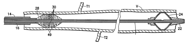
Referring now to Figures 2 and 3, a first embodiment of the present invention
includes a
catheter 10 having three concentric tubes: an innermost tube 12, an outer tube
14, and an
intermediate tube 16. The innermost tube 12 has two lumens: a guide wire lumen
18 and an
inflation lumen 20. The distal end of the innermost tube includes an
atraumatic tip 22 and an
integral inflatable occlusion balloon 24 in fluid communication with the
inflation lumen 20. The
intermediate tube I6 has a single Iumen 26 through which the innermost tube
extends. The
distal end of the intermediate tube 16 is provided with a self expanding
balloon 28 with a
plurality of fluid pores 30 in fluid communication with the lumen 26 of the
intermediate tube.
The outer tube 14 has a single lumen 32 through which the intermediate tube 16
extends.
The proximal end of the innermost tube 12 has a guide wire hub 34 which
provides
access to the guide wire lumen 18 and a fluid port 36 in fluid communication
with the inflation
lumen 20. The proximal end of the intermediate tube 16 is provided with a
fluid port 38 in fluid
communication with the lumen 26 and two fittings 40, 42. The fitting 40 allows
the innermost
tube 12 and intermediate tube 16 to be moved relative to each other while
maintaining a seal of
the annular fluid space between the innermost tube 12 and intermediate tube
16. It will be
appreciated that the proximal end of the tube 16 can be reinforced with metal
tubing such as thin-
walled hypodermic tubing to make it easier to push and provide a more uniform
sealing surface.
The fitting 42 is either press fit or glued to the proximal end of the
intermediate tube 16 or
attaches to a luer hub which is press fit or glued to the proximal end of the
intermediate tube 16.
The proximal end of the outer tube 14 has a fitting 44 that seals the space
between the outer tube
14 and the intermediate tube 16 and also releasably locks their relative
positions.
An exemplary treatment method using the first embodiment of the invention can
be
understood with reference to Figures 4-6. After anesthetizing the patient's
ankle, a guide wire
CA 02452369 2004-O1-05
WO 03/004087 PCT/US02/21132
9
(not shown) is fed into the saphenous vein V from the ankle to the groin of
the patient. The
guide wire can be fed from a cut-down to the vein at the ankle or by using
percutaneous entry
techniques well known in the art which generally involves the use of a
catheter sheath introducer,
and the like. Once the guide wire is in place, the catheter assembly 10 is
threaded over the guide
wire and follows the guide wire up the leg such that the tip 22 of the
occlusion balloon 24 is
located in a desired location, e.g., just proximal to the exit of the
saphenous vein in the vicinity of
the profunda vein. The exact location of the occlusion balloon can be
identified by ultrasound,
palpation, or by angiography, and the like. The occlusion balloon 24 is then
inflated with a gas
or fluid through the port 36. C02 gas is the preferred inflator because it is
easy to see with
ultrasound and it is safely absorbed into the blood stream should any leakage
occur. Once the
occlusion balloon 24 is inflated, blood can no longer pass to or from the
saphenous vein V via
the profunda vein (not shown).
With the inflated occlusion balloon in place, as is shown in Figures 4 and 5,
the fitting 44
of the outermost tube I4 is loosened and the tube 14 is slid backwards over
intermediate tube 16
such that the fitting 44 butts up against connector fitting 42 as shown in
Figure 5. Retracting the
outer tube 14 in this manner releases self expanding balloon 28, which self
expands until it hits
the wall of the varicose vein V.
According to the presently preferred embodiment, the self expanding balloon 28
is made
of a braided mesh, which is braided such that its preferred stable state is
fully expanded. The
wires of the mesh are of a spring material such as stainless steel, cobalt-
chrome-nickel (Elgiloy
wire), nitinol or the like. Alternatively, the self expanding balloon wires
may be made of a plastic
such as PET, PMMA, polyurethane, nylon or the like. The wires may or may not
be heat
hardened to impart greater spring-like properties and memory to the mesh.
Filling the spaces
between the braid wires is a thin membrane 49 made from an elastomeric
material such as
polyurethane, silicone rubber, polyolefin, polyamide copolymers and the like.
The membrane
can be formed by dip molding, insert molding or it can be formed separately
and glued in place.
Those skilled in the art will appreciate that the self expanding balloon both
shortens in length
and widens in diameter as it achieves its preferred expanded state. It will be
appreciated that the
distal end of the balloon 28 is dimensioned such that it can move over the
inner tube 12 but still
maintain a fluid seal between the balloon 28 and the tube 12. As illustrated
in Figures 4-6, a
plurality of fluid pores 30 in the membrane 49 are located about the
circumference of the balloon
28.
CA 02452369 2004-O1-05
WO 03/004087 PCT/US02/21132
When the catheter 10 is in the position shown in Figure 5, a source of
sclerosing agent
(or a microfoam containing a sclerosing agent) is coupled to the port 38 and
operated to allow
sclerosing agent to flow through the annular space between the tubes 12 and
16, into the balloon
28 and out through the pores 30. It will be appreciated that sclerosing agent
so dispensed with
flow directly into the wall of the vein V. According to the invention, as
sclerosing agent is being
dispensed, the self expanding balloon 28 is moved away from the balloon 24 as
shown in Figure
6 by moving the tubes 14 and 16 relative to the tube 12. It will be
appreciated that as the balloon
28 is moved along the length of the tapering vein V, the balloon decreases in
diameter and
lengthens. The sclerosing agent exiting the pores 30 is massaged or
"squeegeed" into the wall
of the vein V by means of the outward pressure exerted by the balloon 28
against the wall of the
vessel V.
Figure 6 shows the occlusion balloon 24 remaining in place as the self
expanding
balloon 28 has traversed along a length of the vessel V. Sclerosing agent is
continually injected
through the balloon 28 as it is withdrawn. It can also be appreciated that as
the balloon 28
passes collateral (tributary) veins such as those identified as T1 and T2,
sclerosing agent fills the
collateral veins to effectively cause them to sclerose. It will be appreciated
that additional
sclerosing agent can be injected into the tributary veins by means of a second
syringe fluidly
coupled to the inlet port 38 by means of a T-connector
According to the presently preferred embodiment, the balloon 28 is withdrawn
through
the entire length of the vein V to be sclerosed. When the entire length is
traversed, the occlusion
balloon 24 is deflated, the catheter 10 is removed and the puncture site is
sealed. Simultaneous
with the removal of the occlusion balloon, or just prior to deflation of the
balloon, or even during
the procedure, the leg of the patient is preferably wrapped with an elastic
compression bandage,
e.g, an ACE BANDAGE, with other compression objects such as foam, etc..
Wrapping the leg
in this manner causes the vein to flatten-out, thereby removing blood from the
vein and allowing
the lumen to fuse to itself in the collapsed embodiment. After a few days of
compression the
bandages are removed and the vein is no longer medically or cosmetically
problematic.
It has been discovered that, due to the "squeegee-like" action of the self
expanding
balloon 28, the sclerosing agent need not be dispensed at the outer
circumference of the balloon.
Thus, the pores 30 of the balloon may be located on the proximal portion of
the balloon.
Alternatively, the balloon need not have any pores, but pores may be provided
at the distal end of
the tube 16. As yet another alternative, neither the balloon nor the
intermediate tube are provided
with pores, but the sclerosing agent is provided via the outer tube 14. In all
cases, the sclerosing
CA 02452369 2004-O1-05
WO 03/004087 PCT/US02/21132
11
agent will flow or be forced toward the wall of the vein and be massaged into
the vein wall by the
balloon 28.
Those skilled in the art will appreciate that if the sclerosing agent is not
injected through
the balloon 28, the balloon need not be self expanding. It could be an
inflatable balloon which is
inflated with a gas or a saline solution from an IV bag, etc. In this
embodiment, the pressure can
be adjusted as the balloon traverses the vein and the diameter of the vein
changes.
As mentioned above, the catheter of the invention can be used to deliver other
types of
intravascular medication directly to the wall of a vein. Although the
treatment of varicose veins
generally involves treating the entire length of the vein, other treatments
may require or prefer
that only a selected portion of the blood vessel be treated. Accordingly, a
second embodiment of
the invention is illustrated in Figure 7 in which two inflatable occlusion
balloons are provided to
isolate a region of a blood vessel for treatment.
As shown in Figure 7, the catheter 110 has four tubes: an innermost tube 112,
an outer
tube 114, and two intermediate tubes 116 and 117. The innermost tube 112 has
an inflatable
balloon 124 with an atraumatic tip 122 at its distal end. The innermost tube
112 is substantially
the same as the tube 12 described above. The outer tube 114 and intermediate
tube 116 are also
substantially the same as the tubes 14 and 16 described above. The additional
tube 117 resides
in the annular space between the tubes 114 and 116. It has an inflatable
balloon 125 at its distal
end and is similar to the tube 112 in that it has two lumen, one of which
carries the tube 116 and
the other of which is used to inflate the balloon 125. The procedure for using
this embodiment
involves inflating one balloon upstream and the other downstream and moving
the self
expanding balloon between them while injecting an intravascular drug. This
allows treatment of
a selected portion of a blood vessel without diluting the treatment drug. It
will also be
appreciated that the catheter 110 can also be used to isolate one or a
plurality of tributary veins
by inflating the balloons on opposite sides of the tributary or tributaries.
Sclerosing agent is
then injected between the balloons and forced into the tributary or
tributaries,
A third embodiment of a catheter 210 according to the invention is shown in
Figure 8.
This embodiment includes an outer tube 214 and an inner tube 216. The distal
end of the inner
tube 216 is provided with a self expanding balloon 228. According to this
embodiment, the
balloon 228 is optionally provided with an abrasive surface 231 and/or pores
(not shown).
When an abrasive surface is provided, the catheter 210 may be used with or
without a sclerosing
agent. In these instances, treatment may consist of abrading the wall of the
blood vessel with the
CA 02452369 2004-O1-05
WO 03/004087 PCT/US02/21132
12
abrasive surface 231 of the balloon 228. In other instances, where pores are
provided in the
balloon, treatment may consist of abrading the wall of the vain (if the
balloon has an abrasive
surface) as the sclerosing agent is injected out of the pores. In yet other
instances, when no
pores are provided in the balloon, treatment may include injecting an
intravascular drug through
the annular space between the outer tube 214 and the inner tube 216 or through
pores (not
shown) at the distal end of the inner tube 216. If the balloon 228 is moved
against the flow of
the drug (from right to left as seen in Figure 8), the drug will be massaged
into the wall of the
blood vessel (which is abraded if the balloon has an abrasive surface) by the
balloon 228.
A fourth embodiment of a catheter 410 according to the invention is shown in
Figures 8a
and 8b. This embodiment includes an outer tube 414 and an inner tube 416. The
distal end of
the inner tube 416 is provided with a brush 430 having a plurality of hollow
bristles, e.g. 430a-
430p and a plug 418. The hollow bristles are in fluid communication with the
inner tube 416
such that a sclerosing agent may be injected through the tube 416 and exit the
ends of the
bristles. The bristles 430a-430p are preferably made of a resilient material
so that they will
expand to the position shown in the Figures, i.e., approximately radial to the
inner tube, when
released from the outer tube. A method for using the fourth embodiment
includes moving the
outer tube and/or inner tube until the bristles 430a-430p are collapsed inside
the outer tube,
delivering the two tubes to a procedural site in a blood vessel, moving the
outer tube and/or inner
tube until the bristles 430a-430p are expanded as shown in the figures, then
moving the inner
tube relative to the blood vessel while injecting a sclerosing agent through
the inner tube and the
bristles.
According to the presently preferred embodiments, a drug dispenser attachment
is
provided to automatically inject the sclerosing agent as the self expanding
balloon is moved
through the vein. Figure 9 shows a three dimensional view of a drug dispenser
300 with a body
302 defining a channel for receiving a disposable syringe 304, an injector cam
306, a string 308
with a hook 309, a winding clutch 310 (shown in further detail in Figure 11)
and slots 312.
Figure 10 is a partially cut away top view of the dispenser 300. As shown in
Figure 10,
the string 308 with hook 309 is wound on spool 314. The spool 314 is rigidly
attached to the
winding clutch 310 and clutchingly attached to an axle 316. The axle 316 is
rigidly attached to a
spur gear 318. The spur gear 318 engages a second spur gear 320, which in turn
is rigidly
attached to an axle 322 which carries a pinion gear 324. The pinion gear 324
engages a rack
326, which is attached to the injector cam 306. As seen in Figure 10, the
injector cam 306 is
aligned with the plunger 305 of the disposable syringe 304.
CA 02452369 2004-O1-05
WO 03/004087 PCT/US02/21132
13
As seen in Figure 11, the winding clutch 310 includes a clutch housing 328 and
a cam
330. The clutch housing 328 defines a center hole 332 through which the axle
(316 in Figure
10) extends. The cam 330 defines a hole 334 with an adjacent keyway 336. The
cam 330 is
spring biased by a spring 338 so that the keyway 336 aligns with the hole 332
and engages flats
(not shown) which are machined on the axle (316 in Figure 10). When the cam
330 is pressed
at 340, the hole 334 lines up with the hole 332 and the flats on the axle are
no longer engaged
causing clutch 310 to rotate without engaging the axle and without dispensing
sclerosing agent
from syringe 304. This feature allows the string 305 to be wound onto the
spool 314. It also
provides the surgeon with a mechanism to lengthen or shorten the string
without dispensing
sclerosing agent from the syringe.
The drug dispenser 300 works as follows. The body 302, with the exit of the
syringe
304 pointed towards the patient's foot, is fastened to the patient's leg,
preferably adjacent the
ankle, via straps such as VELCRO straps which are fed through the slots 312.
The sclerosing
catheter (e.g. 10 in Figure 2) is maneuvered into the patient's vein from a
puncture site in the
patient's ankle. The proximal hub 42 (Fig. 2) of the sclerosing catheter is
attached to the hook
309 at the end of the string 308. As the sclerosing catheter is pulled out of
the vein, tension on
string 308 causes pulley 314 to rotate which causes the pinion 324 to rotate
(via gears 318 and
320) and rack 326 to move. The cam 306 on the rack 326 presses the plunger 305
causing
sclerosing agent to be discharged from syringe 304. Those skilled in the art
will appreciate that
a fluid conduit may be required to couple the fluid exit of the syringe 304 to
the fluid port 38
(Figures 2, 4, and 5). It will also be appreciated that the gear ratios of the
drug dispenser can be
selected such that the appropriate volume of sclerosing agent dispenses as the
catheter is pulled
out of the leg. The main purpose of the injector is to allow the physician to
dispense a
continuous amount of sclerosing agent into the patient as a function of the
withdrawal of the
catheter. It can be appreciated that no sclerosing agent is discharged if the
catheter is not pulled
and that the flow rate of sclerosing agent tracks the speed at which the
catheter is withdrawn. As
mentioned above, the drug dispenser can be used in conjunction with one of the
catheters to
dispense other kinds of infiravascular drugs for different procedures.
Those skilled in the art will appreciate that the drug dispenser may be
attached to the
catheter and the string attached to the patient's leg. Moreover, it will be
appreciated that the
clutch may be omitted if an adjustable length cable is used. It will also be
appreciated that other
types of clutches could be used at any of the gears or axles.
CA 02452369 2004-O1-05
WO 03/004087 PCT/US02/21132
14
There have been described and illustrated herein several embodiments of
methods and
apparatus for sclerosing the wall of a varicose vein. While particular
embodiments of the
invention have been described, it is not intended that the invention be
limited thereto, as it is
intended that the invention be as broad in scope as the art will allow and
that the specification be
read likewise. For example, the catheter can be provided with an integral
guide wire and the
catheter and the guide wire can be inserted simultaneously. Also, the dual
lumen catheter can be
formed by two concentric tubes with the inflation lumen being the annular
space between the
tubes. It will therefore be appreciated by those skilled in the art that yet
other modifications
could be made to the provided invention without deviating from its spirit and
scope as so
claimed.