Note: Descriptions are shown in the official language in which they were submitted.
CA 02452836 2010-11-05
-1-
MENINGOCOCCUS ADHESINS NADA, APP AND ORF 40
TECHNICAL FIELD
This invention is in the field of biochemistry and, in particular, the
biochemistry of the pathogenic
bacteria in the genus Neisseria (e.g. N.meningitidis and N.gonorrhoe'ae).
BACKGROUND ART
International patent applications W099/24578, W099/36544, W099/57280 and.
W000/22430
disclose proteins from Neisseria meningitidis and Neisseria gonorrhoeae. The
complete genome
sequence of serogroup B Mnieningitidis has been published [Tettelin et al.
(2000) Science 287:1809-
1815] and has been subjected to analysis in order to identify vaccine antigens
[Pizza et al. (2000)
Science 287:1816-18201. Approaches to expression of the proteins are disclosed
in W001/64922.
The complete genome sequence of serogroup A Mmeningitidis is also known
[Parkhill et al. (2000)
Nature 404:502-506].
Sequence data alone, however, does not reveal everything about this pathogen.
Objects of the present.
invention include: (a) to provide ways of intervening in Neisseria
biochemistry; (b) to provide new
uses for known Neisseria proteins; (c) to provide. alternative and improved
forms of known Neisseria
proteins, such as enzymatically inactive forms of known proteins or
proteolytic products of known
proteins; and (d) to provide materials useful for studying and modulating
Neisserial adhesion.
DISCLOSURE OF THE INVENTION
Nomenclature used herein
`ORF40' is.disclosed in example 1 of W099/36544. Sequences from serogroups A
and B of
N.meningitidis are disclosed (SEQ IDs 1 to 6 therein). Other forms of the
protein are disclosed in
-W099131132 and W099158683, and can also be found in GenBank (see gi accession
numbers:
11352902, 7228562, 14578015, 12958107, 7228586, 7228572, 7228594, 7228588,
14578013,
7228568, 7228546, 7228548, 7228592, 14578009, 7228558, 7228600, 7228596,
7228542, 7228574,
7228552, 7228554, 14578023, 14578021, 11354080, 7228584 & 7228590).
'App' (adhesion and penetration protein) is disclosed as 'ORF1' in example 77
of W099/24578.
Sequences from serogroups A and B of Mmeningitidis and from N.gonorrhoeae are
disclosed (SEQ
IDs 647 to 654 therein). Other forms of the protein are disclosed in
W099/55873, aad can also be
30' found in GenBank (see gi accession numbers: 11280386, 7227246, 11071865,
6977941, 11071863,
11280387,7379205).
'NadA' (Neisserial adhesin A) from serogroup B of N.meningitidis is disclosed
as protein `961' in
W099/57280 (SEQ IDs 2943 & 2944) and as 'NM 1994' by Tettelin et al. (see also
GenBafik
accession numbers: 11352904 & 7227256) and in Figure 9 herein.
These proteins are preferably expressed other than as a fusion protein (e.g.
without GST, MBP,
his-tag or similar).
CA 02452836 2003-12-31
WO 03/010194 PCT/IB02/03396
-2-
Preferred proteins for use according to the invention are those of serogroup B
N.meningitidis strain
MC58, strain 2996 or strain 394/98 (a New Zealand strain). It will be
appreciated, however, that the
invention is not in general limited by strain - references to a particular
protein (e.g. `ORF40', `App'
etc.) may be taken to include that protein from any strain. In general,
therefore, reference to any
particular protein includes proteins which share sequence identity with one of
the sequences
disclosed above. The degree of `sequence identity' is preferably greater than
50% (eg. 60%, 70%,
80%, 90%, 95%, 99% or more). This includes mutants and allelic variants. In
the context of the
present invention, sequence identity is preferably determined by the Smith-
Waterman homology
search algorithm as implemented in the MPSRCH program (Oxford Molecular),
using an affine gap
search with parameters gap open penalty=12 and gap extension penalty=1.
Typically, 50% identity
or more between two proteins is considered to be an indication of functional
equivalence.
The naming conventions used in W099/24578, W099/36544 and W099/57280 are also
used herein
(e.g. `ORF4', `ORF40', `ORF40-1' etc. as used in W099/24578 and W099/36544;
'm919', 'g919'
and 'a919' etc. as used in W099/57280).
Secreted App
It has been found that, when expressed in E.coli without a GST or his-tag
fusion partner, App is
exported to the outer membrane as a precursor of about 160kDa, where it is
processed and secreted
into the culture.
The invention therefore provides a method for purifying processed App protein,
comprising the steps
of: expressing a gene encoding App protein in a non-Neisserial host cell; and
purifying processed
App protein from the culture medium.
The invention also provides purified protein obtainable by this process.
The App protein preferably includes its wild-type 42 residue signal peptide at
the N-terminus i.e. no
N-terminus fusion partner is used. It is also preferred not to include a C-
terminus fusion partner.
To purify the protein from the culture medium, the culture can be centrifuged
and the protein can be
recovered from the supernatant.
The non-Neisserial host cell is preferably a bacterium and is most preferably
E.coli.
Bacterial expression techniques are known in the art. A bacterial promoter is
any DNA sequence
capable of binding bacterial RNA polymerase and initiating the downstream (3')
transcription of a
coding sequence (eg. structural gene) into mRNA.. A promoter will have a
transcription initiation
region which is usually placed proximal to the 5' end of the coding sequence.
This transcription
initiation region usually includes an RNA polymerase binding site and a
transcription initiation site.
A bacterial promoter may also have a second domain called an operator, that
may overlap an adjacent
RNA polymerase binding site at which RNA synthesis begins. The operator
permits negative
regulated (inducible) transcription, as a gene repressor protein may bind the
operator and thereby
inhibit transcription of a specific gene. Constitutive expression may occur in
the absence of negative
regulatory elements, such as the operator. In addition, positive regulation
may be achieved by a gene
CA 02452836 2003-12-31
WO 03/010194 PCT/IB02/03396
-3-
activator protein binding sequence, which, if present is usually proximal (5)
to the RNA polymerise
binding sequence. An example of a gene activator protein is the catabolite
activator protein (CAP),
which helps initiate transcription of the lac operon in Escherichia coli (E.
coli) [Raibaud et al. (1984)
Annu. Rev. Genet. 18:173]. Regulated expression may therefore be either
positive or negative,
thereby either enhancing or reducing transcription.
Sequences encoding metabolic pathway enzymes provide particularly useful
promoter sequences.
Examples include promoter sequences derived from sugar metabolizing enzymes,
such as galactose,
lactose (lac) [Chang et al. (1977) Nature 198:1056], and maltose. Additional
examples include
promoter sequences derived from biosynthetic enzymes such as tryptophan (trp)
[Goeddel et al.
(1980) Nuc. Acids Res. 8:4057; Yelverton et al. (1981) Nucl. Acids Res. 9:731;
US patent 4,738,921;
EP-A-0036776 and EP-A-0121775]. The g-laotamase (bla) promoter system
[Weissmann (1981)
"The cloning of interferon and other mistakes." In Interferon 3 (ed. I.
Gresser)], bacteriophage
lambda PL [Shimatake et al. (1981) Nature 292:128] and T5 [US patent
4,689,406] promoter
systems also provide useful promoter sequences.
In addition, synthetic promoters which do not occur in nature also function as
bacterial promoters.
For example, transcription activation sequences of one bacterial or
bacteriophage promoter may be
joined with the operon sequences of another bacterial or bacteriophage
promoter, creating a synthetic
hybrid promoter [US patent 4,551,433]. For example, the tac promoter is a
hybrid trp-lac promoter
comprised of both trp promoter and lac operon sequences that is regulated by
the lac repressor
[Amann et al. (1983) Gene 25:167; de Boer et al. (1983) Proc. Natl. Acad. Sci.
80:21]. Furthermore,
a bacterial promoter can include naturally occurring promoters of non-
bacterial origin that have the
ability to bind bacterial RNA polymerase and initiate transcription. A
naturally occurring promoter of
non-bacterial origin can also be coupled with a compatible RNA polymerase to
produce high levels
of expression of some genes in prokaryotes. The bacteriophage T7 RNA
polymerase/promoter
system is an example of a coupled promoter system [Studier et al. (1986) J.
Mol. Biol. 189:113;
Tabor et al. (1985) Proc Natl. Acad. Sci. 82:1074]. In addition, a hybrid
promoter can also be
comprised of a bacteriophage promoter and an E. coli operator region (EPO-A-0
267 851).
In addition to a functioning promoter sequence, an efficient ribosome binding
site is also useful for
the expression of foreign genes in prokaryotes. In E. coli, the ribosome
binding site is called the
Shine-Dalgarno (SD) sequence and includes an initiation codon (ATG) and a
sequence 3-9
nucleotides in length located 3-11 nucleotides upstream of the initiation
codon [Shine et al. (1975)
Nature 254:34]. The SD sequence is thought to promote binding of mRNA to the
ribosome by the
pairing of bases between the SD sequence and the 3' and of E. coli 16S rRNA
[Steitz et al. (1979)
"Genetic signals and nucleotide sequences in messenger RNA." In Biological
Regulation and
Development: Gene Expression (ed. R.F. Goldberger)]. To express eukaryotic
genes and prokaryotic
genes with weak ribosome-binding site [Sambrook et al. (1989) "Expression of
cloned genes in
Escherichia coli." In Molecular Cloning: A Laboratory Manual].
A promoter sequence may be directly linked with the DNA molecule, in which
case the first amino
acid at the N-terminus will always be a methionine, which is encoded by the
ATG start codon. If
CA 02452836 2003-12-31
WO 03/010194 PCT/IB02/03396
-4-
desired, methionine at the N-terminus may be cleaved from the protein by in
vitro incubation with
cyanogen bromide or by either in vivo on in vitro incubation with a bacterial
methionine N-terminal
peptidase (EP-A-0219237).
Usually, transcription termination sequences recognized by bacteria are
regulatory regions located 3'
to the translation stop codon, and thus together with the promoter flank the
coding sequence. These
sequences direct the transcription of an mRNA which can be translated into the
polypeptide encoded
by the DNA. Transcription termination sequences frequently include DNA
sequences of about 50
nucleotides capable of forming stem loop structures that aid in terminating
transcription. Examples
include transcription termination sequences derived from genes with strong
promoters, such as the
trp gene in E. coli as well as other biosynthetic genes.
Usually, the above described components, comprising a promoter, signal
sequence (if desired),
coding sequence of interest, and transcription termination sequence, are put
together into expression
constructs. Expression constructs are often maintained in a replicon, such as
an extrachromosomal
element (eg. plasmids) capable of stable maintenance in a host, such as
bacteria. The replicon will
have a replication system, thus allowing it to be maintained in a prokaryotic
host either for
expression or for cloning and amplification. In addition, a replicon may be
either a high or low copy
number plasmid. A high copy number plasmid will generally have a copy number
ranging from about
5 to about 200, and usually about 10 to about 150. A host containing a high
copy number plasmid
will preferably contain at least about 10, and more preferably at least about
20 plasmids. Either a
high or low copy number vector may be selected, depending upon the effect of
the vector and the
foreign protein on the host.
Alternatively, the expression constructs can be integrated into the bacterial
genome with an
integrating vector. Integrating vectors usually contain at least one sequence
homologous to the
bacterial chromosome that allows the vector to integrate. Integrations appear
= to result from
recombinations between homologous DNA in the vector and the bacterial
chromosome. For example,
integrating vectors constructed with DNA from various Bacillus strains
integrate into the Bacillus
chromosome (EP-A-0127328). Integrating vectors may also be comprised of
bacteriophage or
transposon sequences.
Usually, extrachromosomal and integrating expression constructs may contain
selectable markers to
allow for the selection of bacterial strains that have been transformed.
Selectable markers can be
expressed in the bacterial host and may include genes which render bacteria
resistant to drugs such as
ampicillin, chloramphenicol, erythromycin, kanamycin (neomycin), and
tetracycline [Davies et al.
(1978) Annu. Rev. Microbiol. 32:469]. Selectable markers may also include
biosynthetic genes, such
as those in the histidine, tryptophan, and leucine biosynthetic pathways.
Alternatively, some of the above described components can be put together in
transformation vectors.
Transformation vectors are usually comprised of a selectable market that is
either maintained in a
replicon or developed into an integrating vector, as described above.
CA 02452836 2003-12-31
WO 03/010194 PCT/IB02/03396
-5-
Expression and transformation vectors, either extra-chromosomal replicons or
integrating vectors,
have been developed for transformation into many bacteria. For example,
expression vectors have
been developed for, inter alia, the following bacteria: Bacillus subtilis
[Palva et al. (1982) Proc. Natl.
Acad. Sci. USA 79:5582; EP-A-0 036 259 and EP-A-0 063 953; WO 84/04541],
Escherichia coli
[Shimatake et al. (1981) Nature 292:128; Amann et al. (1985) Gene 40:183;
Studier et al. (1986) J.
Mol. Biol. 189:113; EP-A-0 036 776,EP-A-0 136 829 and EP-A-0 136 907],
Streptococcus cremoris
[Powell et al. (1988) Appl. Environ. Microbiol. 54:655]; Streptococcus
lividans [Powell et al. (1988)
Appl. Environ. Microbiol. 54:655], Streptomyces lividans [US patent
4,745,056].
Methods of introducing exogenous DNA into bacterial hosts are well-known in
the art, and usually
include either the transformation of bacteria treated with CaC12 or other
agents, such as divalent
cations and DMSO. DNA can also be introduced into bacterial cells by
electroporation.
Transformation procedures usually vary with the bacterial species to be
transformed. See eg.
[Masson et al. (1989) FEMS Microbiol. Lett. 60:273; Palva et al. (1982) Proc.
Natl. Acad. Sci. USA
79:5582; EP-A-0 036 259 and EP-A-0 063 953; WO 84/04541, Bacillus], [Miller et
at. (1988) Proc.
Natl. Acad. Sci. 85:856; Wang et al. (1990) J. Bacteriol. 172:949,
Campylobacter], [Cohen et at.
(1973) Proc. Natl. Acad. Sci. 69:2110; Dower et al. (1988) Nucleic Acids Res.
16:6127; Kushner
(1978) "An improved method for transformation of Escherichia coli with ColE1-
derived plasmids. In
Genetic Engineering: Proceedings of the International Symposium on Genetic
Engineering (eds.
H.W. Boyer and S. Nicosia); Mandel et al. (1970) J. Mol. Biol. 53:159; Taketo
(1988) Biochim.
Biophys. Acta 949:318; Escherichia], [Chassy et at. (1987) FEMS Microbiol.
Lett. 44:173
Lactobacillus]; [Fiedler et al. (1988) Anal. Biochem 170:38, Pseudomonas];
[Augustin et al. (1990)
FEMS Microbiol. Lett. 66:203, Staphylococcus], [Barany et at. (1980) J.
Bacteriol. 144:698;
Harlander (1987) "Transformation of Streptococcus lactis by electroporation,
in: Streptococcal
Genetics (ed. J. Ferretti and R. Curtiss III); Perry et al. (1981) Infect.
Ifnmun. 32:1295; Powell et at.
(1988) Appl. Environ. Microbiol. 54:655; Somkuti et at. (1987) Proc. 4th Evr.
Cong. Biotechnology
1:412, Streptococcus].
Adherence proteins
Example 22 of international patent application W001/64922 discloses that
E.coli which expresses
protein NadA can adhere to human epithelial cells. This adherence activity has
been further studied
and it has also been found for App and ORF40.
The invention provides methods for preventing the attachment of Neisserial
cells to epithelial cells.
References to a "Neisserial cell" in this section include any species of the
bacterial genus Neisseria,
including N.gonorrhoeae and N.lactamica. Preferably, however, the species is
N.nzeningitidis. The
N.meningitidis may be from any serogroup, including serogroups A, C, W135 and
Y. Most
preferably, however, it is N.meningitidis serogroup B.
References to an "epithelial cell" in this section include any cell found in
or derived from the
epithelium of a mammal. The cell may be in vitro (e.g. in cell culture) or in
vivo. Preferred epithelial
cells are from the nasopharynx. The cells are most preferably human cells.
CA 02452836 2003-12-31
WO 03/010194 PCT/IB02/03396
-6-
Blocking the Neisseria-epithelium interaction
The invention provides a method for preventing the attachment of a Neisserial
cell to an epithelial
cell, wherein the ability of one or more App, ORF40 and/or NadA to bind to the
epithelial cell is
blocked.
The ability to bind may be blocked in various ways but, most conveniently, an
antibody specific for
App, ORF40 and/or NadA is used. The invention also provides antibody which is
specific for App,
ORF40 or NadA. This antibody preferably has an affinity for App, ORF40 and/or
NadA of at least
10"7 M e.g. 10-$ M, 10M, 10"' M or tighter.
Antibodies for use in accordance with the invention may be polyclonal, but are
preferably
monoclonal. It will be appreciated that the term "antibody" includes whole
antibodies (e.g. IgG, IgA
etc), derivatives of whole antibodies which retain the antigen-binding sites
(e.g. Fab, Fab', F(ab')2 etc.),
single chain antibodies (e.g. sFv), chimeric antibodies, CDR-grafted
antibodies, humanised
antibodies, univalent antibodies, human monoclonal antibodies [e.g. Green
(1999) J Imrnunol
Methods 231:11-23; Kipriyanov & Little (1999) Mol Biotechnol 12:173-201 etc.]
and the like.
Humanised antibodies may be preferable to those which are fully human [e.g.
Fletcher (2001) Nature
Biotechnology 19:395-96].
As an alternative to using antibodies, antagonists of the interaction between
App, ORF40 or NadA
and its receptor on the epithelial cell may be used. As a further alternative,
a soluble form of the
epithelial cell receptor may be used as a decoy. These can be produced by
removing the receptor's
transmembrane and, optionally, cytoplasmic regions [e.g. EP-B2-0139417, EP-A-
0609580 etc.].
The antibodies, antagonists and soluble receptors of the invention may be used
as medicaments to
prevent the attachment of a Neisserial cell to an epithelial cell.
Inhibiting expression of the Neisserial gene
The invention provides a method for preventing the attachment of a Neisserial
cell to an epithelial
cell, wherein protein expression from one or more of App, ORF40 and/or NadA is
inhibited. The
inhibition may be at the level of transcription and/or translation.
A preferred technique for inhibiting expression of the gene is antisense [e.g.
Piddock (1998) Curr
Opin Microbiol 1:502-8; Nielsen (2001) Expert Opin Investig Drugs 10:331-41;
Good & Nielsen
(1998) Nature Biotechnol 16:355-358; Rahman et al. (1991) Antiserzse Res Dev
1:319-327; Methods
in Enzymology volumes 313 & 314; Manual of Antisense Methodology (eds.
Hartmann & Endres);
Antisense Therapeutics (ed. Agrawal) etc.]. Antibacterial antisense techniques
are disclosed in, for
example, international patent applications W099/02673 and W099/13893.
The invention also provides nucleic acid comprising a fragment of x or more
nucleotides from
nucleic acid which encodes App, ORF40 or NadA, wherein x is at least 8 (e.g.
8, 10, 12, 14, 16, 18,
20, 25, 30 or more). The nucleic acid will typically be single-stranded.
The nucleic acid is preferably of the formula 5'-(N)om (X)-(N)b-3', wherein
0>a>15, 0>b>15, N is
any nucleotide, and X is a fragment of a nucleic acid which encodes App, ORF40
or NadA. X
preferably comprises at least 8 nucleotides (e.g. 8, 10, 12, 14, 16, 18, 20,
25, 30 or more). The values
CA 02452836 2003-12-31
WO 03/010194 PCT/IB02/03396
-7-
of a and b may independently be 0, 1, 2, 3, 4, 5, 6, 7, 8, 9, 10, 11, 12, 13,
14 or 15. Each individual
nucleotide N in the -(N)a- and -(N)b-- portions of the nucleic acid may be the
same or different. The
length of the nucleic acid (i.e. a+b+length of X) is preferably less than 100
(e.g. less than 90, 80, 70,
60, 50, 40, 30 etc.).
It will be appreciated that the term "nucleic acid" includes DNA, RNA, DNA/RNA
hybrids, DNA
and RNA analogues such as those containing modified backbones (with
modifications in the sugar
and/or phosphates e.g. phosphorothioates, phosphoramidites etc.), and also
peptide nucleic acids
(PNA) and any other polymer comprising purine and pyrimidine bases or other
natural, chemically or
biochemically modified, non-natural, or derivatized nucleotide bases etc.
Nucleic acid according to
the invention can be prepared in many ways (e.g. by chemical synthesis, from
genomic or cDNA
libraries, from the organism itself etc.) and can take various forms (e.g.
single stranded, double
stranded, vectors, probes etc.).
The antisense nucleic acids of the invention may be used as medicaments to
prevent the attachment
of a Neisserial cell to an epithelial cell.
Knockout of the Neisserial gene
The invention provides a method for preventing the attachment of a Neisserial
cell to an epithelial
cell, wherein one or more of App, ORF40 and/or NadA is knocked out.
The invention also provides a Neisseria bacterium in which one or more of App,
ORF40 and/or
NadA has been knocked out.
Techniques for producing knockout bacteria are well known, and knockout
Neisseria have been
reported [e.g. Moe et al. (2001) Infect. Inunun. 69:3762-3771; Seifert (1997)
Gene 188:215-220; Zhu
et al. (2000) J.Bacteriol. 182:439-447 etc.].
The knockout mutation may be situated in the coding region of the gene or may
lie within its
transcriptional control regions (e.g. within its promoter).
The knockout mutation will reduce the level of mRNA encoding App, ORF40 and/or
NadA to <1%
of that produced by the wild-type bacterium, preferably <0.5%, more preferably
<0.1%, and most
preferably to 0%.
The knockout mutants of the invention may be used as immunogenic compositions
(e.g. as vaccines)
to prevent Neisserial infection. Such a vaccine may include the mutant as a
live attenuated bacterium.
Mutagenesis of the Neisserial gene
The invention provides a method for preventing the attachment of a Neisserial
cell to an epithelial
cell, wherein one or more of App, ORF40 and/or NadA has a mutation which
inhibits its activity.
The invention also provides a mutant protein, wherein the mutant protein
comprises the amino acid
sequence of App, ORF40 and/or NadA, or a fragment thereof, but wherein one or
more amino acids
.35 of said amino acid sequence is/are mutated (e.g. see below for App).
The amino acids which is/are mutated preferably result in the reduction or
removal of an activity of
App, ORF40 and/or NadA which is responsible directly or indirectly for
adhesion to epithelial cells.
CA 02452836 2003-12-31
WO 03/010194 PCT/IB02/03396
-8-
For example, the mutation may inhibit an enzymatic activity or may remove a
binding site in the
protein.
The invention also provides nucleic acid encoding this mutant protein.
The invention also provides a method for producing this nucleic acid,
comprising the steps of: (a)
providing source nucleic acid encoding App, ORF40 or NadA, and (b) performing
mutagenesis (e.g.
site-directed mutagenesis) on said source nucleic acid to provide nucleic acid
encoding a mutant
protein.
Mutation may involve deletion, substitution, and/or insertion, any of which
may be involve one or
more amino acids. As an alternative, the mutation may involve truncation.
Mutagenesis of virulence, factors is a well-established science for many
bacteria [e.g. toxin
mutagenesis described in W093/13202; Rappuoli & Pizza, Chapter 1 of Sourcebook
of Bacterial
Protein Toxins (ISBN 0-12-053078-3); Pizza et al. (2001) Vaccine 19:2534-41;
Alape-Giron et al.
(2000) Eur J Biochem 267:5191-5197; Kitten et al. (2000) Infect lininun
68:4441-4451; Gubba et al.
(2000) Infect hnmun 68:3716-3719; Boulnois et al. (1991) Mol Microbiol 5:2611-
2616 etc.]
including Neisseria [e.g. Power et al. (2000) Microbiology 146:967-979; Forest
et al. (1999) Mol
Microbiol 31:743-752; Cornelissen et al. (1998) Mol Microbiol 27:611-616; Lee
et al. (1995) Infect
Immun 63:2508-2515; Robertson et al. (1993) Mol Microbiol 8:891-901 etc.].
Mutagenesis may be specifically targeted to nucleic acid encoding App, ORF40
and/or NadA.
Alternatively, mutagenesis may be global or random (e.g. by irradiation,
chemical mutagenesis etc.),
which will typically be followed by screening bacteria for those in which a
mutation has been
introduced into App, ORF40 and/or NadA. Such screening may be by hybridisation
assays (e.g.
Southern or Northern blots etc.), primer-based amplification (e.g. PCR),
sequencing, proteomics,
aberrant SDS-PAGE gel migration etc.
The mutant proteins and nucleic acids of the invention may be used as
immunogenic compositions
(e.g. as vaccines) to prevent Neisserial infection.
Screening methods
The invention also provides methods for screening compounds to identify those
(antagonists) which
inhibit the binding of a Neisserial cell to an epithelial cell.
Potential antagonists for screening include small organic molecules, peptides,
peptoids, polypeptides,
lipids, metals, nucleotides, nucleosides, polyamines, antibodies, and
derivatives thereof. Small
organic molecules have a molecular weight between 50 and about 2,500 daltons,
and most preferably
in the range 200-800 daltons. Complex mixtures of substances, such as extracts
containing natural
products, compound libraries or the products of mixed combinatorial syntheses
also contain potential
antagonists.
Typically, App, ORF40 and/or NadA protein is incubated with an epithelial cell
and a test compound,
and the mixture is then tested to see if the interaction between the protein
and the epithelial cell has
been inhibited.
CA 02452836 2003-12-31
WO 03/010194 PCT/IB02/03396
-9-
Inhibition will, of course, be determined relative to a standard (e.g. the
native protein/cell
interaction). Preferably, the standard is a control value measured in the
absence of the test compound.
It will be appreciated that the standard may have been determined before
performing the method, or
may be determined during or after the method has been performed. It may also
be an absolute
standard.
The protein, cell and compound may be mixed in any order.
For preferred high-throughput screening methods, all the biochemical steps for
this assay are
performed in a single solution in, for instance, a test tube or microtitre
plate, and the test compounds
are analysed initially at a single compound concentration. For the purposes of
high throughput
screening, the experimental conditions are adjusted to achieve a proportion of
test compounds
identified as "positive" compounds from amongst the total compounds screened.
Other methods which may be used include, for example, reverse two hybrid
screening [e.g. Vidal &
Endoh (1999) TIBTECH 17:374-381] in which the inhibition of the
Neisseria:receptor interaction is
reported as a failure to activate transcription.
The method may also simply involve incubating one or more test compound(s)
with App, ORF40
and/or NadA and determining if they interact. Compounds that interact with the
protein can then be
tested for their ability to block an interaction between the protein and an
epithelial cell.
The invention also provides a compound identified using these methods. These
can be used to treat or
prevent Neisserial infection. The compound preferably has an affinity for App,
ORF40 and/or NadA
of at least 10-7 M e.g. 10-'M, 10.9 M, 10-10 M or tighter.
The invention also provides a composition comprising (a) an E.coli bacterium
which expresses App
and/or ORF40 (and, optionally, NadA) and (b) an epithelial cell (e.g. a human
epithelial cell).
Expression in outer membrane vesicles (OMVs)
International patent application WO01/52885 discloses that the addition of
further defined
components to OMV vaccines significantly broadens their efficacy.
The preparation of OMVs from NmB is well-known in the art. Methods for
obtaining suitable
preparations are disclosed in, for instance: Claassen et al. [Vaccine (1996)
14:1001-1008]; Cartwright
et al. [Vaccine (1999) 17:2612-2619]; Peeters et al. [Vaccine (1996) 14:1009-
1015]; Fu et al.
[Biotechnology NY (1995) 12:170-74]; Davies et al. [J.Imnaunol.Meth. (1990)
134:215-225];
Saunders et al. [Infect. unman. (1999) 67:113-119]; Draabick et al. [Vaccine
(2000) 18:160-172];
Moreno et al. [Infect. Inmun. (1985) 47:527-533]; Milagres et al. [Infect.
Inamun. (1994) 62:4419-
4424]; Naess et al. [Infect. Innmun. (1998) 66:959-965]; Rosenqvist et al.
[Dev.Biol.Stand. (1998)
92:323-333]; Haneberg et al. [Infect. Inanunn. (1998) 66:1334-41]; Andersen et
al. [Vaccine (1997)
15:1225-34]; Bjune et al. [Lancet (1991) 338:1093-96] etc.
It has now been found that OMVs prepared from E.coli which express a
heterologous Neisseria gene
can give better results in standard immunogenicity tests than the antigens in
purified form.
CA 02452836 2003-12-31
WO 03/010194 PCT/IB02/03396
-10-
The invention therefore provides a method for preparing an OMV from a non-
Neisserial host cell,
characterised in that said cell expresses a gene encoding App, ORF40 or NadA
protein.
The invention also provides (a) OMVs obtainable by this process, and (b) an
outer membrane vesicle
from a non-Neisserial host cell, characterised in that said cell expresses a
gene encoding App, ORF40
or NadA protein.
The non-Neisserial host cell is preferably a bacterium and is most preferably
E.coli.
More generally, the invention provides a method for preparing an OMV from a
non-Neisserial host
cell, characterised in that said cell expresses a gene encoding one or more of
the following proteins:
(A) Even SEQ IDs 2-892 from W099/24578;
(B) Even SEQ IDs 2-90 from W099/36544;
(C) Even SEQ IDs 2-3020 from W099/57280;
(D) Even SEQ IDs 3040-3114 from W099157280;
(E) SEQ IDs 3115-3241 from W099/57280;
(F) The 2160 proteins NMB 0 0 01 to NMB216 0 from Tettelin et al. [supra];
(G) A protein comprising the amino acid sequence of one or more of (A) to (F);
(H) A protein sharing sequence identity with the amino acid sequence of one or
more of (A)
to (F); and
(I) A protein comprising a fragment of one or more of (A) to (F).
Similarly, the invention also provides (a) OMVs obtainable by this process,
and (b) an outer
membrane vesicle from a non-Neisserial host cell, characterised in that said
cell expresses a gene
encoding one or more of proteins (A) to (I) described above.
The degree of `sequence identity' referred to in (H) is preferably greater
than 50% (eg. 60%, 70%,
80%, 90%, 95%, 99% or more) and this includes mutants and allelic variants
The `fragment' referred to in (I) should comprise at least n consecutive amino
acids from one or
more of (A) to (F) and, depending on the particular sequence, n is 7 or more
(eg. 8, 10, 12, 14, 16, 18,
20, 25, 30, 35, 40, 50, 60, 70, 80, 90, 100 or more). Preferably the fragment
comprises an epitope
from one or more of (A) to (F). Preferred fragments are those disclosed in
WO00/71574 and
WO01/04316.
Preferred proteins for (A) to (F) are found in N.meningitidis serogroup B.
Mutants of App
Amino acid 267 of SEQ ID 650 of W099/24578 (SEQ ID 32 herein) is a serine. App
is believed to
be a serine protease and this serine is believed to be a catalytic residue at
its active site. It will be
appreciated that standard sequence alignment techniques will reveal the amino
acid corresponding to
this Ser-267 for any other App sequence (e.g. Ser-260 in SEQ ID 652 of
W099/24578, Ser-267 in
SEQ ID 654 etc.).
The invention provides a protein comprising the amino acid sequence of App,
except that one or
more of amino acids Ser-267, Asp-158 and His-115 (numbered according to SEQ ID
32) is/are
CA 02452836 2003-12-31
WO 03/010194 PCT/IB02/03396
-11-
mutated. The mutation may be a deletion, an insertion or, preferably, a
substitution. The substitution
is preferably with one of the 19 other naturally-occurring amino acids and is
more preferably with
glycine, alanine, tyrosine or lysine.
App is believed to cleaved at a site between amino acids 1063 and 1171
(numbered according to SEQ
ID 32). It will be appreciated that standard sequence alignment techniques
will reveal the amino acids
corresponding to these two residues for any other App sequence.
The invention provides a protein comprising the amino acid sequence of App,
except that one or
more amino acid(s) between Ser- 1064 and Arg- 1171 (numbered according to SEQ
ID 32) is mutated.
The mutation may be a deletion, an insertion, truncation or, preferably, a
substitution. The
substitution is preferably with one of the 19 other naturally-occurring amino
acids. The residue which
is mutated is preferably S-1064, D-1065, K-1066, L-1067, G-1068, K-1069, A-
1070, E-1071, A-
1072, K-1073, K-1074, Q-1075, A-1076, E-1077, K-1078, D-1079, N-1080, A-1081,
Q-1082, 5-
1083, L-1084, D-1085, A-1086, L-1087,1-1088, A-1089, A-1090, G-1091, R-1092, D-
1093, A-1094,
V-1095, E-1096, K-1097, T-1098, E-1099, S-1100, V-1101, A-1102, E-1103, P-
1104, A-1105, R-
1106, Q-1107, A-1108, G-1109, G-1110, E-1111, N-1112, V-1113, G-1114, 1-1115,
M-1116, Q-
1117, A-1118, E-1119, E-1120, E-1121, K-1122, K-1123, R-1124, V-1125, Q-1126,
A-1127, D-
1128, K-1129, D-1130, T-1131, A-1132, L-1133, A-1134, K-1135, Q-1136, R-1137,
E-1138, A-
1139, E-1140, T-1141, R-1142, P-1143, A-1144, T-1145, T-1146, A-1147, F-1148,
P-1149, R-1150,
A-1151, R-1152, R-1153, A-1154, R-1155, R-1156, D-1157, L-1158, P-1159, Q-
1160, L-1161, Q-
1162, P-1163, Q-1164, P-1165, Q-1166, P-1167, Q-1168, P-1169, Q-1170 and/or R-
1171.
App is alternatively believed to cleaved at amino acid 956 and/or amino acid
1178 (numbered
according to SEQ ID 32). It will be appreciated that standard sequence
alignment techniques will
reveal the amino acids corresponding to these residues for any other App
sequence.
The invention provides a protein comprising the amino acid sequence of App,
except that one or
more of amino acids Phe-956, Asn-957, Ala-1178 & Asn-1179 (numbered according
to SEQ ID 32)
is mutated. The mutation may be a deletion, an insertion, truncation or,
preferably, a substitution. The
substitution is preferably with one of the 19 other naturally-occurring amino
acids.
The invention also provides nucleic acid encoding these mutant proteins.
The invention also provides a method for producing this nucleic acid,
comprising the steps of: (a)
providing source nucleic acid encoding App, ORF40 or NadA, and (b) performing
mutagenesis (e.g.
site-directed mutagenesis) on said source nucleic acid to provide nucleic acid
encoding a mutant
protein.
The invention provides mature App.
The invention also provides a protein comprising the amino acid sequence of a
processed App,
wherein said processed App does not comprise the C-terminus domain which is
downstream of an
autoproteloytic cleavage site in full-length App. For example, based on SEQ ID
32 as full-length
App, the invention provides SEQ IDs 33 to 36. C-terminus domains which may be
removed during
autoproteolysis are SEQ IDs 38 and 39.
CA 02452836 2003-12-31
WO 03/010194 PCT/IB02/03396
-12-
The invention also provides a protein comprising the amino acid sequence of a
processed App,
wherein the C-terminus of said processed. App is Phe-956 (numbered according
to SEQ ID 32). For
example, the invention provides SEQ IDs 33 and 35. The amino acid
corresponding to Phe-956 in
other App sequences can be identified by standard sequence alignment
techniques.
The invention also provides a protein comprising the amino acid sequence of a
processed App,
wherein the C-terminus of said processed App is Ala-1178 (numbered according
to SEQ ID 32). For
example, the invention provides SEQ IDs 34 and 36. The amino acid
corresponding to Ala-1178 in
other App sequences can be identified by standard sequence alignment
techniques.
The invention also provides a protein comprising the amino acid sequence of a
processed App,
wherein said processed App does not comprise SEQ ID 37, 38 or 39.
The invention also provides a protein comprising an amino acid sequence
selected from the group
consisting of SEQ IDs 33, 34, 35, 36, 37, 38 & 39.
The invention also provides a protein comprising an amino acid sequence with
at least p% sequence
identity to one or more of SEQ IDs 33, 34, 35, 36, 37, 38 & 39. Depending on
the particular
sequence, the value of p is preferably 50 or more (e.g. 60, 70, 80, 90, 95, 99
or more). These proteins
include homologs, orthologs, allelic variants and functional mutants.
Typically, 50% identity or more
between two proteins is considered to be an indication of functional
equivalence. Identity between
proteins is preferably determined by the Smith-Waterman homology search
algorithm as
implemented in the MPSRCH program (Oxford Molecular), using an affine gap
search with
parameters gap open penalty=12 and gap extension penalty=1.
The invention further provides proteins comprising a fragment of one or more
of SEQ IDs 33, 34, 35,
36, 37, 38 & 39. The fragments should comprise at least q consecutive amino
acids from the
sequences and, depending on the particular sequence, q is 7 or more (e.g. 8,
10, 12, 14, 16, 18, 20, 30,
40, 50, 60, 70, 80, 90, 100 or more). Preferably the fragments comprise one or
more epitopes from
the sequence.
The invention also provides nucleic acid encoding these proteins of the
invention.
Alleles of NadA
The invention provides a protein comprising the amino acid sequence of one or
more of SEQ IDs 1 to 14.
The invention also provides a protein comprising an amino acid sequence having
at least x%
sequence identity to one or more of SEQ IDs 1 to 14. The value of x is at
least 50% (e.g. 60%, 70%,
80%, 85%, 90%, 95%, 97%, 98%, 99%, 99.5% or more). This includes variants e.g.
allelic variants,
homologs, orthologs, paralogs, mutants, etc.
A preferred allele of NadA for use with the present invention is SEQ ID 3 (or
SEQ ID 6).
The invention also provides a protein comprising a fragment of one or more of
SEQ IDs 1 to 14.
These should comprise at least it consecutive nucleotides from one or more of
SEQ IDs 1 to 14,
wherein is is 6 or more (e.g. 7, 8, 9, 10, 11, 12, 14, 15, 18, 20, 25, 30, 35,
40, 50, 60, 70, 80, 90, 100,
CA 02452836 2003-12-31
WO 03/010194 PCT/IB02/03396
-13-
150, 200, 250, 300, 350 or more). The fragment may comprise a sequence which
is common to SEQ
IDs 1 to 14, or may comprise a sequence which is not common to SEQ IDs 1 to
14.
Preferred fragments comprise one or more epitopes from SEQ IDs 1 to 14. Other
preferred fragments
are (a) the N-terminal leader peptides of SEQ IDs 1 to 14, (b) SEQ IDs 1 to
14, but without k
N-terminal amino acid residue(s), wherein k is 1 or more (e.g. 2, 3, 4, 5, 6,
7, 8, 9, 10, 11, 12, 13, 14,
15, 20, 25, 30, 35, 40, 50 etc.), and (c) SEQ IDs 1 to 14, but without l C-
terminal amino acid
residue(s), wherein l is 1 or more (e.g. 2, 3, 4, 5, 6, 7, 8, 9, 10, 11, 12,
13, 14, 15, 20, 25, 30, 35, 40,
50 etc.). Preferred fragments fall within both (b) and (c) i.e. truncation at
both C- and N- termini.
Preferred fragments within category (b) lack the N-terminal leader peptide.
For SEQ IDs 1, 2, 3, 7, 9,
11 & 13 the value of k is thus 23; for SEQ IDs 4, 5, 6, 8, 10, 12 & 14 the
value of k is 25. The leader
peptide may be replaced with the leader peptide from another protein, by
another protein (i.e. to form
a fusion protein) or by an alternative N-terminus sequence to allow efficient
expression.
Preferred fragments within category (c) lack the C-terminal membrane anchor.
The value of l is thus
54. Minor variants of this C-terminal deletion may be used (e.g. where 1 is
45, 46, 47, 48, 49, 50, 51,
52, 53, 55, 56, 57, 58, 59, 60, 61, 62, 63, 64, 65, 66).
Proteins with the N-terminus sequence MKH or MQH are preferred to those with N-
terminus
sequence MSM.
The protein of the invention may include the heptad sequence
(AA,AA2AA3AA4AA5AA6AA7)r
wherein: AA, is Leu, Ile, Val or Met; each of AA2 AA3 AA4 AA5 AA6 and AAA may
independently be
any amino acid; r is an integer of 1 or more (e.g. 1, 2, 3, 4, 5, 6, 7, 8, 9,
10 etc.). Where r is 2 or
more, the meaning of each AA, AA2 AA3 AA4 AA5 AA6 and AAA may be the same or
different in
each of the r heptad repeats. The heptad(s) can form a leucine-zipper domain.
Proteins of the invention can be prepared in many ways e.g. by chemical
synthesis (at least in part),
by digesting longer polypeptides using proteases, by translation from RNA, by
purification from cell
culture (e.g. from recombinant expression), from the organism itself (e.g.
isolation from prostate
tissue), from a cell line source, etc.
Proteins of the invention can be prepared in various forms e.g. native,
fusions, glycosylated,
non-glycosylated, lipidated, non-lipidated etc.
The protein is preferably in the form of an oligomer.
Proteins of the invention may be attached or immobilised to a solid support.
Proteins of the invention may comprise a detectable label e.g. a radioactive
label, a fluorescent label,
or a biotin label. This is particularly useful in immunoassay techniques.
Proteins of the invention are preferably in isolated or substantially isolated
form.
In general, the proteins of the invention are provided in a non-naturally
occurring environment e.g.
they are separated from their naturally-occurring environment. In certain
embodiments, the subject
protein is present in a composition that is enriched for the protein as
compared to a control. As such,
CA 02452836 2003-12-31
WO 03/010194 PCT/IB02/03396
-14-
purified protein is provided, whereby purified is meant that the protein is
present in a composition
that is substantially free of other expressed proteins, where by substantially
free is meant that less
than 90%, usually less than 60% and more usually less than 50% of the
composition is made up of
other expressed proteins.
The term "protein" refers to amino acid polymers of any length. The polymer
may be linear or
branched, it may comprise modified amino acids, and it may be interrupted by
non-amino acids. The
terms also encompass an amino acid polymer that has been modified naturally or
by intervention; for
example, disulfide bond formation, glycosylation, lipidation, acetylation,
phosphorylation, or any
other manipulation or modification, such as conjugation with a labeling
component. Also included
within the definition are, for exampleproteins containing one or more analogs
of an amino acid
(including, for example, unnatural amino acids, etc.), as well as other
modifications known in the art.
Proteins can occur as single chains or associated chains.
Mutants can include amino acid substitutions, additions or deletions. The
amino acid substitutions
can be conservative amino acid substitutions or substitutions to eliminate non-
essential amino acids,
such as to alter a glycosylation site, a phosphorylation site or an
acetylation site, or to minimize
misfolding by substitution or deletion of one or more cysteine residues that
are not necessary for
function. Conservative amino acid substitutions are those that preserve the
general charge,
hydrophobicity/hydrophilicity, and/or steric bulk of the amino acid
substituted. Variants can be
designed so as to retain or have enhanced biological activity of a particular
region of the polypeptide
(e.g. a functional domain and/or, where the polypeptide is a member of a
polypeptide family, a region
associated with a consensus sequence). Selection of amino acid alterations for
production of variants
can be based upon the accessibility (interior vs. exterior) of the amino acid,
the thermostability of the
variant polypeptide, desired disulfide bridges, desired metal binding sites
etc.
The invention also provides nucleic acid encoding a protein of the invention
as defined above. The
invention also provides nucleic acid comprising a fragment of at least n
consecutive nucleotides from
said nucleic acid, wherein n is 10 or more (e.g. 12, 14, 15, 18, 20, 25, 30,
35, 40, 50, 60, 70, 80, 90,
100, 150, 200, 500 or more).
Furthermore, the invention provides nucleic acid which can hybridise to
nucleic acid encoding a
protein of the invention, preferably under "high stringency" conditions (eg.
65 C in a 0.1xSSC, 0.5%
SDS solution).
Nucleic acids of the invention can be used in hybridisation reactions (e.g.
Northern or Southern blots,
or in nucleic acid microarrays or `gene chips') and amplification reactions
(e.g. PCR, SDA, SSSR,
LCR, TMA, NASBA, etc.) and other nucleic acid techniques.
Nucleic acids of the invention can be prepared in many ways e.g. by chemical
synthesis in whole or
part, by digesting longer polynucleotides using nucleases (e.g. restriction
enzymes), from genomic or
cDNA libraries, from the bacterium itself, etc.
Nucleic acids of the invention can take various forms e.g. single-stranded,
double-stranded, vectors,
primers, probes, labelled, unlabelled, etc.
CA 02452836 2003-12-31
WO 03/010194 PCT/IB02/03396
-15-
Nucleic acids of the invention are preferably in isolated or substantially
isolated form.
The invention includes nucleic acid comprising sequences complementary to
those described above
e.g. for antisense or probing, or for use as primers.
The term "nucleic acid" includes DNA and RNA, and also their analogues, such
as those containing
modified backbones, and also peptide nucleic acids (PNA) etc.
Nucleic acid according to the invention may be labelled e.g. with a
radioactive or fluorescent label.
This is particularly useful where the nucleic acid is to be used in nucleic
acid detection techniques
e.g. where the nucleic acid is a primer or as a probe for use in techniques
such as PCR, LCR, TMA,
NASBA, etc.
The invention also provides vectors comprising nucleotide sequences of the
invention (e.g. cloning or
expression vectors, such as those suitable for nucleic acid immunisation) and
host cells transformed
with such vectors.
Immunisation
The invention provides an immunogenic composition comprising (a) a Neisserial
NadA protein and/or
(b) nucleic acid encoding a NadA protein.
The invention also provides a method for raising an antibody response in a
mammal, comprising
administering an immunogenic composition of the invention to the mammal. The
antibody response
is preferably a protective antibody response. The protective antibody
preferably blocks the
attachment of NadA and/or App to epithelial cells.
The invention also provides a method for protecting a mammal against a
Neisserial infection,
comprising administering to the mammal an immunogenic composition of the
invention.
The invention also provides Neisserial NadA protein for use as a medicament.
The invention also provides the use of a NadA protein in the manufacture of a
medicament for
preventing Neisserial infection in a mammal
The invention also provides the use of nucleic acid encoding a NadA protein in
the manufacture of a
medicament for preventing Neisserial infection in a mammal.
The mammal is preferably a human. The human may be an adult or, preferably, a
child.
The NadA protein is preferably a N.meningitidis NadA. It preferably comprises
the amino acid
sequence of one or more of SEQ IDs 1 to 14, or an amino acid sequence having
sequence identity thereto
or comprising a fragment thereof (see above). The NadA protein is preferably
in the form of an oligomer
(e.g. a dimer, trimer, tetramer or higher). Within SEQ IDs 1 to 14, SEQ IDs 1
to 12 are preferred, as
antibodies against these NadA proteins are bactericidal across the various
hypervirulent alleles.
Where an immune response against a non-hypervirulent NadA+ strain is desired,
however, SEQ IDs
13 & 14 are preferred. Of course, NadA mixtures are also possible,
particularly mixtures containing
more than one NadA allele.
CA 02452836 2003-12-31
WO 03/010194 PCT/IB02/03396
-16-
Immunogenic compositions of the invention may be used therapeutically (i.e. to
treat an existing
infection) or prophylactically (i.e. to prevent future infection).
The uses and methods of the invention are particularly useful for
treating/protecting against
infections of Neisseria meningitidis, including serogroups A, B, and C. They
are particularly useful
against strains of N.meningitidis from hypervirulent lineages ET-5, EY-37 and
cluster A4.
The uses and methods are particularly useful for preventing/treating diseases
including, but not
limited to, meningitis (particularly bacterial meningitis) and bacteremia.
Efficacy of therapeutic treatment can be tested by monitoring Neisserial
infection after administration
of the composition of the invention. Efficacy of prophylactic treatment can be
tested by monitoring
immune responses against NadA after administration of the composition.
The composition of the invention may additionally comprise an antigen which,
when administered to a
mammal, elicits an immune response which is protective against a lineage III
strain of N.rneningitidis.
Compositions of the invention will generally be administered directly to a
patient. Direct delivery
may be accomplished by parenteral injection (e.g. subcutaneously,
intraperitoneally, intravenously,
intramuscularly, or to the interstitial space of a tissue), or by rectal,
oral, vaginal, topical,
transdermal, intranasal, ocular, aural, or pulmonary administration.
The invention may be used to elicit systemic and/or mucosal immunity.
Dosage treatment can be a single dose schedule or a multiple dose schedule.
The immunogenic composition of the invention will generally include a
pharmaceutically acceptable
carrier, which can be any substance that does not itself induce the production
of antibodies harmful to
the patient receiving the composition, and which can be administered without
undue toxicity.
Suitable carriers can be large, slowly-metabolised macromolecules such as
proteins, polysaccharides,
polylactic acids, polyglycolic acids, polymeric amino acids, amino acid
copolymers, and inactive
virus particles. Such carriers are well known to those of ordinary .skill in
the art. Pharmaceutically
acceptable carriers can include liquids such as water, saline, glycerol and
ethanol. Auxiliary
substances, such as wetting or emulsifying agents, pH buffering substances,
and the like, can also be
present in such vehicles. Liposomes are suitable carriers. A thorough
discussion of pharmaceutical
carriers is available in Gennaro (2000) Remington: The Science and Practice of
Pharmacy. 20th
edition, ISBN: 0683306472.
Neisserial infections affect various areas of the body and so the compositions
of the invention may be
prepared in various forms. For example, the compositions may be prepared as
injectables, either as
liquid solutions or suspensions. Solid forms suitable for solution in, or
suspension in, liquid vehicles
prior to injection can also be prepared. The composition may be prepared for
topical administration
e.g. as an ointment, cream or powder. The composition be prepared for oral
administration e.g. as a
tablet or capsule, or as a syrup (optionally flavoured). The composition may
be prepared for
pulmonary administration e.g. as an inhaler, using a fine powder or a spray.
The composition may be
CA 02452836 2010-11-05
-17-
prepared as a suppository or pessary. The composition may be prepared for
nasal, aural or ocular
administration e.g. as drops.
The composition is preferably sterile. It is preferably pyrogen-free. It is
preferably buffered e.g. at
between pH 6 and pH 8, generally around pH 7.
Immunogenic compositions comprise an immunologically effective amount of
immunogen, as well
as any other of other specified components, as needed. By `immunologically
effective amount', it is
meant that the administration of that amount to an individual, either in a
single dose or as part of a
series, is effective for treatment or prevention. This amount varies depending
upon the health and
physical condition of the individual to be treated, age, the taxonomic group
of individual to be treated
(e.g. non-human primate, primate, etc.), the capacity of the individual's
immune system to synthesise
antibodies, the degree of protection desired, the formulation of the vaccine,
the treating doctor's
assessment of the medical situation, and other relevant factors. It is
expected that the amount will fall
in a relatively broad range that can be determined through routine trials.
Dosage treatment may be a
single dose schedule or a multiple dose schedule (e.g. including booster
doses). The composition may
be administered in conjunction with other immunoregulatory agents.
The immunogenic composition may include an adjuvant. Preferred adjuvants to
enhance
effectiveness of the composition include, but are not limited to: (A)
aluminium compounds (e.g. an
aluminium hydroxide such as oxyhydroxide, or an aluminium phosphatesuch as
hydroxyphosphate or
orthophosphate, aluminium sulphate etc.), or mixtures of different aluminium
compounds, with the
compounds taking any suitable form (e.g. gel, crystalline, amorphous etc.),
and with adsorption being
preferred; (B) MF59 (5% Squalene, 0.5% Tween 80, and 0.5% Span 85, formulated
into submicron
particles using a microfluidizer); (C) liposomes; (D) ISCOMs, which may be
devoid of additional
detergent; (E) SAP, containing 10% Squalane, 0.4% Tween 80, 5% pluronic-block
polymer L121,
and thr-MDP, either microfluidized into a submicron emulsion or vortexed to
generate a larger
particle size emulsion; (F) RibiTM adjuvant system (RAS), (Ribi Immunochem)
containing 2%
Squalene, 0.2% Tween 80, and one or more bacterial cell wall components from
the group consisting
of monophosphorylipid A (MPL), trehalose dimycolate (TDM), and cell wall
skeleton (CWS),
preferably MPL + CWS (DetoxTM); (G) saponin adjuvants, such as QuilA or QS21,
also known as
StimulonTM; (H) chitosan; (1) complete Freund's adjuvant (CPA) and incomplete
Freund's adjuvant
(IFA); (J) cytokines, such as interleukins (e.g. IL-1, IL-2, IL-4, IL-5, IL-6,
IL-7, IL-12, etc.),
interferons (e.g. interferon-y), macrophage colony stimulating factor, tumor
necrosis factor, etc.; (K)
microparticles (i.e. a particle of -100nm to 150 m in diameter, more
preferably -200nm to 30 m
in diameter, and most preferably -500nm to .-10 m in diameter) formed from
materials that are
biodegradable and non-toxic '(e.g. a poly(a-hydroxy acid), a
polyhydroxybutyric acid, a
polyorthoester, a polyanhydride, a polycaprolactone etc.); (L) monophosphoryl
lipid A (MPL) or 3-
0-deacylated MPL (3dMPL); (M) combinations of 3dMPL with, for example, QS21
and/or oil-in-
water emulsions; (N) oligonucleotides comprising CpG motifs i.e. containing at
least one CG
dinucleotide, with 5-methylcytosine optionally being used in place of
cytosine; (0) a
polyoxyethylene ether or a polyoxyethylene ester, (P) a polyoxyethylene
sorbitan ester surfactant in
*Trade-mark
CA 02452836 2003-12-31
WO 03/010194 PCT/IB02/03396
-18-
combination with an octoxynol or a polyoxyethylene alkyl ether or ester
surfactant in combination
with at least one additional non-ionic surfactant such as an octoxynol; (Q) an
immunostimulatory
oligonucleotide (e.g. a CpG oligonucleotide) and a saponin; (R) an
immunostimulant and a particle of
metal salt; (S) a saponin and an oil-in-water emulsion; (T) a saponin (e.g.
QS21) + 3dMPL + IL-12
(optionally + a sterol); (U) E.coli heat-labile enterotoxin ("LT"), or
detoxified mutants thereof, such
as the K63 or R72 mutants; (V) cholera toxin ("CT"), or detoxified mutants
thereof; (W)
microparticles (i.e. a particle of -I00nm to 150 m in diameter, more
preferably -200nm to -30 m
in diameter, and most preferably '-500nm to 10 m in diameter) formed from
materials that are
biodegradable and non-toxic (e.g. a poly(a-hydroxy acid) such as poly(lactide-
co-glycolide), a
polyhydroxybutyric acid, a polyorthoester, a polyanhydride, a polycaprolactone
etc.); and (X) other
substances that act as immunostimulating agents to enhance the effectiveness
of the composition.
Aluminium salts (aluminium phosphates and particularly hydroxyphosphates,
and/or hydroxides and
particularly oxyhydroxide) and MF59 are preferred adjuvants for parenteral
immunisation. Toxin
mutants are preferred mucosal adjuvants.
Muramyl peptides include N-acetyl-muramyl-L-threonyl-D-isoglutamine (thr-MDP),
N-acetyl-
normuramyl-L-alanyl-D-isoglutamine (nor-MDP), N-acetylmuramyl-L-alanyl-D-
isoglutaminyl-L-
alanine-2-(I'-2'-dipalmitoyl-sn-glycero-3-hydroxyphosphoryloxy)-ethylamine MTP-
PE), etc.
Compositions of the invention may comprise antigens (e.g. protective antigens
against N.meningitidis
or against other organisms) in addition to NadA e.g. DTP antigens, Hib antigen
etc.
Immunogenic compositions of the invention may be used therapeutically (i.e. to
treat an existing
infection) or prophylactically (i.e. to prevent future infection). Therapeutic
immunisation is
particularly useful for treating Candida infection in immunocompromised
subjects.
As an alternative to using proteins antigens in the immunogenic compositions
of the invention,
nucleic acid (preferably DNA e.g. in the form of a plasmid) encoding the
antigen may be used.
Disclaimers
The invention preferably excludes: (a) amino acid and nucleic acid sequences
available in public
sequence databases (e.g. GenBank or GENESEQ) prior to 26th July 2002 and, more
preferably, prior
to 27th July 2001; (b) amino acid and nucleic acid sequences disclosed in
patent applications having
a filing date or, where applicable, a priority date prior to 26th July 2002
and, more preferably, prior
to 27th July 2001. In particular, SEQ ID entries in the following patent
applications may be excluded:
W099/24578; W099/36544; W099/57280; W000/22430; W000/66741; W000/66791;
W000/71574; W000/71725; WO01/04316; WO01/31019; WO01/37863; WO01/38350;
WO01/52885; WO01/64920; WO01/64922.
Definitions
The term "comprising" means "including" as well as "consisting" e.g. a
composition "comprising" X
may consist exclusively of X or may include something additional e.g. X + Y.
CA 02452836 2003-12-31
WO 03/010194 PCT/IB02/03396
-19-
BRIEF DESCRIPTION OF DRAWINGS
Figures 1 to 3 show expression data for (1) ORF40 (2) App (3) NadA.
Figures 4 to 6 show FACS analysis of proteins involved in adhesion to human
cells. In Figures 4 and
(Figure 6), the data are for, from left to right, ORF40 (A), App (0), NadA (+)
and GNA2132 (.).
5 Figures 7 and 8 show homologies of (7) ORF40 and (8) App.
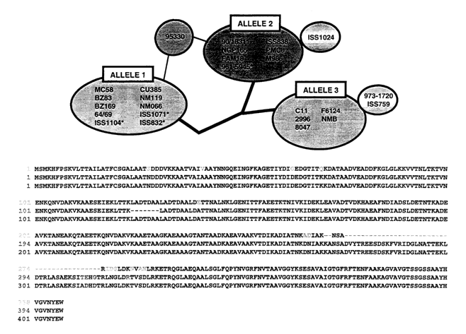
Figure 9 shows an alignment of NadA alleles, and figure 10 shows the
relationship of alleles 1 to 3.
Figure 11 shows predicted secondary structure for NadA.
Figure 12 shows analysis of sequences upstream and downstream of NadA.
Figure 13 shows PCR analysis of NadA expression in different strains of
Naneningitidis.
Figure 14 shows immunoblot analysis of NadA expression in different strains of
N.ineningitidis.
Figure 15 shows variation of NadA expression with culture time.
Figure 16 shows NadA FACS of isogenic capsulated and non-capsulated
Nnaeningitidis cells.
Figure 17 shows immunofluorescence results obtained using anti-NadA against
Chang cells (17A to
17C) or HeLa cells (17D).
Figure 18 shows immunofluorescence results obtained using anti-NadA against
Chang cells after
incubation at (A) 37 C or (B) 4 C.
Figure 19 shows immunofluorescence results for Chang cells treated with
saponin.
Figure 20 shows immunofluorescence results obtained using monocytes.
Figure 21 shows immunofluorescence results obtained using macrophages.
Figure 22 shows IL-a secretion by monocytes in response to NadA treatment.
Figure 23 shows the effect of anti-CD14 on IL-a secretion by monocytes.
Figure 24 shows immunofluorescence results obtained using anti-NadA against
E.coli transformed to
express NadA.
Figure 25 shows staining of the transformed E.coli using (A) anti-NadA (B)
anti-E.coli or (C) both.
Figure 26 is a schematic representation of App features. The N-terminal leader
peptide, the passenger
domain and the C-terminal p-domain are indicated. The positions of the serine
protease active site,
the ATP/GTP binding site, the two Arginine-rich sites and the Proline-rich
region are shown. In BOX
1, cleavage sites are shown. In BOX 2 a comparison of known proteolytic sites
of different
autotransporters is shown and a consensus signature is derived. Arrows
identify the cleavages; X =
any amino acid; hyd = hydrophobic residues; (A,S) = Alanine or Serine.
Figure 27 is a schematic representation of the constructs used for studying
App.
Figure 28 shows a western blot of outer membrane and extracellular proteins in
E.coli.
Figure 29 shows FACS analysis of outer membrane and extracellular proteins in
E.coli.
Figure 30 shows immunofluorescence of outer membrane and extracellular
proteins in E.coli.
Figure 31 shows total E.coli proteins analysed by SDS-PAGE.
Figure 32 shows an immunoblot of crude precipitated culture supernatants using
mouse antiserum
against App-his.
CA 02452836 2003-12-31
WO 03/010194 PCT/IB02/03396
-20-
Figure 33 shows FACS adhesion data using rabbit antiserum against E.coli.
Percentages of cells
positive to adhesion are shown near the fluorescence profiles.
Figure 34 shows immunofluorescence microscopy data showing bacterial adherence
and aggregation.
Figure 35 shows concentration-dependent binding of App-His (=), Appa-His (.)
and NMB2132 (A)
expressed as net Mean Fluorescence Intensity (MFI).
Figure 36 shows the effect on binding of App-His (100 g/m1) of pre-incubation
with pronase (left-
hand columns) or phospholipase A2 (right-hand columns) with increasing
concentration of enzyme.
Pronase was tested at 0, 250, 500, 1000 g/ml; phosholipase A2 was tested at
0, 50, 200, 800 g/ml.
Figure 37 is a comparison of cellular binding specificity of App-His protein
at 100, 25 or 6.25 g/ml
against various different cells.
Figure 38 shows association of wild-type or App-knockout N.meningitidis MC58
bacteria.
Figure 39 shows a western blot analysis of total lysates from N.meningitidis
MC58 harvested at 0.5
or 0.8 OD620nm. Lanes 1 & 3 show wild-type MC58 and lanes 2 & 4 show the App
knockout.
Figure 40 shows a western blot analysis of supernatants in parallel to figure
39.
MODES FOR CARRYING OUT THE INVENTION
NadA homology
NadA shows homology to (a) YadA of enteropathogenic Yersinia, a non-pilus
associated adhesin
implicated in virulence [Cornelis (1998) Microbiol. Mol. Biol. Rev. 62:1315-
1352.] and (b) UspA2 of
Moraxella catarrhalis, a protein involved in serum resistance and a protective
antigen [Chen et al.
(1999) Infect. Inamun. 67:1310-1316.]. Sequence similarity is mainly clustered
in the carboxyl
terminal region (56-63% identity in the last 70 amino acids). Outside this
region the level of identity
drops to 23-25%.
YadA and UspA2 have been identified as adhesins [Hoiczyk et al. (2000) EMBO J
19:5989-5999].
Both proteins form very stable and difficult-to-dissociate high molecular
weight oligomers (150-200
kDa) anchored to the outer membrane. NadA has also been found to form very
stable high molecular
weight aggregates on the outer membrane of meningococcus.
The amino acid sequence of NadA was analysed [Nielsen et al. (1997) Protein
Engineering 10:1-6;
Levin & Garner (1988) Biochina. Biophys. Acta 955:283-295; Berger et al.
(1995) PNAS USA
92:8259-8263; Bornberg-Bauer et al. (1998) Nucleic Acids Res. 26:2740-2746].
Secondary structure
analysis is shown in Figure 11. The globular N-terminus and amphipathic C-
terminus are indicated,
as are the positions of the leader peptide (LP) and a membrane anchor. The
carboxyl-terminal region
(aa 310-362) has a predicted amphipatic (3-structure (0-strands shown in
black) and a terminal
aromatic amino acid, which are typical features of outer membrane anchoring
domains. The amino
terminal region (aa 23-90) has no defined secondary structure, but the rest of
the protein has mainly
a-helix propensity (84.6%). Within this region, residues 90-146 and 183-288
have high probability of
forming coiled coils. In addition, residues 122-143 contain four leucine
residues in the "a" positions
of the heptad repeats (L-x(6)-L-x(6)-L-x(6)-L) that may form a leucine zipper
domain (*94) It is
CA 02452836 2003-12-31
WO 03/010194 PCT/IB02/03396
-21-
known that both coiled coils and leucine zipper sequences are involved in
dimerization and may
mediate oligomerisation of monomers via association of two or more alpha
helices.
Even though primary structure similarity between NadA, YadA and UspA2 is
clustered at the
C-terminus, therefore, the overall similarity between the three proteins is
conserved at secondary
structure level. Putative leucine zippers are present in both NadA and UspA2.
NadA, YadA and
UspA2 have a carboxyl terminal membrane anchor made by four amphipathic fl-
strands and an
internal a-helical region with propensity to form coiled-coils. In YadA and
UspA2 these a-helices
have been shown to form coiled-coils regions, which mediate oligomerisation of
monomers [Hoiczyk
et al. (2000) EMBO J 19:5989-5999; Cope et al. (1999) J. Bacteriol. 181:4026-
4034].
The absence of cysteine residues in the mature forms of NadA is another
feature shared with its
homologues.
The genomic environment of NadA
The 1086bp nadA coding region is flanked at the 3' end by a terminator
sequence while at the 5' end
(Figure 12A) it shows a putative ribosome-binding site (RBS; 5'-AAGG-3') and a
putative promoter
region located 8 and 47 base pairs, respectively, upstream the ATG start
codon.
130 bp upstream the coding region are nine repeats of the tetranucleotide TAAA
(shaded black in
Figure 12A), preceded by a second putative promoter with -10 and -35 regions.
Because of the
presence of the TAAA repeats, the gene had been listed as one of those that
may undergo phase
variation, even though the repeats are not in the coding region [Tettelin et
al.]. The homologous gene
UspA2 has a tetranucleotide repeat (AGAT) located in the same position as in
nadA, which varies in
different strains [Cope et al. (1999) J. Bacteriol. 181:4026-4034].
The G+C content of the nadA gene and its upstream region is lower than average
(45% against an
average of the rest of the genome, 51.5%), suggesting acquisition of the gene
by horizontal transfer.
The NadA gene and its upstream region are not present in the published
sequence of the genome of
serogroup A, strain Z2491 [Parkhill et al. (2000) Nature 404:502-506]. In the
MenA genome, a short
sequence of 16 nucleotides with no homologies in the database, replaces the
nadA gene (Figure 12B),
whereas the upstream and downstream genes (nmb1993 and nmb1995) are well
conserved (91% and
97% identity). Analysis of the sequences immediately adjacent to the nadA
region and absent in the
Z2491 serogroup A strain shows that the segment is flanked by the TCAGAC
direct repeats. This
may indicate a mechanism of recombination. In the A strain the stretch of 16
nucleotides has a
disrupted pair of TCAGAC repeats flanking it.
Variation in NadA genotype
Given the difference in nadA expression between serotypes A and B, 175
different strains of
N.ineningitidis were chosen for analysis - 150 isolates representative of the
five disease-associated
serogroups (A, B, C, Y and W-135) and 25 strains isolated from healthy
carriers. The analysis also
included one strain each of N.gonorrhoeae, N.cinerea and N.lactamica.
CA 02452836 2010-11-05
-22-
Bacteria were grown overnight at 37 C in a humidified atmosphere of 5% CO2 in
air on gonococcus
(GC) medium agar (Difco) supplemented with Kellogg's supplement solution (0.22
M D-glucose,
0.03 M L-glutamine, 0.001 M ferric nitrate, and 0.02 M cocarboxylase) (Sigma-
Aldrich Chemical
Co., St. Louis, Mo.) as previously described (Knapp et al. (1988) Antinzicrob.
Agents Chemother.
32:765-767; Roberts et al. (1977) J. Bacteriol. 131:557-563]. One loopful of
bacteria was dissolved
in 500 I of PBS and chromosomal DNA was prepared as previously described
[Tinsley et al. (1996)
PNAS USA 93:11109-11114].
The bacteria were screened by PCR and/or dot blot hybridization.
PCR amplification of the nadA genes was performed on 10 ng of chromosomal DNA
using primers,
mapping 350 nt upstream and downstream from the coding region (forward primer:
SEQ ID 16;
reverse primer: SEQ ID 17), and Platinum Hifi Taq Polymerase (GIBCO). PCR
conditions were: 30
cycles of denaturation at 95 C for 30 s, annealing at 60 C for 30 s, and
extension at 68 C for 1 min.
PCR products were analysed on 1% agarose gel and the sizes were determined
using a molecular
weight marker 1Kb Plus DNA Ladder (GIBCO). The amplified fragments were
purified on a
Qiaquick*column (Qiagen) and then automated cyclo-sequenced (Applied
Biosystems model 377) by
primer walking on both strands of the amplified fragment.
For dot blotting, the probe used was the whole nadA gene, as amplified from
2996 strain and labelled
with digoxigenin using the Roche DIG High-Prime DNA Labelling and Detection
Kit. 10 pl aliquot
of cell suspension of each strain were boiled for 10 min. and spotted on nylon
membrane
(Boehringer). The membranes underwent cross-linking of DNA by 2' exposure to
UV light and other
standard procedures for preparation and signal detection as reported by the
manufacturer.
The nadA gene was absent in N.gonorrhoeae and in the commensal species
N.lactamica and
Ncinerea. In N.meningtidis, however, 47% of isolates were positive for its
presence.
PCR generated (Figure 13) a product of 1800 bp in NadA+ strains MC58 (lane 1),
90/18311 (lane 2)
and 2996 (lane 3). It gave a product of 400 bp in NadA strain Z2491 and NG3/88
(lane 5). Some
strains (e.g. 93/4286, C4678,2022, ISS 1113) gave a PCR product of 2500 bp
(lane 4: L93/4286).
The presencelabsence of NadA in N.meningitidis was correlated with strain
lineage. Strains. isolated
from invasive meningococcal disease have been classified by multilocus enzyme
electrophoresis
(MLEE) into a small number of hypervirulent lineages: Electrophoretic types
ET37, ET5, cluster
A4, lineage III, subgroups I, III and IV-1 [Achtman (1995) Global epidemiology
of meningococcal
disease. In Meningococcal disease (Cartwight, ed). John Wiley and Sons,
Chichester, England. 159-
175; Caugant (1998) APMIS 106:505-2.51. Recently, a sequence-based
classification, multilocus
sequence typing (MLST), has been introduced, which classifies the above
strains into Sequence
Types ST11, ST32, ST8, ST41, STI, STS, ST4, respectively [Maiden et al. (1998)
PNAS USA
95:3140-3145]. Strains isolated from healthy carriers fall into many different
ET and ST types.
The nadA gene was present in 51 out of 53 strains (96%) of the hypervirulent
lineages ET-5, ET-37
and cluster A4, whereas it was absent in all the tested lineage III strains.
Seven of the 25 carrier
strains were positive. Most of the serogroup C strains tested were positive
even if not belonging to
*Trade-mark
CA 02452836 2003-12-31
WO 03/010194 PCT/IB02/03396
-23-
hyper-virulent lineages. The same was true for the serogroup B strains with
serotype 2a and 2b. For
serogroup A, one strain belonging to subgroup III was positive whereas the
other two strains
belonging to subgroup IV-1 were negative.
Lineage III has only recently been introduced in Europe and USA and the
geographic segregation in
New Zealand for many years could have impaired its ability to acquire novel,
genes. For instance,
mutations may have occurred in the surrounding chromosomal regions preventing
Lineage III from
further recombination events. Another possible explanation is that ET-5, ET-37
and Cluster A4
strains need nadA to achieve peak fitness whereas Lineage III isolates cannot
derive any significant
benefit from nadA insertion, thus undergoing a negative selection.
NadA is thus over-represented in three hypervirulent N.meningitidis lineages.
It appears to be a
foreign gene present in a subset of hypervirulent strains.
NadA alleles
As PCR products were differently sized (Figure 13) and most of the NadA+
strains could be grouped
in three different sizes, genes were sequenced for 36 strains representative
of each size: 26 positive
strains, 4 strains with a long PCR product, and 6 NadA- strains.
In the negative strains, a 16bp sequence was found which was identical to the
sequence present in the
published serogroup A genome sequence.
Analysis of the sequence of the four long PCR product strains revealed an
interruption by a single
copy of IS 1301, interrupting the protein after 162 amino acids with a stop
codon. The insertion site
was identical in all four strains, but the orientation of IS 1301 differed,
indicating independent events.
The target consensus for IS1301, 5'-AYTAG-3' was found within the NadA gene at
nucleotide 472,
generated by, an A->G mutation, and was accompanied by a TA duplication.
In nadA+ strains, gene size ranged from 1086 to 1215 bp, with consequent
variation of the amino acid
sequences of the encoded proteins from 362 to 405 amino acids. It was possible
to cluster 22 of the
26 NadA genes into three well-defined alleles (Figures 9 & 10; Table I). The
sequence of the gene
within each allele is identical and overall identity between the alleles
ranges from 96% to 99%. This
level of conservation is surprising and suggests weak selective pressure
and/or a very recent
acquisition of the nadA gene. The latter possibility is consistent with the
low G+C content of the
genome in this region (see above).
Allele Found in strains SEQ IDs
1 MC58, BZ83, BZ169, NM066, NMI19, CU385, ISS832, ISS1071, ISS1104 1,4
2 90/18311, NGPI65, PMCS, M986, ISS838 and 961-5945 2,5
3 C11, 973-1720, ISS759, F6124, 2996, 8047, NMB 3,6
'30 The sequences shown in Figure 9A assume that the N-terminus amino acid is
the first Met in the
open reading frame (SEQ IDs 4 to 6), but the second Met (residue 3 in SEQ IDs
4 to 6) has a better-
positioned Shine-Dalgarno motif (Figure 9B). Sequences starting from the
second Met codon are thus
preferred (SEQ IDs 1 to 3).
CA 02452836 2003-12-31
WO 03/010194 PCT/IB02/03396
-24-
Allele 1 codes for a protein of 362 amino acids (SEQ ID 1) and includes strain
MC58 and all the
ET-5 positive strains sequenced. The other five strains belonging to allele 1
were very recent isolates
and they have not been ET-typed yet, although serotype and serosubtype
classification (B:15:P1.7
and B:4:P1.15) of these strains suggests affiliation of these strains to the
ET-5 complex.
Allele 2 codes for a protein of 398 amino acids (SEQ ID 2) resulting from the
addition of 2 as after
residue 268 (numbering according to SEQ ID 1), addition of 41 as after residue
271, and deletion of
7 as after residue 122, resulting in the deletion of the first heptad repeat
of the leucine zipper domain.
Leucine residues at a fixed spacing of seven residues commonly identify
leucine zippers. One leucine
in the repeats has frequently been replaced mostly by Met, Val or Ile. In this
case allele 2 could use
the Ile upstream or downstream to form the leucine zipper motif.
Allele 3 codes for a protein of 405 amino acids (SEQ ID 3) and, like allele 2,
contains 43 extra amino
acids at residues 268 and 271 but differs from allele 2 by not having the 7aa
deletion after residue
122. Allele 3 is found in serogroup A, B and C strains.
The remaining 4/26 positive strains (ISS1024, ISS759, 973-1720, 95330; marked
with * in Table 1)
contain minor variants of alleles 1 to 3:
- Serogroup C strain ISS 1024 has a variant of allele 2 with a single heptad
repeat deletion at
residues 229-235 (SEQ IDs 7/8). This sequence was originally classified as a
fourth allele but has
been re-classified as a variant of allele 2. Allele 2 is thus found in all ET-
37 strains, one strain of
cluster A4 and three additional non-ET-typed serogroup C strains.
- Serogroup C strains ISS759 and 973-1720 both contain a variant of allele 3
with a single amino
acid mutation in the leader peptide (SEQ IDs 9/10) resulting from a single
nucleotide mutation.
Among all allele 3 strains, only 973-1720 belongs to a hypervirulent strain
(cluster A4).
- Serogroup B strain 95330 contains a recombinant (chimera) of alleles 1 and 2
(SEQ IDs 11/12),
with nadA being a fusion between the N-terminal portion of allele 2 and the C-
terminal segment
of allele 1. The putative site of recombination is located approximately
between residues 141 and
265 of the protein.
All insertions and deletions happen in the coiled-coil region and involve 7 or
41 amino acids which,
representing 2 or 6 turns of the a-helix, allows for variations in length of
the coiled coil region
without disturbing the overall structure. Furthermore, the deletion in ISS1024
results in the loss of
the first heptad repeat of the leucine zipper domain but does not destroy the
domain because leucine
residues at a fixed spacing of seven residues can be replaced mostly by Met,
Val or Ile. In this case
allele 2 could use the Ile upstream or downstream to form the leucine zipper
motif (Figure 11).
Any of these various NadA sequences and alleles can be used in accordance with
the invention.
When sequence analysis was extended to the putative promoter and terminator
regions (50bp
upstream, 350bp downstream), variations were found only in the in the 5'
region. Three Italian strains
(ISS1071, ISS832 and ISS1104) differed for a single base mutation while in
strain 961-5945 there
was a 7 base differences (indicated with * in Figure 10). Variations were also
found in the 5' regions
CA 02452836 2003-12-31
WO 03/010194 PCT/IB02/03396
-25-
where the TAAA tetranucleotide was repeated from 4 to 12 times in different
strains (Table 1). The
number of repeats was variable also within each allele (Table 1).
Further work was performed on carrier strains isolated from healthy
individuals by oro-pharyngeal
swab. Some strains, even if described as carriers, belong to hypervirulent
clusters, and NadA was
found in all such carrier strains as described above (i.e. allele 1 in the ET-
5 strains and allele 2 in the
ET-37 strains).
NadA was also found in five carrier strains (NGE28, 65/96, 149/96, 16269,
16282) which do not
belong to a hypervirulent cluster. These five strains shared a sequence (SEQ
IDs 13 & 14) which was
not found in strains isolated from patients. This allele is referred to as
`allele C' (carrier).
An alignment of allele C with alleles 1 to 3 is shown in Figure 9C. Disruption
in the coiled-coil
segments of the protein is evident.
Unlike alleles 1 to 3, allele C protein does not readily form a high molecular
aggregate when
expressed in E.coli. Like alleles 1 to 3, however, allele C is exposed on the
surface of N.meningitidis,
because it is a target for bactericidal antibody raised against itself.
However, these antibodies are not
bactericidal against strains carrying alleles 1 to 3; similarly, antibodies
raised against alleles 1 to 3
are not bactericidal against allele C strains.
NadA oligomers on the cell surface
WO01/64922 reports that NadA forms oligomeric structures. To study NadA
oligomers in more
detail, whole cell lysates of N.ineningitidis were probed by Western blot.
Bacterial colonies [strains MC58 (allele 1), 90/18311 (allele 2), 2996 (allele
3), L93/4286 (IS1301
insertion) and NG3/88 (nadA-)] were grown to stationary phase in GC broth
supplemented with 0.3%
glucose. Samples were taken at different times, pelleted by centrifugation at
3000 x g for 10 min, and
resuspended in PBS and thawed/frozen up to bacterial lysis. Equal amounts of
proteins were
subjected to SDS-PAGE on 12.5% polyacrylamide gels and electrotransferred onto
nitrocellulose
membranes.
To prepare anti-NadA polyclonal serum, recombinant NadA was expressed and
purified. Sequences
encoding the three nadA alleles (allele 1: as 24-362; allele 2: as 24-343;
allele 3: as 24-350), were
amplified by PCR on chromosomal DNA and cloned into pET21b+ vector (Novagen).
The plasmids
were transformed in E.coli BL21 (DE3) to express the proteins as C-terminal
histidine fusions.
Protein expression was induced at 30 C by adding lmM IPTG at OD600nm 0.3 and
growing the
bacteria for an additional 3 h; expression was evaluated by SDS-PAGE.
Recombinant fusion proteins
were purified by affinity chromatography on Nit+-conjugated chelating fast-
flow Sepharose 4B resin.
20 pg of purified protein was used to immunise six-week-old CD1 female mice (4
to 6 per group).
Proteins were given intraperitoneally, with complete Freund's adjuvant (CFA)
for the first dose and
' incomplete Freund's adjuvant (IFA) for the second (day 21) and third (day
35) booster doses. Bleed
out samples were taken on day 49 and used for the serological analysis.
CA 02452836 2003-12-31
WO 03/010194 PCT/IB02/03396
-26-
The blots showed a high molecular weight reactive band in strains MC58 (Figure
14, lane 1),
90/18311 (lane 2) and 2996 (lane 3). The band was absent in strain NG3/88
(lane 5). Boiling of the
sample buffer up to 40 minutes did not change the pattern. The different size
of the proteins was
consistent with the size of the alleles. Given the expected size ranging from
35 to 40 kDa of
monomeric proteins, the high MW of the observed band could be explained by the
presence of an
oligomeric form of NadA. This possibility is supported by the fact that in a
strain containing the
IS 1301 insertion, coding for a shorter protein of 162 amino acids and lacking
most of the coiled-coil
region, the high MW reactive band was absent and replaced by a band of 14.5
kDa (Figure 14, lane
4), consistent with the predicted molecular weight of the processed monomeric
protein.
Although the oligomeric protein was found in all strains containing a
functional gene, expression
levels varied from strain to strain (Table I). Moreover, the amount of NadA
protein varied within the
same strain during growth.
Four different strains (MC58, 2996, Cli, F6124), chosen as representative of
diverse overall NadA
expression level, were followed during growth up to stationary phase. Figure
15 shows growth of two
of the tested strains (15A: MC58, with low NadA expression; 15B: 2996, with
high NadA
expression), with the curve showing OD600. Western blots of samples taken at
each point of the OD600
growth curve showed that the NadA band was barely visible at the beginning of
the growth and
became more intense during growth, up to its maximum, at stationary phase. All
strains analysed
showed the same growth-phase dependent behaviour.
High MW NadA was also seen in western blots of outer membrane vesicles,
consistent with NadA
being anchored to the outer membrane.
Similarly, FACS analysis on live bacteria during log-phase growth showed that
NadA was available
for antibody binding on the surface of the bacteria. FACS intensity in a
strain with a poylsaccharide
capsule (strain NMB) was reduced 1 log in comparison to an isogenic non-
encapsulated mutant strain
(M7), but the protein was surface-exposed and available for binding in both
strains (Figure 16).
NadA forms surface-exposed oligomers, which are stable to heat, SDS and
reduction with
(3-mercaptoethanol. As the mature form of the lacks cysteine residues,
disulphide bond formation
cannot be involved in this phenomenon; rather this is consistent with the
predicted coiled-coil
structure and the presence of leucine zipper motifs that might mediate
intermolecular interactions
between monomers [Lupas (1996) Trends Biocheni. Sci. 21:375-382; O'Shea et al.
(1991) Science
254:539-544]. The size of the oligomers is approximately 170 kDa, suggesting a
tetrameric structure
[WO01/64922]. However, a rigid coiled-coil structure is likely to have an
anomalous migration is
SDS PAGE and therefore the 170kDa form may be a trimer.
Protective immunogenicity
Polyclonal anti-NadA serum was tested for bactericidal activity as previously
described [Pizza et al.
(2000); Peeters et al. (1999) Vaccine 17:2702-2712], with pooled baby rabbit
serum (CedarLane)
used as complement source. Serum bactericidal titer, was defined as the serum
dilution resulting in a
50% decrease in colony forming units (CFU) per ml after 60 minutes incubation
of bacteria in the
CA 02452836 2003-12-31
WO 03/010194 PCT/IB02/03396
-27-
reaction mixture, compared to control CFU per ml at time 0. Typically,
bacteria incubated with the
negative control antibody in the presence of complement showed a 150 to 200%
increase in CFU/ml
during the 60 min. of incubation.
Results were as follows:
Strain NadA expression Allele Bactericidal titre
2996 +++ 3 32768
Cil +++ 3 16384
F6124 + 3 4096
MC58 + 1 8192
BZ232 - - <4
NGH38 - - <4
As shown, the serum induced complement-mediated killing of all strains that
have the nadA gene,
and was inactive against the strains that do not have the gene. However,
bactericidal titres varied
between strains. Titres were higher against strains expressing higher amounts
of protein. This result
was confirmed when titres were determined in the early and late phase of
growth (Figure 15).
To check whether the differences in the bactericidal activity were due to
different allele sequences,
immune sera, raised against the three NadA types, were produced and used in a
cross bactericidal
assay. The results obtained with the antisera were similar to those shown
above, suggesting that the
bactericidal activity is not influenced by the allele diversity but rather to
the antigen expression level.
The ability of immune sera to protect animals from bacteremia during infection
was also tested, using
the infant rat model. The sera used were obtained by immunising guinea pigs
with 50 g purified
rNadA (allele 3). Immunisation of outbred Wistar rats (5 to 7 days old) was
performed
subcutaneously together CFA for the first dose and IFA for the further three
doses (days 28, 56, 84).
Bleed out samples were taken on day 105 and used for the animal protection
assay.
Two experiments were performed using two different MenB strains (8047 and
2996). Each strain has
been serially passaged three times in infant rats. In experiment 1, groups of
four rats were challenged
intraperitoneally with 100 l of a mix of (a) bacteria from strain 8047 (7x10³
CFU per rat) and (b)
heat inactivated guinea pig antiserum or anti-capsule control mAb (SEAM 3 [Van
Der Ley et al.
(1992) Infect. Inzinun. 60:3156)). In experiment 2, group of six rats were
treated with the control
mAb or with different dilutions of guinea pig antiserum at time 0. Two hours
later, they were
challenged with the 2996 bacteria (5.6x10³ CFU per rat). In both experiments,
blood cultures were
obtained 18 h after the challenge by puncturing the heart with a syringe and
needle containing
approximately 25 U of heparin without preservative. Bacteria numbers in the
blood cultures were
obtained by plating out 1, 10, and 100 l of blood onto chocolate agar
overnight. For calculation of
geometric mean CFU/ml, animals with sterile cultures were assigned a value of
I CFU/ml.
Results were as follows:
CA 02452836 2003-12-31
WO 03/010194 PCT/IB02/03396
-28-
Blood culture at 18 hours
Expt Treatment Positive/Total CFU/ml (103)
Anti-capsular mAb (2 g/rat) 0/4 <0.001
1 Anti-NadA antiserum (1:5 dilution) 0/4 <0.001
PBS+1%BSA 5/5 40.17
Anti-capsular mAb (20 g/rat) 1/6 0.003
2 Anti-NadA antiserum (1:5 dilution) 1/6 0.002
Anti-NadA antiserum (1:25 dilution) 3/6 0.035
Pre-immune NadA serum 6/6 1.683
Thus anti-NadA antiserum is highly protective in this assay.
Overall, therefore, NadA has several attributes of being a good vaccine
antigen: (i) it is a surface-
exposed molecule, potentially involved in bacterial adhesion; (ii) it is
present in at least 50% of the
disease-associated strains and in almost 100% of three hypervirulent lineages;
(iii) it elicits protective
and bactericidal antibodies in laboratory animals; and (iv) each allele
induces cross-bactericidal
antibodies.
ORF40
ORF40 shows homology to Hsf and its allelic variant Hia, both adhesins of
Haefnophilus influenzae.
The different size among Hia, Hsf and ORF40 is in part explained by the
presence of three copies of
a large repeated domain in Hsf, which is present in single copy in Hia and
only partially in ORF40
(Figure 7). In MenB, ORF40 is found on the outer membrane as a protein of
about 200 kDa (cf.
predicted MW of 59 kDa for mature protein).
App
App shows homology (Figure 8) to the adhesion and penetration protein Hap of
H.influenzae, which
is an adhesin with a serine-protease activity that undergoes autoproteolytic
cleavage and extracellular
release [Hendrixson et al. (1997) Mol Microbiol 26:505-518]. Uncleaved surface-
associated Hap
mediates adherence to epithelial cells and promotes bacterial aggregation and
colonisation.
In Mineningitidis, App is exported to the outer membrane, processed and
secreted. Both Hap and
App belong to the autotransporter family which comprises proteins from gram-
negative bacteria
characterized by a distinct mechanism of secretion. This system was first
described for IgAl protease
of N.gonorrhoeae, which is considered the prototype of this family. Proteins
of the autotransporter
family have been implicated in the virulence of many gram-negative pathogens
[Henderson & Nataro
(2001) Infect linniun 69:1231-1243]. They are synthesized as large precursor
proteins comprising at
least three functional domains: a typical N-terminal leader sequence, an
internal domain (passenger
domain) and a C-terminal domain (translocator domain or R-domain). The leader
sequence mediates
the export (sec-dependent) of the protein to the periplasm. Subsequently the
translocator domain
inserts into the outer membrane forming a j3-barrel pore to allow the export
of the passenger domain.
Once at the bacterial surface, the passenger domain can be cleaved and
released into the
environment. Cleavage can occur by an autoproteolytic event directed by
protease activity in the
CA 02452836 2003-12-31
WO 03/010194 PCT/IB02/03396
-29-
passenger domain itself. Passenger domains of autotransporters are widely
divergent, reflecting their
remarkably disparate roles. On the contrary the 1i-domains display high degree
of conservation
consistent with their conserved function.
App possesses the prevailing domains of the autotransporter proteins as well
as the conserved serine
protease motif (GDSGSP). It has been shown that this motif is responsible for
cleavage of human
IgA by the Neisseria IgAl proteases and for autoproteolytic cleavage of Hap
protein of H.influenzae.
App has been shown to be. a conserved antigen among meningococci, to be
expressed during
infection and carriage, to stimulate B cells and T cells, and to induces a
bactericidal antibody
response [Hadi et al. (2001) Mol. Microbiol. 41:611-623; Van Ulsen et al.
(2001) FEMS Immunol
Med Microbiol 32:53-64].
In serogroup B strain 2996, App has 1454 amino acids and a predicted MW of
159,965 Da. Figure 26
shows the protein's predicted structural features. Three domains can be seen:
domain 1 (amino acids
1-42) is the signal peptide; domain 2 is the passenger domain, which is the
functionally active
protein; domain 3 is the C-terminal translocator domain with (3 barrel
structure.
At the N-terminus of the passenger domain, His-115, Asp-158 and Ser-267
correspond to the serine
protease catalytic triad His-98, Asp-140 and Ser-243 from Hap [Fink et al.
(2001) J Biol Chem
276:39492-39500]. Residues 285-302 are a putative ATP/GTP-binding site (P
loop), which suggests
a mechanism of energy coupling for outer membrane translocation. Towards the C-
terminus of the
passenger domain, two Arg-rich regions are present. The first (RRSRR) is
residues 934-938 and the
second (RRARR) begins at residue 1149. These motifs are reminiscent of known
targets for trypsin-
like proteolytic cleavage sites such as the one in diphtheria toxin and those
upstream of the
auto-cleavage sites of H.influenzae Hap, N.gonorrhoeae IgA-protease and
B.pertussis FhaB (Figure
26, box 1). Downstream of the Arg-rich regions are motifs 954NTL956 and
176NSG1178, which are
identical or similar to the cleavage sites in autotransporters Ssp (Serratia
marcescens), Pm
(Bordetella bronchiseptica), Brka (Bordetella pertussis) [Jose et al. (1995)
Mol. Microbiol. 18:378-
380] and Hap (H.influenzae) (Figure 26, box 2). Together, these sequence
motifs suggest that the two
motifs 954NTL956 and 1176NSG1178 and the RR(A,S,R)2RR pattern could act as
signals for correct
localisation of downstream processing sites.
Further analysis of the App sequence shows a proline-rich region, where the
dipeptide motif PQ is
repeated four times beginning at residue 1156. A search for homology to known
protein sequences
reveals some similarity to the surface proteins of S.pneurnonie PspA and PspC
and to a proline-rich
region of the B.pertussis outer membrane protein p69 pertactin, where the
(PQP)5 motif is located in
a loop containing the major immunoprotective epitope.
Finally, the last three amino acids of App (YRW) are identical to those of Hap
where they have been
described as crucial for outer membrane localisation and protein stability
[Hendrixson et al., 1997].
Expression in E.coli without fusion partners
ORF40, App and NadA full-length genes were cloned in pET21b+ vector and the
plasmids were
transformed in E.coli BL21(DE3) in order to express the genes under control of
T7 promoter.
CA 02452836 2003-12-31
WO 03/010194 PCT/IB02/03396
-30-
Expression was achieved activating the promoter with IPTG or under non-induced
conditions.
Localisation and surface-exposure of the proteins were assayed by cell-
fractionation experiments
(SDS-PAGE and Western blot), FACS analysis and whole-cell immunoblot. As shown
in figures 1 to
3, all the three proteins are translocated to the surface of E.coli:
- ORF40 is expressed as monomeric form and possibly forms also multimers
(Figure 1).
- App is exported to E.coli outer membrane as a precursor of about 160 kDa,
that is processed and
secreted in the culture supernatant (Figure 2).
- NadA is found to the be present in the outer membrane fraction as a single
high molecular weight
band of approximately 180 kDa. This probably corresponds to an oligomeric form
of the protein.
Such a band is absent in E. coli expressing intracellular NadA (Figure 3).
App expression was studied in more detail.
N.meningitidis strain 2996 genomic DNA was prepared as previously described
[Tinsley & Nassif
(1996) PNAS USA 93:11109-11114]. DNA devoid of the sequence coding for the
signal peptide
(amino acids 1 to 42) and of the STOP codon was amplified using PCR primers
SEQ IDs 18 & 19
followed by digestion with N1zeI and Xhol and insertion into the NheIlXhol
sites of the pET-21b
expression vector, to give `pET-App-His' (Figure 27). This plasmid was
introduced into E.coli
BL21(DE3) and used for the expression of a C-terminal His-tagged fusion
protein which was purified
and used to raise antibodies. The full-length app gene was amplified and
cloned in a similar way,
using PCR primers SEQ IDs 20 & 21, to give plasmid 'pET-App'.
Plasmids were introduced into E.coli BL21(DE3) and expression induced by
addition of 1mM IPTG.
The expressed protein was detected by western blotting (Figure 28, lane 1). To
verify that the protein
was exported to the E.coli surface, FACS (Figure 29) and immunofluorescence
microscopy (Figure
30) were used. The FACS analysis showed positive surface expression on the pET-
App
transformants (full-length gene) but no surface expression with App-His (no
signal peptide) or with
the empty vector. The immunofluorescence results agreed with FACS. Therefore
expression of the
full-length app gene resulted in the export of App to the surface of E.coli,
but deletion of the first 42
amino acids abolished surface-localisation.
Western blot analysis of outer membrane proteins from pET-App transformants
revealed a specific
reactive band of -160 kDa (Figure 28, lane 1), corresponding to the predicted
molecular weight of
the full-length protein. A corresponding band was missing in the outer
membrane fraction from
untransformed controls (lane 3). Western blot analysis of culture supernatants
revealed a secreted
protein of -100 kDa with pET-App (lane 2) that was absent with the
untransformed controls (lane 4).
Sometimes a very weak band was also detected at -140 kDa in pET-App
transformants.
Therefore the full length app gene when introduced into E.coli induces
expression of an App protein
which is exported to the outer membrane, cleaved and released into the culture
supernatant.
CA 02452836 2003-12-31
WO 03/010194 PCT/IB02/03396
-31-
Native expression can influence the quality of the immune response
To evaluate the role of protein conformation on induction of an immune
response, outer membrane
vesicles from E.coli expressing ORF40, App or NadA were isolated and used to
immunise mice. Sera
were tested for bactericidal activity and results compared with those obtained
with the fusion
proteins. The bactericidal response (strain 2996) was improved 5-10 fold when
the proteins are
produced in their "native" form in OMVs:
Bactericidal titres
Antigen
Fusion protein E.coli OMV
ORF40 256 2048
App 64 1024
NadA 32768 >65536
* Titres expressed as the reciprocal of the serum
dilution yielding -50% bacteria killing
App autoproteolytic cleavage
E.coli pET-App transformants secrete a lOOkDa product into culture supernatant
and show a 160kDa
surface product. To test whether the secreted App product derives from an
autoproteolytic process, one
of the putative catalytic residues (Ser-267) was replaced with Ala.
The pET-AppS267A mutant was obtained by site-directed mutagenesis using the
QuikChange kit
(Stratagene) and primers SEQ IDs 22 & 23.
SDS-PAGE analysis of total proteins from pET-AppS267A transformants (figure
31, lane 2) showed a
protein similar in size to pET-App transformants (lane 1). The protein was
shown to be surface exposed
by FACS analysis (Figure 29).. Western blot analysis of culture supernatants
showed App in pET-App
transformants (Figure 32, lane 1) but not in pET-AppS267A transformants (lane
2).
Mutation of Ser-267 to Ala thus abolishes processing and secretion of the App
precursor, which remains
cell-associated. These data suggest that App has a serine protease activity
that is responsible for
autoproteolytic processing and release in the supernatant of the secreted App
domain.
Cleavage at 954NTL956 would leave a fragment with predicted molecular weight
of 104190 Da. Cleavage
at 1176NSG1178 would give a 128798 Da fragment. These two predicted fragments
may match the two
bands of -140 and -100 kDa observed in culture supernatants. Cleavage may
occur first to give the
-140 kDa fragment and then second to give the 100 kDa fragment. The (3 domain
of App would thus
begin at residue 1177.
NadA, ORF40 and App function as adhesins
Example 22 of international patent application WO01/64922 discloses that NadA
expression in E.coli
makes the transformed bacterium adhere to human epithelial cells. The adherent
phenotype has been
further studied for NadA and also for App and ORF40.
E.coli BL21(DE3) bacteria (108 CFU), grown under non-induced or induced
conditions, were
inoculated onto Chang human epithelial monolayers (105 cells) and incubated at
37 C for 1 or 2
CA 02452836 2003-12-31
WO 03/010194 PCT/IB02/03396
-32-
hours. Cells were then incubated with rabbit anti-E.coli and PE-conjugate
secondary antibody.
Adhesion was detected by FACS as specific fluorescence intensity associated to
Chang cells. Positive
controls were E.coli DH5 expressing hsf (DH5/pDC601)); negative controls were
BL21(DE3)/pET2lb and DH5a/pT7-7. The results in figure 4 show that the ability
of the
recombinant E.coli strains to adhere to cultured epithelial cells is
associated with expression of these
three proteins.
To confirm that these three proteins are able to promote interaction with host
cells, the recombinant
proteins themselves were investigated for binding to epithelial cells. 105
Chang human epithelial cells
(Wong-Kilbourne derivative, clone 1-5c-4, human conjunctiva) were incubated at
4 C for 30 minutes
with medium alone or with different concentration of ORF40 (150 g/ml), App
(150 g/ml) or NadA
(300 g/m1), or with GNA2132 (300 g/ml) as negative control [see Pizza et al.
(2000)]. Binding was
detected by FACS using polyclonal antisera against the single recombinant
proteins and a secondary
PE-conjugate antibody. The FACS signal shifts (Figure 5) show that the three
proteins are able to
bind to human epithelial cells, whereas purified GNA2132 (negative control)
does not.
Figure 6A shows that binding increases in a dose-dependent manner. Binding of
NadA reaches a
plateau at around 200 g/ml. GNA2132 fails to bind even at 400 g/ml (Figure
6B). Data in Figure 6
are mean fluorescent intensity (MFI) values plotted against protein
concentration ( g/ml).
Using FACS, binding of NadA to cells was also seen with Hep-2 and MOLT-4
cells, but not with
HeLa, A549, Hec-1B, Hep-G2, CHO or HUVEC cells. Adhesion to Chang cells could
be abolished
by treating the cells with pronase, indicating that the human receptor for
NadA is a protein.
Adhesion of purified NadA protein to Chang conjunctiva cells was also observed
using
immunofluorescence microscopy. The protein (lacking its C-terminal anchor
domain) was incubated
with Chang cells at 37 C in complete culture medium for 3 hours at various
concentrations. Cells
were then washed, fixed, and analysed by laser confocal microscopy after
staining with anti-NadA
mouse polyclonal antibodies and secondary Texas-red coupled anti-mouse IgG
antibodies. No
binding was seen at OnM (figure 17A), but binding was evident at 170nM (17B)
and 280nM (17C),
with clustering evident at higher concentrations. In contrast, no binding of
NadA was seen with HeLa
cells, even at 280nM protein (17D).
Binding was much more evident at 37 C (figure 18A) than at 4 C (figure 18B).
The dot-like
structures seen at 4 C, compared to clusters at 37 C, suggest that lateral
interactions between NadA
monomers are temperature-dependent (influenced by membrane fluidity).
To distinguish surface and endocytosed protein, saponin detergent was added
during the staining
procedure. Intracellular clusters having the size of endosomes were more
evident (arrow) when
saponin was used, but a high proportion of protein remained on the cell
surface (figure 19).
Immunofluorescence also revealed that NadA binds to monocytes (figure 20A).
NadA alone (no
staining antibody; 20B) and NadA stained with pre-immune serum (20C) were not
visible. At high
magnification, evidence of uptake into vesicles (either endosomes or
phagosomes) was seen.
CA 02452836 2003-12-31
WO 03/010194 PCT/IB02/03396
-33-
Figure 21 shows that murine macrophages (raw 264.7) bind and endocytose NadA
(125nM, 3 hours,
37 C; cells cultured in DMEM).
Heating NadA at 95 C for 15 minutes prior to incubation removed its ability to
bind to monocytes, as
measured by secretion of IL-a by the cells (figure 22). The stimulatory
activity of NadA preparations
is thus heat-labile. Stimulatory activity was also blocked by the use of anti-
CD 14 (figure 23) or by
the removal of NadA from the preparations using bead-immobilised anti-NadA.
Immunofluorescence microscopy was also used to detect binding of E.coli
expressing NadA.
Transformed E.coli bound strongly (figure 24A) whereas untransformed bacteria
did not (24B). IL-a
release by monocytes was over 1.5x higher using the transformed E.coli than
the untransformed
bacteria at a bacteria/monocyte ratio of 40:1.
Transformed E.coli were bound to glass cover slips, fixed and double-stained
with anti-NadA
(figure 25A) and anti-E.coli antibodies (25B). When both were used, patches of
anti-NadA were
visible, suggesting that NadA tends to form aggregates on the bacterial
surface, which hamper the
interaction of antibodies with other surface antigens.
Looking at App, recombinant Ecoli strains were incubated with monolayers of
Chang conjunctiva
epithelial cells (Wong-Kilbourne derivative, clone 1-5c-4 [human conjunctiva],
ATCC CCL 20.2)
and adhesion was analysed using FACS. Cells obtained from confluent monolayers
were seeded at
105 cells per well in 12-well tissue culture plates and incubated for 24
hours. Cultures of bacteria
after IPTG induction were washed twice in PBS and resuspended in DMEM+l % FBS
to a
concentration of 5x10⁸ bacteria per ml. Aliquots of 1 ml of each strain were
added to monolayer
cultures of Chang cells and incubated for 3 hours at 37 C in 5% CO2. Non-
adherent bacteria were
removed by washing three times with PBS, and 300 gl of cell dissociation
solution (Sigma) were
added to each microtitre well. Incubation was continued at 37 C for 10
minutes. Cells were harvested
and then incubated for 1 hour at 4 C with rabbit polyclonal anti-E.coli
antiserum (DAKO). Cells
were washed twice in PBS+5% FBS and incubated for 30 mintues at 4 C with R-
Phycoerythrin-
conjugated anti-rabbit IgG (Jackson ImmunoResearch Laboratories). Cells were
then washed in
PBS+5% FBS and resuspended in 100pl PBS. Fluorescence was measured with
FACSCalibur flow
cytometer (Becton Dickinson). For each of fluorescence profile, 10000 cells
were analysed.
The results reported in Figure 33 show pET-App transformants were able to
adhere to Chang cells,
giving a fluorescence shift of 90.3%. S267A transformants were also able to
adhere (91.0%).
Untransformed E.coli were unable to adhere to Chang cells (bottom FACS plot).
As for NadA, FACS results were in agreement with immunofluorescence microscopy
data. As shown
in Figures 34A & 34B, pET-App transformants incubated with monolayers
demonstrated high levels
of adhesion to epithelial cells and visible bacteria-bacteria aggregation. For
the S267A mutant,
adhesion and bacterial aggregation were increased (34C & 34D). Untransformed
controls showed no
adhesion (34G). Deletion of the first 42 amino acids also abolished adhesion.
In contrast to Chang epithelial cells, no adhesion was seen when HUVEC
endothelial cells were
tested with pET-App transformants. To cause sepsis and meningitis,
N.meningitidis has to interact
CA 02452836 2003-12-31
WO 03/010194 PCT/IB02/03396
-34-
with human endothelial cells. App may thus be involved in the first step of
colonisation at the level
of human respiratory epithelial mucosa, rather than in pathological
endothelial colonisation.
Localization and specificity of App binding activity.
To identify the binding region of App, a chimeric protein named App(3 was
used. This protein
consists of the C-terminal domain of App (amino acids 1077 to 1454) fused to
the leader peptide of
IgAl protease of N.gonorrhoeae. The gonococcal leader sequence was chosen
because it has been
well characterized and is functional in E.coli. Plasmid pET-App(3 contains a
1.1 kbp DNA fragment
amplified by PCR using SEQ IDs 26 & 27.
The pET-Appp construct was introduced into E.coli BL21(DE3). FACS localisation
studies
confirmed that App(3 was localized on the E.coli surface. The in vitro
adhesion assay using Chang
epithelial cells showed adhesion by immunofluorescence (Figure 34E & 34F).
FACS analysis
showed that the pET-App(3 transformants were still able to adhere to
epithelial cells but at lower
levels (74.2% shift) than pET-App transformants.
These results indicate that the App binding domain is located in its C-
terminal region, in the 100mer
fragment between residues 1077 and 1176.
Purified recombinant proteins were also studied. App-a-His consists of the N-
terminal portion of
App (amino acids 43-1084) fused to a poly-His tag. Plasmid pET-Appa-His
contains a NheIlXhol 3.1
kbp fragment amplified by PCR with SEQ IDs 24 & 25. The binding activity of
the purified
recombinant App-a-His was compared to that of App-His by FACS binding assays.
Chang cells were
incubated with increased concentrations of recombinant App proteins or
lipoprotein NMB2132-His
(negative control). Binding of App-His (t) increased in a dose-dependent
manner and reached a
plateau at a concentration of 50 g/ml whereas the binding of Appa-his (=) was
very low (Figure
35). The control NMB2132-His (A) failed to bind Chang cells.
To explore the biochemical nature of the molecule involved in interaction with
App, the Chang cells
were treated with pronase or phospholipase A2 before the binding experiments..
105 cells per well
were placed in microplates and incubated in FCS-free DMEM at 37 C in 5% CO2
for 30 minutes
with (a) pronase at 250, 500, or 1000 Vg/ml or (b) phospholipase A2 at 50,
200, or 800 Vg/ml. After
enzymatic incubation, an equal volume of complete medium was added to each
well to stop the
reaction. Cells were subsequently mixed with 100 g/ml App-His or medium alone
and incubated for
1 hours at 4 C. As shown in Figure 36, pronase treatment (left-hand columns)
markedly reduced the
binding of App-His protein to Chang cells, while treatment with phospolipase
A2 (right-hand
columns) did not.reduce the binding. The receptor for App on Chang cells is
thus proteinaceous.
Adhesion to different cell lines were also tested (Figure 37). After
incubation of cultured cells with
three different concentrations of App-His (100, 25 & 6.25 Vg/ml) high level
binding to Chang cells
and HepG2 cells was seen, a moderate level of binding to A-549 cells, and
minimal binding to HeLa
cells. No binding was observed to Hec-1-B, Hep-2, 16HBE14o epithelial cell
lines or to HUVEC
endothelial cells.
CA 02452836 2003-12-31
WO 03/010194 PCT/IB02/03396
-35-
App knockout
After the- work on E.coli suggesting an adhesin role for App, an isogenic
mutant strain of
Mmeningitidis was constructed. The starting strain was MC58. Its app gene was
truncated and
replaced with an antibiotic cassette by transforming the parent strain with
the plasmid pBSUDAppERM,
which contains a truncated app gene and the ermC gene (erythromycin
resistance) for allelic exchange.
Briefly, 600bp of the upstream flanking region including the start codon and
700 bp downstream
flanking region including the stop codon were amplified from MC5 8 using
primers SEQ IDs 28 to 31.
Fragments were cloned into pBluescript and transformed into E.coli DH5 using
standard techniques.
Once all subcloning was complete, naturally competent N.meningitidis strain
MC58 was transformed by
selecting a few colonies grown overnight on GC agar plates and mixing them
with 20 l of 10mM
TrisHCl pH8.5 containing 1 gg of plasmid DNA. The mixture was spotted onto a
GC agar plate,
incubated for 6 hrs at 37 C, 5% C02 then diluted in PBS and spread on GC agar
plates containing 5
pg/ml erythromycin. The deletion app gene in the genome of MC58 was confirmed
by PCR. Lack of
App expression was confirmed by Western blot analysis.
Adhesion of wildtype MC58 and the isogenic MC58Aapp mutant strain was
evaluated on Chang
cells. There was a -10 fold reduction (ranging from 3- to 27-fold in different
experiments) of the
association of the knockout mutant compared with the wild type strain (Figure
38). No difference
was observed between the app mutant and the parental strain with Hep2 and
16HBE14o cell lines
and with HUVEC endothelial cells, confirming that App does not mediate
adhesion to these cells.
No non-pilus adhesins which contribute to adhesion of N.meningitidis in a
capsulated background
have previously been reported.
App expression was studied in N.meningitidis MC58. Colonies from plates grown
overnight were
diluted in GC broth and incubated at 37 C with 5% CO2. Samples were taken when
OD620nm = 0.5
(mid log phase) and 0.8 (stationary phase) and analysed by western blot. Two
bands with apparent
molecular weights -160 and -140 kDa were detected in whole cells lysates of
log phase bacteria
(Figure 39, lane 1), while stationary phase bacteria showed only a faint band
at -140 kDa (lane 3).
As expected, no App was observed in the AApp mutant (lanes 2 & 4).
In marked contrast, supernatant samples of wild-type MC58 showed a band at -
140 kDa and its
amount was higher in stationary phase than in log phase (Figure 40, lanes 3 &
1). The stationary
phase sample also showed a reactive band at -100 kDa.
It will be understood that the invention is described above by way of example
only and modifications
may be made whilst remaining within the scope and spirit of the invention.
CA 02452836 2003-12-31
WO 03/010194 PCT/IB02/03396
-36-
TABLE I - Characteristics of 26 N.rneningitidis strains and their nadA gene
allele
Strain Serogroup Clonal group nadA (TAAA) NadA
type:subtype allele repeats expression
64/69 NG:15:P1.7,16 ET-5 1 4 +
BZ83 B:15 ET-5 1 5 +++
CU385 B:4:P1.15 ET-5 1 6 ++
MC58 B:15:P1.7,16b ET-5 1 9 +
BZ169 B:15:P1.16 ET-5 1 12 ++
95330* B:4:P1.15 ET-5 1 9 nd
ISS1104 B:15:P1.7,16 nd 1 4 +
ISS1071 B:15:P1.7,16 nd 1 5 +++
ISS832 B:15:P1.7 nd 1 5 ++
NM119 B.4.P1.15 nd 1 6 nd
NM066 B:15:P1.7,16 nd 1 12 nd
90/18311 C:NT:P1.5 ET-37 2 9 ++
NGP165 B:NT:P1.2 ET-37 2 9 ++
FAM18 C:2a:P1.5,2 ET-37 2 9 nd
M986 B:2a:P1.5,2 ET-37 2 12 ++
ISS1024* C:2b:P1.5 nd 2 9 ++
ISS838 C:2a:P1.5,2 nd 2 6 ++
PMC8 C: nd 2 10 ++++
961-5945 B:2b:P1.21,16 A4 2 12 +++
ISS759* C:2b:P1.2 nd 3 8 ++++
F6124 A Subgroup III 3 9 +
NMB B:2b:P1.5,2 nd 3 12 ++
8047 B:2b:P1.2 nd 3 12 +++
2996 B:2b:P1.5-1,2 nd 3 12 +++
C11 C:NT:P1.1 nd 3 12 +++
973-1720* C:2b:P1.2 A4 3 12 +++
* indicates that the strain carriers a minor variant of the relevant allele
nd = not done
CA 02452836 2003-12-31
WO 03/010194 PCT/IB02/03396
-37-
TABLE II - Characteristics of N.fneningitidis strains analysed for NadA
expression
ST ET Strain Year Serogroup:type:subtype Country Disease iVadA gene
74 ET5 MC58 1985 B:t5:P1.7,16b UK case 4-
32 ET5 H44/76 1976 B:15:P1.7,16 Norway case +
32 ET5 BZ169 1985 .B:15:P1.16 Netherlands case +
32 ET5 30/00 2000 13:15T I.7,16 Norway case +
33 ET5 N44/89 1989 B:4,7:P1.19,15 Brazil case +
34 ET5 BZ83 1984 .8:1.5 Netherlands case +
- ET5 72/00 2000 B:15:P1.7.13 Norway case +
ET5 39/99 1999 C:I5:P1.7.16 Norway case +
ET5 M4102 1996 B:ND USA case +
- ET5 95330 1995 B:4;P1.15 Canada case +
- ET5 2201731 1993 NG:4:P1.15 Iceland carrier +
- ET5 64/96 1996 NG:15:P1.7,16 Norway carrier +
- ET5 CU385 1980 B:4:P1.15 Cuba case +
- ET5 8680 1987 B Chile case +
ET5 204/92 1992 B Cuba case +
- ET5 EG329 1985 B Germany case +
- ET5 NG080 1981 B Norway case +
- ET5 NG144/82 1982 B Norway case +
- ET5 NG PB24 1985 B Norway case +
- ET5 196/87 1987 C Norway case +
- ET5 Mk521 /99 1999 13 Ivory Coast case +
- ET5 GR 4/00 2000 - Greece case +
11 ET37 FAM18 1983 C:2a:P1.5,2 USA case +
11 ET37 L93/4286 1993 C UK case +
ET37 'NGP165 1974 B:NT PI.2 Norway - +
ET37 M986 1963 B:2a:P1.5,2 USA case A-
- ET37 C4678 1998 C:2a:P1.5.2 Germany case +
- ET37 95N477 1995 B:2a:P1.2 Australia case -
- ET37 :BRAZIO 1976 C Brazil case +
- ET37 F1576 1984 C Ghana case +
CA 02452836 2003-12-31
WO 03/010194 PCT/IB02/03396
-38-
ET37 M597 1988 C Israel case +
ET37 500 1984 C Italy case +
ET37 DI 1989 C Mali case +
ET37 NO P20 1969 B Norway case +
ET37 MA-5756 1985 C Spain case +
ET37 38VI 1964 B USA carrier +
ET37 N 1/99 1999 C:2a Norway case +
ET37 N28/00 2000 W-135:2a Norway case 4-
66 A4 973-1720 1997 .C:2b:P1.2 Australia case +
153 A4 961-5945 1996 B:2b:P1.21,16 Australia case +
A4 5/99 1999 B:2b:P1.5,2 Norway case +
- A4 312294 1995 .C:2b:P.1.5,2 UK case +
A4 96217 1996 B:2b:P1.5,10 Canada case +
A4 G2136 1986 B UK case +
A4 312 901 1996 C UK case +
A4 AK22 1992 B Greece case +
A4 BZIO 1.967 B Holland case +
A4 BZ163 1979 B Holland case +
A4 136116/77 1977 B Iceland case +-
A4 94/155 1994 C New Zealand case +
A4 SB25 1990 C South Africa case +
A4 N53/00 2000 C:2b:P1.5,2 Norway case +
A4 N62/00 2000 C:2b:1'1.5,2 Norway case -+-
41 Lin.111 BZ198 1986 B:NT Netherlands case -
42 Lin.111 M7 98/254 1998 B:4:P 1.4 New Zealand case -
158 Lin.111 972-0319 1997 B:NT:P1..4 Australia case -
159 Lin.111 980-2543 1998 B:NT:P1.4 Australia case -
1127 Lin.111 67/00 2000 B:4,7 Norway case -
- Lin.11l 93/11.4 1993 C:4:P1.4 Belgium case -
Lin.111 M198/172 1998 B:4:P1.4 New Zealand case -
Lin.111 347/97 1997 B:4:P1.4 New Zealand case -
- Lin. ITl 386/98 1998 B:4:P1.4 NewZealand case -
Lin.111 389/98 1998 B:4:P1.4 New-Zealand case -
Lin.111 392/98 1998 B:4:P 1.4 New Zealand case -
CA 02452836 2003-12-31
WO 03/010194 PCT/IB02/03396
-39-
Lin.111 394/98 1998 B:4:P1.4 New Zealand case -
- Lin.111 400 1991 B Austria case -
Lin.il1 M40/94 1994 B Chile case -
Lin.111 AKSO 1992 B Greece case
Lin.111 M-101/93 1993 B Iceland case
Lin.111 931905 1993 B Netherlands case -
Lin.111 91/4{) 1991 B NewZealand case
Lin.111 50/94 1994 B Norway case -
Lin.111 N45/96 1996 B Norway case
Lin.111 88/03415 1988 B Scotland case
1 s l BZ133 1977 B:NT Netherlands case -
s III F6124 1988 A Chad case +
4 s IV-1 205900 1990 A 4 21:P1.7:.1 Mali case -
4 s IV-1 Z2491 1983 A Gambia case -
12 other N03/88 1988 B:8(2):P 1..1. Norway case
13 other NG6/88 1988 B:NT:P.1.1 Norway case
14 other NGF26 1988 B:NT:P1.16 Norway carrier -
other NGE31 1988 B:NT Norway carrier
18 other 528 1989 B: nd Russia case
other 1000 1988 B: NT:P1.5 Russia case -
22 other A22 1986 W-135 Norway carrier -
26 other NGE28 1988 B:4 Norway carrier +
29 other 860800 1986 Y Netherlands case
31 other E32 1988 Z Norway carrier -
35 other SWZI.07 1986 B:4:P1.2 Switzerland case -
36 other NGH38 1988 B:NT:PI.3 Norway carrier -
38 other BZ232 1964 B:NT:P1.2 Netherlands case -
39 other E26 1988 X Norway carrier -
43 other NGHIS 1988 B:8:P1.15 Norway carrier -
47 other NGH36 1988 B:8:P1.2 Norway carrier -
48 other BZ147 1963 B:NT Netherlands case -
49 other 297-0 1987 B:4:PI.15 Chile carrier
540 other 2996 1975 B:2b:P1.5-1,2 UK case +
1034 other 96/1101 1996 C:14:P.1.1.7 Belgium case -
CA 02452836 2003-12-31
WO 03/010194 PCT/IB02/03396
-40-
other 15 1990 B:14,19:P1.9,15 Slovenia case -
other M1090 1996 B:4 Israel case -
other M1096 1996 C:NT:P1.5 Israel case -
other B3937 1995 B:22:PI.16 Germany case +
- other. 31 1993 B:4 Finland case -
other 95074 1995 B:NT:PI.1-1 Canada case +
- other. 660/94 1994 B:4:P1.6 Algeria case -
other 30/93 1993 B:14:P1.14 Argentina case -
other 24370 1996 BIND South Africa case -
other 2411751 1993 NG:21:P1.16 Iceland carrier -
- other 1712741 1993 NG:15:- Iceland carrier -
- other 65/96 1996 B:4:P1.14 Norway carrier +
- other 66/96 1996 B:17:P1.15 Norway carrier -
- other 149196 1996 B:1,19:P1..5,2 Belgium carrier +
- other 16060 1991 B:4:P1.14 Belgium carrier -
- other 16489 1991 NG:21:P.1.1 Norway carrier -
- other 16990 1991 NG:14:P1.5,2,6 Norway carrier -
- other 2022 1991 NG:4:P1.10 Norway carrier +
- other M136 1968 B:11TP1.15 USA case -
- other 860060 1988 X Holland case -
- other NG H41 1986 B Norway carrier -
- other NG G40 1.988 B Norway carrier -
- other N04/88 1988 B Norway case -
- other EG 327 1.985 B DDR case -
- other EG 328 1985 B DDR case -
- other 3906 1977 B China case -
- other NG E30 1988 B Norway carrier -
other 71/94 1.994 Y Norway case -
other DK24 1940 B Denmark case -
- Cl l 1.965 C :16 :Pl.7a,1 Germany - +
- pmc8 - C - - +
- NM.B 1968 B:2b:.P.1.5,2 USA case +
- 8047 1978 B:2b:P1.2 USA case +
- S3446 1972 B. 14:P.1.23,14 USA case -
CA 02452836 2003-12-31
WO 03/010194 PCT/IB02/03396
-41-
- - ISS 749 1996 B:14:P1.13 Italy case -
- ISS 759 1996 C:2b:P1.2 Italy case +
- ISS 832 1997 B:15:P1.7 Italy case +
- I:SS 838 1997 C:2a:P1.5,2 Italy case +
- - ISSIOO1 1999 B:14:P1.13 Italy case -
- ISS1024 2000 C:2b:P1.5 Italy case +
- ISS1.026 2000 B:4:P1.1.3 Italy case -
- ISS1071 2000 B;15:P1.7,16 Italy case +
- 1SS1102 2000 B:15'.P1.4 Italy case -
I.SS1.104 2000 B:I5:P1.7,16 Italy case +
- ISS1106 2000 B:4:P1.4 Italy case -
- ISS1113 2000 C:2a:P1.5 Italy case +
- NI002/90 - - Brazil - +
- 1MC2135 - Brazil - +
- NM001 - B:4:P1.4 UK case -
- - NM002 - B:NT:P1.16 UK case -
- NM004 - B:NT:P1.14 UK case -
- NM008 - B:4:P1.4 UK case -
NMO09/10 B:4:P13,6 UK case -
NM021 - B:4:P1.16 'UK case -
NM036 C:2a:P1.10 UK case +
NM037 - B:2b:P1.10 UK case +
NM050 B:NT:P1.9 UK case -
- NM058 - B:NT:NST UK case -
NM066 B:15:P1.7,16 UK case +
NM067 - C:2a:NST UK case +
NM069 - B:15:P1.7,16 UK case +
NM081 - C:2a:P1.5,2 UK case +
- - NM088 - C:2a:P1.5,2 UK case +
NM092 - B:4:P1.4 UK case -
- - NM 106 - B:NT:P 1.4 'UK case -
NM 107/8 - BAT IA UK case -
- - NMI17 - 8:21;P1,9 UK. case -
NMI 19 - B:4:P 1.15 UK case +
CA 02452836 2003-12-31
WO 03/010194 PCT/IB02/03396
-42-
- NM131 - B UK case
- NM 145 - C UK case +
- NM154 - C:NT:.P1.5,2 UK case +
- NM156 - B:15:P1.16 UK case +
- NM167 - B UK case
NMI 84 - B:NT:P1.5,2 UK case
NM186 - B UK case
NM 188 - B UK case +
- - NM200 - B:4:P1.4 UK case
TABLE III - SEQUENCE LISTING
SEQ ID NO: Description
1 allele 1 of 961
2 allele 2 of 961
3 allele 3 of 961
4 allele I of 961 (first-ATG start)
5 allele 2 of 961 (first-ATG start)
6 allele 3 of 961 (first-ATG start)
7 variant allele 2 of 961 in strain ISS1024
8 variant allele 2 of 961 (first-ATG start) in strain ISS 1024
9 variant allele 3 of 961 in strains 973-1720 and ISS759
variant allele 3 of 961 (first-ATG start) in strains 973-1720 and ISS759
11 961 allele 1/2 chimera (strain 95330)
12 961 allele 1/2 chimera (strain 95330) (first-ATG start)
13 961 allele C
14 961 allele C (first-ATG start)
coding sequence for SEQ ID 13
16-31 PCR primers
32 SEQ ID 650 from W099/24578
33-39 Domain derivatives of SEQ ID 32