Note: Descriptions are shown in the official language in which they were submitted.
CA 02453252 2004-O1-05
WO 03/034034 PCT/US02/33356
AUTOMATED SYSTEM AND METHOD
FOR PROCESSING MULTIPLE LIQUID-BASED SPECIMENS
CROSS-REFERENCE TO RELATED APPLICATIONS
This application claims the benefit of commonly owned U.S. provisional
application Nos. 60/330,092, filed October 19, 2001, 60/372,080, filed April
15, 2002, and
60/373,658, filed April 19, 2002, all of which are incorporated herein by
reference. This
application also is related to commonly owned U.S. non-provisional application
No.
10/122,151, filed April 15, 2002, which is also incorporated herein by
reference.
TECHNICAL FIELD
The present disclosure is directed to apparatus and methods for collecting and
processing specimens of particulate matter-containing liquid, e.g., biological
fluid,
including collecting and depositing onto a microscope slide or other surface a
uniform
layer of particulates therefrom (e.g., cells) suitable for examination (e.g.,
use in cytology
protocols).
BACKGROUND ART
Diagnostic cytology, particularly in the area of clinical pathology, bases
cytological interpretations and diagnoses on examination of cells and other
microscopic
objects. The accuracy of the screening process and diagnosis, and the
preparation of
optimally interpretable samples from specimens typically depends upon adequate
specimen and sample preparation. In this regard the ideal sample would consist
of a
monolayer of substantially evenly spaced cells, which enables
cytotechnologists,
cytopathologists, other medical professionals, and automated screening and
diagnostic
equipment to view or image the cells more clearly so that abnormalities can be
identified
more readily, more accurately and more reproducibly. Newer methodologies such
as
immunocytochemistry and cytometric image analysis require preparation
apparatus and
methods that are safe, effective, accurate, precise, reproducible,
inexpensive, efficient, fast
and convenient.
Cytological examination of a sample begins with obtaining specimens including
a
sample of cells from the patient, which can typically be done by scraping,
swabbing or
brushing an area, as in the case of cervical specimens, or by collecting body
fluids, such as
those obtained from the chest cavity, bladder, or spinal column, or by fine
needle
aspiration or fine needle biopsy. In a conventional manual cytological
preparation, the
cells in the fluid are then transferred directly or by centrifugation-based
processing steps
CA 02453252 2004-O1-05
WO 03/034034 PCT/US02/33356
onto a glass microscope slide for viewing. In a typical automated cytological
preparation,
a filter assembly is placed in the liquid suspension and the filter assembly
both disperses
the cells and captures the cells on the filter. The filter is then removed and
placed in
contact with a microscope slide. In all of these endeavors, a limiting factor
in the sample
preparation protocol is adequately separating solid matter from its fluid
carrier, and in
easily and efficiently collecting and concentrating the solid matter in a form
readily
accessible to examination under a microscope.
Currently, biological specimens are collected for cytological examinations
using
special containers. These containers usually contain a preservative and
transport solution
for preserving the cytology specimen during shipment from the collection site
to the
diagnostic cytology laboratory. Further, cytology. specimens collected from
the body
cavities using a swab, spatula or brush are also preserved in special
containers with
fixatives (e.g., alcohol or acetone fixatives) prior to transfernng cells onto
the slide or
membrane for staining or examination. Specimen containers are known that allow
a
liquid-based biological specimen to be processed directly in the container so
as to obtain a
substantially uniform layer of cells on a collection site (in a filter housing
defining a
particulate matter separation chamber) that is associated with the container
itself. See, for
example, U.S. patent Nos. 5,301,685; 5,471,994; 6,296,764; and 6,309,362, of
Raouf A.
Guirguis, all of which are incorporated herein by reference.
The filtration techniques taught in these patents in practice have yielded
fairly
good results in terms of obtaining close to a monolayer of cells on slides,
but there is room
for improvement. Further, the types of specimen containers disclosed in these
patents
require specially configured apertured covers and adapters therefor that are
designed to
mate with the filter housing, and with suction equipment (e.g., a syringe or a
mechanized
vacuum source) used to aspirate liquid from the container and draw it through
the filter. In
addition, extraction of the filter so that it can be pressed against a
microscope slide to
transfer collected cells to the slide requires disassembly of the cooperating
parts of the
cover and/or adapters associated therewith. If the processing is done by
automated
equipment, special handling devices are required to. carry out such
disassembly. All of
tlus complexity adds time, and material and labor cost to the processing
required prior to
the actual cytology examination.
In general, automated equipment thus far developed for processing liquid-based
specimens have not performed with sufficient consistency, reliability, speed
and
automation to satisfy current and projected needs in cancer screening and
other cytology-
2
CA 02453252 2004-O1-05
WO 03/034034 PCT/US02/33356
based medical, analytical, screening and diagnostic procedures. The vial-based
automated
processing system disclosed herein provides a safe, elegant and effective
solution to these
problems.
SLT1VEV1ARY DISCLOSURE OF THE INVENTION
The specimen vial disclosed herein houses a complete processing assembly,
typically one for mixing the liquid-based specimen therein and for holding a
filter~on
which a uniform layer of cells can be collected from the specimen. It is
expected that the
specimen vial would be prepackaged with a liquid preservative solution, as is
commonplace, and sent to the point-of care site for specimen collection.
The processing assembly is coupled to a simple cover for the vial by means of
a
simple and inexpensive releasable coupling. When the cover is removed at the
point-of
care site (physician's office, clinic, hospital, etc.), the processing
assembly remains with
the cover to allow medical personnel easy access to the container interior for
insertion of a
biological specimen into the vial. The cover, along with the attached
processing assembly,
is then replaced to seal the vial. The vial may then be sent to a laboratory
for processing.
When the vial is manipulated in a simple way while still closed, the
processing
assembly detaches from the cover and remains in the vial for access by
automated or
manual laboratory equipment when the cover is subsequently removed. In a
preferred
embodiment, a downward force on the center of the cover is all that is
required to detach
the processing assembly from the cover. In contrast with the prior art
specimen vials
discussed above, the vial of the present invention requires no further
interaction with the
cover, which can be removed by a simple uncapping device and is discarded to
avoid
contamination. Ribs inside the vial support the processing assembly in the
proper position
for access during processing. This self contained vial and processing assembly
arrangement minimizes human operator exposure to biohazards, such as
tuberculosis or
other pathogens in sputum or in other specimens types, such as urine, spinal
tap fluid,
gastric washings, fine-needle aspirates, and gynecological samples.
The automated specimen processing apparatus disclosed herein is referred to as
the
"LBP" device (for liquid-based preparation), and is designed to produce slides
of high
quality and consistency. The LBP device also can be interfaced with a device
for
detecting and/or quantifying multiple morphologic, cytochemical, and/or
molecular
changes at the cellular level.
During the past two years or so, a review of the literature and reanalysis of
existing
data have led to the identification of a panel of molecular diagnostic
reagents that are
3
CA 02453252 2004-O1-05
WO 03/034034 PCT/US02/33356
capable of detecting and characterizing lung cancer, which is the most common
cancer,
with high sensitivity and specificity. See, for instance, commonly owned U.S.
patent
application Nos. 10/095,297 and,l0/095,298, both filed March 12, 2002, and No.
10/241,753, filed September 12, 2002. Here, the cells can be reacted with
antibodies and
or nucleic-acid "probes" that identify a pattern of changes that is consistent
with a
diagnosis of cancer. The molecular system can utilize algorithms fine tuned
for that tumor
heterogeneity.
Identifying molecular changes at the cellular level is one of the ways cancer
can be
detected early and at a more curable stage. Such molecular diagnostic devices
can be used
for early detection and diagnosis with the necessary sensitivity and
specificity to justify
their use as population-based screens for individuals who are at-risk for
developing cancer.
Such a molecular diagnostic device also can be used to characterize the tumor,
thereby
permitting the oncologist to stratify his/her patients, to customize therapy,
and to monitor
patients in order to assess therapeutic efficacy and disease regression,
progression or
recurrence. The availability of such tests will also foster the development of
new and
more effective therapeutic approaches for the treatment of early stage
disease.
Such molecular diagnostics are designed to balance cost and test performance.
While screening tests must exhibit high sensitivity and specificity, cost is
always a critical
factor, as the tests are typically directed to performing on a large number of
individuals
who; while at-risk, do not typically have symptomatic evidence of the disease.
In this
xespect, the present LBP device can be interfaced with a molecular diagnostic
device to
develop a system for automatically diagnosing cancer, with a minimum or no
human
intervention. Alternatively, the present LBP device can be interfaced with a
pathology
work station, where medical professionals can observe individual slides
prepared by the
LBP device. The resulting diagnosing system, regardless whether an automated
device or
a manual observation device is interfaced, can be interfaced with an
integrated data
management system based on specialized software and a computer operating
system to
manage data entry and exchange of information, and network with the laboratory
and
hospital information systems.
The present LBP device transports multiple specimen vials of the novel type
mentioned above sequentially through various processing stations and produces
fixed
specimens on slides, each slide being bar-coded and linked through a data
management
system to the vial and the patient from which it came. Fresh slides are
automatically
removed one at a time from a cassette, and each is returned to the same
cassette after a
4
CA 02453252 2004-O1-05
WO 03/034034 PCT/US02/33356
specimen is fixed thereon. Multiple slide cassettes can be loaded into the LBP
device, and
the device will automatically draw fresh slides from the next cassette after
all of the slides
of the preceding one have been used. The slide cassettes preferably are
configured for
liquid immersion and interfacing with automated staining equipment that will
stain the
specimens without having to remove the slides from the cassette. In this
regard the
cassettes preferably have slots that allow for liquid drainage, and slots or
other means that
cooperate with the hooks normally used in the staining equipment to suspend
other types
of slide holders. The same slide cassettes are also configured to interface
with automated
diagnostic equipment and other devices that are part of an integrated system.
While specimen vials can be loaded into the transport manually, the full
benefits of
automation can be realized by using an optional vial handling system that
automatically
loads specimen vials for processing, and removes each one after its processing
is
complete. In one example of such a handling system the vials initially are
loaded
manually into special space-saving trays that hold up to forty-one vials each.
LTp to eight
trays can be loaded into the LBP device, and the device will process all of
them
sequentially, removing one at a time from a tray and reW ruing processed (and
resealed)
vials to a tray. The trays also can be used for storing and retrieving
processed vials.
Each vial is transported through the LBP device on a computer-controlled
conveyor, in its own receptacle. (In the example disclosed the conveyor has
thirty
receptacles.) The vials and the receptacles are keyed so that the vials
proceed along the
processing path in the proper orientation, and cannot rotate independently of
its respective
receptacle. They first pass a bar code reader (at a data acquisition station),
where the vial
bar code is read, and then proceed stepwise through the following processing
stations of
the LBP device: an uncapping station including a cap disposal operation; a
primary
mixing or dispersal station; a filter loading station; a specimen acquisition
and filter
disposal station; a cell deposition station; and a re-capping station. There
is also a slide
presentation station, at which a fresh microscope slide is presented to the
specimen
acquisition station for transfer of the specimen to the slide. Each of the
stations operates
independently on the vial presented to it by the conveyor, but the conveyor
will not
advance until all of the operating stations have completed their respective
tasks.
The vial uncapping station has a rotary gripper that unscrews the cover from
the
vial, and discards it. Before doing so, however, the uncapping head presses on
the center
of the cover to detach the internal processing assembly from the cover. The
primary
mixing station has an expanding collet that grips the processing assembly,
lifts it slightly
5
CA 02453252 2004-O1-05
WO 03/034034 PCT/US02/33356
and moves (e.g., spins) it in accordance with a specimen-specific stirnng
protocol (speed
and duration). The filter loading station dispenses a specimen-specific filter
type into a
particulate matter separation chamber (manifold) at the top of the processing
assembly.
The specimen acquisition station has a suction head that seals to the filter
at the top of the
processing assembly arid first moves the processing assembly slowly to re-
suspend
particulate matter in the liquid-based specimen. Then the suction head draws a
vacuum on
the filter to aspirate the liquid-based specimen from the vial arid past the
filter, leaving a
monolayer of cells on the bottom surface of the filter. Thereafter the
monolayer specimen
is transferred to a fresh slide, and the vial moves to the re-capping station,
where a foil seal
is applied to the vial.
An improved filter system ensures that the highest quality monolayer specimens
axe produced. Specimen liquid flows through the filter as well as
substantially across the
front surface of the filter. Specifically, the specimen liquid is made to have
a secondary
flow component across the filter surface. The secondary flow is designed to
flow radially
outwardly or have a substantial radial component, which creates a shearing
action that
flushes or washes clusters of relatively weakly adhering partieulates so that
a more
uniformly distributed and thinner layer can be formed on the front surface of
the filter. In
this respect, the present system includes a peripheral outlet through which
specimen liquid
can flow from the area adjacent the front surface of the filter.
The filter assembly preferably has a holder, a frit seated in the holder, and
a
membrane filter positioned over and in contact with the outer surface of the
frit. The frit
can extend beyond the end of the holder. The membrane filter can be attached
to the
holder. The sidewall portion extending beyond the holder forms an area through
which
the specimen liquid can flow, creating a secondary flow. The holder can be
configured so
that the frit is slightly bowed outwardly at the center so that when pressure
is applied to a
slide during the specimen transfernng step, the central portion of the frit
flattens to more
evenly contact the membrane filter to the slide for more effective transfer.
The manifold at the upper end of the processing assembly seats the filter
assembly
with the membrane filter side facing down. The manifold preferably has a
substantially
conically.configured bottom wall that rises from the central inlet (which
communicates
with the depending suction tube portion of the processing assembly). The
filter assembly
and the conically configured bottom wall form a manifold chamber that has a
slight gap at
its periphery, forming a peripheral outlet, by virtue of raised members or
standoffs that act
6
CA 02453252 2004-O1-05
WO 03/034034 PCT/US02/33356
as spacers. The standoffs can have channels between them through which the
specimen
liquid can flow out of the manifold chamber.
Various preferred materials and possible alternatives are specified herein for
several components of the system. It is to be understood that material choices
are not
limited to the specific materials mentioned, and that the choice of an
alternate material is
governed by many factors, among them functionality, molding accuracy,
durability,
chemical resistance, shelf life, cost, availability, and/or optical clarity
(e.g., to address user
requirements or marketing issues).
In its most basic aspect the invention claimed herein is directed to an
automated
. method and automated apparatus for individually processing multiple
specimens of
particulate matter-containing liquid in respective containers. The containers
are
transported seriatim along a processing path to present them to, at least, a
preprocessing
apparatus adapted to preprocess the specimen fluid in any container presented
to it, and
then to a specimen acquisition apparatus adapted to remove preprocessed
specimen fluid
from any container presented to it for subsequent analytical testing or
evaluation. Each
apparatus is actuated in response to presentation of a container thereto so as
to carry out its
respective operation independently. The specimens may contain biological
material.
The preprocessing apparatus may act on the specimen to disperse particulate
components of the specimen fluid, e.g., by mixing, while the specimen
acquisition
apparatus may collect a sample of the particulates, e.g., on a filter. In the
case of filtration,
a filter loading head upstream of the specimen acquisituon apparatus dispenses
a filter into .
a container-borne particulate matter separation chamber, the filter loading
head operating
independently of the other apparatus in response to any container presented
thereto. The
filter-borne sample may be transferred to a slide.
According to other aspects of the invention, the method may involve additional
operations, including container uncapping prior to specimen preprocessing, and
recapping
of containers after processing is complete. The apparatus similarly may
include additional
heads or other devices for performing these operations. Each operation is
carried out
independently in response to presentation of container to the respective
operating station.
BRIEF DESCRIPTION OF THE DRAWING FIGURES
Preferred embodiments of the disclosed system and the invention, including the
best mode for carrying out the invention, are described in detail below,
purely by way of
example, with reference to the accompanying drawing, in which:
7
CA 02453252 2004-O1-05
WO 03/034034 PCT/US02/33356
Fig. 1 is a vertical sectional view through a specimen vial for use with the
LBP
device, showing the processing assembly (stirrer) in the vial coupled to the
cover;
Fig. 2a is a.front elevational view,of the container portion of the vial;
Fig. 2b is a top plan view of the container, shown with the stirrer removed;
Fig. 3 is a top plan view of the stirrer;
Fig. 4 is a bottom plan view of the liner that fits within the cover;
Fig. 5 is an exploded vertical sectional view of the stirrer and a filter
assembly
adapted for use in the stirrer;
Fig. 6 is a vertical sectional view of the upper portion of the stirrer,
showing the
filter assembly in place in the particulate matter separation chamber; , ,
Fig. 7a is a partial schematic view of the arrangement depicted in Fig. 6,
showing
the flow of liquid and particulate matter separated therefrom;
Fig. 7b is a view similar to Fig. 7a, showing liquid flow in a prior art
filter system;
Fig. 8 is an exploded, cross-sectional view of the filter assembly;
Fig. 9 is a schematic illustration of the dimensional configuration of the
flow
manifold;
Fig. 10 is a vertical sectional view of the specimen vial similar to Fig. 1,
but
showing the stirrer detached from the cover;
Fig. 10a is a partial vertical sectional view similar to Fig. 10, showing a
modification of the stirrer;
Fig. 11 is a top plan.view of the LBP device;
Fig. 11 a is a schematic diagram of.the operating sequence of the LBP device;
Fig. 12 is a front perspective view of the LBP device, with certain parts
removed
for clarity;
Fig. 13 is a reax perspective view of a portion of the LBP device, showing the
auto
loader/unloader mechanism;
Fig. 14 is a top plan view of the auto loader/unloader mechanism;
Fig. 15 is a front elevational view of the auto loader/unloader mechanism;
Fig. 15a is a detail sectional view taken along line 15a-lSa in Fig. 14;
Fig. 16 is an elevational view of an alternative embodiment of a gripper for
the
auto loader/unloader mechanism;
Fig. 17 is a perspective view of a specimen vial tray used in the auto
loader/unloader mechanism;
Fig. 18 is an enlarged detail view taken at encircling line 18 in Fig. 17;
8
CA 02453252 2004-O1-05
WO 03/034034 PCT/US02/33356
Fig. 19 is a bottom perspective view of the specimen.vial tray of Fig. 17;
Fig. 20 is a perspective view of three stacked specimen vial trays;
Fig. 21 is a block diagram showing specimen vial handling and data flow;
Fig. 2la.is a pictorial diagram showing an overall laboratory system
incorporating
the LBP device;
Fig. 21b is a relational database table;
Fig. 22 is a block diagram showing a computer or work station;
Fig. 23 is a facsimile of a computer screen;
Fig. 24 zs a facsimile of another computer screen;
Fig. 25 is a facsimile of two computer screens;
Fig. 26 is a vertical sectional view of a specimen vial being uncapped;
Fig. 27 is a front elevational view, partly in section, of a specimen vial
engaged by
the uncapping head of the LBP device;
Fig. 28 zs a top plan view of the uncapping head, taken along line 28-28 in
Fig. 27;
Fig. 29 is a side elevational view of the uncapping station of the LBP device;
Fig. 30 is a sectional view taken along line 30-30 in Fig. 29;
Fig. 31 is a top plan view of the uncapping station of Fig. 29;
Fig. 32 is a vertical sectional view of a specimen container showing
engagement
. by the primary stirring head;
Fig. 33 is a side elevational view of the primary stirring station of the LBP
device;
Fig. 34 is a front elevational view of the primary stirring station;
Fig. 35 is a top plan view of the primary stirring station;
Fig. 36 is a vertical sectional view of a specimen container during filter
loading;
Fig. 37 is a side elevational view of the magazine portion of the filter
loading
station of the LBP device;
Fig. 38 is a front elevational view of the pusher portion of the filter
loading station;
Fig. 39 is a top plan view of the pusher portion of the filter loading
station;
Fig. 40 is a top plan view of the magazine portion of the filter loading
station;
Fig. 41 is a vertical sectional view of a specimen container during specimen
acquisition;
Fig. 42 is a vertical sectional view of a specimen container during specimen
transfer to a slide;
Fig. 43 is a side elevational view of the specimen acquisition station of the
LBP
device;
9
CA 02453252 2004-O1-05
WO 03/034034 PCT/US02/33356
Fig. 44 is a front elevational view of the lower portion of the specimen
acquisition
station;
Fig. 45 is a top plan view of the specimen acquisition station, partly in
section,
taken along line 45-45 in Fig. 43;
Fig. 46 is a top plan view of the specimen acquisition station;
Fig. 47 is a schematic of a bubble flow meter used in the specimen acquisition
station;
Fig. 47a is a schematic of a modification of the flow meter of Fig. 47;
Fig. 48 is a schematic of a vacuum system used in the specimen acquisition
station;
Fig. 49 is an operation chart for the vacuum system of Fig. 48;
Fig. 50 is a front perspective view of the re-capping station of the LBP
device;
Fig. 51 is a side elevational view of the re-capping station;
Fig. 52 is a front perspective view of a slide cassette used in the LBP
device;
Fig. 53 is a detail perspective view of the slide cassette taken from Fig. 52;
Fig. 54 is a rear perspective view of the slide cassette;
Fig. 55 is a side elevational view of the slide cassette;
Fig. 56 is a top plan view of the slide presentation system of the LBP device;
and
Fig. 57 is a side elevational view of the slide presentation system.
DETAILED DESCRIPTION OF BEST MODE
A full description of this vial-based specimen handling and processing system
must
begin with the vial itself, which consists of a container, a cover and a
processing assembly
(stirrer) in the vial.
SPECIMEN VIAL
Referring to Figs. 1, 2a and 2b, the vial 10 comprises a container 20, a cover
30
and a processing assembly 40. Processing assembly 40 is designed to carry out
several
functions, among them mixing, and for this preferred rotary embodiment will be
referred
to as a stirrer for the sake of convenience. Container 20 preferably is molded
of a
translucent plastic, preferably polypropylene, and has a substantially
cylindrical wall 21,
surrounding its longitudinal axis, joined to a conical bottom wall 22.
Possible alternative
plastics include ABS and polycyclohexylenedimethylene terephthalate, glycol
(commercially available from Eastman Kodak Co. under the name EASTAR~ DN004).
A
small portion 24 of wall 21. preferably is flat, the outer surface of the flat
portion adapted
to receive indicia, e.g., a bar code label, containing information concerning
the specimen
placed in the vial. Although only one flat portion is shown, the container
could be
CA 02453252 2004-O1-05
WO 03/034034 PCT/US02/33356
configured without a flat portion, or with two or more flat portions, each
adapted to
receive indicia. Alternatively, the indicia could be located on a curved
portion of wall 21.
The bottom end of flat portion 24 has an arcuate notch 25 which acts to keep
the container
in a proper orientation when handled by the LBP device, which as noted is
designed to
cradle the container and move it through various processing stations. A
differently shaped
notch (e.g., V-shaped) can be used as long as the notch properly mates with
the LBP
device. Other suitable mating structures can be used instead.
Four longitudinal ribs 26 proj ect inwardly from wall 21. The upper ends 27 of
ribs
26 form rests for the stirrer 40 when it is detached from cover 30 (see Fig.
10). The top of
container 20 has an opening 28 and a standard right-hand helical thread 29
that preferably
extends for one and one half turns and mates with a similar thread on cover
30. Other
types of cover-to-container coupling may be used, such as a bayonet coupling,
snap-fit
arrangement, etc.
Cover 30 comprises a commercially available simple molded plastic threaded cap
31, and a novel liner 32 retained in the cap. Cap 30 preferably is molded of
polypropylene, but ABS and EASTAR~ DN004, among others, are alternative
plastic
material choices. Cap 31 has a flat solid top, and an externally knurled
depending flange
with an internal helical thread 33 that mates with thread 29 on container 20.
Referring to
Fig. 4, liner 32 is molded of plastic material, preferably polyethylene, and
has a
substantially flat base 34 sized to fit snugly within cap 31, behind thread
33, so that the
liner is not readily separated from the cap. As seen in Fig. 1, liner base 34
serves as a
gasket-type seal between the cap 31 and the rim of the container wall 21.
Liner base 34 has a coupler in the form of an annular projection 35 that
preferably
is slightly conical in shape, preferably forming an angle of about 5°
to its central axis. In
other words, the inner diameter of annular coupler 35 is greater at its
proximal end, where
it joins liner base 34, than at its distal end. Liner base 34 also has a
central annular boss
36 that projects further from base 34 than annular coupler 35 so as to
interact with stirrer
40, as described below. While the use of a separate liner mated to a standard
cap is
preferred, the cover could be integrally molded in one piece to include the
annular coupler
35 and the central annular boss 36. Such a one-piece cover (or even the two-
piece cover
described above) could instead be configured to act as a plug-type seal by
projecting into
and sealing against the inside of the rim of container wall 21.
11
CA 02453252 2004-O1-05
WO 03/034034 PCT/US02/33356
Referring to Figs. 1, 3 and 5, stirrer 40 is molded of plastic, preferably
polypropylene, and has a circular base or bottom wall 41, sloped at its
center, with a
central inlet port 42; a central depending suction tube 43 with two
diametrically opposed
suction ports 44 near the bottom of the tube; and a dispersing (mixing)
element in the form
of laterally extending vanes 45. The upper portion of the stirrer 40 has a cup-
shaped
particulate matter separation chamber or manifold 46 defined by base 41 and an
upstanding annular wall 47. The upper edges of wall 47 are beveled, the inner
edge 48
preferably being beveled to a greater degree to facilitate placement of a
filter assembly F
in manifold 46, as described below. Possible alternative plastic material for
the stirrer
include ABS and EASTAR~ DN004.
Annular wall 47 serves as a coupler for releasably coupling the stirrer 40 to
cap
liner 32, and is therefore dimensioned to fit snugly within annular coupler 35
(see Fig. 1).
Specifically, there is a friction or press fit between couplers 35 and 47 such
that normal
handling of the closed vial, and normal handling of cover 30 when removed from
~ container 20 (e.g., to place a biological specimen in the container) will
not cause
separation of the stirrer from the cover. Coupler 47 is dimensioned relative
to coupler 35
so that there is a very slight initial diametrical interference, preferably
about 0.31 mm.
Coupler 47 is stiffer than coupler 35, so assembly of the stirrer to the cover
involves slight
deformation principally of coupler 35, resulting in a frictional force that
beeps the stirrer
and the cover engaged. Application of an external force to the vial that
overcomes this
frictional retention force will cause stirrer 40 to detach from cover 30 and
drop by gravity
further into container 20 (see Fig. 10).
The external separation force preferably is applied to the central portion of
cover
(see the arrow in Fig. 10), which deflects cap 31 and liner 32 inwardly. As
illustrated
25 in Fig. 1, central boss 36 on liner 32 is dimensioned such that its distal
end just contacts or
lies very close to base 41 of the stirrer. Thus, when the central portion of
the cover is
depressed, central boss 36 will deflect further than annular coupler 35 on
liner 32 and push
stirrer 40 out of engagement with coupler 35. Inward deflection of liner 32
also causes
coupler 35 to spread outwardly, thereby lessening the retention force and
facilitating
30 detachment of the stirrer. The separation force applied to cover 30 and
required to detach
the stirrer should be in the range of 5 to 30 lbs., preferably about 12 lbs.
Once detached from the cover 30, stirrer 40 comes to rest on the upper ends 27
of
ribs 26. See Fig. 10. The particulate matter separation chamber (manifold) 46
thus is
stably supported near the container opening and easily accessed by the LBP
processing
12
CA 02453252 2004-O1-05
WO 03/034034 PCT/US02/33356
heads, which will manipulate the stirrer so as to process the specimen
directly in the
container. At least three ribs 26 are required to form a stable support for
the stirrer, but
four are preferred because that number seems to promote more thorough
dispersion of the
particulate matter in the liquid during stirnng. Should the stirrer
inadvertently become
detached from the cover at the point-of care site, the physician or an
assistant simply
places the stirrer loosely in the vial so that it descends into the specimen
and then screws
the cover on as usual. This is not difficult because the ribs in the vial
allow insertion of
the stirrer in only one direction. Once the vial is closed with the specimen
inside, the
stirrer remains in the vial throughout processing and is sealed therein when
the vial is re-
capped.
A small percentage of patient specimens, as may be found in gynecological Pap
test and other specimen types, contain large clusters of cells, artifacts, and
/or cellular or
noncellular debris. Some of these large objects, if collected and deposited on
a slide, can
obscure the visualization of diagnostic cells and, consequently, result in a
less accurate
interpretation or diagnosis of the slide sample. Since most of these features
are not of
diagnostic relevance, their elimination from the sample is, in general,
desirable. To
achieve this result, the side suction ports 44 in the stirrer suction tube 43
preferably are
eliminated (see Fig. 10a) in favor of close control of the interface between
the bottom of
the suction tube 43 and the small projection 23 at the center of bottom wall
22 of the
container 20. This interface effectively forms a metering valve whose geometry
(orifice)
23a is created when the stirrer 40 rests on the ribs 26 of the container 20
(see Fig. 10).
Proper sizing of the annular flow orifice 23a prevents large objects from
entering the
suction tube 43, while allowing the passage of smaller objects that may be
diagnostically
useful. While the orifice 23a has a thin passage section and a small metering
area,
clogging is not an issue-due to its large diameter. The annular orifice 23a
preferably has
an outside diameter on the order of 0.105 in. and an inside diameter on the
order of 0.071
in., yielding a passage width on the order of 0:017 in. This orifice size is
optimized for
gynecological specimens.
FILTER SYSTEM
Figs. 6 and S illustrate one embodiment of a filter assembly F according to
the
present invention. Figs. 3 and 6 illustrate one embodiment of a manifold 46
(in stirrer 40)
according to the present invention. The filter system includes the filter
assembly F and the
manifold 46.
13
CA 02453252 2004-O1-05
WO 03/034034 PCT/US02/33356
Refernng to Figs. 6 and 8, the filter assembly F comprises a filter housing or
holder 200, a porous frit 202, and a porous membrane filter 205. Fig. 8 shows
these
components more clearly in an exploded view. The holder 200.can be cup- or
container-
shaped, having a recess or cavity 206 for seating the frit 202 and a chamber
207 between
the frit 202 and the holder 200. The frit 202 and the membrane filter 205 can
be made of
the materials disclosed in the Guirguis patents identified above, namely U.S.
Patent Nos.
5,301,685 and 5,471,994, the disclosures of which are incorporated herein by
reference.
In the present filter assembly F the membrane filter 205, the frit 202, and
the
holder 200 are assembled together as a unit. The frit 202, which has a
cylindrical shape, is
first seated in the holder 200. Then the membrane filter 205 is permanently
affixed,
adhered, joined, or fused to the holder 200. In the illustrated embodiment,
the outer
perimeter or edge of the membrane filter 205 is fused to the holder 200. In
this regard, the
holder 200 has a bevel or chamfer 208 formed around an outer circumferential
corner 209.
The chamfer 208 provides an angled surface to which the membrane filter 205
can be
attached using a conventional bonding technique, such as ultrasonic welding.
The holder
200 and the membrane filter 205 should be made of materials that will fuse
together.
Preferably both axe made of polycarbonate, although an ABS holder will work
with a
polycarbonate membrane filter. Thermoplastic polyester could be used for the
holder if
the membrane filter is made of the same material. The frit 202 preferably is
made of
polyethylene.
Referring to Fig. 8, the holder 200 preferably is cylindrical and comprises a
substantially cup-shaped body having a bottom wall or base 210 and a
substantially
upright cylindrical sidewall 211 extending therefrom and terminating in a rim
211a. The
sidewall 211 has an annular shoulder 212 extending radially inwardly, toward
the center.
The shoulder 212 acts as a seat that accurately positions the frit 202. Frit
202 preferably is
dimensioned so that the frit's outer or front face 213 is proud of (extends
beyond) the rim
211 a when the peripheral portion of the frit's rear face abuts the shoulder
212.
The inner diameter of the sidewall 21 J. can be dimensioned to frictionally
engage
and hold the frit 202 in place. In this respect, the frit's outer diameter can
substantially
correspond to the inner diameter of the sidewall 211 to mechanically, i.e.,
frictionally,
hold the frit 202 in place. However, since the membrane filter 205 covers the
frit 202, the
frit need not be frictionally held to the holder. That is, the frit 202 can be
loosely seated in
the holder. Frictionally seating the frit 202 in the holder 200, however,
maintains the frit
202 in place so that attachment of the member filter 205 can be done at a
remote site. It
14
CA 02453252 2004-O1-05
WO 03/034034 PCT/US02/33356
also simplifies and reduces the cost of mass production of filter assemblies
because the
holder 200 and the frit 202 can be joined to make a secure subassembly and
stored for later
attachment of the membrane filter 205.
After the frit 202 is seated in the holder 200, the membrane filter 205 is
draped
over the frit's outer face 213 and the exposed portion 214 of the frit's side
wall 215 that
extends beyond the holder 200, and is attached to the chamfer 208, as is
better seen in Fig.
6. The frit's exposed outer sidewall portion 214 provides an annular surface
area through
which the specimen liquid can flow to provide a dual flow path, as
schematically
illustrated in Fig. 7a.
The filter assemblies F can be coded to denote different pore size and pore
density
(number of pores per unit cross-sectional area) as may be required for
specific processing
protocols. Color coding of filter assemblies is preferred, although any form
of machine-
detectable~coding can be used, including distinguishing projections, such as
small nipples,
for tactile-based sensor recognition. The LBP device is provided with a sensor
that can
discriminate between these colors or other codes to ensure proper filter
selection. The
filter assemblies also can be provided in paper carriers for easy insertion
into the LBP
device.
Referring back to Fig. 8, the holder's bottom wall 210 has a central opening
204
through which vacuum can be applied to draw specimen liquid therethrough. The
holder
200 further includes a central projection or protrusion 216 extending into the
holder from
the bottom wall 210. The central protrusion 216 is aligned with the opening
204 and
positioned in the chamber 207, which is defined by the frit'~ inner face 218,
the inner face
219 of the bottom wall 210 and the inner side 220 of the sidewall 211. The
protrusion 216
is substantially hollow and has a plurality of side openings 221 that
distribute vacuum to
the chamber 207 and provide a substantially symmetrical flow through the
chamber. The
specimen liquid drawn through the membrane filter 205 and the frit 202 fills
the chamber
207 and exits the chamber 207 through the side openings 221 and the central
opening 204.
The protrusion 216 has an abutting surface 217 that faces and extends towaxd
the
holder's open face., The abutting surface 217 is configured to abut against
the frit's rear
face 218. In particular, the abutting surface 217 is slightly proud of the
annular shoulder
212. That is, the abutting surface 217 lies slightly above or beyond the level
of the annular
shoulder 212 so that the frit's outer face 213 bows slightly outwardly when
the frit is
installed in the holder. For example, the abutting surface 217 can extend
beyond the
height of the annular shoulder 212 by about 0.002 inch. The resulting slight
bow created
CA 02453252 2004-O1-05
WO 03/034034 PCT/US02/33356
by the protrusion pushing out the central portion of the frit 202 ensures that
the central part
of the membrane filter 205 contacts the slide. The pressure applied to the
slide during
imprinting flattens the frit's front surface 213, ensuring full contact of the
membrane filter
205 with the slide to more effectively transfer the collected particulates to
the slide and
minimizing any deposition artifacts. If this slightly bowed configuration is
desired, the frit
202 preferably is securely seated in the holder 200, such as by friction as
previously
explained.
Due to the bowed frit configuration, the membrane filter 205 need not be taut.
This simplifies the manufacturing process, reduces cost, and reduces the
rejected part rate.
Anything short of a major wrinkle can work effectively. As noted, the frit 202
preferably
is slightly deformable, its compliance allowing it to flex and flatten against
a glass slide
post aspiration to transfer cells and other objects of interest from the
filter to the slide. To
accomplish this the frit should have an elasticity that allows it to be
crushed flat by
application of a force of 8 lbs. through a displacement of 0.0016 in. Good
frit materials
include sintered polyethylene and sintered polyester. The frit 202 may be a
porous
material, with spatially random pores, typically with pore sizes in the range
of about 50-
micrometer to 70-micrometer. A significant attribute of this material is that
it is of low
fluidic impedance relative to the material of the thin membrane filter 205
(which typically
has pore sizes of about 5-micrometer to 8-micrometer). In other words, the
pressure drop
across the frit 202 is much less than the pressure drop across the membrane
filter 205.
Thus, fluid that passes through the filter flows freely through the frit.
Alternatively,
instead of having randomly positioned pores,, the frit 202 may be made of a
material or
structure that has many parallel channels of small (e.g., 50-micrometer to 70-
micrometer)
inner diameters through which aspirated fluid and particulates may flow. Such
a parallei-
channel arrangement would behave as an inner fluid-pervious medium with an
apparent
low fluidic impedance. In fact, any material or device with the proper low
fluidic
impedance and deformability/resilience characteristics may be used in the
specimen
acquisition station, whether it has pores or not. '
It has been found that flowing the specimen liquid substantially or mostly in
an
axial direction, i.e., perpendicular to the membrane filter, can accumulate
layers or clusters
of particulates, as schematically illustrated in Fig. 7b, particularly if the
vacuum is applied
through the membrane filter for a longer period than necessary. This can
happen even
with the Guirguis dual flow design, which provides some secondary flow
components that
are radially directed. See, for example, Figs. 4 and 12 of Guirguis' U.S.
Patent Nos.
16
CA 02453252 2004-O1-05
WO 03/034034 PCT/US02/33356
5,471,994 and 5,301,685. It seems that the secondary flow generated by that
configuration
is insufficient to create an effective flushing, or shearing action across the
membrane filter.
An earlier Guirguis patent, namely U.S. Patent No. 5,137,031, discloses a
funnel- or cone
shaped manifold. In that arrangement, however, there is no secondary radial
outflow at its
periphery. As there is no flow other than directly through the filter itself,
there is no
substantial radial flow component. Accordingly, the specimen liquid only flows
substantially perpendicularly to the membrane filter.
Referring to Fig. 6, the inner diameter of the upright wall 47 of the manifold
46 at
the top of stirrer 40 is dimensioned to be slightly larger than the outer
diameter of the filter
assembly F, namely the holder's sidewall 211, so that the manifold 46 can
receive and seat
the filter assembly F, with the membrane filter 205 facing down, as
illustrated. The filter
assembly F can be loosely seated in the manifold 46. When the filter assembly
F is seated
in the manifold 46, the outer peripheral edge of the membrane filter 205 rests
on the
bottom wall 41. The bottom wall 41 is configured to have a well or recess that
forms a
manifold chamber M when the filter assembly F is seated in the manifold 46.
The
chamber M is thus bounded by the outer surface of the membrane filter 205 and
the upper
surface 41 S of the bottom wall 41.
The present dual flow arrangement solves the problem of particulate build-up
or
accumulation on the face of the membrane filter. This arrangement causes a
shearing
force or action across the front face of the membrane filter that is
sufficient to flush the
particulates aside and keep them from building up or layering. Built-up or
layered
particulates have a weaker bond to the layer underneath them as they build up,
because the
suction power decreases as the pores of the membrane filter 205 become covered
with
particulates. A shearing force is created' by imparting a tangential or
substantially radial
flow. component to the specimen liquid across the front face of the membrane
filter 205.
This flow component is substantially parallel to the front face of the
membrane filter, i.e.,
it is perpendicular to the built-up direction of the layers, and flushes the
particulates
radially outwardly, away from the front face of the membrane filter.
To provide a secondary or radial flow path, the manifold 46 is configured to
provide a small spacing or gap G (see Fig. 6) at the periphery of the manifold
chamber M,
between the front face of the membrane filter 205 and the upper surface 41 S
of the bottom
wall 41, to allow flushed particulates to exit the manifold chamber M, away
from the front
face of the membrane filter. The gap G must be large enough to prevent the
particulates
from clogging it. That is, if the gap G is made too small for the particulates
being filtered,
17
CA 02453252 2004-O1-05
WO 03/034034 PCT/US02/33356
the gap G can get clogged, cutting off the secondary flow. The minimum size of
the gap
ultimately depends on the particulate size, the viscosity of the specimen
liquid, and the
temperature of the specimen liquid. It has been determined that the gap G
should be at
least 0.004 in. to prevent clogging by cellular particulates.
Referring to Figs. 3 and 6, to create the gap G, which forms an outflow
nozzle, the
bottom wall 41 of manifold 46 includes a plurality of spaced standoffs or
raised ribs 48a
around the periphery of the manifold 46. The spaces 49 between the ribs 48a
provide a
passage for specimen liquid to exit the chamber M. In the illustrated
preferred
embodiment, the manifold 46 .has an inner diameter of 23.4 mm, and has thirty-
six ribs
48a, evenly spaced at 10°. The ribs are 0.150 mm high and arcuately
blend into the
surrounding shoulder with a radius R of 0.63 mm, as illustrated. Of course,
the present
invention contemplates other configurations of spaced ribs or standoffs, which
are
intended to precisely space the filter assembly from the bottom wall 41 so
that a precise
outflow area is created. Depending on the number and thickness of ribs or
standoffs, the
total outflow area can be reduced as much as 50% as compared. to the inlet
area.
It has been observed in the Guirguis type filter arrangement referred to above
that
specimen liquid traveling ra.dially outwardly loses velocity. The present dual
flow filter
system compensates for the velocity slowdown by providing a shallow,
substantially
conical surface across which the specimen liquid flows. This surface forms a
substantially
conical distribution manifold chamber M confronting the membrane filter 205.
The
chamber M according to the present invention has an annular radial outlet O,
through
spaces 49, having an area that is about equal to or smaller than the maximum
area of the
central inlet I. Refernng to Fig. 9, the "face" area of the radially directed
annular flow
passage is cylindrical and is defined (bounded) at any given radius Rl, RX,
Ry, . .., Ra by
the front surface of the membrane filter 205 and the conical surface 41 S of
the manifold.
As the specimen liquid travels outwardly, the radius increases while the
manifold height
' decreases. The manifold chamber M can be configured so that the height Hl,
HX, Hy, ..:,
Hz decreases at a rate which maintains the face area of the annular passage
substantially
uniform from the inlet I to the outer perimeter outlet O of the manifold,
yielding a
substantially linear radial flow velocity across the face of the membrane
filter 205.
In this regard, still referring to Fig. 9, the maximum theoretical radial flow
area of
a round manifold inlet I can be defined as the circumference (2~R1) multiplied
by the
height of the manifold chamber Hl . In this instance, 2~R1H1 defines the total
18
CA 02453252 2004-O1-05
WO 03/034034 PCT/US02/33356
circumferential area of the manifold inlet I. The maximum circumferential flow
area of a
round manifold' outlet O can be defined as 2~RZH2. If the outlet flow area is
to equal the
inlet flow area, then the inlet and outlet areas can be expressed as:
2~R1H1 = 2~R2H2
RiHi=RaH2
Using this expression, the heights, e.g., HX, Hy, can be defined at their
given radii, e.g., RX,
Ry from the inlet I to the outlet O. If the heights Hl, ..., HX, ..., Hy,
...HZ from the inlet to
the outlet are plotted, the resulting surface 41S would be curved, not linear.
However, it
has been observed that a significantly curved lower manifold surface does not
work as
effectively as a linear surface 415. Accordingly, the present preferred
embodiment
contemplates a linear or substantially or nearly linear surface 41 S (which
can be slightly
curved) extending from the inlet to the outlet. Also, there is a minimum
height HZ of
about 0.006 inch clearance for the specimen liquid to effectively flow. Based
on this
requirement, the minimum Rl can be defined as 0.006R2/Hl inches. With this
configuration, as the specimen liquid is drawn through the filter, the
specimen liquid
traverses the front face of the membrane filter 205 in a direction that is
substantially
parallel'to or approaching nearly parallel to the front face of the membrane
filter, creating
the desired shearing action.
Empirical study has revealed that for a linear conical surface 41 S, the area
of the
outlet O preferably should be less than or equal to the maximum area of the
inlet I. That
is, R1H1 I~Ha. For example, the exemplary manifold can have the following
dimensions
(all units here in mm): Rl = 1.24, Hl = 1.32, R2 = 10.00, HZ = G = 0.15. The
maximum
inlet area would thus be 3.27 mm2 and the outlet area 3.00 mm2, which is
slightly less
than the maximum inlet area, but greater than the average inlet area, which
can be defined
as 50% of the maximum inlet area (1.64 mrna). Thus, the outlet area can fall
between
the maximiun inlet area and the average inlet area. Another example can have
the
following dimensions (all units here in inches): Rl = 0.040, Hl = 0.060, R2 =
0.400, HZ =
0.006. The maximum inlet axea would thus be 0.004~n in2, which is equal to the
outlet
area.
In summary, the manifold chamber M that confronts the substantially flat
membrane filter should have a shallow, funnel-shaped configuration and a
peripheral
outlet so as to create -a substantial radial flow across the outer surface of
the membrane
filter. The radial flow creates a shearing action that washes or flushes away
any
19
CA 02453252 2004-O1-05
WO 03/034034 PCT/US02/33356
particulates that are relatively weakly attached so as to leave a very thin
layer of
particulates - a monolayer - on the surface of the membrane filter.
LBP DEVICE AND METHOD
Figs. 11-57 illustrate a preferred embodiment of an LBP device according to
the
present invention. The LBP device is an automated machine for preparing slides
for
viewing, imaging or optical analysis. The LBP device can use the above-
described dual
flow filtering system (Figs. 6, 7a, 9) to collect monolayers or thin layers of
cells and
transfer them onto slides.
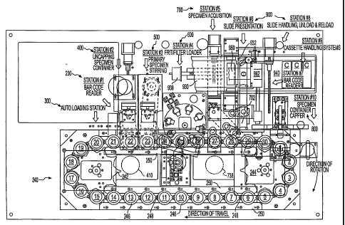
Referring to Fig. 1 l, the illustrated embodiment of the LBP device can be
compartmentalized into at least six discrete processing stations: data
acquisition station
(bar code reader) 230; uncapping station 400; primary stirnng station 500;
filter placement
station 600; specimen acquisition station 700; and re-capping station 800.
These six
stations are structured for parallel processing, meaning that all these
stations can operate
simultaneously and independently of the other. The I,BP device also includes a
separate
data reading station, a slide presentation station, a slide handling station,
and a cassette
handling station, all of which can be incorporated as an integrated system
900. The LBP
device further includes a transport mechanism 240 for moving the specimen
containers to
the various operating stations. It can further incorporate an auto loading
mechanism 300
that automatically loads and unloads specimen vials onto and from the
transport
mechanism. All stations are computer-controlled. Fig. l l a shows the
operating sequence
of the LBP device. This is the top-level table from which the operating
software is
structured.
Fig. 12 shows the basic structural elements of the LBP device, namely a frame
260
preferably made of extruded aluminum, preferably on casters (not shown) for
mobility,
and a machined aluminum base plate 262 supported by the frame and on which the
main
operating mechanisms are mounted. Beneath the base plate is a compressor 264
for
supplying compressed air for powering some of the components; a vacuum pump
(not
shown) which provides a vacuum source for various components; stainless steel
shelves
for holding the vial trays used in the auto loading mechanism 300; and
electrical
components, including power supplies and controllers, and miscellaneous
equipment. A
compressor would not be required if electrically-powered actuators were used
instead of
air-powered actuators. A user interface, e.g. a touch-sensitive LCD display
(not shown), is
mounted to the left of the transport mechanism 240 and gives the technician
control over
machine operation beyond the normal automated processing protocols. See Fig.
25, which
CA 02453252 2004-O1-05
WO 03/034034 PCT/US02/33356
shows examples of a log-in screen (top) and a navigation screen (bottom) as
they might
appear on the user interface. Of course, other screens would be presented to
the user as
he/she interacts with the user interface.
An "economy" version of the LBP device can take the form of a counter-top
model
for processing a more limited number of specimens at a time. In such a model
certain
components can be eliminated, such as frame 260 and auto loading mechanism
300, while
other components can be scaled back, such as the capacity of filter placement
station 600.
External sources of vacuum and compressed air could be used to power such a
device,
while other components (power supplies, controllers, etc.) could be
repositioned to one or
more modules adjacent to or on a modified machine base plate. Various ways of
implementing these modifications will be readily apparent to those skilled in
the art.
TRANSPORT MECHANISM
Referring to Fig. 11, the transport mechanism 240 comprises an endless link-
belt
conveyor 242 driven by a stepper motor (not shown) around precision sprockets
242, 244.
The conveyor has a plurality of receptacles or carriers 246, linked by pins
248, for
receiving a corresponding number of specimen vials. The illustrated embodiment
in Fig.
11 has 30 receptacles, numbeYed 1 through 30. Depending on the sample vial
size and the
length of the conveyor, the LBP device can use fewer than or greater than 30
receptacles,
as desired or feasible, sufficiently long to permit all processing to be
completed in a single
line.
The receptacles 246 of the link-belt conveyor are guided between the sprockets
by
pairs of guide rails 250 forming tracks, and has a conventional position
correction system .
(not shown) to accurately position the receptacles. The LBP device can track
the position
of each receptacle and step-drive or index them in a conventional manner. For
instance,
the LBP device can include linear position sensors, such as optical sensors or
a photo-
interrupter on each link, that can feed the position to a controller for
registering carrier
position and precisely indexing each carrier at each of the processing
stations along the
processing path. The manner of driving the conveyor for precise alignment and
positioning is conventional and thus will not be described further.
The guide rails 250 that form tracks in Z and Y axes engage slots machined in
the
sides of the receptacles. See, for example, Figs. 29, 33, 37 and 43. The
mechanical tracks
and drive sprockets can be constructed of a self lubricating plastic for
operation without
the need to add an external lubricant. The receptacles 246 each can have a
window 247
(see Fig. 12) for allowing access to laser or optical scanning of the bar code
on the
21
CA 02453252 2004-O1-05
WO 03/034034 PCT/US02/33356
specimen containers. The conveyor can be hard-coated aluminum, o-impregnated
with
PTFE7 for easy cleaning. The link pins 248 can be precision ground and
hardened. The
link pins can be axially fixed in location in the non-rotating link bore.
Rotating link bores
can be fitted with a suitable bearing material capable of operation without
additional
lubricant. For operator safety, the conveyor operation can be interlocked with
the cover of
the machine (not shown).
The receptacles 246 are also configured so that they receive or seat the
specimen
vials in a particular orientation. That is, the specimen vials and the
receptacles are
complementarily configured or keyed so that the vials can only be seated in
the receptacles
10, in a particular orientation. For example, the vials can be "D" shaped,
namely having a flat
side (see Figs. 2a, 2b), and the receptacles can be "D" shaped so that the
flat sides align
with each other. In this way the vials do not rotate relative to the
receptacles, while
allowing unrestricted vertical movement relative to the receptacles. In
addition to the D
shape, each vial can have a bottom notch 25 (see Fig. 2a), and the receptacles
can have a
mating peg or stud (not shown) that keys into the notch 25. While the
illustrated notch
and peg are arcuate, they can take on other mating shapes (e.g., V-shaped).
VIAL L(aADING/IJNLOADING MECHANISM
Figs. 12, 13 and 14 show the automated vial loading and unloading mechanism
300. A pivoted pick-and-place arm 304 is mounted on an elevator carriage 306
driven by
a vertical (Y-axis) lead screw motor 308 atop a vertical standard 310. Arm 304
has a
conventional electrically- or pneumatically-operated jaw-type gripper 312
adapted to grasp
and move specimen vials 10 in three degrees of freedom. Arm motion in
horizontal planes
is afforded by lateral lead screw motor 314, which is pivotally mounted in a
clevis-type
bracket 316 to elevator carnage 306. Instead of a jaw-type gripper as shown,
the pick-
and-place arm can be equipped with a conventional pneumatically operated
suction-head
type gripper as shown in Fig. 15. Such a gripper has a silicone rubber bellows
318 which
seals against the cover 30 of a vial when placed againsfthe cover and subject
to suction
through a suction line 320. Whether mechanical or pneumatic, actuation of the
gripper is
accomplished through the programmed operation of the machine as is understood
by those
skilled in the art.
Referring to Figs. 17-20, specimen vials 10 are stored in special injection
molded
plastic vial trays 330 that slide into the machine on shelves 320 (see Fig.
12). To avoid
confusion, it should be pointed out that Figs. 13-15 show a different form of
tray (made of
stamped steel), but the operation of the mechanism that rotates the trays,
regardless of
22
CA 02453252 2004-O1-05
WO 03/034034 PCT/US02/33356
their construction, is the same. The plastic vial trays 330 are the preferred
form, and are
preferably made of polypropylene. The term "tray" as used herein is not
limited to the
embodiments shown, and should be construed to cover any type of carrier,
rimmed or
rimless, that can support and move a generally planar array of discrete
articles generally in
the manner described herein.
Each tray 330 has forty-one circular recesses 332 sized and configured to
receive
specimen vials 10 only in one orientation. The upper edge of each recess 332
preferably
has a beveled edge 333, Which facilitates smooth insertion of vials. The
recesses are
arranged in a close-pack array of four concentric rows, preferably as follows.
The
outermost row has sixteen recesses; the next row in has eight recesses; the
third row in has
nine recesses; and the innermost row has eight recesses. The receptacles of
adjacent rows
are offset for closer spacing. The receptacles of the second row are radially
aligned with
the receptacles of the fourth (innermost) row. The receptacles of the
outermost row are
spaced at 18° on center. The receptacles of each of the other rows are
spaced at 36° on
center. Of course, other receptacle arrays could be used as long as they
permit access of
all vials by the pick-and-place arm 304. Each receptacle has a unique and
addressable
location, so that any vial can be accessed at will and in any sequence.
As noted above, orientation of specimen vials during the processing is
critical, so
the proper orientation of the stored vials in these trays ensures that the
pick-and-place arm
304 will properly position each vial in a conveyor receptacle 246.
Accordingly, each
recess 332 has at its bottom (see Fig. 19) a fixed indexing peg 334 that is
sized to fit into
notch 25 in the vial. The pegs 334 are installed, e.g., by adhesive, in
grooves 335 that are
molded into the tray adjacent the bottoms of the recesses 332. Some of the
pegs have been
omitted from Fig. 19 for illustrative purposes.
The pegs 334 are arranged at specific angles with respect to the median plane
of
the tray 330 such that each vial removed from the tray is delivered to a
transport receptacle
with its notch aligned with the mating peg in that receptacle, and vice versa.
Each of these
angles is dictated by the rotational position of the tray 330 when a vial in a
specific recess
332 is to be accessed by the pick-and-place arm 304, and the angular rotation
of the pick-
and-place arm from the point of vial pick-up to the point of vial placement in
the conveyor
receptacle 246. The determination of these angles is considered to be within
the abilities
of one of ordinary skill in the art.
23
CA 02453252 2004-O1-05
WO 03/034034 PCT/US02/33356
The tray 330 also has three upstanding guide posts 336, each with a spring-
loaded
ball 338 at its tip, which cooperate with guides (not shown) above each shelf
302 and
serve to guide the tray into the machine as it is inserted and ensure its
proper orientation.
The guide posts 336 also serve as stacking posts when the trays are stacked
for storage
(see Fig. 20), the balls 338 engaging dimples 339 (see Fig. 19) in the bottom
of the
superior tray.
The tray 330 also has a large flared notch 340 which is oriented toward the
machine when the tray is inserted on a shelf 302. The innermost portion of the
notch 340
has opposed keyways 342 which are adapted for engagement by floating keys, as
described below. The keyways preferably are formed in a milled brass hub
insert 343 that
is recessed flush with the top of the tray and secured thereto by screws.
Referring to Figs. 14, 15 and 15a, a rotary outer spindle 350 is journaled at
its top
and its bottom in bearings 352, 354, respectively. Outer spindle 350 engages
and rotates
only one tray at a time so that the pick=and-place arm 304 can access vials
therefrom by
moving downwardly through an opening 266 in base plate 262 and past any idle
trays via.
their homed notches 340. Fig. 14 shows the home positions of the trays in
dashed lines,
with their notches 340 aligned and embracing outer spindle 350. Spindle 350 is
rotated in
a precision manner from the bottom by a computer-controlled rotation stepper
motor 356
and a timing belt 358 engaging timing gears 360, 362. A downwardly facing
optical
rotary position sensor 363 located over the aligned tray notches detects when
and how far
a tray is rotated from. its home position and provides control feedback for
rotation of
stepper motor 356.
Within outer spindle 350 is an inner spindle 364 carrying eight pairs of
opposed
keys 365, one pair for each tray. The keys 365 project from outer spindle 350
through
opposed slots 366 in the outer spindle (see Fig. 15a, which is a sectional
view through the
spindles and the center portions of the. bottom two trays). The inner spindle
364 is moved
vertically within the outer spindle 350 by an internal lead screw 372. Lead
screw 372 is
rotated by lead screw stepper motor 374 through a timing belt 376 and timing
gears 378,
380. A key "home" sensor 382 (see Fig. 15) is located at the top of inner
spindle 364 to
provide a reference point, i.e., when the machine is turned on, it will "home"
the inner
spindle to the key home sensor 382 and then reference its movements from
there.
The even vertical spacing of the pairs of keys can be seen in Fig. 15. This
spacing,
or pitch, differs from the pitch of the keyways 342 in a full complement of
installed trays
330. Accordingly, which keyways are engaged by the keys depends on the
vertical
24
CA 02453252 2004-O1-05
WO 03/034034 PCT/US02/33356
position of inner spindle, and only one pair of lceyways (tray) can be engaged
at any time.
The enlarged view of Fig. 15a shows that the keyways 342 of bottom tray 330-1
are
engaged by keys 365, while the keyways of the tray above it, 330-2, are not
engaged by
any keys. Movement of inner spindle 364 by one-eighth the pitch difference
disengages
one tray and engages the immediately adjacent tray. The operation of the
loading and
unloading mechanism is unaffected by the absence of one or more trays from the
tray
slots, which are defined by shelves 302.
When a selected tray is to be accessed by the pick-and-place arm 304 (as
determined by the computer controller), the lead screw motor 374 moves the
inner spindle
the appropriate distance so that the appropriate keys engage the keyways of
the selected
tray. The rotation motor 356 then rotates the keyed tray to the proper angular
position so .
the arm 304 can access a particular recess 332. The superposed arrangement of
the trays,
the way in which a selected tray is accessed by the gripper 312 through the
flared notches
340 of superior trays, and the close-pack spacing of the recesses 332 in each
tray make for
an extremely compact, high capacity and efficient vial handling system that is
readily
incorporated into the compact base of the LBP device.
In the embodiment shown, the LBP device can accommodate up to eight trays
holding forty-one specimen vials each. One of the forty-one recesses can be
reserved for a
cleaning vial, which would contain a cleaning solution and be run through the
LBP device
to clean the various parts of the device that normally come into contact with
specimen
fluid. Alternatively, the forty-first vial could contain a typical control
specimen for
calibration purposes. Thus the LBP device can accommodate up to at least 320
vials
containing specimens to be processed. The device is therefore capable of
operating
continuously unattended for a long duration - at least eight hours - so that
specimen
~5 processing can be earned out even when laboratory personnel axe not
normally present,
such as at night.
When the trays 330 are bar-coded or otherwise labeled with machine-readable
identifying data, they can be used in an automated storage device that can
access a
particular tray on command. The tray-identifying data can be input into the
integrated data
management system so that the location of any specimen vial in tray storage
can be readily
ascertained.
A cost reduction in tray-based storage of specimen vials can be achieved by
using
a liner-type system in conjunction with trays 330. For example, vials can be
supported
and stored in thin sheet-like liners (not shown) that conform to trays 330 and
slip readily
CA 02453252 2004-O1-05
WO 03/034034 PCT/US02/33356
into recesses 332. The liners are stiff enough to be self supporting when
fully loaded, can
be stacked, and can be housed in wheeled carts for ease of mobility.
DATA ACCESSIONING AND SPECIMEN MANAGEMENT
It is, of course, important to keep track of each specimen vial and the
specimen
slides produced from each vial. Accordingly, the LBP device typically
communicates
with the integrated data management system (DMS) 104 through an accessioning
station
102 or other computer. Fig. 21 schematically illustrates specimen vial
handling and the
flow of data that is integrated into to operation of the LBP device. The
communication
link between the LBP device and the DMS can be made via ethernet or other
protocol
using a direct peer-to-peer connection, or through a server-based network.
The specimen processing operation begins with collecting or transferring data
from
the labeled specimen vial, e.g. via a bar code reader on a data entry terminal
or
accessioning station, to the DMS via either a direct connection or over a
network.
Specimen traclcing data can include, for example, the patient's name, test
identification .
(ID) number, patient data, and any special processing instructions. For
example, the bar-
coded specimen vial can be linked to the patient information initially by a
paper
requisition form and subsequently by an assigned, unique numerical ID in the
database. In
a preferred embodiment, the patient and test information including the vial
bar code can be
entered into the networked DMS database at the point-of care site (e.g.,
physician's
office), thereby eliminating entirely the need for a paper requisition form.
U.S. patent No.
5,963,368 (incorporated herein by reference), which is assigned to AccuMed
International,
Inc. (now Molecular Diagnostics, Inc., or MDI) discloses a similar concept as
applied to a
computer-controlled instrument for analyzing biological specimens (a
microscope) and
storing data from each analysis. The '368 patent is exclusively licensed to
MonoGen, Inc..
(the owner of this application) in the field of liquid-based cytology in
combination with or
for use with non-fluorescence based image analysis devices, processes, systems
and/or
instruments. MonoGen's commercially available pathology work station and data
management system implement the concept disclosed in the '368 patent.
Each specimen vial includes an identification (ID) symbol or~label (e.g., bar
code)
and/or a stored information label or symbol such as a hologram or a memory
chip or
device. The present embodiment contemplates reading an ID label using an
optical reader,
such as a bar code reader, which provides the information to a DMS for sharing
information between different work stations or instruments at the same or
different
locations, such as laboratories; doctors' offices, hospitals, or other patient
care providers.
26
CA 02453252 2004-O1-05
WO 03/034034 PCT/US02/33356
Fig. 21a depicts an overall laboratory system wherein the DMS is expanded to
link
specimen/patient data through a server to a variety of specimen processing
devices and/or
computerized work stations for fully integrated specimen management.
A separate bar code reader 230 (see Fig. 11) is mounted on the LBP machine
itself,
and scans all specimen vials prior to processing through a slit in each
transport receptacle
246. Each of the transport receptacles 246 is tracked using this symbol or
code, such as a
bar code that can be read with a conventional optical reading device. The bar
code readers
used in the LBP device can be any commercially available type, such as
I~eyence BL-600,
with a minimum BCR target code capability of Interleaved 2 of 5, Code 128c, or
EAN-
128. The bar code readers preferably are sealed in liquid-tight enclosures for
operator
protection. After reading, specimen vial/transport receptacle ID data are
transmitted to the
DMS of the host database or work station. The host database or local work
station can
then transmit back to the LBP device the specific processing protocol to be
performed on
that individual specimen.
~ Some of the most important functions of the data management system (DMS)
include:
Obtaining data on the patient and the specimen during accessioning, and making
this available to each instrument as required to set processing parameters and
to
provide medical data to the slide reviewer;
Maintaining chain of custody of specimens and slides to ensure data integrity;
Compiling data and printing required forms for regulatory, compliance, and
laboratory management reports;
Generating medical reports and ensuring integrity using safeguarded digital
electronic signatures;
Managing billing for instruments on "per use" charges;
Storing optimal processing protocols for each process and supplying to the
instrument in accordance with the specimen type and/or user requirements; and
Facilitating remote diagnostics and repair, and providing user manuals and
troubleshooting guides.
Fig. 21b shows an example of a relational database table that can be used to
accomplish
these tasks.
The DMS can provide paper-free data flow among the different stages of the
cytology process, saving a significant amount of personnel time and cost,
reducing
transcription errors, improving accuracy, and eliminating the space required
to store paper
27
CA 02453252 2004-O1-05
WO 03/034034 PCT/US02/33356
records. By automating and managing data acquisition, storage and retrieval,
each
operation becomes more efficient, significantly reducing the turn-around time
for
specimens. Specimen quality is enhanced by automated calibration and cross-
checking
routines that identify potential problems early. Flexible foreign language
support for
worldwide sales assists laboratories in multicultural environments.
The DMS provides a common user interface that provides detailed information on
the operation of each connected laboratory device and work station, and
together with
online user manuals and training aids eases use and minimizes training. The
DMS handles
the exchange of all relevant patient and specimen data with the users' own LIS
(or other
data management systems) through a provided software interface. Moreover,
remote
instrument diagnostic capabilities ensure maximum interruption-free operation.
The
reduction in paperwork, ready cross-compatibility with other instruments and
existing
computer networks, and integration with the central hospital or laboratory
information
system provides significant user benefits.
In typical operation, the laboratory: (1) receives a requisition from the
healthcare
provider along with the pre-bar-coded specimen vial, (2) assigns a unique ID
number
(accession number) to the specimen, and (3) based on information on the
requisition,
enters a specific LBP test ID to specify the process to be used. Fig. 23 shows
an example
of the accessioning (data entry) screen that is presented to the technician,
into which the
vial bar code, accession number and LBP process code are entered. When the
specimen
vial is loaded into the LBP device for processing, the LBP device
automatically reads the
bar code on the specimen vial and transmits the bar code number (106) to the
DMS, which
sends back the processing parameters for the selected test, and the number of
slides to be
produced. The LBP device returns an acknowledgment (108) and processes the
specimen,
making one or more slides as instructed via the DMS. Immediately before the
LBP device
imprints a specimen slide with material filtered from a specimen vial, the LBP
device
reads the bar code from the pre-bar-coded slide that is to receive the
specimen sample.
The LBP device sends each slide bar code (110) and its associated vial bar
code to the
DMS which updates the patient database with the slide bar code number, cross-
references
30~ it to the correct vial number, and signals (112) the LBP device to
proceed. The LBP
device then imprints a cytological sample from the specimen onto one or more
slides and
readies the onboard data log for the next specimen to be processed. Fig. 24
shows an
example of a DMS menu screen showing data items that are now linked in the DMS
database, including the vial number, slide numbers) and patient data. The DMS
can
28
CA 02453252 2004-O1-05
WO 03/034034 PCT/US02/33356
produce a printable report listing slide m numbers and associated vial m
numbers, patient
data and processing protocols.
At a minimum the protocol variables include specimen mixing parameters
(stirnng
speed and time) and filter selection. Typically, primary stirring speed can be
varied from
500 rpm to 3,000 rpm selectable in 50 rpm steps. The stirring interval can be
varied from
5 to 120 seconds, selectable in 5 second increments. Choice of filter type is
based on
average pore size diameter: either 5 micron (red housing), e.g. for non-
gynecological
specimens, such as sputum specimens, or 8 micron (white housing), e.g. for
gynecological
specimens, depending on the test protocol selected.
~ The LBP device is capable of processing mixed sample-runs (i.e., runs that
may
include vials containing various types of specimens) interchangeably and
without the need
for batch processing of same-type specimens. Specimen processing can include
at least
100 different processing protocols resident within the DMS and accessible to
users.
Predefined procedure codes (test ID's) such as the following can be used to
simplify
operator input and specify which processing protocol is used:
1 breast cyst, L
2 breast cyst, R
3 bronchial brushing
4 bronchial washing
' S bronchoalveolar lavage
6 cerebrospinal fluid
7 colonic brushing/wash
8 esophageal brushing/wash
9 gastric brushing/wash
10 gingival (buccal)
scrape
11 gyn PAP test
12 intestinal brushing/wash
13 nipple discharge, L
14 nipple discharge, R
15 ovarian cyst, L
_.. 16 ovarian cyst, R
17 pericardial effusion
18 peritoneal effusion
19 pleural effusion
29
CA 02453252 2004-O1-05
WO 03/034034 PCT/US02/33356
20 rectal brushing/wash
21 sputum, induced
22 sputum, spontaneous
23 urine, catheterized
.. 5 24 urine, voided
Each specimen is processed with a new filter to prevent the possibility of
cross
contamination. In the present embodiment, either of two or more different
filter types can
be specified for versatility in test selection (the device's eight filter
tubes allow for up to
eight different filter types). Processing parameters for each type of specimen
preparation
can be determined remotely and in advance, and communicated to the processing
device
using a bi-directional communication link utilizing the specimen vial bar code
as the key
identifier. The LBP device can utilize default (pre-loaded into the DMS)
process
protocols as well as laboratory-generated process protocols that users can add
to the DMS.
An overfilled-vial sensor (not shown) can be positioned at or just downstream
of
the bar code reader 230 to detect whether an excessive amount of fluid is
present in each
translucent vial. Opening and processing an overfilled vial can result in
hazardous
spillage or ejection of biological fluid. Accordingly, if an overfilled vial
is detected, the
DMS will be so notified and the complete LBP processing protocol for that vial
will be
canceled, allowing the overfilled vial to proceed through the processing path
unopened.
Alternatively, an overfilled condition can be sensed at the conveyor holder
246 into which
vials are loaded by the vial loading mechanism 300. If an overfilled vial is
detected there,
the DMS will be so notified and the loading mechanism will be instructed
immediately to
return the overfilled vial to its tray 330.
A similar approach can be used to deal with other anomalies detected as each
vial
is loaded into the conveyor. For example, a sensor (not shown) can be used to
detect an
unreadable bar code on the vial, or detect when a vial is improperly position
in the holder
246. When any such condition is detected, the DMS will be so notified and the
loading
mechanism will be instructed immediately to return the overfilled vial to its
tray 330.
Fig. 22 is a block diagram showing the components of a general purpose
computer
system or work station 270, which can be used to run the DMS. The computer
system 270
typically includes a central processing unit (CPU) 272 and a system memory
274. The
system memory 274 typically contains an operating system 276, a BIOS driver
278, and
application programs 271, such as a DMS. In addition, the computer system 270
can
CA 02453252 2004-O1-05
WO 03/034034 PCT/US02/33356
include input devices 273, such as mouse, keyboard, microphone, joystick,
optical or bar
code reader, etc., and output devices, such as a printer 275P, and a display
monitor 275M.
The computer system or work station can be connected to an electronic network
280, such as a computer network. The computer network 280 can be a public
network,
such as the Internet or Metropolitan Area Network (MAN), or other private
network,, such
as a corporate Local Area Network (LAN) or Wide Area Network (WAN), or a
virtual
private network. In this respect, the computer system 270 can include a
communications
interface 277, such as ethernet, USB, or Firewire, which can be used to
communicate with'
the electronic network 280. Other computer systems 279, such as a remote host
database,
other types of work stations including automated analyzers, and computers or
databases
(e.g., LIS) of a hospital, laboratory, or other medical establishment, can
also be linked to
the electronic network 280. Other LBP devices, as well as other types of
specimen
processing instruments (e.g., automated slide stainers and coverslippers) 279a
can also be
connected to each other and the DMS via the network.
One skilled in the art would recognize that the above-described system
includes
typical components of a general purpose computer system connected to an
electronic
network. Many other similar configurations can be used to control the LBP
device and its
processes. Further, it should be recognized that the computer system and
network
disclosed herein can be programmed and configured by one skilled in the art to
implement
the methods, system, and software discussed herein, as well as provide
requisite computer
data and electronic signals to implement the present invention.
In addition, one skilled in the art would recognize that the "computer"
implemented invention described further herein may include components that are
not
computers per se, but include devices such as Internet appliances and
Programmable Logic
Controllers (PLCs) that may be used to provide one or more of the
functionalities
discussed herein. Furthermore, while "electronic" networks are generically
used to refer
to the communications network connecting the processing sites of the present
invention,
one skilled in the art would recognize that such networks could be implemented
using
optical or other equivalent technologies. One skilled in the art would
recognize that other
system configurations and data structures can be provided to implement the
functionality
of the present invention. All such configurations and data structures are
considered to be
within the scope of the present invention. In this context, it is also to be
understood that
the present invention may utilize known security and information processing
measures for
transmission of electronic data across networks. Therefore, encryption,
authentication,
31
CA 02453252 2004-O1-05
WO 03/034034 PCT/US02/33356
verification, compression and other security and information processing
measures for
transmission of electronic data across both public and private networks are
provided,
where necessary, using techniques that are well known to those spilled in the
art.
UNCAPPING STATION
One of the advantages of the present vial-based LBP device and system is that
it
minimizes operator exposure to the specimens, which can contain potential
biohazards.
Refernng to Figs. 26-31, the LBP device has an uncapping mechanism 400 that
first
automatically separates the stirrer 40 in the vial from cover 30, and then
removes and
discards the cover - all without intervention by an operator. See Fig. 26,
which shows the
stirrer resting on vial ribs 26 after the cover 30 is removed.
A closed specimen vial 10 which has arrived at the uncapping station in its
transport receptacle 246 is met by an uncapping head 402 which is lowered onto
the cover
30 of the specimen vial. See Figs. 27 and 28. Uncapping head 402 has four
tapered legs
404 that form a tapered gripping cavity having chisel-like inner edges 406
spaced and
sized to progressively tighten onto cover 30 as head 402 is lowered. Once the
cover is
tightly engaged by the legs, a central spindle or plunger 408 is lowered into
contact with
the center of cover 30 and applies a downward force to the cover to cause the
stirrer 40 to
detach from the cover 30, as described above, and drop down in the vial onto
ribs 26.
Then the plunger is retracted and the uncapping head 402 is rotated
counterclockwise (Fig.
28) to unscrew cover 30 and remove it from container 20. Thereafter the
uncapping head
with the removed cover in its grip moves laterally to the position shown in
dashed lines
410 in Figs. 29 and 11, and plunger 408 is again lowered, this time to eject
cover 30,
which falls into a waste chute or bin (not shown) beneath the uncapping head.
Alternatively, a movable waste chute can be brought beneath the uncapping head
to catch
the ejected cover, so that lateral movement of the uncapping head is not
required. Covers
are not reused to eliminate the possibility of cross-contamination.
The plunger 408 is driven by a pneumatic cylinder 412, mounted on an L-bracket
415 at the top of the uncapping head, that can apply a force on the cover of
up to about 30
lbs. A coil spring 413 returns the plunger to its retracted position when
cylinder 412 is
deactivated. The head 402 is capable of applying an uncapping torque through
the
gripping legs of up to about 10 lb-ft, which is sufficient to loosen the
cover. The gripping
legs can be of the self energizing type so that precise alignment with the
cover or small
variations in cover geometry do not frustrate their grip.
32
CA 02453252 2004-O1-05
WO 03/034034 PCT/US02/33356
The uncapping mechanism has a mounting frame 414 supported on blocks 416 that
slide laterally of the processing path on rails 418. A Y-axis stepper motor
420 and lead
screw 422 effect lateral motion. The uncapping head 402 is rotatably mounted
in a
bearing block 424. Bearing block 424 is secured to a C-frame 426 that is
vertically
slidable on mounting frame 414. Vertical movement of C-frame 426 and, hence,
uncapping head 402 is effected by Z-axis stepper motor 428 and lead screw 430.
Lead
screw 430 can be vertically compliant to accommodate upward movement of the
cover 30
as it is unscrewed. However, it is preferred that stepper motor 428 be
actuated during the
uncapping sequence so that head 402 rises at about the same rate as, but no
faster than, the
unthreading cover. Uncapping head 402 is rotatably driven by uncapper motor
432 .
through a gear reduction unit 433, a timing belt 434 and timing pulleys 436,
438.
The uncapping head described above would also work with vials having a
conventional "press and turn" bayonet-type coupling between the container and
the cover.
The downward force of the plunger 408 would be sufficient to release the
internal anti-
turn lock of the coupling, allowing the gripper to rotate and remove the
cover. Vials
having covers that do not require rotation for removal, e.g., a snap-on cover,
would require
a differently designed uncapping head, tailored to the type of cover
connection involved.
Alternatives to the above-described plunger 408 can be employed at or upstream
of
the uncapping station for applying the required external force to the covered
vial to effect
separation of the stirrer from the cover. For example, a cam, lever arm or
other movable
mechanical element can contact and press down on the cover. Alternatively, an
abrupt
upward external force can be applied to the vial to yield an acceleration
force that
overcomes the frictional retention force between couplers 35 and 47,
effectively pulling
the stirrer out of engagement with the cover. This can be done by, e.g.,
moving the closed
vial rapidly downwardly to rap the bottom of the container 20 against a rather
hard
surface, e.g., by mechanically and/or pneumatically thrusting the closed vial
into the
transport carrier 246 that will hold the vial during the subsequent processing
steps, or by
dropping the vial down a chute into the carrier a~sufficient distance to
dislodge the stirrer.
Another way to exert an abrupt upward external force on the vial is to strike
the bottom of
the container 20 with a striking member. This can be accomplished by, e.g.,
cradling the
container 20 and momentarily thrusting a striker against the bottom of the
container, e.g.
through a bottom opening in the vial carrier 246, by pneumatic and/or
mechanical means
The design of these and other variants of suitable automated mechanisms for
accomplishing these tasks is within the grasp of those skilled in the
mechanical arts.
33
CA 02453252 2004-O1-05
WO 03/034034 PCT/US02/33356
PREPROCESSING (PRIMARY STIRRING) STATION
After uncapping is completed, the transport mechanism indexes the specimen
container to a station where preprocessing occurs. The preprocessing station
is the
location at which preprocessing operations, such as specimen dispersal within
its
container, are performed prior to the container and its specimen moving to the
specimen
acquisition station. The preprocessing station typically performs a dispersal
operation. In
the preferred embodiment, the dispersal operation is performed by a mechanical
mixer,
which rotates at a fixed speed and for a fixed duration within the specimen
container. In
this example, the mixer serves to disperse large particulates and microscopic
particulates,
such as human cells, within the liquid-based specimen by homogenizing the
specimen.
Alternatively, the specimen may contain subcellular sized objects such as
molecules in
crystalline or other conformational forms. In that case, a chemical agent may
be
introduced to the specimen at the preprocessing station to, for example,
dissolve certain
crystalline structures and allow the 'molecules to be dispersed throughout the
liquid-based
specimen through chemical diffusion processes without the need for mechanical
agitation.
In this example, the chemical preprocessing station introduces its dispersing
agent through
the preprocessing head.
In the illustrated preferred embodiment preprocessing occurs at the primary
stirring
station 500, which uses a specified or instructed stirring protocol to stir
the specimen, if
needed, using the stirrer 40 in the container, at a specified speed (rpm) for
a specified
duration. The stirring protocol chiefly depends on the specimen, as described
above, and
is~ normally intended to disaggregate any mucous material and disperse it
and/or other
particulate material in the specimen liquid.
Referring to Figs. 32-35, the primary stirring station 500 has a stirring head
502 in
the form of an expanding steel collet. The collet is formed at the lower end
of a shaft 503
which splits into six flexible fingers 504 defined by six equally spaced slits
506. 'Shaft 503
is rotatable in a bearing block 508 secured to a C-frame 510 that is
vertically slidable on a
mounting frame 512. Vertical movement of C-frame 510 and, hence, stirring head
502 is
effected by a Z-axis stepper motor 514 and a lead screw 516. Stirring head 502
is
rotatably driven by a stirnng motor 518 through a timing belt 520 and timing
pulleys 522,
524.
The inner surfaces of the collet fingers 504 taper uniformly inwardly toward
the
lower end of the collet. A central plunger 526, movable vertically by a
pneumatic cylinder
528 atop a bracket 530, expands the fingers 504 outwardly when it descends and
34
CA 02453252 2004-O1-05
WO 03/034034 PCT/US02/33356
encounters the narrowing passage defined by the tapering forgers. Thus the
diameter of
the lower end of the stirring head (collet) 502 increases when the plunger
descends. This
end is sized to fit loosely but closely within the annular wall 47 at the top
of stirrer 40
when the collet is not expanded. When plunger 526 descends, the forgers 504
expand
outwardly to wedge against the inside of wall 47, in manifold M, securely
engaging the
stirrer.
In operation, the stirnng head 502 is first lowered so that the collet enters
the
manifold M. The dashed motor and bracket lines in Figs. 33 and 34 indicate
this lowered
position. Then plunger 526 descends to lock the stirring head to the stirrer.
Then the
stepper motor 514 is operated to slightly raise the stirring head and the
attached stirrer 40.
This vertical movement need only be very small, such as 0.050 in., just to
free the stirrer
from the ribs 26 and prevent interference with the container during stirnng.
Then DC
stirring motor 518 is operated in accordance with the specimen-specific
stirring protocol.
Stirring speed can vary, and is usually in the range of about 500 rpm to about
3,000 rpm.
The stirnng time can vary from about 5 seconds to about 90 seconds. The base
or bottom
wall 41 of the stirrer acts as a slinger to thrust any liquid that may rise
along the stirrer
against the container wall, and prevents the escape of liquid from the
container.
Withdrawing the plunger 526 from the collet releases the stirrer 40 from the
collet 502 so
the specimen container can move on to the next station.
A contracting collet could be used instead of expanding collet 502. In that
case,
the collet fingers would fit around the outside of annular wall 47, and would
be squeezed
together to clamp around the wall by a descending sleeve that surrounds the
fingers.
FILTER PLACEMENT STATION
At the filter placement station 600 an appropriate filter assembly F (see Fig.
5) is
loaded into the open manifold M at the top of the stirrer 40. Filter
assemblies can come in
different filter configurations for automated machine recognition. For
example,.one set of
filter assemblies can be colored red (5 micrometers); another set white (8
micrometers),
each having different filtering properties, and a color sensor can detect
which type of filter
is before it and cause the proper filter to be loaded. The filter assemblies
are dispensed by
' a pusher from a magazine having multiple filter tubes.
Figs. 36-40 show the structure and operation of the filter placement station.
Referring to Figs. 37 and 40, a filter dispensing head 610 comprises a filter
magazine in
the form of a turret 612 rotatable on a spindle 614 by a stepper motor 616.
Vertical post
611 provides the main support for the turret. Turret 612 has a top support
plate 618 with
CA 02453252 2004-O1-05
WO 03/034034 PCT/US02/33356
eight equally spaced holes 620 near its periphery, each hole opening through
the edge of
the plate 618 with a slot 622. A bottom guide plate 624 on spindle 614 has a
similar
arrangement of holes that are aligned with the holes and slots in the top
support plate.
Eight steel filter tubes 626, each having an upper support shoulder 628, are
supported vertically in holes 620 and the aligned holes beneath them, with
shoulders 628
resting on the top of top plate 618. Each filter tube 626 has a full-length
slot 630, and its
bottom portion is split into four springy fingers 632 by slots 634. Just above
the bottom
end the forgers 632 curve inwardly, forming rounded inner shoulders 636
against which a
filter assembly F rests. The filter tube is dimensioned such that the
shoulders 636 keep up
to a full stack of filter assemblies F from falling out of the tube, but
deflect to allow a filter
assembly to pass when the stack is pushed downwardly without damage to the
filter
assembly. Fingers 632 thus form a springy choke.
Fig. 39 shows the position of the filter magazine 612 in relation to the
processing
path and the adjacent processing stations,~namely the primary stirring station
500 to the
left, and the specimen acquisition station 700 to the right, all located on
one side of the
processing path as defined by guide rails 250. On the other side of the
processing path
opposite the filter magazine 612 is the assembly that supports and drives a
pusher arm
640. This assembly comprises a support post 642 supporting a Z-axis lead screw
644
driven by a stepper motor (not shown) which moves a shuttle 646 that carries
pusher arm
640. A filter sensor 650 positioned opposite bottom guide plate 624 monitors
the passage
(drop) of the lowest filter assembly F in the filter tube presented to (i.e.,
directly above)
the specimen container. Sensor 650 also detects when the filter tube is empty.
A second
sensor 651 monitors filter type.
Filter assemblies of the same type are stacked in the proper orientation, with
the
membrane filter side (beveled edge) facing down, in each tube. For example, 54
filter
assemblies can be housed in each tube; thus a total of 432 filter assemblies
can be loaded
into the magazine. Fifty-four filter assemblies can be prepackaged in a stack
that is
inserted into a filter tube with a wrapper tab projecting from slot 630, and
unwrapped by
pulling the tab outwardly. Alternatively, filter assemblies of the same type
can be dumped
onto a vibratory feeder, which can recognize their orientation by geometric
configuration,
and properly orient and feed the filter assemblies onto the tubes. Several of
these feeders
can be used, one for each type of filter assembly.
In operation, with the pusher arm 640 in its home (top) position, indicated by
the
dashed shuttle outline in Fig. 38, the filter magazine 612 is rotated by
stepper motor 616
36
CA 02453252 2004-O1-05
WO 03/034034 PCT/US02/33356
until sensor 650 detects the presence of the specified type of filter assembly
in the filter
tube before it. Shuttle 646 then moves downwardly with pusher arm 640 moving
through
slot 630 to press the stack of filter assemblies in that tube downwardly,
until the lowest
filter assembly drops from the tube into the manifold M in stirrer 40. When
filter drop is
sensed, the shuttle 646 with its pusher arm 640 stops its advance. In an
alternative
arrangement, a weight sensor can be used to monitor the weight of the filter
stack, ,and
detect by weight change when a filter assembly has dropped from the stack and
when the
filter tube is empty.
The use of eight filter tubes 626 in magazine 612 enables unattended
processirig.of
all of the specimens housed in the trays of the vial autoloader 300. For a
counter-top
model of the type described above, however, a single filter tube supported in
a fixed
position above the processing path would suffice for processing specimens that
require the
same type of filter.
SPECIMEN ACQUISITION AND CELL DEPOSITION STATION
Referring to Fig. 41, specimen acquisition station 700 has a suction head 702
that
descends to engage the upper portion of the stirrer 40. Before drawing a
vacuum on the
specimen through the filter assembly F, the suction head grips, slightly lifts
and rotates the
stirrer 40, this time more slowly than at the primary stirring station
(typically no more than
500 rpm for a 5 second interval), to re-suspend the particulate matter in the
specimen
liquid. The re-stir motor can be a Maxon 24 volt DC planetary gear-reduced
type. Then
suction is applied through suction line 750 to aspirate specimen liquid from
the container
20 through suction tube 43, into the particulate matter separation chamber
(manifold) 46
and through the filter assembly F, leaving a monolayer or thin layer of
uniformly
deposited cells on the bottom surface of the filter as described above. It may
also be
possible to rotate the stirrer slowly while the specimen liquid is being
aspirated.
Fig. 6 shows how the suction head cooperates with the annular wall 47 of the
stirrer manifold and the filter assembly F therein. The outer portion 704 of
the suction
head envelops the wall 47 and has an O-ring 760 that seals against the outside
of wall 47.
The inner portion 706 of the suction head has two concentric O-rings 762, 764
that seal
against the top of filter holder 200. Suction applied through port 750 creates
a vacuum
around central opening 204 and within filter holder 200, which draws liquid
into the
manifold 46 and through the filter 202. An O-ring 766 is,interposed between
the inner and
outer portions of the suction head.
37
CA 02453252 2004-O1-05
WO 03/034034 PCT/US02/33356
Referring to Fig. 42, when aspiration of the specimen is complete, the suction
head
702 is raised. The inner portion 706 of the suction head is extended at the
same time by
action of a pneumatic cylinder (not shown) mounted above the suction head. As
the
suction head 702 is raised, the outer portion 704 disengages from the stirrer
40, but the
filter assembly F is retained on the inner portion 706 by application of a
vacuum through
suction line 752 to the annular space between O-rings 762 and 764. Thus the
suction head
702 removes filter:assembly F from the stirrer, and can continue to apply
light suction via
suction line 750 through the filter to effect a desired degree of moisture
control of the
cellular material on the filter.
The suction head 702 then moves laterally away from the transport conveyor by
pivoting 90° about a vertical axis to the cell transfer position "P"
shown in Fig. 46, to
position the filter assembly F over a microscope slide S delivered from a
slide cassette at
slide presentation station 9f0. This pivoting movement of suction head 702 can
also be
seen in Figs. 11 and 39. The inner portion 706 of the suction head 702 then
moves
downwardly to press the filter against the slide S with a tamping force in the
range of 4 to
8 lbs. and transfer the monolayer of cells thereto. The phantom lines in Fig.
42 show this
change in position of suction head 702 and contact of the filter with slide S.
Instead of
being pivotally mounted, the suction head 702 could be mounted for rectilinear
movement
to and from a different deposition site where slides are presented, e.g.,
above the
processing path.
Referring to Figs. 43-46, suction head 702 is rotatably mounted on a boom 716
that
also carries the re-stirnng motor 718, which rotates suction head 702 through
a timing belt
720. Boom 716 is pivotally supported about a vertical axis 721 on a slide 722,
which is
vertically movable along frame support 724 by means of a Z-axis stepper~motor
726 and a
lead screw 728. Motor 726 thus moves the entire suction head vertically.
Pivoting motion
of boom 716 is effected by stepper a motor 717 operating through a gear train
(not shown).
Vertical motion of the inner portion 706 of the suction head is effected by a
pneumatic
cylinder and return spring (not shown) mounted above the suction head to an L-
bracket
719, substantially identical to the arrangement~412; 413, 415 (see Fig. 29)
used to move
the plunger 408 of the uncapping head 402.
The frame support 724 is mounted on a slide 730 so as to be movable laterally
of
the transport path. A Y-axis stepper motor 732 and a lead screw 734 effect
this
movement. After the slide is printed the suction head is raised by the Z-axis
motor, and
38
CA 02453252 2004-O1-05
WO 03/034034 PCT/US02/33356
the Y-axis stepper motor 732 advances the entire assembly to the dashed line
position "X"
shown in Fig. 43. Then the suction head pivots back to its original
orientation, transverse
to the transport path (position "S" in Fig. 46). The Y-axis stepper motor 732
then pulls the
entire assembly back toward its original position (solid lines in Fig. 43). As
the suction
head 702 moves (to the right as seen in Fig. 43), the still-retained filter
assembly F is
"scraped" off the suction head by the edge 736 of an open-top used filter
(waste) tube 738
(see also Figs. 11 and 39). This leaves suction head 702 free to engage a
fresh filter
assembly.
The vacuum 'source that communicates with the suction head 702 pulls a slight
vacuum, e.g., in the range of 3 in. to 10 in. Hg (adjustable by, a regulator),
through suction
line 750 to aspirate specimen liquid and draw it through the filter assembly
F. The
separately regulated vacuum applied through suction line 752 for holding the
filter
assembly to the suction head 702 is higher, on the order of 20 in. Hg.
Formation of high-quality specimens on microscope slides depends critically on
the deposition of a monolayer of cells of specified concentration (i.e.,
number of cells per
unit area) on the surface of the filter that will contact the slide. That, in
turn, depends
critically on the aspiration rate and/or the aspirated flow volume. Since.cell
concentration
on the filter surface is a function of the number of filter pores blocked by
the solids
suspended in the specimen liquid, the percent of flow reduction from the
maximum open
filter condition correlates to the blockage or amount of accumulation on the
filter.
Because of the~nature of biological specimens, solid particle concentration is
a significant
variable in the process and must be taken into consideration. Also, it is
important to
identify the total volume of material filtered on a real time basis for other
processing
operations.
The specimen acquisition station thus further includes a deposition control
system
for controlling the liquid draw vacuum duration by monitoring the flow rate
and/or
aspirated volume. The monitored flow rate or aspirated volume can be used to
signal
vacuum cut-off and/or suction head retraction, which correlates to the
specified
concentration of cells collected on the membrane filter surface. If a
specified
concentration factor is not achieved before a specified volume of fluid is
aspirated, the
system can also issue a retract signal.
Different types of deposition control systems or modules can be used for these
purposes. Fig. 47 schematically shows one such system, which has a meter in
the form of
a digital level detector positioned along a fluid column. This "bubble flow"
system can
39
CA 02453252 2004-O1-05
WO 03/034034 PCT/US02/33356
use sensors in the form of a plurality of LED emitters and corresponding
number of
photosensors, such as Omron sensor, EE-SPX613 GaAs infrared LED, placed along
the
length of the column. Any other type of sensors may be used. Alternatively,
LED sensors
such as the. Omron sensors mentioned above can be used without corresponding
emitters
when they are positioned just at the edge of a glass tube. The meniscus edge
of the liquid
in the tube diffracts the light passing through the tube, and the sensor will
detect the
shifted light pattern when the rising meniscus edge reaches the sensor.
The fluid column is formed in a vertically extending transparent tube or
cylinder
770, e.g., one made of Pyrex glass 9 mm in diameter by l mm thick. The
aspirated
specimen fluid is drawn from the specimen container through the membrane
filter, and
pulled into the glass cylinder 770 via suction line 750 and a 3-way valve 778,
by means of
a vacuum source 772 connected to the top of the cylinder. The sensors 774 are
positioned
evenly along the length of the cylinder 770, preferably at 1.5 ml capacity
intervals, and are
interfaced with a controller or microprocessor 776.
In operation, in the normal state, with no fluid in the tube 770, the sensor
relay line
is "low." Vacuum begins to draw fluid into the tube through the filter, and
the controller
marks the beginning of the draw sequence. When the fluid reaches the first
sensor, the
first sensor relay line goes "high." The controller marks the time it took for
the fluid to
reach the first sensor, indicating the nearly free-flow condition of the
filter, and the
relative viscosity of the fluid in the test. When an additional 1.5 ml of
fluid is drawn into
the tube, the second sensor relay line goes "high." The time interval for the
first 1.5 ml of
fluid (between the first and second sensors) is noted by the controller, and
this becomes
the reference time base. As each additional 1.5 ml of fluid is drawn into the
system (and is
detected by succeeding sensors), the time base for that increment is computed.
When the
incremental time base reaches an empirically derived percentage (e.g., 120%)
of the
original (reference) time base, the controller indicates that cell collection
is completed, and
a stop signal is transmitted, preferably to retract the suction head 702 from
the manifold in
the specimen container. The empirically derived figure mentioned above is
variable with
the protocol and directly controls the cellularity of the specimen sample.
The best approximation of the free-flow condition of the filter is obtained if
the
time it takes for the fluid to reach the first sensor 774 is kept to a
practical minimum. This
can be accomplished by incorporating the first sensor into the suction head
itself, as
schematically illustrated in Fig. 47a. In this embodiment, inner portion 706
of the suction
head carries an emitter 774a and an opposed sensor 774b, which 'detects the
leading edge
CA 02453252 2004-O1-05
WO 03/034034 PCT/US02/33356
of the fluid column very close to the filter assembly F. The outer portion
704, which has
teeth 775 engaged by timing belt 720 (not shown), is rotatable about the inner
portion 706
(note interposed bearing 773) to rotate the stirrer (not shown) and stir the
specimen prior
to aspiration.
During the specimen drawing operation, the controller records the cumulative
or
total aspirated volume. If the cumulative volume'reaches a predetermined level
before
reaching the predetermined flow rate reduction from the reference flow, the
controller will
also issue a stop signal and a flag indicating that the stop signal issued not
as a result of
desired reduced flow, but by reaching the maximum liquid draw limit. A slide
formed
under the flagged condition will likely form a hypo-cellular condition. The
controller can
imprint the slide and indicate to the DMS that a hypo-cellular condition
likely exists.
Accordingly, if the flagged condition exists, the controller issues a signal
to purge the
liquid in the cylinder 770 and initiate a second draw. The cylinder is purged
of all liquid
after each sample is taken.
Refernng to Fig. 48, the deposition control system can have a purge value so
that
when the draw cycle is completed, the stop signal generated by the controller
776 will
open the purge valve to vent the vacuum supply line to the atmosphere and
divert the
liquid remaining in the cylinder 770 into a waste container. The cylinder 770
can be
maintained under a negative pressure. The system is then ready for the next
cycle.
Specifically, the system can have a 2-way solenoid valve V-3 in the suction
line with one
port 780 open to the atmosphere. The bottom of the cylinder 770 is connected
to a valve
manifold 782 with two solenoid valves V-2, V-4. The solenoid valves can be Lee
LF
series designed for use in vacuum systems, 2-way valve LFVA 2450110H, viton
seal, 24
volt and 3-way valve, LFRX 0500300B, viton seal, 24 volt. The 2-way valve V-4
can port
the specimen liquid to the bubble flow cylinder 770, or to vacuum by-pass 784.
The 2-
way valve V-2 can control the filter dehydration vacuum source. Fig. 49
illustrates the
valve logic.
The deposition control system can use an analog level indicator instead of the
digital sensors 774. The analog level indicator senses capacitance of the
aspirated liquid.
The difference is only in the method of sensing the volume and fill rate of
the liquid in the
cylinder 770. Here two spaced electrodes are used, one around the outside of
the cylinder
770 and the other positioned down the center of the cylinder the cylinder,
separated from
the aspirated liquid by a dielectric. ~ A high frequency, such as 10 kHz, low
voltage current
is applied across the electrodes. Capacitance in this system is measured by a
bridge
41
CA 02453252 2004-O1-05
WO 03/034034 PCT/US02/33356
circuit, which provides an analog indication of capacitance in the circuit. As
fluid fills the
column, capacitance in the circuit increases. A lOX differential in direct
capacitance is
easily obtained with this system. Capacitance is indicated on a real time
basis and can be
sampled frequently enough to provide control of the sampling system. This
arrangement,
like the first two, uses a computer or microprocessor and a bubble flow
technology to
measure the flow rate and the total fluid volume in real time. The
predetermined volume
increment for these arrangements can be in the range of about 0.1 ml to 5.0
ml, and .
preferably is in the range of about 1.0 to 2.0 ml.
A different system can use an ultrasonic indicator for measuring fluid
movement
through a tube. The ultrasonic system uses ultrasonic wave propagation through
a moving
liquid. In this regard, the third system employs an ultrasonic emitter and
detector clamped
across the liquid draw tube (suction line 750) operating on the distal end of
the filter
assembly F. This system provides a digital indication of fluid flow in the
tube, the total
volume aspirated through the tube being calculated by a flow interval
calculation. It
measures phase shift from the ultrasonic wave generator source to a detector
for measuring
flow speed.
Another way to measure aspirated fluid volume and control the duration of the
specimen draw is to detect the change in the weight of the specimen vial. This
can be
accomplished by using a sensor that makes a high-precision measurement of the
weight or
mass of the vial containing the specimen that is being aspirated. Vial weight
or mass is
repeatedly measured at a high frequency such that the rate of change of the
weight or mass
of the vial is accurately determined. Specimen aspiration is completed when
the rate of
change in weight or mass has diminished by a predetermined amount or
percentage from
the initial rate. The weight sensor can be, e.g., a load cell in each conveyor
receptacle 246,
or a single load cell beneath the conveyor at the specimen acquisition head
that rises to
engage the container.above it. In either case, the specimen acquisition head
can be raised
slightly during aspiration to unload the container so that the load cell can
measure only the
combined weight of the container and the remaining specimen.
Although specimen acquisition preferably is accomplished through aspiration
(using a vacuum), it can also be accomplished by pressurizing the container 20
through an
appropriate head that seals against the top of the container and forces
specimen liquid up
through tube 43 and through the' filter assembly by means of positive
pneumatic pressure.
The fluid volume control schemes and mechanisms described above would also
work in
conjunction with such a pressurized specimen acquisition system.
42
CA 02453252 2004-O1-05
WO 03/034034 PCT/US02/33356
The cell concentration can be selected from low to high by defining flow
control
cut-off. For a typical low cellularity result, the cut-off can be 80% of the
120% reference
discussed above, and for high cellularity the cut-off can be set at 60% of
the.reference,
selectable in 5% increments. The number of slides per specimen can range from
one to
three. Some of the typical default protocols are as follows:
GYN: 1,000 RPM stir, 30 second interval, 8-micrometer filter, 60% - high
cellularity, one slide.
Urine: 1,000 RPM stir, 20 second interval, 5-micrometer filter, 70% - medium
cellularity, one slide.
Lung sputum: 3,000 RPM stir, 120 second interval, 5-micrometer filter, 80% -
high
cellularity, two slides.
RE-CAPPING STATION
After completing the specimen processing cycle, the specimen container is
resealed
with the stirrer still inside the container. It is preferred to use a thin,
polypropylene-coated
aluminum foil to form the new cap, which is available in roll form. The foil
is drawn
across the open end of the specimen container, thermally bonded to the
container at a seal
temperature of about 365° F applied for about 3 seconds with a seal
force of 3 pounds, and
cut from the roll. Of course, any other type of re-capping material can be
used as long as
it is compatible with the vial material and creates a safe and reliable seal.
For example, a
foil bacleed with a thermosetting resin adhesive could be used; a sticky-
backed foil could
be used that does not require heat to effect a seal; or a plastic seal
material can be bonded
to the container ultrasonically. To enhance unattended operation, an automatic
threader
could be included for threading a new roll of sealing material into the re-
capping
mechanism. Cutting caps from a roll can be eliminated if roll-mounted pre-die-
cut closures
having peel-off tabs are fed to the re-capping mechanism.
Referring to Figs. 50 and 52, the re-capping mechanism 800 has a side support
plate 802 secured to the machine base plate. The side support plate carnes a
main frame
810 having a top plate 812 with slots 814, 816, and two side plates 818, 820.
A driver
capstan 822 is journaled in side plates 818, 820. A foil advance motor 824,
mounted on a
bracket 826, drives the capstan. A pressure roller 828 is pivotally mounted to
the main
frame 810 and resiliently engages the capstan under the influence of a spring
830.
Capstan 822 and pressure roller 828 define between them a throat through which
the foil
runs, and have resilient surfaces which grip the foil for positive feed. A
handle 832 allows
43
CA 02453252 2004-O1-05
WO 03/034034 PCT/US02/33356
the throat to be opened manually to allow the end of the foil to be fed into
the throat after
first passing through slot 814. A spindle 804, carried side support plate 802,
supports a
replaceable roll of foil.
Fig. 51 shows the foil path 834 through the throat. An L-shaped cutter 836 is
pivoted at its elbow to the rear of main frame 810. One end of a single-acting
pneumatic
cutter actuator cylinder 838 is mounted on a bracket 840, and the other end of
the cylinder
is linked to the upper leg 842 of cutter 836. The lower leg of the cutter has
a blade 844
that normally rests above the foil path downstream of the throat, held in that
position by a
spring 845 linked between the upper leg 842 and the support plate 802.
A rear post 850 pivotally supports an arm 852 that extends forwardly toward
main
frame 810. Arm 852 carnes a heated platen 854 and a foil guide fork 856 having
two tines
that extend toward the throat and are spaced apart so as to allow the platen
854 to pass
between them. Arm 852 is kept elevated, in the rest position shown in Fig. 51,
by a spring
858. During the re-capping operation a single-acting pneumatic cylinder 860
pulls down
on the arm 852 to lower the platen 854 and the guide fork 856. Note the
position of a
container 20 in a transport receptacle (not shown) beneath the platen 854.
In operation, the foil advance motor turns the capstan 822 to feed a measured
length of foil past the cutter blade 844, into the fork 856, and to the
position shown by the
dashed line in Fig. 51. A photocell 862 detects the leading edge of the foil
and signals the
motor to stop. Then cylinder 838 is actuated to cut the foil, and cylinder 860
is actuated to
pull arm 852 down to the seal position. The cut length of foil is sandwiched
between the
platen 854 and the container 20, and the container is sealed. After about
three seconds
cylinder 860 is deactivated and the arm 852 rises, returning to its rest
position. A vacuum
assist (not shown) optionally may be used to help hold the cut length of foil
in position on
the platen prior to sealing.
The foil caps applied by the re-capping mechanism are approximately square in
shape. The corners of the foil caps can protrude from the vials and interfere
with other re-
capped vials that are returned to the trays 330. Accordingly, a foil folding
ring 870 (seen
in phantom lines in Fig. 51) preferably is provided which acts to fold the
edges and
corners of each foil cap down along the side of the container. The foil
folding ring 870
preferably is mounted to act on the vial in the transport position.immediately
downstream
of the re-capping mechanism, i.e., position "FF" in Fig. 51, and may be
mounted on the re-
capping mechanism itself, e.g., to main frame 810, so that actuation of
cylinder 860 serves
simultaneously to apply a foil cap to one container and fold the edges and
corners of the
44
CA 02453252 2004-O1-05
WO 03/034034 PCT/US02/33356
foil cap of the preceding (downstream) container. Alternatively, the foil
folding ring or an
equivalent foil folding mechanism can be mounted further downstream of the re-
capping
mechanism so as to act independently thereof.
Foil folding ring 870 is a metal ring having an inner diameter that is
slightly larger
than the outside diameter of the threaded portion of the container 20. The
ring 870 is
mounted on an arm (not shown) that moves downwardly when actuated to lower the
ring
870 over the upper end of the container. As the ring encircles the container,
it folds the
overhanging portions 872 of the foil cap against the side of the container.
When the ring
rises after folding the foil, the container is held in position in its
transport receptacle by a
pin (not shown) that is mounted on a leaf spring (not shown) and is situated
in the center
of the ring 870. The leaf spring is carried by the arm that holds the ring, so
the pin
resiliently presses down against the center of the foil cap until the arm and
the ring retract
fully.
The foil seals applied to the processed containers are easily punctured by a
syringe
or a pipette to obtain further liquid specimen samples. The seals are very
durable,
however, withstanding rough handling and preventing leakage in low ambient
pressure
conditions, e.g., in aircraft flying as high as 40,000 ft. Further, the
appearance of the foil
seal makes it readily distinguishable from the cover of an unprocessed vial,
making
handling by low-skilled operators virtually foolproof. To avoid the potential
of puncturing
the foil seal inadvertently, the re-sealed container can be capped with an
unused screw-on
cover of a distinct color.
SLIDE HANDLING AND PRESENTATION
The LBP device can use 30 and 40 slide plastic magazines (cassettes), which
can
accept standard 25 mm x 75 mm x 1 mm and 1 x 3 x 0.040 in. slides. Metric and
inch
based slides can be used interchangeably. Figs. 52-55 show a 40-slide cassette
C suitable
for use in the LBP device. The slide cassette is in some respects similar to
that disclosed
in U.S. patent No. 5,690,892 (incorporated herein by reference), but is
specially adapted
for use in other devices as well, such as an automated stainer, an automated
image
analyzer, and a pathology work station, so that the slides do not have ~to be
unloaded and
reloaded into different magazines for use in those devices. Machine-readable
indicia on
the cassette, such as a bar code or an embedded microchip, provides cassette
information
that can be linked by the DMS to thebar codes on the slides in the cassette so
that the
location and status of any cassette and any slide in that cassette can be
tracked in a
laboratory system. The cassettes are stackable for compact storage and easy
retrieval.
CA 02453252 2004-O1-05
WO 03/034034 PCT/US02/33356
Specifically, the slide cassette is molded of plastic and has a generally
rectangular
shape with an open front 902, a rear wall 904, a top wall 906, a bottom wall
908 and side
walls 910. The top wall 906 bears bar-coded information 909. A guide flange
912
extends laterally outwardly from each side wall. Rear wall 904 has a
rectangular central
opening 914 through which a slide shuttle can pass (see below) to extract and
return one
slide at a time. An inwardly projecting ridge 916 around the central opening
acts as a stop
against which the slides abut when they are inserted into the cassette. The
preferred
material for the cassette is ABS plastic; alternative choices include
polyurethane,
thermoplastic polyester, and polypropylene. The open front face is sized to
accommodate
the rear of another like cassette so as to be stackable.
The slides are supported on shelves 918 at each side of the cassette. In the
illustrated embodiment there are 41 pairs of left and right shelves, and each
pair (except
for the top pair) supports one slide that spans the space between the shelves.
Refernng to
the detailed view in Fig. 53, each shelf (except for the top and bottom
shelves) has a raised
top ledge 920 on which the slide rests and an underside beam spring 922 for
applying a
force to pinch and thereby frictionally.restrain the slide against the top
ledge directly
beneath it. This arrangement keeps the slides from falling,out of the
cassette, even when
the cassette is held face down, yet enables each slide to be moved out of and
back into, the
cassette by the slide presentation apparatus, described below, without
blocking, scratching
or interfering with the slide-mounted specimens. Each shelf 918 also has a
lead-in ramp
924 which guides the slide during insertion into the cassette. Each shelf 918
(including
spring 922) preferably is integrally molded into the cassette and is attached
to both the rear
wall 904 and a side wall 910. However, separately fabricated springs, plastic
or metal,
may be inserted between the shelves instead.
Each side wall is provided with multiple drainage ports 926 which allow fluid
to
drain from the cassette after removal from a staining bath. The last (top and
bottom)
drainage ports 923 on each side also cooperate with a hanger assembly of a
stainer for
moving the cassette from one staining bath to another. During the staining
operation the
cassette is oriented generally on its side, hung from the last two drainage
ports on the
upper side. An all-plastic construction makes the cassette compatible with
acid baths and
all types of staining bath compositions.
Refernng to Fig. 54, rear wall 904 has two rows of apertures 927 that form two
integrally molded gear racks 928, which are adapted to engage pinion gears 936
(see
below) for moving the cassette longitudinally so that each slide can be
accessed by the
46
CA 02453252 2004-O1-05
WO 03/034034 PCT/US02/33356
slide shuttle. Two spaced parallel racks and two pinion gears enhance the
smoothness and
accurate positioning of the cassette, as compared to a single rack and single
pinion. Also
integral with the rear wall is a row of 40 cassette position sensing slots 929
extending
through the rear wall and coincident with the positions of the slides to allow
for optical
sensing of each slide. Further, rear wall 904 has a row of 40 blind recesses
925 (these do
not extend completely through the rear wall) that allow for accurate sensing
of cassette
position when it is driven via the gear racks 928.
The molded cassette preferably is supplied wrapped in sealed plastic for
cleanliness, with slides installed. It is therefore well suited for shipping,
relatively low in
cost, disposable yet reusable. It has a high storage capacity and is stackable
with others,
thus providing high density storage for specimen samples.
Slide cassettes populated with slides are manually loaded into the LBP device
in an
elevated in-feed track 930 (see Fig. 11) located behind the filter loading
station 600 and
the specimen acquisition station 700. No latching is required to enter
cassettes into the
system. Up to ten unprocessed cassettes can be loaded in the LBP device at any
one time,
but only in a single orientation. The cassettes can be marked with a top
indicator, and will
not be accepted if they are installed backwards or upside down. The cassettes
are loaded
with their open fronts facing to the right as seen in Fig. 11, with the lead
cassette between
vertical rails 932.
The lead cassette moves down incrementally whenever a new slide is to be
withdrawn from the cassette for specimen printing. This is accomplished by a
stepper
motor (not shown) driving pinion gears 936 that engages the racks 928 on the
back of the
cassette C (see Fig. 54). When all slides in the cassette have been processed,
the cassette
descends all the way to outfeed track 940, and a stepper motor/lead screw
pusher 938
moves the cassette to the right into outfeed track 940, and then retracts.
Then the next
cassette in the infeed track 930 is advanced by a motor/lead screw pusher (not
shown) to
the front position between vertical rails 932, where it is engaged by the
pinion gears 936
and moved downwardly until the first (lowest) slide comes into position for
extraction.
Each of the feed tracks can have a home sensor, which can be Omron self
contained
shutter type, and a cassette full sensor, which can be Keyence fiber optic.
Figs. 11, 56 and 57 show the slide presentation system, which uses a slide
shuttle
feed system 960, e.g. AM Part No. 5000-1, for extracting one slide at a time
from the
cassette along the X-axis and placing it on a Y-axis handler, which moves the
slide to the
pressing (print) position. The aforementioned U.S. patent No. 5;690,892
discloses a
47
CA 02453252 2004-O1-05
WO 03/034034 PCT/US02/33356
similar slide cassette and shuttle arrangement used in a pathology work
station
(microscope). The Y-axis handler 962 has a slide platen 964 secured to a
follower 966,
967. The handler is driven by a stepper motor 970 and a lead screw 972, guided
along a
rail 968. A slide is held to the platen under a fixed shoulder 974 (against a
spring 976) and
a pivoted arm 978 which is spring-biased in the counterclockwise direction as
seen in Fig.
56.
When the handler 962 moves to the left, arm 978 moves off an adjustable stop
980
and rotates over the slide. ' The full Y-axis slide travel (shown as "T" in
Fig. 57) brings the
center of the slide to the print position "P" (note the dashed line position
of the slide and
the handler in Fig. 56). On its way to the print position the bar code number
on the slide is
. acquired by a bar code reader 982 and transmitted to the host data base.
When the print
position is reached the suction head 702, which has pivoted along arc "A"
about axis 721,
lowers the filter assembly F into contact with the slide, as described above,
depositing
(printing) the specimen on the slide. Vacuum on the filter is maintained
throughout the
1 S printing cycle to prevent over-hydration of the sample and unintentional
dripping.
After printing the slide moves back to the right, pausing under a fixative
dispensing head 984. Here a solenoid-driven pump (not shown), such as Lee LPL
X
OSOAA, 24V, 20 microliter per pulse, yielding 12 microliters per pulse
(maximum of 2
pulselsecond), applies fixative to the specimen. The total volume can be
determined by
the number of solenoid cycles. The total fixative volume dispensed is
programmable in 20
microliter increments. It can have a flexible connection to a dispensing
sapphire jet nozzle
with a 0.030 in. orifice. The liquid can be gravity-fed from a reservoir to
the pump. The
reservoir can be a tank and can have a "fluid low" sensor connected to the
operating.
' system. More than one fixative dispenser can be employed to provide
alternative fixatives
as determined by processing protocols.
After the specimen is fixed, the completed slide moves all the way to the
right,
where it is transferred by the slide shuttle mechanism back to its original
position in the
cassette. When the cassette is fully processed, the entire cassette is ejected
into the
outfeed track 940, as described above.
A COMPLETE LABORATORY SYSTEM
The present LBP device does not require that specimens be pre-processed before
loading, and can automate every step of the slide preparation process.
Moreover, the
device does not require the operator to open any of the specimen containers -
an important
48
CA 02453252 2004-O1-05
WO 03/034034 PCT/US02/33356
operator safety feature. The LBP device can automatically prepare high quality
cytology
slides from all specimen types, including mucous-containing GYN and non-GYN
specimens, using the integral high-speed, high-shear mixing station that
facilitates mucous
disaggregation. The incorporated dual-flow filter system allows production of
slides with
optimal cell separation, cell concentration, cell dispersion, and optimal
preservation of
antigens, DNA, and morphologic characteristics to enhance the performance of
subsequent
testing. The slide cassettes, containing up to 40 slides each, will be
utilized in the follow-
on laboratory processing devices to avoid the labor-intensive need to transfer
slides to
different racks before continuing with slide processing. Data on the patient,
the specimen,
the vial, the cassette and the slide can be transferred automatically to the
LIS over the
user's network, via a DMS software interface.
The present LBP device can provide eight hours of unattended operation. Thus,
if
the operator re-loads the device before leaving for the day, a single-shift
laboratory can
produce two shifts of output per day without added personnel or equipment
costs. The
total throughput can exceed 160,000 slides per year, at a per-test cost
significantly below
that of the current leading LBP system.
The LBP device also has the capability to process specimens for current and
future
molecular diagnostic tests including quantitative DNA analyses, and tests
utilizing
markers & probes. Features built into the device include the capacity to
employ multiple
fixative dispensers in order to provide non-routine fixatives that may be
required for
special molecular diagnostic tests.
The complete laboratory system, illustrated, e.g., in Fig. 21a, includes a
pathology
review station, a computer-aided microscopy work station used by pathologists
to review
specimen slides and sign out cytology cases. As with all components of the
laboratory
system, the pathology review stations are networked to the DMS and thereby to
all other
devices on the system, for rapid access to patient data and specimen
processing
information. The pathology review station accepts slide cassettes for
automated loading
and review of specimen slides. Computerized, fully automated image analyzers
will
perform quantitative analyses of DNA and molecular diagnostic tests, receiving
their
operating instructions and reporting their results via specimen bar codes
using the integral
DMS. See, for example, AccuMed/MDI U.S. patent Nos. 5,963,368; 6,091,842; and
6,148,096, which are incorporated herein by reference.
The laboratory system will also include, for example, slide autostainers and
autocoverslippers (and/or combination automated stainer/coverslipper devices)
controlled
49
CA 02453252 2004-O1-05
WO 03/034034 PCT/US02/33356
via the DMS that utilize the same slide cassette as the present LBP device.
Cassettes
containing processed slides can be utilized directly in these additional
devices without the
need to unload slides and reload them into separate racks.
The inter-connectivity and high degree of automation of the processing and
, analytical devices malting up the laboratory system will enable high-
quality, high-
throughput specimen processing and analysis at relatively low cost.
INDUSTRIAL APPLICABILITY
The above disclosure presents a safe, effective, accurate, precise,
reproducible,
inexpensive, efficient, fast and convenient vial-based system and method for
collecting,
handling and processing liquid-based cellular specimens, providing fully
integrated
specimen and information management in a complete diagnostic cytology
laboratory
system.