Note: Descriptions are shown in the official language in which they were submitted.
CA 02453409 2004-O1-08
WO 03/006949 PCT/US02/21951
TIME-DEPENDENT DIGITAL SIGNAL SIGNAL SCALING PROCESS
CROSS-REFERENCES TO RELATED APPLICATIONS
[O1] This application claims priority from provisional application No.
60/305,427,
filed July 13,_ 2001, the disclosure of which is incorporated herein by
reference in its
entirety for all purposes.
BACKGROUND OF THE INVENTION
[02J Time-of flight mass spectrometry (TOFMS) is an analytical process that
determines the mass-to-charge ratio (m/z) of an ion by measuring the time it
takes a
given ion to travel a fixed distance after being accelerated to a constant
final velocity.
There are two fundamental types of time-of flight mass spectrometers: those
that
accelerate ions to a constant final momentum and those that accelerate ions to
a
constant final energy. Because of various fundamental performance parameters,
constant energy TOF systems are preferred.
[03] A previously known constant kinetic energy TOF mass spectrometer is shown
in FIG. 1A. Ions are created in a region typically referred to as the ion
source. Two
ions with masses M1 and M2 have been created as shown in FIG. 1A. A uniform
electrostatic field created by the potential difference between repeller lens
10 and
ground aperture 11 accelerates ions M1 and M2 through a distance s. After
acceleration, ions pass through ground aperture 11 and enter an ion drift
region where
they travel a distance x at a constant final velocity prior to striking ion
detector 12.
[04] The time-of flight of the ions can be measured to calculate their mass-to-
charge
ratio values. For example, referring to FIG. 1A, within the ion optic
assembly,
accelerating electrical field (E) is taken to be the potential difference (V)
between the
two lens elements (10 and 11) as applied over acceleration distance s, (E =
V/s) .
CA 02453409 2004-O1-08
WO 03/006949 PCT/US02/21951
2
Equation (1) defines the final velocity (v) for ion M1 with charge z. The
final velocity
of ion M2 is determined in a similar manner.
tia
v _ 2sEz (1)
M,
[OS] Inverting equation (1) and integrating with respect to distance s yields
equation (2), which describes the time spent by ion Ml in the acceleration
region (t$)
va
is = M' ~ cps)
C 2Esz
[06] The total time-of flight for ion M1 (tt) is then derived by adding is to
the time
spent during flight along distance x (the ion drift region). Time is equals
the product
of the length of free flight distance x with 1/v, as shown in Equation (3).
t~ = M' Ja (2s+x)z
C 2Esz
[07] Rearranging equation (3) in terms of Ml/z yields equation (4)
_M' = 2traEs ~ (4)
z (2s+x)a
[08] For all TOFMS systems, E, s, and x are intentionally held constant during
analysis, thus equation (4) can be reduced to equation (5).
~' = ktra (5)
z
[09] Equations (1) - (5) simplify the TOFMS process by assuming that all ions
are
created at the same time, within the same location, and have no initial
velocity prior to
acceleration. Routinely, this is not the case and in many instances,
variations in
CA 02453409 2004-O1-08
WO 03/006949 PCT/US02/21951
3
formation time, original location, and initial velocity (also referred to as
initial energy)
are often demonstrated for various ions of a given m/z population. Such
variation
ultimately limits the mass resolving power of the instrument. Mass resolving
power is
typically defined as the ability to determine subtle differences in m/z.
[10] For a TOFMS system, mass resolving power R is mathematically defined by
equation (6), where dm and dt are the respective full mass or full temporal
width of a
measured signal at its half magnitude.
__m __T
R dm 2dt
[11] Ultimately, factors that limit mass resolving power are dictated by the
ionization means, geometry of the ionization source, geometry and stability of
the
TOF mass spectrometer, as well as the nature of the sample itself. Various
strategies
have been adapted to improve mass resolving power in time-of flight mass
spectrometry.
[12] Another example of a TOF mass spectrometer is shown in FIG. 1B. The TOF
mass spectrometer shown in FIG. 1B is an orthogonal extraction device. In the
device, ions are generated from ion source 20 and directed to repeller lens 22
via RF
ion guide 21. A uniform electrostatic field created between repeller lens 22,
extractor
lenses 29, and ground apertures 28 accelerate ions. After acceleration, ions
pass
through ground apertures 28 and enter an ion drift region along path 35 where
they
travel through reflectron 27. Reflectron 27 functions to narrow ion energy
spread, and
then it redirects the ions to detector 26.
(13] The output signal of ion detector 26 can be an analog signal, which is
then
converted to a digital signal. The analog-to-digital conversion may be
accomplished,
for example, using a time-interval recording device, such as a time-to-digital
converter
(TDC). For instance, detector 26 outputs a signal to high speed time-to-
digital
converter (TDC) 24 when an ion impacts its detecting surface. TDC 24 converts
CA 02453409 2004-O1-08
WO 03/006949 PCT/US02/21951
4
analog signals from detector 26 to digital information suitable for software
processing
at stage 25. TDC 24 records a single impulse when the detector 26 output
signal
exceeds a predetermined threshold. HV pulser 23 indicates to TDC 24 the'start
of an
ion detection cycle when the repeller lens 22 starts to accelerate the ions.
[14] Previously known systems have employed means for providing gain in the
output signal of detector 26 prior to digitization. Such gain has been
provided by
primary ion to secondary product or primary ion to secondary electron
conversion
prior to striking an electromissive detector surface. Primary ions are
converted to
secondary products through the mechanisms of surface induced dissociation,
generating ion and neutral fragments, andlor fast ion bombardment of solid
surfaces,
creating sputtered products. Primary ions can also be converted to secondary
electrons by directing them to strike a metal of low work potential,
ultimately
releasing low energy electrons. These secondary products are then directed to
strike
an electromissive device, creating an amplification cascade provided by the
generation
of secondary, tertiary, quaternary, etc. electrons.
[15] The probability of producing an output signal from the detector 26
decreases
with increasing time-of flight (and also increasing m/z values). As shown in
FIG. 2 as
ion m/z increases, the ion-to-electron conversion probability decreases.
[16] Ions are more likely to be detected by a detector if they have high
velocities.
Ions with high m/z values have greater mass and have lower velocities than
ions with
low m/z values. Consequently, ions with high m/z values have a lower
probability of
generating secondary charged particles such as electrons in the detector and
have a
lower probability of being detected by the detector than ions with low m/z
values. For
example, FIG. 2 depicts the ion to electron conversion probability for ions of
various
mass-to-charge ratio values (mlz) at two different kinetic energy levels: 50
KeV (line
30) and 25 KeV (line 31). As shown in FIG. 2, the ions with higher kinetic
energy
(line 30) are more likely to produce electrons than ions with low kinetic
energy (line
31).
CA 02453409 2004-O1-08
WO 03/006949 PCT/US02/21951
[17] Also, ions are less likely to arrive at the detector if they remain in
flight for
longer periods of time. Ions with high m/z values have a higher mass and take
a
longer time to arnve at the detector than ions with low mlz values. Because
ions with
high m/z values remain in flight longer than ions with low m/z values, there
is an
increased chance that the ions may not arrive at the detector. Accordingly,
the
probability of transporting ions to the detector decreases as the mlz value of
an ion
increases. The decreased probability often results in shorter peaks in the
mass
spectrum signal at high m/z values than would be the case if all ions had the
same
chance of reaching the detector.
[18] Furthermore, in TOF mass spectra, empirical data indicate that peaks tend
to
widen with increasing with time-of flight values (and m/z values). A number of
factors can contribute to increasing peak widths including differences in the
initial
velocity of the ions of a given m/z value, differences in the initial spatial
distributions
of the ions, slight differences in the chemical composition of the analytes,
etc. As ions
are in flight for longer periods of time, it is believed that factors such as
initial
velocity distributions can become more pronounced resulting in wider time-of
flight
distributions in the mass spectrum signal. If left uncorrected, the resulting
peaks in
the mass spectrum signal are shorter and wider at the end of the mass spectrum
signal
than at the beginning of the mass spectrum signal, even though the areas of
all peaks
may indicate that substantially the same number of analyte ions were detected
for each
of the peaks.
[19] In sum, the peaks in the mass spectrum can be short and wide at high mlz
values, and tall and thin at low m/z values. This visual distribution of peak
shapes can
be problematic as one of the crucial steps in analyzing a mass spectrum signal
is
identifying peaks of potential analyte ions in the mass spectrum signal. The
thinner,
longer peaks at the beginning of the mass spectrum signal tend to dominate the
visual
presentation of the mass spectrum signal and the viewer's eyes. The visual
presentation gives the impression that the peaks at higher m/z values are not
present
even though the areas of those peaks would show that the ions forming those
peaks
CA 02453409 2004-O1-08
WO 03/006949 PCT/US02/21951
were detected in substantially equal number as the ions forming the longer,
thinner
peaks at the beginning of the mass spectnzm signal. It is possible that some
peaks, and
consequently some analytes at high mlz values may not be identified.
[20] Even a "peak picking" algorithm may not be able to identify the shorter,
wider
peaks at the end of the mass spectrum signal. A "peak picking" algorithm can
automatically identify peaks in a mass spectrum signal using predetermined
criteria
such as a minimum signal-to-noise ratio. The shorter, wider peaks can blend
with
noise thus making it difficult for a peak picking algorithm to find peaks of
potential
significance. Automated peak picking algorithms are desirable, but
optimization of
the algorithms, for example, to function well both for high intensity, narrow
peaks at
short time-of flight values and low-intensity broad peaks at long time-of
flight values
is difficult.
[21] In view of these problems, it would be desirable to produce mass spectrum
signal data with more clearly defined peaks, especially at high m/z values so
that the
peaks can be identified more easily by a user or an algorithm.
[22] Embodiments of the invention address these and other problems.
SUMMARY OF THE INVENTION
[23] Embodiments of the invention are directed to methods for processing a
signal
that is indicative of the mass-to-charge ratio values of ions from a detector.
Other
embodiments of the invention are directed to computer readable media and mass
spectrometers.
[24] One embodiment of the invention is directed to a method for digitally
processing time-dependent signal data, the method comprising: (a) receiving
the
time-dependent signal data in memory, wherein the time-dependent signal data
represent a time-dependent signal, and wherein the time-dependent signal data
include
CA 02453409 2004-O1-08
WO 03/006949 PCT/US02/21951
7
representations of time-of flight values of ions, or values derived from time-
of flight
values of ions; and (b) scaling the time-dependent signal data with a time-
dependent
scaling function.
[25] Another embodiment of the invention is directed to a computer readable
medium comprising: (a) code for receiving time-dependent signal data in
memory,
wherein the time-dependent signal data represent a time-dependent signal, and
wherein the time-dependent signal data include representations of time-of
flight
values of ions, or values derived from time-of flight values of ions; and (b)
code for
scaling the time-dependent signal data with a time-dependent scaling function.
[26] Another embodiment of the invention is directed to a mass spectrometer
system
comprising: (a) an ionization source that generates ions; (b) a mass analyzer
that
receives the ions from the ionization source, and focuses and accelerates the
ions
using electrostatic fields toward an ion detector; (c) an ion detector with a
detecting
surface that detects the ions and produces a time-dependent signal; (d) a
digital
converter adapted to convert the time-dependent signal from the ion detector
into
time-dependent signal data; (e) a digital computer including a memory, the
digital
computer configured to process the time-dependent signal data according to the
steps
of (i) receiving the time-dependent signal data in the memory, wherein time-
dependent signal includes representations of the time-of flight values of the
ions, or
values derived from time-of flight values of the ions, and (ii) scaling the
time-
dependent signal data with a time-dependent scaling function.
[27] These and other embodiments of the invention are described in further
detail
below.
BRIEF DESCRIPTION OF THE DRAWINGS
[28] FIG. 1A shows a schematic diagram of a time-of flight mass spectrometer.
CA 02453409 2004-O1-08
WO 03/006949 PCT/US02/21951
8
[29] FIG. 1B shows a schematic diagram of an orthogonal extraction time-of
flight
mass spectrometer.
[30] FIG. 2 shows a graph of the ion-to-electron conversion probability for
ions
with different mass-to-charge ratio values at 25 and 50 KeV of total kinetic
energy.
[31] FIG. 3 is a block diagram of a mass spectrometer according to an
embodiment
of the invention.
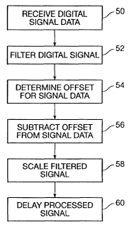
[32] FIG. 4 is a flowchart for a process according to an embodiment of the
invention.
(33] FIG. 5(a) shows a signal that is indicative of mass-to-charge ratio
values of
ions that impact a surface of an ion detector over a time period.
[34] FIG. 5(b) shows the signal shown in FIG. 5(a) after a time-dependent
scaling
function is applied to the signal.
[35] FIG. 5(c) shows the signal in FIG. 5(a) after a time-dependent Gaussian
filter
function is applied to the signal.
[36] FIG. 5(d) shows the signal in FIGS. 5(a) after a time-dependent scaling
function and a time-dependent Gaussian filter function is applied to the
signal.
(37] FIG. 6 shows a graph of scaling factor vs. ion m/z.
DETAILED DESCRIPTION
[38] As noted above, the overall detection efficiency for ions in a typical
time-of
flight mass spectrometer generally decreases as the molecular weight of the
ions
increase. Consequently, a given population of low molecular weight ions
produces
stronger detection signals when compared to an identical number of higher
molecular
CA 02453409 2004-O1-08
WO 03/006949 PCT/US02/21951
weight ions. Also, as noted above, the probability that ions will arnve at a
detector
decreases with increasing m/z values. In addition to these problems, there is
a
significant amount of noise in raw mass spectrum signal data that can obscure
analyte
ion peaks.
[39] It would be desirable to provide for a scaling and filtering scheme that
scales
and preferably filters a signal at various mass-to-charge ratio values (m/z)
in TOFMS.
For low m/z ions, ion-to-electron conversion efficiency and the probability of
arrival
at the detector are high, thus diminishing the need for significant additional
peak
scaling. For high m/z ions, ion-to-electron conversion efficiency and the
probability
of arrival at the detector are low, thus creating a need for further signal
scaling.
Furthermore, if mass resolving power for low molecular weight ions is to be
preserved, any attendant scaling is desirably achieved without diminishing any
required frequency response. Signal data scaling preferably takes place
without undue
scaling of extraneous high frequency noise.
[40] Embodiments of the invention address these concerns. One embodiment of
the
invention is directed to a method for digitally processing time-dependent
signal data.
The method comprises receiving the time-dependent signal data in memory. The
time-dependent signal data can represent a time-dependent signal. The time-
dependent signal data include representations of time-of flight values of
ions, or
values derived from tilde-of flight values of ions. After the time-dependent
signal
data are received, it is scaled with a time-dependent scaling function.
[41] "Values derived from time-of flight values" include any higher order
values
that~originate from time-of flight values. For example, as noted above, a mass-
to-
charge ratio value is a value that is derived from a time-of flight value.
[42] Also, in discussing some embodiments of the invention, "m/z values" are
often
used to illustrate specific examples. It is understood that other values that
are
proportional to m/z values, such as time-of flight values, can be used in
place of mlz
values in any of the specifically described invention embodiments (and vice-
versa).
CA 02453409 2004-O1-08
WO 03/006949 PCT/US02/21951
For instance, specific examples discussed below describe scaling peaks at
specific m/z
values. Alternatively, peaks can be scaled at one or more time-of flight
values.
I. Obtaining Digital Signal Data
(43] Embodiments of the invention may be used with various mass spectrometers
5 including time-of flight mass spectrometers (TOFMS) and various TOF tandem
hybrid systems such as quadrapole-TOFMS, an ion trap-TOFMS, an electrostatic
analyzer-TOFMS, and a TOF-TOF MS. A block diagram of a time-of flight mass
spectrometer is shown in FIG. 3. The mass spectrometer of FIG. 3 may be
configured
as a parallel extraction device or an orthogonal extraction device.
10 [44] A sample containing matter that is to be analyzed by the mass
spectrometer is
introduced through sample inlet system 70. The sample may be introduced as a
solid,
liquid, or gas. The sample is transferred into ion optics 72. Ionization
source 60
causes a portion of the sample to become an ionized gas in ion optics 72.
Ionization
source 60 may comprise a laser desorption ionization device, a plasma
desorption
1 S ionization device, a fast atom bombardment ionization device, ari electron
ionization
device, a chemical ionization device, or an electrospray ionization device. A
laser
desorption device may be used to perform laser desorption/ionization, surface-
enhanced laser desorption/ionization, and/or matrix-assisted laser
desorption/ionization (MALDI).
[45J Although a laser desorption process is described in detail, any suitable
ionization technique can be used in embodiments of the invention. The
ionization
techniques may use, for example, electron ionization, fast atom/ion
bombardment,
matrix-assisted laser desorption/ionizatior< (MALDI), surface enhanced laser
desorptionlionization, or electrospray ionization. These ionization techniques
are well
known in the art.
[46] In preferred embodiments, a laser desorption time-of flight mass
spectrometer
is used. Laser desorption spectrometry is especially suitable for analyzing
high
CA 02453409 2004-O1-08
WO 03/006949 PCT/US02/21951
molecular weight substances such as proteins. Fox example, the practical mass
range
for a MALDI or a surface enhanced laser desorption/ionization process can be
up to
300,000 daltons or more. Moreover, laser desorption processes can be used to
analyze
complex mixtures and have high sensitivity. In addition, the likelihood of
protein
fragmentation is lower in a laser desorption process such as a MALDI or a
surface
enhanced laser desorption/ionization process than in many other mass
spectrometry
processes. Thus, laser desorption processes can be used to accurately
characterize and
quantify high molecular weight substances such as proteins.
[47] Surface-enhanced laser desorption/iorlization, or SELDI, represents a
significant advance over MALDI in terms of specificity, selectivity and
sensitivity.
SELDI is described in U.S. Pat. No. 5,719,060 (Hutchens and Yip). SELDI is a
solid
phase method for desorption in which the analyte is presented to the laser
while on a
surface that enhances analyte capture and/or desorption.
[48] Again refernng to FIG. 5, ion optics 72 accelerates ions toward mass
analyzer
74. Ion optics 72 may, for example, comprise electrostatic lenses such as a
repeller
lens and ground aperture as discussed above. Mass analyzer 74 directs the ions
to ion
detector 76. In a TOF mass spectrometer, the mass analyzer 74 is a free flight
region
where the ions "fly" after they are accelerated. TOF mass spectrometer
analyzers may
comprise a linear system, in which ion free-flight occurs with rectilinear
motion.
Alternatively, the analyzers may include a reflected system, in which ions are
turned
about in an ion mirror or reflectron by an array of electrostatic sectors. Ion
detector
76 may comprise, for example, a microchannel plate detector, mufti-stage
electron
multiplier, or a hybrid combination of these. Ion detector 76 detects ions
that impact
its detecting surface and passes an output signal indicative of the mass-to-
charge ratio
of the detected ions to signal amplifier 78.
[49] An optional signal amplifier 78 outputs a signal to the data acquisition
device
80, which converts the analog output from the amplifier 78 to digital signal
data. The
data acquisition device 80 may include any suitable digital converter device
that
produces digital signal data. Analog-to-digital conversion may be
accomplished, for
CA 02453409 2004-O1-08
WO 03/006949 PCT/US02/21951
12
example, using a time-interval recording device, such as a time-to-digital
converter, in
an orthogonal extraction mass spectrometer. Alternatively, a time array
recording
device such as a transient recorder or a digital oscilloscope could be used in
a parallel
extraction mass spectrometer. Data acquisition device 80 then transfers that
digital
signal data to the computer 82 where the digital signal data are stored. The
computer
82 may include a memory (not shown) such as a RAM (random access memory),
ROM (read only memory), EPROM (erasable programmable read only memory), etc.,
The digital signal data may be received and stored in the memory temporarily,
permanently, or semi-permanently. After the computer 82 receives the digital
signal
data, one or more processors (e.g., a microprocessor, a digital signal
processor {DSP),
etc.) (not shown), andlor hardware circuitry {not shown), in the computer 82
can then
digitally process the digital signal data. A computer readable medium such as
a
magnetic, optical, or electromagnetic information storage medium (e.g., a hard
disk
drive) in the computer 82 can include any suitable code for directing the
processor to
process the digital signal data.
II. Processing the digital signal data
[50] After the digital signal data have been received by the digital computer,
a
process such as the one illustrated in the flowchart shown in FIG. 4 can be
performed
on the digital signal data. Referring to FIG. 4, digital signal data are first
received in
memory from, for example, an analog-to-digital converter (step 50) and is then
stored
in memory. Optionally, the signal data can be filtered (step 52). Then, an
offset is
calculated for the digital signal data (step 54). Then, the offset can be
subtracted from
the digital signal data (step 56). After filtering and subtracting the offset,
the signal
data can be scaled (step 58). After scaling, the processed signal can be
displayed (step
60). Each of these steps is described in greater detail below.
[51) Although the previously described steps 52, 54, 56, 58, 60 are shown in a
particular order, it is understood that in embodiments of the invention, the
steps may
be performed in any suitable order to produce processed digital signal data.
For
example, in some embodiments, any suitable combination of filtering the signal
data
CA 02453409 2004-O1-08
WO 03/006949 PCT/US02/21951
13
52, subtracting the offset from the signal data 56, and scaling the signal
data 58 can be
performed on each data point in the signal data before processing other data
points.
Alternatively, one-of filtering the signal data 52, subtracting the offset
from the signal
data 56, or scaling the signal data 58 can be performed on all data points in
the digital
signal data before performing the other steps.
[52] Moreover, although a specific set of steps is shown in FIG. 4, all of the
steps
need not be performed. For example, in some embodiments, a signal can be
filtered
with analog circuitry before it is digitized. Thus, in these embodiments,
digitally
filtering the digital signal data are optional. Additionally, some of the
steps, or
portions of steps, can be performed by hardware rather than implemented by a
processor. For example, a digital filtering circuit can perform the filtering
step 52
with filter coefficients, for example, provided by a processor, stored in a
memory, etc.
Moreover, one or more processors can be used to implement the steps shown in
FIG.
4. For example, a digital signal processor (DSP) can be used to implement the
filtering the signal data 52, subtracting the offset from the signal data 56,
and/or
scaling the signal data 58, while a general purpose microprocessor, video
processor, or
the like, can be used to display the processed signal 60. Therefore, the term
"digital
computer", as used herein, is intended to include a "computer" having one or
more
processors, and/or hardware circuitry for processing digital data as described
above,
[53] The products of the various processing steps shown in FIG. 4 can be
described
with reference to FIGS. 5(a) to 5(d). In each of FIGS. 5(a) to 5(d), two types
of
displays are shown. A first type of display 200 is a graph of signal intensity
vs. time-
of flight (or m/z). A second type of display 201 is a gray-scale image where
signal
intensity is represented by a line, a color, or a shade of color. High signal
intensities
may be represented by a specific color or a specific color intensity.
[54] FIG. 5(a) shows digital signal data that have not been filtered or
scaled. FIG.
5(b) shows the raw digital signal data in FIG. 5(a) after it has been scaled
with a
time-dependent scaling function according to an embodiment of the invention.
FIG.
CA 02453409 2004-O1-08
WO 03/006949 PCT/US02/21951
14
S(c) shows the raw signal data in FIG. 5(a) after it has been filtered with a
time-
dependent Gaussian filter function.
(55] While improvements to the raw mass spectrum signal data shown in FIG.
5(a)
are made by scaling or filtering alone, better signal data are produced when a
time-
dependent scaling function and a time-dependent filtering function are both
used to
process the signal data. For example, FIG. 5(d) shows the raw signal data in
FIG. 5(a)
after it has been both scaled with a time-dependent scaling function and
filtered with a
time-dependent, Gaussian filtering function. High frequency noise is removed,
while
scaling peaks in the signal data. As shown in FIG. 5(d), clearly identifiable
peaks are
present at m/z values above 100,000 Daltons. Such peaks do not appear to be
readily
discernable to the human eye in the graphs in FIGS. 5(a) to 5(c).
[56] Embodiments of the invention provide a number of advantages. For example,
in some embodiments of the invention, the peak heights in the digital signal
data
reflect the number of particles detected without a priori identification of
the peaks.
Peaks that might otherwise go undetected in the past can readily be identified
using
embodiments of the invention. Peak identification prior to scaling is not
required in
these embodiments. Moreover, the visual presentation of the peaks is markedly
improved using embodiments of the invention. For example, as shown in FIG.
5(d),
using embodiments of the invention, a user or a peak picking algorithm can
readily
identify analyte ion peaks in the signal data (e.g., above 100,000 Daltons)
that might
otherwise go unnoticed. Also, embodiments of the invention compensate for the
time-
dependent decrease in the probability of detecting high mass ions, and the
time-
dependent reduction in signal intensity for detected ions. This makes the
processed
data more informative to the user than the raw signal data that does not make
such
compensations. The peaks in the processed signal data generally have heights
that are
proportional to the amount of analyte ions being ionized. The relative heights
of the
peaks can accurately represent the relative amounts of ions at particular mlz
values.
Moreover, because the processing of the signal is performed by a digital
computer, the
processing of the signal can be easily changed without affecting the mass
spectrometer
CA 02453409 2004-O1-08
WO 03/006949 PCT/US02/21951
hardware. Accordingly, embodiments of the invention are more easily designed,
tested, implemented, optimized, or adjusted, than if the same functions were
implemented in hardware.
A. Determining An Offset and Adjusting the Signal Data Using the Offset
5 [57] In embodiments of the invention, a DC (direct current) offset can be
determined for the digital signal data. The digital signal data can be
adjusted using the
determined DC offset. For example, after obtaining the digital signal data,
the DC
offset can be subtracted from the digital signal data.
[58] Subtracting the DC offset from the digital signal data are desirable,
since the
10 inclusion of the DC offset can cause excessive scaling of the digital
signal data when
the scaling step is performed. For example, the DC offset for digital signal
data may
be 5 V. During the scaling step, data points forming peaks in the digital
signal data
may be multiplied to different values so that they are scaled in a time
dependent
manner. For instance, a time-dependent scaling function may scale data points
15 forming two different peaks by 1 V and 2V, respectively. The additional DC
offset
value for the digital signal data may cause data points forming the peaks to
scale by
SV and lOV respectively, thus disproportionately scaling the data points
forming the
peaks. Accordingly, before scaling the two peaks by 1V and 2V, SV may be
subtracted from each data point in the digital signal data so that the DC
offset for the
digital signal data are essentially zero.
[59] The DC offset for the digital signal data may be determined in any
suitable
manner. For example, in some embodiments, the signal offset may be determined
by
analyzing only the signal data in the last SO% or less of the time period over
which the
digital signal data are obtained. For example, the signal offset can be
estimated using
the average signal of the last 30% of the spectrum. It is believed that the
digital signal
data in the last 50% or less of the time period over which the digital signal
data are
obtained is more stable and has less fluctuations than the digital signal data
in the first
50% of the time period over which the digital signal data are obtained. In the
last 50%
CA 02453409 2004-O1-08
WO 03/006949 PCT/US02/21951
16
of the time period over which the digital signal is obtained, a baseline DC
offset for
the digital signal data can be determined, and this baseline DC offset can be
subtracted
from each data point of the digital signal data to remove the DC offset from
the digital
signal data. This particular process for determining the DC offset is
relatively simple
and can be implemented relatively quickly.
[60) The determination of the appropriate DC offset could be easily improved.
For
example, average data points with signal greater than two standard deviations
away
from the mean could be excluded from the determination of the DC offset. Data
points that are greater than two standard deviations from the mean may be
produced
by ions and can skew the DC offset upward. Removing such data points from the
DC
offset determination produces a more accurate DC offset.
B. Filtering the Signal Data
[61) Time-of flight mass spectrometers typically have several sources of
signal
noise including sampling noise, Johnson noise, flicker noise, and high
frequency noise
created by the detection apparatus. Noise is typically modeled as a wide
bandwidth
additive signal. Thus, the signal data can be described as desired signal
data, which
represents detection of ions generated from the sample, added with a wide
bandwidth
noise signal.
[62) It is desirable to reduce the noise and increase the signal-to-noise
ratio (SNR),
24 thus, making the peaks in the digital signal data more discernable to the
user. The
bandwidth of the desired signal data are bandwidth limited while the noise
signal is
not. Therefore, by applying a bandwidth limiting filter to the signal data,
the noise
can be reduced while only minimally effecting the desired signal. Thus,
applying a
bandwidth limiting filter to the signal data increases the SNR of the signal
data.
Accordingly, in some embodiments, before or after the DC offset is determined
and/or
the digital signal data are adjusted with the determined DC offset, the
digital signal
data are filtered. As described above, such filtering may also be implemented,
prior to
digitizing the signal data, with an analog filter.
CA 02453409 2004-O1-08
WO 03/006949 PCT/US02/21951
17
[63] SNR can be defined as the peak height divided by the standard deviation
of the
noise. The area of a peak is proportional to the number of ions detected, so
the peak
heights for equal numbers of ions detected at different mlz values decrease
with
increasing m/z because the peak widths increase while the area of the peak is
held
S constant. Additionally, it has been found that noise exhibited in mass
spectrometers is
not a strong function of m/z at high mlz. Since the peak height decreases with
time,
while the standard deviation of noise tends to remain unchanged, the SNR falls
with
increasing time.
[64] As shown in the following table, ion populations with lower mass-to-
charge
ratio values produce detection signals that have comparatively higher
frequency
components than ions with larger mass-to-charge ratio values as shown in the
following table that describes typical ion flight time, target resolution, and
major
frequency components (as determined by required peak width to obtain target
resolution).
TABLE 1
Mass-to-ChargeIon Flight Major ComponentPeak Width Mass
Ratio Time Frequency At Half HeightResolution
(m/z) (microseconds)(MHz) (microseconds)
500 10.2 740 0.0010 5000
1,000 14.4 500 0.0016 4500
2,000 20.4 250 0.0034 3000
5,000 32.2 70 0.0134 1200
15,000 55.8 19 0.0254 1100
40,000 91.1 2 0.3037 150
150,000 176.3 .290 1.7600 SO
250,000 227.6 .130 3.8000 30
500,000 321.9 .063 8.0500 20
CA 02453409 2004-O1-08
WO 03/006949 PCT/US02/21951
18
[65] As described above, digital filtering can be applied to oversampled raw
data to
improve the SNR. Typically, a digital filter is a linear shift invariant
system for
computing a discrete output sequence form a discrete input sequence. Often,
digital
filtering is implemented by the convolution of a smoothing function (filter)
with the
signal data. As is well-known to those skilled in the art of digital signal
processing,
convolution can be implemented in time-space or frequency-space. Additionally,
it is
typically more computationally efficient to implement convolution in frequency-
space. However, as is described below, in some embodiments of the invention,
it
appears to be more practical to perform the convolution of the filter with the
signal
data in time-space. Particularly, in some embodiments, a filter having a
bandwidth
that narrows with time is applied to the signal data.
[66] A commonly used digital filter is a finite impulse response (FIR). The
filtering
of signal data with an FIR filter can be mathematically described as,
NH
.Yln) - ~ k~~~ k) a
k=-Nt
where x(n) is the input data sequence to the digital filter, y(n) is the
filtered data sequence,
f( NUJ, . . . , f(N~ are the filter coefficients, and NL + NH + 1 is the width
of the filter.
[67] In the specific embodiment, a different filter is applied to obtain each
filtered
output valuey(n). Thus, in this embodiment the signal data are filtered as,
Ntr(n) r_
k=-Nt (n)
[68] Here, fn is the digital filter applied to obtain the filtered output
y(n), and NL(~) +
NH(n) + 1 is the width of the filter f". Each filter fn has a different
bandwidth
corresponding to the bandwidth of the data signal at that particular time, and
each
filter therefore has a different set of NL(n) + NH(n) + 1 filter coefficients.
[69] As described above, the SNR of the unfiltered signal data decreases with
time
because peak heights decrease with time while the standard deviation of noise
remains
constant. If the signal data are filtered with a filter having a constant
bandwidth, the
CA 02453409 2004-O1-08
WO 03/006949 PCT/US02/21951
19
SNR of the signal data are increased overall. However, the SNR of the signal
data
still decreases with time. But, if a digital filter, whose bandwidth decreases
with time
to match the decreasing bandwidth of the signal, is applied to the signal
data, then the
SNR of the signal data can be increased and can also be made more constant
with
time.
[70] In a specific embodiment, a Gaussian filter function is used to filter
the digital
signal data. The Gaussian filter results in a gradual pass band roll off and
has a
response curve (magnitude vs. frequency) that approximates an ideal Gaussian
curve.
The Gaussian distribution can be defined by the following equation.
ct_~~Z
G~t) ~- 2~' a z°~Z (9)
[71] In the formula above, "Q" is the standard deviation, "t" is time, and "~"
is a
constant. A gaussian filter whose bandwidth corresponds to the width of a peak
in the
data signal at half height is:
.93943 cx-
' w k = a .3607sZwZ , 1
SW
fox k = -int[w], . . . , -1, 0, l, .. . int[w], where w is a measured or
expected peak width,
and-where s is a constant that can be adjusted for a desired degree of
smoothing. As
will be described in more detail below, the expected peak width increases with
time.
Thus, refernng to equation (8), equation (10) can be used to generate a
different filter
fn for each n.
[721 Other details regarding filtering with filters whose bandwidths vary with
time
are described in U.S. Provisional Patent Application No. 60/134,072 filed May
13,
1999, and U.S. Non-Provisional U.S. Patent Application No. 09/569,158, filed
May
CA 02453409 2004-O1-08
WO 03/006949 PCT/US02/21951
11, 2000. Both of these U.S. patent applications are assigned to the same
assignee as
the present invention.
[73] In the above-described embodiments, a different filter fn is applied to
the signal
data to obtain each filtered signal data y(h). However, in other embodiments,
a f rst
5 filter having a first bandwidth can be used to generate a first subset of
filtered signal
data, a second filter having a second bandwidth can be used to generate a
second
subset of filtered signal data, etc. The first bandwidth of the first filter
can correspond
to the bandwidth of a first subset of the unfiltered signal data, the second
bandwidth of
the second filter can correspond to the bandwidth of a second subset of the
unfiltered
10 signal data,' etc. Additionally, it is to be understood that other types of
filters besides a
FIR filter can be used. For example, an infinite impulse response (IIR)
filter, a non-
linear filter, etc., can also be used.
C. Scaling the Signal Data
[74] As previously explained for a given TOF geometry and acceleration
potential,
15 low molecular weight ions have shorter times of flight than larger
molecular weight
ions. Therefore, low molecular weight ions impact the ion detector before
larger
molecular weight ions. Ion detection signal scaling preferably increases for
higher
molecular weight ions to compensate for the fact that higher molecular weight
ions
possess comparatively diminished detection efficiency with respect to low m/z
ions.
20 In embodiments of the invention, signal intensity scaling generally
increases as a
function of time. Thus, as the molecular weights of ions striking the ion
detector
increase, the digital computer scales the signal more.
[75] The digital signal data may be scaled by any suitable amount using a
time-dependent scaling function. Data points forming the peaks in the digital
signal
data are scaled using the time-dependent scaling function so that the scaled
intensity
values increase as function of time. The peaks can be scaled so that the
heights of the
peaks are proportional to the quantity of ions that are detected.
CA 02453409 2004-O1-08
WO 03/006949 PCT/US02/21951
21 .__......__ _~ .___ ...-.
[76] The digital signal data may be scaled using any suitable process. q
Suitable time-
dependent scaling functions can be proportional to time. In some embodiments,
the
time-dependent scaling function can be proportional to the square of time, or
the cube
of time. Moreover, the time-dependent scaling function can include a step
function.
For example, the scaling function can increase stepwise in at least one step
so that sets
of peaks in the digital signal data are scaled according to discrete values.
For
instance, in some embodiments, specific ranges of time-of flight values could
be
multiplied by scaling factors that are specific for those ranges. An example
of an
embodiment of this type is described below. However, in other embodiments, the
time-dependent scaling function can be a continuous function.
[77] In some embodiments, the digital signal data may be scaled using an
expected
peak dimension such as expected or measured peak widths. In other embodiments,
the digital signal data may be scaled using the ion conversion efficiency in
the system
as a function of particle impact velocity. In yet other embodiments, the
digital signal
data may be scaled using the relative detection efficiencies of the mass
spectrometer
as calculated using various test compounds. Further details about each of
these
exemplary scaling process examples are provided below.
[78] In some embodiments of the invention, the expected peak widths may be
used
to scale the signal data. First, the expected peak width value at a particular
time-of
flight value (or a value derived from a time-of flight value such as an m/z
value) can
be determined. An "expected" peak width for a peak can be the width of a peak
in a
mass spectrum that is predicted to be produced at a given time-of flight value
(or
value derived from a time-of flight value) by the mass spectrometer that is
currently
being used for a given number of ions. In general, the expected peak widths
increase
as mlz values or time-of flight values increase.
[79] The expected peak width can be the expected width at any suitable point
along
the height of a peak. In some embodiments, the expected peak width may be the
expected width of the base of a peak, or at a point between the apex and base
of each
peak. For instance, the peak widths that are used may be the peak widths at
half the
CA 02453409 2004-O1-08
WO 03/006949 PCT/US02/21951
22
height of each peak. In another example, for a series of peaks in a mass
spectrum
signal, the expected peak widths can be at a point between the apex and the
base of
each peak at the same distance from the baseline forming the bases of the
peaks. In
both cases, the expected peak width generally increases as the m/z values
increase.
[80] The expected peak widths can be theoretically or empirically derived. For
example, a mass spectrum signal with a number of peaks corresponding to
different
analytes with known mlz values can be created, wherein the number of each of
the
different analytes is known to be approximately the same. The average time-of
flight
value associated with each peak and the width of the peak can be recorded in a
table
of expected peak widths using analytes with known m/z values. An exemplary
table
of expected peak widths is shown in Table 3.
TABLE 3
Table of Expected Peak Widths
Time-of flight Expected Peak Width
(nanoseconds)
(microseconds) .
0 4
60 80
94 600
132 2000
188 4000
CA 02453409 2004-O1-08
WO 03/006949 PCT/US02/21951
23
[81] Using the values in Table 3, a best-fit curve can be created to fit the
values in
Table 3 and the function forming the curve can be used to scale the signal
data.
Alternatively, linear interpolation can be used to form a linear function that
represents
the data. In each of these embodiments, the intensity values associated with
data
points corresponding to higher time-of flight values would be increased more
than the
intensity values corresponding to lower time-of flight values.
_ [82] The determined expected peak width value could then be used to adjust
the
intensity value at the time-of flight value. When the expected peak width is
used to
scale the intensity, the resulting peak heights in the processed signal data
become
proportional to the number of detected particles for each of the peaks. The
relative
heights of the peaks can accurately represent the relative amounts of analytes
within a
particular sample being ionized.
(83] The signal intensity value corresponding to that data point may then be
scaled
in an amount proportional to the expected peak width value for that data
point. For
instance, referring to Table 3 above, each data point in the digital signal
data can be
scaled as follows: from 0 to 60 microseconds, each data point is scaled by 4;
from
above 60 to 94 microseconds, each data point is scaled by 80; from above 94 to
132
microseconds, each data point is scaled by 600; from above 132 microseconds to
188
microseconds, each data point is scaled by 2000; and above 188 microseconds,
each
data point is scaled by 4000. The values 4, 80, 60, 2000, and 4000 can be
considered
scaling factors the proportionally scale data points forming peaks. The
absolute
scaling values may be determined by the user if desired.
[84] In some embodiments, the signal intensity value corresponding to a data
point
may be multiplied by an amount equal to about "1.00 + expected peak width" to
produce a scaling factor. If the expected peak width at a data point is zero,
the
intensity value that is associated with that data point is multiplied by 1.0
so that it is
. . not scaled. The data point may even be scaled by an additional "intensity
factor" that
is input by the user to adjust the degree of scaling even further if even
greater peak
differentiation is desired by the user. In these embodiments, each data point
may be
CA 02453409 2004-O1-08
WO 03/006949 PCT/US02/21951
24
amplified by an amount equal to about "1.00 + expected peak width * intensity
factor".
[85] Peaks in the digital signal data may also be scaled using peaks widths
that are
determined from a set of peaks in the time-dependent digital signal data. That
is, peak
width information in the obtained digital signal data that is to be scaled can
be used to
scale the peaks in the digital signal data. In these embodiments, the peaks in
the
digital signal data are identified before scaling takes place. In a typical
example, a set
of peaks can first be identified in the digital signal data using any number
of known
techniques. After the peaks are identified, peak widths can be determined for
each of
the peaks in the set of peaks. After determining the peak widths for the peaks
in the
set, the respective peaks can be scaled based on their respective measured
peak
widths.
[86] Peaks in the digital signal data may additionally be scaled based on the
ion
conversion efficiency in the system as a function of particle impact velocity.
As noted
above, the particle impact velocity is proportional to the ion m/z values. The
ion
conversion efficiency of a detector as a function of particle impact velocity
(or ion
m/z) could be determined by experiment. Such an experiment would be done by
comparison with a cryogenically operated phonon-detecting ion detector. The
inverse
function could be used to scale the digital signal data as a function of time-
of flight.
For example, as shown in FIG. 6, the curve 33 is an inverted curve of curve 31
in FIG.
2. The curve in FIG. 6 can be used to identify an appropriate scaling amount
for a
given mlz value and compensates for changes in the ion conversion efficiency
as the
m/z values of the ions increase. As shown in FIG. 6, a scaling factor with a
greater
magnitude is used for ions with high m/z values than for ions with low mlz
values.
[87] Peaks in the digital signal data may also be scaled based on the relative
detection efficiency of the instrument. The relative detection efficiency of
the
instrument may be empirically derived using various test compounds. Using the
test
compounds, the detection efficiency of the instrument as a function of mlz may
be
determined. For example, a mass spectrum signal including a number of peaks
CA 02453409 2004-O1-08
WO 03/006949 PCT/US02/21951
corresponding to known analyte ions with different m/z values and in known
quantity
may be formed. The detection efficiencies of the mass spectrometer at each of
the m/z
values can be determined. A function of detection efficiency vs. m/z value can
be
created using the determined detection efficiencies. The inverse of this
function could
5 then be used to scale data points in the signal data.
[88] Any of the above-described steps can be embodied by any suitable computer
code that can be executed by any suitable computational apparatus, such as,
for
example, a microprocessor, a DSP, etc. The computational apparatus may be
incorporated into the mass spectrometer or may be separate from and
operatively
10 associated with the mass spectrometer. Any suitable computer readable media
including, for example, magnetic, electronic, or optical disks or tapes, flash
memory,
etc. can be used to store the computer code. The code may also be written in
any
suitable computer programming language including, for example, Fortran,
Pascal, C,
C++, assembly language, etc. Accordingly, embodiments of the invention can be
15 automatically performed without significant intervention on the part of the
user.
[89] Appendix A contains source code that provides an example of code for
processing digital signal data in a time-of flight mass spectrometry process
in
accordance with an embodiment of the invention. The source code is written in
C++.
[90] The terms and expressions which have been employed herein are used as
terms
20 of description and not of limitation, and there is no intention in the use
of such terms
and expressions of excluding equivalents of the features shown and described,
or
portions thereof, it being recognized that various modifications are possible
within the
scope of the invention claimed. Moreover, any one or more features of any
embodiment of the invention may be combined with any one or more other
features of
25 any other embodiment of the invention, without departing from the scope of
the
invention.
[91] All publications and patent documents cited in this application are
incorporated
by reference in their entirety for all purposes to the same extent as if each
individual
CA 02453409 2004-O1-08
WO 03/006949 PCT/US02/21951
26
publication or patent document were so individually denoted. By their citation
of
various references in this document Applicants do not admit that any
particular
reference is "prior art" to their invention.