Note: Descriptions are shown in the official language in which they were submitted.
CA 02453528 2008-05-02
Novel Fusion Proteins And Assays For Molecular Binding
Field of the Invention
The invention relates to cell and molecular biology, recombinant DNA
technology, and recombinant protein technology.
Background
Interactions among molecules such as proteins are fundamental to cell
biology. Protein binding to a wide variety of cellular components, including
proteins,
nucleic acids, carbohydrates, and lipids, has been recognized as an important
drug
target due to its integral nature within signal transduction and biological
pathways.
Such binding can be correlated to a variety of intracellular events, including
protein
expression, the availability of an active state of a protein, and, directly or
indirectly, to
protein catalytic activity. For instance, in the cytoplasm the protein kinase
MAPK,
when complexed with MEK1, is inactive. Upon activation, MEK1 and MAPK
dissociate, leading to free, activated MAPK. Detection of the activated MAPK
by
virtue of its ability to bind to a binding domain in a target substrate
indicates the
presence of the active enzyme, and is indirectly related to the MAPK activity
of
phosphorylating substrates.
1
CA 02453528 2004-01-09
WO 03/012068 PCT/US02/24572
Current methods for analyzing cellular molecular binding events, such as two-
hybrid systems and variants thereof, substrate complementation systems,
immunoprecipitation assays, in vivo incorporation of radiolabeled moieties,
and the
use of antibodies specific for a given modification (such as phosphorylation),
suffer
from numerous drawbacks. Such drawbacks include the need to construct two or
more chimeric proteins; the inability to monitor biochemical events in live,
intact cells
or in fixed cells; the requirement for considerable time to conduct the
assays; and the
need for specialized and expensive equipment. Thus, improved reagents and
methods
for detecting and measuring specific binding events are needed.
A very significant improvement would be a flexible design for reagents and
assays that can be used to detect molecular binding events that occur within
living
cells. Such reagents would preferably comprise a single chimeric protein, and
would
be applicable to monitoring molecular binding events in live and fixed end
point cell
preparations as well as to making kinetic measurements of the binding events
in cells.
Such reagents would preferably possess detectable signals that permit easy
detection
of molecular binding events of interest, and also provide the ability to
combine the
molecular binding event assay with other cell-based assays.
Summary of the Invention
The present invention fulfills the need in the art for novel reagents, and
assays
using such reagents, for detecting molecular binding events that do not suffer
from the
drawbacks of previous reagents and assays for the detection of molecular
binding.
In one aspect, the present invention provides a recombinant fusion protein
comprising a detection domain; a first localization domain; and a binding
domain for
the molecule of interest; wherein the detection domain, the first localization
domain,
2
CA 02453528 2004-01-09
WO 03/012068 PCT/US02/24572
and the binding domain for the molecule of interest are operably linked;
wherein the
binding domain for the molecule of interest is separated from the first
localization
domain by 0-20 amino acid residues; and wherein the first localization domain
and
the binding domain for the molecule of interest do not all occur in a single
non-
recombinant protein, or do not all occur in a single non-recombinant protein
with the
same spacing as in the recombinant fusion protein for detecting binding of a
molecule
of interest.
In a preferred embodiment, the recombinant fusion protein further comprises a
second localization domain, wherein the binding domain for the molecule of
interest
is separated from the second localization domain by more than 20 amino acid
residues; wherein the first localization domain and the second localization
domain do
not target the recombinant fusion protein to an identical subcellular
compartment; and
wherein the first localization domain, the second localization domain, and the
binding
domain for the molecule of interest do not all occur in a single non-
recombinant
protein, or do not all occur in a single non-recombinant protein with the same
spacing
as in the recombinant fusion protein.
In a further preferred embodiment, the binding site for the molecule of
interest
does not contain a "cleavage site," wherein "cleavage site" is defined as an
amino
acid sequence within the binding domain that is targeted for cleavage by a
proteolytic
enzyme.
In another aspect the invention provides recombinant nucleic acid molecules
encoding a recombinant fusion protein for detecting binding of a molecule of
interest,
comprising the following operably linked regions in frame relative to each
other: a
first nucleic acid sequence encoding a detection domain; a second nucleic acid
3
CA 02453528 2004-01-09
WO 03/012068 PCT/US02/24572
sequence encoding a first localization domain; and a third nucleic acid
sequence
encoding a binding domain for the molecule of interest; wherein the third
nucleic acid
sequence is separated from the second nucleic acid sequence by 0-60
nucleotides, and
wherein the second nucleic acid sequence and the third nucleic acid sequence
do not
all occur in a single non-recombinant nucleic acid molecule, or do not all
occur in a
single non-recombinant nucleic acid molecule with the same spacing as in the
recombinant nucleic acid molecule encoding a recombinant fusion protein for
detecting binding of a molecule of interest.
In a preferred embodiment, the recombinant nucleic acid molecules further
comprise a fourth nucleic acid sequence encoding a second localization domain,
wherein the fourth nucleic acid sequence is separated from the third nucleic
acid
sequence by more than 60 nucleotides; wherein the first localization domain
and the
second localization domain do not target the recombinant fusion protein to an
identical subcellular compartment; and wherein the second nucleic acid
sequence, the
third nucleic acid sequence, and the fourth nucleic acid sequence do not all
occur in a
single non-recombinant nucleic acid molecule, or do not all occur in a single
non-
recombinant nucleic acid molecule with the same spacing as in the recombinant
nucleic acid molecule encoding the recombinant fusion protein.
In another aspect, the present invention provides recombinant nucleic acid
molecules comprising the following operably linked regions in frame relative
to each
other: a first nucleic acid sequence encoding a detection domain; a second
nucleic
acid sequence encoding a first localization domain; and a third nucleic acid
sequence
that comprises one or more restriction enzyme recognition sites that are not
present
elsewhere in the recombinant nucleic acid molecule; wherein the third nucleic
acid
sequence is separated from the second nucleic acid sequence by 0-60
nucleotides; and
4
CA 02453528 2004-01-09
WO 03/012068 PCT/US02/24572
wherein the second nucleic acid sequence and the third nucleic acid sequence
do not
both occur in a single non-recombinant nucleic acid molecule, or do not both
occur in
a single non-recombinant nucleic acid molecule with the same spacing as in the
recombinant nucleic acid molecule.
In a preferred embodiment, the recombinant nucleic acid molecules further
comprise a fourth nucleic acid sequence encoding a second localization domain,
wherein the fourth nucleic acid sequence is separated from the third nucleic
acid
sequence by more than 60 nucleotides; wherein the first and second
localization
domains do not target the recombinant fusion protein to an identical
subcellular
compartment; and wherein the second nucleic acid sequence, the third nucleic
acid
sequence, and the fourth nucleic acid sequence do not all occur in a single
non-
recombinant nucleic acid molecule, or do not all occur in a single non-
recombinant
nucleic acid molecule with the same spacing as in the recombinant nucleic acid
molecule.
In another aspect the invention provides recombinant expression vectors
comprising the nucleic acid molecules of the invention, and cells transfected
with
such expression vectors.
In another aspect the invention provides kits containing the fusion proteins,
the
nucleic acid molecules, the expression vectors and/or the host cells of the
invention,
and instructions for their use in detecting the binding of a molecule of
interest to the
fusion protein in a cell.
In another aspect the invention provides methods for identifying compounds
that alter the binding of a molecule of interest in a cell comprising
providing cells that
contain the recombinant fusion proteins of the invention, obtaining optically
detectable signals from the detection domain, comparing the subcellular
distribution
5
CA 02453528 2004-01-09
WO 03/012068 PCT/US02/24572
of the recombinant fusion protein in the presence and absence of one or more
test
compounds, and identifying one or more compounds that alter the subcellular
distribution of the recombinant fusion protein, wherein such altering of the
subcellular
distribution of the recombinant fusion protein indicates that the one or more
test
compounds have altered the binding of the molecule of interest to the
recombinant
fusion protein in the cells, and/or have altered the expression of the
molecule of
interest in the cells.
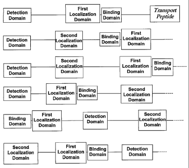
Description of the Figures
Figure 1 is a pictoral depiction of various possible fusion protein
arrangements.
Figure 2 is a table of subcellular compartment localization sequences.
Figure 3 is a table of binding domains.
Figure 4 is a table of nuclear localization signals and nuclear export
signals.
Figure 5 is a table of further nuclear localization signals.
Figure 6 is a table of further experimentally verified nuclear localization
signals.
Figure 7 is a table of detection domains.
Figure 8 is a table of protein-derived transport peptides.
Figure 9 is a table of RNA binding domains.
Figure 10 is a table of further nuclear export signals.
Figure 11 is a table of post-translational modification sites.
Figure 12A shows the sequence of the Plekstrin Homology (PH) domain from PLC-
beta2.
Figure 12B shows the sequence of the diacylglycerol binding domain (DBD) from
protein kinase C.
6
CA 02453528 2004-01-09
WO 03/012068 PCT/US02/24572
Detailed Description of the Invention
Within this application, unless otherwise stated, the techniques utilized may
be
found in any of several well-known references such as: Molecular Cloning: A
Laboratory Manual (Sambrook, et al., 1989, Cold Spring Harbor Laboratory
Press),
Gene Expression Technology (Methods in Enzymology, Vol. 185, edited by D.
Goeddel, 1991. Academic Press, San Diego, CA), "Guide to Protein Purification"
in
Methods in Enzymology (M.P. Deutshcer, ed., (1990) Academic Press, Inc.); PCR
Protocols: A Guide to Methods and Applications (Innis, et al. 1990. Academic
Press,
San Diego, CA), Culture of Animal Cells: A Manual of Basic Technique, 2nd Ed.
(R.I.
Freshney. 1987. Liss, Inc. New York, NY), Gene Transfer and Expression
Protocols
(pp. 109-128, ed. E.J. Murray, The Humana Press Inc., Clifton, N.J.), and the
Ambion
1998 Catalog (Ambion, Austin, TX).
In one aspect, the present invention provides fusion proteins for detecting
binding of a protein of interest, comprising
a) a detection domain;
b) a first localization domain; and
c) a binding domain for the molecule of interest;
wherein the detection domain, the first localization domain, and the binding
domain for the molecule of interest are operably linked;
wherein the binding domain for the molecule of interest is separated from the
first localization domain by 0-20 amino acid residues; and
wherein the first localization domain and the binding domain for the molecule
of interest do not both occur in a single non-recombinant protein, or do not
both occur
in a single non-recombinant protein with the same spacing as in the
recombinant
fusion protein for detecting binding of a molecule of interest.
7
CA 02453528 2004-01-09
WO 03/012068 PCT/US02/24572
In a preferred embodiment, the fusion protein further comprises a second
localization domain, wherein the detection domain, the first localization
domain, the
second localization domain, and the binding domain for the molecule of
interest are
operably linked; wherein the binding domain for the molecule of interest is
separated
from the second localization domain by more than 20 amino acid residues;
wherein
the first localization domain and the second localization domain do not target
the
recombinant fusion protein to an identical subcellular compartment; and
wherein the
first localization domain, the second localization domain, and the binding
domain for
the molecule of interest do not all occur in a single non-recombinant protein,
or do not
all occur in a single non-recombinant protein with the same spacing as in the
recombinant fusion protein for detecting binding of a molecule of interest.
As used herein, "separated by" means that the recited number of residues must
be present between the domains, thus separating the domains.
As used herein, "binding of a molecule of interest" means binding of the
molecule of interest to the binding domain. Binding may be by covalent or non-
covalent interaction. Detection of such binding demonstrates that the molecule
of
interest has been expressed by the cells, and demonstrates that the molecule
of interest
is in a state capable of binding to the binding domain. Such binding may
indicate that
the molecule of interest has undergone a post-translational modification, such
as a
conformational change or phosphorylation, allowing such binding. Such binding
may
also indicate that the molecule of interest is active. Furthermore, such
binding may
indicate that the binding domain has undergone a covalent modification via an
enzymatic reaction.
The molecule of interest can be any chemical or biological molecule capable
of binding to the binding domain and thus inhibiting the activity of the first
8
CA 02453528 2004-01-09
WO 03/012068 PCT/US02/24572
localization domain via steric hindrance. In a preferred embodiment, the
binding
domain comprises a binding domain for a molecule of interest selected from the
group
consisting of nucleic acid, protein, and lipid. In a most preferred
embodiment, the
binding domain comprises a binding domain for a protein of interest.
As used herein, "fusion protein" means a non-naturally occurring protein
product, wherein the domains of the fusion protein are derived from one or
more other
proteins or artificially derived sequences. For example, each domain can be
derived
from a different naturally occurring protein sequence, or mutant/variant
thereof, that
possesses the desired properties. Alternatively, the domains can all be
derived from a
naturally occurring protein, wherein the spacing of the binding domain
relative to the
first and (if present) the second localization domains has been modified with
respect
to their spacing in the naturally occurring protein. Many other variations on
this
theme will be apparent to one of skill in the art.
The fusion protein may be constructed by a variety of mechanisms including,
but not limited to, standard DNA manipulation techniques and chemical assembly
via
subunit parts of the fusion protein. The chemical assembly may lead to an
equivalent
form as the molecular genetic form or alternative associations with equivalent
function. In a preferred embodiment, the fusion protein is produced by
standard
recombinant DNA techniques.
The basic principle of the fusion proteins of the present invention is that
the
distribution of the fusion protein changes upon being bound by the molecule of
interest. The unbound fusion protein is distributed based on the subcellular
distribution directed by the first localization domain (in the embodiment with
only
one localization domain), or based on the subcellular distribution between two
subcellular compartments as directed by the first and second localization
domains,
9
CA 02453528 2004-01-09
WO 03/012068 PCT/US02/24572
respectively, in a ratio based upon the relative strengths of the first and
the second
localization domains. Thus, in the two localization domain embodiment, in the
unbound state, there may be an equilibrium in the distribution of the fusion
protein
between the two targeted subcellular compartments, or either one or the other
localization domain may bias the distribution of the fusion protein.
Upon binding of the molecule of interest to the binding domain of the fusion
protein, the ability of the first localization domain to direct the fusion
protein to the
subcellular compartment normally targeted by the first localization domain is
inhibited, due to steric hindrance caused by the proximity of the bound
molecule of
interest. Thus, the distribution of the fusion protein within the cell will be
either
without bias within the cell in the embodiment with only the first
localization domain,
or will be determined mainly by the second localization domain in the
embodiment
with both a first and second localization domain, reflecting in both cases a
change in
the distribution of the bound fusion protein within a cell, which can be
detected by a
change in the distribution of the detectable signal from the detection domain
of the
fusion protein within the cell.
The exact order of the domains in the fusion protein, as well as the presence
and/or length of any other sequences located between the domains, is not
generally
critical, as long as: (a) the required spacing between the binding domain and
the first
localization domain and second localization domain (if present) are
maintained; (b)
the first and second localization domains function independently; and (c) the
function
of each domain is retained. Generally, this requires that the two-dimensional
and
three-dimensional structure of any intervening protein sequence does not
preclude the
binding or interaction requirements of the domains of the fusion protein,
except as
contemplated herein. One of skill in the art will readily be able to optimize
the fusion
CA 02453528 2004-01-09
WO 03/012068 PCT/US02/24572
protein for these parameters using the teachings herein. Examples of fusion
protein
arrangements may be found in Figure 1.
As recited herein, for each domain it will be understood that more than one
copy of the sequence that imparts the required function may be present. For
example,
as used herein, "localization domain" means an amino acid sequence that
imparts a
restriction on the cellular distribution of the fusion protein to a particular
subcellular
compartment of the cell. Thus, the first localization domain and the second
localization domain may each individually comprise 1, 2, or more such amino
acid
sequences that impart a restriction on the cellular distribution of the fusion
protein.
The first and second localization domains do not target the recombinant fusion
protein to the identical subcellular compartment. In the unbound state, the
fusion
protein will distribute between the two subcellular compartments targeted by
the first
and second localization domains as described above. For example, where the
first
localization domain comprises a nuclear localization signal (NLS) with an
adjacent
binding domain, and the second localization domain comprises a nuclear export
signal
(NES), the unbound fusion protein will distribute between the nucleus and the
cytoplasm in a ratio based upon the relative strengths of the first and the
second
localization domains. Upon binding of the molecule of interest to the binding
domain, the NLS will be inhibited, NES targeting will then predominate over
NLS
targeting, and the fusion protein will be primarily localized in the
cytoplasm.
As used herein, "subcellular compartment" refers to any sub-structural
macromolecular component of the cell whether it is made of protein, lipid,
carbohydrate, or nucleic acid. It could be a macromolecular assembly or an
organelle
(a membrane delimited cellular component). Subcellular compartments include,
but
are not limited to, cytoplasm, nucleus, nucleolus, inner and outer surface of
the
11
CA 02453528 2004-01-09
WO 03/012068 PCT/US02/24572
nuclear envelope, regions within the nucleus with localized activities, such
as
transcription, cytoskeleton, inner leaflet of the plasma membrane, outer
leaflet of the
plasma membrane, outer leaflet of the mitochondrial membrane, inner leaflet of
the
mitochondrial membrane, inner or outer leaflet of the inner mitochondrial
membrane,
Golgi, endoplasmic reticulum, and extracellular space.
In a preferred embodiment, the first localization domain is selected from the
group consisting of SEQ ID NO:l, SEQ ID NO:3, SEQ ID NO:5, SEQ ID NO:7, SEQ
ID NO:9, SEQ ID NO:11, SEQ ID NO:13, SEQ ID NO:15, SEQ ID NO:17, SEQ ID
NO:19, SEQ ID NO:21, SEQ ID NO:23, SEQ ID NOS:145-287, and SEQ ID
NOS:315-325 (See Figures 2, 4, 5, 6, and 10). In a further preferred
embodiment,
either the first or the second localization domain is a nuclear localization
signal, while
the other localization domain is a nuclear export signal, resulting in a
fusion protein
that is distributed between the nucleus and the cytoplasm. Selection of the
most
appropriate localization domains can be accomplished by one of skill in the
art using
the teachings herein.
It is possible to maximize the signal-to-noise ratio from the fusion protein
by
using localization domains that bias distribution of the fusion protein to the
subcellular compartment where the binding event is most likely to occur (i.e.
where
the molecule of interest is most likely to be present). For example,
deacetylases, such
as histone deacetylases, are often found in the nucleus, where they are
involved in
chromatin reorganization. Using a fusion protein with a binding domain for a
histone
deacetylase, a strong NLS as the first localization sequence, such as the SV40
NLS
(SEQ ID NO: 145), with a relatively weak NES as the second localization
sequence,
such as the MAPKAP-2 NES (SEQ ID NO:317), will result in an equilibrium bias
distribution of the unbound fusion protein favoring nuclear distribution.
Optically
12
CA 02453528 2004-01-09
WO 03/012068 PCT/US02/24572
detectable signals from the fusion protein in the cytoplasm will be relatively
low in
intensity. Upon binding of the deacetylase to the fusion protein binding
domain
proximal to the NLS, nuclear import will be blocked, resulting in accumulation
of the
fusion protein in the cytoplasm. Since the cytoplasm starts out with a
relatively low
intensity of detectable signal, relatively small increases in intensity are
more readily
detected than if the intensity of the unbound fusion protein in the cytoplasm
were
higher.
In another example, for a protein generally limited to the cytoplasm, such as
ras, a fusion protein composed of a binding domain for ras (example, from c-
raf), a
relatively strong NES, such as from MEIN 1 (SEQ ID NO:17) as a first
localization
sequence, and a weaker NLS, such as from NFkB (SEQ ID NO:5) as a second
localization sequence results in an equilibrium bias distribution of the
unbound fusion
protein favoring the cytoplasm. Optically detectable signals from the fusion
protein in
the nucleus will be relatively low in intensity. Upon ras binding to the
fusion protein
in the cytoplasm, nuclear export is blocked, and the nuclear intensity of the
optically
detectable signals from the fusion protein will increase. Since the nucleus
starts out
with a relatively low intensity of detectable signal, relatively small
increases in
intensity are more readily detected than if the intensity of the unbound
fusion protein
in the nucleus were higher. When the compartment where the binding event of
the
molecule of interest is unknown, or when the molecule of interest is
relatively evenly
distributed between compartments, using an NES and NLS combination where the
equilibrium bias is a fairly equal distribution between the two subcellular
compartments avoids the need for any prior knowledge of the
compartmentalization
of the target protein. One of skill in the art will readily be able to
optimize the design
of the localization domains using the teachings herein.
13
CA 02453528 2004-01-09
WO 03/012068 PCT/US02/24572
As used herein, "binding domain" refers to one or more amino acid sequences
to which the molecule of interest binds. The binding domain may be a naturally
occurring binding domain, a mutant, variant, or fragment thereof, or an
artificial
domain. It is to be understood that the binding domain can comprise a binding
site for
any molecule of interest. Thus, the fusion protein of the present invention
can detect
binding of any type of molecule that binds to a binding domain comprising an
amino
acid sequence. In a preferred embodiment, the binding domain is a binding
domain
for a molecule of interest selected from the group consisting of nucleic acid,
protein,
and lipid. In a most preferred embodiment, the binding domain is a binding
domain
for a protein of interest. (For examples, see Figure 3.) In one embodiment,
such
proteins are those involved in post-translational modifications, including,
but not
limited to, protein kinases, protein phosphatases, and proteins promoting
protein
glycosylation, acetylation, and ubiquitination, fatty acid acylation, and ADP-
ribosylation.
The binding domain can comprise (a): an amino acid sequence for non-
covalent binding (such as protein-protein interaction sites), referred to as a
"non-
covalent binding site"; (b) an amino acid sequence for covalent binding,
defined as
the amino acid or amino acid sequence at which the molecule of interest
effects an
enzymatic reaction (ie: covalent binding), and referred to as a "covalent
binding site";
or (c) a combination of one or more covalent binding sites and one or more non-
covalent binding sites. An exarnple of a covalent binding site is an amino
acid(s) that
is/are phosphorylated by a kinase.
In a most preferred embodiment, the binding domain does not contain a
"cleavage site", wherein "cleavage site" is defined as an amino acid sequence
within
the binding domain that is targeted for cleavage by a proteolytic enzyme.
Since the
14
CA 02453528 2004-01-09
WO 03/012068 PCT/US02/24572
recombinant fusion proteins of the invention are used to detect binding of the
molecule of interest to the binding domain, and since such detection relies on
steric
hindrance of the first localization domain by the bound molecule of interest,
it is
highly preferred that the recombinant fusion proteins remain intact, and that
binding
of the molecule of interest does not result in cleavage of the fusion protein.
Furthermore, the recombinant fusion proteins of the present invention are
capable of
permitting reversible detection of binding. The non-covalent binding is
generally
reversible due to equilibrium considerations, while the covalent binding can
be
reversible by action of enzymes that reverse a given post-translational
modification,
such as phosphatases, deacetylases, etc. The presence of a cleavage site
within the
binding domain would eliminate such reversible measurements.
In one embodiment, the binding domain consists of a binding domain for a
nucleic acid of interest. In a more preferred embodiment, the nucleic acid of
interest
is an RNA of interest. In a further preferred embodiment, the binding domain
for the
RNA of interest has an amino acid sequence selected from the group consisting
of
SEQ ID NOS:310-314 (see Figure 9). In a further preferred embodiment, the
nucleic
acid of interest is a DNA. In a preferred embodiment, the binding domain for
the
DNA of interest has an amino acid sequence selected from the group consisting
of
SEQ ID NO:338 and SEQ ID NO:339.
In a further embodiment, the binding domain consists of a binding domain for
a lipid of interest. For example, the pleckstrin homology (PH) (SEQ ID NO:364,
encoded by SEQ ID NO:363) domain from phospholipases that binds PIP2
phospholipids (Wang et al.,2000, J. Biol. Chem. 275:7466-7469; Singer et al.,
1997,
Annu. Rev Biochem 66:475-509), or the diacylglycerol binding domain (DBD) from
protein kinase C (SEQ ID NO:366, encoded by SEQ ID NO: 365), can be used to
CA 02453528 2004-01-09
WO 03/012068 PCT/US02/24572
detect generation of PIP2 phospholipids or diacyglycerol, respectively, at the
plasma
membrane. Insertion into the fusion protein of the PH domain or DBD as the
binding
domain wherein the first localization sequence comprises an NLS would lead to
blockage of nuclear import of the fusion protein upon the generation of PIP2
phospholipids at the plasma membrane. The bound fusion protein would not
translocate from the cytoplasm to the nucleus, but would accumulate at the
plasma
membrane. Thus, analysis could entail measurements at the cytoplasm, nucleus,
and
plasma membrane.
In a further preferred embodiment, the binding domain is not a binding
domain for a protease, and the molecule of interest is not a protease.
In embodiments wherein the binding domain consists of a non-covalent
binding site but does not include a covalent binding site, the fusion protein
serves to
detect binding events only, without detection of subsequent enzymatic
reactions.
Thus, for example, the fusion protein can be used to detect expression and
appropriate
secondary and tertiary structure of a protein kinase, but is not biased by
other post-
translational modifications that counteract the enzymatic activity of the
protein kinase
(for example, protein phosphatase activity). In one such embodiment, the
binding
domain is a binding domain for a protein, and has an amino acid sequence
selected
from the group consisting of SEQ ID NO:25, SEQ ID NO:27, SEQ ID NO:29, SEQ
ID NO:31, SEQ ID NO:33, SEQ ID NO:35, SEQ ID NO:37, SEQ ID NO:39, SEQ ID
NO:41, SEQ ID NO:43, SEQ ID NO:45, SEQ ID NO:47, SEQ ID NO:49, SEQ ID
NO:51, SEQ ID NO:53, SEQ ID NO:55, SEQ ID NO:57, SEQ ID NO:59, SEQ ID
NO:61, SEQ ID NO:63, SEQ ID NO:65, SEQ ID NO:67, SEQ ID NO:69, SEQ ID
NO:71, SEQ ID NO:73, SEQ ID NO:75, SEQ ID NO:77, SEQ ID NO:79, SEQ ID
NO:81, SEQ ID NO:83, SEQ ID NO:85, SEQ ID NO:87, SEQ ID NO:89, SEQ ID
16
CA 02453528 2004-01-09
WO 03/012068 PCT/US02/24572
NO:91, SEQ ID NO:93, SEQ ID NO:95, SEQ ID NO:97, SEQ ID NO:99, SEQ ID
NO:101, SEQ ID NO:103, SEQ ID NO:105, SEQ ID NO:107, SEQ ID NO:109, SEQ
ID NO:111, SEQ ID NO:113, and SEQ ID NO:115, SEQ ID NO:117, SEQ ID
NO:119, SEQ ID NO:120, SEQ ID NO:122, SEQ ID NO:124, SEQ ID NO:126, SEQ
ID NO:128, SEQ ID NO:130, SEQ ID NO:132, SEQ ID NO:134, SEQ ID NO:136,
SEQ ID NO:138, SEQ ID NO:140, SEQ ID NO:141, SEQ ID NO:143, SEQ ID
NO:341, SEQ ID NO:343, SEQ ID NO:345, SEQ ID NO:347, SEQ ID NO:349, SEQ
ID NO:350, SEQ ID NO:352, SEQ ID NO:354, SEQ ID NO:356, SEQ ID NO:358,
SEQ ID NO:360, SEQ ID NO:362, SEQ ID NO:364, and SEQ ID NO:366 (see
Figures 3 and 11).
In a further embodiment wherein the binding domain consists of a non-
covalent binding site but does not include a covalent binding site, the
binding domain
is a binding domain for a protein kinase. In a further embodiment, the binding
domain for the protein kinase has an amino acid sequence selected from the
group
consisting of SEQ ID NO:25, SEQ ID NO:27, SEQ ID NO:29, SEQ ID NO:31, SEQ
ID NO:33, SEQ ID NO:35, SEQ ID NO:37, SEQ ID NO:39, SEQ ID NO:41, SEQ ID
NO:43, SEQ ID NO:45, SEQ ID NO:47, SEQ ID NO:49, SEQ ID NO:51, SEQ ID
NO:53, SEQ ID NO:55, SEQ ID NO:57, SEQ ID NO:59, SEQ ID NO:61, SEQ ID
NO:63, SEQ ID NO:65, SEQ ID NO:67, SEQ ID NO:69, SEQ ID NO:71, SEQ ID
NO:73, SEQ ID NO:75, SEQ ID NO:77, SEQ ID NO:79, SEQ ID NO:81, SEQ ID
NO:83, SEQ ID NO:85, SEQ ID NO:87, SEQ ID NO:89, SEQ ID NO:91, SEQ ID
NO:93, SEQ ID NO:95, SEQ ID NO:97, SEQ ID NO:99, SEQ ID NO:101, SEQ ID
NO:103, SEQ ID NO:105, SEQ ID NO:107, SEQ ID NO:109, SEQ ID NO:111, SEQ
ID NO:113, SEQ ID NO:115, SEQ ID NO:117, SEQ ID NO:119, SEQ ID NO:120,
SEQ ID NO:122, SEQ ID NO:124, SEQ ID NO:126, SEQ ID NO:128, SEQ ID
17
CA 02453528 2004-01-09
WO 03/012068 PCT/US02/24572
NO:341, SEQ ID NO:343, SEQ ID NO:345, SEQ ID NO:347, SEQ ID NO:349, SEQ
ID NO:350, SEQ ID NO:352.
In a further embodiment wherein the binding domain consists of a non-
covalent binding site but does not include a covalent binding site, the
binding domain
is a binding domain for an acetyl transferase. In a preferred embodiment, the
binding
domain for a histone acetyl transferase has an amino acid sequence selected
from the
group consisting of SEQ ID NO:132, SEQ ID NO:134, SEQ ID NO:136, SEQ ID
NO:354, and SEQ ID NO: 356.
In a further preferred embodiment wherein the binding domain consists of a
non-covalent binding site but does not include a covalent binding site, the
binding
domain is a binding domain for a histone deacetylase. In a preferred
embodiment, the
binding domain for the histone deacetylase has an amino acid sequence of SEQ
ID
NO: 138.
In a further preferred embodiment wherein the binding domain consists of a
non-covalent binding site but does not include a covalent binding site, the
binding
domain is a binding domain for an ubiquitin ligase. In a further preferred
embodiment, the binding domain for the ubiquitin ligase has an amino acid
sequence
selected from the group consisting of SEQ ID NO:140 and SEQ ID NO:141.
In embodiments wherein the binding domain is a non-covalent binding site but
does not include a covalent binding site, the binding domain for the molecule
of
interest is separated from the first localization domain by 0-20 amino acid
residues,
and the binding domain for the molecule of interest is separated from the
second
localization domain (if present) by more than 20 amino acid residues. In
preferred
embodiments, the binding domain for the molecule of interest is separated from
the
first localization domain by 0-15 amino acids, and more preferably by 0-10
amino
18
CA 02453528 2004-01-09
WO 03/012068 PCT/US02/24572
acids. This spacing dictates that the molecule of interest can act to
sterically hinder
the effect of the first localization domain, while minimizing any potential
steric
hindrance on the second localization domain. Thus, for example, the binding
domain
can partially or completely overlap with the first localization domain. The
same is
true for embodiments of the binding domain with only the covalent binding
site,
which can also overlap with the first localization domain, or with both the
covalent
binding site and the non-covalent binding site.
Thus, according to these various embodiments wherein the binding domain
comprises a non-covalent binding site, but does not include a covalent binding
site,
the non-covalent binding site is preferably separated from the first
localization
domain by 0, 1, 2, 3, 4, 5, 6, 7, 8, 9, 10, 11, 12, 13, 14, 15, 16, 17, 18,
19, or 20 amino
acid residues.
In embodiments wherein the binding domain is a covalent binding site but
does not include a non-covalent binding site, the covalent binding site is
preferably
separated from the first localization domain by 0, 1, 2, 3, 4, 5, or 6 amino
acid
residues. In a preferred embodiment, the binding domain is preferably
separated from
the first localization domain by 0-4, and more preferably by 0-2 amino acid
residues.
Preferred embodiments of such binding domains include amino acid sequences
selected from the group consisting of SEQ ID NOS: 341, 343, 345, 347, 349,
350, 352
(all of which are binding domains for kinases), 354, 356 (both of which are
binding
domains for acetylases), 358, 360, and 362 (all of which are binding domains
for
farnesylases).
In these embodiments, the covalent binding resulting from the enzymatic
reaction, including but not limited to phosphorylation, acetylation,
ubiquitination, or
famesylation, inhibits activity of the first localization domain via steric
hindrance,
19
CA 02453528 2004-01-09
WO 03/012068 PCT/US02/24572
leading to a change in the distribution of the fusion protein, as described
above. In
these embodiment, the change in distribution of the recombinant fusion protein
provides direct evidence for post-translational modification of the binding
domain by
the molecule of interest, and thus provides a different functionality from the
embodiment wherein the binding domain does not include the covalent binding
site.
In these embodiments, wherein the fusion protein further comprises a second
localization domain, the covalent binding site is preferably separated from
the second
localization domain by more than 6 amino acid residues; preferably by at least
10
amino acid residues, and more preferably by at least 20 amino acid residues.
In embodiments wherein the binding domain is both a covalent binding site
and a non-covalent binding site, either or both of the above spacing
requirements are
satisfactory. Thus, the covalent binding site in the binding domain is
preferably
separated from the first localization domain by 0, 1, 2, 3, 4, 5, or 6 amino
acid
residues. In a preferred embodiment, the binding domain is preferably
separated from
the first localization domain by 0-4, and more preferably by 0-2 amino acid
residues.
Alternatively, or in addition, the non-covalent binding site for the molecule
of interest
is separated from the first localization domain by 0-20 amino acid residues,
preferably
0-15 amino acid residues, and more preferably by 0-10 amino acid residues. It
is to
be understood that in this embodiment, the covalent binding site and the non-
covalent
binding site do not have to be contiguous, although they may be contiguous.
Thus,
there may be amino acid residues present between the covalent binding site and
the
non-covalent binding site. The length of such intervening sequences is
variable, and
may be determined readily by one of skill in the art. This embodiment provides
added
functionality to the fusion proteins of the invention, as the presence of the
non-
covalent binding site adds specificity to the enzymatic reaction occurring at
the
CA 02453528 2004-01-09
WO 03/012068 PCT/US02/24572
covalent binding site. For example, a covalent binding site for a kinase may
be
common to multiple kinases. Thus, including a non-covalent binding site for a
specific kinase increases specificity and efficiency of the enzyme at the
covalent
binding site.
In all of these embodiments, it is most preferred that the binding domain does
not include a cleavage site, that the binding domain is not a binding domain
for a
protease, and that the molecule of interest is not a protease.
As used herein, "detection domain" means one or more amino acid sequence
that can be detected. This includes, but is not limited to, inherently
fluorescent
proteins (e.g. Green Fluorescent Proteins and fluorescent proteins from
nonbioluminescent Anthozoa species), cofactor-requiring fluorescent or
luminescent
proteins (e.g. phycobiliproteins or luciferases), and epitopes recognizable by
specific
antibodies or other specific natural or unnatural binding probes, including,
but not
limited to, dyes, enzyme cofactors and engineered binding molecules, which are
fluorescently or luminescently labeled. Such detection domains include, but
are not
limited to, amino acid sequences selected from the group consisting of SEQ ID
NOS:288-295 (see Figure 7). Also included are site-specifically labeled
proteins that
contain a luminescent dye. Methodology for site-specific labeling of proteins
includes, but is not limited to, engineered dye-reactive amino acids (Post, et
al., J
Biol. Chem. 269:12880-12887 (1994)), enzyme-based incorporation of luminescent
substrates into proteins (Buckler, et al., Analyt. Biochem. 209:20-31 (1993);
Takashi,
Biochemistry. 27:938-943 (1988)), and the incorporation of unnatural labeled
amino
acids into proteins (Noren, et al., Science. 244:182-188 (1989)).
21
CA 02453528 2004-01-09
WO 03/012068 PCT/US02/24572
As used herein, the term "operably linked" refers to an arrangement of
elements wherein the components so described are configured so that they
function as
a unit for their intended purpose.
As used herein, "target" or "targeted" means to direct the fusion protein to a
particular subcellular compartment.
In a preferred embodiment, the fusion protein further comprises a transport
peptide domain for delivery into the cell. As used herein, "transport peptide
domain"
means one or more amino acid sequences that drive transport of the fusion
protein
into a cell. Examples of such transport peptide domains include, but are not
limited to
SEQ ID NOS: 291-304 (see Figure 8).
In another aspect, the present invention provides a recombinant nucleic acid
molecule encoding a recombinant fusion protein for detecting binding of a
molecule
of interest, as described above. In a preferred embodiment, the recombinant
nucleic
acid molecule comprises the following operably linked regions in frame
relative to
each other:
a) a first nucleic acid sequence encoding a detection domain;
b) a second nucleic acid sequence encoding a first localization domain; and
c) a third nucleic acid sequence encoding a binding domain for the molecule
of interest;
wherein the third nucleic acid sequence is separated from the second nucleic
acid sequence by 0-60 nucleotides, and wherein the second nucleic acid
sequence and
the third nucleic acid sequence do not all occur in a single non-recombinant
nucleic
acid molecule, or do not all occur in a single non-recombinant nucleic acid
molecule
with the same spacing as in the recombinant nucleic acid molecule encoding a
recombinant fusion protein for detecting binding of a molecule of interest.
22
CA 02453528 2004-01-09
WO 03/012068 PCT/US02/24572
In a preferred embodiment the third nucleic acid sequence is separated from
the second nucleic acid sequence by 0-45 nucleotides, and more preferably by 0-
30
nucleotides. Thus, in these various preferred embodiments, the third nucleic
acid
sequence is separated from the second nucleic acid sequence by 0, 1, 2, 3, 4,
5, 6, 7,
8, 9, 10, 11, 12, 13, 14, 15, 16, 17, 18, 19, 20, 21, 22, 23, 24, 25, 26, 27,
28, 29, 30,
31, 32, 33, 34, 35, 36, 37, 38, 39, 40, 41, 42, 43, 44, 45, 46, 47, 48, 49,
50, 51, 52, 53,
54, 55, 56, 57,58, 59, or 60 nucleotides.
In a preferred embodiment, the recombinant nucleic acid molecule further
comprises a fourth nucleic acid sequence encoding a second localization
domain,
wherein the fourth nucleic acid sequence is operably linked to the first,
second, and
third nucleic acid sequences, wherein the fourth nucleic acid sequence is
separated
from the third nucleic acid sequence by more than 60 nucleotides; wherein the
first
localization domain and the second localization domain do not target the
recombinant
fusion protein to an identical subcellular compartment; and wherein the second
nucleic acid sequence, the third nucleic acid sequence, and the fourth nucleic
acid
sequence do not all occur in a single non-recombinant nucleic acid molecule,
or do
not all occur in a single non-recombinant nucleic acid molecule with the same
spacing
as in the recombinant nucleic acid molecule encoding a recombinant fusion
protein
for detecting binding of a molecule of interest.
In embodiments wherein the third nucleic acid sequence encodes a binding
domain that is a non-covalent binding site but does not include a covalent
binding
site, the third nucleic acid sequence is separated from the second nucleic
acid
sequence encoding the first localization domain by 0-60 nucleotides,
preferably 0-45
nucleotides, and more preferably 0-30 nucleotides, and the third nucleic acid
sequence
23
CA 02453528 2004-01-09
WO 03/012068 PCT/US02/24572
is separated from the fourth nucleic acid sequence encoding the second
localization
domain (if present) by more than 60 nucleotides.
In embodiments wherein the third nucleic acid sequence encodes a binding
domain comprising a covalent binding site but no non-covalent binding site,
the
nucleic acid sequence encoding the covalent binding site is preferably
separated from
the nucleic acid sequence encoding the first localization domain by 0-18
nucleotides,
more preferably by 0-12 nucleotides, and even more preferably by 0-6
nucleotides.
Preferred embodiments of such nucleic acid sequences encode an amino acid
sequence selected from the group consisting of SEQ ID NOS: 341, 343, 345, 347,
349, 350, 352, 354, 356, 358, 360, and 362. In a further preferred embodiment,
the
third nucleic acid sequence is selected from the group consisting of SEQ ID
NOS:340,
342, 344, 346, 348, 351, 353, 355, 357, 359, and 361. In these embodiments,
wherein
the recombinant nucleic acid molecule further comprises a fourth nucleic acid
sequence encoding a second localization domain, the third nucleic acid
sequence is
preferably separated from the fourth nucleic acid sequence by more than 18
nucleotides, preferably by at least 30 nucleotides, and more preferably by at
least 60
nucleotides.
In embodiments wherein the third nucleic acid encodes a binding domain with
a covalent binding site and a non-covalent binding site, either or both of the
above
spacing requirements are satisfactory. Thus, the nucleic acid sequence
encoding the
covalent binding site in the binding domain is preferably separated from the
second
nucleic acid sequence encoding the first localization domain by 0-18,
preferably 0-12,
and more preferably 0-6 nucleotides. Alternatively, or in addition, the
nucleic acid
sequence encoding the non-covalent binding site for the molecule of interest
is
separated from the second nucleic acid sequence encoding the first
localization
24
CA 02453528 2004-01-09
WO 03/012068 PCT/US02/24572
domain by 0-60 nucleotides, preferably 0-45 nucleotides, and more preferably
by 0-30
nucleotides. It is to be understood that in this embodiment, the nucleic acid
sequences encoding the covalent binding site and the non-covalent binding site
do not
have to be contiguous within the third nucleic acid sequence.
In all of these embodiments, it is most preferred that the third nucleic acid
sequence does not encode a binding domain with a cleavage site, and that the
molecule of interest is not a protease.
A nucleic acid sequence is operably linked to another nucleic acid coding
sequence when the coding regions of both nucleic acid sequences are capable of
expression in the same reading frame. The nucleic acid sequences need not be
contiguous, so long as they are capable of expression in the same reading
frame.
Thus, for example, intervening coding regions can be present between the
specified
nucleic acid coding sequences, and the specified nucleic acid coding regions
can still
be considered "operably linked"
The nucleic acid molecule of the invention can comprise DNA or RNA, and
can be single stranded or double stranded.
In a preferred embodiment, the third nucleic acid sequence encodes a binding
domain for a molecule of interest selected from the group consisting of
nucleic acid,
protein, and lipid.
Thus, the third nucleic acid sequence may encode an amino acid sequence
comprising a sequence selected from the group consisting of SEQ ID NO:25, SEQ
ID
NO:27, SEQ ID NO:29, SEQ ID NO:31, SEQ ID NO:33, SEQ ID NO:35, SEQ ID
NO:37, SEQ ID NO:39, SEQ ID NO:41, SEQ ID NO:43, SEQ ID NO:45, SEQ ID
NO:47, SEQ ID NO:49, SEQ ID NO:51, SEQ ID NO:53, SEQ ID NO:55, SEQ ID
NO:57, SEQ ID NO:59, SEQ ID NO:61, SEQ ID NO:63, SEQ ID NO:65, SEQ ID
CA 02453528 2004-01-09
WO 03/012068 PCT/US02/24572
NO:67, SEQ ID NO:69, SEQ ID NO:71, SEQ ID NO:73, SEQ ID NO:75, SEQ ID
NO:77, SEQ ID NO:79, SEQ ID NO:81, SEQ ID NO:83, SEQ ID NO:85, SEQ ID
NO:87, SEQ ID NO:89, SEQ ID NO:91, SEQ ID NO:93, SEQ ID NO:95, SEQ ID
NO:97, SEQ ID NO:99, SEQ ID NO:101, SEQ ID NO:103, SEQ ID NO:105, SEQ ID
NO:107, SEQ ID NO:109, SEQ ID NO: 111, SEQ ID NO: 113, and SEQ ED NO: 115,
SEQ ID NO:117, SEQ ID NO:119, SEQ ID NO:120, SEQ ID NO:122, SEQ ID
NO:124, SEQ ID NO:126, SEQ ID NO:128, SEQ ID NO:130, SEQ ID NO:132, SEQ
ID NO:134, SEQ ID NO:136, SEQ ID NO:138, SEQ ID NO:140, SEQ ID NO:141,
SEQ ID NO:143, SEQ ID NO:341, SEQ ID NO:343, SEQ ID NO:345, SEQ ID
NO:347, SEQ ID NO:349, SEQ ID NO:350, SEQ ID NO:352, SEQ ID NO:354, SEQ
ID NO:356, SEQ ID NO:358, SEQ ID NO:360, SEQ ID NO:362, SEQ ID NO:364,
and SEQ ID NO:366.
In a further preferred embodiment, the third nucleic acid sequence encodes a
binding domain for a protein kinase with an amino acid sequence selected from
the
group consisting of SEQ ID NO:25, SEQ ID NO:27, SEQ ID NO:29, SEQ ID NO:31,
SEQ ID NO:33, SEQ ID NO:35, SEQ ID NO:37, SEQ ID NO:39, SEQ ID NO:41,
SEQ ID NO:43, SEQ ID NO:45, SEQ ID NO:47, SEQ ID NO:49, SEQ ID NO:51,
SEQ ID NO:53, SEQ ID NO:55, SEQ ID NO:57, SEQ ID NO:59, SEQ ID NO:61,
SEQ ID NO:63, SEQ ID NO:65, SEQ ID NO:67, SEQ ID NO:69, SEQ ID NO:71,
SEQ ID NO:73, SEQ ID NO:75, SEQ ID NO:77, SEQ ID NO:79, SEQ ID NO:81,
SEQ ID NO:83, SEQ ID NO:85, SEQ ID NO:87, SEQ ID NO:89, SEQ ID NO:91,
SEQ ID NO:93, SEQ ID NO:95, SEQ ID NO:97, SEQ ID NO:99, SEQ ID NO:101,
SEQ ID NO:103, SEQ ID NO:105, SEQ ID NO:107, SEQ ID NO:109, SEQ ID
NO:111, SEQ ID NO:113, SEQ ID NO:115, SEQ ID NO:117, SEQ ID NO:119, SEQ
ID NO:120, SEQ ID NO:122, SEQ ID NO:124, SEQ ID NO:126, SEQ ID NO:128,
26
CA 02453528 2004-01-09
WO 03/012068 PCT/US02/24572
SEQ ID NO:341, SEQ ID NO:343, SEQ ID NO:345, SEQ ID NO:347, SEQ ID
NO:349, SEQ ID NO:350, SEQ ID NO:352.
In a further preferred embodiment, the third nucleic acid sequence is selected
from the group consisting of SEQ ID NO:26, 28,30, 32, 34, 36, 38, 40, 42, 44,
46, 48,
50, 52, 54, 56, 58, 60, 62, 64, 66, 68, 70, 72, 74, 76, 78, 80, 82, 84, 86,
88, 90, 92, 94,
96, 98, 100, 102, 104, 106, 108, 110, 112, 114, 116, 118, 121, 123, 125, 127,
129,
131, 133, 135, 137, 139, 142, 144, 340, 342, 344, 346, 348, 351, 353, 355,
357, 359,
and 361.
In another embodiment, the third nucleic acid sequence encodes a binding
domain for an acetyl transferase. In this embodiment, it is preferred that the
third
nucleic acid sequence encodes an amino acid sequence selected from the group
consisting of SEQ ID NO:132, SEQ ID NO:134, SEQ ID NO:136, SEQ ID NO:354,
and SEQ ID NO: 356.
In another embodiment, the third nucleic acid sequence encodes a binding
domain for a histone deacetylase. In this embodiment, it is preferred that the
third
nucleic acid sequence encodes an amino acid sequence selected from the group
consisting of SEQ ID NO: 138.
In another embodiment, the third nucleic acid sequence encodes a binding
domain for an ubiquitin ligase. In this embodiment, it is preferred that the
third
nucleic acid sequence encodes an amino acid sequence selected from the group
consisting of SEQ ID NO:140 and SEQ ID NO:141.
In another embodiment, the third nucleic acid sequence encodes a binding
domain for a nucleic acid of interest. In a preferred embodiment, the nucleic
acid of
interest is an RNA of interest. In this embodiment, it is preferred that the
third
27
CA 02453528 2004-01-09
WO 03/012068 PCT/US02/24572
nucleic acid sequence encodes an amino acid sequence selected from the group
consisting of SEQ ID NOS:310-314.
In any of these embodiments, the second nucleic acid sequence preferably
encodes a first localization domain selected from the group consisting of SEQ
ID
NO:1, SEQ ID NO:3, SEQ ID NO:5, SEQ ID NO:7, SEQ ID NO:9, SEQ ID NO:11,
SEQ ID NO:13, SEQ ID NO:15, SEQ ID NO:17, SEQ ID NO:19, SEQ ID NO:21,
SEQ ID NO:23, SEQ ID NOS:145-287, and SEQ ID NOS:315-325. Selection of
nucleic acid sequences encoding the most appropriate localization domains to
be used
in conjunction with a given nucleic acid sequence encoding a binding domain
can be
readily accomplished by one of skill in the art using the teachings herein.
In a further preferred embodiment, the second and fourth nucleic acid
sequences encode amino acid sequences selected from the group consisting of
SEQ
ID NO: 1, 3, 5, 7, 9, 11, 13, 15, 17, 19, 21, 23, and 145-287.
In each of these embodiments, the first nucleic acid sequence encodes a
detection domain as described above. In any of the above embodiments, the
recombinant nucleic acid molecule can also further comprise nucleic acid
sequence
that encodes a transport peptide domain, as described above.
In another aspect, the present invention provides a recombinant nucleic acid
molecule comprising the following operably linked regions in frame relative to
each
other:
a) a first nucleic acid sequence encoding a detection domain;
b) a second nucleic acid sequence encoding a first localization domain; and
c) a third nucleic acid sequence that comprises one or more restriction enzyme
recognition sites that are not present elsewhere in the recombinant nucleic
acid
molecule;
28
CA 02453528 2004-01-09
WO 03/012068 PCT/US02/24572
wherein the third nucleic acid sequence is separated from the second nucleic
acid sequence by 0-60 nucleotides; and
wherein the second nucleic acid sequence and the third nucleic acid sequence
do not both occur in a single non-recombinant nucleic acid molecule, or do not
both
occur in a single non-recombinant nucleic acid molecule with the same spacing
as in
the recombinant nucleic acid molecule.
In various preferred embodiments, the third nucleic acid sequence is separated
from the second nucleic acid sequence by 0-45 and 0-30 nucleotides. Thus, in
these
various preferred embodiments, the restriction enzyme recognition site in the
third
nucleic acid sequence that is closest to the second nucleic acid sequence is
separated
from the second nucleic acid sequence by 0, 1, 2, 3, 4, 5, 6, 7, 8, 9, 10, 11,
12, 13, 14,
15, 16, 17, 18, 19, 20, 21, 22, 23, 24, 25, 26, 27, 28, 29, 30, 31, 32, 33,
34, 35, 36, 37,
38, 39, 40, 41, 42, 43, 44, 45, 46, 47, 48, 49, 50, 51, 52, 53, 54, 55, 56,
57,58, 59, or
60 nucleotides.
In a preferred embodiment, the recombinant nucleic acid molecule further
comprises a fourth nucleic acid sequence encoding a second localization domain
that
is operably linked to the first, second, and third nucleic acid sequences,
wherein the
fourth nucleic acid sequence is separated from the third nucleic acid sequence
by
more than 60 nucleotides; wherein the first and second localization domains do
not
target the recombinant fusion protein to an identical subcellular compartment;
and
wherein the second nucleic acid sequence, the third nucleic acid sequence, and
the
fourth nucleic acid sequence do not all occur in a single non-recombinant
nucleic acid
molecule, or do not all occur in a single non-recombinant nucleic acid
molecule with
the same spacing as in the recombinant nucleic acid molecule.
29
CA 02453528 2004-01-09
WO 03/012068 PCT/US02/24572
In this aspect of the invention, the preferred embodiments for the first,
second,
and fourth nucleic acid sequences are as described above.
This aspect of the invention permits the custom design of a fusion protein for
detecting binding of any molecule of interest, and the above embodiments are
particularly appropriate for designing fusion proteins wherein the binding
domain
consists of a non-covalent binding site, or both a covalent binding site and a
non-
covalent binding site.
In a further embodiment, the recombinant nucleic acid molecule of this aspect
of the invention is as described above, with the exception that the third
nucleic acid
sequence is separated from the second nucleic acid sequence by 0-18
nucleotides, and
wherein the third nucleic acid sequence is separated from the fourth nucleic
acid
sequence (if present) by more than 18 nucleotides. This embodiment is
particularly
appropriate for designing fusion proteins wherein the binding domain consists
of a
covalent binding site, or both a covalent binding site and a non-covalent
binding site.
The third nucleic acid sequence may consist of a single restriction enzyme
site, may comprise multiple restriction enzyme sites (i.e.: a "polynucleotide
linker")
or variations thereof. The third nucleic acid may comprise more than one copy
of a
given restriction enzyme recognition site, as long as the restriction enzyme
recognition site is not present elsewhere in the recombinant nucleic acid
molecule.
As used herein, the phrase "one or more restriction enzyme recognition sites
that are not present elsewhere in the recombinant nucleic acid molecule"
refers to the
presence of restriction enzyme recognition sites within the third nucleic acid
sequence
that can be cleaved by restriction enzymes using standard techniques, to
provide a
suitable ligation site for one of skill in the art to use for cloning of a
binding domain
of a molecule of interest within a given distance from the second nucleic acid
CA 02453528 2004-01-09
WO 03/012068 PCT/US02/24572
sequence encoding the first localization domain. As used herein, the
limitation that
the "third nucleic acid sequence is separated from the second nucleic acid
sequence
by 0-60 nucleotides" means that the restriction enzyme recognition site in the
third
nucleic acid sequence closest to the second nucleic acid sequence and not
present
elsewhere in the recombinant nucleic acid molecule must be within 0-60
nucleotides
of the second nucleic acid sequence. Thus, other restriction enzyme
recognition sites
in the third nucleic acid sequence and not present elsewhere in the
recombinant
nucleic acid molecule may be more than 60 nucleotides from the second nucleic
acid
sequence. For example, if the third nucleic acid sequence comprises a
polynucleotide
linker containing 7 restriction enzyme recognition sites that are not present
elsewhere
in the recombinant nucleic acid molecule, only the restriction enzyme
recognition site
in the polynucleotide linker that is closest to the second nucleic acid
sequence is
required to be 60 nucleotides or fewer from the second nucleic acid sequence.
Alternatively, all, or more than one, of the restriction enzyme recognition
sites maybe
within 60 nucleotides of the second nucleic acid sequence.
In this embodiment, the location of the restriction enzyme recognition sites
in
the third nucleic acid sequence that are not present elsewhere in the
recombinant
nucleic acid molecule permit the cloning of a sequence encoding a binding
domain of
the molecule of interest within 60 nucleotides or less of the second nucleic
acid
sequence encoding the first localization domain into the recombinant nucleic
acid
molecule. This can be accomplished by cloning directly into a single
restriction
enzyme recognition site that is within 60 nucleotides of the second nucleic
acid, or
may, by way of a non-limiting example, involve restriction enzyme digestion at
two
or more of the restriction sites in the third nucleic acid sequence and
removal of a
portion of the third nucleic acid sequence in order to clone in a nucleic acid
encoding
31
CA 02453528 2004-01-09
WO 03/012068 PCT/US02/24572
a binding domain to be within 60 nucleotides of the second nucleic acid
sequence.
Such cloning strategies and implementation are well known in the art.
In another aspect the invention provides recombinant expression vectors
comprising DNA control sequences operably linked to the recombinant nucleic
acid
molecules of the present invention, as disclosed above. "Control sequences"
operably
linked to the nucleic acid sequences of the invention are nucleic acid
sequences
capable of effecting the expression of the recombinant nucleic acid molecules.
The
control sequences need not be contiguous with the individual nucleic acid
sequences,
as long as they function to direct the expression thereof. Thus, for example,
intervening untranslated yet transcribed sequences can be present between a
promoter
sequence and the nucleic acid sequences and the promoter sequence can still be
considered "operably linked" to the coding sequence. Other such control
sequences
include, but are not limited to, polyadenylation signals, and termination
signals.
In another aspect the invention provides genetically engineered host cells
that
have been transfected with the recombinant expression vectors of the
invention. Such
host cells can be prokaryotic, for example, to produce large quantities of the
recombinant nucleic acid molecules or proteins of the invention.
Alternatively, such
host cells can be eukaryotic cells, particularly for use in the methods of the
invention
described below.
In another aspect the invention provides kits containing the fusion proteins,
the
nucleic acid molecules, the expression vectors or the host cells of the
invention and
instructions for their use in the detection of binding of a molecule of
interest to the
fusion protein in a cell.
In another aspect, the invention provides methods for detecting binding of a
molecule of interest to a fusion protein in a cell, comprising providing host
cells that
32
CA 02453528 2004-01-09
WO 03/012068 PCT/US02/24572
contain one or more of the fusion proteins of the invention, obtaining
optically
detectable signals from the detection domain of the fusion protein, and
determining
the subcellular distribution of the optically detectable signals, wherein the
subcellular
distribution of the optically detectable signals correlates with the
subcellular
distribution of the fusion protein. Changes in the subcellular distribution of
the fusion
protein indicate a change in the binding of the molecule of interest to the
binding
domain in the fusion protein, or may indicate direct binding of a test
compound of
interest to the binding domain. For example, the binding of a test compound to
the
recombinant fusion protein of the invention can be used to identify those
compounds
that mimic binding of the molecule of interest to the binding domain.
Preferably, such
an assay would be conducted using cells that do not express the molecule of
interest,
including but not limited to knock out cell lines and cells that have
otherwise been
manipulated to not express the molecule of interest.
As discussed above, the unbound fusion protein is distributed based on the
subcellular distribution directed by the first localization domain (in the
embodiment
with only one localization domain), or based on the subcellular distribution
between
two subcellular compartments as directed by the first and the second
localization
domains, in a ratio based upon the relative strengths of the first and the
second
localization domains. Thus, in the two localization domain embodiment, in the
unbound state, there may be an equilibrium in the distribution of the fusion
protein
between the two targeted domains, or either one or the other localization
domain may
bias the distribution of the fusion protein.
Upon binding of the molecule of interest (or, possibly, a test compound) to
the
binding domain of the fusion protein, the ability of the first localization
domain to
direct the fusion protein to the subcellular compartment normally targeted by
the first
33
CA 02453528 2004-01-09
WO 03/012068 PCT/US02/24572
localization domain is inhibited, due to steric hindrance caused by the
proximity of
the bound molecule of interest. Thus, the distribution of the fusion protein
in the cell
will be without bias in the embodiment with only the first localization
domain, or will
be determined mainly by the second localization domain in the embodiment with
both
a first and second localization domain, causing a change in the distribution
of the
bound fusion protein within a cell, which can be detected by a change in the
distribution of detectable signal from the detection domain of the fusion
protein
within the cell.
In a further preferred embodiment, the method further comprises contacting
the host cells with one or more test compounds, comparing the sub cellular
distribution
of the fusion protein in the presence and absence of one or more test
compounds, and
identifying those compounds that alter the subcellular distribution of the
fusion
protein, wherein such altering of the subcellular distribution of the fusion
protein
indicates that one or more of the test compounds have altered the binding of
the
molecule of interest to the fusion protein in the cells, either directly or
indirectly, or
that the test compound itself has bound to the binding domain of the fusion
protein.
The one or more test compounds can be of any nature, including, but not
limited to,
chemical and biological compounds, environmental samples, and cultured cell
media.
The one or more test compounds may also comprise a plurality of compounds,
including, but not limited to, combinatorial chemical libraries and natural
compound
libraries. Contacting of the cells with the one or more test compounds can
occur
before, after, and/or simultaneously with obtaining optically detectable
signals from
the detection domain, depending on the assay design. For example, in order to
carry
out kinetic screening, it is necessary to obtain optically detectable signals
from the
34
CA 02453528 2004-01-09
WO 03/012068 PCT/US02/24572
detection domain at multiple time points, and the user may obtain such signals
before,
at the time of, and after contacting of the cells with the test compound.
In a preferred embodiment, the binding domain comprises a binding domain
for a molecule of interest selected from the group consisting of nucleic acid,
protein,
and lipid. In a most preferred embodiment, the binding domain comprises a
binding
domain for a protein of interest.
The fusion protein may be expressed by transfected cells or added to the cells
via non-mechanical modes including, but not limited to, diffusion, facilitated
or active
transport, signal-sequence-mediated transport, and endocytotic or pinocytotic
uptake;
or combinations thereof, at any time during the screening assay. Mechanical
bulk
loading methods, which are well known in the art, can also be used to the
fusion
proteins into living cells (Barber et al. (1996), Neuroscience Letters 207:17-
20; Bright
et al. (1996), Cytometry 24:226-233; McNeil (1989) in Methods in Cell Biology,
Vol.
29, Taylor and Wang (eds.), pp. 153-173). These methods include, but are not
limited
to, electroporation and other mechanical methods such as scrape-loading, bead-
loading, impact-loading, syringe-loading, hypertonic and hypotonic loading.
Optically detectable signals from the detection domain may be obtained by
any method able to resolve the distribution of the detectable signals in
cells. Such
detection involves recording one or more of the presence, position, and amount
of the
signal, and is accomplished via any means for so recording the presence,
position,
and/or amount of the signal. The approach may be direct, if the signal is
inherently
fluorescent, or indirect, if, for example, the signal is an epitope that must
be
subsequently detected with a labeled antibody. Modes of detection include, but
are not
limited to: (1) intensity; (2) polarization; (3) lifetime; (4) wavelength; (5)
energy
transfer; and (6) recovery after photobleaching.
CA 02453528 2004-01-09
WO 03/012068 PCT/US02/24572
In a preferred embodiment, obtaining optically detectable signals from the
detection domain comprises obtaining images of fluorescent signals at
subeellular
resolution, wherein the cellular localization of the fluorescent signals is
determined.
Such "high content" images comprise a digital representation of the
fluorescent
signals from the detection domain, and do not require a specific arrangement
or
display of the digital representation. In preferred embodiments, well known
formats
for such "images" are employed, including, but not limited to, dib, .tiff,
.jpg, and
.bmp. In further preferred embodiments, the images are analyzed
algorithmically,
and/or displayed to provide a visual representation of the image.
In another preferred embodiment, changes in the distribution of the fusion
protein between the cytoplasm and nucleus are detected. Such changes include,
but
are not limited to, increase or decrease of signal, changes in the difference
of signal in
the two compartments, changes in the ratio of signal between the two
compartments,
and changes in the ratio of signal relative to the same cell at different time
points. In
a preferred embodiment, the cells also possess a nuclear stain, such as
Hoechst 33342,
to identify the nuclei of individual cells. A nuclear image is acquired and
preferably
thresholded to create a nuclear mask. A cytoplasmic image is created using
either the
nuclear image (for example, by dilation), or the fluorescent signals from the
detection
domain of the fusion protein. Redistribution of the fluorescent signal between
the
nucleus and the cytoplasm can then be determined by detecting fluorescent
signals
from the detection domain in the nuclear mask and cytoplasmic mask in the
presence
and absence of one or more test compounds. One of skill in the art will
understand
that various such assays can be employed to measure the distribution of the
fusion
protein in the cell, depending on the subcellular domains targeted by the
first and the
36
CA 02453528 2004-01-09
WO 03/012068 PCT/US02/24572
second localization domains. Such other assays are disclosed, for example, in
WO
98/38490, WO 00/03246, and W000/70342.
In a preferred embodiment, the optically detectable signals are obtained on a
high content screening (HCS) system. As used herein, "high content screening
system" means a device capable of automatically acquiring and analyzing
optically
detectable signals at a subcellular level, such as that disclosed in U.S.
Patent No.
5,989,835.
Benefits of the fusion proteins and associated methods of the present
invention
include, but are not limited to: 1) the ability to concentrate the signal in
order to
achieve a high signal to noise ratio (the target compartment, such as the
nucleolus,
may be very small in order to concentrate the signal into a very small area);
2) the
ability to assay either living or fixed cells without changing the assay
format; 3) the
need for only a single fluorescent signal, thus limiting the range of spectrum
required
for measuring one activity, particularly for multiparameter assays; 4) the
arrangement
of the domains of the fusion protein is flexible and applicable to the
development of
fusion proteins for many different assays; 5) the ability, with the use of
different
localization domains, to monitor multiple binding events using the same
detection
signal wavelength, wherein the color would be the same but the spatial
position of the
different fusion proteins would provide discrimination; and 6) the ability to
alter the
sensitivity of the assay by adjusting the relative strengths of the first
localization
domain and the second localization domain.
The present invention may be better understood in light of the following
examples.
37
CA 02453528 2004-01-09
WO 03/012068 PCT/US02/24572
Examples
The following abbreviations may be found throughout this section:
CREB cAMP- Response Element Binding Protein
GFP Green Fluorescent Protein
JNK/SAPK c-Jun N-terminal Kinase/Stress Activated Protein Kinase
MAPK Mitogen Activated Protein Kinase
MAPKAP2/MK2 Mitogen Activated Protein Kinase-Activated Protein Kinase 2
MEK1/2 MAP Kinase Kinasel/2
NES Nuclear Export Signal
NLS Nuclear Localization Signal
PKA cAMP-dependent Protein Kinase
PKI Protein Kinase A Inhibitor
PMA Phorbol-12-Myristate- l 3-Acetate
RSKI/2 Ribosomal S Kinase 1/2
SV40 Simian Virus 40
Example 1: cAMP-Dependent Protein Kinase Interaction Fusion Protein
Introduction
In this example, a fusion protein for detecting the availability for specific
binding of the catalytic domain of cAMP-dependent protein kinase (cPKA), the
protein of interest, is based on the distribution of the fusion protein
between the
cytoplasm and nucleus. It is constructed such that the detection domain is a
GFP, the
first localization domain is the NLS from SV40 large T-antigen, the second
localization domain is the NES from MAPKAP2, and the binding domain is from
CREB. The NLS and the binding domain are separated by 2 amino acids. The
fusion
protein is introduced into cells via DNA transfection or retrovirus infection.
The
catalytic domain cPKA binds to the regulatory domain of PKA (rPKA) in the
absence
of cAMP. With an increase in the concentration of cAMP, cPKA dissociates from
rPKA, enabling cPKA to bind to the fusion protein. Operationally, the binding
of
cPKA to the binding domain blocks the localization of this fusion protein into
the
nucleus. Thus, the GFP fluorescence intensity of the nucleus will decrease
upon
38
CA 02453528 2004-01-09
WO 03/012068 PCT/US02/24572
binding cPKA with a comparable increase in the cytoplasmic GFP fluorescence.
The
ratio of these intensities can be readily measured. This fusion protein can
detect
cPKA binding in either the nucleus or cytoplasm. Contacting the cell with a
compound that causes a separation of cPKA from rPKA, such as forskolin, via an
increase cAMP, will shift the distribution of the fusion protein from the
nucleus to the
cytoplasm.
In an alternative to genetic introduction, the fusion protein may be
introduced
into the cells by external delivery. The fusion protein is produced using, for
example,
a baculovirus-insect cell system. The fusion protein can be labeled with a
sulfhydryl-
specific reactive fluorescent dye, such as Alexa 568-maleimide, to provide the
detectable signal of the detection domain. In this situation, the fusion
protein contains
a protein transport peptide sequence that facilitates the incorporation of the
fusion
protein into living cells. The purified labeled fusion protein is then
delivered into cells
by mixing with cells. After incubation and washing, the fusion protein will
reach an
equilibrium distribution within the cells.
Construction of Fusion Protein
As shown below, this fusion protein was constructed using a GFP, a nuclear
localization signal from the SV 40 large T antigen (SEQ ID NO: 145), the
sequence
from Proline 315 to Serino 362 of MAPKAP2 (Genbank accession number X76850),
which includes a nuclear export signal (between D328 to E351), and the
sequence from
1104 to A164 of CREB (SEQ ID NO:105, accession number X55545).
39
CA 02453528 2004-01-09
WO 03/012068 PCT/US02/24572
GFP MK2 (from Proline 315 to Serino 362 includes NES)
+1 P Q T P L H T S R V L K E D K
1 CCT CAG ACT CCA CTG CAC ACC AGC CGT GTC CTG AAG GAG GAC AAG
MK2
+1 E R W E D V K E E M T S A L A
46 GAA CGA TGG GAG GAT GTC AAG GAG GAG ATG ACC AGT GCC TTG GCC
MK2
+1 T M C V D Y E Q I K I K K I E
91 ACG ATG TGT GTT GAC TAT GAG CAG ATC AAG ATA AAG AAG ATA GAA
NLS SV40 Large T antigen 1104 CREB
MK2 (S362)
+1 D A S P K K K R K V L E I A E
136 GAC GCA TCC CCA AAG AAG AAG CGA AAG GTG CTC GAG ATT GCA GAA
CREB
+1 S E D S Q E S V D S V T D S Q
181 AGT GAA GAT TCA CAG GAG TCA GTG GAT AGT GTA ACT GAT TCC CAA
CREB
+1 K R R E I L S R R P S Y R K I
226 AAG CGA AGG GAA ATT CTT TCA AGG AGG CCT TCC TAC AGG AAA ATT
CREB
+1 L N D L S S D A P G V P R I E
271 TTG AAT GAC TTA TCT TCT GAT GCA CCA GGA GTG CCA AGG ATT GAA
CREB A164
+1 E E K S E E E T S A (SEQ ID NO:326)
316 GAA GAG AAG TCT GAA GAG GAG ACT TCA GCA (SEQ ID NO:327)
Response to Test Compounds
Protocol: HeLa cells were transiently transfected with recombinant nucleic
acid
expfression vectors expressing the fusion protein. Cells were serum starved
for 24 h
prior to treatment. Cells were exposed to forskolin for 2 h or PMA for 1 h.
Cells
were then treated with 3.7% formaldehyde & Hoechst for 20 min. to fix and
stain the
nuclei, and then washed. Data was collected on ArrayScan II (Cellomics, Inc.
Pittsburgh, PA).
CA 02453528 2004-01-09
WO 03/012068 PCT/US02/24572
Results: The baseline distribution of the fusion protein was biased to the
nucleus due
to the strong NLS used. Activation by serum or PMA, both growth stimulants, or
forskolin, a stimulator of cAMP production, led to a change in localization of
the
fusion protein to the cytoplasm. Most if not all of the fusion protein was
affected. The
change in distribution is inhibitable by a 2 hour pretreatment of the cells
with 10 uM
PKI, a specific inhibitor of both the regulatory domain binding and the
catalytic
activity of PKA.
Variation in Separation Distance between the First Localization Domain and the
Binding Domain
Three variants of the cAMP-dependent protein kinase interaction fusion
protein were created wherein the first localization domain and the binding
domain
were separated by 6, by 8, and by 10 amino acids. In all cases, the
distribution of the
fusion protein between the nucleus and the cytoplasm changed upon test
compound-
induced activation in a manner similar to that of the original fusion protein
(NLS and
binding domain separated by 2 amino acids), although with increasing
separation
distance between the first localization domain and the binding domain, the
magnitude
of the change decreased.
Example 2: MAPK Binding Fusion Protein
Introduction
In this example, a fusion protein for detecting the availability for specific
binding of MAPK, the protein of interest, is based on the distribution of the
fusion
protein between the cytoplasm and nucleus. It is constructed such that the
detection
41
CA 02453528 2004-01-09
WO 03/012068 PCT/US02/24572
domain is a GFP, the first localization sequence is a mutated NLS from n-myc,
the
second localization sequence is an NES from MAPKAP2, and the binding domain is
the MAPK binding domain from RSK-1. The NLS and MAPK binding domain are
separated by 2 amino acids. Operationally, the binding of MAPK to the binding
domain blocks the localization of this fusion protein into the nucleus. Thus,
the GFP
fluorescence intensity of the nucleus will decrease upon binding MAPK with a
comparable increase in cytoplasmic GFP fluorescence. Contacting the cell with
a
compound that activates MAPK, such as PMA or serum, will activate MAPK by
inducing a dissociation of MAPK from MEK1, thus making MAPK available for
binding to the RSK-l binding domain in the fusion protein, and will shift the
distribution of the fusion protein from the nucleus to the cytoplasm.
Construction of Fusion Protein
As shown below, this fusion protein was constructed using a GFP, a mutated
nuclear localization signal from n-myc, based on human n-myc (Genbank
accession
number Y00664), the sequence from Proline 315 to Alanine 361 of MAPKAP2
(Genbank accession number X76850), which includes a nuclear export signal
(between D328 to E351, SEQ ID NO:317), and the MAPK binding domain from RSK1
spanning from S718 to T733 (Genbank accession number L07597).
42
CA 02453528 2004-01-09
WO 03/012068 PCT/US02/24572
GFP P315 MK2
P Q T P L H T
1 CCT CAG ACT CCA CTG CAC ACC
MK2
+1 S R V L K E D K E R W E D V K
46 AGC CGT GTC CTG AAG GAG GAC AAG GAG CGA TGG GAG GAT GTC AAG
MK2
+1 E E M T S A L A T M R V D Y E
91 GAG GAG ATG ACC AGT GCC TTG GCC ACG ATG CGT GTT GAC TAT GAG
Mutated n-myc NLS
MK2
+1 Q I K I K K I E D A Q K K R K
136 CAG ATC AAG ATA AAG AAG ATA GAA GAC GCA CAG AAG AAG CGT AAG
RSK I sequence between S718 T733
+1 S S I L A Q R R V R K L P S T
181 ACT AGT ATC TTG GCC CAG CGT CGA GTC CGA AAG CTG CCT TCC ACT
RSKI
+1 T L A H * (SEQ ID NO:328)
226 ACT TTG GCC CAC TGA (SEQ ID NO:329)
A variant of this MAPK binding fusion protein was also prepared. This fusion
protein was constructed using a GFP, a nuclear localization signal from the SV
40
large T antigen (SEQ ID NO:145, Genbank accession number J02400), specifically
P126 to V132, the sequence from Glutamic Acid 327 to Isoleucine 353 of MAPKAP2
(Genbank accession number X76850), which includes a nuclear export signal
(between D328 to E351, SEQ ID NO:317), and the MAPK binding domain from RSK1,
spanning from S718 to T733 (Genbank accession number L07597).
43
CA 02453528 2004-01-09
WO 03/012068 PCT/US02/24572
MK2 sequence E327-1353
+1 E F G A G D E D K E R W E D V
1 GAA TTC GGA GCT GGC GAC GAG GAC AAG GAG CGG TGG GAG GAC GTG
MK2
+1 K E E M T S A L A T M R V D Y
46 AAG GAG GAG ATG ACC AGC GCC CTG GCC ACC ATG CGG GTG GAC TAC
MK2
+ 1 E Q I L A G Q P K A N P G A G
91 GAG CAG ATT CTA GCC GGA CAG CCA AAG GCC AAC CCC GGC GCC GGA
+1 D G Q P K A N P K R V D P L E
136 GAT GGT CAA CCT AAA GCT AAT CCT AAA CGC GTG GAT CCT CTC GAG
SV40 NLS RSKI
+1 P K K K R K V K D L S S I L A
181 CCA AAG AAG AAG CGG AAG GTG AAA GAT CTA TCA TCC ATC CTG GCC
RSK1 sequence S718-T733
+1 Q R R V R K L P S T T L V D L
226 CAG CGG CGA GTG AGG AAG TTG CCA TCC ACC ACC CTG GTC GAC CTG
+1 A H * (SEQ ID NO:330)
271 GCC CAC TAA AGC GGC CGC (SEQ ID NO:331)
Response to Test Compounds
First Variant
Protocol: HeLa cells were transiently transfected with the fusion protein.
Cells were
serum starved for 24 hours prior to treatment. Cells were then exposed to PMA
(1.5
nM) or sorbitol (200mM) for 1 hour. For inhibitor treatment, cells were
pretreated for
2 hours with 10 uM inhibitor,and then treated with PMA, in parallel with non-
inhibitor-treated cells. Cells were then treated with 3.7% formaldehyde &
Hoechst
33342 for 20 minutes to fix and stain the nuclei, and then washed. Data was
collected
on ArrayScan II (Cellomics, Inc. Pittsburgh, PA).
44
CA 02453528 2004-01-09
WO 03/012068 PCT/US02/24572
Results: The baseline distribution of the fusion protein was somewhat biased
to the
nucleus, due to the relatively strong NLS used. Activation by serum, PMA, or
forskolin led to a change in localization of the fusion protein to the
cytoplasm.
Stimulation by sorbitol, a stress kinase activator, did not induce a change in
the
localization of the fusion protein. Most, if not all, of the fusion protein
was affected.
The change in distribution was inhibitable by pretreatment of 10 uM PD98059, a
specific inhibitor of MAPK activation, but not by SB203580, a specific
inhibitor of
p38 MAPK activation.
Second Variant
Protocol: HeLa cells were transiently transfected with the fusion protein.
Cells were
serum starved for 24 hours prior to treatment. Cells were exposed to serum
(20%) or
PMA (200 riM) for 2 hours. Cells were then treated with 3.7% formaldehyde &
Hoechst 33342 for 20 minutes to fix and stain the nuclei, and then washed.
Data was
collected on ArrayScan II (Cellomics, Inc. Pittsburgh, PA).
Results: The baseline distribution of the fusion protein was biased to the
nucleus due
to the strong NLS used. Activation by serum or PMA led to a change in
localization
of the fusion protein to the cytoplasm.
CA 02453528 2004-01-09
WO 03/012068 PCT/US02/24572
Example 3: c-Jun N-terminal Protein Kinase Fusion Protein
Introduction
In this example, a fusion protein for detecting the availability of specific
binding of c-Jun N-terminal kinase (JNK), the protein of interest, is based on
the
distribution of the fusion protein between the cytoplasm and nucleus. It is
constructed
such that the detection domain is a GFP, the first localization domain is a
modified
SV40 T antigen NLS, the second localization domain is an NES from MAPKAP2,
and the binding domain is the JNK binding domain from c-jun. Operationally,
the
binding of JNK blocks the localization of this fusion protein into the
nucleus. Thus,
the fluorescence intensity of the nucleus will decrease upon binding JNK with
a
comparable increase in cytoplasmic fluorescence. Contacting the cell with a
compound that activates JNK-related stress pathways will shift the
distribution of the
fusion protein from the nucleus to the cytoplasm. This example illustrates the
detection and monitoring of a binding event induced to occur within the cell.
Construction of Fusion Protein
As shown below, this fusion protein was constructed using GFP, a modified
nuclear localization signal from SV40 T antigen, the sequence from Proline 315
to
Serine 362 of MAPKAP2 (Genbank accession number X76850), which includes a
nuclear export signal (between D328 to E351, SEQ ID NO:317), and the sequence
from
P30 to L60 of c-Jun (accession number J041 11), which serves as the JNK
binding
domain.
46
CA 02453528 2004-01-09
WO 03/012068 PCT/US02/24572
GFP MK2 (P315 to S362) includes MK2 NES (D328 to E351)
+1 P Q T P L H T S R V L K E
1 CCT CAG ACT CCA CTG CAC ACC AGC CGT GTC CTG AAG GAG
+1 D K E R W E D V K E E M T S A
46 GAC AAG GAA CGA TGG GAG GAT GTC AAG GAG GAG ATG ACC AGT GCC
+1 L A T M R V D Y E Q I K I K K
91 TTG GCC ACG ATG CGT GTT GAC TAT GAG CAG ATC AAG ATA AAG AAG
c-Jun (P 30 to L 60)
+1 I E D A S N P S R P K I L K Q
136 ATA GAA GAC GCA TCC AAC CCT TCT AGA CCC AAG ATC CTG AAA CAG
+1 S M T Q N L A V P V G S L K P
181 AGC ATG ACC CAG AAC CTG GCC GTC CCA GTG GGG AGC CTG AAG CCG
Modified SV40 T-antigen NLS
+1 H L C A K N S D L K R R K K A
226 CAC CTC TGC GCC AAG AAC TCG GAC CTC AAG CGT CGT AAG AAG GCC
+1 H * (SEQ ID NO: 332)
271 CAC TGA (SEQ ID NO: 333)
Response to Test Compounds
Protocol: HeLa cells were transiently transfected with the fusion protein.
Cells were
serum starved for 24 hours prior to treatment. Cells were exposed to test
compounds
known to induce cell stress, including anisomycin (500 nM), sorbitol (300 mM),
TNF
(tumor necrosis factor, 100 ng/ml), or staurosporine (1 uM), for 1 hour. For
inhibitor
treatment, cells were pretreated for 2 hours with 10 uM inhibitor then treated
with
anisomycin, in parallel with non-inhibitor-treated cells. Cells were then
treated with
3.7% formaldehyde & Hoechst 33342 for 20 minutes to fix and stain the nuclei,
and
then washed. Data was collected on ArrayScan II (Cellomics, Inc. Pittsburgh,
PA).
47
CA 02453528 2004-01-09
WO 03/012068 PCT/US02/24572
Results: The baseline distribution of the fusion protein was balanced between
the
nucleus and cytoplasm due to the relatively equal strengths of the NLS and NES
used.
Activation by anisomycin, staurosporine, sorbitol, and to some extent TNF led
to a
change in localization of the fusion protein to the cytoplasm. Exposure to
sorbitol, a
stress kinase activator, did not induce a change in localization of the fusion
protein.
The change in distribution was not affected by pretreatment with 10 uM
SB203580, a
specific inhibitor of p38 MAPK activation.
Example 4: Fusion Proteins for the Detection of the Availability of a Specific
Sequence on DNA
In this example, a fusion protein for indicating structural changes in
chromatin
is prepared. It is constructed such that the detection domain is a GFP, the
first
localization domain is a nuclear localization signal (NLS) from NFkB (SEQ ID
NO:336, Genbank accession # M58603, amino acids Q360-K365), the second
localization domain is a nuclear export signal (NES) from MEK1 (SEQ ID NO:337,
Genbank accession # L11284, amino acids L33-L42), and the binding domain is
the
DNA binding domain from Spl (SEQ ID NO:338, Genbank accession # AF252284;
amino acids K619_K71 ). (See table below.) The fluorescence intensity within
the
nucleus relative to that of the cytoplasm will change with the availability of
the DNA
binding sequence 5'-GGG-GCG-GGG-C-3' (SEQ ID NO:334) in the chromatin in
response to various treatments.
In another variation, a similar fusion protein is constructed, with the
difference
that the binding domain is the DNA binding domain from Zif268 (SEQ ID NO:339,
Genbank accession # NM007913, amino acids Y346_H416). The changes in the
fluorescence intensity within the nucleus relative to that of the cytoplasm
will report
48
CA 02453528 2004-01-09
WO 03/012068 PCT/US02/24572
changes of the availability of the sequence 5'-GCG-TGG-GCG-3' (SEQ ID NO:335)
in the nucleus in response various treatments.
Genbank SEQ Notes
Name Accession Sequence ID
Number NO.
NFkB M58603 QRKRQK 336 Q -K ;
NLS
MEK1 L11284 LQKKLEELEL 337
NES
Spl AF252284 KKKQHICHIQGCGKVYGKTSHLRAH 338 K -K ;
LRWHTGERPFMCTWSYCGKRFTRSD DNA
ELQRHKRTHTGEKKFACPECPKRFM binding
RSDHLSKHIKTHQNKK domain
Zif268 NM 0079 YACPVESCDRRFSRSDELTRHIRIHTG 339
13 QKPFQCRICMRNFSRSDHLTTHIRTH DNA
TGEKPFACDICGRKFARSDERKRHTK binding
IH domain
Example 5: Fusion Proteins for the Screening of an Exogenous Library
For screening libraries for potential binders to specific binding domains,
cells
may be transfected with a cDNA library of interest. The resulting cell library
is then
loaded with an externally deliverable fusion protein containing the
appropriate
binding domain. The cells are then screened for relative distribution of
fusion protein.
Those cells that show a distribution of the fusion protein different from
cells that do
not express the cDNA represent candidates of proteins that interact with the
chosen
binding domain. Alternatively, a cell line could be developed that stably
expresses a
fusion protein comprising the selected binding domain. That cell line could be
used
as the basis for transfecting, for example, transiently, the cDNA library.
Analysis of
the distribution of the detectable signal would identify binding partners. No
specific
modifications of the cDNA library are required in this example of the
invention.
49
CA 02453528 2004-01-09
WO 03/012068 PCT/US02/24572
Example 6: Fusion Proteins for Monitoring Protein and RNA Expression
Fusion proteins based on the invention can be used to detect and measure the
expression of either a protein of interest or the mRNA encoding the protein of
interest. To detect expression of a protein of interest, the fusion protein
would contain
a binding domain for the protein of interest such that upon expression of the
protein of
interest, the protein of interest would bind to the fusion protein and induce
a change in
the subcellular distribution of the fusion protein. An expression reporter
gene
analogous to those expressing luciferase or b-lactamase could be constructed
by
placing, for example, a single-chain antibody, specific to a particular
epitope, under
the control of a promoter. A fusion protein for detecting increased expression
driven
by the chosen promoter would have as its binding domain the epitope specific
for the
antibody. Thus, as the gene is translated and new antibody molecules are
expressed,
they would bind to the fusion protein, thereby inducing redistribution of the
fusion
protein.
To detect the mRNA of interest, the fusion protein would contain a domain
capable of binding a specific sequence of RNA, and the specific RNA would bind
to
the fusion protein and induce a change in the subcellular distribution of the
fusion
protein. An expression reporter gene analogous to using luciferase or b-
lactamase
could be constructed by utilizing the specific DNA sequence that, when
transcribed
would be expressed in the resultant mRNA, as the sequence specific for the
binding
domain of the fusion protein. Thus, as the gene is transcribed and the mRNA
molecules are expressed, they would bind to the fusion protein, thereby
inducing
redistribution of the fusion protein.
CA 02453528 2004-01-09
WO 03/012068 PCT/US02/24572
Example 7: Fusion Proteins for Monitoring Concentrations of Cofactors and
Metabolites
In another example, this invention could be used to detect metabolites, such
as
cAMP, within living cells. By exploiting the dependence of particular binding
interactions on the availability of a metabolite, the amount of binding is an
indirect
measure of the amount of metabolite. For example, the catalytic domain of PKA
(cPKA) binds to the regulatory domain of PKA (rPKA) in the absence of cAMP.
With an increase in the concentration of cAMP, cPKA dissociates from rPKA. A
fusion protein could be designed wherein the binding domain is the rPKA-
binding
domain from cPKA and is located proximal to an NLS, such that upon binding
rPKA
localization of the fusion protein into the nucleus is blocked. This would
provide a
means by which to measure the relative changes in the concentration of cAMP by
monitoring the relative distribution of the fusion protein. Since cAMP binds
to rPKA
preventing it from binding to the rPKA-binding domain in the fusion protein,
the
degree of binding reflects the relative concentration of cAMP within the cell
requiring
only a single detection domain. One of skill in the art would understand that
other
fusion proteins based on systems of interacting proteins that are dependent on
the
amount of specific metabolites present can be constructed.
Example 8: Fusion Proteins for Monitoring Post-Translational Modifications
In an example analogous to the previous example, the invention can be used to
monitor post-translational modifications. A post-translational event, such as
phosphorylation, can be monitored indirectly by monitoring any protein binding
interaction dependent on the post-translational modification. Thus, for
example,
when binding of a protein can only occur if the binding domain is
phosphorylated,
51
CA 02453528 2004-01-09
WO 03/012068 PCT/US02/24572
then the relative distribution of a fusion protein comprising the binding
domain
reflects the level of phosphorylation activity.
In another variant for monitoring post-translational modification, a fusion
protein is constructed such that the detection domain is a GFP, the first
localization
domain is a nuclear localization signal (NLS), the second localization domain
is a
nuclear export signal (NES), and the binding domain is the phosphorylation
site from
Elk-1 specific for MAPK mediated phosphorylation. The covalent attachment of a
phosphate proximal to the NLS sterically blocks the nuclear localization. The
fluorescence intensity within the nucleus relative to that of the cytoplasm
will change
upon phosphorylation by MAPK in response to various treatments.
In yet another variant of a fusion protein for monitoring post-translational
modification, a fusion protein is constructed such that the detection domain
is a GFP,
the first localization domain is an NLS, the second localization domain is an
NES, and
the binding domain includes a covalent binding site, the phosphorylation site
from
Elk-1 specific for MAPK mediated phosphorylation, and a non-covalent binding
site,
the binding domain of RSK-1, separated from the NLS and NES. The fluorescence
intensity within the nucleus relative to that of the cytoplasm will change
upon
phosphorylation of the fusion protein by MAPK in response to various
treatments.
The benefit of this variant is the non-covalent binding of MAPK to the fusion
protein,
placing it in close proximity to the covalent binding domain.
52
CA 02453528 2004-01-09
WO 03/012068 PCT/US02/24572
References Cited
Bessert, D.A., Gutridge, K.L., Dunbar, J.C. and Carlock, L.R. (1995) The
identification of a functional nuclear localization signal in the Huntington
disease
protein. Brain Res Mol Brain Res, 33, 165-73.
Blauer, M., Husgafvel, S., Syvala, H., Tuohimaa, P. and Ylikomi, T. (1999)
Identification of a nuclear localization signal in activin/inhibin betaA
subunit;
intranuclear betaA in rat spermatogenic cells. Biol Reprod, 60, 588-93.
Bonifaci, N., Moroianu, J., Radu, A. and Blobel, G. (1997) Karyopherin beta2
mediates nuclear import of a mRNA binding protein. Proc Natl Acad Sci USA, 94,
5055-60.
Bouvier, D. and Baldacci, G. (1995) The N-terminus of fission yeast DNA
polymerase alpha contains a basic pentapeptide that acts in vivo as a nuclear
localization signal. Mol Biol Cell, 6, 1697-705.
Carriere, C., Plaza, S., Caboche, J., Dozier, C., Bailly, M., Martin, P. and
Saule, S.
(1995) Nuclear localization signals, DNA binding, and transactivation
properties of
quail Pax-6 (Pax-QNR) isoforms. Cell Growth Differ, 6, 1531-40.
Chan, C.K., Hubner, S., Hu, W. and Jans, D.A. (1998) Mutual exclusivity of DNA
binding and nuclear localization signal recognition by the yeast transcription
factor
GAL4: implications for nonviral DNA delivery. Gene Ther, 5, 1204-12.
Chang, D., Haynes, J.I.d., Brady, J.N. and Consigli, R.A. (1992a)
Identification of a
nuclear localization sequence in the polyomavirus capsid protein VP2.
Virology, 191,
978-83.
Chang, D., Haynes, J.I.d., Brady, J.N. and Consigli, R.A. (1992b) The use of
additive
and subtractive approaches to examine the nuclear localization sequence of the
polyomavirus major capsid protein VP1. Virology, 189, 821-7.
Chang, S.C., Yen, J.H., Kang, H.Y., Jang, M.H. and Chang, M.F. (1994) Nuclear
localization signals in the core protein of hepatitis C virus. Biochem Biophys
Res
Commun, 205, 1284-90.
Dang, C.V. and Lee, W.M. (1989) Nuclear and nucleolar targeting sequences of c-
erb-A, c-myb, N-myc, p53, HSP70, and HIV tat proteins. JBiol Chem, 264, 18019-
23.
Eguchi, H., Ikuta, T., Tachibana, T., Yoneda, Y. and Kawajiri, K. (1997) A
nuclear
localization signal of human aryl hydrocarbon receptor nuclear
translocator/hypoxia-
inducible factor lbeta is a novel bipartite type recognized by the two
components of
nuclear pore-targeting complex. JBiol Chem, 272, 17640-7.
Gao, M. and Knipe, D.M. (1992) Distal protein sequences can affect the
function of a
nuclear localization signal. Mol Cell Biol, 12, 1330-9.
Gilmore, T.D. and Temin, H.M. (1988) v-rel oncoproteins in the nucleus and in
the
cytoplasm transform chicken spleen cells. J Virol, 62, 703-14.
Hall, M.N., Hereford, L. and Herskowitz, I. (1984) Targeting of E. coli beta-
galactosidase to the nucleus in yeast. Cell, 36, 1057-65.
Hicks, G.R. and Raikhel, N.V. (1995) Nuclear localization signal binding
proteins in
higher plant nuclei. Proc Natl Acad Sci USA, 92, 734-8.
53
CA 02453528 2004-01-09
WO 03/012068 PCT/US02/24572
Hsieh, J.C., Shimizu, Y., Minoshima, S., Shimizu, N., Haussler, C.A., Jurutka,
P.W.
and Haussler, M.R. (1998) Novel nuclear localization signal between the two
DNA-
binding zinc fingers in the human vitamin D receptor. J Cell Biochem, 70, 94-
109.
Ide, Y., Zhang, L., Chen, M., Inchauspe, G., Bahl, C., Sasaguri, Y. and
Padmanabhan,
R. (1996) Characterization of the nuclear localization signal and subcellular
distribution of hepatitis C virus nonstructural protein NS5A. Gene, 182, 203-
11.
Irie, Y., Yamagata, K., Gan, Y., Miyamoto, K., Do, E., Kuo, C.H., Taira, E.
and Miki,
N. (2000) Molecular cloning and characterization of Amida, a novel protein
which
interacts with a neuron-specific immediate early gene product arc, contains
novel
nuclear localization signals, and causes cell death in cultured cells. JBiol
Chem, 275,
2647-53.
Kalderon, D., Roberts, B.L., Richardson, W.D. and Smith, A.E. (1984) A short
amino
acid sequence able to specify nuclear location. Cell, 39, 499-5 09.
Kaneko, H., Orii, K.O., Matsui, E., Shimozawa, N., Fukao, T., Matsumoto, T.,
Shimamoto, A., Furuichi, Y., Hayakawa, S., Kasahara, K. and Kondo, N. (1997)
BLM (the causative gene of Bloom syndrome) protein translocation into the
nucleus
by a nuclear localization signal. Biochem Biophys Res Commun, 240, 348-53.
Kato, G.J., Lee, W.M., Chen, L.L. and Dang, C.V. (1992) Max: functional
domains
and interaction with c-Myc. Genes Dev, 6, 81-92.
Knuehl, C., Seelig, A., Brecht, B., Henklein, P. and Kloetzel, P.M. (1996)
Functional
analysis of eukaryotic 20S proteasome nuclear localization signal. Exp Cell
Res, 225,
67-74.
Koike, M., Ikuta, T., Miyasaka, T. and Shiomi, T. (1999) The nuclear
localization
signal of the human Ku70 is a variant bipartite type recognized by the two
components of nuclear pore-targeting complex [published erratum appears in Exp
Cell Res 1999 Nov 25;253(1):280]. Exp Cell Res, 250, 401-13.
Kukolj, G., Katz, R.A. and Skalka, A.M. (1998) Characterization of the nuclear
localization signal in the avian sarcoma virus integrase. Gene, 223, 157-63.
Liang, S.H. and Clarke, M.F. (1999) The nuclear import of p53 is determined by
the
presence of a basic domain and its relative position to the nuclear
localization signal.
Oncogene, 18, 2163-6.
Liu, M.T., Hsu, T.Y., Chen, J.Y. and Yang, C.S. (1998) Epstein-Barr virus
DNase
contains two nuclear localization signals, which are different in sensitivity
to the
hydrophobic regions. Virology, 247, 62-73.
Lyons, R.H., Ferguson, B.Q. and Rosenberg, M. (1987) Pentapeptide nuclear
localization signal in adenovirus Ela. Mol Cell Biol, 7, 2451-6.
Mattaj, I.W. and Englmeier, L. (1998) Nucleocytoplasmic transport: the soluble
phase. Annu Rev Biochem, 67, 265-306.
Michael, W.M., Eder, P.S. and Dreyfuss, G. (1997) The K nuclear shuttling
domain: a
novel signal for nuclear import and nuclear export in the hnRNP K protein.
Embo J,
16, 3587-98.
Miyamoto, Y., Imamoto, N., Sekimoto, T., Tachibana, T., Seki, T., Tada, S.,
Enomoto, T. and Yoneda, Y. (1997) Differential modes of nuclear localization
signal
54
CA 02453528 2004-01-09
WO 03/012068 PCT/US02/24572
(NLS) recognition by three distinct classes of NLS receptors. JBiol Chem, 272,
26375-81.
Mizuno, T., Okamoto, T., Yokoi, M., Izumi, M., Kobayashi, A., Hachiya, T.,
Tamai,
K., Inoue, T. and Hanaoka, F. (1996) Identification of the nuclear
localization signal
of mouse DNA primase: nuclear transport of p46 subunit is facilitated by
interaction
with p54 subunit. J Cell Sci, 109, 2627-36.
Moede, T., Leibiger, B., Pour, H.G., Berggren, P. and Leibiger, I.B. (1999)
Identification of a nuclear localization signal, RRMKWKK, in the homeodomain
transcription factor PDX-1. FEBSLett, 461, 229-34.
Moreland, R.B., Langevin, G.L., Singer, R.H., Garcea, R.L. and Hereford, L.M.
(1987) Amino acid sequences that determine the nuclear localization of yeast
histone
2B. Mol Cell Biol, 7, 4048-57.
Moreland, R.B., Nam, H.G., Hereford, L.M. and Fried, H.M. (1985)
Identification of
a nuclear localization signal of a yeast ribosomal protein. Proc Natl Acad Sci
USA,
82, 6561-5.
Nederlof, P.M., Wang, H.R. and Baumeister, W. (1995) Nuclear localization
signals
of human and Thermoplasma proteasomal alpha subunits are functional in vitro.
Proc
Natl Acad Sci USA, 92, 12060-4.
Palmeri, D. and Malim, M.H. (1999) Importin beta can mediate the nuclear
import of
an arginine-rich nuclear localization signal in the absence of importin alpha.
Mol Cell
Biol, 19,1218-25.
Prieve, M.G., Guttridge, K.L., Munguia, J. and Waterman, M.L. (1998)
Differential
importin-alpha recognition and nuclear transport by nuclear localization
signals
within the high-mobility-group DNA binding domains of lymphoid enhancer factor
1
and T-cell factor 1. Mol Cell Biol, 18, 4819-32.
Rhee, S.K., Icho, T. and Wickner, R.B. (1989) Structure and nuclear
localization
signal of the SKI3 antiviral protein of Saccharomyces cerevisiae. Yeast, 5,
149-58.
Richardson, W.D., Roberts, B.L. and Smith, A.E. (1986) Nuclear location
signals in
polyoma virus large-T. Cell, 44, 77-85.
Robbins, J., Dilworth, S.M., Laskey, R.A. and Dingwall, C. (1991) Two
interdependent basic domains in nucleoplasmin nuclear targeting sequence:
identification of a class of bipartite nuclear targeting sequence. Cell, 64,
615-23.
Rubtsov, Y.P., Zolotukhin, A.S., Vorobjev, I.A., Chichkova, N.V., Pavlov,
N.A.,
Karger, E.M., Evstafieva, A.G., Felber, B.K. and Vartapetian, A.B. (1997)
Mutational
analysis of human prothymosin alpha reveals a bipartite nuclear localization
signal.
FEBSLett, 413, 135-41.
Schmidt -Zachmann, M.S. and Nigg, E.A. (1993) Protein localization to the
nucleolus: a search for targeting domains in nucleolin. J Cell Sci, 105, 799-
806.
Schreiber, V., Molinete, M., Boeuf, H., de Murcia, G. and Menissier-de Murcia,
J.
(1992) The human poly(ADP-ribose) polymerase nuclear localization signal is a
bipartite element functionally separate from DNA binding and catalytic
activity.
Embo J, 11, 3263-9.
CA 02453528 2004-01-09
WO 03/012068 PCT/US02/24572
Schwemmle, M., Jehle, C., Shoemaker, T. and Lipkin, W.I. (1999)
Characterization
of the major nuclear localization signal of the Borna disease virus
phosphoprotein. J
Gen Virol, 80, 97-100.
Shoya, Y., Kobayashi, T., Koda, T., Ikuta, K., Kakinuma, M. and Kishi, M.
(1998)
Two proline-rich nuclear localization signals in the amino- and carboxyl-
terminal
regions of the Boma disease virus phosphoprotein. J Virol, 72, 9755-62.
Sock, E., Enderich, J., Rosenfeld, M.G. and Wegner, M. (1996) Identification
of the
nuclear localization signal of the POU domain protein Tst-1/Oct6. JBiol Chem,
271,
17512-8.
Somasekaram, A., Jarmuz, A., How, A., Scott, J. and Navaratnam, N. (1999)
Intracellular localization of human cytidine deaminase. Identification of a
functional
nuclear localization signal. JBiol Chem, 274, 28405-12.
Sudbeck, P. and Scherer, G. (1997) Two independent nuclear localization
signals are
present in the DNA-binding high-mobility group domains of SRY and SOX9. JBiol
Chem, 272, 27848-52.
Tinland, B., Koukolikova-Nicola, Z., Hall, M.N. and Hohn, B. (1992) The T-DNA-
linked VirD2 protein contains two distinct functional nuclear localization
signals.
Proc Natl Acad Sci USA, 89, 7442-6.
Truant, R. and Cullen, B.R. (1999) The arginine-rich domains present in human
immunodeficiency virus type 1 Tat and Rev function as direct iraportin beta-
dependent nuclear localization signals. Mol Cell Biol, 19, 1210-7.
Truant, R., Fridell, R.A., Benson, R.E., Bogerd, H. and Cullen, B.R. (1998)
Identification and functional characterization of a novel nuclear localization
signal
present in the yeast Nab2 poly(A)+ RNA binding protein. Mol Cell Biol, 18,
1449-58.
Underwood, M.R. and Fried, H.M. (1990) Characterization of nuclear localizing
sequences derived from yeast ribosomal protein L29. Embo J, 9, 91-9.
Vandromme, M., Cavadore, J.C., Bonnieu, A., Froeschle, A., Lamb, N. and
Fernandez, A. (1995) Two nuclear localization signals present in the basic-
helix 1
domains of MyoD promote its active nuclear translocation and can function
independently. Proc Natl Acad Sci USA, 92, 4646-50.
Vihinen -Ranta, M., Kakkola, L., Kalela, A., Vilja, P. and Vuento, M. (1997)
Characterization of a nuclear localization signal of canine parvovirus capsid
proteins.
EurJBiochem, 250, 389-94.
Wang, P., Palese, P. and O'Neill, R.E. (1997) The NPI-1/NPI-3 (karyopherin
alpha)
binding site on the influenza a virus nucleoprotein NP is a nonconventional
nuclear
localization signal. J Virol, 71, 1850-6.
Wang, Y., MacDonald, J.I. and Kent, C. (1995) Identification of the nuclear
localization signal of rat liver CTP:phosphocholine cytidylyltransferase.
JBiol Chem,
270, 354-60.
Weber, F., Kochs, G., Gruber, S. and Haller, O. (1998) A classical bipartite
nuclear
localization signal on Thogoto and influenza A virus nucleoproteins. Virology,
250, 9-
18.
56
CA 02453528 2004-01-09
WO 03/012068 PCT/US02/24572
Welch, K., Franke, J., Kohler, M. and Macara, I.G. (1999) RanBP3 contains an
unusual nuclear localization signal that is imported preferentially by
importin-alpha3.
Mol Cell Biol, 19, 8400-11.
Wu, J., Zhou, L., Tonissen, K., Tee, R. and Artzt, K. (1999) The quaking 1-5
protein
(QKI-5) has a novel nuclear localization signal and shuttles between the
nucleus and
the cytoplasm. JBiol Chem, 274, 29202-10.
Wychowski, C., Benichou, D. and Girard, M. (1986) A domain of SV40 capsid
polypeptide VP1 that specifies migration into the cell nucleus. Embo J, 5,
2569-76.
Wychowski, C., Benichou, D. and Girard, M. (1987) The intranuclear location of
simian virus 40 polypeptides VP2 and VP3 depends on a specific amino acid
sequence. J Virol, 61, 3862-9.
Youssoufian, H., Gharibyan, V. and Qatanani, M. (1999) Analysis of epitope-
tagged
forms of the dyskeratosis congenital protein (dyskerin): identification of a
nuclear
localization signal. Blood Cells Mol Dis, 25, 305-9.
Yu, Z., Lee, C.H., Chinpaisal, C. and Wei, L.N. (1998) A constitutive nuclear
localization signal from the second zinc-finger of orphan nuclear receptor
TR2. J
Endocrinol, 159, 53-60.
Zacksenhaus, E., Bremner, R., Phillips, R.A. and Gallic, B.L. (1993) A
bipartite
nuclear localization signal in the retinoblastoma gene product and its
importance for
biological activity. Mol Cell Biol, 13, 4588-99.
57