Note: Descriptions are shown in the official language in which they were submitted.
CA 02454017 2004-O1-15
WO 03/008030 PCT/US02/23201
1
CARDIAC IMPLANT DEVICE TETHER
SYSTEM AND METHOD
[0001] This application claims the benefit of U.S.
provisional application No. 60/306,178, filed July 18,
2001, which is hereby incorporated by reference in its
entirety herein.
Background of the Invention
Field of the Invention
l0 [00021 The invention relates to apparatus for
implanting devices in atrial appendages. The implanted
devices may be used to filter or otherwise modify blood
flow between the atrial appendage and an associated
atrium of the heart to prevent thrombi from escaping from
the atrial appendage into the body's blood circulation
system. In particular the invention relates to apparatus
for percutaneous delivery and implantation of such
devices.
Description of the Related Art
[0003] There are a number of heart diseases (e. g.,
coronary artery disease, mitral valve disease) that have
various adverse effects on a patient's heart. An adverse
CA 02454017 2004-O1-15
WO 03/008030 PCT/US02/23201
2
effect of certain cardiac diseases, such as mitral valve
disease, is atrial (or auricular) fibrillation. Atrial
fibrillation leads to depressed cardiac output. A high
incidence of thromboembolic (i.e., blood clot
particulate) phenomena is associated with atrial
fibrillation, and the left atrial appendage (LAA) is
frequently the source of the emboli (particulates).
[00041 Thrombi (i.e., blood clots) formation in the
LAA may be due to stasis within the fibrillating and
inadequately emptying LAA. Blood pooling in the atrial
appendage is conducive to the formation of blood clots.
Blood clots may accumulate, and build upon themselves.
Small or large fragments of the blood clots may break off
and propagate out from the atrial appendage into the
atrium. The blood clot fragments can then enter the
body's blood circulation and embolize distally into the
blood stream.
[00057 Serious medical problems result from the
migration of blood clot fragments from the atrial
appendage into the body's blood stream. Blood from the
left atrium and ventricle circulates to the heart muscle,
the brain, and other body organs, supplying them with
necessary oxygen and other nutrients. Emboli generated
by blood clots formed in the left atrial appendage may
block the arteries through which blood flows to a body
organ. The blockage deprives the organ tissues of their
normal blood flow and oxygen supply (ischemia), and
depending on the body organ involved leads to ischemic
events such as heart attacks (heart muscle ischemia) and
strokes (brain tissue ischemia).
[00061 It is therefore important to find a means of
preventing blood clots from forming in the left atrial
appendage. It is also important to find a means to
CA 02454017 2004-O1-15
WO 03/008030 PCT/US02/23201
3
prevent fragments or emboli generated by any blood clots
that~~may have formed in the atrial appendages, from
propagating through the blood stream to the heart muscle,
brain or other body organs.
[0007] Some recently proposed methods of treatment are
directed toward implanting a plug-type device in an
atrial appendage to occlude the flow of blood therefrom.
[0008] Another treatment method for avoiding
thromboembolic events (e.g., heart attacks, strokes, and
other ischemic events) involves filtering out harmful
emboli from the blood flowing out of atrial appendages.
Co-pending and co-owned U.S. patent application No.
09/428,008, U.S. patent application No. 09/614,091, U.S.
patent application No. 09/642,291, U.S.
patent application No. 09/697,628, U.S.
patent application No. 09/932,512, U.S.
patent application No. 09/960,749, and U.S.
patent application No. 10/094,730, all of which are
hereby incorporated by reference in their entireties
herein, describe filtering devices which may be implanted
in an atrial appendage to filter the blood flow
therefrom.
[0009] Common catheterization methods (including
transseptal procedures) may be used to implant the
devices in the atrial appendages. A narrow diameter
catheter delivery tube is passed through the patient's
vasculature to provide a conduit or pathway to the
patient's atrial appendage. The implant devices
generally have an elastic or compressible structure.
This structure allows a device to be reversibly compacted
to a small size that is suitable for insertion in the
narrow diameter catheter delivery tube. A compacted
device is attached to a guide wire or a push rod, and
CA 02454017 2004-O1-15
WO 03/008030 PCT/US02/23201
4
moved through the catheter delivery tube to a deployment
position within the patient's heart cavity. Then by
remote manipulation, the compacted device may be expanded
in situ, and detached from the push rod or guide wire to
serve as an atrial appendage implant.
[0010] The success of the atrial implant treatment
procedure depends on the deployment of the implant device
in an appropriate position and orientation (relative to
the atrial appendage). To be effective the device must
intercept all of the blood flow through the atrial
appendage. For example, for a filter device implant to
be successful, the device should be positioned and
oriented so that all of the atrial appendage blood flow
is directed through device filter elements, and so that
there is no seepage around the device.
10011] However, the percutaneous catheterization
delivery techniques used for implant delivery (which
often rely on operator dexterity) may not be sufficiently
precise to place the device in a desirable orientation at
the first attempt. Inadvertent movement or instability
in the position or orientation of the device delivery
catheter tube may make precise placement of an atrial
appendage implant device difficult. Placing a device in
a suitable deployment position with a desirable
orientation may in some cases require repeated position
probing or adjustment. Further, properly placed
compacted devices, may during subsequent in situ
expansion or detachment become dislodged or misoriented.
Under some conditions, it may even be desirable to
withdraw a delivered device.
[0012] Co-pending and co-owned U.S. patent application
No. 09/932,512 describes a catheterization apparatus
having a positioning device or guide, which enables
CA 02454017 2004-O1-15
WO 03/008030 PCT/US02/23201
position probing and readjustment of as-delivered implant
device positions. Consideration is now being given to
additional catheterization apparatus features to enable
controlled recovery or repositioning of implanted
5 devices.
Summary of the Invention
[0013] The invention provides a catheterization
apparatus having a system by which implant devices are
attached to a tether during device delivery and
deployment. The catheterization apparatus includes a
delivery tube that provides a conduit or a pathway for
moving implant devices through a patient's~vasculature to
internal body cavities. The implant devices may be moved
through the delivery tube, and expelled or released from
the distal end of the delivery tube for deployment in the
internal body cavities. Conventional mechanisms such as
a tubular push rod or shaft may be used to move a device
through the catheter delivery tube.
[0014] The tether system provides remote mechanical
control over implant devices, which are expelled or
released from the distal end of a catheter delivery tube
into the internal body cavities of a patient. This
mechanical control over post deployment devices enables a
physician to recover and reposition implant devices as
needed.
[0015] In one embodiment of the invention, the tether
system includes a wire-dispensing hub connected to the
device push rod or shaft. The tether system maybe used
with implant devices that have (or those that can be
fitted with a suitable wire-connection feature, for
example, an eye hole. A flexible wire (or line) is
dispensed by the hub. The dispensed wire is threaded
CA 02454017 2004-O1-15
WO 03/008030 PCT/US02/23201
6
through push rod and the implant device wire-connection
feature to form a wire loop. A wire leg of the loop
extends from the hub, through the tubular device push
rod, to the implant device. Another wire leg extends
from the implant device back to the hub. The hub may
have an anchor post or fixture to which a wire end may be
attached or fixed to securely anchor one leg of the wire
loop. The hub also may have other securement means, for
example, an adjustable line lock, to hold the other
"free" leg of the wire loop as needed during the implant
catheterization procedure.
[0016] During the implant procedure, the tethered
implant device is moved through the catheter delivery
tube using the push rod. Additional lengths of wire may
be dispensed to lengthen the wire loop as the implant
device is moved through and out of the catheter delivery
tube if needed. The implant device remains attached or
tethered to the wire loop even after it has been expelled
from the catheter delivery tube and is deployed in a body
cavity.
[0017] Deployed implant devices, which, for example,
are not satisfactorily positioned, may be retracted into
catheter delivery tube by retracting the push rod with
both wire legs securely anchored in the hub. The
retracted device may be redeployed or may be completely
withdrawn as appropriate. Implant devices which are
satisfactorily deployed may be untethered by first
deactivating the line lock in the hub to free one wire
end of the loop, and by then retracting the push rod so
that the free end of the wire loop slides clear of the
implant device wire connection feature.
[00181 Other embodiments of the tether system may have
other configurations of wires (and wire securement
CA 02454017 2004-O1-15
WO 03/008030 PCT/US02/23201
7
means), which allow mechanical control over a tethered
implant device.
[0019] Further features of the invention, its nature
and various advantages will be more apparent from the
accompanying drawings and the following detailed
description.
Brief Description of the Drawings
[0020] FIG. 1 is a partial cross sectional view of a
heart illustrating a conventional catheter entering a
left atrial appendage using a transseptal catheterization
procedure.
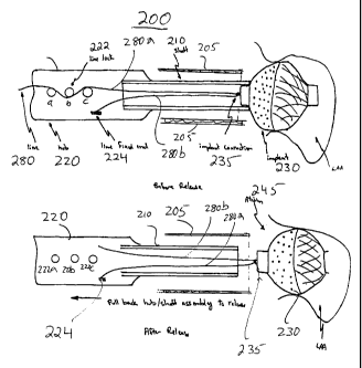
[0021] FIG. 2 is a schematic cross-sectional view of a
catheterization apparatus having a device tether system,
which includes a wire-dispensing hub connected to a
tubular push rod that is used for moving an implant
device through a catheter delivery tube in accordance
with the principles of the invention. Also, an exemplary
filter implant device tethered to a wire loop is shown
deployed in an atrial appendage. The two wire legs of
the wire loop are, respectively, shown as being anchored
at an anchoring post and at a line lock mechanism in the
wire-dispensing hub
[0022] FIG. 3 is a schematic cross-sectional view of a
catheterization apparatus of FIG. 2 showing the line lock
mechanism deactivated to release a leg of the wire loop
in preparation for untethering the deployed implant
device in accordance with the principles of the
invention.
Description of the Preferred Embodiments
[0023] Implant devices for filtering or otherwise
modifying blood flow between an atrial appendage and its
CA 02454017 2004-O1-15
WO 03/008030 PCT/US02/23201
8
atrium may be attached to a push rod or shaft, and then
be~percutaneously delivered to the appendage through a
catheter delivery tube inserted in a blood vessel leading
to the heart.
[0024] FIG. 1 illustrates, for example, catheter 21
inserted through a femoral vein (not shown) entering the
right atrium of the heart through the inferior vena cava
18, and then passing into left atrium 11 through the
fossa ovalis 19 or through the septum 29 before entering
the left atrial appendage 13. Alternatively (not shown
in FIG. 1), catheter 21 may enter the left ventricle 16
of the heart through the aorta 12, and then pass through
mitral valve 17 to reach left atrial appendage 13. An
implant device (not shown) attached to catheter 21 may be
used to prevent thrombus 30 or emboli generated therefrom
from migrating into atrium 11.
[0025] The implant devices generally include materials
having suitable properties (e. g., radio-opacity) that
make it possible to monitor the in-vivo device position
during and after the catheterization procedure using
external imaging techniques such as radiography or
fluoroscopy, echocardiography, and ultrasound. However,
the circuitous path of the catheter delivery tube through
the patient's vasculature across the cardiac septum may
make precise placement of an implant device difficult,
even when the operating physician has the benefit of
using external imaging techniques to monitor the implant
device position during the catheterization procedure.
10026] The present invention provides catheterization
apparatus having a device tether system in addition to
the conventional features of known catheterization
apparatus (e. g., previously disclosed catheterization
apparatus described in U.S. patent application No.
CA 02454017 2004-O1-15
WO 03/008030 PCT/US02/23201
9
09/960,749, and U.S. patent application No. 60/351,898).
A basic feature common to known catheterization apparatus
is a device delivery tube, which provides a conduit or
pathway for insertion of the implant device into the
patient's body. Another basic feature common to known
catheterization apparatus is a mechanism such as a push
rod or shaft for carrying or moving the implant device
through the delivery tube. It will be understood that
the inventive catheterization apparatus may in general
have one or more nested tubes, wires or shafts, and other
features (e. g. the positioning guides that are described
in U.S. patent application No. 09/960,749). However for
clarity in the description of the present invention
herein, and to simplify understanding of the invention,
reference will made only to the two previsouly mentioned
basic conventional features of the inventive
catheterization apparatus.
(0027] In the inventive tether system, the implant
device is tethered to a length of flexible line or wire
extending through a tubular push rod or shaft. The
tether wire allows an operating physician to retain
mechanical control over an implant device after it has
been expelled from the catheter delivery tube into a body
cavity. This mechanical control over post deployment
devices enables the physician to recover and reposition
implant devices as needed.
10028] The tether system may be used with implant
devices that have (or those that can be fitted with) a
suitable wire connection feature such as an eye hole. It
will also be understood that the device materials have
suitable properties (e.g., radio-opacity) that make it
possible to monitor the in-vivo device position during
and after the catheterization procedure using external
CA 02454017 2004-O1-15
WO 03/008030 PCT/US02/23201
imaging techniques, for example, radiography or
fluoroscopy, echocardiography, and ultrasound. Exemplary
devices, which may be implanted using inventive tether
system, are the reversibly expandable filter implant
5 devices having elastic structures described in U.S.
patent application No. 09/428,008, U.S.
patent application No. 09/614,091, U.S.
patent application No. 09/642,291, U.S.
patent application No. 09/697,628, U.S.
10 patent application No. 09/932,512, U.S.
patent application No. 09/960,749, and U.S.
patent application No. 10/094,730. It will be understood
that the tether system may also be used with any other
type or kind of implant devices, which are amenable to
delivery through catheter tubes.
[0029] In one embodiment of the invention, the tether
system includes a wire-dispensing hub connected to the
distal end of the tubular push rod or shaft. A flexible
wire (line, cord, or string) is dispensed in the hub.
The wire may be made of any suitable material, for
example, metals, polymers or a combination thereof. A
wire of suitable strength may be fabricated from a single
strand or from multiple strands of material. The wire
passes through the tubular push rod and out of the
proximal end of the push rod. The dispensed wire
extending out of the push rod is threaded through the
implant device wire-connection feature, and passed back
through the push rod to the hub. The wire loop thus
formed has a wire leg extending from the hub to the
implant device, and another leg extending from the
implant device back to the hub. Both ends of the wire
loop may be anchored or fixed securely at anchoring
fixtures that are provided in the hub. The tethered
CA 02454017 2004-O1-15
WO 03/008030 PCT/US02/23201
11
device may be held firmly against (and carried on) the
distal end of the push rod by suitably adjusting the
length of the wire loop legs.
[00301 In a catheterization implant procedure, the
push rod carrying a tethered device on its (push rod's)
distal end may be used to transfer the implant device
from outside the patient's body into a body cavity
through a pathway formed by the catheter delivery tube.
The implant device may, for example, be a self-expanding
device. The device is deployed in the body cavity by
pushing it through past the distal end of the catheter
delivery tube. The implant device remains tethered to
the wire loop even after it has been expelled from the
catheter delivery tube.
10031] External imaging techniques may be used to
verify the position of the deployed device. Alternative
diagnostic means, for example, electronic monitoring of
the patient's physiological parameters may also be used
to assess the suitability of the deployed device.
[0032] Deployed implant devices, which, for example,
are not satisfactorily positioned or oriented, may be
retracted into catheter delivery tube by pulling the push
rod out of the catheter delivery tube. The backward
motion of the push rod causes the wire loop to
mechanically pull the tethered device into the catheter
delivery tube. Because of its elastic structure the
implant device is compressed to its compact size as it is
retracted into the delivery tube. The operating
physician may attempt to reposition and redeploy the
retracted device in a more satisfactory position or
orientation by moving the push rod forward to again expel
the retracted device from the catheter delivery tube.
Before attempts to redeploy the retracted device are
CA 02454017 2004-O1-15
WO 03/008030 PCT/US02/23201
12
made, the catheter delivery tube itself may be suitably
repositioned or stabilized as necessary.
10033] Alternatively, if medically appropriate, the
retracted device may be retrieved from the patient's body
by pulling back the push rod completely out of the
catheter delivery tube.
[0034] Implant devices which are satisfactorily
deployed may be untethered by first deactivating the line
lock in the hub to free one wire end of the loop, and
then retracting the push rod so that the free end of the
wire loop slides clear of the implant device wire
connection feature.
[0035] FIG. 2 schematically illustrates.portions of
catheterization apparatus 200 having a device tether
system. Catheterization apparatus 200 includes a hollow
tubular shaft or push rod 210, and a catheter device
delivery tube 205. Catheter delivery tube 205 and push
rod 210 may be fabricated from any suitable material
including metals and polymeric materials, for example,
stainless steel and PTFE (e. g., Teflon). Catheter
delivery tube 205 may be used to establish a percuatneous
passage to a body cavity. Push rod 210 is designed to
slide through catheter device delivery tube 205. Push
rod 210 may be used to push or carry a compacted implant
device through the device delivery tube 205 into a body
cavity.
[0036] For example, FIG. 2 schematically shows
delivery tube 205 forming a conduit to atrium 235.
Further, FIG 2.shows filter implant device 230, which has
expelled through device delivery tube 205, and deployed
in a patient's left atrial appendage 240. Implant device
230 is provided with a eye hole 235 at its distal end.
CA 02454017 2004-O1-15
WO 03/008030 PCT/US02/23201
13
[0037] A wire-dispensing hub 220 is mechanically
connected to the proximal end of push rod 210. Hub 220
has a container-like structure, and may be fabricated
from any suitable materials including metals and
polymeric materials. Wire post 224 and line lock fixture
222, are disposed on an interior wall of hub 220. Line
lock fixture 222 includes posts 222a, 222b, and 222c.
Hub 220 may be provided with a removable access cover
(not shown) 'to provide access to the interior of hub 220.
(0038] Implant device 230 is tethered by cable 280.
Cable 280 is fixed to wire post 224, for example, by a
conventional screw and washer arrangement (not shown).
Cable 280 may, for example, be a polyester~or nylon
string. Alternatively, cable 280 may be fabricated from
other suitable natural or synthetic fibers. Cable 280
extends from wire post 224 through push rod 210 lumen to
implant device 230. Cable 280 passes through eye hole
235 disposed on device 230, and returns through push rod
230 lumen to hub 220. The return end of cable 280 may be
wrapped around line lock posts 222a-222c, to anchor cable
280, and to thereby firmly tether implant device 230 on
the distal end of push rod 210. In alternative designs
of hub 220, line lock 222 may include moving levers,
reels, rollers, or other mechanical structures to grip,
pinch, or other wise hold and anchor the return end of
cable 280. In this fashion, implant device 230 is
tethered by the wire loop that is formed by cable 280
with leg 280a extending from wire post 224 to implant
device 230, and leg 280b extending from the device 230 to
hub 220. Implant device 230 remains tethered after it
has been expelled from catheter delivery tube 205 and
deployed in atrial appendage 240, as shown in FIG. 2.
CA 02454017 2004-O1-15
WO 03/008030 PCT/US02/23201
14
[0039] To untether implanted device 230, the end of
leg 280 may be unwrapped from around posts 222a, b and c,
to free leg 280b from line lock 222. Push rod 210 (with
connected hub 220) may then be pulled back out of
catheter delivery tube 205. This back ward movement
causes cable 280 to slide out of eye hole 235 and to
thereby untether device 230. Fig. 3 schematically
illustrates the portions of catheterization apparatus 200
shown in FIG. 2 during the untethering procedure. In
FIG. 3, cable leg 280b is shown as free and unattached to
line lock 222. Push rod 210 is shown as having moved
back into cathter device delivery tube 205, and
disengaged from device 230. Further, back ward movement
of push rod 210 into catheter device delivery tube 205
would cause the free end of cable 280 to completely slide
out of eye hole 235 (not shown).
[0040] It will be understood that the foregoing is
only illustrative of the principles of the invention, and
that various modifications can be made by those skilled
in the art without departing from the scope and spirit of
the invention. It will be understood that terms like
"distal" and "proximal", "forward" and "backward",
"front" and "rear", and other directional or
orientational terms are used herein only for convenience,
and that no fixed or absolute orientations are intended
by the use of these terms.