Note: Descriptions are shown in the official language in which they were submitted.
CA 02454045 2004-O1-16
WO 03/008977 PCT/US02/22714
MATRIXES, ARRAYS, SYSTEMS AND METHODS
FIELD OF THE INVENTION
The present invention relates to matrixes, arrays, systems and methods
for preparing, sorting, amassing, or analyzing biomolecules based on
separation in one
dimension according to their isoelectric points in an electrical field, and
optionally
followed by a second analysis technique in a second dimension, and uses
therefor.
BACKGROUND OF THE INVENTION
The basic principle behind isoelectric focusing or focusing in a pH
gradient is that a charged molecule will become immobilized in a electric
field when it
migrates to a position in the pH gradient that is equal to its isoelectric
point (zero net
charge). This process occurs independently of the initial location of a
specific protein
in the solution. It is the result of the disappearance of the effective
electrical charge of
the protein when migrating to the region where pH is equal to pI.
Various techniques for determining the isoelectric point of a protein
have been described. Typically, the protein of interest is injected or
administered
directly into a gel containing a pH gradient, wherein the pH gradient is
parallel to the
direction of the electric field, and the protein can only be separated from
other proteins
by traveling uni-directionally through many different pH environments before
reaching
a pH environment that is equivalent to its isoelectric point. These techniques
suffer
from the disadvantages that (1) they require a relatively long time to
separate the
protein because the velocity of the fraction tends to zero asymptotically; (2)
they
require relatively high voltage's (typically 1000V and higher), and (3) they
require a
cooling mechanism. Traditional IEF methods are labor intensive, time
consuming,
non-standardized, expensive and not sensitive. Another practical limitation of
traditional isoelectric focusing gels is that it is difficult to manufacture
gels having
CA 02454045 2004-O1-16
WO 03/008977 PCT/US02/22714
incrementally small pH changes within a pH gradient to improve the linear
dispersion
of the proteins.
Two dimensional analysis of proteins that use the above described
isoelectric focusing step suffer from the same problems. For example, Zuo et
al.,
(2000) Analytical Biochemistry 284:266-278, describe the separation of
proteins based
on their isoelectric point by unidirectional travel through a pH range
followed by
sodium dodecyl sulfate polyacrylamide gel electrophoresis (SDS-PAGE). Becker,
et
al., (1998) J. Micromech. Microeng. 8:24-28 suggests the unidirectional travel
of
proteins through a pH range followed by a second dimension separation on a
planar
chip. See also, US 6,254,754 (Ross).
Because of these limitations, only certain cell, lane, and matrix designs
and orientations of the cells, lanes, and matrixes in a chamber, and certain
systems for
one and two dimensional analysis are possible thereby limiting the development
of
faster, more sensitive, more accurate, more flexible and less expensive
methods for one
and two dimensional analyses of samples, including automated, high throughput
analysis systems. Better tools and methods for one and two dimensional
analysis of
biomolecule are useful for, e.g., drug development, medical research, and the
pre-
diagnosis and/or diagnosis of diseases. In particular, better,tools and
methods are need
for proteomic analysis. The present invention solves these and other problems.
SUMMARY OF THE INVENTION
The present invention relates to matrixes, arrays, systems and methods
for analyzing or preparing biomolecules by their isoelectric point in one
dimension
and, optionally, in combination with other methods for analysis. The
assortment of
cells, matrixes, arrays and systems provided herein have unique configurations
and
combinations of elements.
According to one embodiment, a biomolecule moves through the
running buffer of a chamber of this invention and becomes trapped in an IEF
buffer or
a cell comprising an IEF buffer of this invention. According to another
embodiment, a
biomolecule that is trapped in an IEF buffer or cell of this invention remains
trapped in
the IEF buffer or cell while biomolecules having pI values that are not the
same as the
pH values of the IEF buffer are removed by alternating the direction of the
electric
CA 02454045 2004-O1-16
WO 03/008977 PCT/US02/22714
field. If the IEF buffer or cell is closed so that the electrical current is
preventing from
exiting out the opposite side of its entry into the IEF buffer or cell, then,
according to
one preferred embodiment, the electric field is reversible. If the lEF buffer
or cell is
open, then the electrical field can be unidirectional. According to another
embodiment
of this invention, the movement of the biomolecule in the running buffer is
increased
by the convection heat generated by the electrical field.
According to another embodiment of this invention, the movement of
the biomolecule in the running buffer is increased by a device that circulates
the
running buffer comprising the biomolecule across the IEF buffer (e.g., by stir
bar, by
pump or by the movement of the IEF buffer relative to the running buffer).
According to yet another embodiment, the pH range of the IEF buffer
can be ultra-narrow (e.g, spanning 0.1 pH units or less; 0.02 pH units or
less; or 0.01
pH units or less). According to one embodiment, the chamber comprising the
running
buffer further comprises a plurality of IEF buffers and/or cells that are
isolated from
each other either by physical separation or by a substrate that substantially
prevents the
movement of biomolecules directly from one IEF buffer/cell to another rather
than
through the running buffer. Thus, the biomolecule primarily moves through the
running buffer to reach a different IEF buffer or cell. In another embodiment,
the IEF
buffers or cells have the same or different pH values. According to yet
another
embodiment, the present invention comprises a vast plurality of discrete,
isolated IEF
buffers having ultra-narrow, substantially non-overlapping pH ranges such that
resulting image of the separated material is comparable positionally to an
image from a
traditional IEF gel but has greater resolution than a traditional IEF gel.
According to this invention, biomolecules of this invention can be
separated as a single entity or as part of a complex based on their
isoelectric point. For
example, the biomolecule ("target biomolecule") can form a complex with
another
molecule that specifically recognizes it ("target recognition molecule"). The
complex
can be separated from other non-complexed biomolecules based on the
isoelectric
point of the complex using the matrixes, arrays, systems and methods of this
invention.
According to one embodiment of this invention, an improved one- or
two dimensional analysis method using a plurality of discrete, isolated IEF
buffers with
narrow pH ranges and steps, e.g., 0.1 pH units or less, is provided. More
preferably,
the pH range or step is 0.02 pH units or less. It is an object of this
invention to provide
CA 02454045 2004-O1-16
WO 03/008977 PCT/US02/22714
improved one and two dimensional methods for analyzing biomolecule having pI
values that are 0.02 pH or less units apart. According to one configuration of
the
system, diffusion ofbiomolecule from one cell into an adjoining cell is
avoided, e.g.,
between cells that comprise IEF buffers with slightly different pH values, by
using
membranes or materials that are impermeable to the biomolecule. For example,
each
IEF buffer or cell comprising said IEF buffer can be physically separated,
noncontinuous, discrete entities.
It is an object of this.invention to provide an analysis method that
allows the use of a high electric field at a low applied voltage, optionally
avoiding the
use of a kV range power supply. In one preferred embodiment, a device for
reversibly
directing an electrical field in and out of the IEF buffer in the cells and,
optionally,
device for circulating buffer around a plurality of cells simultaneously is
used in the
methods and systems of this invention. Another object of this invention is to
provide
an analysis method that requires little or no device for cooling the chamber.
Yet
another object of this invention is to provide a two dimensional matrix that
requires
minimal or no manipulation of the biomolecule during the first and second
dimension
separations, thereby saving time and effort and minimizing the loss of the
biomolecule
being tested.
Another embodiment of this invention provides an IEF technique
suitable for use in combination with a second dimension analysis (e.g, high
pressure
liquid chromatography (HPLC), mass spectrometry, affinity chromatography, gel
electrophoresis, etc. ). Yet another object of the invention is systems or
methods
capable of separating and /or purifying small or large quantities of a
specific
biomolecule, such as a protein or nucleic acid molecule. This invention also
provides
methods for detecting a target biomolecule complexed to a target recognition
biomolecule. The target biomolecule can form a complex with the TB in
environments
that encourage or discourage complex formation. In one embodiment of the
invention,
a labeled target recognition biomolecule is placed directly into the IEF
buffer cell, lane,
or matrix prior to the introduction of the target molecule-containing sample.
Yet another embodiment of this invention is to provide systems and/or
methods for one and two dimensional analysis that can be miniaturized and
automated
for high throughput analysis of samples for drug screening, medical research
such as
enhanced detection of biological response patterns for drug discovery,
monitoring of
CA 02454045 2004-O1-16
WO 03/008977 PCT/US02/22714
drug therapies, genetic or proteome analysis, and clinical diagnosis, and
diagnostics,
e.g., proteome analysis. A system of this invention can be constructed to have
automated interacting components, for example, titrators for filling of
channels with
pH solutions or gels (immobilines, ampholyte mixtures etc.), extractors for
recovering
biomolecule from the cells comprising IEF buffers, devices for staining the
biomolecule, devices for detecting and scanning the biomolecule, devices for
recording
and analyzing the images. A system and/or method of this invention can be
automated
for high throughput screening of candidates useful for a desired drug effect.
This invention provides methods for enhancing detection of
biomolecule in the response to various perturbations and stimuli, such as the
response
to a drug, a drug candidate or an experimental condition designed to probe
biological
pathways as well as changes in a animal or human that correspond to a
particular
disease or disease state, or to a treatment of a particular disease or disease
state.
BRIEF DESCRIPTION OF THE DRAWITTGS
FIG. 1 depicts (A) a cell of this invention containing an IEF buffer and
having a protein and ion permeable membrane on opposing sides of the cell; and
(B) a
matrix comprising a plurality of cells of this invention.
FIG. 2 depicts an apparatus of this invention comprising two electrode
plates on either side of a chamber and a matrix comprising a plurality of
cells in
between the electrodes. The chamber is on top of a magnetic stirrer. The
direction of
the electrical field is reversible.
FIG. 3 depicts an apparatus of this invention comprising a matrix
suspended over the bottom of the chamber between two electrode plates, wherein
the
running buffer can flow around the matrix aided by the stir bar. The direction
of the
electrical field is reversible.
FIG. 4 depicts an apparatus of this invention wherein the matrix rotates
to distribute the biomolecule in the running buffer across the cell openings.
Optionally, the chamber may also have a stir bar to circulate the running
buffer. The
direction of the electrical field is reversible.
FIG. 5 depicts three apparatuses of this invention: (A) a plurality of
cells are individuallyvand randomly mounted on a insulated support in the
running
CA 02454045 2004-O1-16
WO 03/008977 PCT/US02/22714
buffer in the chamber and between two electrode plates; (B) a plurality of
cells are free
floating in the chamber between two electrode plates; or (C) a plurality of
cells are
attached to each other and rotate between two electrode plates. A stir bar is
used to
circulate the running buffer. The direction of the electric field is
reversible.
FIG. 6 depicts a top view of a chamber comprising a plurality of cells
adjoined in series, separated by membranes that substantially maintain the pH
range
present in each cell, and arranged (A) in parallel or (B) perpendicular to the
direction
of the electrical field. A stir bar is used to circulate the running buffer.
The direction
of the electric field is reversible.
FIG. 7 depicts biomolecule in the cells of a matrix of this invention
being subjected to SDS-PAGE capillary electrophoresis in a second dimension.
FIG. ~ depicts a matrix of this invention, wherein the cells are capable
of being adjusted into a linear series for attaching to an SDS polyacrylamide
gel for
electrophoresis in a second dimension.
FIG. 9 depicts matrix of this invention comprised of an agarose gel with
channels comprising a plurality of IEF buffers. Each vertical column of
channels
contain the same IEF buffer, except for the fourth column of channels which
contain
no buffer. Ferritin, phycocyanin (first band), phycocyanin (second band), and
hemoglobin accumulated in the first, second, third and fifth vertical columns,
respectively.
FIG.10 is an image of a chip according one embodiment of this
invention. The chip as drawn herein has been designed to analyze three target
biomolecule in a sample. Each IEF buffer in each cell of the chip in the
first, second
and third rows has a pH that is the same as the pI of the complexes comprising
various
TBs/TRMs, e.g., TB1/TRM1, TB2/TRM2 or TB3/TRM3, respectively. The fourth
row comprises cells that are designed to receive non-TB molecules that have
been
added to the running buffer for use as a control, standard or data point for
developing a
calibration curve. Accordingly, the IEF buffers in the fourth row have a pH
value that
is the same as the pI value of the non-TB biomolecule.
FIG. 11 is an image of a chamber comprising a multicell chip located
between two electrode plates according to one embodiment of this invention.
The
chamber can be attached to a power supply capable of reversing the polarity of
the
CA 02454045 2004-O1-16
WO 03/008977 PCT/US02/22714
electrical field. The chamber can further comprise a mechanisms) for stirring
the
running buffer in both compartments on either side of the multicell chip.
FIG. 12 is an image of a detection device for a multicell chip according
to one embodiment of this invention. After one or more complexes are received
into a
plurality of cells in the chip, the chip can be placed in a detection device.
Fluorescently labeled complexes in a multicell chip can be stimulated by a
light source
(e.g., monochomatic light source) for detection, then the light emitted from
the label
can be captured by a photodiode, converted into an electronic signal (read out
unit),
and analyzed by a computer. The chip can be encased in a holder that is
movable
relative to the light source or diode. Alternatively, the light source and
diode can be
moveable relative to the chip.
FIG. 13 is a graphical representation of the absorption of hemoglobin at
610nm at various concentrations. Each data point represents a reading of
absorption
taken from a standard cuvette with rectangular shape filled with solutions
having
different concentrations of hemoglobin.
FIG. 14 is a graphical representation of a calibration curve for a blood
sample being tested for diabetes. The X axis is the ratio of molar
concentration of
glycated hemoglobin to the sum of the molar concentration of glycated and non-
glycated hemoglobin, expressed as a percentage. The Y axis is the ratio in
percentage
of the absorption of the glycated hemoglobin to the sum of the absorption of
glycated
and non-glycated hemoglobin.
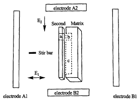
FIG.15 depicts examples of matrixes and an array according to this
invention. FIG.15A depicts side views of four examples of matrixes of this
invention
each containing an IEF buffer ("b"). Matrixes A and D have grooves comprising
IEF
buffer in it. Matrixes B and C have IEF buffers set on the surface of the
matrix.
Matrixes C and D have an area designated "c," which is a lane. The matrixes A-
D can
be used in combination with a second layer in the system according to this
invention
for two dimensional analysis. The direction of the electrical field for the
isoelectric
focusing step is indicated as "El." A portion of "b" in matrix A and B can
also serve
as a lane for the second dimension separation according to this invention,
(e.g., by
adding sodium dodecyl sulfate to the running buffer during the second
dimension
separation). According to another embodiment of this invention, matrixes A-D
can be
rotated 90 degrees in any direction in the same El field during the
isoelectric focusing
CA 02454045 2004-O1-16
WO 03/008977 PCT/US02/22714
step (not shown). FIG.1 SB depicts an array wherein a second layer comprising
a
perforation can be placed on a matrix such that the IEF buffer in the matrix
is capable
of contacting a running buffer during the separation in the first dimension.
FIG.16 relates to an example of array of this invention. (A) is a top
view of an example of a second layer of an array. In this example, the second
layer is
manufactured from a material that is impermeable to biological molecules but
is
permeable to ions in the running buffer. (B) is a top view of an example of a
matrix
according to this invention. The matrix comprises a rectangular groove that is
filled
with gel to form the lane and a circular groove that is filled with IEF
buffer. (C) is a
top view of the second layer of (A) aligned on top of the matrix of (B) so
that the IEF
buffer is exposed through the perforation in the second layer. (D) is an side
view of
one of the IEF/lane units of the array of (C). (E) is an enlargement of a side
view of a
portion of the array of (C) (dotted circle). The top layer is the second layer
(A) and the
bottom layer is the matrix (B).
FIG.17 depicts top view of an example of a system of this invention
comprising an array according to this invention in a chamber comprising
running
buffer. A stir bar is in the chamber to allow circulation of the biomolecule
across the
IEF buffers) or cells in "b." Electrodes A1 and B 1 create an electric field
that
periodically reverses direction during the isoelectric focusing step.
FIG.18 is an electronic scan of a silver-stained gel of a commercially
available protein standard prepared as described in Example 1, ihf~a.
FIG.19 is an optical scan of a silver-stained gel of a human plasma
prepared as described in Example 2, iafi°a.
FIG.20 is a top view of a matrix used in the separation of human plasma
proteins having pI's of 7.5 to 8.5 using the methods of this invention. The
matrix is
lxlcm lucite chip. Each line on the matrix was drawn by a modified inkjet
printer that
deposited IEF buffer and acrylamide mixtures such that parallel lanes of gels,
wherein
each lane had a uniform width and thickness of 100 micron and a length of 1
cm, but a
different pH (i.e., fifty lanes starting with pH 7.50, 7.52, 7.54, 7.56, 7.58,
etc. up to
8.50). The matrix placed in some running buffer in a chamber so that one tip
of each
of the lanes was immersed in the rumung buffer. A human plasma sample was
circulated in the running buffer across the tips of the lanes with a stir bar
while an
electrical field that periodically reversed direction was applied to the
running buffer.
CA 02454045 2004-O1-16
WO 03/008977 PCT/US02/22714
After some minutes, the electric field was switched off, a 3% SDS solution was
added
to the running buffer, and the entire chip was immersed in the running buffer.
Then, a
unidirectional electrical field parallel to the lanes was applied in a
direction away from
the tip of the IEF buffer used in the isoelectric focusing step and down the
length of the
lane. The chip was then silver-stained. The grey and black spots observable in
Fig.20
are proteins that have been silver-stained.
FIG.21 is a comparison of the human plasma prepared according to the
two dimensional analysis of this invention and according to a traditional two
dimensional IEF-SDS-PAGE analysis. (A) is a digitalized, optical scan of the
silver-
stained chip used in Example 3. The scan has been enlarged to scale for
comparison
with the Swiss Protein 2D image. (B) is a published, silver-stained two
dimensional
gel of human plasma proteins having pI's 7.50 to 8.50.
FIG.22 is a comparison of the human plasma prepared according to the
two dimensional analysis of this invention and according to a traditional two
dimensional IEF-SDS-PAGE analysis. (A) is a published, silver-stained two
dimensional gel of human plasma proteins having pI's 5.50 to 6.00. (B) is a
digitalized, optical scan of the silver-stained chip used in Example 4. The
scan has
been enlarged ten times.
FIG.23 is an optical scan of a single capillary SDS-PAGE lane. The
darkened areas in the lane are silver-stained human plasma proteins having a
pI of
approximately 7Ø Human plasma protein was subjected to separation using an
array
of this invention prepared as described in Example 5.
FIG.24 is an optical scan of a single capillary SDS-PAGE lane. The
darkened areas in the lane are silver-stained human plasma proteins having a
pI of
approximately 7Ø Human plasma protein was subjected to separation using an
array
of this invention prepared as described in Example 5 in a three-electrode
system as
described in Example 6.
FIG.25 is a schematic of an apparatus that can make an IEF buffer area
on a matrix. A device is used to mix an acidic and basic solution to form an
buffer
having the desired pH value ("titrator"). The buffer is combined with a
monomer (e.g.,
acrylamide) and polymerizing agent and loaded into another device ("matrix
printer")
that lays the IEF buffer in a desired position on the array.
CA 02454045 2004-O1-16
WO 03/008977 PCT/US02/22714
FIG.26 is a schematic of an apparatus that can make the lanes on a
matrix. An acrylamide solution and a polymerizing agent is loaded into a
device
("matrix printer") that lays lanes in a desired position on the array.
FIG.27 is a schematic example of one automated system of this
invention. The system provides, for example, a device for feeding a sample
into the
two dimensional electrophoretic analysis chamber of this invention ("sample
feeder"),
a device for removing waste ("waste disposal"), a device for adding new
rumiing
buffer ("buffer feeder"), a device for staining the matrix after two
dimensional analysis
("staining reagent feeder"), a device for bringing the stained chip to a
scanner ("array
handling system"), a device for scanning the chip ("scanner"), a device for
receiving
and recording the scanned image ("computer"), a device for analyzing the
recorded
image ("software"), and a device for displaying the recorded image
("display").
FIG.28 is a diagram of an example of a three-electrode system
according to this invention.
FIG.29 is a chart demonstrating the efficiency of separation within the
IEF system by plotting calibrated fluorescence as determined by the dispersion
of a
labeled protein through a gel vs. the observed fluorescence of a labeled
antibody after
10 minutes of isoelectric focusing. The numbers represent the total number of
protein
molecules in the sample, demonstrating near 100% efficiency in protein
separation.
DETAILED DESCRIPTION
An IEF buffer comprises components that have a buffering capacity
around a given pH value (buffering agent) or components that organize to form
a pH
gradient (e.g., ampholytes, immobilines or a combination of buffering agents).
The
IEF buffer according to this invention is in the form of a liquid or slurry or
a gel such
that a biomolecule can pass through IEF buffer unless the pI of the
biomolecule is in
the pH range of the IEF buffer. An IEF buffer according to this invention can
comprise
other components such as urea, detergent and a reducing agent as needed. See,
e.g.,
Malloy, et al., Anal. Biochem. 280: pp. 1-10 (2000). It is desirable that the
IEF buffers
according to this invention are functionally stable under the influence of an
electric
field.
The IEF buffer or cell comprising the IEF buffer can be formed by hand
or by various devices. For example, the IEF buffer can be deposited (e.g,
coated,
to
CA 02454045 2004-O1-16
WO 03/008977 PCT/US02/22714
printed or spotted) on the surface of a substrate or in a groove or channel of
a substrate.
The substrate can be a matrix as described below or a bead made of the same
material
as the matrix. According to one embodiment of this invention, the IEF buffer
can be
made by a device that mixes an acidic and basic solution to form an buffer
having the
desired pH value ("titrator"). The buffer is combined with a monomer (e.g.,
acrylamide) and polymerizing agent and loaded into another device ("matrix
printer")
that lays the IEF buffer in a desired position on the matrix. See, e.g.,
Fig.25. These
devices can be incorporated into an automated system of this invention.
Ampholines according to this invention are a set of various oligo-amino
and/or oligocarboxylic acids that are amphoteric (i.e., positively charged in
acidic
media and negatively charged in basic media), soluble and have Mr values from
approximately 300 up to 1000 u. Ampholytes used in this invention can be
prepared or
purchased. For example, several carrier ampholytes are known in the art (e.g.,
pages
31-50, Righetti, P.G., (1983) Isoelectric Focusin : Theory Methodology and
Applications, eds., T.S. Work and R. H. Burdon, Elsevier Science Publishers
B.V.,
Amsterdam; US patent 3,485,736). Alternatively, purchased ampholytes include
Ampholines (LIMB), Servalytes (Serva), Biolytes or Pharmalytes (Amersham
Pharmacia Biotech, Uppsala, Sweden).
Imrnobilines are non-amphoteric, bifunctional acrylamido derivatives of
the general formula: CHz CH-CO-NH-R. Immobilines useful according to this
invention can be prepared or purchased. For example, methods for synthesizing
immobilines are known in the art (Bjellquist et al., (1983) J. Biochem. Bioph
Methods., 6:317). The immobilines can be copolymerized with the acrylamide to
form
IPG's (immobilized pH gradients). IPG's can be prepared by methods known in
the art
or can be purchased.
pH gradients according to this invention can be formed by mixing
amphoteric or non-amphoteric buffers. For example, such buffers combinations
are
described in Allen, RC et al., Gel Electrophoresis and Isoelectric Focusing of
Proteins'
Selected Techn~ues, Berlin:Walter de Gruyter & Co. (1984); and in US 5,447,612
(Bier). Some IEF buffering agents include those are selected from the group
consisting of 50 mM glycine,l4 mM NaOH; SOmM HEPES, l2mM NaOH; SOmM
THMA, 44.6mM HCl; 52mM citrate acid, 96mM Na2HP04; SOmM BICINE, l8mM
NaOH; and SOmM DMGA, 40mM NaOH. The pH gradient created by the IEF buffer
11
CA 02454045 2004-O1-16
WO 03/008977 PCT/US02/22714
in each cell can have a narrow or a wide pH range (e.g., pH 6.8-pH 7.8 or pH
6.8-pH
12.8, respectively).
An IEF buffer of this invention can have an extremely narrow pH range,
e.g. 5.50-5.60 (0.1 pH unit or less difference) or ultra narrow pH range,
e.g., 5.52-5.54
(0.02 pH unit difference or less). This is possible because an IEF buffer
according to
this invention can be one buffering agent that has been adjusted to a certain
pH value.
In this case, the pH range of the IEF buffer is equivalent to the buffering
capacity of the
buffering agent around the pH value to which the buffering agent had been
adjusted.
The term "interval" refers to the incremental difference in a pH value
within the pH gradient created by the IEF buffer. The term "step" refers to
the
incremental difference in pH value between two different IEF buffers. For
example,
within one cell, the intervals can be as small as 0.02 pH units through the
full pH range
in that cell (e.g., pH 6.8, pH 7.0, pH 7.2, etc., in that cell). In another
example, the pH
"step" between an IEF buffer in cell #1 and cell #2 can be O.lpH unit. For
example,
the IEF buffer in cell #1 can have a pH gradient starting at pH 6.8 and ending
at pH 7.8
and the IEF buffer in cell #2 can have a pH gradient starting at pH 7.9 and
ending at
pH 8.9 (i.e., pH 7.9 minus pH 7.8). The term "pH range" refers to the highest
to the
lowest pH values in an IEF buffer or a cell comprising an IEF buffer (e.g., pH
7.9-pH
8.9), or the difference between the highest and lowest pH values in a IEF
buffer or a
cell comprising an IEF buffer (e.g., 1.0 pH units). According to this
invention, the
intervals within a cell do not have to be uniform. Further, the pH steps
between two
cells of a plurality of cells do not have to be uniform. According to one
embodiment,
the matrix comprises IEF buffers or cells with IEF buffers having an extremely
narrow
or ultra narrow pH range and small pH steps between each cell.
According to one embodiment of this invention, the pH range of an IEF
buffer in a cell is a narrow pH gradient, e.g., less than one pH unit or up to
a few pH
units. According to another embodiment, the pH gradient in the cell is over
several pH
units. According to one embodiment of this invention, the pH interval of an
IEF
buffer is 0.1 pH unit or less. In another embodiment, the pH interval of an
IEF buffer
is 0.02 unit or less. According to one embodiment of this invention, the pH
steps
between two or more lEF buffers are 0.01 units or less. According to another
embodiment of this invention, the pH steps between two or more IGF buffers are
0.02
units or less.
12
CA 02454045 2004-O1-16
WO 03/008977 PCT/US02/22714
A cell according to this invention is a hollow structure that has an IEF
buffer in it and/or integrated into a wall of it. The cell can have any shape
including a
sphere, a triangle, square, rectangle and a cylinder. A cell can have one or
more walls
depending on the shape of the structure. The walls of the cell have an inner
side that
faces towards the center of the structure and an outer side that faces towards
the
outside of the cell. See e.g., Fig. 1. Depending upon the desired use, a wall
of a cell of
this invention can be made of a membrane, mesh or solid that is biomolecule
permeable, biomolecule impermeable, and/or penetrable or impenetrable by an
electric
field.
Some of the walls of the cell can be impenetrable to an electrical field.
However, the cell walls should be constructed so that an electrical current
can pass into
the cell. An IEF buffer can be integrated into a wall of the cell. For
example, a
Whatman GF/D glass fiber filter disc can be immersed in acrylamide that is
allowed to
polymerize into a gel and then soaked in an IEF buffer. The disc can than be
used to
form a wall of the cell. Thus, a biomolecule can be trapped in a cell that has
a wall
soaked in an IEF buffer that is the same pH value as the pI value of the
biomolecule.
If a sample comprising a biomolecule(s) of interest is added to the
running buffer in the system, then at least one wall of the cell should be
permeable to
one or more of the biomolecule of interest. In one embodiment, all the walls
of the cell
are permeable to the biomolecule of interest. In another embodiment, all but
one wall
of the cell is biomolecule impermeable and/or impenetrable by an electric
field. In a
embodiment, the walls of the cell that face into and in the same direction as
the
electrical field in the first dimension are permeable to the biomolecule of
interest.
According to an alternative embodiment, a wall or the walls of the cell
can be substantially impermeable to the biomolecule of interest if the
biomolecule of
interest are being prepared by (1) adding a sample comprising the biomolecule
of
interest to the IEF buffer in a cell in the system and (2) allowing
biomolecule and/or
ions that are not of interest to migrate out of the cell. In this way, the
cell can be used
in the first dimension step in combination with the matrixes, arrays, systems
and
methods of this invention.
The cells can be arranged spatially in several ways. For example, the
cells can be contiguously arranged, e.g., wherein a biomolecule-permeable or a
biomolecule-impermeable material separates one cell adjoined to another. See
e.g., Fig.
13
CA 02454045 2004-O1-16
WO 03/008977 PCT/US02/22714
6a and b. According to one embodiment, the IEF buffers or cells are "isolated"
such
that biomolecule substantially travel from one IEF buffer to another by
migrating
through the running buffer circulating around the IEF buffers or cells rather
than
through one IEF buffer directly into another IEF buffer or through the wall of
one cell
directly into another cell. See e.g., Fig. 5a, b or 2. According to one
embodiment, the
isolated cells are adjoined but have biomolecule-impermeable material
separating
them. See. e.g.,Fig. 6. Alternatively, the isolated IEF buffers or cells are
not adjoined.
According to an embodiment of this invention, if the IEF buffers or cells are
contiguously arranged, at least one of the walls of the IEF buffers or cells
that is not
adjoined to another IEF buffer or cell contacts the running buffer and is
permeable to
the biomolecule being tested. According to one embodiment of the invention,
the IEF
buffers or cells form part of a matrix. According to one embodiment, the IEF
buffers
or cells are not adjoined in series to each other when the cells are arranged
in parallel
to the electrical field of the first dimension.
The biomolecule-permeable or biomolecule-impermeable material can
be a membrane depending on the desired result. According to one embodiment of
this
invention, the membrane can be prepared so that it has virtually no net charge
in the
electric field at the pores of the membrane. In an alternative embodiment, the
pH of
the membrane can be a pH value intermediate between the pHs on both sides of
the
membrane. This is desirable to minimize bulk fluid flow through the membranes
caused by the presence or acquisition of an electrical charge on the membrane
(electroendoosmosis). Depending on the desired result, membranes useful
according to
this invention include those described in US 4,243,507 (Martin).
Alternatively,
membranes according to this invention can include membranes covalently bonded
with
immobilines as described in US 4,971,670 (Faupel).
The cells according to this invention can be directly or indirectly
attached to the chamber as long as the cells are capable of contacting the
running
buffer. For example, the cells may be attached directly to the bottom or sides
of the
chamber or mounted to the chamber by an insulating support. See e.g, Fig. 5a
or Sc.
Alternatively, the cells may be placed in a matrix that is attached to the
chamber or
attached to a post that is attached to the chamber. In yet another embodiment,
the cells
comprising an IEF buffer that is a buffering agent may float freely in the
running
buffer, but the cells should be distinguished to indicate the pH range of the
IEF buffer
14
CA 02454045 2004-O1-16
WO 03/008977 PCT/US02/22714
in the cell. See e.g., Fig. 5b. According to this invention, the matrix or the
individual
cells can be attached to the chamber such that they rotate within the chamber.
The sensitivity of the methods and systems of this invention will
increase as the size of the IEF buffer or cell decreases. According to one
embodiment
of this invention, the size of the IEF buffer or cell, particularly the length
IEF buffer or
cell, is as small as possible. The IEF buffer or cell length refers to the
widest cross-
section of the IEF buffer or cell that is parallel to the direction of the
electric field in
the second dimension. The IEF buffer or cell width refers to the widest cross-
section
of the IEF buffer or cell that is perpendicular to the direction of the
electric field in the
second dimension. In one embodiment of this invention, the cell length can be
any
size, e.g., 10 microns to S.Omm. In another embodiment of this invention, the
cell
width can be any size, e.g., 10 microns to lO.Omm.
The lane according to this invention can be various sizes. According to
one embodiment, the width of the lane is 20 microns to 1 mm. For example, the
width
of the lane can be 100 microns. According to another embodiment, the length of
each
lane is 3-lOmm.
The lane can comprise materials suitable for separation techniques (e.g.,
by size, shape, charge, affinity or combination thereof). Such as material can
include
those suitable for chromatography, electrophoresis such as SDS-PAGE, zone
electrophoresis, affinity electrophoresis, capillary electrophoresis, and
electro-
chromatography. Accordingly, in one embodiment, the lane can be a capillary
tube
that is filled with chromatographic substances (e.g., liquid chromatography)
or
substances useful for electrophoresis (e.g, capillary zone electrophoresis,
capillary gel
electrophoresis using cross-linked and uncross-linked gels), and capillary
isoelectric
focusing. According to one embodiment, the second dimension is an electric
field-
mediated separation technique.
A lane according to one embodiment of this invention can comprise a
gel-like material that is suitable for electrophoretic separation, e.g., US
6,197,173
(Kirpatrick). The gel-like material can be comprised of monomers that have
been
polymerized. The gel can be denaturing or non-denaturing for the biomolecule
of
study. The gel can have various pore sizes. Accordingly, the lane can comprise
additional components such as urea, detergent and a reducing agent as needed.
See,
e.g., Malloy, et al., Anal. Biochem. 280: pp.1-10 (2000). The lane itself can
comprise
CA 02454045 2004-O1-16
WO 03/008977 PCT/US02/22714
an IEF buffer for further separation of the biomolecule that have accumulated
in the
IEF buffer of the first dimension. Alternatively, a lane that comprises an IEF
buffer
can be converted into an SDS-containing gel by the addition of SDS to the
running
buffer, hence the biomolecule can separate in the second dimension based on
molecular weight.
According to one embodiment, the lane is premade to comprises sodium
dodecylsulfate (SDS) and polyacrylamide gel. According to another embodiment,
the
length of the lane is sandwiched between two biomolecule impermeable layers.
According to a further embodiment, if the lane comprises SDS and
polyacrylamide gel,
the lane can be sandwiched between a matrix layer and another layer, wherein
both
layers can be biomolecule impermeable and ion impermeable as long as an
electrical
field can penetrate the lane and direct an electric field down the lane away
from the IEF
buffer. See e.g., Fig. 16
The lane can be formed by hand or by various devices. For example, an
acrylamide solution and a polymerizing agent can be loaded into a device
("matrix
printer") that lays lanes in a desired position on an array. See, e.g.,
Fig.26. A modified
office inkjet printer is one example. Such devices can be incorporated into an
automated system of this invention.
Various monomers can be used in addition to the conventional
acrylamide/ bis-acrylamide solution or agarose solutions to make a gel for use
in the
first and/or the second dimension steps according to this invention. It is
known in
conventional chemically-polymerized gels to use hydroxyethylmethacrylate and
other
low-molecular weight acrylate-type compounds as monomers; these have been
commercialized as "Lone-Ranger" gels. Use of polymers substituted with one or
more
acrylate-type groups has also been described in the literature (Zewert and
Harrington,
Electrophoresis 13: pp.824-831, (1992)), as especially suitable for
separations in mixed
solvents of water with miscible organic solvents, such as alcohol or acetone.
Gel-
forming monomers can also be any substantially water-soluble molecule
containing a
photo-polymerizable reactive group, in combination with a material which can
form
cross-links, provided that the combination, once polymerized, forms a gel
suitable for
the particular type of electrophoresis.
Exemplary materials include acrylamide, in combination with
methylene-bis-acrylamide or other known crosslinkers; hydroyethylmethacrylate
and
16
CA 02454045 2004-O1-16
WO 03/008977 PCT/US02/22714
other low-molecular weight (less than about 300 daltons) derivatives of
acrylic acid,
methacrylic acid, and alkyl-substituted derivatives thereof, such as crotonic
acid; vinyl
pyrrolidone and other low-molecular weight vinyl and allyl compounds; vinylic,
allylic, acrylic and methacrylic derivatives of non-ionic polymers, including
such
derivatives of agarose ("Acrylaide" crosslinker, FMC Corp.), dextran, and
other
polysaccharides and derivatives, such as cellulose derivatives including
hydroxyethyl
cellulose; polyvinyl alcohol; monomeric, oligomeric and polymeric derivatives
of
glycols, including polymers of ethylene oxide, propylene oxide, butylene
oxide, and
copolymers thereof; acryl, vinyl or allyl derivatives of other water-
compatible
polymers, such as polyHEMA (polyhydroxyethyl acrylic acid), polymeric N-
isopropyl
acrylamide (which is temperature-sensitive), malefic-acid polymers and
copolymers,
partially hydrolysed EVAC (polymer of ethylene with vinyl acetate),
ethyleneimine,
polyaminoacids, polynucleotides, and copolymers of the subunits of these with
each
other and with more hydrophobic compounds such as pyridine, pyrrolidone,
oxazolidine, styrene, and hydroxyacids. The polymerizable materials need not
be
entirely water-soluble, especially when solvents or surfactants are included
in the gel-
forming solution.
Methods for making polymerizable derivatives of common polymers are
known in the art; for example, addition of allyl glycidyl ether to hydroxyl
groups is
known, as is esterification of hydroxyls with acids, anhydrides or acyl
chlorides, such
as acrylic anhydride. Amines are readily derivatized with acyl anhydrides or
chlorides.
Many of the derivatized polymers described above will contain more than one
reactive
group, and so are self crosslinking. Addition of a crosslinking agent, which
contains on
average more than one reactive group per molecule, is required for formation
of gels
from monomers which have only one reactive group, such as acrylamide. These
include, in addition to multiply-derivatized polymers, methylene bis-
acrylamide,
ethylene glycol diacrylate, and other small molecules with more than one
ethylenically-
unsaturated functionality, such as acryl, vinyl or allyl.
Candidate non-acrylamide monomers can include, e.g., allyl alcohol,
HEMA (hydroxyethyl(meth)acrylate), polyethylene glycol monoacrylate,
polyethylene
glycol diacrylate, ethylene glycol monoacrylate, ethylene glycol diacrylate,
vinylcaprolactam, vinylpyrrolidone, allylglycidyl dextran, allylglycidyl
derivatives of
polyvinylalcohol and of cellulose and derivatives, vinyl acetate, and other
molecules
17
CA 02454045 2004-O1-16
WO 03/008977 PCT/US02/22714
containing one or more acryl, vinyl or allyl groups.
An IEF/lane unit according to this invention is an IEF buffer or a cell
comprising an IEF buffer together with a lane according to this invention. In
one
embodiment, the IEF/lane unit is premade such that the IEF buffer is contacted
to the
S lane. See e.g., Fig. 16D. In another embodiment, the IEF/lane unit can be
premade so
that the IEF buffer and lane are separate, but can be caused to be connected
to each
other. For example, the IEF buffer and lane can be movable in the matrix so
that they
can be forced together at the desired time. In another example, the IEF buffer
and lane
can be connected via a gel plug that joins the two together. When an IEF
buffer or cell
is connected to a lane, the connection between the IEF buffer or cell and lane
must be
permissive for transfer of biomolecule of interest or of study.
A matrix (or matrix layer) according to this invention is a solid material
or a semi-solid material, e.g, a ceramic, a glass, polystyrene, poly(methyl
methacrylate)
such as lucite, or a gel, that comprises one or a plurality of cells and / or
IEF/lane
units. According to one embodiment, the material forming the matrix is poorly
conductive. According to another embodiment, the matrix is, in part or in
whole, made
of a material that is biomolecule impermeable and ion impermeable (BIA) to
contact
the length of the lane. An IEF/lane unit can be set on the surface of the
matrix e.g., as
a gel, Fig. I5, matrix B or C, can be set in a groove etched in the matrix
layer or can
extend through the matrix layer as long as the IEF buffer or cell can contact
the running
buffer in the first dimension. According to one embodiment of the invention,
if the
IEF buffer, cell or lane extends through the matrix, then, one of the sides of
the IEF
buffer, cell or lane that contacts the running buffer is covered with a
biomolecule
impermeable layer. The matrix can be movable or within the chamber
The matrix can be made, for example, by a drilling holes) through one
side of the matrix out through to the opposing side of the matrix, filling the
channel
with an IEF buffer and sealing the openings in the channel with an ion-
permeable,
protein-permeable membrane. Alternatively, the channels can be filled with a
polymer,
such as agarose or polyacrylamide gel, mixed with an IEF buffer that
solidifies into a
gel having a particular pH range. The cells in the matrix can also be made by
creating
a groove or a plurality of grooves, which do not extend through the opposing
side of
the matrix, e.g., Fig.lS, matrix A or D. The grooves can be made on any side
of the
18
CA 02454045 2004-O1-16
WO 03/008977 PCT/US02/22714
matrix. According to one embodiment, the grooves are on one side of the
matrix. The
grooves can be filled with one or a plurality of IEF buffers.
According to one embodiment of the invention, the IEF buffers or cells
are isolated so that biomolecule substantially travel from IEF buffer or cell
to another
IEF buffer or cell via the running buffer instead of directly between each
other. When
a cell extends through one side of the matrix to the opposite side of the
matrix, it can
be referred to as a channel. The channels in the matrix are typically arranged
in
parallel to each other. According to one embodiment, the matrix layer
comprises a
plurality of identically orientated IEF/lane units. According to another
embodiment,
the plurality of identically orientated IEF/lane units can be arranged in
parallel and/or
in tandem to each other. According to another embodiment, the matrix or chip
of this
invention is pre-designed to include a-subset of cells comprising IEF buffers
for use in
creating a calibration curve or having a standard to compare with the results
from the
other cells, e.g., Fig.lS. According to another embodiment, the matrix or chip
of this
invention is pre-made diagnostic tool comprising a pre-selected set of IEF
buffers
having pH values that correspond to the pIs of known biomolecule of interest
(e.g., a
biomolecule marker for a disease state) or series of biomolecule indicative of
a disease
state.
Calibration curves according to this invention are useful for determining
the amount of a target biomolecule ("TB") in a sample. A quantitative
calibration
curve can be generated by mixing in known concentrations of known biomolecule
or
complexes comprising known biomolecule and evaluating the accumulation of the
known biomolecule or complexes in cells having the appropriate IEF buffer.
Preferably, if the TB and / or target recognition molecule ("TRM") is
commercially
available or readily obtainable, the commercially available or readily
obtainable TB is
used as the known biomolecule or is contacted with the commercially available
or
readily obtainable TRM to form the complex for the calibration curve. The
known
biomolecule can be labeled or can be present in a complex that is labeled.
Preferably,
the same label is used in the process of generating the calibration curve and
testing the
sample. The concentration of the known biomolecule or complex that was added
to
the running buffer can be graphed against the quantity of the signal in the
cell in which
it accumulated. The graph can be used as a means for extrapolating the
concentration
19
CA 02454045 2004-O1-16
WO 03/008977 PCT/US02/22714
of the TB in a sample based on the quantity of the signal in the cell in which
it
accumulated. See, e.g., Fig. 29.
After the complexes are separated into each cell, the complex can be
subjected to analysis in a second dimension, i.e. analysis outside of the
cell. For
example, second dimension analysis include methods of analysis such as SDS
PAGE,
mass spectrometry, and HPLC chromatography.
The electrical field in the first dimension should be able to pass into the
lEF buffer. The angle between the direction of electrical field and the matrix
can be
between +90 to -90 degrees relative to each other so long as the electrical
field can pass
into the IEF buffer in the channels. In one embodiment, the angle is +90
degrees. In
another embodiment of this invention, the electric current is not reversible
and flows in
a single direction across a system wherein the IEF buffer and cell are non-
adjoined and
walls permeable to the biomolecule in question are oriented perpendicular to
the
direction of the electric current with the sample added directly to the
running buffer. In
another embodiment, said system is provided with a stirnng means, for
instance, a
magnetic stir bar. In still another embodiment of this invention, the
convection current
generated within the running buffer during the experiments are the sole means
by
which the system is stirred.
In one embodiment, for high throughput screening of samples, it is
useful if the matrix comprising the cells is a small chip-like structure. The
chip can be
made of any material that can be micro-fabricated, e.g., dry etched, wet
etched, laser
etched or machined, molded or embossed, to have desired miniaturized surface
features. The chip can be a polymer, a ceramic, a glass, a composite thereof,
a
laminate thereof, or the like. The use of micro-fabrication techniques such
as, but not
limited to, bulk etching, surface micro-machining, thick film processing,
laser ablation,
laser etching, molding and embossing, in the practice of the invention allows
for a high
degree of precision in the alignment of micro-scale components and structures,
e.g.,
E.W. Becker et al., (1986) Microelectronic Engineering 4:35-56. In one
embodiment,
the chip comprises a plurality of cells, wherein two or more cells have
different IEF
buffers. See e.g., Fig. 1l. In a another embodiment, the pH values of the IEF
buffers
of the subset of cells are not the same as the pI3 of the IEF buffers of the
cells in which
the complexes comprising the TB are accumulating.
CA 02454045 2004-O1-16
WO 03/008977 PCT/US02/22714
The dimensions of the matrix can be, for example, lxlcm to 1Ox10cm.
According to one embodiment, the matrix is SxScm or 4x10cm, depending on the
number of laales, the length of those lanes and the spacing between them.
According to
one embodiment, the thickness of the matrix is 1 mm.
An array according to this invention is a matrix that additionally
comprises a second layer. The second layer generally functions to cover one
side of
the lane to prevent substantial amounts of biomolecule in a sample from
localizing in
the lane during the IEF separation step but allows an electrical field to
penetrate the
lane during the second dimension step. Accordingly, the second layer comprises
a lane
screening area (LSA) that is the same length and width of the lane or larger,
wherein
the LSA is impermeable to a biomolecule and is permeable to ions. The second
layer
can be made entirely of the LSA material or it can be constructed to have
portions of
LSA material with the dimensions of the lane. According to one embodiment of
this
invention, the LSA is not conductive. According to another embodiment, the
lane is
sandwiched between the matrix layer and the LSA.
Materials that are biomolecule impermeable, yet ion permeable are
known in the art, e.g., cellophane, polyether sulfone, nylon, cellulose
acetate,
polyvinylidene fluoride (PVDF), perfluorosulphonate cation exchange membranes
(e.g., Nafion membranes) and other perflorinate ion exchange membranes.
A second layer according to this invention may optionally additionally
comprise a perforation through the plane of the second layer. The perforation
an be
arranged so that the perforation is positioned over the IEF buffer or cell
comprising the
IEF buffer. The function of the perforation is to allow biomolecule in the
sample have
access to the IEF buffer or cell during the IEF separation step. See, e.g.,
Fig.l6a and b.
The second layer can be detachable from the matrix, permanently attached to
the
matrix or not connected to the matrix at all. Examples of arrays according to
this
invention can be seen in Figs. I SB and 16C and the combination of the
"second" and
"matrix" in Fig.l7.
A chamber according to this invention is a container comprising a
running buffer. See e.g., Fig. 2. According to one embodiment of this
invention, the
chamber is designed to hold a small volume of running buffer, i.e., the
minimal amount
needed to contact the cells and electrodes so that an electrical field can
pass into the
IEF buffer or cell and the lane. According to another embodiment of this
invention,
21
CA 02454045 2004-O1-16
WO 03/008977 PCT/US02/22714
the outside of the chamber further comprises connectors to allow electrical
current to
pass through into the chamber to the electrodes. According to yet another
embodiment
of this invention, the chamber is disposable.
A running buffer according to this invention is a solution in the chamber
that can carry an electrical current. For example, the running buffer can be
O.O1M
KZS04. The running buffer can comprise other agents, e.g., those useful for
the
maintaining the activity andlor stability of the biomolecule such as protease
inhibitors
or detergents. The running buffer used in the first dimension can be
optionally
changed to the same or different buffer in the second dimension step.
Alternatively, no
running buffer is present in the second dimension step. According to one
embodiment,
the running buffer is optimized for the pH range of the IEF buffers and
biomolecule of
interest to allow the complexes to accumulate in the appropriate cells. A
running
buffer can be adjusted to be a pH value that increases or decreases the
mobility of a
biomolecule entering an IEF buffer or cell.
A device for directing an electrical field through the IEF buffer or cell
according to this invention can, e.g., include the use of a cathode electrode
and an
anode electrode and a voltage power supply. According to one embodiment, the
device
is capable of generating an alternating electric field. The electrodes can be
placed on
opposite sides of the IEF buffer or cell such that the electrical field passes
into the IEF
buffer or the cell. According to one embodiment of this invention, the
electrodes axe
wires. According to another embodiment of this invention the electrodes are
parallel
sets of wires or thin plates. See e.g., Figs. 2-6. The device can supply AC or
DC
voltage. If the IEF buffer or cell is in a closed system (e.g., the electrical
field cannot
pass through one side of the IEF buffer or cell and out the opposing side of
the IEF
buffer or cell), then it is advantageous that the device is capable of
directing an
electrical field in and out of the IEF buffer or cell comprising the IEF
buffer (i.e., an
alternating electrical field). The orientation of the alternating field does
not have to be
perpendicular to the plane of the array or the face plane of the IEF buffer or
cell. It can
be between +90 and -90 degrees relative to the plane of the array, always
preserving a
field component parallel to the axis of the IEF buffer or cell.
According to one embodiment, the electrodes are made of
platinum or titanium or coated with platinum or titanium. According to one
embodiment of this invention, the electrodes are between 0.5 to 10 cm distance
apart.
22
CA 02454045 2004-O1-16
WO 03/008977 PCT/US02/22714
According to another embodiment, the electrodes are 5 cm apart. According to a
further embodiment, the distance between the electrodes is the minimal
distance that
still allows the running buffer to circulate across the cells. According to a
fiuther
embodiment, the electrodes are approximately the same distance apart as the
matrix or
as the length of the cell.
The voltage applied to the running buffer can be DC or AC. If the IEF
buffer or cell is closed and voltage applied is DC, then there must be a way
for
manually or automatically alternating the direction of the electrical field so
that the
electrical field is directed in and out of the IEF buffer or cell. According
to an
embodiment of this invention, the direction of the electrical field can be
changed, e.g.,
by manually or automatically switching the polarity of the applied voltage or
rotating
the IEF buffer or cell by 180 degrees in a constant electric field. According
to one
preferred embodiment, the voltage is AC.
A device for circulating the running buffer across the IEF buffer or cell
simultaneously includes, e.g., a stir bar placed in the chamber controlled by
a magnetic
plate or other devices for circulating liquid known in the art (e.g., pumps,
vibrators,
e.g., piezo vibrator, agitators, tilting devices). See Fig. 2. In another
embodiment, the
device for circulation can be a mechanism for moving the IEF buffer or cell
relative to
the running buffer. For example, the IEF buffer or cell can be rotated in the
running
buffer. The activity of such devices is useful during the first dimension step
(IEF step)
in the methods and systems of this invention. According to this invention, the
circulation of the running buffer or cells relative to the running buffer
promotes high
rate of exposure of the biomolecules of interest to their respective IEF
buffers or cells.
Alternatively, the methods and systems of this invention can be devoid of such
a
circulating device. In another embodiment of this invention, the circulation
is solely
provided by the convection currents naturally generated during the isoelectric
focusing.
According to one embodiment of this invention, the amount of convection energy
that
is sufficient to circulate the biomolecule is 10-'° joules per 1 cm3 of
running buffer.
A device for directing an electrical field down the length of the lane
away from the IEF buffer or the cell can comprise several different,
arrangements of
components. This device functions to move the biomolecules within the IEF
buffer
into and down the lane. Accordingly, the direction of the electric field in
the second
dimension that involved a lane separation should be predominantly away from
the IEF
23
CA 02454045 2004-O1-16
WO 03/008977 PCT/US02/22714
buffer and down the lane. For example, one electrode can be place at one end
of the
IEF/lane unit (e.g., at the tip of the IEF buffer) and another at the other
end of the
IEF/lane unit (e.g., at the end of the lane). Alternatively, one electrode can
be placed at
the end of the lane and the other electrode can be one that was used in the
prior IEF
separation step. See e.g., Fig.28. The voltage being supplied can be DC. Power
supplies and electrodes that can supply a DC current are commercially
available and
known in the art.
A system according to this invention comprises several components that
can be sold as a kit disassembled or assembled. Components of the kit include:
an IEF
buffer, cell, matrix or an array according to this invention; and optionally a
device for
directing an electrical field in and out of the IEF buffer and cell and/or a
chamber
comprising a running buffer. The system also optionally includes a device for
directing
an electrical field down the length of a lane away from the IEF buffer or
cell. The
system optionally further includes a device for circulating the running buffer
across the
IEF buffer or cell. Examples of systems according to this invention include
Fig.l7 and
Fig.28. According to one embodiment of this invention, the chamber is
disposable and
has connectors that are attached to the bottom or side of the chamber to
contact the
voltage supply.
A system according to this invention may further comprise any one of
the following: a device for detecting the biomolecules of the sample in a cell
or lane; a
device for receiving the data from the detection device; and a device for
processing the
data received. According to one embodiment, a scanning microdensitometer
detects,
receives and processes the signal from the cells) or lane(s).
One or more of the devices necessary for detecting the biomolecules of
the sample in the cell or lane, receiving the data from the detection device,
and
processing the data received can be packaged into a computer.
A detection device can be designed to project electromagnetic radiation
that is a spectrum of wavelengths, a plurality of wavelengths or one
wavelength onto a
lane simultaneously or sequentially. According to one embodiment, the
illuminating
light source is monochromatic. For example, the detection device can be a
custom-
made photometer that quickly, sequentially reads the absorption magnitude from
each
IEF buffer, cell or lane at a specific wavelength after a narrow spectrum of
light is
projected onto each IEF buffer, cell or lane. Alternatively, the detection
device can be
24
CA 02454045 2004-O1-16
WO 03/008977 PCT/US02/22714
designed to read each IEF buffer, cell or lane simultaneously and/or take
readings
relating to the electromagnetic radiation emitted from each IEF buffer, cell
or lane at
several wavelengths.
Suitable detection devices, including, but not limited to, the naked eye,
spectrophotometric, chemiluminescent, photometric/densitometric,
electrochemical or
radiochemical detecting instruments depending on whether the biomolecule is
labeled
and the type of label. The label can require other components to cause a
reaction that
produces a signal or to enhance the signal that is detectable according to the
above-
mentioned methods. A detailed discussion of suitable signal producing systems
can be
found in Ullman, et al., U.S. Pat. No. 5,185,243, colurmis 11-13, incorporated
herein
by reference. Details of techniques for attaching labels are known in the art.
See, for
example, Matthews, et al., Anal. Biochem. (1985) 151:205-209 and Engelhardt,
et al.,
European Patent Application No. 0302175.
According to one embodiment of the invention, the computer contains a
module that is capable of causing the computer to execute the steps of (a)
receiving
experimental data from the lanes) and (b) generating a profile representative
of the
biomolecules in the sample and/or the biomolecules of interest in the sample.
Such
module can be useful in rapidly identifying, triaging and selecting more
functionally
annotated drug targets in disease. According to another embodiment of the
invention,
the computer contains a module that is capable of causing the computer to
execute the
steps of (a) receiving experimental data from the lanes) and generating a
profile of
biomolecules in the sample; (b) receiving a reference profile; and (c)
calculating an
objective measurement of the similarity between the two profiles. The
reference
profiles can be values known in the art or values programmed by the
researcher.
The computer can be linked to a network, which can be part of an
Ethernet link to other local computer systems, remote computer systems, or
wide area
communication networks such as the Internet. The network link allows the
computer
to share data and processing tasks with other computer links. The access to
shared data
is particularly useful for genetic or proteome analysis for diagnostic, pre-
diagnostic or
general research purposes. For example, the computer can be preset to
recognize
particular profile (e.g., protein or RNA expression patterns) that is
indicative of a
particular disease state or susceptibility to a particular disease state using
known
CA 02454045 2004-O1-16
WO 03/008977 PCT/US02/22714
information. Then, a sample from a subject can be tested using the system of
this
invention to determine if the biomolecules in the sample exhibit the same
profile.
Further still, the system according to this invention can additionally
include at least one, a combination or all of the following: a sample feeder,
a waste
disposal, a buffer feeder, a staining reagent feeder, an array handling system
and a
display. A sample feeder of this invention can be programmed to add an aliquot
of a
sample to the chamber. A waste disposal of this invention can be programmed to
remove waste material (e.g., running buffer after its use) at any time during
the
analysis. A buffer feeder of this invention can be programmed to release new
or
different buffer at any time during the analysis. A staining reagent feeder
can be
programmed to release and expose the biomolecules to stain for a set period of
time.
An array handling system can be programmed to move the matrix, array or
chamber as
necessary during the analysis. A display according to this invention can be a
screen or
other device that provides information related to the results of the one or
two
dimensional analysis. See, e.g., Fig. 27.
According to one embodiment of the invention, the system is automated
in whole or in part so that one or many samples can be analyzed according to
the
methods of this invention. For example, sample could be added to the system
that is
programmed to carry out all the steps of one- or two-dimensional analysis, to
collect an
image of the lanes and to receive, process and determine whether a specific
biomolecule or pattern of biomolecules is present.
A system of this invention can be constructed to have additional,
automated, interacting components, for example, titrators for filling channels
with pH
solutions or gels (immobilines, ampholyte mixtures etc.) and extractors for
recovering
biomolecules from the IEF buffers, cells or lanes. According to one
embodiment, the
system of this invention is automated for high throughput screening of
samples,
compounds or drugs.
Biomolecules according to this invention include any organic molecule
present in a biological sample having a charge such as peptides, proteins,
oligosaccharides, lipids, steroids, prostaglandins, prostacyclines, and
nucleic acids
(including DNA and RNA). As used herein, the term "biomolecule" includes
unmodified, glycated, unglycated, phosphorylated, unphosphorylated and
otherwise
modified biomolecules. For example, a biomolecule of this invention can be
labeled
26
CA 02454045 2004-O1-16
WO 03/008977 PCT/US02/22714
prior to separation in the first dimension (e.g., by 35S-methionine labeling
or 3zP-
labeling). According to one embodiment of this invention, the biomolecules are
proteins. According to another embodiment of this invention, a biomolecule can
be
man-made or naturally occurring. According to yet another embodiment of this
invention, a protein is a peptide which can be of a length selected from the
group
consisting of, but not limited to, less than 500 residues, less than 300
residues, less
than 200 residues, less than 100 residues, less than 50 residues, less than 25
residues,
and less than 15 residues.
A target biomolecule ("TB") according to this invention is a
biomolecule of interest that is specifically recognizable by a target
recognition
molecule ("TRM"). In one embodiment, the target biomolecule is a marker for a
disease or condition. The target biomolecule can be a biomolecule that is
endogenous
to the sample or exogenous to the sample, i.e., added to the sample or running
buffer.
The biomolecule of interest can be modified so that it is a TB that has
affinity or
greater affinity to a TRM. For example, the biomolecule of interest can be
covalently
modified to additionally comprise a peptide containing an epitope that
specifically
binds to a TRM such as an antibody.
A TRM is a molecule that specifically binds to a portion of the TB.
TRM can be useful for providing or amplifying a signal for detection and/or
providing
a signal distinguishable over background. For example, a TRM can be a labeled
antibody that specifically recognizes the TB such as a monovalent
(monoepitopic) or
polyvalent (polyepitopic)) polyclonal antibody, a monoclonal antibody or an
antibody
fragment (e.g., Fab, Fv and F(ab')2, Fab', and the like). In addition,
aggregates,
polymers, and conjugates of immunoglobulins or their fragments can be used
where
appropriate so long as binding affinity for a particular target biomolecule is
maintained.
i
In the case where the target biomolecule is an antibody, antibodies that
specifically
bind that antibody can be used. In another example, the TRM can be a ligand or
a
receptor that binds to the TB when the TB is a receptor or a ligand,
respectively. In yet
another example, the TRM can be a single-stranded nucleic acid molecule that
specifically binds to or hybridizes to TB when the TB is a nucleic acid
molecule.
Alternatively, the TRM can be a nucleic acid binding protein such as a
transcription
factor, splicing factor, histone or the like that binds to a nucleic acid
molecule. In yet
27
CA 02454045 2004-O1-16
WO 03/008977 PCT/US02/22714
another example, a TRM can be a molecule that specifically binds to the active
site of
an enzyme when TB is that enzyme.
Accordingly, the TB and TRM can be selected from the group
consisting of polynucleotides such as m-RNA, r-RNA, t-RNA, DNA, DNA-RNA
duplexes; polynucleotide binding agents, such as, but not limited to
restriction
enzymes, activators, repressors, nucleases, polymerases, histones, repair
enzymes,
chemotherapeutic agents; antigens and antibodies; non-immunological pairs such
as
avidin and biotin; receptors and ligands including membrane bound receptors
such as
G-protein receptors (e.g., muscarinic, adrenergic, prostaglandin and dopamine
such as
the D2 receptor), tyrosine kinase (insulin-like IGF, epidermal EGF, nerve NGF,
fibroblast FGF growth factors), ion channels and T-cell receptors.
The biomolecules can be tagged prior to the IEF separation step or after
separation in the second dimension for detection by, e.g., the naked eye,
spectrophotometric, chemiluminescent, photometric/densitometric,
electrochemical or
radiochemical means or by surface plasmon resonance imaging. The tag can be a
detectable molecule such as a compound, nucleic acid or protein (e.g.,
antibody) that is
labeled or is endogenously detectable and specifically recognizes the
biomolecule. In
the case where the biomolecule is tagged prior to the IEF separation step, the
pI of the
biomolecule will likely change because of the presence of the tag, thus the pH
values
of the IEF buffers or cells will change accordingly to capture the complex.
See, e.g.,
United States provisional patent application 60/340,698, filed October 29,
2001,
incorporated by reference herein. The biomolecule can be tagged after the
second
dimension by methods known in the art such as, e.g., western blotting.
Several non-specific stains for biomolecules are known in the art and
are useful for tagging the biomolecules according to this invention, e.g.,
coomassie
blue, silver staining, Hoechst dye and 4', 6- diamidoino- 2-phenylindole
(DAPI7. The
stain and the tags used will vary according the biomolecule of interest.
Labels useful for detecting biomolecules can include fluorophores,
substrates, electron transfer agents, coenzymes, enhancers, enzymes,
substances that
react with enzymic products, catalysts, activators, cofactors, inhibitors,
scavengers,
metal ions, and a specific binding substance required for binding of signal
generating
substances. Examples of a fluorophore useful according to this invention
include
fluorescein, cyanine dyes, coumarins, phycoerythrin, phycobiliproteins, dansyl
28
CA 02454045 2004-O1-16
WO 03/008977 PCT/US02/22714
chloride, isothiocyanate, rhodamine compounds, phycocyanin, allophycocyanin,
o-phthaldehyde, and fluorescamine and Texas Red. Suitable labels include, by
way of
illustration and not limitation, enzymes such as alkaline phosphatase,
glucose-6-phosphate dehydrogenase ("G6PDH") and horseradish peroxidase;
promoters; dyes; electroluminescent labels such as ruthenium chelates;
chemiluminescers such as isoluminol; sensitizers; coenzymes; enzyme
substrates;
radiolabels such as'ZSI, '3'I, 'aC, 3H, 5'Co and'SSe. Suitable enzymes and
coenzymes
are disclosed in Litman, et al., U.S. Pat. No. 4,275,149, columns 19-28, and
Boguslaski, et al., U.S. Pat. No. 4,318,980, columns 10-14; suitable
fluorescers and
chemiluminescers are disclosed in Litman, et al., U.S. Pat. No. 4,275,149, at
columns
30 and 31; which are incorporated herein by reference.
According to one embodiment, an antibody developed against a
biomolecule of interest is labeled with a fluorophore. According to one
embodiment,
the fluorophore is selected to have a high absorption coefficient at a
specific
wavelength or a high fluorescent yield.
According to one embodiment, label free detection (e.g., surface
plasmon resonance) may be used to detect single biomolecules or complexes. For
example, cells or matrices may sit upon a solid support within the chamber
which has
been biotinylated in a manner such that cells of varying pH ranges sit upon
one or more
biotin molecules. Target biomolecules or target recognition molecules may be
modified to be bound to streptavidin and then introduced into the cells or
into the
running buffer. Complexes of the proper pI will diffuse into one or more cells
bearing
the proper pH and will form a tertiary complex with the present biotin
molecules. Such
binding can be detected by a surface plasmon resonance sensor underneath the
solid
state support, with the resonance signal detected and processed by said
detection
means.
A complex comprising a TB and a TRM, wherein the components of
the complex are covalently or non-covalently bound to each other, can comprise
only
TB and TRM or additionally comprise other molecules, such as other
biomolecules,
metal ions, detection moieties or labels. The overall pI value of the complex
will
dictate whether the complex accumulates in a particular cell or in none of the
cells of
the apparatus. If there are a plurality of complexes of interest to be
monitored in a
29
CA 02454045 2004-O1-16
WO 03/008977 PCT/US02/22714
single sample, then it is highly desirable that the complexes do not have the
same pI
values nor use the same label.
A sample according to this invention refers to any solid or fluid sample
obtained from, excreted by or secreted by any living organism, including
single-celled
micro-organisms (such as bacteria and yeasts) and multicellular organisms
(such as
plants and animals, for instance a vertebrate or a mammal, and in particular a
healthy
or apparently healthy human subject or a human patient affected by a condition
or
disease to be diagnosed or investigated). A biological sample can be a
biological fluid
obtained from any site (e.g. blood, plasma, serum, urine, bile, synovial
fluid,
cerebrospinal fluid, amniotic fluid, semen, cervical mucus, sputum, saliva,
gingival
fluid, aqueous or vitreous humor, or any bodily secretion), a transudate, an
exudate
(e.g. fluid obtained from an abscess or any other site of infection or
inflammation), or
fluid obtained from a joint (e.g. a normal joint or a joint affected by
disease such as
rheumatoid arthritis, osteoarthritis, gout or septic arthritis).
Alternatively, a sample can be obtained from any organ or tissue
(including a biopsy or autopsy specimen) or may comprise cells (whether
primary cells
or cultured cells) or medium conditioned by any cell, tissue or organ. If
desired, the
biological sample can be subjected to preliminary processing, including
preliminary
separation techniques. For example, cells or tissues can be extracted and
subjected to
subcellular fractionation for separate analysis of biomolecules in distinct
subcellular
fractions, e.g. proteins or drugs found in different parts of the cell. See
Deutscher (ed.),
Methods In Enz~nolo~, vol. 182, pp. 147-238 (1990) (incorporated herein by
reference in its entirety).
The matrix, axrays, systems and methods of this invention are useful for
quickly determining the pI of a biomolecule by allowing the researcher to test
different
broad or narrow ranges of pH values. In one embodiment of this invention, a
biomolecule of interest within a sample may be placed directly into a
plurality of cells
with different pH ranges and analyzed manually or automatically by means of
the
aforementioned detection devices and systems. In another embodiment of this
invention, a biomolecule of interest within a sample may be placed directly
into the
running buffer with the IEF buffers/ cells or lanes analyzed manually or
automatically
by means of the aforementioned detection devices and systems.
CA 02454045 2004-O1-16
WO 03/008977 PCT/US02/22714
A biomolecule that has been sorted, separated, characterized,
quantitated and/or compared to other molecules using the matrixes, arrays,
systems and
methods of this invention can be further evaluated by methods and commercial
systems
known in the art (e.g., silver-staining, immunostaining, high pressure liquid
chromatography (HPLC), affinity chromatography, capillary electrophoresis,
polyacrylamide electrophoresis, SDS-PAGE, centrifugation, gradient gel
electrophoresis, isoelectric focusing techniques, excision of the protein-
containing
region followed by other methods of analysis known in the art, e.g., mass
spectrometry,
e.g., mass spectrometry (PE Biosystems, PerSeptive DE-STR MALDI-TOF-MS and
Broker Esquire Ion-Trap MS ). Unlike traditional IEF focusing followed by
MALDI
TOF spectrometry, the present invention allows the detection of small amounts
of
biomolecules. For instance, the present invention is capable of detecting a
low
femtomole amounts of protein, high attomole amounts of protein or 1-200pg of
protein
in the standard range of 10-200kD using silverstaining.
According to one embodiment, this can be achieved by performing a
small scale two dimensional analysis using a Scm x Scm matrix or smaller
comprising
IEF buffer/lane units, comparing the separated biomolecules to biomolecules
that have
been separated on a traditional, larger IEF focusing gel and using the smaller
scale
analysis to determine the location of the appropriate area to be excised on
the
traditional IEF focusing gel for further analysis by another technique (e.g.,
mass
spectrometry). A special holder with magnifying glass and a customized cutter
can
facilitate the excision of the location containing the biomolecule of
interest. The
amount of protein in the excised portion, can be estimated, e.g., by using an
optical
density calibration scale prepared by staining known amounts of biomolecule
separated
by the methods of this invention.
Thus, the present invention expands the range of detectable
biomolecules by mass spectrometry and enables the analysis of proteins that
tend to be
expressed at low levels in the cell. Accordingly, a protein in the range of
sub pico-
gram quantities can be detected and visualized by mass spectrometry. The
present
invention provides an accurate and reproducible method for observing a
biomolecule's
pI value and its mass.
When the protein in the sample derived from blood or tissue sample is
subjected to the methods of this invention, disease-specific proteins can be
separated in
31
CA 02454045 2004-O1-16
WO 03/008977 PCT/US02/22714
one or two dimensions and the those proteins in the lanes can be evaluated by
methods
known in the art (e.g., silver-staining, irnmunostaining, high pressure liquid
chromatography (HPLC), affinity chromatography, capillary electrophoresis,
polyacrylamide electrophoresis, SDS-PAGE, centrifugation, gradient gel
electrophoresis, isoelectric focusing techniques, excision of the protein-
containing
region followed by other methods of analysis known in the art, e.g., mass
spectrometry,
etc.).
Using the methods of this invention, one or more biomolecules can be
focused according to pI, sorted, separated, purified, characterized,
quantitated and/or
compared to other biomolecules. The concentration of the biomolecules can
increase
or decrease in a sample or can be physically modified in response to an event.
For
example, the methods and systems of this invention can quantitatively and/or
qualitatively monitor a change in a biomolecule in response to a disease
state, drug
treatment, life cycle, or other stimulus. For example, the phosphate
modification of a
protein or a protein level can be monitored before and after treating the
protein or the
environment around the protein with a stimulus. The system and methods of this
invention can be used to observe the accumulation of the protein in a cell
having a
different pH value after treatment with the stimulus.
Anomalous expression of proteins in the sample from an animal or plant
relative to a non-diseased animal or plant can be the hallmark of a specific
disease.
The relative abundance of biomolecules can be compared to a normal pattern for
diagnosis or pre-diagnosis of a disease (e.g., cancer ). The relative
abundance of
biomolecule in a cells can be normalized by introducing a known quantity of a
specific
protein into the sample before separation and comparing the optical density
measurements to the optical density of the added protein. In an alternative
embodiment, the appearance or disappearance or shift in the physical
characteristics of
a biomolecule in the test animal or plant compared to a normal animal or plant
can be
used to diagnose or pre-diagnose a diseased state. In yet another embodiment,
the
modification of a biomolecule in the test animal or plant compared to a normal
animal
or plant can used to diagnose or pre-diagnose a diseased state.
A diseased state according to this invention is any disease that can be
detected by a change in a biomolecule in a sample (e.g., hemoglobin in
diabetic
subjects) or the deletion or addition of a biomolecule (e.g., proteins from
bacterial
32
CA 02454045 2004-O1-16
WO 03/008977 PCT/US02/22714
infections). For example, the following genetic diseases can be diagnosed by a
change
in at the DNA or protein level: Huntington's disease, prostate cancer, Fragile
X
syndrome type A, myotonic dystrophy type I Kennedy's disease, Machado-Joseph
disease, dentatorubral and pallidolyusian atrophy, and spino-bulbar muscular
atrophy.
The disease or condition also can be associated with a gene such as genes
encoding
BRCAl, BRCA2, APC; a gene encoding dystrophin, .beta.-globin, Factor IX,
Factor
VIII, ornithine-d-amino-transferase, hypoxanthine guanine phosphoribosyl
transferase,
or the cystic fibrosis transmembrane receptor (CFTR); or a proto-oncogene.
Examples
of proteins that can be monitored are prostate specific antigens (PSA) for
prostate
cancer and cardiac enzymes for heart disease. Alternatively, a group of
proteins can be
monitored. In yet another embodiment of this invention, specific mRNA
concentration
profiles can be analyzed to determine a potential disease state.
In yet another alternative embodiment, the sample can be obtained from
a cell culture or an in vitro assay that is cell-less. For example, samples
from assays
that comprise the use of cell fractions can be subjected to the one- or two-
dimensional
analysis of this invention before and after the cell fraction is perturbed by
a drug or
some other stimulus. In another embodiment of this invention, extracts from a
developing organism or from the tissue of a subject can be analyzed to
understand the
changes that occur in the organism or animal during its life cycle. For
example,
compounds that are inhibitors, enhancers or initiators of a biological event
can be
identified. In all these assays, the TB and TRM can be added to the extract
after a
stimulus is applied to the extract, subject, or organism to be tested. In one
embodiment, a method for high throughput screening of candidate molecules,
including proteins and compounds, that cause a biological event comprising the
step of
detecting a TB or TBs in a sample using a system or method of this invention
is
contemplated.
According to an embodiment of this invention, the TRM can be added
to the sample before or after the sample comprising the TB is added to the
running
buffer. According to one embodiment of this invention, the TRM is added to the
sample comprising the TB before the sample is added to the running buffer. In
all
cases, it is most desirable to expose the TB and TRM to each other under
conditions
that will not inhibit or disrupt their binding to one another.
33
CA 02454045 2004-O1-16
WO 03/008977 PCT/US02/22714
The term proteome refers to all the proteins expressed by a genome.
Proteomics involves the identification and study of proteins in the body and
the
determination of their role in physiological and patho-physiological
functions. The
methods, matrixes, arrays and systems of this invention enable quicker, more
sensitive
techniques for monitoring the proteome. With this invention, more detailed and
accurate functional proteomic maps of cellular activity can be developed,
leading to a
better understanding of diseases and discoveries of new medicines.
The term "monitor" as used herein is intended to include continuous
measuring as well as end point measurement. In some embodiments, the
biomolecules
of the samples are measured continuously. In other embodiments, the
biomolecules are
analyzed before and after a cell or subject is stimulated or otherwise
perturbed (e.g., by
the addition of a drug or a change in the environment around the cell or
subject). In
still other embodiments, the biomolecules are measured in a control group of
samples
that have not been perturbed, and the cellular constituents of several
experimental
1 S groups are measured and compared with those of the control group. It is
apparent to
those skilled in the art that other experimental designs are also suitable for
the method
of this invention to detect the change in biomolecules in response to
perturbations.
The invention provides a method for sorting biomolecules in a sample
away from each other. According to one embodiment of this method, a sample
comprising the biomolecules is added to a system of this invention and
circulated
across an IEF buffer or cell, exposed to an alternating reversible electrical
field in and
out of the IEF buffer or cell; and then, optionally, separated in a second
dimension,
e.g., by exposing the biomolecules to an electric field that is directed down
the lane
away from the IEF buffer or cell.
The invention provides a method for characterizing a biomolecule in a
sample. According to one embodiment of this invention, the biomolecule(s) of
interest
can be separated, prepared and / or analyzed in one dimension by adding the
sample to
the running buffer in an IEF apparatus of this invention, generating the
electric field,
circulating the running buffer, and periodically reversing the direction of
the electric
field. Alternatively, the sample could be added directly to the cell, channel,
or lane.
According to another embodiment of this method, a sample comprising the
biomolecule is added to a system of this invention and circulated across an
IEF buffer
or cell, exposed to an alternating electrical field in and out of the IEF
buffer or cell;
34
CA 02454045 2004-O1-16
WO 03/008977 PCT/US02/22714
separated in a second dimension, e.g., by exposing the biomolecules to an
electric field
that is directed down the lane away from the IEF buffer or cell and then
determining
the position or quantity of the biomolecule in the lane. The identification of
the
position or quantity of the biomolecule in the lane can be useful in
determining the pI
of the biomolecule, the molecular weight of the biomolecule and/or its state
of
modification. A change in the position of a biomolecule in a test sample
compared to
the same biomolecule in a control sample can be indicative of a modification
of the
biomolecule (e.g., phosphorylation, etc.). A change in the quantity of a
biomolecule in
a test sample compared to the same bimolecule in a control sample can be
indicative of
an increase or a decrease in the amount of the tested biomolecule due to,
e.g., a change
in the expression of the biomolecule or stability of the biomolecule.
The invention provides a method for quantitating the amount of a
biomolecule in a sample using a matrix, array or system of this invention. The
amount
of a biomolecule in a lane according to this invention can be detected
instruments
known in the art as discussed above. Alternatively, the biomolecule in the
lane can be
bound with another molecule that is detectable by instruments known in the
art. The
quantity of the biomolecule can be extrapolated from a standard curve using
known
amounts of the same biomolecule or similar types of biomolecules (BSA for
protein
determination). The biomolecule in the lane can also be excised from the lane
for
analysis and quantitation.
According to one embodiment of this method, one biomolecule is
monitored. According to another embodiment of this method, a plurality of
biomolecules are monitored by a computer of this invention. According to a
further
embodiment of this invention, the data corresponding to the biomolecule or
plurality of
biomolecules of interest in the test sample are compared to the data
corresponding to
the biomolecule or plurality of biomolecules of interest from a subject that
does not
have the disease or is not predisposed to having the disease (i.e., a normal
subject). It
is desirable that computer according to this invention is able to compaxe and
calculate
the similarities and differences between the two sets of data. The parameters
of the
computer can be set to reach a threshold above which a positive or negative
result is
declared.
According to another embodiment, a system according to this invention
can be used to remove biomolecules and/or ions in a sample away from a
biomolecule
CA 02454045 2004-O1-16
WO 03/008977 PCT/US02/22714
of interest by adding the sample comprising the biomolecule of interest into a
cell in
the system. In this case, the IEF buffer in the cell would have a pH values)
that
encompassed the pI of the biomolecule of interest. Thus, if the reversible
electric field
and circulating means in the system are applied, other biomolecules or ions
would
migrate out of the cell but the biomolecule of interest would remain in the
cell. The
biomolecule of interest can be recovered from the IEF buffer or cell.
Alternatively, a
sample may be placed in a cell and an electric current passed through an IEF
buffer or
cell in a single direction, perpendicular to the plane of the biomolecule
permeable
walls of the buffer or cell.
An subject according to this invention includes a plant, animal or a
human. In one embodiment, the subject is a human.
Throughout the specification, the word "comprise," or variations such
as "comprises" or "comprising," will be understood to imply the inclusion of a
stated
integer or group of integers but not the exclusion of any other integer or
group of
integers.
All references, patents and patent applications cited herein are
incorporated by reference. United States provisional application nos.
60/305,802, filed
July 16, 2001; 60/310,316, filed August 6, 2001; 60/340,698, filed October 28,
2001
and 60/377,044, filed April 30, 2002 are incorporated by reference herein.
While a number of embodiments of this invention have been provided,
it is apparent that the basic construction can be altered to provide other
embodiments
which utilize the compositions and methods of this invention. Therefore, it
will be
appreciated that the scope of this invention is encompassed by the embodiments
of the
inventions recited herein and the specification rather than the specific
examples which
axe exemplified below.
EXAMPLE 1
Molecular Weight Standards
A groove of dimensions O.1x0.1x3.0 mm was engraved in a thin
para-methoxymethylamphetamine (PMMA) plate. The groove as filled with 10% SDS
polyacrylamide gel. At one end of the lane, a spot of IEF buffer having an
immobilized
pH of 8.80 (arbitrarily chosen) was deposited. On hundred ngs of a protein
ladder
marker RPN 800 by Amersham Pharmacia Biotech (10 to 250 kD in molecular
weight)
36
CA 02454045 2004-O1-16
WO 03/008977 PCT/US02/22714
were inj ected into the spot. A metallic electrode was connected to the
immobilized pH
gradient gel and another metallic electrode was connected to the end of the
lane
furthest away from the pH.
The plate was immersed in a 1%SDS SOmM tris-glycine buffer (pH
8.3). A voltage of 36 Volts (field ~ 100v/cm) was applied to the electrodes
for 5
minutes. Next, the lane was silver stained and fixed. Fig. I8 is an optical
scan of the
separated bands as would be viewed under a microscope.
EXAMPLE 2
Two Dimensional Analysis Of The Proteins of Blood Plasma at pH 8.65 X0.05
A 100 micron diameter spot of a pH gel with pH value of 8.65 X0.05
(polyacrylamide gel mixed with a buffer solution) was deposited on a polymer
wafer,
such as PMMA that is lcm x lcm x 0.3mm. The wafer was placed in a small
chamber
comprising a stir bar and filled with 1M sodium sulphate buffer as a running
buffer.
100 ng of blood plasma sample was introduced into the chamber (lcm x lcm x
0.2cm)
comprising 200u1 of running buffer. A voltage of SOV was applied perpendicular
to
the plane of the wafer for 5 minutes width 180 degree changes in the direction
of the
current every 0.5 minutes. Next, the wafer was removed from the running buffer
and
rinsed in distilled water. The gel spot was removed from the wafer and placed
at one
end of a lane (10% SDS-PAGE) on a plate as described in Example 1.
The plate was immersed in a 1%SDS SOmM tris-glycine buffer (pH
8.3). A voltage of 36 Volts (field ~ 100v/cm) was applied to the electrodes
for 5
minutes. Next, the lane was silver stained and fixed. Fig.l9 is a optical scan
of the
proteins in the plasma sample that have an isolectric point at approximately
8.65 X0.05
and were separated by SDS polyacrylamide gel electrophoresis in the lane. The
lane is
as it would appear if viewed under a microscope.
EXAMPLE 3
Two Dimensional Analysis Of Blood Plasma at pH 7.50 to 8 50
(1) The IEF Buffers
Fifty IEF buffers having pH values of 7.50 up to 8.50 in steps of 0.02
pH units were prepared.
a. IEF buffers pH 7.50-7.68 10% uol~rylamide
37
CA 02454045 2004-O1-16
WO 03/008977 PCT/US02/22714
25 mls O.1M N-2- hydrosyethylpiperisine-N-3-propansulfonic acid
(EPPS) (25,232g/L) was mixed with 5 grams polyacrylamide (Biorad) and a volume
of
O.1M NaOH as indicated below. The volume of the mixture was increased up to 50
mls with water at 25 degrees C.
pH=7.50 O.1M NaOH 5.9 ml
pH=7.52 O.1M NaOH 6.1 ml
pH=7.54 O.1M NaOH 6.3 ml
pH=7.56 O.1M NaOH 6.7 ml
pH=7.58 O.1M NaOH 6.9 ml
pH=7.60 O.1M NaOH 7.0 ml
pH=7.62 O.1M NaOH 7.2 ml
pH=7.64 O.1M NaOH 7.3 ml
pH=7.66 O.1M NaOH 7.4 ml
pH=7.68 O.1M NaOH 7.5 ml
b. IEF buffers having pH 7.70-7 88 7% polyacrylamide
mls O.1M N,N-bis(2-hydroxymethyl) glycine (BICINE) (16,317g/L)
was mixed with 3.5 grams polyacrylamide gel (Biorad) and a volume of O.1M NaOH
as indicated below. The volume of the mixture was increased up to 50 mls with
water
at 25 degrees C.
20 pH=7.70 0.1M NaOH 6.5 ml
pH=7.72 0.1 M NaOH 6.6 ml
pH=7.74 O.1M NaOH 6.7 ml
pH=7.76 O.1M NaOH 7.9 ml
pH=7.78 O.1M NaOH 7.2 ml
25 pH=7.80 O.1M NaOH 7.5 ml
pH=7.82 O.1M NaOH 8.7 ml
pH=7.84 0.1 M NaOH 8.0 ml
pH=7.86 O.1M NaOH 8.2 ml
pH=7.88 O.1M NaOH 8.6 ml
c. IEF buffers havin~pH 7 90-8 26 10% polvacrylamide
38
CA 02454045 2004-O1-16
WO 03/008977 PCT/US02/22714
50 mls O.1M Tris-aminomethan (Tris) (12.114g/L) was mixed with 7.0
grams polyacrylamide gel (Biorad) and a volume of O.1M HCl as indicated below.
The
volume of the mixture was increased up to 100 mls with water.
pH= 7.90 O.1M HCl 34.5 ml
pH=7.92 0.1M HCl 34.3 ml
pH=7.94 O.1M HCl 34.1 ml
pH=7.96 O.1M HCl 34.0 ml
pH=7.98 O.1M HCl 33.6 ml
pH=8.00 O.1M HCl 36.5 ml
10pH=8.02 O.1M HCl 33.2 ml
pH=8.04 O.1M HCl 33.0 ml
pH=8.06 0.1M HCl 32.9 ml
pH=8.08 O.1M HCl 32.7 ml
pH=8.10 O.1M HCl 32.5 ml
15pH=8.12 O.1M HCl 32.2 ml
pH=8.14 O.1M HCl 32.0 ml
pH=8.16 O.1M HCl 31.8 ml
pH=8.18 O.1M HCl 31.7 ml
pH=8.20 O.1M HCl 31.5 ml
20pH=8.22 O.1M HCl 31.3 ml
pH=8.24 O.1M HCl 31.2 ml
pH=8.26 O.1M HCl 31.1 ml
pH=8.26 O.1M HCl 30.8 ml
d. IEF buffers having_pH 8 30-8 50 8% pol a~crylamide
25 100m1s 0.04M 5,5-diethylbarbitural Na (veronal Na) was mixed with
8.9 grams polyacrylamide gel (Biorad) and a volume of 0.2M HCl as indicated
below.
pH=8.30 0.2M HCl 13.4 ml
pH=8.32 0.2M HCl 13.1 ml
pH=8.34 0.2M HCl 12.9 ml
30pH=8.36 0.2M HCl 12.7 ml
pH=8.38 0.2M HCl 12.5 ml
pH=8.40 0.2M HCl 12.1 ml
pH=8.42 0.2M HCl 11.7 ml
pH=8.44 0.2M HCl 11.2 ml
39
CA 02454045 2004-O1-16
WO 03/008977 PCT/US02/22714
pH=8.46 0.2M HCl 11.0 ml
pH=8.50 0.2M HCl 10.74 ml
The IEF buffers were individually mixed with ammonium persulfate
and deposited in parallel lines on a thin Lucite chip, each line having a
width and
thickness of 100 micron and a length of 1 cm and a different pH. The
dimensions of
the chip on which the IEF buffers were deposited were 1.2cm x 1.2cm.
(2) The Runnin Bg offer
Five mls of a solution having the final concentration 7M Urea, 2.2M
thiourea, 1.1 % (w/v) tetradecanoylamidopropyldimethylammoniopropanesulfonate
(ASB14), 10% (w/v) dimethylacetamide (DMAc), O.SSmM ethylenediaminetetra-
acetic acid (EDTA) in deionized and distilled water (ddH20) was mixed at
30°C on a
rotating table for 30 minutes until dissolved. Five mls ddH20 was added if
needed for
dissolving. Next, 0.4g amberlite was added and the solution was rotated at
32°C for 10
minutes. The solution was then passed through a 0.45 ~m syringe filter without
creating foam. Next, the following ingredients were added with water to a
final
concentration of 2% citrate acid-citrate Na buffer (pH 4.0) (w/v), 2mM Tri(2-
carboxyethyl)phosphine hydrochloride (TCEP); 2mM acrylamide; 1 % glycerin.
Final
volume: l Oml. The final concentration of some of the components of the
running
buffer is as filed: 7.7M urea, 2.2M thiourea, 1.1 % ASB 14, 11 % DMAc, 0.5 5
mM
EDTA
The pH of the running buffer was adjusted to 4.0 with hydrogen chloride.
(3) The First Dimension Separation: Isoelectric Focusing Step
To perform the first dimension separation (lEF), the chip having the
deposited IEF buffers described in above was placed in a chamber between two
electrodes that were spaced 1 cm apart. The chamber had a 2 ml volume
capacity.
However, in this step, it was only filled with an amount of running buffer
such that an
end of the Lucite chip was submerged in running buffer to a depth of 0.2mm. As
a
result, a small area at the ends of each of the lanes, but not the entire area
of the lanes
was submerged in the running buffer. ~ne microgram of a human plasma protein
mixture was added to the running buffer. An electric field was applied to the
chip.
The field was generated by a rectangular waveform of +80V to -80V and
frequency or
CA 02454045 2004-O1-16
WO 03/008977 PCT/US02/22714
1HZ. During the 10 minutes, a stir bar was used to circulate the plasma
proteins
around the chamber and to the IEF buffers.
(4) The Second Dimension Separation: SDS-Pol~acrylamide Electrophoresis
After performing the first dimension separation, the chip was
reorientated in the chamber between two electrodes spaced 1.3 cm apart and
submerged in the same running buffer in such a way that an electrical field
generated
between the electrodes would be parallel to the lanes and would be directed
away from
the area on the lanes where the proteins accumulated in the IEF step towards
the
opposite ends of the lanes. A 3% SDS solution was added to the running buffer
for a
final concentration of 2% SDS running buffer. Immediately after adding the
SDS, the
electric field was generated using 100V for approximately 5 minutes.
After the second dimension separation, the chip was immersed in a
silver nitrite solution and exposed to UV light for fast silver staining.
After silver
staining, the chip was scanned by a commercial office scanner (UMAX ASTRA
2200). The scanned image was digitalized and enlarged 10 times (FIG.7A).
FIG.7B is
a optical scan of a silver-stained gel of human plasma having a pI of 7.50-
5.50 using a
traditional isoelectric focusing method (obtained from the Swiss Data Base).
The position of many of the proteins in Fig. 21A and 21B are the same.
Fig.2lA illustrates that the use of the matrixes, arrays, systems and methods
of this
invention yield much better results than traditional methods. The resolution
and
sensitivity of two dimensional IEF-SDS analysis is much improved.
E~~AMPLE 4
Two Dimensional Analysis Of Blood Plasma at pH 5 50 to 6 00
(1) The IEF Buffers
Twenty-five IEF buffers having pH values of 5.50 up to 6.00 in steps of
0.02 pH units were prepared.
a. IEF buffers pH 5 50-5 72 7% polyacrylamide
25 mls 0.1M 2-(IV-morfolin) ethanolsulfonic acid (MES) was mixed
with 3.5 grams polyacrylamide gel (Biorad) and a volume of O.1M NaOH as
indicated
below. The volume of the mixture was increased up to 50 mls with water.
41
CA 02454045 2004-O1-16
WO 03/008977 PCT/US02/22714
pH=5.50 4.4 ml O.1M NaOH
pH=5.52 4.8 ml 0.1 M NaOH
pH=5.54 5.1 ml O.1M NaOH
pH=5.56 5.3 ml 0.1M NaOH
pH=5.58 5.5 ml O.1M NaOH
pH=5.60 5.7 ml O.1M NaOH
pH=5.62 5.9 ml 0.1 M NaOH
pH=5.64 6.0 ml O.1M NaOH
pH=5.66 6.4 ml O.1M NaOH
pH=5 .68 6.3 5 ml 0.1 M NaOH
pH=5.70 6.7 ml O.1M NaOH
pH=5.72 6.8 ml O.1M NaOH
b. IEF buffers having_pH 5 74-5 98 9% pol~rylamide
25 mls O.1M tris(hydroxymethil)aminomethan-maleat (Tris-maleat) was
mixed with 9 grams polyacrylamide (Biorad) and a volume of 0.2M NaOH as
indicated
below. The volume of the mixture was increased up to 100 mls with water.
pH=5.74 7.8 ml 0.2M Na.OH
pH=5.76 7.9 ml 0.2M NaOH
pH=5.78 8.1 ml 0.2M NaOH
pH=5.80 8.2 ml 0.2M NaOH
pH=5.82 8.4 ml 0.2M NaOH
pH=5.84 8.8 ml 0.2M NaOH
pH=5.86 9.3 ml 0.2M NaOH
pH=5.88 9.8 ml 0.2M NaOH
pH=5.90 10.5 ml 0.2M NaOH
pH=5.92 11.6 ml 0.2M NaOH
pH=5.94 12.0 ml 0.2M NaOH
pH=5.96 12.5 ml 0.2M NaOH
pH=5.98 12.97 ml 0.2M NaOH
The IEF buffers were individually mixed with ammonium persulfate
and deposited in parallel lines on a thin Lucite chip, each line having a
width and
thickness of 100 micron and a length of lcm and a different pH. The dimensions
of
the chip on which the IEF buffers were deposited were 1.2cm x l.2cm.
42
CA 02454045 2004-O1-16
WO 03/008977 PCT/US02/22714
(2) The Runnin Buffer
The running buffer was the same as described in Example 3.
(3) The First Dimension Separation: Isoelectric Focusing Step
To perform the first dimension separation (IEF), the chip having the
deposited IEF buffers described in above was placed in a chamber between two
electrodes that were spaced 1 cm apart. The chamber had a 2 ml volume
capacity.
However, in this step, it was only filled with an amount of running buffer
such that an
end of the lucite chip was submerged in running buffer to a depth of 0.2mm. As
a
result, a small area at the ends of each of the lanes, but not the entire area
of the lanes
was submerged in the running buffer. One microgram of a human plasma protein
mixture was added to the running buffer. An electric field was applied to the
chip.
The field was generated by a rectangular waveform of +80V to -80V and
frequency or
1HZ for 10 minutes. During the 10 minutes, a stir bar was used to circulate
the plasma
proteins around the chamber and to the IEF buffers.
(4) The Second Dimension Separation' SDS-Pol~rylamide Electro horesis
After performing the first dimension separation, the chip was
reorientated in the chamber between two electrodes spaced 1.3 cm apart and
submerged in the same running buffer in such a way that an electrical field
generated
between the electrodes would be parallel to the lanes and would be directed
away from
the area on the lanes where the proteins accumulated in the IEF step towards
the
opposite ends of the lanes. A 3% SDS solution was added to the running buffer
for a
final concentration of 2% SDS in the running buffer. Immediately after adding
the
SDS, the electric field was generated using 100V for approximately 5 minutes.
After the second dimension separation, the chip was immersed in a
silver nitrite solution and exposed to UV light for fast silver staining.
After silver
staining, the chip was scanned by a commercial office scanner (UMAX ASTRA
2200). The scanned image was digitalized and enlarged 10 times (Fig.22B).
Fig.22A
is a optical scan of a silver stained gel of human plasma having a pI of 5.50-
6.00 using
a traditional isoelectric focusing method (obtained from the Swiss Data Base).
The position of many of the proteins in Fig.22A and 22B are the same.
Fig.22B illustrates that the use of the matrixes, arrays, systems and methods
of this
43
CA 02454045 2004-O1-16
WO 03/008977 PCT/US02/22714
invention yield much better results than traditional methods. The resolution
and
sensitivity of two dimensional IEF-SDS analysis is much improved.
EXAMPLE 5
Two Dimensional Analysis Using An Array
An array containing one IEF/lane unit was constructed and used in a
two dimensional separation according to this invention.
( 1 ) The Array
A 10% polyacrylamide gel having 2% SDS was deposited as a narrow
lane on a 1 mm thick Lucite wafer. The dimensions of the lane was l Omm
length,
O.lmm width and 50 microns thick. The lane was then covered with a 100 micron
thick cellophane layer that had a 200 micron diameter, circular perforation.
The
cellophane layer was aligned with the lane so that the circular perforation
was
positioned at one end of the lane.
An IEF buffer was prepared by mixing polyacrylamide gel, pH 7.00
buffer and ammonium persulfate to form a 7% polyacrylamide gel. The pH 7.00
buffer
was prepared by mixing SOmI of N-ethylmorfolin-HCl (Sigma) with 8 mls 1M HCl
and
42 ml of water. A drop of IEF buffer was deposited in the perforation and
allowed to
solidify.
(2) The First Dimension Separation: Isoelectric Focusing
The array prepared as described above was placed in a chamber
containing running buffer (pH 6.9), a stir bar and electrodes. The running
buffer
consisted of 1 ~,MI~ZS04 mixed with HCI. A sample comprising lmicrogram of
human
plasma protein was added to the running buffer. A square waveform voltage of
+30V
to -30V and frequency of 1Hz was generated for 8 minutes while the running
buffer
was circulated.
(3) The Second Dimension Separation' SDS-PAGE
For the second dimension separation, a DC voltage of 30 V was applied
along the lane for 5 minutes.
44
CA 02454045 2004-O1-16
WO 03/008977 PCT/US02/22714
After the second dimension separation, the array was immersed in a
silver nitrite solution to stain the lane. The lane was silver stained for
three minutes
under ultraviolet illumination.
Figure 23 shows a magnified, optical scan of the silver-stained wafer.
The darkened areas are stained human plasma proteins having pIs approximately
7.0 in
value.
EXAMPLE 6
Two dimensional Analysis Using Three Electrodes
An array as described in Example S was prepared.
(1) The First Dimension Separation: Isoelectric Focusing
The array placed in a chamber containing running buffer (pH 6.9), a stir
bar and three electrodes. The running buffer consisted of 1~MI~zSO~ mixed with
HCI.
A sample comprising 1 microgram of human plasma protein was added to the
running
buffer. Using two electrodes that were parallel to the plane of the array, a
square
waveform voltage of +30V to -30V and frequency of 1Hz was generated for S
minutes
while the running buffer was circulated.
(2) The Second Dimension Separation: SDS-PAGE
For the second dimension separation, a DC voltage of 30 V was applied
along the lane for 5 minutes. This was accomplished by detaching one of the
electrodes used in the IEF step from the power supply, reattaching it to the
end of the
lane that is farthest from the IEF buffer and activating the electric field,
etc., Fig.28. In
this case, although there is a small, undesirable electrical force that is not
parallel to the
lane, there is significantly more electrical force being applied down the lane
in a
direction away from the IEF buffer that allows significant and efficient
separation of
the proteins in the lane.
After the second dimension separation, the array was immersed in a
silver nitrite solution to stain the lane. The lane was silver stained for
three minutes
under ultraviolet illumination.
CA 02454045 2004-O1-16
WO 03/008977 PCT/US02/22714
Figure 24 shows a magnified, optical scan of the silver-stained wafer
that has been magnified. The darkened areas are stained human plasma proteins
having pIs approximately 7.0 in value.
EXAMPLE 7
Separation of Protein Mixture Components using Lucite Matrix
Twenty-five holes, lmm in diameter and 2mm deep, were drilled into
the face of a rectangular, plastic (Lucite) plate and filled with 2% agarose
gel. The
holes (hereinafter, "channels") were arranged in a grid-like fashion,
approximately 3
mm distance from each other. One side of each channel was sealed with a
protein ion
transparent membrane made of commercial nylon membrane (ICN, Irvine CA).
Isofocusing buffers having a pH value corresponding to the isoelectric
point (p1) of cytochrome C, deoxy-hemoglobin, hemoglobin cc2[32, C-
phycocyanin,
lentil lectin and ferritin (BioRad) were prepared. The channel buffers
(hereinafter,
"IEF buffers") were prepared by mixing either glycine, HEPES (N-2-
hydroxyethylpiperasin-N'-ethansulfonic acid), tris(hydroxymethyl)aminmetan
(THMAM), citrate acid, BICINE (N,N-bis(2-hydroxymethyl)glycine or ~i,~i-
dimethylglutaric acid (DMGA) with water and titrating each IEF buffer to a
different
pH using sodium hydroxide, hydrochloride or Na2HP04. See Table I below.
Table I. IEF buffers and corresponding proteins
BufferIEF Buffer Protein pI of ProteinColor of
the
N' Concentration protein in
(aq,) buffer
1 50 mM glycine; cytochrome 9.28 ~ 0.02 red
C
14 mM NaOH
2 SOmM HEPES dexoxi- 7.07 ~ 0.02 brown red
l2mM NaOH hemoglobin
253 SOmM THMAM hemoglobin 7.2 ~ 0.04 red
a+Z~i+z
44.6mM HCL
4 52mM citrate C-phycocyanin 4.65 ~ 0.02 blue
acid
96mM Na2HP0~
46
CA 02454045 2004-O1-16
WO 03/008977 PCT/US02/22714
SOmM BICINE lentil lectin 8.2 ~ 0.07 colorless
18mM NaOH
6 S OmM DMGA ferntin 4.6 ~ 0.05 red
40mM NaOH
Each vertical series of channels in the matrix were filled with 3~,1 of one
type of IEF
5 buffer from Table I. The filled channels were sealed with a protein ion
transparent
membrane (same as described above).
The matrix comprising the channels and IEF buffers was placed
between two platinum electrodes in a chamber having the internal dimensions 50
mm
width, 100mm height and SOmm length. Each electrode had approximately the same
dimensions as the matrix and was arranged in parallel geometry approximately'S
cm
from each other. The chamber fi~rther comprised SOmls of O.O1M KaS04 running
buffer such that the matrix and the electrodes were immersed in the running
buffer. A
stir bar was present in the chamber to ensure circulation of the running
buffer across
the channels. The chamber was placed on a magnetic stirrer.
One microgram of any one of the following proteins were added to the
running buffer in the chamber: cytochrome C, deoxy - hemoglobin, hemoglobin
a+2~3+2,
C-phycocyanin, lentil lectin and ferritin (BioRad). While the proteins were
being
stirred in the chamber at 25°C, a voltage of 100V DC (E= 20V/cm) was
applied to the
electrodes for 2 minutes. After this time, the direction of the electric field
was reversed
for another 2 minutes. This process was repeated 5 times.
Protein sorting of each of the proteins was observed by watching the
accumulation of the protein into one of the vertical series (columns) of
channels.
Generally, protein sorting was complete within 10 minutes.
EXAMPLE 8
A 25-channel matrix was prepared as described in Example 7 except
that each of the five vertical series of channels were filled with the
following: buffer
no. 6, buffer no. 4, and buffer no. 3, from Table I. See Fig. 9 from left to
right,
respectively. The matrix was then placed in a chamber with running buffer as
47
CA 02454045 2004-O1-16
WO 03/008977 PCT/US02/22714
described in Example 7. One microgram of ferritin, phycocyanin (1St band of
the IEF
standard in BioRad, Cat. No. 161-0310), phycocyanin (2°d band of the
IEF standard in
BioRad, Cat. No. 161-0310) and hemoglobin a+Z~i+2 were added to the running
buffer.
While the proteins were being stirred in the chamber at 25°C, a voltage
of 100V DC
(E= 20V/cm) was applied to the electrodes for 2 minutes. After this time, the
direction
of the electric field was reversed for another 2 minutes. This process was
repeated 5
times.
Protein sorting of each of the proteins was observed by watching the
accumulation of ferritin into the first vertical series~of channels,
phycocyanin (1St band)
into the second vertical series of channels, phycocyanin (2"d band) into the
third
vertical series of channels, and hemoglobin a+Z~i+z into the fifth vertical
series of
channels. See Fig. 9. The negative control, i.e., the fourth vertical series
of channels,
showed no accumulation of any of the proteins. Furthermore, little or no
accumulation
of protein was observed in channels containing lEF buffers with pH values that
did not
correspond to the isoelectric points of the protein. Generally, protein
sorting was
complete within 10 minutes.
E~~AMPLE 9
Method for Dia osin Diabetes
A hallmark for the diagnosis of diabetes is an increase in the amount of
glycated hemoglobin in the blood of the patient being tested. The quantity of
glycated
hemoglobin in the blood can expressed as a percentage ratio of the glycated
hemoglobin to total hemoglobin. Accordingly, the concentrations of glycated
hemoglobin (HbAlc) and non-glycated hemoglobin (HbAl) should be measured for
diagnosing diabetes.
The pI of glycated and non-glycated hemoglobin was determined to be
pH 6.95 and pH 7.22, respectively, using traditional IEF gel electrophoresis.
Accordingly, a diagnostic chip containing two pH compartments, i.e., pH 6.95
and pH
7.22, was prepared.
The chip was made by drilling two holes, 2mm in diameter and lmm
deep, into the face of a rectangular, plastic (Lucite) plate and filled with
3%
polyacrylamide gel. The holes (hereinafter, "channels") were arranged
approximately
48
CA 02454045 2004-O1-16
WO 03/008977 PCT/US02/22714
3 mm distance from each other. One side of each channel was sealed with a
protein
ion transparent membrane made of commercial nylon membrane (ICN, Irvine CA).
An isoelectric focusing buffer having a pH value of 6.95 was prepared
by mixing 22.4m1 of O.1M NaOH with SOmI of O.1M KHZP04 and adding H20 for
total volume of 100 ml. An isoelectric focusing buffer having a pH value of pH
7.22
was prepared by mixing 36m1 of 0.2M Na2HP04 with 14m1 of 0.2M NaHZP04 and
adding H20 for total volume of 100 ml. The IEF buffers were mixed with
acrylamide
persulfate. The mixtures were added to separate channels and polymerized
therein.
The filled channels were sealed with a protein ion transparent membrane (same
as
described above).
The chip was placed in separation chamber between two platinum
electrodes as shown in Fig. 2. The internal dimensions of the separation
chamber were
50 mm width, 100mm height and SOmm length. Each electrode had approximately
the
same dimensions as the matrix and was arranged in parallel geometry
approximately 5
cm from each other. The chamber further comprised 10 mls of 10% HEPES buffer
(pH 7.44) running buffer such that the matrix and the electrodes were immersed
in the
running buffer. A stir bar was present in the chamber to ensure circulation of
the
running buffer across the channels. The chamber was placed on a magnetic
stirrer.
The electrodes were attached to a power supply that could reverse polarity.
Glycated hemoglobin was purchased from Abbot [N1A86-10]. Non-
glycated hemoglobin H-3883 was purchased from Sigma [9008-02-0], respectively,
for
use in forming calibration curves. Prior to preparing the mixtures of each for
a
calibration curve, the extinction coefficients of glycated and non-glycated
hemoglobin
were measured at several concentrations to confirm that they remained constant
over
the range used for the calibration curve. See, for exafnple, Table 1 below and
Fig. 13.
The extinction coefficient for each was constant through two orders of
magnitude of
the concentration. The measurement was performed using a commercial
spectrophotometer LKB 2202 made by Pharmacia.
49
CA 02454045 2004-O1-16
WO 03/008977 PCT/US02/22714
Absorption
C (Mole/L) (arbitrary
units)
1 E-6 0.015
3E-6 0.044
4E-6 0.063
SE-6 0.077
6E-6 0.092
7E-6 0.107
8E-6 0.121
9E-6 0.138
1E-5 0.151
2E-5 0.317
3E-5 0.456
4E-5 0.611
SE-5 0.748
6E-5 0.901
7E-5 1.06
8E-5 1.196
9E-5 1.377
1 E-4 1.497
To generate the calibration curve, six multicell chips each having two
cells -- a pH 6.95 and a pH 7.22 cell -- were exposed to mixtures having
different ratios
of commercially available hemoglobin and glycated hemoglobin. For example,
mixtures of glycate Hb to non-glycated Hb were resuspended in a constant
volume of
HEPES-NaOH (pH7.5). See Table 2. Absorbancy readings at 610nm were taken for
each cell. The percentage absorbance was calculated by dividing the absorbance
reading for glycated hemoglobin by the sum of the absorbance reading for
glycated
hemoglobin and hemoglobin. The percentage concentration of glycated hemoglobin
for each mixture (x-axis) was calculated by dividing the known molar
concentration of
so
CA 02454045 2004-O1-16
WO 03/008977 PCT/US02/22714
glycated hemoglobin in each mixture with the sum of the total known molar
concentration of glycated hemoglobin and non-glycated hemoglobin in each
mixture.
A line that is correlative to the percentage absorption vs. percentage
concentration of
glycated hemoglobin was drawn from the calculated data. The "x" marks where
the
percentage absorbance calculated from the absorbance readings for the sample
at pH
6.95 and pH 7.22 occurs on the calibration curve. The percentage of glycated
hemoglobin over total hemoglobin in the sample was extrapolated from the
calibration
curve. See Table 2.
Table 2. Calibration using various concentrations of HbAlc and HbAl
10Conc. C1 AbsorptionConc. Absorptio C1/(Cl Al/(A1+A2
HbAlc (arb. units)C2 n +C2) )
(moles/1) HbAl (arb. (%) (%)
(moles!) units)
1 e-6 0.0157 1 e-4 1.495 0.99 1.03
2e-6 0.037 7e-5 1.090 2.77 3.28
154e-6 0.661 1 e-4 1.5 3.8 4.22
Se-6 0.070 1e-4 1.461 4.76 4.57
7e-6 0.111 9e-5 1.333 7.2 7.69
9e-6 0.141 1 e-4 1.421 8.25 9.02
For each mixture, 10 u1 of sample was added to a chamber comprising a chip as
20 described above and subject to 150 volts for 10 minutes. The direction of
the electrical
field was reversed 1 time/minute. Hemoglobin has a strong absorption at 610nm,
which could be detected in this assay. Therefore, no TRM or label needed to be
added
to detect glycated or non-glycated hemoglobin.
The optical absorption of the glycated and non-glycated hemoglobin
25 that accumulated in the pH 6.95 and pH 7.22 compartments, respectively, was
measured using a spectrophotometer. The results were expressed as percentage
concentration versus percentage absorbance of glycated hemoglobin to total
hemoglobin (X vs. Y axis, respectively). See Fig.l4. Specifically, the
percentage
absorbance of the glycated hemoglobin in the tested mixture was calculated by
30 dividing the absorbance reading from the pH 6.95 compartment by the sum of
the
absorbance readings from the pH 6.95 and pH 7.22 compartments and multiplying
by
s1
CA 02454045 2004-O1-16
WO 03/008977 PCT/US02/22714
100. The percentage concentration of glycated hemoglobin for each mixture was
calculated by dividing the known concentration of glycated hemoglobin in each
mixture with the sum of the total known concentration of glycated hemoglobin
and
non-glycated hemoglobin in each mixture. A line that is correlative to the
percentage
absorption vs. percentage concentration of glycated hemoglobin could be drawn
from
the calculated data. See Fig. l4.
Next, a sample of blood was taken from a patient. The blood was
centrifuged at 500 g. A 10u1 aliquot of the supernatant was added to a chamber
comprising a chip as described above and subject to 150 volts for 10 minutes.
The
percentage absorbance of glycated hemoglobin in the sample was calculated as
described above ("Y" value). The percentage concentration of glycated
hemoglobin
for the sample was approximately 5.2% based on extrapolation from the line
drawn in
Fig. 14. This result indicates that the person being tested has diabetes. This
percentage is typical for people with diabetes under control but is higher
than the
normal range of 3-4%.
EXAMPLE 10
Method for determining collection efficiency for isoelectric focusing
As a means of determining the efficiency with which proteins are
separated by the isoelectric focusing process of this invention, known
quantities of
Alexa Fluor 594 goat antimouse IgG (heavy and light chains; Jackson Immuno
Research Laboratories; pI = 8.2) were deposited into the running buffer of a
system of
this invention. The chamber contained a "separation chip": a single channel of
a
diameter of 100 microns and a total volume of 1 n1. Each channel had an IEF
buffer
with a pH range of pH 8.2 ~ 0.05 pH units. The IEF buffer was made by mixing
Tris
Glycine (pH 8.20 +/- 0.05, Biorad, catalog number 161-0771) into a standard
polyacrylamide gel.
To calibrate the system, 10,000, 50,000, 100,000, 300,000, 700,000, and
1,000,000 molecules of the above IgG were added to acrylamide and polymerized
in
individual pH 8.2 channels. Their fluorescence was measured with a Zeiss
Axiovert
200 fluorescent microscope using Axiovision 2.05 to quantify the fluorescence
intensity. A calibration curve of fluorescence intensity vs. concentration of
the
s2
CA 02454045 2004-O1-16
WO 03/008977 PCT/US02/22714
fluorescent marker was generated for comparison with experimental fluorescence
intensities.
Next, 50,000, 100,000, and 1,000,000 molecules of the above IgG were
tested in the above system. Each amount was individually deposited into a
running
buffer of a chamber. Isoelectric focusing was performed for each amount under
fixed
separation conditions using identical separation chips as described above (10
minutes
through the narrow pH channels in 0.1 ml of a Tris Glycine (pH 4.8) buffer at
30V DC
current). After each experiment, the fluorescent intensity was measured with
the Zeiss
Axiovert 200 fluorescent microscope and compared with the calibrated results.
The results shows that the collection efficiency of the process is close to
100% with a correlation coefficient of r = 0.9999, indicating that alinost all
of the
proteins are separated into their pH chambers in during the 10 minute duration
of the
experiment. See Fig. 29. The S value for the fit was 45.85. The abscissa
represents
the fluorescence value measured in the calibration run while the ordinate
represents the
fluorescence obtained after the isoelectric focusing runs. The numbers
represent the
total number of protein molecules in the sample demonstrating the high
sensitivity of
isoelectric focusing in narrow pH channels.
53