Note: Descriptions are shown in the official language in which they were submitted.
CA 02454300 2004-01-16
WO 03/010326 PCT/1B02/02791
PARALLEL OR ANTIPARALLEL, HOMOLOGOUS OR COMPLEMENTARY
BINDING OF NUCLEIC ACIDS OR ANALOGUES THEREOF TO FORM
DUPLEX, TRIPLEX OR QUADRUPLEX COMPLEXES
SPECIFICATION
BACKGROUND OF THE INVENTION
1. FIELD OF INVENTION
The invention relates to nucleobase binding in complexes,
such as duplexes, triplexes and quadruplexes, and more
particularly to methods wherein such complexes are formed by
specific binding between single-stranded or double-stranded
nucleobase-containing probes and single-stranded or
double-stranded nucleobase-containing target sequences.
2. DESCRIPTION OF RELATED ART
The Watson-Crick model of nucleic acids has been the
accepted standard in molecular biology for nearly fifty years.
As recounted by James Watson in his book entitled "A Personal
Account of the Discovery of the Structure of DNA," (1968), the
Watson-Crick model, which won Watson and Crick the Nobel
Prize, arose from the ashes of their abandoned theory that
bases bind to like bases on opposing strands (Watson at
p.125). Watson described
how he abandoned his "briefly
considered like-with-like pairing" model when he realized the
advantages of a model based on A:T and G:C binding. Id.
Although antiparallel nucleic acid duplexes first
suggested by Watson and Crick are the most widely studied type
of multiple-strand nucleic acid structures, it has been
discovered that nucleic acids also form triplex structures and
quadruplex structures under certain conditions.
Until recently, binding among three nucleic acid strands
to form a triplex was widely believed to be confined to very
limited species of nucleic acids (e.g., polypurine or
polypyrimidine sequences). See, e.g., Floris et al., "Effect
of cations on purine-purine-pyrimidine triple helix formation
in mixed-valence salt solutions," 260 Eur. J. Biochem. 801-809
(1999). Moreover, canonical triplex binding or hybridization
1
CA 02454300 2009-08-25
was thought to be based on Hoogsteen binding between limited
varieties of adjacent nucleobases, rather than Watson-Crick
base pairing.
See, e.g., Floris et al. and U.S. Patent No.
5,874,555 to Dervan et al.
However, the inventors have
recently disclosed in several patent applications that
specifically bound mixed base sequence triplex nucleic acids
based on Watson-Crick base pairing can be created and used as
the basis for a highly accurate and sensitive assay for
specific binding.
See U.S. Patent Nos. 6,420,115 and
6,403,313.
Zhurkin et al., 239 J. Mol. Biol. 181 (1994) discloses
the possibility of parallel DNA triplexes; however, these
triplexes are said to be created by the third strand binding
in the major groove of the duplex in the presence of
recombination proteins, such as RecA protein.
As has been the case with triplex nucleic acids, the
conventional wisdom regarding quadruplex nucleic acids has
been that such peculiar structures only exist under relatively
extreme conditions for a narrow class of nucleic acids. In
particular, Sen et al. (Nature 334:364-366 (1988)) disclosed
that guanine-rich oligonucleotides can spontaneously
self-assemble into four-stranded helices in vitro. Sen et al.
(Biochemistry 31:65-70 (1992)) disclosed that these
four-stranded complexes can further associate into
superstructures composed of 8, 12, or 16 oligomers.
Marsh et al. (Biochemistry 33:10718-10724 (1994), and
Nucleic Acids Research 23:696-700 (1995)) disclosed that some
guanine-rich oligonucleotides can also assemble in an offset,
parallel alignment, forming long "G-wires".
These higher-
order structures are stabilized by G-quartets that consist of
four guanosine residues arranged in a plane and held together
through Hoogsteen base pairings.
According to Sen et al.
(Biochemistry 31:65-70 (1992)), at least three contiguous
2
CA 02454300 2004-01-16
WO 03/010326 PCT/1B02/02791
guanines within the oligomer are critical for the formation of
these higher order structures.
It has been suggested that four-stranded DNAs play a role
in a variety of biological processes, such as inhibition of
HIV-1 integrase (Mazumder et al., Biochemistry 35:13762-13771
(1996)), formation of synapsis during meiosis (Sen et al.,
Nature 334:364-366 (1988)), and telomere maintenance
(Williamson et al., Cell 59:871-880 (1989)); Baran et al.,
Nucleic Acids Research 25:297-303 (1997)). It
has been
further suggested that controlling the production of guanine-
rich quadruplexes might be the key to controlling such
biological processes. For example, U.S. Patent No. 6,017,709
to Hardin et al. suggests that telomerase activity might be
controlled through drugs that inhibit the formation of guanine
quartets.
U.S. Patent No. 5,888,739 to Pitner et al. discloses that
G-quartet based quadruplexes can be employed in an assay for
detecting nucleic acids.
Upon hybridization to a
complementary oligonucleotide, the G-quartet structure unfolds
or linearizes, thereby increasing the distance between donor
and acceptor moieties on different parts of the G-quartet
structure, resulting in a decrease in their interaction and a
detectable change in a signal (e.g., fluorescence) emitted
from the structure.
U.S. Patent No. 5,912,332 to Agrawal et al. discloses a
method for the purification of synthetic oligonucleotides,
wherein the synthetic oligonucleotides hybridize specifically
with a desired, full-length oligonucleotide and concomitantly
form a multimer aggregate, such as quadruplex DNA.
The
multimer aggregate containing the oligonucleotide to be
purified is then isolated using size-exclusion techniques.
Despite the foregoing developments, a need has continued
to exist to systematically investigate and catalogue all
specific interactions between mixed base sequence nucleic
3
CA 02454300 2009-08-25
acids and to create new, effective and rapid methods for
producing and analyzing specific interaction between nucleic
acids and/or nucleic acid analogues.
BRIEF SUMMARY OF THE INVENTION
The invention provides a complex comprising: (1) a probe
containing a heteropolymeric probe sequence of nucleic acids
or nucleic acid analogues; and (2) a target containing a
heteropolymeric target sequence of nucleic acids or nucleic
acid analogues, wherein: (a) at least one of the probe and the
target is purified or synthetic; and (b) the heteropolymeric
probe sequence is bonded to the heteropolymeric target
sequence by Watson-Crick complementary base interaction or by
homologous base interaction, provided that when the complex is
a duplex and the heteropolymeric probe sequence is
antiparallel to the heteropolymeric target sequence, the
heteropolymeric probe sequence is bonded to the
heteropolymeric target sequence by homologous base
interaction, and provided that when the complex is a triplex,
the complex is free of recombination proteins.
Also provided is a method for assaying a target, the
method comprising: (1) providing a sample comprising the
target containing a heteropolymeric target sequence of nucleic
acids or nucleic acid analogues; (2) providing a probe
containing a heteropolymeric probe sequence of nucleic acids
or nucleic acid analogues; (3) providing a hybridization
mixture comprising the target, the probe, water, and a buffer;
(4) incubating the hybridization mixture for an incubation
time effective to bind the heteropolymeric target sequence to
the heteropolymeric probe sequence to provide a complex; and
(5) detecting a signal correlated with binding affinity
between the probe and the target to assay the target, wherein
the heteropolymeric probe sequence is bonded to the
4
CA 02454300 2004-01-16
WO 03/010326 PCT/1B02/02791
heteropolymeric target sequence by Watson-Crick complementary
base interaction or by homologous base interaction, provided
that when the complex is a duplex and the heteropolymeric
probe sequence is antiparallel to the heteropolymeric target
sequence, the heteropolymeric probe sequence is bonded tc the
heteropolymeric target sequence by homologous base
interaction, and provided that when the complex is a triplex,
the complex is free of recombination proteins.
BRIEF DESCRIPTION OF SEVERAL VIEWS OF THE DRAWINGS
The invention will be described in conjunction with the
following drawings in which like reference numerals designate
like elements and wherein:
Figs. 1A, 1B, IC, 2A, 2B, 3A, 3B, 4, 5A, 5B, 6, 7, 8, 9,
10A, 10B, I1A, 11B, 12A, 12B, I3A and 13B are composite graphs
of fluorescent intensity plotted as a function of wavelength
for each sample analyzed; and
Figs. 14A, 14B and 14C are composite graphs of
fluorescent intensity plotted as a function of time for each
sample analyzed.
DETAILED DESCRIPTION OF THE INVENTION
The invention flows from our elucidation of the specific
binding properties of heteropolymeric nucleic acid strands.
We have previously disclosed the specific binding of a
heteropolymeric strand to duplex nucleic acid and the specific
binding of duplex nucleic acid to other duplex nucleic acid.
We now disclose that heteropolymeric nucleic acids (and/or
their analogues) can specifically bind to each other by
homologous base bonding as well as by Watson-Crick base
interaction, and that base bonding is not limited to strands
having antiparallel directionality relative to each other.
Thus, heteropolymeric nucleic acids (and/or their analogues)
can specifically bind to each other with parallel or
antiparallel directionality, wherein the bases bond by
homologous base bonding and/or Watson-Crick base bonding
5
CA 02454300 2004-01-16
WO 03/010326 PCT/1B02/02791
rules.
The invention is more than merely the disclosure of
unorthodox binding properties of nucleic acids. The invention
encompasses novel compounds, as well as methods for the
analysis of nucleic acids, diagnostic methods, therapeutic
methods, prophylactic methods, gene therapy and genetic
engineering.
The invention encompasses novel duplex, triplex and
quadruplex complexes of nucleic acids (and/or analogues
thereof).
Nucleic acid strands have ,inherent directionality. The
conventional wisdom holds that strands of opposite
directionality, i.e., which are antiparallel in their
orientation to one another, can form a duplex through
Watson-Crick complementary binding of their respective bases.
Certain duplexes according to the invention, on the other
hand, comprise two strands of nucleic acid (and/or nucleic
acid analogues) hybridized in parallel relation to one
another, wherein specific binding is either through homologous
base pairing or Watson-Crick base pairing. Conventional
wisdom holds that such duplexes do not exist, or at least
would be extremely unstable due to, e.g., backbone
irregularities necessitated by the conformational requirements
of parallel base bonding.
Even more surprising is our
discovery that under appropriate hybridization conditions,
homologous bonding demonstrates specificity and stability
rivaling that of Watson-Crick complementary antiparallel
duplex.
The invention also encompasses duplexes containing two
strands of nucleic acid (and/or nucleic acid analogues)
hybridized in antiparallel relation to one another, wherein
specific binding is through homologous base pairing.
As used herein, the terms "Watson-Crick base pairing",
"complementary base pairing" and the like are intended to
6
CA 02454300 2004-01-16
WO 03/010326 PCT/1B02/02791
define specific association between opposing or adjacent pairs
of nucleic acid and/or nucleic acid analogue strands via
matched bases (e.g., A:T; G:C and/or A:U). In the context of
non-canonical complexes described herein, including parallel
duplexes, parallel and antiparallel triplexes, and parallel
and antiparallel quadruplexes, terms like "Watson-Crick base
bonding" and "complementary base bonding" are intended to
denote bonding between A and T, A and U and/or G and C, but
not necessarily in the edgewise, planar conformation first
described by Watson and Crick. In addition to the
conventional binding motif first proposed by Watson and Crick
(the "W-C motif"), and conformational variants thereof
encompassed by the foregoing definition of Watson-Crick base
bonding, the present invention encompasses complexes formed by
homologous base bonding. In homologous base bonding, bases
bond specifically with identical bases rather than
complementary bases.
Thus, in the "homologous motif",
homologous base pairs include A:A, G:G, C:C, T:T and U:U.
The binding by the bases of nucleic acid strands is
affected or conditioned by a number of factors, particularly
the binding potential of the strands pursuant to either the W-
C motif or homologous motif, and ionic conditions (e.g., salt
concentration and/or type). Salty conditions tend to favor
the formation of Watson-Crick bonding over homologous bonding.
Homologous motif quadruplexes are favored over W-C motif
quadruplexes under identical buffer conditions probably
because the localized environment can become relatively
low-salt, based on the presence of the charged backbones of
the two duplex nucleic acids.
Each strand in a complex of the invention can comprise
any sequence of nucleobases and/or nucleobase analogues,
provided the nucleobases are related to the nucleobases to
which they are to specifically bind by either the W-C motif or
the homologous motif. Contrary to certain teachings of the
7
CA 02454300 2004-01-16
WO 03/010326 PCT/1B02/02791
prior art, the target and probe need not be homopolymeric to
achieve binding, even in the case of triplex or quadruplex
formation.
Thus, in certain embodiments, the probe
nucleobases are arranged in a heteropolymeric probe sequence
of interspersed purines and pyrimidines, and the target
nucleobases are arranged in a target sequence at least
partially complementary or partially homologous to the probe
sequence. For example, the probe sequence can contain 25% to
75% purine bases and 75% to 25% pyrimidine bases in any order.
Complexes of the invention can form from heteropolymeric
sequences, which as defined herein, means sequences containing
at least one purine nucleobase or purine analogue and at least
one pyrimidine nucleobase or pyrimidine analogue in at least
their hybridizing segments.
Heteropolymeric sequences
preferably lack homopolymeric fragments greater than 5 bases
long.
Other nucleobases are also suitable for use in the
invention, such as, e.g., synthetic analogues of naturally
occurring bases which have specific Watson-Crick and/or
homologous binding affinities to other bases.
In addition to duplexes, complexes of the invention also
include triplexes and quadruplexes, wherein opposing
heteropolymeric strands are linked by Watson-Crick
complementary bases or by homologous bases, and the relative
directionality of the bound sequences is parallel or
antiparallel to one another.
A probe strand can specifically bind in the major or
minor groove of a double-stranded target. Further, the bases
of a single-stranded probe can interact specifically with
bases on one or both strands of a double-stranded target.
Similarly, the bases of each strand of a double-stranded probe
can interact specifically with bases on one or both strands of
a double-stranded target in quadruplex complexes of the
invention.
8
CA 02454300 2004-01-16
WO 03/010326 PCT/1B02/02791
In certain triplex and quadruplex embodiments, each
nucleobase binds to one or two other nucleobases. Thus, in
addition to the traditional duplex Watson-Crick base pairs and
the duplex homologous base pairs described above, such
embodiments include the following Watson-Crick base binding
triplets: A:T:A, T:A:T, U:A:T, T:A:U, A:U:A, U:A:U, G:C:G
and/or C:G:C (including C4.:G:C, and/or any other ionized
species of base), and/or the following homologous base
triplets: A:A:T, T:T:A, U:U:A, T:U:A, A:A:U, U:T:A, G:G:C
and/or C:C:G (including C:C+:G, and/or any other ionized
species of base).
Thus, in certain quadruplex embodiments wherein the probe
is defined as a duplex of a first and a second strand and the
target is defined as a duplex of a third and a fourth strand,
it is believed that the bases of the first and third strands
also bind to each other, in addition to: (a) the binding
between opposing bases of the first and second strands; (b)
the binding between opposing bases of the third and fourth
strands; and (c) the binding between opposing bases of the
second and fourth strands.
In certain embodiments of the triplex and quadruplex
structures of the invention, no binding sequence of bases is
contiguous with another binding sequence of bases. That is,
there are at least three separate strands. Although folded
conformations and the like (e.g., hairpin turns, etc.) are
within the scope of the invention, folded portions of a single
strand do not make the strand count more than once toward the
minimum of three separate strands.
Complexes of the invention preferably do not rely on
Hoogsteen bonding or G-G quartets for maintenance of the
complex structure, although Hoogsteen bonding and/or G-G
quartets may be present. That is, complexes of the invention
are preferably substantially free of Hoogsteen bonding, and
substantially free of G-G quartets.
9
CA 02454300 2004-01-16
WO 03/010326 PCT/1B02/02791
Each strand of the complex independently comprises a
nucleic acid having a deoxyribose phosphate or ribose
phosphate backbone (e.g., DNA, RNA, mRNA, hnRNA, rRNA, tRNA or
cDNA) or a nucleic acid analogue.
Preferred nucleic acid
analogues contain an uncharged or partially charged backbone
(i.e., a backbone having a charge that is not as negative as a
native DNA backbone), and include, e.g., PNA and LNA. Certain
embodiments are free of PNA.
At least a portion of the complex is isolated, purified,
artificial or synthetic.
In embodiments, a portion of the complex is a PCR
amplified product.
The complexes of the invention can be present in
solution, on a solid support, in vitro, in vivo or in silico.
The solid support can be electrically conductive (e.g., an
electrode) or non-conductive. In addition, the complexes can
be optically mapped or sequenced after being elongated, as
taught in U.S. Patents Nos. 6,147,198 and 5,720,928 to
Schwartz.
Complexes of the invention can be provided by a method
comprising: (a) providing a hybridization mixture comprising a
target containing a heteropolymeric target sequence of nucleic
acids or nucleic acid analogues, a probe containing a
heteropolymeric probe sequence of nucleic acids or nucleic
acid analogues, water, and a buffer; and (b) incubating said
hybridization mixture for an incubation time effective to
hybridize said heteropolymeric target sequence to said
heteropolymeric probe sequence to provide the complex.
The hybridization mixture can include any conventional
medium known to be suitable for preserving nucleotides. See,
e.g., Sambrook et al., "Molecular Cloning: A Lab Manual,"
Vol. 2 (1989). For example, the medium can comprise
nucleotides, water, buffers and standard salt concentrations.
When divalent cations are used exclusively to promote triplex
CA 02454300 2009-08-25
or quadruplex formation, chelators such as EDTA or EGTA should
not be included in the reaction mixtures.
Specific binding between complementary bases occurs under
a wide variety of conditions having variations in temperature,
salt concentration, electrostatic strength, and buffer
composition.
Examples of these conditions and methods for
applying them are known in the art.
Our U.S. Patent No.
6,927,027 discloses conditions particularly suited for use in
this invention.
Unlike many Hoogsteen-type complexes, which are unstable
or non-existent at pH levels above about 7.6, the complexes of
the invention are stable over a wide range of pH levels,
preferably from about pH 5 to about pH 9.
Complexes of the invention can be provided for analytic,
diagnostic, therapeutic and/or engineering purposes. The
complexes can be used to analyze, diagnose and/or treat
conditions associated with infection by an organism or virus.
The organism or virus can be quantitated, if desired.
Complexes of the invention can be formed under
conventional hybridization conditions, under triplex
hybridization conditions, under quadruplex hybridization
conditions or under conditions of in situ hybridization. It
is preferred that complexes be formed at a temperature of
about 2 C to about 55 C for about two hours or less.
In
certain embodiments, the incubation time is preferably less
than five minutes, even at room temperature. Longer reaction
times are not required, but incubation for up to 24 hours in
most cases does not adversely affect the complexes. The fast
binding times of the complexes of the invention contrast with
the much longer binding times necessary for Hoogsteen bound
complexes.
The promoter in the hybridization medium is
preferably an intercalating agent or a
cation,
as disclosed in U.S. Patent No. 6,420,115.
The
11
CA 02454300 2004-01-16
WO 03/010326 PCT/1B02/02791
intercalators are optionally fluorescent. The intercalating
agent can be, e.g., a fluorophore, such as a member selected
from the group consisting of YOYO-1, TOTO-1, YOYO-3, TOTO-3,
POPO-1, BOBO-1, POPO-3, BOBO-3, LOLO-1, JOJO-1, cyanine
dimers, YO-PRO-1, TO-PRO-1, YO-PRO-3, TO-PRO-3, TO-PRO-5, P0-
PRO-1, BO-PRO-1, PO-PRO-3, BO-PRO-3, LO-PRO-1, JO-PRO-1,
cyanine monomers, ethidium bromide, ethidium homodimer-1,
ethidium homodimer-2, ethidium derivatives, acridine, acridine
orange, acridine derivatives, ethidium-acridine heterodimer,
ethidium monoazide, propidium iodide, SYTO dyes, SYBR Green 1,
SYBR dyes, Pico Green, SYTOX dyes and 7-aminoactinomycin D.
Suitable cations include, e.g., monovalent cations, such
as Na+ (preferably at a concentration of 40 mM to 200 mM),
K+(preferably at a concentration of 40 mM to 200 mM), and
other alkali metal ions; divalent cations, such as alkaline
earth metal ions (e.g., Mg+2 and Ca+2) and divalent transition
metal ions (e.g., Mn+2, Ni+2, Cd+2, Co+2 and Zn+2); and cations
having a positive charge of at least three, such as C0(1\1113)63,
trivalent spermidine and tetravalent spermine.
Mn+2 is
preferably provided at a concentration of 10mM to 45mM. Mg+2
is preferably provided at a concentration of 10mM to 45mM.
Ni+2 is preferably provided at a concentration of about 20mM.
In embodiments, Mg+2 and Mn+2 are provided in combination at a
concentration of 1mM each, 2mM each, 3mM each ... 40mM each
(i.e., 1-40 mM each).
The amount of cation added to the medium in which the
complex forms depends on a number of factors, including the
nature of the cation, the concentration of probe, the
concentration of target, the presence of additional cations
and the base content of the probe and target. The preferred
cation concentrations and mixtures can routinely be discovered
experimentally. For triplexes, it is preferred to add
cation(s) to the medium in the following amounts: (a) 10mM-
30mM Mn+2; (b) 10mM-20mM Mg4-2; (c) 20mM Ni+2; or (d) 1mM-30mM of
12
CA 02454300 2004-01-16
WO 03/010326 PCT/1B02/02791
each of Mn+2 and Mg+2. For quadruplexes, it is preferred to
add cation(s) to the medium in the following amounts:
(a)
1 OmM-45mM Mn+2; (b) 10mM-45mM Mg+2; or (c) 1 OmM-40mM of each of
Mn+2 and Mg+2.
Although not required, other promoters include, e.g.,
single stranded binding proteins such as Rec A protein, T4
gene 32 protein, E. coli single stranded binding protein,
major or minor nucleic acid groove binding proteins, viologen
and additional intercalating substances such as actinomycin D,
psoralen, and angelicin. Such facilitating reagents may prove
useful in extreme operating conditions, for example, under
abnormal pH levels or extremely high temperatures. Certain
methods for providing complexes of the invention are conducted
in the absence of protein promoters, such as Rec A and/or
other recombination proteins.
The invention provides a rapid, sensitive,
environmentally friendly, and safe method for assaying
binding. The inventive assay can be used to, e.g., identify
accessible regions in folded nucleotide sequences, to
determine the number of mismatched base pairs in a
hybridization complex, and to map genomes.
The inventive assay not only detects the presence of
specific probe-target binding, but also provides qualitative
and quantitative information regarding the nature of
interaction between a probe and target. Thus, the imention
enables the practitioner to distinguish among a perfect match,
a one base pair mismatch, a two base pair mismatch, a three
base pair mismatch, a one base pair deletion, a two base pair
deletion and a three base pair deletion arising between a
sequence in the double-stranded probe or single-stranded probe
and in a sequence in the double-stranded or single-stranded
target.
Embodiments of the invention comprise calibrating the
measured signal (e.g., optical,
fluorescence,
13
CA 02454300 2004-01-16
WO 03/010326 PCT/1B02/02791
chemiluminescence, electrochemiluminescence, electrical or
electromechanical properties) for a first probe-target mixture
against the same type of signal exhibited by other probes
combined with the same target, wherein each of the other
probes differs from the first probe by at least one base.
A calibration curve can be generated, wherein the
magnitude of the measured signal (e.g., fluorescent intensity)
is a function of the binding affinity between the target and
probe. As
the binding affinity between the target and a
plurality of different probes varies with the number of
mismatched bases, the nature of the mismatch(es) (e.g., A:G
vs. A:C vs. T:G vs. T:C, etc. in the W-C motif), the location
of the mismatch(es) within the complex, etc., the assay of the
invention can be used to sequence the target.
In embodiments, the signal measured can be the
fluorescent intensity of a fluorophore included in the test
sample. In such embodiments, the binding affinity between the
probe and target can be directly or inversely correlated with
the intensity, depending on whether the fluorophore signals
hybridization through signal quenching or signal
amplification.
Under selected conditions, the fluorescent
intensity generated by intercalating agents can be directly
correlated with probe-target binding affinity, whereas the
intensity of preferred embodiments employing a
non-intercalating fluorophore covalently bound to the probe
can be inversely correlated with probe-target binding
affinity.
The fluorescent intensity decreases for
non-intercalating fluorophores as the extent of matching
(e.g., the amount of matches vs. mismatches and/or the types
of mismatches) between the probe and target increases,
preferably over a range inclusive of 0-2 mismatches and/or
deletions, more preferably over a range inclusive of 0-3
mismatches and/or deletions.
14
CA 02454300 2004-01-16
WO 03/010326 PCT/1B02/02791
The invention enables quantifying the binding affinity
between probe and target. Such information can be valuable
for a variety of uses, including designing antisense drugs
with optimized binding characteristics.
The assay of the invention is preferably homogeneous.
The assay can be conducted without separating free probe and
free target from the hybridization complex prior to detecting
the magnitude of the measured signal. The assay does not
require a gel separation step, thereby allowing a great
increase in testing throughput.
Quantitative analyses are
simple and accurate. Consequently the binding assay saves a
lot of time and expense, and can be easily automated.
Furthermore, it enables binding variables such as buffer, pH,
ionic concentration, temperature, incubation time, relative
concentrations of probe and target sequences, intercalator
concentration, length of target sequences, length of probe
sequences, and possible cofactor (i.e., promoter) requirements
to be rapidly determined.
The assay can be conducted in, e.g., a solution within a
well or microchannel, on an impermeable surface or on a
biochip. In certain embodiments, the target is provided in
the hybridization medium before the probe, and the probe is
provided in dehydrated form prior to rehydration by contact
with the hybridization medium.
In certain embodiments, the inventive assay is conducted
without providing a signal quenching agent on the target or on
the probe.
The invention obviates the need to denature the target
prior to assaying. It is surprising that the inventors have
been able to specifically assay heteropolymeric triplexes and
quadruplexes, wherein the interaction between the probes and
targets is based on Watson-Crick or homologous base
interaction (at least in the sense that A binds to T (or U, in
the case of RNA) and G binds to C), rather than the very
CA 02454300 2004-01-16
WO 03/010326 PCT/1B02/02791
limited Hoogsteen model of complex hybridization of, e.g.,
Pitner et al., supra.
Suitable targets are preferably 8 to 3.3 X 10⁹ base pairs
long, and can be single or double-stranded.
Probes of the invention are preferably 2 to 75 bases long
(more preferably 5 to 30 bases long), and can be single or
double-stranded.
Thus, suitable probes for use in the
inventive assay include, e.g., ssDNA, RNA, ssPNA, LNA, dsDNA,
dsRNA, DNA:RNA hybrids, dsPNA, PNA:DNA hybrids and other
single and double-stranded nucleic acids and nucleic acid
analogues having uncharged, partially-charged, sugar phosphate
and/or peptide backbones.
The length of the probe can be
selected to match the length of the target.
The instant invention does not require the use of
radioactive probes, which are hazardous, tedious and
time-consuming to use, and need to be constantly regenerated.
Probes of the invention are preferably safe to use and stable
for years. Accordingly, probes can be made or ordered in
large quantities and stored.
The complex is preferably detected by a change in at
least one label. The at least one label can be attached to
the probe and/or the target, and/or can be free in the test
medium.
The at least one label can comprise at least two
moieties.
The label is preferably at least one member selected from
the group consisting of a spin label, a fluorophore, a
chromophore, a chemiluminescent agent, an electro-
chemiluminescent agent, a radioisotope, an enzyme, a hapten,
an antibody and a labeled antibody. Preferably, the complex
is detected by at least one emission from the label or by
monitoring an electronic characteristic of the complex.
The labeled antibody can be, e.g., a labeled anti-nucleic
acid/nucleic acid antibody, which can be labeled with a
detectable moiety selected from the group consisting of a
16
CA 02454300 2009-08-25
fluorophore, a chromophore, a spin label, a radioisotope, an
enzyme, a hapten, a chemiluminescent agent and an
electro-chemiluminescent agent.
The complex can be detected under at least one varied
condition, such as disclosed in U.S. Patent No. 6,265,170.
Suitable varied conditions include, e.g., (a) a change in
nonaqueous components of the test medium, (b) a change in a pH
of the test medium, (c) a change in a salt concentration of
the test medium, (d) a change of an organic solvent content of
the test medium, (e) a change in a formamide content of the
test medium, (f) a change in a temperature of the test medium,
and (g) a change in chaotropic salt concentration in the test
medium. In
addition, the varied condition can be the
application of a stimulus, such as, e.g., electric current (DC
and/or AC), photon radiation (e.g., laser light), or
electromagnetic force. The stimulus can be applied constantly
or pulsed. Detection can be accomplished through the use of a
single varied condition, or through a combination of
conditions varied serially.
The response of a characteristic of the complex in the
test medium to the varied condition or stimulus can be
monitored to detect the complex. The characteristic can be,
e.g., electrical conductance or Q (a resonant structure of a
transmission line or changes in phase or amplitude of a signal
propagated in the transmission line in the test medium).
In embodiments, the detection method comprises:
(a)
detecting a signal from a label, wherein the signal is
correlated to a binding affinity between said probe and said
target; (b) varying a condition of a test medium; (c)
detecting a subsequent signal; and (d) comparing the signal
and the subsequent signal. The varying and the detecting can
be repeated at least once or performed only once.
17
CA 02454300 2004-01-16
WO 03/010326 PCT/1B02/02791
The label is preferably a fluorophore.
Both
intercalating and non-intercalating fluorophores are suitable
for use in the invention.
The fluorophore can be free in
solution, covalently bound to the probe and/or covalently
bound to the target. When the fluorophore is covalently bound
to the probe, it is preferably bound to the probe at either
end. Preferred fluorescent markers include biotin, rhodamine,
acridine and fluorescein, and other markers that fluoresce
when irradiated with exciting energy.
Suitable non-
intercalating fluorophores include, e.g., alexa dyes, BODIPY
dyes, biotin conjugates, thiol reactive probes, fluorescein
and its derivatives (including the "caged probes"), Oregon
Green, Rhodamine Green and QSY dyes (which quench the
fluorescence of visible light excited fluorophores).
The excitation wavelength is selected (by routine
experimentation and/or conventional knowledge) to correspond
to this excitation maximum for the fluorophore being used, and
is preferably 200 to 1000 nm.
Fluorophores are preferably
selected to have an emission wavelength of 200 to 1000 nm. In
preferred embodiments, an argon ion laser is used to irradiate
the fluorophore with light having a wavelength in a range of
400 to 540 nm, and fluorescent emission is detected in a range
of 500 to 750 nm.
The assay of the invention can be performed over a wide
variety of temperatures, such as, e.g., from about 2 to about
60 C. Certain prior art assays require elevated temperatures,
adding cost and delay to the assay. On the other hand, the
invention can be conducted at room temperature or below (e.g.,
at a temperature below 25 C)
The reliability of the invention is independent of
guanine and cytosine content in either the probe or the
target. In the
traditional W-C motif, since G:C base pairs
form three hydrogen bonds, while A:T base pairs form only two
hydrogen bonds, target and probe sequences with a higher G or
18
CA 02454300 2004-01-16
WO 03/010326 PCT/1B02/02791
C content are more stable, possessing higher melting
temperatures.
Consequently, base pair mismatches that
increase the GC content of the hybridized probe and target
region above that present in perfectly matched hybrids may
offset the binding weakness associated with a mismatched
probe.
The inventive assay is extremely sensitive, thereby
obviating the need to conduct PCR amplification of the target.
For example, it is possible to assay a test sample having a
volume of about 20 microliters, which contains about 10
femtomoles of target and about 10 femtomoles of probe.
Embodiments of the invention are sensitive enough to assay
targets at a concentration of 5x10⁻⁹ M, preferably at a
concentration of not more than 5x10⁻¹ M. Embodiments of the
invention are sensitive enough to employ probes at a
concentration of 5x10⁻⁹ M, preferably at a concentration of
not more than 5x10⁻¹ M. It should go without saying that the
foregoing values are not intended to suggest that the method
cannot detect higher concentrations.
The ratio of probe to target is preferably about 1:1 to
about 1000:1.
Unlike certain prior art assays, the invention not only
detects the presence of hybridization (i.e., binding), but
also provides qualitative and quantitative information
regarding the nature of binding between a probe and target.
Thus, the invention enables the practitioner to: (a) detect
the presence of the target in the test medium; (b) detect
allelic or heterozygous variance in the target; (c) quantitate
the target; (d) detect an extent of complementarity (in the
case of binding in the W-C motif) or homologousness (in the
case of binding in the homologous motif) between the probe and
the target; and (e) detect haplotypes.
We have noticed that duplexes which complex parallel
strands of nucleic acid containing complementary base
19
CA 02454300 2004-01-16
WO 03/010326 PCT/1B02/02791
sequences bind to form triplexes at a different rate and bind
as a culmination of a very different process than do bases in
a double helix formed by nucleic acid strands of opposite
directionality.
Strands of opposite directionality (i.e.,
antiparallel strands) readily present regularly spaced bases
in a planar orientation to the bases opposite with minimal
backbone distortion.
The various complexes of the invention comprise a probe
containing a heteropolymeric probe sequence of nucleobases
and/or nucleobase analogues, and a target containing a
heteropolymeric target sequence of nucleobases and/or
nucleobase analogues. The complex is synthetic or purified in
that at least one of either the probe or the target is
synthetic or purified.
The backbone of the probe is a
deoxyribose phosphate backbone such as in DNA, or a peptide-
like backbone such as in PNA, or is of some other uncharged or
partially charged (negatively or positively) moieties.
In certain embodiments, the probe and target are single-
stranded and the complex is a duplex. When said probe and
target are a duplex they have parallel directionality with W-C
complementary or homologous binding, or have antiparallel
directionality with homologous binding.
In other embodiments, either the probe or the target is
single-stranded and the other of said probe or target is
double-stranded and the resulting complex is a triplex. This
complex can be free of PNA.
In certain embodiments, the triplex contains a
heteropolymeric probe sequence parallel to a heteropolymeric
target sequence, wherein the heteropolymeric probe sequence is
bonded to the heteropolymeric target sequence by homologous
base binding or Watson-Crick complementary base binding.
In
certain other embodiments, the heteropolymeric probe sequence
is antiparallel to the heteropolymeric target sequence and the
heteropolymeric probe sequence is bonded to the
CA 02454300 2004-01-16
WO 03/010326 PCT/1B02/02791
heteropolymeric target sequence by homologous base binding or
Watson-Crick complementary base binding.
In certain embodiments of the triplex complex, the target
includes a first strand containing a heteropolymeric target
sequence and a second strand containing a second
heteropolymeric target sequence that is Watson-Crick
complementary and antiparallel to the first heteropolymeric
target sequence. The heteropolymeric probe sequence is bonded
to the first heteropolymeric target sequence by homologous
base bonding and is also bonded to the second heteropolymeric
target sequence by Watson-Crick complementary base bonding.
In certain other embodiments of the triplex complex, the
target includes a first strand containing a heteropolymeric
target sequence and a second strand containing a second
heteropolymeric target sequence that is Watson-Crick
complementary and antiparallel to the first heteropolymeric
target sequence. The heteropolymeric probe sequence is bonded
to the first heteropolymeric target sequence by Watson-Crick
complementary base bonding and is also bonded to the second
heteropolymeric target sequence by homologous base bonding.
In certain embodiments, the probe and the target are
double-stranded and the resulting complex is a quadruplex.
This complex can be free of PNA.
In certain embodiments, the quadruplex contains a
heteropolymeric probe sequence parallel or antiparallel to a
heteropolymeric target sequence, wherein the heteropolymeric
probe sequence is bonded to the heteropolymeric target
sequence by homologous base binding or Watson-Crick
complementary base binding. In such embodiments,
the
quadruplex complex contains a first probe strand containing
said heteropolymeric probe sequence and a second probe strand
containing a second heteropolymeric probe sequence that is
complementary and antiparallel to the first probe sequence.
The target includes a first target strand containing a
21
CA 02454300 2009-08-25
heteropolymeric target sequence and a second target strand
containing a second heteropolymeric target sequence that is
complementary and antiparallel to the first.
In such quadruplex embodiments, the heteropolymeric probe
sequence can bond to the heteropolymeric target sequence by
Watson-Crick complementary or homologous base binding and the
heteropolymeric probe sequence can optionally and additionally
bond to the second heteropolymeric target sequence by
homologous or Watson-Crick complementary base binding,
respectively. Thus, when the heteropolymeric probe sequence
bonds to the heteropolymeric target sequence by homologous
base bonding, the heteropolymeric probe sequence optionally
bonds to the second heteropolymeric target sequence by Watson-
Crick complementary base bonding, and when the heteropolymeric
probe sequence bonds to the heteropolymeric target sequence by
Watson-Crick complementary base bonding, the heteropolymeric
probe sequence optionally bonds to the second heteropolymeric
target sequence by homologous base bonding.
The invention will be illustrated in more detail with
reference to the following Examples, but it should be
understood that the present invention is not deemed to be
limited thereto.
EXAMPLES
Example 1
Complementary sense and antisense 50-mer ssDNA target
sequences, derived from exon 10 of the human cystic fibrosis
gene (Nature 380, 207 (1996)) were synthesized on a DNA
synthesizer (ExpediteTM 8909, PerSeptive Biosystems) and
purified by HPLC. SsDNA oligonucleotides were dissolved in
ddH20 and diluted to a concentration of 1 pmole/ 1. Equimolar
amounts of complementary oligonucleotides were heated at 95 C
for 10 min and allowed to anneal gradually in the presence of
10 mM Tris, pH 7.5, 1 mM EDTA and 100 mM NaCl, as the
22
CA 02454300 2004-01-16
WO 03/010326 PCT/1B02/02791
temperature cooled to 21 C over 1.5 hours.
DsDNA
oligonucleotides were diluted in ddH20 at a concentration of 1
pmole/ 1.
The sequence for the sense strand of the wild-type target
DNA (SEQ ID NO:1) was: 5'-TGG CAC CAT TAA AGA AAA TAT
CAT CTT TGG TGT TTC CTA TGA TGA ATA TA-3'.
The sequence for the antisense strand of the wild-type
target DNA (SEQ ID NO:1) was: 5'-TAT ATT CAT CAT AGG AAA
CAC CAA AGA TGA TAT TTT CTT TAA TGG TGC CA-3'.
SEQ ID NO:2 was a 50-mer mutant dsDNA target sequence
identical to wild-type target DNA (SEQ ID NO:1) except for a
one base pair mutation (underlined) at amino acid position 507
at which the wild-type sense strand sequence CAT was changed
to CGT.
The sequence for the sense strand of SEQ ID NO:2 was:
5'-TGG CAE CAT TAA AGA AAA TAT CGT CTT TGG TGT TTC
CTA TGA TGA ATA TA-3'.
The sequence for the antisense strand of SEQ ID NO:2 was:
5'-TAT ATT CAT CAT AGG AAA CAC CAA AGA CGA TAT TTT
CTT TAA TGG TGC CA-3'.
SEQ ID NO:3 was a 50-mer mutant dsDNA target sequence
identical to wild-type target DNA (SEQ ID NO:1) except for a
consecutive two base pair mutation (underlined) at amino acid
positions 506 and 507 at which the wild-type sense strand
sequence CAT was changed to ACT.
The sequence for the sense strand of SEQ ID NO:3 was:
5'-TGG CAC CAT TAA AGA AAA TAT ACT CTT TGG TGT TTC
CIA TGA TGA ATA TA-3'.
The sequence for the antisense strand of SEQ ID NO:3 was:
5'-TAT ATT CAT CAT AGG AAA CAC CAA AGA GTA TAT TTT
CTT TAA TGG TGC CA-3'.
SEQ ID NO:4 was a 50-mer mutant dsDNA target sequence
identical to wild-type target DNA (SEQ ID NO:1) except for a
consecutive three base pair mutation (underlined) at amino
23
CA 02454300 2004-01-16
WO 03/010326 PCT/1B02/02791
acid positions 506 and 507 at which the wild-type sense strand
sequence CAT was changed to ACG.
The sequence for the sense strand of SEQ ID NO:4 was:
5'-TGG CAC CAT TAA AGA AAA TAT ACG CTT TGG TGT TTC
CTA TGA TGA ATA TA-3'.
The sequence for the antisense strand of SEQ ID NO:4 was:
5t-TAT ATT CAT CAT AGG AAA CAC CAA AGC GTA TAT TTT
CTT TAA TGG TGC CA-3'.
SEQ ID NO:5 was a 50-mer dsDNA target sequence modified
from SEQ ID NO:1, wherein the percent GC content was changed
from 30% to 52%.
The sequence for the sense strand of the wild-type target
DNA (SEQ ID NO:5) was: 5,-GAG CAC CAT GAC AGA CAC TGT
CAT CTC TGG TGT GTC CTA CGA TGA CTC TG-3'.
The sequence for the antisense strand of the wild-type
target DNA (SEQ ID NO:5) was: 5'-CAG AGT CAT CGT AGG ACA
CAC CAG AGA TGA CAG TGT CTG TCA TGG TGC TC-3'.
SEQ ID NO:6 was a 50-mer mutant dsDNA target sequence
identical to SEQ ID NO:5, except for a one base pair mutation
(underlined), at which the sense strand sequence CAT was
changed to CGT.
The sequence for the sense strand of mutant SEQ ID NO:6
was: 5'-GAG CAC CAT GAC AGA CAC TGT CGT CTC TGG TGT
GTC CTA CGA TGA CTC TG-3'.
The sequence for the antisense strand of mutant SEQ ID
NO:6 was: 5'-CAG AGT CAT CGT AGG ACA CAC CAG AGA CGA
CAG TGT CTG TCA TGG TGC TC-3'.
SEQ ID NO:7 was a 50-mer mutant dsDNA target sequence
identical to SEQ ID NO:5, except for a one base pair mutation
(underlined), at which the sense strand sequence CTC was
changed to CTT.
The sequence for the sense strand of mutant SEQ ID NO:7
was: 5'-GAG CAC CAT GAC AGA CAC TGT CAT CTT TGG TGT
GTC CTA CGA TGA CTC TG-3'.
24
CA 02454300 2004-01-16
WO 03/010326 PCT/1B02/02791
The sequence for the antisense strand of mutant SEQ ID
NO:7 was: 5'-CAG AGT CAT CGT AGG ACA CAC CAA AGA TGA
CAG TGT CTG TCA TGG TGC TC-3'.
SEQ ID NO:8 was a 50-mer mutant dsDNA target sequence
identical to SEQ ID NO:5, except for a consecutive two base
pair mutation (underlined), at which the sense strand sequence
CAT was changed to ACT.
The sequence for the sense strand of mutant SEQ ID NO:8
was: 5'-GAG CAC CAT GAC AGA CAC TGT ACT CTC TGG TGT
GTC CTA CGA TGA CTC TG-3'.
The sequence for the antisense strand of mutant SEQ ID
NO:8 was: 5'-CAG AGT CAT CGT AGG ACA CAC CAG AGA GTA
CAG TGT CTG TCA TGG TGC TC-3'.
SEQ ID NO:9 was a 47-mer mutant dsDNA target sequence
identical to wild-type target DNA (SEQ ID NO:1) except for a
consecutive three base pair deletion (indicated by three dots)
at amino acid positions 507 and 508 at which the wild-type
sense strand sequence CTT is deleted.
The sequence for the sense strand of SEQ ID NO:9 was:
5'-TGG CAC CAT TAA AGA AAA TAT CAT . . . TGG TGT
TTC CTA TGA TGA ATA TA-3'.
The sequence for the antisense strand of SEQ ID NO:9 was:
5'-TAT ATT CAT CAT AGG AAA CAC CA . . . A TGA TAT
TTT CTT TAA TGG TGC CA-3'.
SEQ ID NO:10 was a 50-mer mutant dsDNA target sequence
identical to SEQ ID NO:5, except for a one base pair mutation
(underlined), at which the sense strand sequence CAT was
changed to CTT.
The sequence for the sense strand of mutant SEQ ID NO:10
was: 5,-GAG CAC CAT GAC AGA CAC TGT CTT CTC TGG TGT
GTC CTA CGA TGA CTC TG-3'.
The sequence for the antisense strand of mutant SEQ ID
NO: 10 was: 5'-CAG AGT CAT CGT AGG ACA CAC CAG AGA
AGA CAG TGT CTG TCA TGG TGC TC-3'.
CA 02454300 2004-01-16
W003/010326 PCT/1B02/02791
SEQ ID NO:11 was a 50-mer mutant dsDNA target sequence
identical to SEQ ID NO:5, except for a one base pair mutation
(underlined), at which the sense strand sequence CTC was
changed to CCC.
The sequence for the sense strand of mutant SEQ ID NO:11
was: 5'-GAG CAC CAT GAC AGA CAC TGT CAT CCC TGG TGT
GTC CTA CGA TGA CTC TG-3'.
The sequence for the antisense strand of mutant SEQ ID
NO: 11 was: 5'-CAG AGT CAT CGT AGG ACA CAC CAG GGA
TGA CAG TGT CTG TCA TGG TGC TC-3'.
The PNA probes were synthesized, HPLC purified and
confirmed by mass spectroscopy by Commonwealth
Biotechnologies, Inc. (Richmond, VA, USA). PNA probes were
first dissolved in 0.1% TFA (trifluoroacetic acid) to a
concentration of 10 mg/ml, and then diluted to 1 mg/ml by the
addition of ddH20. Final PNA stock solutions were prepared in
ddH20 at a concentration of 1 pmole/ 1.
Probe No. 1 was a 15-mer PNA probe designed to be
completely complementary to a 15 nucleotide segment of the
sense strand of the 50-mer wild-type target DNA (SEQ ID NO:1),
overlapping amino acid positions 505 to 510 (Nature 380, 207
(1996)).
The directionality of the probe was opposite or
antiparallel to that of the sense strand in the target.
The sequence for Probe No. 1 (SEQ ID NO:12) was: 5'-H-
CAC CAA AGA TGA TAT-Lys-CONH2-3'=
Probe No. 2 was a 15-mer PNA probe identical in sequence
to Probe No. 1, but was of the same directionality, or
parallel to that of the sense strand in the dsDNA target.
The sequence for Probe No. 2 (SEQ ID NO:13) was: 5'-H-
TAT AGT AGA AAC CAC-Lys-CONH2-3'.
The 15-mer ssDNA probes were synthesized and purified by
HPLC as above. SsDNA probes were dissolved in ddH20 at a
concentration of 1 pmole/ 1.
Probe No. 3 was a 15-mer ssDNA probe designed to be
26
CA 02454300 2004-01-16
WO 03/010326 PCT/1B02/02791
completely complementary to a 15 nucleotide segment of the
sense strand of the 50-mer wild-type target DNA (SEQ ID NO:5).
The directionality of the probe was opposite or antiparallel
to that of the sense strand in the target.
The sequence for Probe No. 3 (SEQ ID NO:14) was: 5'-CAC
CAG AGA TGA CAG-3'.
Probe No. 4 was a 15-mer ssDNA probe identical in
sequence to Probe No. 3, but was of the same directionality,
or parallel to that of the sense strand in the dsDNA target.
The sequence for Probe No. 4 (SEQ ID NO:15) was: 5'-GAC
AGT AGA GAC CAC-3'.
Probe No. 5 was a 15-mer antiparallel ssDNA probe
identical to Probe No. 3, except it had an attached
fluorescein moiety at the 5' position.
The sequence for Probe No. 5 (SEQ ID NO:16) was: 5'-Flu-
CAC CAG AGA TGA CAG-3'.
Probe No. 6 was a 15-mer parallel ssDNA probe identical
to Probe No. 4, except it had an attached fluorescein moiety
at the 5' position.
The sequence for Probe No. 6 (SEQ ID NO:17) was: 5'-Flu-
GAC AGT AGA GAC CAC-3'.
Probe No. 7 was a 15-mer ssDNA probe, with an attached
fluorescein moiety at the 5' position, designed to be
completely complementary to a 15 nucleotide segment of the
sense strand of the 50-mer wild-type target DNA (SEQ ID NO:1).
The directionality of the probe was opposite or antiparallel
to that of the sense strand in the target.
The sequence for Probe No. 7 (SEQ ID NO:18) was: 5'-Flu-
CAC CAA AGA TGA TAT-3'.
Probe No. 8 was a 15-mer ssDNA probe designed to be
completely complementary to a 15 nucleotide segment of the
sense strand of the 50-mer wild-type target DNA (SEQ ID NO:1).
The directionality of the probe was antiparallel to that of
the sense strand in the target.
27
CA 02454300 2004-01-16
WO 03/010326 PCT/1B02/02791
The sequence for Probe No. 8 (SEQ ID NO:19) was: 5'-CAC
CAA AGA TGA TAT-3'.
Probe No. 9 and Probe No. 10 were 15-mer mutant ssDNA
probes identical in sequence to wild-type Probe No. 8, except
for a one base mutation (underlined).
The sequence for Probe No. 9 (SEQ ID NO:20) was: 5'-CAC
GAA AGA TGA TAT-3'.
The sequence for Probe No. 10 (SEQ ID NO:21) was:
5'-CAC CAA ACA TGA TAT-3'.
It is well known that ssDNA strands of mixed base
sequence readily form ssPNA:ssDNA duplexes on a Watson-Crick
pairing basis when reacted with either antiparallel or
parallel synthesized ssPNA strands at room temperature.
We
have previously shown that such ssPNA:ssDNA complexes
containing perfectly matched sequences can reliably be
distinguished from ssPNA:ssDNA complexes containing a 1 bp
mismatch when assayed in the presence of the DNA intercalator,
YOYO-1 (Molecular Probes, Eugene, OR, USA), and that the order
of assembly of the PNA strand has a significant bearing on its
ability to specifically bind a ssDNA target. Example 1
compares the efficiency of formation of dsDNA duplexes when
wild-type or mutant ssDNA target sequences are reacted with
Watson-Crick complementary antiparallel ssDNA probes or with
homologous, that is to say identical parallel, ssDNA probes.
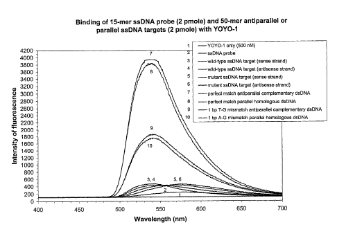
The hybridization reaction mixtures giving rise to the
data illustrated in Fig. 1A, each contained the following
mixture: 2 pmoles of ssDNA target, 2 pmoles of ssDNA probe,
0.5 x TEE and 500 nM of YOYO-1 in a final volume of 40 1. The
reaction mixtures were incubated at room temperature (21 C)
for 5 minutes, placed into a quartz cuvette, irradiated with
an argon ion laser beam having a wavelength of 488 nm and
monitored for fluorescent emission. The intensity of
fluorescence was plotted as a function of wavelength for each
sample analyzed.
28
CA 02454300 2004-01-16
WO 03/010326 PCT/1B02/02791
In Figs. 1B and 1C, the hybridization reaction mixtures
(40 1) each contained the following: 2
pmoles of ssDNA
target, 2 pmoles of 5'-fluorescein labeled ssDNA probe, 10 mM
Tris-HC1, pH 7.5, and 1 mM EDTA. The reaction mixtures were
incubated at room temperature (21 C) for 30 minutes or 90
minutes. Following incubation, each sample was placed into a
quartz cuvette, irradiated with an argon ion laser beam having
a wavelength of 488 nm and monitored for fluorescent emission.
The maximum fluorescent intensities occurred at a wavelength
of 525 nm, the emission wavelength for fluorescein. The
intensity of fluorescent emission was plotted as a function of
wavelength for each sample analyzed.
When the ssDNA Probe No. 3 was reacted with the 50-mer
wild-type sense strand of SEQ ID NO:5 or with the 50-mer
mutant sense strand of SEQ ID NO:7 in the presence of YOYO-1,
antiparallel complementary ssDNA:ssDNA duplexes were formed
(Fig. 1A). The fluorescent intensity emitted by the 1 bp T-G
mismatched antiparallel complementary duplex (sense strand of
SEQ ID NO:7 + Probe No. 3) was 56% lower than that obtained by
the perfectly matched antiparallel complementary duplex (sense
strand of SEQ ID NO:5 + Probe No. 3).
When the ssDNA Probe No. 3 was reacted with the 50-mer
wild-type antisense strand of SEQ ID NO:5 in the presence of
YOYO-1, the efficiency of parallel homologous ssDNA:ssDNA
duplex formation was only 3% lower than the efficiency of
antiparallel complementary ssDNA:ssDNA duplex formation (Fig.
1A). This result was completely unanticipated. The 1 bp A-G
mismatched parallel homologous duplex formed when the 50-mer
mutant antisense strand of SEQ ID NO:7 was reacted with the
ssDNA Probe No. 3 in the presence of YOYO-1, produced a
fluorescent emission intensity that was 56% lower than that
emitted by the perfectly parallel homologous duplex (Fig. 1A).
Control samples comprising each 50-mer ssDNA target plus 500
nM YOYO-1 exhibited levels of fluorescence which ranged from
29
CA 02454300 2004-01-16
WO 03/010326 PCT/1B02/02791
91% to 92% lower than that observed with the perfectly matched
duplexes (Fig. 1A). The level of fluorescence emitted by the
15-mer ssDNA Probe No. 3 plus 500 nM YOYO-1 was slightly
greater than that produced by YOYO-1 alone. The shift in
fluorescent emission wavelength observed with the ssDNA
targets and probe is typical of YOYO-1's emission profile in
the presence of ssDNA.
YOYO-1 facilitated DNA complex formation between a ssDNA
probe and a complementary base sequence in an antiparallel
ssDNA target, or between a ssDNA probe and an identical base
sequence in a parallel ssDNA target, with similar efficacy, to
allow differentiation between perfectly matched complexes and
those containing a 1 bp mismatch. In the parallel homologous
complexes, the 1 bp mismatch was a non-homologous base pair.
The comparative efficiency of antiparallel complementary
and parallel homologous dsDNA duplex formation was further
examined using ssDNA targets and ssDNA-F probes in the absence
of complex promoting agents such as YOYO-1 or cations. When
the ssDNA-F Probe No. 5 was incubated for 30 minutes in Tris
buffer at room temperature with the 50-mer wild-type sense
strand of SEQ ID NO:5, the Watson-Crick complementary
antiparallel ssDNA:ssDNA-F duplexes were formed very
efficiently, resulting in a 53% reduction in fluorescent
emission compared to that emitted by Probe No. 5 alone (Fig.
13). By
contrast, antiparallel complementary ssDNA:ssDNA-F
complexes that contained a 1 bp T-G mismatch (sense strand of
SEQ ID NO:7 + Probe No. 5) were less stable, resulting in only
a 40% decrease in fluorescent emission compared to that
emitted by Probe No. 5 alone after a 30 minute incubation
(Fig. 1B).
Parallel homologous ssDNA:ssDNA-F complexes were formed
when the ssDNA Probe No. 5 was reacted with the 50-mer wild-
type antisense strand of SEQ ID NO:5 or with the 50-mer mutant
antisense strand of SEQ ID NO:7, generating fluorescent
CA 02454300 2004-01-16
WO 03/010326 PCT/1B02/02791
emission intensities that were 44% and 37% lower,
respectively, than that emitted by ssDNA Probe No. 5 alone
after a 30 minute incubation (Fig. 13). The avid formation of
parallel homologous ssDNA:ssDNA-F complexes in the absence of
a promoting agent was completely unanticipated. The
discrimination between signals emitted from perfectly matched
duplexes and 1 bp mismatched duplexes in the absence of
complex promoting agents, was not as dramatic as that observed
when YOYO-1 was present and served as the promoter and
signaling agent (compare Figs. 1A and 13). This was the case
for both antiparallel and parallel duplexes. Slightly less
discrimination between perfectly matched and 1 bp mismatched
DNA complexes was observed when a parallel homologous ssDNA
target was used than when an antiparallel complementary ssDNA
target was used to produce the ssDNA:ssDNA-F complexes (Fig.
1B).
After a 90 minute incubation, Watson-Crick antiparallel
dsDNA:ssDNA-F complexes consisting of perfectly complementary
sequences (sense strand of SEQ ID NO:5 + Probe No. 5) or 1 bp
T-G mismatched sequences (sense strand of SEQ ID NO:7 + Probe
No. 5) produced a 39% and 30% decrease, respectively, in
fluorescent emission intensity compared to that emitted by
Probe No. 5 alone (Fig. IC). Remarkably, parallel homologous
ssDNA:ssDNA-F complexes exhibited the same level of stability
after 90 minutes of incubation as did the Watson-Crick
antiparallel ssDNA:ssDNA-F complexes.
The fluorescent
intensities for a perfectly parallel homologous duplex
(antisense strand of SEQ ID NO:5 + Probe No. 5) and a 1 bp A-G
mismatched parallel homologous duplex (antisense strand of SEQ
ID NO:7 + Probe No. 5) were 40% and 25% lower, respectively,
than that emitted by ssDNA Probe No. 5 alone after a 90 minute
incubation (Fig. 1C).
The mechanism of recognition and binding of the
homologous bases in the parallel dsDNA duplexes is unknown at
31
CA 02454300 2004-01-16
WO 03/010326 PCT/1B02/02791
this time. Nevertheless, recognition and binding of parallel
homologous ssDNA sequences occurred in a configuration which
allowed the discrimination between perfectly matched
ssDNA:ssDNA complexes and those containing a 1 bp or 2 bp
mismatch. In these parallel homologous complexes, the 1 bp
mismatch was a non-homologous base pair.
Example 2
In Example 1, the remarkable efficiency of parallel
homologous ssDNA:ssDNA duplex formation was demonstrated both
in the presence of a complex promoting agent such as YOYO-1
and in the absence of any complex promoting agent.
The
recognition and binding of the homologous bases in the
parallel dsDNA duplexes was such as to allow easy
discrimination between perfectly homologous base sequences and
parallel homologous sequences that contained a 1 bp mismatch.
These parallel homologous 1 bp mismatches were also clearly
recognizable as mismatches based on Watson-Crick complementary
recognition and binding rules.
Example 2 examines the
recognition and binding efficiency of parallel homologous
dsDNA duplexes that contain A-T or G-C base pairings, to
determine whether these Watson-Crick complementary pairings
appear as mismatches in a parallel homologous binding
reaction.
Each hybridization reaction mixture (40 1) contained the
following: 2 pmoles of ssDNA target, 2 pmoles of ssDNA probe,
0.5 x TBE and 500 nM of YOYO-1. The reaction mixtures were
incubated at room temperature (21 C) for 5 minutes, placed
into a quartz cuvette, irradiated with an argon ion laser beam
having a wavelength of 488 nm and monitored for fluorescent
emission.
The intensity of fluorescence was plotted as a
function of wavelength for each sample analyzed.
When the ssDNA Probe No. 3 (with a 53% GC content) was
reacted with the 50-mer wild-type antisense strand of SEQ ID
32
CA 02454300 2004-01-16
WO 03/010326 PCT/1B02/02791
NO:5 or with the 50-mer mutant antisense strand of SEQ ID
NO:10 in the presence of YOYO-1, parallel homologous
ssDNA:ssDNA duplexes were formed (Fig. 2A). The fluorescent
intensity emitted by the 1 bp A-T mismatched parallel
homologous duplex (antisense strand of SEQ ID NO:10 + Probe
No. 3) was 72% lower than that obtained by the perfectly
parallel homologous duplex (antisense strand of SEQ ID NO:5 +
Probe No. 3) (Fig. 2A).
This dramatic decrease in
fluorescent emission by the parallel homologous duplex
containing a 1 bp A-T, strongly suggested that the Watson-
Crick A-T binding was hindered by the spatial and/or charge
configuration imposed on the A and T bases when part of
parallel homologous strands attempting to achieve stable
duplex. Control samples comprising each 50-mer ssDNA target
plus 500 nM YOYO-1 exhibited levels of fluorescence which
ranged from 96% to 97% lower than that observed with the
perfectly matched duplexes (Fig. 2A).
The level of
fluorescence emitted by the 15-mer ssDNA Probe No. 3 plus 500
nM YOYO-1 was slightly greater than that produced by YOYO-1
alone. The shift in fluorescent emission wavelength observed
with the ssDNA targets and probe is typical of YOYO-1's
emission profile in the presence of ssDNA.
Parallel homologous ssDNA:ssDNA duplexes were also formed
when the 50-mer wild-type antisense strand of SEQ ID NO:1
(with a 33% GC content) was reacted with the wild-type ssDNA
Probe No. 8 or with the mutant ssDNA Probes No. 9 and 10, in
the presence of YOYO-1 (Fig. 2B). The fluorescent intensities
emitted by the 1 bp G-C mismatched parallel homologous duplex
(antisense strand of SEQ ID NO:1 + Probe No. 9) and the 1 bp
C-G mismatched parallel homologous duplex (antisense strand of
SEQ ID NO:1 + Probe No. 10) were 67% and 66% lower,
respectively, than that obtained by the perfectly parallel
homologous duplex (antisense strand of SEQ ID NO:1 + Probe No.
8) (Fig. 2B). The configuration of the interacting bases in
33
CA 02454300 2004-01-16
WO 03/010326 PCT/1B02/02791
the parallel homologous duplexes was unfavorable for Watson-
Crick complementary G-C binding, resulting in a decrease in
fluorescent emission indicative of a 1 bp mismatch. Control
samples consisting of the 50-mer ssDNA target plus 500 nM
YOYO-1 or each of the 15-mer ssDNA probes plus 500 nM YOYO-1
resulted in levels of fluorescence that were slightly greater
than that produced by YOYO-1 alone (Fig. 2B).
Therefore, the interacting base pairs in parallel
homologous dsDNA duplexes, formed in the presence of YOYO-1,
adopt a configuration that is unfavorable for binding between
Watson-Crick complementary base pairs, resulting in such
duplexes appearing to contain 1 bp mismatches.
We are led to envisage how mismatches in binding
sequences, whether occurring as part of a hairpin or
multistrand complex can cause energetic and repeated motion as
the base sequences try to achieve the stability of the ideal
binding configuration under either binding motif. It
is
expected that binding strength of base pairs upstream or
downstream of nucleation sites, metal ions and other factors
will have a bearing on the attempts to achieve bonding.
Example 3
This example examines the efficiency of antiparallel
homologous ssDNA:ssDNA duplex formation facilitated by YCY0-1
or by monovalent cations.
The hybridization reactions, giving rise to the data
illustrated in Fig. 3A, each contained the following mixture:
2 pmoles of ssDNA target, 2 pmoles of ssDNA probe, 0.5 x TEE
and 500 nM of YOYO-1 in a final volume of 40 1. The reaction
mixtures were incubated at room temperature (21 C) for 5
minutes, placed into a quartz cuvette, irradiated with an
argon ion laser beam having a wavelength of 488 nm and
monitored for fluorescent emission. The intensity of
fluorescent emission was plotted as a function of wavelength
34
CA 02454300 2004-01-16
WO 03/010326 PCT/1B02/02791
for each sample analyzed.
In Fig. 3B, the hybridization reaction mixtures (40 1)
each contained the following: 2 pmoles of ssDNA target, 2
pmoles of 5'-fluorescein labeled ssDNA probe, 10 mM Tris-HC1,
pH 7.5, and 50 mM NaCl. The reaction mixtures were incubated
at room temperature (21 C) for various lengths of time ranging
from 1 minute to 60 minutes. Following incubation, samples
were placed into a quartz cuvette, irradiated with an argon
ion laser beam having a wavelength of 488 nm and monitored for
fluorescent emission. The intensity of fluorescent emission
was plotted as a function of wavelength for each sample
analyzed.
Incubation of ssDNA Probe No. 4 with the 50-mer wild-type
antisense strand of SEQ ID NO:5 in the presence of YOYO-1
resulted in antiparallel homologous ssDNA:ssDNA complex
formation (Fig. 3A). Although the efficiency of antiparallel
homologous complex formation was only 65% that of conventional
antiparallel complementary dsDNA formation (compare Figs. 1A
and 1A), recognition and binding of antiparallel homologous
ssDNA sequences did occur, facilitated by YOYO-1. This result
was completely unanticipated.
Furthermore, antiparallel
homologous ssDNA:ssDNA complexes comprising wild-type
sequences were clearly distinguished from those comprising 1
bp or 2 bp mismatches. The fluorescent intensities emitted by
the 1 bp A-G mismatched DNA complex (antisense strand of SEQ
ID NO:7 + Probe No. 4), the 1 bp C-T mismatched DNA complex
(antisense strand of SEQ ID NO:6 + Probe No. 4), and the
consecutive 2 bp mismatched DNA complex (antisense strand of
SEQ ID NO:8 + Probe No. 4) were 25%, 65% and 71% lower,
respectively, than that obtained by the perfect antiparallel
homologous complex (antisense strand of SEQ ID NO:5 + Probe
No. 4) (Fig. 1A). As the degree of homology between the probe
and target decreased, the level of fluorescent emission
decreased.
Control samples comprising each 50-mer ssDNA
CA 02454300 2004-01-16
WO 03/010326 PCT/1B02/02791
target plus 500 nM YOYO-1 exhibited levels of fluorescence
which ranged from 88% to 90% lower than that observed with the
perfectly matched complexes (Fig. 3A).
The level of
fluorescence emitted by the 15-mer ssDNA Probe No. 4 plus 500
nM YOY0-1 was slightly greater than that produced by YOYO-1
alone.
Antiparallel homologous ssDNA:ssDNA complex formation was
further examined using ssDNA targets and ssDNA-F probes both
in the presence and absence of 50 mM NaCl. After 15 minutes
of incubation of ssDNA-F Probe No. 6 with the 50-mer wild-type
antisense strand of SEQ ID NO:5 in the presence of 50 mM NaC1,
antiparallel homologous ssDNA:ssDNA-F complexes were formed,
as indicated by the 34% decrease in fluorescence observed
compared to that emitted by Probe No. 6 alone (Fig. 33). The
efficiency of antiparallel homologous complex formation was
62% that of antiparallel complementary complex formation
following a 15 minute incubation (data not shown). By
contrast, antiparallel homologous ssDNA:ssDNA-F complexes that
contained a 1 bp A-G mismatch (antisense strand of SEQ ID NO:7
+ Probe No. 6), a 1 bp C-T mismatch (antisense strand of SEQ
ID NO:6 + Probe No. 6), a 1 bp A-T mismatch (antisense strand
of SEQ ID NO: 10 + Probe No. 6), and a consecutive 2 bp
mismatch (antisense strand of SEQ ID NO:8 + Probe No. 6),
produced a 24%, 26%, 23% and a 13% decrease in fluorescence,
respectively, compared to that emitted by Probe No. 6 alone
after a 15 minute incubation (Fig. 3B). The configuration of
the interacting bases in the antiparallel homologous duplexes
was apparently unfavorable for Watson-Crick complementary A-T
binding, resulting in a change in fluorescent emission
indicative of a 1 bp mismatch. Less antiparallel homologous
complex formation occurred following a 30 minute incubation in
the presence of 50 mM NaC1 (data not shown). No
complex
formation was evident after 45 minutes of incubation. Similar
rates of antiparallel homologous complex formation and
36
CA 02454300 2004-01-16
WO 03/010326 PCT/1B02/02791
stability were observed in Tris buffer without NaC1 (data not
shown).
Promoted by YOYO-1 or NaCl, recognition and binding of
antiparallel homologous ssDNA sequences occurred in a
configuration which allowed the discrimination between
perfectly matched ssDNA:ssDNA complexes and those containing a
1 bp or 2 bp mismatch. The interaction of the base pairs in
the antiparallel homologous duplex resulted in a conventional
Watson-Crick A-T base pair being destabilizing as a mismatch.
Example 4
This example demonstrates the efficiency of parallel
complementary ssDNA:ssDNA complex formation promoted by
monovalent cations.
The hybridization reaction mixtures
(40 1) each contained the following: 2 pmoles
of ssDNA
target, 2 pmoles of 5'-fluorescein labeled ssDNA probe, 10 mM
Tris-HC1, pH 7.5, and 50 mM NaCl. The reaction mixtures were
incubated at room temperature (21 C) for various lengths of
time ranging from 1 minute to 60 minutes.
Following
incubation, samples were placed into a quartz cuvette,
irradiated with an argon ion laser beam having a wavelength of
488 nm and monitored for fluorescent emission. The intensity
of fluorescent emission was plotted as a function of
wavelength for each sample analyzed.
After a 15 minute incubation in the presence of 50 mM
NaCl, ssDNA:ssDNA-F duplexes consisting of perfectly
complementary sequences (sense strand of SEQ ID NO:5 + Probe
No. 6) formed readily, resulting in a 41% decrease in
fluorescent emission intensity compared to that emitted by
Probe No. 6 alone (Fig. 4). This high efficiency of parallel
complementary duplex formation was completely unexpected. By
contrast, incompletely complementary ssDNA:ssDNA-F complexes
containing a 1 bp T-G mismatch (sense strand of SEQ ID NO:7 +
Probe No. 6), a 1 bp G-T mismatch (sense strand of SEQ ID NO:6
37
CA 02454300 2004-01-16
WO 03/010326 PCT/1B02/02791
+ Probe No. 6), a 1 bp T-T jmismatch (sense strand of SEQ ID
NO: 10 + Probe No. 6), and a consecutive 2 bp mismatch (sense
strand of SEQ ID NO:8 + Probe No. 6), generated an 18%, 20%,
10% and 16% decrease, respectively, in fluorescent emission
intensity compared to that exhibited by Probe No. 6 alone
(Fig. 4).
Once formed in the presence of 50 mM NaC1, the perfectly
matched parallel complementary duplexes were very stable,
resulting in a 40% and 47% decrease in fluorescent emission
after 30 minutes and 45 minutes of incubation, respectively,
compared to that emitted by Probe No. 6 alone (data not
shown). The 1 bp and 2 bp mismatched parallel complementary
complexes were much less stable after 30 minutes and 45
minutes of incubation in the presence of 50 mM NaC1 (data not
shown). The rate and efficiency of parallel complementary
ssDNA:ssDNA-F formation was very similar to that of
antiparallel complementary ssDNA:ssDNA-F formation during the
first 45 minutes of incubation in the presence of 50 mM NaC1
(data not shown). While antiparallel complementary complexes
continued to form easily after 60 minutes of incubation in 50
mM NaC1, no parallel complementary complex formation was
evident at this time (data not shown).
NaC1 facilitated DNA complex formation between a ssDNA-F
probe and an antiparallel complementary ssDNA target, or
between a ssDNA-F probe and a parallel complementary ssDNA
target, with similar efficacy, to allow differentiation
between perfectly matched complexes and those containing a 1
bp or 2 bp mismatch.
Example 5
Examples 1 to 4 demonstrated alternate base recognition
and binding motifs occurring between antiparallel or parallel
ssDNA probes, and complementary or homologous ssDNA targets to
generate ssDNA:ssDNA duplexes, other than the conventional
38
CA 02454300 2004-01-16
WO 03/010326 PCT/1B02/02791
antiparallel Watson-Crick complementary dsDNA complexes. This
example will show that bases are capable of recognizing and
interacting with both complementary and homologous bases at
the same time.
Samples of two pmoles of ssDNA Probe No. 3 were heated at
95 C for 10 minutes and allowed to cool to room temperature
for 30 minutes in the presence of various concentrations of a
free base, resulting in ssDNA probes containing conjugated
bases. Duplicate samples of ssDNA Probe No. 3 were similarly
denatured and cooled in the absence of added free bases to
generate non-conjugated ssDNA probes.
Two pmoles of these
conjugated or non-conjugated ssDNA probes were then mixed with
2 pmoles of ssDNA target in the presence of 500 nM YOYO-1 and
0.5 x TEE in a final reaction volume of 40 1. The reaction
mixtures were incubated at room temperature (21 C) for 5
minutes, placed into a quartz cuvette, irradiated with an
argon ion laser beam having a wavelength of 488 nm, and
monitored for fluorescent emission.
The intensity of
fluorescence was plotted as a function of wavelength for each
sample analyzed.
When the non-conjugated ssDNA Probe No. 3 was reacted
with the 50-mer wild-type sense strand of SEQ ID NO:5 or with
the 50-mer mutant sense strand of SEQ ID NO:7, in the presence
of YOYO-1, antiparallel complementary ssDNA:ssDNA complexes
were formed (Fig. 5A). The fluorescent intensity emitted by
the 1 bp T-G mismatched antiparallel complementary duplex
(sense strand of SEQ ID NO:7 + Probe No. 3) was 45% lower than
that obtained by the perfectly matched antiparallel
complementary duplex (sense strand of SEQ ID NO:5 + Probe No.
3). Control samples comprising each 50-mer ssDNA target plus
500 nM YOYO-1 exhibited levels of fluorescence which ranged
from 92% to 93% lower than that observed with the perfectly
matched duplexes (Fig. 5A). The level of fluorescence emitted
by the 15-mer ssDNA Probe No. 3 plus 500 nM YOYO-1 was
39
CA 02454300 2004-01-16
WO 03/010326 PCT/1B02/02791
slightly greater than that produced by YOYO-1 alone.
When the ssDNA Probe No. 3 was reacted with the 50-mer
wild-type antisense strand of SEQ ID NO:5 in the presence of
YOYO-1, the efficiency of parallel homologous ssDNA:ssDNA
duplex formation was 14% lower than the efficiency of
antiparallel complementary ssDNA:ssDNA duplex formation
(compare Figs. 5A and 5B). The 1 bp A-G mismatched parallel
homologous duplex formed when the 50-mer mutant antisense
strand of SEQ ID NO:7 was reacted with the ssDNA Probe No. 3
in the presence of YOYO-1, produced a fluorescent emission
intensity that was 47% lower than that emitted by the
perfectly parallel homologous duplex (Fig. 5B).
The 15-mer ssDNA Probe No. 3 contains six adenine bases.
Conjugation of 2 pmoles of ssDNA Probe No. 3 with 3 pmoles of
free thymine could result in 25% of the complementary A or
100% of the homologous T within Probe No. 3 bound to the added
thymine. Complementary A-T binding is energetically
preferred. Reaction of 2 pmoles of ssDNA Probe No. 3
(conjugated with 3 pmoles of thymine) with 2 pmoles of the
wild-type antisense strand of SEQ ID NO:5 in the presence of
YOYO-1 resulted in dramatically enhanced parallel homologous
ssDNA:ssDNA complex formation (Fig. 5B). Twenty-five percent
conjugation of the ssDNA probe with 3 pmoles of thymine
increased parallel homologous complex formation between the
perfectly homologous sequences by 78%. This augmentation of
parallel homologous complex formation can be linked to the
ability of the adenines in Probe No. 3 to interact
simultaneously with the conjugated complementary thymine
bases, as well as with the homologous adenines in the ssDNA
target. Moreover, interaction with available complementary
bases was not deleterious to the homologous binding
configuration adopted by the homologous bases and their
neighbors.
By contrast, the efficiency of formation of parallel
CA 02454300 2004-01-16
WO 03/010326 PCT/1B02/02791
homologous complexes containing a 1 bp A-G mismatch (antisense
strand of SEQ ID NO:7 + Probe No. 3) were increased by 16%
when Probe No. 3 was conjugated 25% with thymine than when
non-conjugated Probe No. 3 was used (Fig. 5B).
This
corresponded to a 65% reduction in fluorescent emission
intensity for the 1 bp A-G mismatched parallel homologous
complex compared to that observed for the perfectly matched
parallel homologous complex when the T-conjugated Probe No. 3
was used.
Conjugation of the ssDNA probe increased the
specificity in discriminating between perfectly matched
parallel homologous complexes and 1 bp mismatched parallel
homologous complexes.
Remarkably, perfectly matched antiparallel complementary
ssDNA:ssDNA complex formation was enhanced by 48% when Probe
No. 3 conjugated 25% with thymine was reacted with the sense
strand of SEQ ID NO:5 in the presence of YOYO-1 (Fig. 5A).
The simultaneous interaction of an adenine in Probe No. 3 with
the conjugated complementary thymine and the complementary, T
in the ssDNA target augmented formation of the perfectly
matched antiparallel complementary complex. Remarkably,
formation of the 1 bp T-G mismatched antiparallel
complementary complex was very inefficient when T-conjugated
Probe No. 3 was used, resulting in an 88% decrease in
fluorescent emission intensity compared to that generated by
the perfectly matched antiparallel complementary complex
containing conjugated T (Fig. 5A). It is also remarkable that
discrimination between perfectly matched and 1 bp mismatched
antiparallel complementary ssDNA:ssDNA complexes was greatly
enhanced by use of conjugated ssDNA probes in the presence of
YOYO-1.
Twenty-five percent conjugation of Probe No. 3 with
cytosine or guanosine also increased the efficiency of both
antiparallel complementary and parallel homologous ssDNA:ssDNA
complex formation in the presence of YOYO-1, as well as
41
CA 02454300 2004-01-16
WO 03/010326 PCT/1B02/02791
improved the specificity in differentiation between perfectly
matched complexes and 1 bp mismatched complexes (data not
shown).
Formation of ssDNA:ssDNA complexes comprising conjugated
bases proves that the bases in a sequence can recognize and
interact specifically and simultaneously with both
complementary and homologous bases provided the conjugated
base is a Watson-Crick complement to a base on the strand
which binds specifically to another strand. The recognition
and binding configurations between bases in a ssDNA probe,
conjugated bases and bases in a ssDNA target may be similar to
the base configurations formed in antiparallel and parallel
dsDNA:ssDNA complexes described herein.
Example 6
Example 6 demonstrates quadruplex DNA formation between
dsDNA targets containing mixed base sequences and homologous
dsDNA probes labeled with fluorescein.
Quadruplex DNA
formation is enhanced by the presence of monovalent cations
added to the reaction.
Complementary sense and antisense 15-mer ssDNA sequences
were synthesized, purified by HPLC and annealed as above to
generate 15-mer dsDNA probes. DsDNA probes were diluted in
ddH20 at a concentration of 1 pmole/ 1.
Probe No. 11 was a 15-mer dsDNA probe with an attached
fluorescein moiety at each 5' position, and was designed to be
completely homologous to a central 15 bp segment of the 50-mer
wild-type target DNA (SEQ ID NO:5).
The sequence for the sense strand of Probe No. 11 (SFQ ID
NO:22) was: 5'-Flu-CTG TCA TCT CTG GTG-3'.
The sequence for the antisense strand of Probe No. 11
(SEQ ID NO:22) was: 5'-Flu-CAC CAG AGA TGA CAG-3'.
Each hybridization reaction mixture (40 1) contained the
following: 0.4 pmoles of target dsDNA, 4 pmoles of 5'-
42
CA 02454300 2004-01-16
WO 03/010326 PCT/1B02/02791
fluorescein labeled dsDNA Probe No. 11, 10 mM Tris-HC1, pH 7.5
and 100 mM KC1. The reaction mixtures were incubated at room
temperature (21 C) for 1 hour, without prior denaturation.
Samples were placed into a quartz cuvette, irradiated with an
argon ion laser beam having a wavelength of 488 nm and
monitored for fluorescent emission. The maximum fluorescent
intensities occurred at a wavelength of 525 nm, the emission
wavelength for fluorescein. Fig. 6 shows the intensity of
fluorescence plotted as a function of wavelength for each
sample analyzed.
In the absence of KC1, no binding between the dsDNA
targets and Probe No. 11 was detected, resulting in similar
fluorescent intensities observed when wild-type dsDNA target
(SEQ ID NO:5) or mutant dsDNA target (SEQ ID NO:7) were mixed
with dsDNA Probe No. 11 or when dsDNA Probe No. 11 was present
alone (data not shown).
After a 1 hour incubation at 21 C in the presence of 100
mM KC1, dsDNA:dsDNA-F quadruplexes consisting of perfectly
homologous sequences on dsDNA target (SEQ ID NO:5) and dsDNA
Probe No. 11 formed readily, resulting in a 62% decrease in
the intensity of fluorescent emission compared to that emitted
by dsDNA Probe No. 11 alone (labeled dsDNA-F) (Fig. 6). In
contrast, incompletely homologous dsDNA:dsDNA-F quadruplexes
(SEQ ID NO:7 + Probe No. 11), containing a 1 base pair
mismatch were less stable in these reaction conditions,
yielding only an 18% decrease in fluorescent intensity
compared to that exhibited by dsDNA Probe No. 11 alone.
The presence of monovalent cations, such as le, at
specific concentrations was sufficient to allow quadruplex
formation between dsDNA targets and dsDNA probes labeled with
fluorescein in the absence of prior denaturation. Quadruplex
formation occurred on the basis of homologous base pair
affinities, with a measurable and significantly greater amount
of quadruplex formation between fully homologous duplex
43
CA 02454300 2004-01-16
WO 03/010326 PCT/1B02/02791
strands. Moreover, the reaction occurred at room temperature
within just 1 hour of incubation at a ratio of probe to target
of 10 to 1, using natural dsDNA. The dsDNA targets and dsDNA
probe used in this example were homologous, contained 53% GC
content, and did not contain homopurine or homopyrimidine
stretches on any DNA strand. The assay of the invention was
able to identify perfectly homologous dsDNA sequences and
those containing a pair of mismatched bases, using a dsDNA
probe.
Example 7
The quadruplex DNA assays performed in Example 6 were
facilitated by the addition of monovalent cations in the
reaction mixtures. The specificity of the assay was further
examined utilizing divalent cations to facilitate quadruplex
DNA formation with dsDNA targets and dsDNA-F probes possessing
53% GC content.
Each hybridization reaction mixture (40 1) contained the
following: 0.4 pmoles of target dsDNA, 4 pmoles of 5'-
fluorescein labeled dsDNA Probe No. 11, 10 mM Tris-HC1, pH
7.5, 20mM MnC12 and 20 mM MgC12. The reaction mixtures were
incubated at room temperature (21 C) for 1 hour, without prior
denaturation. Samples were placed into a quartz cuvette,
irradiated with an argon ion laser beam having a wavelength of
488 nm and monitored for fluorescent emission. Fig. 7 shows
the intensity of fluorescence plotted as a function of
wavelength for each sample analyzed.
When dsDNA-F Probe No. 11 (with a 53% GC content) was
incubated with the 50-mer wild-type dsDNA target (SEQ ID NO:5)
or the mutant dsDNA target (SEQ ID NO:7) in the presence of 20
mM MnC12 and 20 mM MgC12, quadruplexes were formed at room
temperature under non-denaturing conditions. While perfectly
homologous DNA quadruplexes yielded the maximum decrease in
fluorescent intensity, a 34% decrease, the less favourable
44
CA 02454300 2004-01-16
WO 03/010326 PCT/1B02/02791
dsDNA:dsDNA-F quadruplexes containing a 1 bp mismatch (SEQ ID
NO:7 + Probe No. 11) produced a fluorescent intensity that was
about the same as that observed with dsDNA Probe No. 11 alone
(Fig. 7).
The presence of divalent cations such as Mn+2 and Me
facilitated quadruplex formation under non-denaturing
conditions to allow accurate discrimination between fully
homologous dsDNA target and dsDNA probe quadruplexes, and
quadruplex sequences containing a pair of bases which are
mismatched.
Example 8
The quadruplex DNA assays performed in Examples 6 and 7
were facilitated by the addition of either monovalent cations
or divalent cations in the reaction mixtures. The next
Example demonstrates the specificity of the homologous
quadruplex DNA assay when the DNA intercalator, YOYO-1, is
employed.
Complementary sense and antisense 15-mer ssDNA sequences
were synthesized, purified by HPLC and annealed as above to
generate 15-mer dsDNA probes. DsDNA probes were diluted in
ddH20 at a concentration of 1 pmole/ 1.
Probe No. 12 was a 15-mer dsDNA probe identical in
sequence to Probe No. 11, but without the attached 5'
fluorescein moieties.
The sequence for the sense strand of Probe No. 12 (SEQ ID
NO:23) was: 5'-CTG TCA TCT CTG GTG-3 .
The sequence for the antisense strand of Probe No. 12
(SEQ ID NO: 23) was: 5'-CAC CAG AGA TGA CAG-3'.
Each hybridization reaction mixture (40 1) contained the
following: 0.4 pmoles of dsDNA target, 4 pmoles of dsDNA Probe
No. 12, 0.5 x TEE and 100 nM of YOYO-1. The reaction mixtures
were incubated at 21 C for 5 minutes, placed into a quartz
cuvette, irradiated with an argon ion laser beam having a
CA 02454300 2004-01-16
WO 03/010326 PCT/1B02/02791
wavelength of 488 nm and monitored for fluorescent emission.
The intensity of fluorescent emission was plotted as a
function of wavelength for each sample analyzed.
The fluorescent intensities observed when no target or
probe was present (YOY0-1 only) are shown in Fig. 8. Fig. 8
also shows the fluorescent intensities observed when the
reaction mixtures combined dsDNA Probe No. 12 with wild-type
50-mer dsDNA target (SEQ ID NO:5) which contained homologous
sequences, or with four other dsDNA targets which, but for one
mismatched pair of bases, contained sequences which were
homologous to the base sequences in the dsDNA Probe No. 12.
Homologous wild-type dsDNA target (SEQ ID NO:5) when present
in the reaction mixture with the dsDNA Probe No. 12 produced
the greatest fluorescent intensity. Mismatched dsDNA targets
when incubated with dsDNA Probe No. 12 in the reaction mixture
yielded lesser fluorescent intensity values ranging from 20%
less for dsDNA target (SEQ ID NO:10) to 80% less for dsDNA
target (SEQ ID NO:11), compared to that achieved by perfectly
matched quadruplexes (Fig. 8).
It was observed that homologous quadruplexes, stabilized
by YOYO-1 intercalation, formed more readily between a dsDNA
target and a dsDNA probe when that probe contained perfectly
homologous sequences, than when there was a single pair of
bases which were not homologous, that is to say identical, to
a pair of bases in the dsDNA target. The quadruplex complexes
described in the foregoing three examples are referred to by
us as mirror homologous.
Example 9
In this example, 50-mer dsDNA targets were exposed to a
53% GC 15-mer dsDNA probe (Probe No. 13), wherein Watson-Crick
complementarity exists between bases of the strands of the
probe and proximal bases of the strands of the target when the
major groove of one duplex is placed in the minor groove of
46
CA 02454300 2004-01-16
WO 03/010326 PCT/1B02/02791
the other duplex. The sequences of bases in the duplex probe
are not homologous but are inverted in relation to those in
the duplex target. The duplexes, when nested major groove
into minor groove, are parallel to one another, and referred
to by us as nested complementary.
Complementary sense and antisense 15-mer ssDNA sequences
were synthesized, purified by HPLC and annealed as above to
generate 15-mer dsDNA probes. DsDNA probes were diluted in
ddH20 at a concentration of 1 pmole/ 1.
The sequence for the sense strand of Probe No. 13 (SEQ ID
NO: 24) was: 5'-GAC AGT AGA GAC CAC-3'.
The sequence for the antisense strand of Probe No. 13
(SEQ ID NO :24) was: 5'-GTG GTC TCT ACT GTC-3'.
Each hybridization reaction mixture (40 1) contained the
following: 0.4 pmoles of target dsDNA, 4 pmoles of dsDNA Probe
No. 13, 0.5 x TBE and 100 nM of YOYO-1. The reaction mixtures
were incubated at room temperature (21 C) for 5 minutes,
placed in a quartz cuvette, irradiated with an argon ion laser
beam having a wavelength of 488 nm and monitored for
fluorescent emission. The intensity of fluorescent emission
was plotted as a function of wavelength for each sample
analyzed.
Fig. 9 illustrates that in the absence of prior
denaturation, the highest fluorescent intensities were
achieved when the wild-type 50-mer dsDNA target (SEQ ID NO:5)
was reacted with the 15-mer dsDNA Probe No. 13, which was a
perfect match on a nested complementary basis to the dsDNA
target (SEQ ID NO:5). The fluorescent intensity is indicative
of DNA binding taking place, in this case quadruplex formation
between the dsDNA target and the nested complementary dsDNA
probe.
Mutant dsDNA targets which were mismatched with the
duplex probe by a single pair of bases when matching was
assessed on the inverted homology basis of nested
47
CA 02454300 2004-01-16
WO 03/010326 PCT/1B02/02791
complementarity, formed measurably fewer quadruplex complexes
with the dsDNA probe, than did the fully complementary wild-
type dsDNA target. The various mismatches, which were assayed
on a mirror homologous basis in Example 8 were assayed on a
nested complementary basis in this example.
As shown in Fig. 9, the fluorescent intensities produced
by the quadruplexes formed with the 1 bp mismatched dsDNA
targets plus dsDNA Probe No. 13, ranged from 8% to 16% less
than that achieved by perfectly matched quadruplexes (SEQ ID
NO:5 + Probe No. 13).
Greater discrimination in fluorescence was observed
between perfectly homologous and partially homologous
quadruplexes in Example 8.
This suggests that fully
complementary or 1 base pair mismatched nested complementary
dsDNA probes bind less discriminately to dsDNA targets than do
mirror homologous dsDNA probes, which bind with greater
specificity.
This example shows that Watson-Crick quadruplex binding
between nested complementary DNA duplexes readily occurs in
the presence of YOYO-1.
Example 10
Example 10 demonstrates that the assay of the invention
can discriminate between perfectly matched, Watson-Crick
complementary dsDNA:ssPNA complexes and dsDNA:ssPNA complexes
containing 1 bp, 2 bp and 3 bp mismatches when a cationic
decondensing agent, such as the DNA intercalator, YOYO-1 is
present.
Each hybridization reaction mixture (40 1) contained the
following: 2 pmoles of target dsDNA, 2 pmoles of ssPNA probe,
0.5 x TBE and 500 nM of YOYO-1. The reaction mixtures were
incubated at room temperature (21 C) for 5 minutes, placed
into a quartz cuvette, irradiated with an argon ion laser beam
having a wavelength of 488 nm and monitored for fluorescent
48
CA 02454300 2004-01-16
WO 03/010326 PCT/1B02/02791
emission.
The intensity of fluorescence was plotted as a
function of wavelength for each sample analyzed.
The fluorescent intensities observed when no DNA or PNA
was present (YOY0-1 only), or when wild-type SEQ ID NO:1,
mutant SEQ ID NO:2 or mutant SEQ ID NO:3 were reacted with
antiparallel PNA Probe No. 1 or parallel PNA Probe No. 2 are
shown in Figs. 10A and 10B, respectively.
DsDNA:ssPNA
complexes consisting of perfectly complementary sequences (SEQ
ID NO:1 + Probe No. 1) allowed maximum interaction between
YOYO-1 and the complexes, yielding the highest fluorescent
intensities (Fig. 10A). The fluorescent intensities for e one
base pair mismatched dsDNA:ssPNA complex (SEQ ID NO:2 + Probe
No. 1) and a two base pair mismatched dsDNA:ssPNA complex (SEQ
ID NO:3 + Probe No. 1) was 97% and 99% lower, respectively,
than the perfectly matched dsDNA:ssPNA complex (Fig. 10A).
Similarly, when parallel PNA Probe No. 2 was bound to the
target dsDNA sequences, the one and two base pair mismatched
dsDNA:ssPNA complexes exhibited fluorescent intensities that
were 92% and 97% lower, respectively, than the perfectly
complementary dsDNA:ssPNA complexes (SEQ ID NO:1 + Probe No.
2) (Fig. 10B).
Three base pair mismatched dsDNA:ssPNA
complexes consisting of SEQ ID NO:4 and Probe No. 1, or SEQ ID
NO:4 and Probe No. 2 produced fluorescent intensities that
were 99% and 97% lower, respectively, than the perfectly
matched dsDNA:ssPNA complexes (data not shown). Control
samples comprising 50-mer dsDNA targets plus 500 nM YOYO-1
exhibited levels of fluorescence which were at or below the
level of fluorescence observed with 3 bp mismatched complexes
(data not shown). The level of fluorescence emitted by either
ssPNA probe plus 500 nM YOYO-1 together was identical to that
emitted by YOYO-1 alone (data not shown). As the degree of
mismatch between the probe and the target increased, the level
of interaction of YOYO-1 with the mismatched complexes
diminished. Hence the intensity of fluorescent emission
49
CA 02454300 2004-01-16
WO 03/010326 PCT/1B02/02791
decreased.
This relationship held whether or not an
antiparallel or parallel PNA probe was used.
The
characteristic level of fluorescence emitted by each complex
was monitored over time and was stable between 5 minutes and
24 hours.
Interestingly, when 15-mer target dsDNA sequences were
reacted with 15-mer PNA probe sequences, larger differences in
fluorescent emission were observed between perfectly matched
complexes and 1 or 2 bp mismatched complexes when parallel PNA
probes were used, than when antiparallel PNA probes were used
(data not shown).
Therefore, the fluorescent intensity assay measuring
dsDNA:ssPNA complex formation is able to distinguish between
wild-type sequences and those containing 1 bp, 2 bp or 3 bp
mutations, without prior denaturation of the duplex DNA
target.
Example 11
The specificity of the assay measuring triplex formation
promoted by YOYO-1 was further investigated by reacting wild-
type and mutant dsDNA targets of mixed base sequence with
antiparallel and parallel ssDNA probes in the absence of prior
denaturation of dsDNA targets.
Each hybridization reaction mixture (40 1) contained the
following: 2 pmoles of target dsDNA, 2 pmoles of ssDNA probe,
0.5 x TEE and 500 nM of YOYO-1. The reaction mixtures were
incubated at room temperature (21 C) for 5 minutes, placed
into a quartz cuvette, irradiated with an argon ion laser beam
having a wavelength of 488 nm and monitored for fluorescent
emission. The
intensity of fluorescence was plotted as a
function of wavelength for each sample analyzed.
When the antiparallel ssDNA Probe No. 3 (with a 5396 GC
content) was reacted with the 50-mer wild-type dsDNA target
(SEQ ID NO:5) and mutant dsDNA targets (SEQ ID NO:6 and SEQ ID
CA 02454300 2004-01-16
WO 03/010326 PCT/1B02/02791
NO:8), dsDNA: ssDNA complexes were formed at room temperature
under non-denaturing conditions (Fig. 11A). While perfectly
matched DNA complexes emitted the highest fluorescent
intensities, incompletely complementary complexes with a 1 bp
mismatch (SEQ ID NO:6 + Probe No. 3) and a consecutive 2 bp
mismatch (SEQ ID NO:8 + Probe No. 3) produced fluorescent
intensities that were 63% and 95% lower, respectively, than
that observed with the perfectly matched sequences
(Fig.
11A). The level of fluorescence diminished as the degree of
mismatch between the probe and target increased. The
characteristic fluorescent intensity exhibited by each complex
was monitored over time and was stable between 5 minutes and
24 hours.
Control samples comprising 50-mer dsDNA targets
plus 500 nM YOYO-1 exhibited levels of fluorescence which were
below the level of fluorescence observed with 2 bp mismatched
DNA complexes (data not shown). The level of fluorescence
generated by the ssDNA probe plus 500 nM YOYO-1 was identical
to that achieved by YOYO-1 alone (data not shown). Very
similar results were obtained when 15-mer antiparallel ssDNA
probes were reacted with wild-type or mutant 50-mer dsDNA
targets having 33% GC and 73% GC contents under the same
reaction conditions, demonstrating the reliability of the
dsDNA:ssDNA complex formation assay utilizing antiparallel
ssDNA probes, independent of the percent GC content of the
ssDNA probes and dsDNA targets (data not shown).
Similarly, in the presence of YOYO-1, dsDNA:ssDNA
complexes were formed when the parallel ssDNA Probe No. 4 was
reacted with the 50-mer wild-type dsDNA target (SEQ ID NO:5)
and mutant dsDNA targets (SEQ ID NO:6 and SEQ ID NO:8). The
fluorescent intensities for a 1 bp mismatched DNA complex (SEQ
ID NO:6 + Probe No. 4) and a consecutive 2 bp mismatched DNA
complex (SEQ ID NO:8 + Probe No. 4) were 48% and 65% lower,
respectively, than that obtained by the perfectly matched
sequences (Fig. 11E). As the degree of mismatch between the
51
CA 02454300 2004-01-16
WO 03/010326 PCT/1B02/02791
probe and target increased, the level of fluorescent emission
decreased. Slightly less discrimination between perfectly
matched and mismatched DNA complexes was observed when a
parallel ssDNA probe was used than when an antiparallel ssDNA
probe was used to generate the dsDNA:ssDNA complexes.
YOYO-1 facilitated DNA complex formation between an
antiparallel ssDNA probe and dsDNA targets, and between a
parallel ssDNA probe and dsDNA targets, to allow
differentiation between perfectly matched complexes and those
containing 1 bp or 2 bp mismatches, without the requirement
for prior denaturation of dsDNA targets.
Example 12
The complexes formed in Examples 10 and 11 were
stabilized by the DNA intercalator, YOYO-1 present in the
reaction mixtures. The specificity of the assay was further
examined utilizing divalent cations to promote and stabilize
complex formation with dsDNA targets and ssDNA-F probes.
Each hybridization reaction mixture (40 1) contained the
following: 0.4 pmoles of target dsDNA, 4 pmoles of 5'-
fluorescein labeled ssDNA probe, 10 mM Tris-HC1, pH 7.5, and 1
mM to 20 mM each of MgCl2 and MnC12. The reaction mixtures
were incubated at room temperature (21 C) for various lengths
of time ranging from 1 minute to 2 hours, without rrior
denaturation of dsDNA targets. Following incubation, samples
were placed into a quartz cuvette, irradiated with an argon
ion laser beam having a wavelength of 488 nm and monitored for
fluorescent emission. The maximum fluorescent intensities
occurred at a wavelength of 525 nm, the emission wavelength
for fluorescein. The intensity of fluorescent emission was
plotted as a function of wavelength for each sample analyzed.
When the antiparallel ssDNA-F Probe No. 5 was incubated
for 1 hour with the 50-mer wild-type dsDNA target (SEQ ID
NO:5) in the presence of 15 mM MgC12 and 15 mM MnC12,
52
CA 02454300 2004-01-16
WO 03/010326 PCT/1B02/02791
perfectly complementary dsDNA:ssDNA-F complexes were formed
very efficiently, generating a 74% decrease in fluorescence
compared to that achieved by Probe No. 5 alone (Fig. 12A.). By
contrast, dsDNA:ssDNA-F complexes that contained a 1 bp T-G
mismatch (SEQ ID NO:7 + Probe No. 5) were much less stable in
the presence of 15 mM MgC12 and 15 mM MnC12, yielding a 15%
decrease in fluorescence compared to that emitted by Probe No.
5 alone after a 1 hour incubation (Fig. 12A). When Probe No.
5 (containing a 53% GC content) was reacted with the dsDNA
target SEQ ID NO:9 (containing a 33% GC content), a 3%
increase in fluorescence was observed compared to that
obtained by Probe No. 5 alone (Fig. 12A), indicative of no DNA
complex formation. This result was expected considering this
probe and target combination would result in a 5 bp mismatch.
In the presence of 10 mM MgC12 and 10 mM MnC12, the
dsDNA:ssDNA-F complexes possessing a 53% GC content and
containing perfectly complementary sequences (SEQ ID NO:5 +
Probe No. 5) or a 1 bp T-G mismatch (SEQ ID NO:7 + Probe No.
5) generated fluorescent intensities that were 68% and 20%
lower, respectively, after an 1 hour incubation, and 76% and
16% lower, respectively, after a 30 minute incubation, than
that emitted by Probe No. 5 alone (data not shown).
The
addition of 5 mM MgC12 and 5 mM MnC12 (or lower
concentrations) was insufficient to allow complex formation
between the antiparallel ssDNA-F Probe No. 5 and all dsDNA
targets tested following a 1 hour incubation (data not shown).
DsDNA:ssDNA complexes were also formed when the parallel
ssDNA Probe No. 6 was reacted with the 50-mer wild-type dsDNA
target (SEQ ID NO:5) and mutant dsDNA target (SEQ ID NO:7).
In this case DNA complex formation was promoted with much
lower concentrations of MgCl2 and MnC12 (i.e. 1-5 mM each)
requiring shorter incubation periods.
Incubation in the
presence of 1 mM MgCl2 and 1 mM MnC12, or 2 mM MgC12 and 2 mM
MnC12 for 15 minutes was sufficient to generate DNA complexes
53
CA 02454300 2004-01-16
WO 03/010326 PCT/1B02/02791
(data not shown). The fluorescent intensities for a perfectly
matched DNA complex (SEQ ID NO:5 + Probe No. 6) and a 1 bp
mismatched DNA complex (SEQ ID NO:7 + Probe No. 6) were 29%
and 6% lower, respectively, than that obtained by parallel
ssDNA Probe No. 6 alone in the presence of 3 mM MgC12 and 3
mM MnC12 after a 45 minute incubation (Fig. 12B).
Although DNA complexes formed readily at 10 mM MgC12 and
mM MnC12 after a 1 hour incubation, no discrimination
between perfectly matched and mismatched complexes was
10 observed when a parallel ssDNA probe was used. Concentrations
above 15 mM MgC12 and 15 mM MnC12 were inhibitory for DNA
complex formation with a parallel ssDNA probe (data not
shown).
The addition of salt bridging, condensing agents such as
divalent cations promoted DNA complex formation between non-
denatured dsDNA targets and fluorescently-labeled antiparallel
or parallel ssDNA probes, to allow accurate and reliable
discrimination between perfectly complementary sequences and
those containing 1 bp mutations. The reactions occurred at
room temperature within 15-60 minutes of incubation at a ratio
of probe to target of 10 to 1. The dsDNA targets and ssDNA
probes did not contain homopurine or homopyrimidine stretches
of DNA.
Despite the presence of 5 pyrimidine bases
interspersed within the 15 nucleotide ssDNA probes, DNA
complexes formed readily in a sequence specific manner.
Example 13
The utility of probes of varying directionality was also
evaluated when monovalent cations were employed to promote and
stabilize complex formation with dsDNA targets.
Each hybridization reaction mixture (40 1) contained the
following:
0.4 pmoles of target dsDNA, 4 pmoles of 5'-
fluorescein labeled ssDNA probe, 10 mM Tris-HC1, pH 7.5, and
10 mM to 150 mM NaCl. The reaction mixtures were incubated at
54
CA 02454300 2004-01-16
W003/010326 PCT/1B02/02791
room temperature (21 C) for various lengths of time ranging
from 1 minute to 2 hours, without prior denaturation of dsDNA
targets.
Following incubation, samples were placed into a
quartz cuvette, irradiated with an argon ion laser beam having
a wavelength of 488 nm and monitored for fluorescent emission.
The maximum fluorescent intensities occurred at a wavelength
of 523 nm, the emission wavelength for fluorescein.
The
intensity of fluorescent emission was plotted as a function of
wavelength for each sample analyzed.
In the absence of NaCl or presence of 10 mM or 25 mM
NaCl, no binding between the dsDNA targets (SEQ ID NO:1 or SEQ
ID NO:2) and the antiparallel ssDNA-F Probe No. 7 was
detected, after all incubation periods (data not shown).
After a 1 hour incubation in the presence of 50 mM NaCl,
dsDNA:ssDNA-F complexes consisting of perfectly complementary
sequences (SEQ ID NO:1 + Probe No. 7) formed readily,
resulting in a 49% decrease in fluorescent emission intensity
compared to that emitted by the control Probe No. 7, which was
similarly incubated in the reaction mixture (Fig. 13A). By
contrast, incompletely complementary dsDNA:ssDNA-F complexes
containing a 1 bp G-T mismatch (SEQ ID NO:2 + Probe No. 7)
yielded a 11% decrease in fluorescent emission intensity
compared to that exhibited by the Probe No. 7 control sample.
The presence of 75 mM, 100 mM and 125 mM NaCl in the
reaction mixture also resulted in fluorescent emission
quenching consistent with significant amounts of complex
formation between the perfectly matched SEQ ID NO:1 target and
antiparallel Probe No. 7, and significantly less quenching
when the 1 bp G-T mismatched SEQ ID NO:2 target and Probe No.
7 were present, producing similar fluorescent intensities to
that observed in the presence of 50 mM NaC1 (data not shown).
DsDNA:ssDNA complexes were also formed when the parallel
ssDNA Probe No. 6 was reacted with the 50-mer wild-type dsDNA
target (SEQ ID N :5) and mutant dsDNA target (SEQ ID N :7) in
CA 02454300 2004-01-16
WO 03/010326 PCT/1B02/02791
the presence of 50 mM, 75 mM, 100 mM or 150 mM NaCl. Optimum
results were obtained in the presence of 100 mM NaCl. After a
75 minute incubation at room temperature in a reaction mixture
containing 100 mM NaC1, the fluorescent emission intensities
for a perfectly matched DNA complex (SEQ ID NO:5 + Probe No.
6) and a 1 bp mismatched DNA complex (SEQ ID NO:7 + Probe No.
6) were 53% and 9% lower, respectively, than that obtained by
the control parallel ssDNA Probe No. 6 reacted under the same
conditions (Fig. 13B). 50
mM NaC1 promoted maxiMum
discrimination between perfectly matched and mismatched
complexes in an incubation period of 45 minutes (data not
shown). In general, complexes containing either antiparallel
or parallel ssDNA probes seemed to form with similar
efficiencies at similar NaC1 concentrations and incubation
periods.
Use of monovalent cations, which are known DNA condensing
agents, facilitated DNA complex formation between non-
denatured dsDNA targets and fluorescently-labeled antiparallel
or parallel ssDNA probes, to allow reliable differentiation
between complexes containing perfectly complementary sequences
and those containing 1 bp mismatches.
Example 14
DsDNA:ssDNA complexes facilitated by YOYO-1 readily form
at room temperature within 5 minutes of incubation and
generate fluorescent emissions at the same level of intensity
for hours. Complexes containing base pair mismatches
similarly emit fluorescent signals which persist, indicating
the same level of complex formation over time. To examine the
rate of formation, stability and rate of disassociation of
dsDNA:ssDNA complexes formed in the presence of condensing
agents such as cations, time course experiments were
performed.
Each hybridization reaction mixture (40 1) contained the
56
CA 02454300 2004-01-16
WO 03/010326 PCT/1B02/02791
following: 0.4 pmoles of
non-denatured target dsDNA, 4
pmoles of 5'-fluorescein labeled ssDNA probe, 10 mM Tris-HC1,
pH 7.5, 10 mM MgC12 and 10 mM MnC12. The reaction mixtures
were incubated at room temperature (21 C) for various periods
ranging from 1 minute to 2 hours.
Following incubation,
samples were placed into a quartz cuvette, irradiated once
with an argon ion laser beam having a wavelength of 488 nm and
monitored for fluorescent emission.
Further fluorescent
measurements were taken of the same samples after subsequent
multiple laser irradiation, at the indicated times (Fig. 14).
The intensity of fluorescence was plotted as a function of
time for each sample analyzed.
The fluorescence emitted by control samples comprising 4
pmoles of Probe No. 5 plus 10 mM MgC12 and 10 mM MnC12, in the
absence of target dsDNA, dramatically decreased 3-fold within
just 5 minutes of incubation (data not shown), and then
steadily declined at a much slower rate within the next few
hours (Fig. 14A).
This effect we refer to as "Cationic
Quench".
This inhibition of fluorescence, associated with
increased incubation periods of ssDNA-F probes with specific
cations, occurred routinely in the presence of divalent
cations, but not in the presence of monovalent cations (data
not shown). This observation makes evident the importance of
incubating the control sample in an experiment under exactly
the same conditions that the test samples of an experiment are
reacted. Multiple lasing of each ssDNA-F control sample after
varying periods of incubation inhibited further quenching of
the fluorophore, resulting in a steady level of fluorescence
thereafter (Fig. 14A).
This result was entirely
unanticipated.
When the antiparallel ssDNA-F Probe No. 5 was incubated
with the 50-mer wild-type dsDNA target (SEQ ID NO:5) in the
presence of 10 mM MgCl2 and 10 mM MnC12, dsDNA:ssDNA-F complex
formation was evident after 15 minutes of incubation resulting
57
CA 02454300 2004-01-16
WO 03/010326 PCT/1B02/02791
in a decrease in fluorescence, which was 6% greater than the
progressive cationic quench of the control Probe No. 5
(compare Figs. 14A and 143). Complex formation was greatly
indicated after 30 and 60 minutes of incubation of SEQ ID NO:5
with Probe No. 5 in the presence of 10 mM MgCl2 and 10 mM
MnC12, generating a 76% and 61% decrease in fluorescence,
respectively, compared to that achieved by the cationically
quenched Probe No. 5 alone (Fig. 143).
After 90 and 120
minutes of incubation in the presence of 10 mM MgC12 and 10 mM
MnC12, no complex formation was being signaled (Fig. 143).
The level of fluorescent emission seen at 90 and 120 minutes
was wholly attributable to the cationic quench effect (compare
Figs. 14A and 14B).
By contrast, dsDNA:ssDNA-F complexes that contained a 1
bp T-G mismatch (SEQ ID NO:7 + Probe No. 5) formed at a slower
rate and were much less stable once formed in the presence of
10 mM MgC12 and 10 mM MnC12. The 1 bp T-G mismatched complex
was first observed after 30 minutes of incubation, and
appeared to have been eliminated after 60 minutes of
incubation (Fig. 14C). Once again, the probe was antiparallel
to the complementary strand in the duplex (Fig. 14C).
Multiple laser irradiation of perfectly complementary
dsDNA:ssDNA complexes (SEQ ID NO:5 + Probe No. 5) formed after
minutes or 60 minutes of incubation in the presence of 10
25 mM MgC12 and 10 mM MnC12 resulted in fluorescent emissions
consistent with the destruction of these complexes at a rate
characteristic for DNA complexes containing an antiparallel
ssDNA probe (Fig. 14B). When a subsequent measurement was
made at 45 minutes after lasing of the perfectly complementary
30 complex at 30 minutes, the emission intensity level was 1869,
testimony to the rapidity with which the complex was destroyed
(data not shown). The level of fluorescent emission, after
multiple lasing, returned to the cationically quenched values
observed by the uncomplexed Probe No. 5 alone control (compare
58
CA 02454300 2004-01-16
WO 03/010326 PCT/1B02/02791
Figs. 14A and 14B).
The only exception was the perfectly
matched complexes formed after 15 minutes of incubation and
repeatedly irradiated thereafter (Fig. 14B). In this case the
fluorescent emission was not consistent with the destruction
of the complexes (Fig. 14B), even though further cationic
quench of Probe No. 5, when multiply irradiated after a 15
minute incubation, was totally inhibited (Fig. 14A).
DsDNA:ssDNA complexes containing a 1 bp T-G mismatch (SEQ ID
NO:7 + Probe No. 5) were similarly apparently destroyed by
multiple lasing (Fig. 14C).
An experiment was performed to determine the basis for
the effect of multiple lasing on the complexes. It was found
that when fresh cations were added to the reaction mixture
which had been lased twice, the inhibition of cationic quench
in fluorescence emitted by the ssDNA-F probe could not be
reversed and further cationic quench did not occur upon
further incubation, strongly suggesting that the ssDNA-F probe
was inactivated by multiple irradiation, by a yet unknown
mechanism (data not shown).
Similarly, when fresh ssDNA-F
probes were added to the reaction mixture which had been lased
twice, after normalizing for the increased fluorescent
emission of the fresh probe, no subsequent progressive
cationic quenching was observed upon further incubation of the
reaction mixture, strongly suggesting that the lased cations
were somehow disabled (data not shown).
In the foregoing examples and description, we have
elucidated that heteropolymeric nucleic acid strands can
specifically bind on the basis of homologous base pairing.
Such binding can occur between parallel or antiparallel
strands.
We have also elucidated that nucleic acid bases bound in
a Watson-Crick complementary duplex are not quiescent as
regards the bases of proximal nucleic acid strands and that
such bases can be interacted with on the basis of Watson-Crick
59
CA 02454300 2004-01-16
WO 03/010326 PCT/1B02/02791
complementary base pairing or homologous base pairings,
depending on the binding potential of the proximal sequence of
bases determined by either of the possible binding motifs.
This is true whether the bases in the duplex are interacting
with bases in a third strand to form a specifically bound
triplex structure or whether the bases of the duplex are
specifically interacting with proximal bases which are
themselves coupled into a Watson-Crick complementary duplex.
Accordingly the invention comprises the discovery that Watson-
Crick coupled bases remain reactive as specific bases to
interact and bind to proximal bases on other strands and do so
with great specificity and alacrity. While all of this is
remarkable, it is considered especially remarkable that A:T
and G:C pairings are detected as mismatches in binding
reactions wherein the homologous binding motif is dominant and
being enforced on all base pairs by a strand-wide imperative.
It is likewise remarkable that homologous quadruplex binding
is more specific than is Watson-Crick complementary quadruplex
binding. Of necessity quadruplex binding occurs between the
major groove side of a duplex-coupled base or base pair and
the minor groove side of a duplex-coupled base or base pair.
Heretofore, while the potential of further binding by a base
already complexed in a duplex was unknown, it had been
postulated that third strand recognition of bases in a duplex
occurred solely in the major groove of the duplex. This we
show is not the case. We have also demonstrated that putative
backbone repulsion is no barrier to duplex : duplex
interaction.
Our invention relates to readily achieved binding
reactions which are typically achieved with short incubation
periods at room temperature and which do not depend on molar
excess of a reagent to drive a reaction. Accordingly the
invention is shown to be not only readily achieved, but
obviously biologically relevant.
CA 02454300 2004-01-16
WO 03/010326 PCT/1B02/02791
Finally we have shown that partial Watson-Crick
complementary conjugation with free bases can contribute to
increased duplex binding and increased specificity.
The invention constitutes a substantial addition to the
knowledge of base binding and as such will be central to the
elucidation of many biological functions whose mechanisms are
currently mysterious, such as gene silencing.
It is most remarkable to detect specific homologous
recognition and binding by bases previously and stably coupled
into Watson-Crick complementary duplex.
We believe that what we have elucidated will require the
abandonment of many "canonical" ideas and the reopening of the
question of nucleic acid binding capability.
While the invention has been described in detail and with
reference to specific examples thereof, it will be apparent to
one skilled in the art that various changes and modifications
can be made therein without departing from the spirit and
scope thereof.
61
CA 02454300 2004-01-16
1/7
SEQUENCE LISTING
<110> Ingeneus Research
<120> Parallel or Antiparallel, Homologous or Complementary Binding of
Nucleic Acids or Analogues Thereof to Form Duplex, Triplex and Quadruplex
Complexes
<130> 08899623CA
<140>
<141> 2002-07-15
<150> US 09/909,496
<151> 2001-07-20
<160> 24
<170> PatentIn version 3.0
<210> 1
<211> 50
<212> DNA
<213> Exon 10 of Human Cystic Fibrosis Gene
<400> 1
tggcaccatt aaagaaaata tcatctttgg tgtttcctat gatgaatata 50
<210> 2
<211> 50
<212> DNA
<213> Exon 10 of Human Cystic Fibrosis Gene
<400> 2
tggcaccatt aaagaaaata tcgtctttgg tgtttcctat gatgaatata 50
<210> 3
CA 02454300 2004-01-16
2/7
<211> 50
<212> DNA
<213> Exon 10 of Human Cystic Fibrosis Gene
<400> 3
tggcaccatt aaagaaaata tactctttgg tgtttcctat gatgaatata 50
<210> 4
<211> 50
<212> DNA
<213> Exon 10 of Human Cystic Fibrosis Gene
<400> 4
tggcaccatt aaagaaaata tacgctttgg tgtttcctat gatgaatata 50
<210> 5
<211> 50
<212> DNA
<213> Exon 10 of Human Cystic Fibrosis Gene
<400> 5
gagcaccatg acagacactg tcatctctgg tgtgtcctac gatgactctg 50
<210> 6
<211> 50
<212> DNA
<213> Exon 10 of Human Cystic Fibrosis Gene
<400> 6
gagcaccatg acagacactg tcgtctctgg tgtgtcctac gatgactctg 50
<210> 7
<211> 50
<212> DNA
<213> Exon 10 of Human Cystic Fibrosis Gene
CA 02454300 2004-01-16
3/7
<400> 7
gagcaccatg acagacactg tcatctttgg tgtgtcctac gatgactctg 50
<210> 8
<211> 50
<212> DNA
<213> Exon 10 of Human Cystic Fibrosis Gene
<400> 8
gagcaccatg acagacactg tactctctgg tgtgtcctac gatgactctg 50
<210> 9
<211> 47
<212> DNA
<213> Exon 10 of Human Cystic Fibrosis Gene
<400> 9
tggcaccatt aaagaaaata tcattggtgt ttcctatgat gaatata 47
<210> 10
<211> 50
<212> DNA
<213> Exon 10 of Human Cystic Fibrosis Gene
<400> 10
gagcaccatg acagacactg tcttctctgg tgtgtcctac gatgactctg 50
<210> 11
<211> 50
<212> DNA
<213> Exon 10 of Human Cystic Fibrosis Gene
<400> 11
gagcaccatg acagacactg tcatccctgg tgtgtcctac gatgactctg 50
<210> 12
CA 02454300 2004-01-16
4/7
<211> 15
<212> DNA
<213> Exon 10 of Human Cystic Fibrosis Gene
<220>
<221> misc_feature
<223> 5' end begins with H, 3' end ends with lysine-CONH2
<400> 12
caccaaagat gatat 15
<210> 13
<211> 15
<212> DNA
<213> Exon 10 of Human Cystic Fibrosis Gene
<220>
<221> misc_feature
<223> 5' end begins with H, 3' end ends with lysine-CONH2
<400> 13
tatagtagaa accac 15
<210> 14
<211> 15
<212> DNA
<213> Exon 10 of Human Cystic Fibrosis Gene
<400> 14
caccagagat gacag 15
<210> 15
<211> 15
<212> DNA
CA 02454300 2004-01-16
5/7
<213> Exon 10 of Human Cystic Fibrosis Gene
<400> 15
gacagtagag accac 15
<210> 16
<211> 15
<212> DNA
<213> Exon 10 of Human Cystic Fibrosis Gene
<220>
<221> misc_feature
<223> fluorescein moiety attached at the 5' position
<400> 16
caccagagat gacag 15
<210> 17
<211> 15
<212> DNA
<213> Exon 10 of Human Cystic Fibrosis Gene
<220>
<221> misc_feature
<223> fluorescein moiety attached at 5' end
<400> 17
gacagtagag accac 15
<210> 18
<211> 15
<212> DNA
<213> Exon 10 of Human Cystic Fibrosis Gene
CA 02454300 2004-01-16
6/7
<220>
<221> misc_feature
<223> fluorescein moiety attached at 5' end
<400> 18
caccaaagat gatat 15
<210> 19
<211> 15
<212> DNA
<213> Exon 10 of Human Cystic Fibrosis Gene
<400> 19
caccaaagat gatat 15
<210> 20
<211> 15
<212> DNA
<213> Exon 10 of Human Cystic Fibrosis Gene
<400> 20
cacgaaagat gatat 15
<210> 21
<211> 15
<212> DNA
<213> Exon 10 of Human Cystic Fibrosis Gene
<400> 21
caccaaacat gatat 15
<210> 22
<211> 15
<212> DNA
<213> Exon 10 of Human Cystic Fibrosis Gene
CA 02454300 2004-01-16
7/7
<220>
<221> misc_feature
<223> fluorescein moiety attached at 5' end
<400> 22
ctgtcatctc tggtg 15
<210> 23
<211> 15
<212> DNA
<213> Exon 10 of Human Cystic Fibrosis Gene
<400> 23
ctgtcatctc tggtg 15
<210> 24
<211> 15
<212> DNA
<213> Exon 10 of Human Cystic Fibrosis Gene
<400> 24
gacagtagag accac 15