Note: Descriptions are shown in the official language in which they were submitted.
CA 02455769 2004-O1-27
WO 03/011143 PCT/US02/20454
DILATION DEVICES AND METHODS FOR REMOVING
TISSUE SPECIMENS
FIELD OF INVENTION
[0001] This invention relates generally to medical devices, particularly
devices
for removing tissue specimens from within a patient's body, such as biopsy
devices, and methods for using such devices.
BACKGROUND OF THE INVENTION
[0002] In diagnosing and treating certain medical conditions, such as
potentially cancerous tumors, it is often medically desirable to remove a
tissue
mass. For example, during a biopsy a specimen of suspicious tissue may be
removed for pathological examination and analysis, and in a lumpectomy a
suspicious mass is removed from a patient's breast to preclude spread of
malignant tissue. Tissue that is removed during a biopsy, lumpectomy, or other
procedure may include all or part of the identified tissue mass, and may also
include a surrounding margin of healthy tissue. In order to minimize bleeding,
trauma to the patient, and for cosmetic reasons, the path through which the
biopsy instrument passes into a patient's body is preferably a small one.
However, in order to collect enough tissue to allow for a proper diagnosis, or
to
insure that no malignant tissue remains within a patient's body, it is often
desirable to remove a mass of tissue that is wider than the entry path. In
many
cases removal of this large tissue mass requires further trauma to the
patient,
including cutting or tearing the skin and tissue of the patient in order to
enlarge
the exit path for removal of the tissue.
[0003] Accordingly, devices and methods for removing a tissue specimen
without cutting or causing unnecessary additional trauma to the patient are
desired.
-1-
CA 02455769 2004-O1-27
WO 03/011143 PCT/US02/20454
SUMMARY OF THE INVENTION
L0004] This invention is directed to a tissue removing device, such as a
biopsy
device, and method of use thereof and, more specifically, a device for the
relatively non-traumatic removal of a tissue mass through a tissue passageway
leading to the tissue mass which has smaller transverse dimensions than the
tissue mass.
L0005] The tissue removal device embodying features of the invention
generally has an elongated shaft with a distal region for securing the tissue
mass to be removed and a tissue expander proximal to the tissue securing
region to expand the tissue passageway to facilitate the tissue mass removal.
(0006] The tissue expander surrounding the shaft may take a variety of
forms. For example, the expander may be in the form of an inflatable balloon
secured to the shaft and having an interior in fluid communication with a
passagevvay in the elongated shaft to deliver inflation fluid to the balloon.
Other forms include dilation plates which deploy radially away from a shaft,
elongated members which expand arcuately away from a shaft or which
expand spirally away from a shaft, and a meshwork which expands radially
away from a shaft.
L0007] The invention provides devices and methods for use in removing tissue
samples and suspect tissue masses from within a patient's body while
minimizing trauma to the patient. The devices and methods of the invention
expand and widen the entry path leading to the tissue to be removed, thereby
aiding in its removal form the patient's body. Such expansion or widening of
the entry path, also termed dilation, is accomplished by devices that exert
outward pressure on the walls of the tissue path. In one embodiment, devices
having a balloon or balloons dilate the tissue path with balloon inflation. In
another embodiment, arms inserted into the path, which may have end plates
or other specialized shapes configured to engage tissue, exert outward
pressure
on the walls of the tissue path ~by outward movement of the arms so that the
arms and end plates press outwardly on the path to widen it.
-2-
CA 02455769 2004-O1-27
WO 03/011143 PCT/US02/20454
(0008] In an embodiment, the invention provides an intracorporeal device
having an elongate shaft with a cutting surface attached to a distal portion
of
the shaft, and an inflatable balloon attached to the elongate shaft. In
embodiments of the invention, the inflatable balloon is attached to the shaft
proximal of the distal end of the shaft. )n further embodiments, the device
has
a plurality of inflatable balloons attached to the shaft. In embodiments of
the
invention, the plurality of balloons includes a distal balloon attached to a
distal
portion of the elongate shaft, and a proximal balloon attached to the elongate
shaft proximal of the distal balloon.
(0009] The devices of the invention may include a cutting surface or a
plurality of cutting surfaces. For example, a device embodying features of the
invention may have a cutting surface attached to its distal end. A device with
a plurality of cutting surfaces may have a cutting surface attached to its
distal
end and another cutting surface attached to the elongate shaft proximal of the
distal end. Thus, intracorporeal devices having a single cutting surface or a
plurality of cutting surfaces may include a balloon or a plurality of
balloons,
where the balloons include a distal balloon attached to a distal portion of
the
elongate shaft, and a proximal balloon attached proximally of the distal
balloon.
Devices embodying features of the invention may also include an anchoring
device, which may include at least one extendable element configured to
deploy from a retracted position adjacent the elongate shaft to an extended
position.
(0010] In yet further embodiments, the invention provides an intracorporeal
device including an elongate shaft with a plurality of cutting surfaces,
including
a distal cutting surface attached to the distal end and a side cutting surface
extendably and retractably attached to the elongate shaft proximal of said
distal
end; and having a balloon or a plurality of balloons attached to the elongate
shaft. The plurality of balloons may include a distal balloon attached to a
distal
portion of the elongate shaft, and a proximal balloon attached to the elongate
shaft proximal of the distal balloon. The side cutting surface may be attached
to the elongate shaft between proximal and distal balloons. The device may
further have an anchoring device, which may include at least one extendable
-3
CA 02455769 2004-O1-27
WO 03/011143 PCT/US02/20454
element configured to deploy from retracted position adjacent said elongate
shaft to an extended position. The anchoring device may be attached to the
elongated shaft between a distal balloon and a proximal balloon.
C0011] The invention also provides methods for removing a tissue specimen
from a tissue bed within a patient's body with a device having a balloon or
balloons. The methods include positioning a device embodying features of the
invention along a path in a tissue bed within a patient's body; inflating a
balloon
effective to dilate the path; and removing the tissue specimen. The methods of
the invention may further include anchoring the tissue specimen to said
intracorporeal device. According to the methods of the invention, a balloon,
or
a plurality of balloons, including, e.g., a proximal balloon and a distal
balloon,
may be inflated to aid in the removal of the tissue specimen. In embodiments,
inflation of a proximal balloon is effective to dilate a path leading to a
tissue
mass within a patient's body. In other embodiments, inflation of a distal
balloon is effective to aid in the removal of a tissue specimen from a tissue
bed.
[0012] Devices embodying features of the invention may have means other
than balloons for dilating a path leading to a tissue mass within a patient.
In
yet further embodiments, the invention provides a dilation device for dilating
a
path within a patient's body, including a proximal handle portion; a distal
dilation portion having an expandable transverse dimension and at least one
inner surface configured to enclose at least a portion of a shaft; and a
dilation
mechanism effective to enlarge the transverse dimension of the dilation
portion.
In embodiments, the dilation portion includes at least two arms each having
distal ends with outer surfaces, the outer surfaces being configured to engage
tissue. The dilation mechanism may include a pivot configured to separate the
arms. In further embodiments, the handle portion may include a pair of legs
operably connected to the pivot and arms effective that motion of the legs
together is effective to separate the two arms.
[0013] In further embodiments, the invention provides a method for dilating a
path within a patient's body, where the path contains an elongated shaft at
-4-
CA 02455769 2004-O1-27
WO 03/011143 PCT/US02/20454
least partly therethrough, including the steps of enclosing at least a portion
of
the elongated shaft with at least a distal dilation portion of a dilation
device
having an expandable transverse dimension, at least one inner surface
configured to enclose at least a portion of an elongated shaft, and at least
one
outer surface configured to engage tissue; and enlarging the transverse
dimension of said dilation portion effective to dilate a path through the
tissue
bed. The distal dilation portion may include at least two arms each having
distal ends with outer surfaces, the outer surfaces being configured to engage
tissue, further comprising a step of separating the at least two distal ends
effective to engage tissue within said tissue bed; the methods may further
include steps of compressing the handle portion effective to separate said at
least two distal ends, and where the dilation mechanism includes a pivot, the
compression of the handle portion is effective to rotate said at least two
arms
about said pivot effective to separate the arms. The handle portion may be a
single handle connected to both legs of the device, or may be separate handle
portions for each leg.
[0014 The devices and methods of the invention are useful for enlarging an
exit path for removal of tissue samples from within a patient's body. In some
embodiments, the devices include balloons, and in other embodiments, the
devices include mechanical devices including a pivot. The balloon and
mechanical devices and methods may be used individually, or may be used
together, to provide the advantage of enabling the removal of tissue samples
without need for cutting or tearing the skin, or for creating large entry
wounds
to remove tissue. Thus, the devices and methods of the invention minimize
trauma to a patient during biopsy, lumpectomy, or other such procedures, and
reduce resulting physical and cosmetic damage to the patient.
ERIEF DESCRIPTION OF THE DRAWINGS
[0015 FIG. 1 A is a perspective view of a device embodying features of the
invention having a balloon, showing the balloon in a deflated configuration.
[00161 FIG. 1 B is a perspective view of a device embodying features of the
invention having a balloon, showing the balloon in an inflated configuration.
-5-
CA 02455769 2004-O1-27
WO 03/011143 PCT/US02/20454
[0017] FIG. 1 C is a cross-sectional view of the device of Fig. 1 B taken
along line
1 C-1 C.
[0018] FIG. 1 D is a cross-sectional view of the device of Fig. 1 B taken
along line
1 D-1 D.
[0019] FIG. 2A is a perspective view of a device embodying features of the
invention having two balloons, showing the balloons in a deflated
configuration.
[0020] FIG. 2B is a perspective view of a device embodying features of the
invention having two balloons, showing the proximal balloon in an inflated
configuration and the distal balloon in a deflated configuration.
[0021] FIG. 2C is a perspective view of a device embodying features of the
invention having two balloons, showing the balloons in inflated
configurations.
[0022] FIG. 2D is a cross-sectional view of the device of Fig. 2C taken along
line
2D-2D.
[0023] FIG. 2E is a cross-sectional view of the device of Fig. 2C taken along
line
2E-2E.
[0024] FIG. 2F is a cross-sectional view of the device of Fig. 2C taken along
line
2F-2F.
[0025] FIG. 2G is a cross-sectional view of the device of Fig. 2C taken along
line
2G-2G.
[0026] Fig. 3A is a perspective view of a device embodying features of the
invention having a balloon in a deflated configuration, anchored in place in a
breast
shown in phantom.
[0027] Fig. 3B is a perspective view of the device of Fig. 3A with a balloon
in an
inflated configuration, during removal of a tissue specimen from within a
breast
shown in phantom.
[0028] Fig. 4A is a perspective view of a device embodying features of the
invention having two balloons, shown in a deflated configuration, anchored in
place
in a breast shown in phantom.
-6-
CA 02455769 2004-O1-27
WO 03/011143 PCT/US02/20454
[0029] Fig. 4B is a perspective view of the device of Fig. 4A with the
proximal
balloon in an inflated configuration and dilating a path out from within a
breast
shown in phantom.
(0030] Fig. 4C is a perspective view of the device of Fig. 4A with both
balloons in
inflated configurations during removal of a tissue specimen from within a
breasfi
shown in phantom.
[0031] Fig. 5A is a perspective view of a mechanical dilating device embodying
features of the invention showing the device in a closed configuration
partially
enclosing the shaft of a tissue-cutting device.
[0032] Fig. 5B is a perspective view of a mechanical dilating device embodying
features of the invention shown in an opened configuration around the shaft of
a
tissue-cutting device.
[0033] Fig. 6A is a perspective view of a mechanical device embodying features
of the invention shown in a closed configuration shown in place around a
tissue-
cutting device in a breast shown in phantom.
[0034] Fig. 6B is a perspective view of the device of Fig. 6A with shown in an
open configuration, during removal of a tissue specimen from within a breast
shown
in phantom.
[0035] Fig. 7A is a perspective view of a device embodying features of the
invention having a plurality of expandable plates, showing the plates in a
partially expanded configuration.
[0036] Fig. 7B is a perspective view of the device of Fig. 7A, showing the
plates in a maximally expanded configuration.
[0037] Fig. 8A is a perspective view of a device embodying features of the
invention having a plurality of radially expandable bands, showing the bands
in
an expanded configuration.
[0038] Fig. 8B is a perspective view of a device embodying features of the
invention having a plurality of spirally expandable bands, showing the bands
in
an expanded configuration.
_7_
CA 02455769 2004-O1-27
WO 03/011143 PCT/US02/20454
[0039] Fig. 8C is a perspective view of a device embodying features of the
invention having an expandable meshwork, showing the meshwork in an
expanded configuration.
DETAILED DESCRIPTION OF THE INVENTION
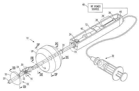
[0040] Figures 1A and 1 B show a balloon dilation device 10 including a handle
12, and an elongate shaft 14 oriented along a longitudinal axis 15 defining a
distal
direction away from handle 12 and a proximal direction towards handle 12. The
elongate shaft 14 has a distal portion 16 with a tip 18. A distal cutting
surface 20 is
shown in this embodiment to be an arcuate electrosurgical cutter spaced
distally
from tip 18. Slots 22 along shaft 14 house anchor elements 24 (retracted so
not
shown in Fig. 1A) shown in an extended configuration in Fig. 1 B. Anchor
elements
24 may extend from slots 22 to anchor the device 10 to adjacent tissue. Side
cutting surface 30, shown in Fig. 1A in an extended configuration and in Fig.
1 B in a
retracted configuration, may be used to cut tissue to isolate a mass of tissue
from a
surrounding tissue bed within a patient's body. Side cutting surface 30 may be
an
electrosurgical cutter as illustrated in Figs. 1A and 1 B.
[0041] Device 10 illustrated in Fig. 1A and Fig. 1B has a single balloon,
proximal
balloon 26, located proximal of the tip 18 along shaft 14 and shown in a
deflated
configuration in Fig. 1A and in an inflated configuration in Fig. 1 B.
Proximal balloon
26 may be inflated by passage of a fluid, which may be either a gas or a
liquid,
along inflation tube 28 into proximal balloon 26. The fluid is caused to flow
into and
inflate balloon 26 by pressure from an inflation mechanism 32, illustrated in
Figs. 1A
and 1 B as a syringe connected to the device 10 by a tube.
[0042] Fig. 1 B shows device 10 with proximal balloon 26 in an inflated
configuration. Inflation of balloon 26 while device 10 is in place within a
path
leading to a tissue specimen within a' patient's body presses balloon 26
against
body tissue, effective to compress and displace the body tissue and to expand
the
path. Tissue specimens may be removed through the path as the device is
withdrawn from the body. Fixation of the tissue mass 52 to the shaft 14 by
anchor
elements 24 brings the tissue mass 52 out of the tissue bed 58 as the device
10 is
withdrawn from the patient's body. The expanded path makes possible the
removal
of larger tissue specimens than would otherwise be possible, and eases the
_g_
CA 02455769 2004-O1-27
WO 03/011143 PCT/US02/20454
removal of all tissue specimens regardless of size while minimizing trauma and
damage to the patient.
10043] Side cutting surface 30 may be extended and retracted by movement of
side-cutter deployment shaft 44 effected by side-cutter shuttle 42. Handle 12
includes within it rotary mechanism 34 effective to rotate shaft 14, and
anchor
element extension mechanism 36 for extending anchor elements 24 into tissue,
comprising shuttles 38 visible in the view shown in Figs 1A and 1 B. Shuttles
38
connect with deployment shafts 40 and 41 shown in Figs. 1 C and 1 D. Conductor
46 provides electrical power to distal cutting surtace 20 by connecting power
source
48 with distal cutting surface 20. Power source 48 may be a source of
radiofrequency (RF) power.
(0044] Distal cutting surface 20 is effective to cut tissue and so to aid in
the entry
of device 10 into, and the passage through, a patient's body. In preferred
embodiments, distal cutting surface 20 and side cutting surface 30 are
electrosurgical cutting elements receiving RF power from power source 48
effective
to cut tissue. In addition, cutting surtaces 20 and 30 may be used to
cauterize
tissue when desired. Rotation of shaft 14 while side cutting element 30 is in
a
deployed configuration is effective to cut a path through surrounding tissue
and to
isolate a tissue specimen around the shaft 14 of device 10.
(0045] In embodiments of the invention, the device 10 may have a plurality of
balloons. As illustrated in Figs. 2A-2G, a device 10 embodying features of the
invention has two balloons, a proximal balloon 26 and a distal balloon 50.
Both
balloons 26 and 50 are shown in a deflated configuration in Fig. 2A; proximal
balloon 26 is shown inflated, and distal balloon 50 deflated, in Fig. 2B; and
both
balloons 26 and 50 are shown inflated configurations in Fig. 2C. Fluid may
flow into
balloons for inflation. Fluid flows into proximal balloon 26 via tube 28 and
into distal
balloon 50 via tube 51.
L0046] The device 10 having two balloons 26 and 50 is shown in cross section
in
Figs. 2D-2G. Fig. 2D is a cross-sectional view of the device of Fig. 2C taken
along
line 2D-2D, and shows the outer circumference of distal balloon 50 and
elements of
shaft 14, including anchor elements 24, anchor element deployment shaft 40,
and
distal cutting surface conductor 46. FIG. 2E is a cross-sectional view of the
device
_g_
CA 02455769 2004-O1-27
WO 03/011143 PCT/US02/20454
of Fig. 2C taken along line 2E-2E, showing side cutting surface 30 and the
region of
shaft 14 between balloons 26 and 50. FIG. 2F is a cross-sectional view taken
along
line 2F-2F showing the outer circumference of proximal balloon 26 and shaft 14
and
elements in the interior of shaft 14. A cross-sectional view of the device 10
proximal
of balloon 26, taken along line 2G-2G, is shown in FIG. 2G. Proximal balloon
inflation tube 28 and distal balloon inflation tube 51 are effective to carry
fluid for
inflation of proximal balloon 26 and distal balloon 50, respectively. Outflow
of fluid
is effective to allow balloons to deflate.
[0047] Devices and methods of the invention may be used to remove tissue
specimens from any suitable location within a patient's body. Figures 3A and
3B
illustrate the removal of a tissue mass 52 from within the breast 54 of a
patient. A
device 10 having a single balloon, proximal balloon 26, is shown in place in a
breast
54 in Figs. 3A and 3B. Anchor elements 24 are shown anchoring the isolated
tissue
mass 52 to the device 10. Proximal balloon 26 is in the deflated configuration
in
Fig. 3A, and in an inflated configuration in Fig. 3B. Inflation of proximal
balloon 26
presses walls 57 of path 56 outwardly, dilating the path so it can accommodate
the
passage of tissue mass 52. Path 56 crosses tissue bed 58 and skin 59 and
dilation
of path 56 is effective to compress and stretch tissue bed 58 and skin 59 but
does
not tear or cause undue trauma to these parts of the patient's body. The
dilation of
path 56 reduces or eliminates the need for cutting tissue bed 58 or skin 59 to
remove the tissue mass 52. Removal of the tissue mass 52 anchored to the shaft
14 by anchor elements 24 is accomplished by proximal movement of the device
10.
[0048] In Figs. 3A and 3B, the elongate shaft 14 of device 10 is shown
inserted
into the patient with tip 18 having passed through the suspect tissue mass 52
location within a patient's body and with shaft 14 occupying a position within
a path
56 through tissue bed 58, the path 56 leading to the tissue mass 52 that is to
be
removed. Distal cutting surface 20 is used to aid in making a path to the
desired
location within a patient's body and in positioning the device in a desired
location.
Deployment of side cutting surtace 30 and rotation of shaft 14 rotated causes
side
cutting surface 30 to cut tissue effective to isolate a tissue mass 52 from
the
surrounding tissue bed 58. Distal cutting surface 20 and side cutting surface
30
may be provided with, e.g., RF power, and used as electrosurgical cutting
elements.
Anchoring elements 24 are shown deployed from slots 20 to anchor the isolated
-10-
CA 02455769 2004-O1-27
WO 03/011143 PCT/US02/20454
tissue mass 52 to the device 10. Proximal balloon 26 is shown in its inflated
configuration in Fig. 3B, effective to press on tissue and expand the diameter
of the
path 56 leading to the isolated tissue mass 52. The expanded path more easily
accommodates the removal of the isolated tissue mass 52 from within the breast
54.
L00491 Figs. 4A, 4B and 4C illustrate a device 10 having two balloons anchored
in
place in a breast 54 shown in phantom, showing the balloons deflated (Fig.
4A), the
proximal balloon inflated and the distal balloon deflated (Fig. 4B), and both
proximal
and distal balloons inflated (Fig. 4C). In Fig. 4A, the shaft 14 is shown in
place
within path 56 extending through the isolated tissue mass 52, which is
anchored to
the shaft 14 by anchoring elements 24. Fig. 4B illustrates the device 10 of
Fig. 4A
with the inflated proximal balloon 26 shown dilating path 56 to aid the
removal of
isolated tissue mass 52. Fig. 4C illustrates the device of Fig. 4A with both
balloons
in the inflated configuration during removal of a tissue specimen from within
a
breast shown in phantom.
[00501 Figs. 4A-4C illustrate one method of use of a device 10 having two
balloons 26 and 50. In use during a diagnostic or therapeutic procedure to
remove
tissue from a patient, the sequence of inflation of balloons 26 and 50 would
follow
the sequence shown in Figs. 2A, 2B and 2C. Thus, the elongate shaft 14 of
device
would be inserted into the patient to bring tip 18 near to or through the
desired
location within a patient's body, with shaft 14 occupying a position within a
path
leading to the tissue mass that is to be removed. Distal cutting surface 20
may be
used to aid in making a path to the desired location within a patient's body
and in
positioning the device in a desired location. Side cutting surface 30 may be
deployed and shaft 14 rotated, thereby rotating the other elements of the
device,
including side cutting surface 30, and causing side cutting surface 30 to cut
tissue
and to isolate a tissue mass from the surrounding tissue bed. Anchoring
elements
24 may be deployed from slots 20 to anchor device 10 to the isolated tissue
mass
52. Proximal balloon 26 is next inflated, pressing on tissue and expanding the
diameter of the path 56. The expanded path 56 may more easily accommodate the
removal of the isolated tissue mass 52. Distal balloon 50 is next inflated,
pressing
on tissue and urging the device and attached tissue mass out along the path
56,
further aiding in the removal of the isolated tissue mass 52. In preferred
-11-
CA 02455769 2004-O1-27
WO 03/011143 PCT/US02/20454
embodiments, and as illustrated in Figs. 2A-2G, tip-cutting surface 20 and
side
cutting surface 30 may be provided with electrical power, such as RF power,
and
used as electrosurgical cutting elements.
[0051] The devices of the invention may also be dilation devices having
pivots,
with and without inflatable balloons. Examples of dilation devices 102 without
balloons are shown in Figs. 5 and 6. Such mechanical devices 102 may be used
together with balloon dilation devices 10 to form a tissue removal system 100.
in
addition, a tissue removal system 100 may include a tissue cutting device 60
and a
dilation device 102. Fig. 5A is a perspective view of a dilation device 102
embodying features of the invention showing the device in a closed
configuration
partially enclosing the shaft of a tissue-cutting device 60. The tissue
cutting devices
shown in Figs. 5 and 6 have features described in co-pending applications
Serial
Nos. 09/057,303; 09/146,185; 09/159,467; 09!238,965; and 09/356,187 named and
incorporated supra. Thus, the tissue cutting devices 60 illustrated in Figs. 5
and 6
are effective to isolate a tissue mass 52 and to anchor the mass to the device
60.
f0052~ As shown in the tissue removal system 100 illustrated in Fig. 5A and
5B, a
dilation device 102 having features of the invention includes a pair of arm
portions
104 having distal ends 106 configured to dilate tissue. As illustrated in
Figs. 5 and
6, distal ends 106 have dilation plates 108 for engaging and dilating tissue
along a
path within a patient's body. A pivot 110 joins the two arm portions 104 and
joins
leg portions 112 effective that lateral motion of leg portions 112 moves arm
portions
104. As shown in Figs. 5A, 5B, 6A, and 6B, leg and arm portions are continuous
and shaped from a single piece of material, hinged by pivot 110. However, in
embodiments of the invention, arm portions 104 and leg portions 112 may be
formed of different pieces of material and joined together during manufacture
of a
dilating device 102. Leg portions 112 have handles 114 at their distal ends
116.
The distance 120 between leg portions 112 is a variable distance depending on
the
position of leg portions 112 with respect to pivot 110. In embodiments, a
single
handle 114 may join legs 112.
f 00531 In Fig. 5A, leg portions 110 form an angle 118 with arm portions 104
effective that movement of leg portions 110 towards each other (so as to
reduce
distance 120) causes arm portions 104 to separate. The result of such movement
reducing the distance 120 is shown in Fig. 5B, showing arms 104 separated.
Such
-12
CA 02455769 2004-O1-27
WO 03/011143 PCT/US02/20454
separation of arms 104, when dilation plates 108 are in place within a path 56
within
a patient's body, is effective to dilate a path 56 and to aid in the removal
of a tissue
specimen.
(0054] In a scissors, a blade portion is aligned with an axis parallel to a
handle
axis so that motion around a central pivot causes congruent motion of the
blade and
handle. Thus, in a scissors, separating the handles separates the blades. Such
movement, where two blades are separated when two handle portions are open, is
termed "scissor-like." Alternatively, where a scissors has blades and handles
that
are not aligned, but meet at an angle so that the handle portions are
separated
when the blade portions are together, squeezing the handle portion separates
the
blades. Such an arrangement is not scissor-like.
[0055] The configuration of arm portions 104 and leg portions 112 around pivot
110 in the device illustrated in Figs. 5A and 5B, where arms 104 are analogous
to
the blades of a scissors, is not scissor-like. Connected arm portions 104 and
leg
portions 112 form an obtuse angle 118 around pivot 110, so that when arm
portions
104 are in contact, leg portions 112 are separated, and conversely, when leg
portions 112 are close together or in contact, arm portions 104 are separated.
An
operator holding the leg portions 112 of a dilation device 102 as illustrated
in Figs.
5A and 5B can dilate tissue in contact with dilation plates 108 by squeezing
together
legs portions 112. Distance 120 is larger in Fig. 5A than in Fig. 5B, the leg
portions
112 shown in Fig. 5A are shown farther apart than in Fig. 5B.
[0056] Fig. 6A shows a tissue removal system 100 including a dilation device
102
shown with dilation plates 108 in a closed configuration in place around a
tissue
cutting device 60 within a breast. In this embodiment, the handles 114 and
arms
104 are in a scissor-like configuration. Anchor elements 24 are shown
anchoring
tissue cutting device 60 to an isolated tissue mass 52. Dilation plates 108
are in
place within path 56 leading to isolated (issue mass 52. The mechanical
dilation
device 102 illustrated in Figs. 6A and 6B differs from the dilation device 102
illustrated in Figs. 5A and 5B in the orientation of arm portions 104, pivot
110 and
leg portions 112, the devices having different angles 118 and behaving
differently in
response to reduction in distance 120 between leg portions 112. Reduction in
distance 120 effects separation of arm portions 104 in the device 102
illustrated in
Figs. 5A and 5B. However, the dilation device 102 illustrated in Figs. 6A and
6B is
-13
CA 02455769 2004-O1-27
WO 03/011143 PCT/US02/20454
configured so that increasing distance 120 between leg portions 112 is
effective to
separate arm portions 104 and to press dilation plates 108 into path 56 so as
to
press on walls 57 effective to dilate path 56 to enable the removal of
isolated tissue
mass 52, as shown in Fig. 6B. The arm portions 104 and leg portions 112 are
configured in a scissor-like configuration around pivot 110 in the dilation
device 102
illustrated in Fig. 6A and 6B. Thus, an operator holding the dilation device
102
illustrated in Figs. 6A and 6B can dilate tissue in contact with dilation
plates 108 by
moving legs portions 112 apart. Distance 120 is larger in Fig. 6B than in Fig.
6A,
the leg portions 112 shown in Fig. 6B are shown farther apart than in Fig. 6A.
10057] In embodiments of the invention, devices having other forms of tissue
expanders instead of balloons, or in addition to balloons, may be used to
dilate a
path through tissue. For example, a dilation device 130 embodying features of
the
invention may have tissue expanders in the form of dilation plates 132
configured to
deploy from a retracted position adjacent a shaft 14 to configurations, as
shown in
Figs. 7A and B, that are partially expanded (Fig. 7A) and maximally expanded
(as
shown in Fig. 7B) as deployment struts 134 rotate around pivots 136.
Deployment
shuttles 138 may be connected by hollow shafts, rods, bands, or other
elements housed within shaft 14 to deployment struts 134 so as to move
deployment struts 134 in order to deploy and to retract dilation plates 132.
[0058] Other examples of devices with tissue expanders are shown in Figs. 8A-
8C. In Fig. 8A, a device 140 with tissue expanders having a plurality of
radially
expandable bands 142 is shown, with the bands in an expanded configuration
spaced radially away from the shaft 14. The bands are effective to contact
and to dilate tissue along a tissue path when the bands 142 are deployed to
assume an expanded configuration. Coliars 144 at the ends of the bands 142
may be moved longitudinally towards each other to effect deployment of the
bands 142, and may be moved longitudinally away from each other to effect
retraction of the bands 142. Movement of expandable bands 142 may be
effected by, e.g., movement of deployment shuttles 138 that are connected by
hollow shafts, rods, bands, or other elements housed within shaft 14 to
collars
144. In Fig. 8B, a device 150 with tissue expanders is illustrated having a
plurality of spirally expandable bands 152. The spirally expandable bands 152
- 14-
CA 02455769 2004-O1-27
WO 03/011143 PCT/US02/20454
are shown in an expanded configuration spaced away from the shaft 14. The
spirally expandable bands 152 are effective to contact and to dilate tissue
along a tissue path when the spiral bands 152 are deployed to assume an
expanded configuration. The spirally expandable bands 152 may be deployed
by movement of a collar 144 or of collars 144, where such movement may be
rotational movement around the longitudinal axis 15 of shaft 14, longitudinal
movement along the longitudinal axis 15 of shaft 14, or a combination of such
movements. Fig. 8C shows a device 160 with tissue expanders having an
expandable meshwork 162, the meshwork 162 being shown in an expanded
configuration. The meshwork 162 is effective to contact and to dilate tissue
along a tissue path when the meshwork 162 is deployed to assume an
expanded configuration. The meshwork 162 may assume a retracted
configuration adjacent shaft 14, and may be deployed to dilate a path into an
expanded configuration as shown by, e.g., longitudinal movement of collars
144 towards each other along shaft 14.
[00591 A method for removing a tissue specimen 52 from a tissue bed 58 within
a
patient's body is illustrated by Figs. 3A and 3B. Elongate shaft 14 of device
10 may
be inserted into the patient to bring tip 18 near to or through the desired
location
within a patient's body, with shaft 14 occupying a position within a path 56
leading
to the tissue mass that is to be removed. Distal cutting surtace 20 may be
used to
aid in making a path 56 to the desired location within a patient's body and in
positioning the device in a desired location. Side cutting surface 30 may be
deployed and shaft 14 rotated, thereby rotating the other elements of the
device
including side cutting surface 30 and causing side cutting surface 30 to cut
tissue
and to isolate a tissue mass 52 from the surrounding tissue bed 58. Anchoring
elements 24 may be deployed from slots 22 to anchor device 10 to the isolated
tissue mass 52. Proximal balloon 26 may then be inflated by fluid flow through
inflation tube 28, pressing on path walls 57 and expanding the diameter of the
path
56, aiding in the removal of the isolated tissue mass 52. In preferred
embodiments,
distal cutting surface 20 and/or side cutting surface 30 may be provided with
electrical power, such as RF power, and used as electrosurgical cutting
elements.
-15-
CA 02455769 2004-O1-27
WO 03/011143 PCT/US02/20454
(00601 Another method for removing a tissue specimen 52 from a tissue bed 58,
using a device 10 having two balloons 26 and 50, is illustrated by Figs. 4A-
4C. In
use during a diagnostic or therapeutic procedure to remove a tissue mass 52
from a
patient, the sequence of inflation of balloons 26 and 50 would follow the
sequence
shown in Figs. 2A, 2B and 2C. Thus, following insertion of the device 10 into
the
patient to bring tip 18 near to or through the desired location within a
patient's body,
side cutting surface 30 may be deployed and shaft 14 rotated, causing side
cutting
surface 30 to cut tissue and to isolate a tissue mass from the surrounding
tissue
bed. Anchoring elements 24 may be deployed from slots 22 to anchor device 10
to
the isolated tissue mass. Proximal balloon 26 may then be inflated, pressing
on
path walls 57 and expanding the diameter of the path 56. The expanded path 56
may more easily accommodate the removal of the isolated tissue mass 52. Distal
balloon 50 may next be inflated by fluid flow via inflation tube 51, pressing
on tissue
and urging the device 10 and attached tissue mass 52 out along the path 56,
further
aiding in the removal of the isolated tissue mass 52. In preferred
embodiments, and
as illustrated in Figs. 2A-2G, distal cutting surface 20 and side cutting
surface 30
may be provided with electrical power, such as RF power from a RF power source
48, and used as electrosurgical cutting elements.
(00611 In preferred embodiments, the distal cutting surface 20 and side
cutting
surface 30 are electrosurgical cutting surfaces. Power to these cutting
surfaces
may be from any suitable source of electrical power, preferably a source of RF
power 48. In one embodiment of the invention, the source of RF power 48 can
operate at frequencies from about 200 kiloHertz (kHz) to about 10 megaHertz
(MHz). Preferably, side cutting electrode 30 receives RF power at a frequency
of,
for example, between about 2.5 MHz and about 7.5 MHz, preferably at a
frequency
of about 5 MHz, at a voltage of between about 450V to about 550V and at a
power
of up to about 400 Watts (W). In embodiments of the invention, a distal
cutting
surface receives RF power at a frequency of about 300 kHz to about 1.5 MHz,
preferably about 500 kHz to about 1000 kHz, more preferably about 700 kHz to
about 900 kHz, at a power of, for example, between about 50 W to about 150 W,
and more specifically, at a power of from about 80 W to about 100 W. The
distal
cutting surface and side cutting surface are also effective to cauterize
tissue when
sufficient amounts of power (typically greater than the amounts listed above)
are
- 16-
CA 02455769 2004-O1-27
WO 03/011143 PCT/US02/20454
supplied to them. Examples of suitable power sources are disclosed in co-
owned,
co-pending U.S. Patent Application No.09/752,978, filed December 28, 2000, the
disclosure of which is hereby incorporated by reference in its entirety.
[0062] Devices 10, 130, 140, 150, 160 and devices 102 of the invention may be
used together for removing a tissue specimen from a tissue bed within a
patient's
body. Thus, one embodiment of a method for removing a tissue specimen includes
the steps of positioning a device 10 as illustrated in Figs. 1-4, having a
shaft 14
with an inflatable balloon 26 adjacent a tissue specimen 52 along a path 57 in
a
tissue bed 58 within a patient's body; enclosing a portion of the shaft 14
with a
portion of a dilation device 102 as illustrated in Figs. 5 and 6; inflating a
balloon 26
effective to dilate the path 56 through the tissue bed 58; enlarging the
transverse
dimension of the dilating device 102, as by manipulating the arms 104 having
dilation plates 106 to separate the legs 112 of the dilating device 102,
effective to
dilate a path 56 through the tissue bed 58; and removing said tissue specimen
52.
(0063] Balloons may be made from any suitable material or materials, including
polymers, rubber (both natural and synthetic such as latex and silicon
rubber). For
example, balloons may be made from polymers and polymer blends, including
polymers such as polyamides, polyesters, polyethylene, polyimides,
polytetrafluoroethylene (Teflon~), polyurethane, polyvinyl chloride,
polynitrile,
polyethylene terephthalate and polyolefin polymers. Balloons may be made from
flexible and foldable materials such as woven material, braided material, knit
material, web material, mesh material, film material, flexible laminate
material;
and/or elastic material.
L0064] Balloons may be inflated by increasing internal pressure within the
balloon,
as by flow of a fluid into the balloon to fill and expand the balloon. The
fluid may be
a gas or liquid. In one embodiment, a balloon and balloon inflation tube is
connected by a conduit to a syringe filled with a liquid, such as water,
saline,
mineral oil, or other substantially incompressible liquid. Pressure on the
plunger of
the syringe forcing the fluid out of the syringe, through the conduit, and
into the
balloon is effective to inflate the balloon. The conduit may be a flexible
tube, such
as one made from Tygon° tubing, and may pass through a valve or be
fitted with a
clip or clamp. Closure of a valve or clamp or placement of a clip onto the
conduit,
such as a dialysis clip, is effective to prevent fluid flow after inflation of
the balloon
-17-
CA 02455769 2004-O1-27
WO 03/011143 PCT/US02/20454
so as to maintain the balloon in an inflated configuration as long as is
desired.
Alternatively, a balloon may be connected to a source of high pressure air or
gas,
and may be inflated by allowing air or gas into the balloon until a desired
amount of
inflation and internal pressure has been achieved.
(0065] Dilation devices embodying features of the invention may be made from
any suitable material, including metals, composites, plastics, and ceramics.
For
example, devices 102 embodying features of the invention may be made stainless
steel, which may be coated with a biocompatible material, or from a
biocompatible
polymer, composite, such as a glass-reinforced nylon, high density
polyethtlene
(HDPE), or other durable material. Devices 10, 130, 140, 150, and 160 may
similarly be made from any suitable material, including metals, composites,
plastics,
ceramics, and combinations of such materials, and are typically made from more
than a single material.
(0066] Preferably, the devices embodying features of the invention are made
from
materials that are suitable for sterilization, including ultraviolet, chemical
and
radiation sterilization. Such sterilizable materials include stainless steel
and
other metals, ceramics, composites, plastics, and such polymers as
polyethylene, polypropylene, a fluorinated ethylene polymer, or other
material.
[0067] Devices embodying features of the invention may also include other
useful features that may aid placement or removal of tissue masses from within
a patient. For example, markings 31, as shown in Fig. 1 A, may be placed
along shaft 14 of a device 10, 130, 140, 150 or 160 to aid an operator in
determining the depth of tip 18. Other useful features may be included in a
device 10, 102, 130, 140, 150, 160, or other devices and systems embodying
features of the invention. Thus, while particular forms of the invention have
been
illustrated and described, it will be apparent that various modifications can
be made
without departing from the spirit and scope of the invention. Accordingly, it
is not
intended that the invention be limited, except as by the appended claims.
Reference to the terms "members," "elements," "sections," "expanders," and
terms
of similar import in the claims which follow shall not be interpreted to
invoke the
provisions of 35 U.S.G. ~112(paragraph 6) unless reference is expressly made
to
the term "means" followed by an intended function.
-18-