Note: Descriptions are shown in the official language in which they were submitted.
CA 02455826 2004-O1-29
WO 03/011147 PCT/US02/19921
METHODS AND DEVICES FOR
INTERBODY SPINAL STABILIZATION
Field of the Invention:
The present invention relates generally to instruments and devices for spinal
surgery, more particularly to methods and devices for spinal disc space
preparation and
interbody spinal stabilization.
BACKGROUND OF THE INVENTION
There are prior art interbody devices that are fabricated prior to
implantation and
then inserted into the patient's spinal disc space during surgery. It is also
known to insert
one or more pre-fabricated devices from anterior, antero-lateral, lateral,
postern-lateral,
transforaminal, posterior, posterior mid-line or any other known approach to
the disc
space. These pre-fabricated devices can require the surgeon to modify the
interbody
device, the vertebral bodies, and/or the vertebral endplates to achieve a
desired fit between
the spinal anatomy and the interbody device. While some pre-fabricated devices
can be
modified before and during surgery by the surgeon, this is a time consuming
task and also
does not always result in a desired or optimum ~t with the natural or altered
spinal
anatomy. Further, the various approaches and instruments required to insert
pre-fabricated
devices can be invasive and traumatic to the nervature, vasculature, and
tissue between the
skin and the disc space.
What is therefore needed are methods and devices for providing interbody
devices
in a disc space between vertebral bodies that allow the surgeon to achieve a
desired or
optimum fit between the device and the natural or altered spinal anatomy. What
is also
needed are devices and methods for preparing a disc space for an interbody
device while
minimizing invasion into the tissue between the skin and the subject disc
space. What is
further needed are improved devices and methods for performing spinal surgery.
What is
also needed are methods and devices for providing interbody fusion utilizing
minimally
invasive approaches and instruments. The present invention is directed toward
meeting
these needs, among others.
CA 02455826 2004-O1-29
WO 03/011147 PCT/US02/19921
2
SUMMARY OF THE INVENTION
According to one aspect of the present invention, there is provided a form
positionable in a spinal disc space and an interbody device made from material
that has a
first condition allowing placement around the form and in contact with the
vertebral
endplates and thereafter the material has a second condition that provides
structural
support between the endplates.
According to another aspect of the invention, there is provided a distractor
for a
disc space that has a reduced-size configuration for insertion into a disc
space and an
enlarged configuration for distracting the disc space and for defining a void
between the
enlarged portion and the inner wall of the disc space annulus.
According to yet another aspect of the invention, a spinal disc space
distractor
provides an intradiscal form around which an interbody device is placed.
According to a further aspect of the invention, a spinal disc space distractor
having
an enlargeable portion is provided.
According to a further aspect of the invention, a spinal disc space distractor
having
an enlargeable portion with upper and lower vertebral endplate contact
surfaces with
predetermined areas is provided.
According to another aspect of the invention, a surgeon inserts a distractor
in a
spinal disc space and places a first material around the distractor and
between the vertebral
endplates. When the first material cures, the distractor is withdrawn and a
second material
is placed in the disc space in the space that was occupied by the distractor.
According to a further aspect of the invention, multiple distractors having
enlargeable distracting portions are inserted in the disc space to form a void
for receiving a
first material
According to another aspect of the invention, a disc space is bi-laterally
distracted
by inserting an enlargeable portion of a first distractor at a ftrst lateral
disc space location
and an enlargeable portion of a second distractor at a second lateral disc
space location.
Scoliosis can be addressed by providing the enlargeable portions with
different distraction
heights.
According to a further aspect of the invention, a spinal disc space distractor
having
an enlargeable portion of a predetermined shape is provided. The predetermined
shape is
CA 02455826 2004-O1-29
WO 03/011147 PCT/US02/19921
selected from one of the following: vertically-oriented cylinder, horizontally-
oriented
cylinder, sphere, cylindrical center portion with frusto-conical tapered ends;
banana-
shaped, and pear shaped.
These and other aspects, forms, features and advantages will be apparent from
the
following description of the illustrated embodiments.
BRIEF DESCRIPTION OF THE DRAWINGS
Fig. 1 is diagrammatic illustration in the axial plane of a spinal disc space
with
instruments positioned therein for performing a discectomy procedure.
Fig. 2a is a diagrammatic illustration of the disc space of Fig. 1 with a
distractor
having an enlargeable portion positioned therein.
Fig. 2b is a diagrammatic illustration looking in the direction transverse to
the
sagittal plane of the spinal column segment encompassing the disc space and
the distractor
of Fig. 2a.
Fig 3a is a diagrammatic illustration of the disc space of Fig. 2a with the
distractor
disposed therein along with a material delivery instrument.
Fig. 3b is a diagrammatic illustration of the disc space of Fig. 3a with a
first
material being delivered around the enlarged portion of the distractor.
Fig. 3c is a sectional view of an alternate embodiment enlargeable distractor
and
material delivery instrument according to the present invention.
Fig. 4 is a diagrammatic illustration of the disc space of Fig. 3b after the
first
material has cured and the enlargeable portion of the distractor in a reduced
size
configuration for removal from the disc space.
Fig. 5 is a diagrammatic illustration of the disc space of Fig. 4 with a
second
material in the disc space within the cured material.
Fig. 6 is a diagrammatic illustration of in partial section through line 6-6
of Fig. 5.
Fig. 7 is a diagrammatic illustration of the partial sectional view of Fig. 7
showing
posterior stabilization instrumentation secured to the spinal column segment
across the
disc space.
Fig. 8 is a diagrammatic illustration in the axial plane of a spinal disc
space having
a pair of distractors having enlargeable portions for bi-lateral distraction
of the disc space.
CA 02455826 2004-O1-29
WO 03/011147 PCT/US02/19921
4
Fig. 9 is a diagrammatic illustration of a spinal disc space having another
arrangement for dual distractors along with a first material positioned at a
first lateral
location in the disc space.
Figs. 10a-1 Oc show a side view, an end view and a plan view, respectively, of
one
embodiment of an inflatable distractor.
Figs. 11 a-11 c show a side view, an end view and a plan view, respectively,
of
another embodiment inflatable distractor.
Figs. 12a-12c show a side view, an end view and a plan view, respectively, of
another embodiment inflatable distractor.
Figs. 13a-13c show a side view, an end view and a plan view, respectively, of
another embodiment inflatable distractor.
Figs. 14a-14c show a side view, an end view and a plan view, respectively, of
another embodiment inflatable distractor.
Figs. 15a-15c show a side view, an end view and a plan view, respectively, of
another embodiment inflatable distractor.
Figs. 16a-16c show a side view, an end view and a plan view, respectively, of
another embodiment inflatable distractor.
Figs. 17a-17c show a side view, an end view and a plan view, respectively, of
another embodiment inflatable distractor.
Fig. 18 is a graphical representation of the load applied to the vertebral
endplates
versus inflation pressure for inflatable distractors having various vertebral
endplate contact
areas.
DESCRIPTION OF THE ILLUSTRATED EMBODIMENTS
For the purposes of promoting an understanding of the principles of the
invention,
reference will now be made to the embodiments illustrated in the drawings and
specific
language will be used to describe the same. It will nevertheless be understood
that no
limitation of the scope of the invention is thereby intended. Any such
alterations and
further modifications in the illustrated devices, and any such further
applications of the
principles of the invention as illustrated herein are contemplated as would
normally occur
to one skilled in the art to which the invention relates.
The present invention provides techniques for forming interbody devices in a
disc
CA 02455826 2004-O1-29
WO 03/011147 PCT/US02/19921
space of the spinal column. It is contemplated that techniques of the present
invention
utilize minimally invasive endoscopic instruments and methods for performing
discectomy
and other disc space preparatory procedures. However, open surgical techniques
and other
visualization instruments and techniques are also contemplated. In techniques
where the
interbody device is part of a spinal fusion procedure, percutaneous
stabilization and
fixation techniques through the pedicles or facets are also possible after
completing
insertion of the interbody device. The present invention further provides
minimally
invasive techniques for segmental stabilization of a spinal disc space to
repair a spinal disc
space due to, for example, disc space collapse or progressive mono-segmental
instability
which are normally repaired via discectomy procedures that do not include
interbody
fusion. The present invention has application from any approach to any disc
space along
the spinal column, including LS-S 1. Further, the present invention has
application in a bi-
portal, postern-lateral approach to one or disc spaces in the lumbar region of
the spine.
Reference will now be made to Figs. 1-7 to describe methods, instruments and
materials according to the present invention to provide an interbody device
formed in situ
in the disc space that conforms with the patient's vertebral endplate anatomy.
Fig. 1
shows an outline in plan view of a spinal disc space and lower vertebral body
l Ob in plan
view during a discectomy procedure. The anterior aspect of the spinal column
is indicated
by "A" and the posterior side is indicated by "P." The lateral aspects of the
spinal column
extend between A and P on each side the spinal column. As shown further in Fig
2b, the
subject spinal disc space is located between an upper vertebra 10a having an
inferior
endplate l la and a lower vertebral lOb having a superior endplate l 1b. The
disc space has
a nucleus 12 that is surrounded by an annulus 14. First and second pedicles
16a extend
posteriorly from upper vertebral body 10a, and first and second pedicles 16b
extend
posteriorly from lower vertebral body 10b. The spinal cord or dura 17 extends
along the
posterior aspect of vertebrae 10a, 10b.
In Fig. 1 there are shown instruments inserted via a bi-portal approach to the
disc
space that are useful in completing a nucleotomy or a discectomy of the spinal
disc. The
instruments for performing this procedure can include a scope 20 and a
discectomy
instrument 22. In the illustrated embodiment, discectomy instrument 22 and
scope 20 are
inserted through first access port 18 and second access port 19, respectively,
in a postero-
CA 02455826 2004-O1-29
WO 03/011147 PCT/US02/19921
6
lateral approach to the disc space. Access ports 18, 19 can each be a working
channel
cannula to provide a protected first and second postern-lateral access ports
to the disc
space. It is to be understood that aspects of the present invention
contemplate approaches
and combinations of approaches to the disc space other than a postern-lateral
approach,
such as a lateral approach, anterior approach, or antero-lateral approaches.
It should be
understood that uni-portal disc space access is contemplated, as well as bi-
portal disc
space access from the same side of the spinal disc space or from differing
approaches,
such as a lateral approach and a postern-lateral approach. It is further
contemplated that
open surgical procedures could be utilized for the discectomy.
In one specific surgical technique used with the present invention, the disc
space in
the lumbar region of the spine is accessed endoscopically via a foraminal or
postero-
lateral, bi-portal approach. Cannulas and dilators can be used for access
ports 18, 19 and
catheters inserted therethrough for visualization, discectomy procedures,
distraction, and
material delivery. In these approaches, the outer cannulas can have an outside
diameter of
up to 7.5 millimeters and more typically in the range of about 6.5
millimeters. However,
any sized cannula is contemplated so long as there is an acceptable level of
trauma to the
tissue and nerve structures.
To provide access ports 18, 19 in this specific technique, insertion begins 9
to 13
centimeters from the midline with a guidewire or discogram needle. The facet
joint at the
dome of the facet is initially targeted and palpated by the tip of the needle.
The needle is
withdrawn and re-angulated to go inside the dome, thus missing the exiting
nerve root.
The posterior vertebral bodyline is imaged fluoroscopically to document its
resting
position. The fluoro machine is then moved to an A-P position and the resting
zone is
either on the mid or lateral pendicular starting position for a postern-
lateral approach or
the medial pendicular midline for a foraminal approach. Needle insertion into
the disc
space can be completed simultaneously on the left and right hand sides. The
needles can
be triangulated to touch one another in the posterior central portion of the
disc space or
alignment can be adjusted and conformed via discography.
One or more dilators of increasing diameter are then sequentially placed over
each
of the needles to the annulus, and a cannula is placed over each of the final
dilators to land
on the annulus. The final dilators are removed and a trephine used through
each cannula
CA 02455826 2004-O1-29
WO 03/011147 PCT/US02/19921
7
to cut holes in the annulus to allow for entry into the disc space. An
endoscope can be
used at any time throughout the procedure to document the presence of nerve
roots or to
observe the annulus prior to cutting. The final dilator is then re-inserted
into each of the
cannulas and impacted through the hole in the annulus and into the disc space.
The final
dilator thus secures the cannula into position and obstructs the annulus
opening to ensure
material is delivered into the disc space without excursion out of the disc
space. The
cannulas and dilators are then used as access portals to the disc space for
completion of the
remaining procedures, and also allow for the interchange of instruments
between the left
and right sides. Either one of the access ports 18, 19 can then be used for
endoscopic
visualization and the other access portal 18, 19 can be used for disc material
removal with
manual, automated, ultrasonic, laser, or any other disc material removal
instruments
desired by the surgeon.
After discectomy there is a prepared disc space 24. It can also be desired by
the
surgeon to expose and gently remove endplate cartilage and to remove all soft
tissue and
debris from within the disc space to expose the inner wall of the annulus.
Inner portions
of a minimally appropriate amount of the inner wall laminates of annulus 14
surrounding
the removed nucleus can be removed to increase the lateral and anterior-
posterior extent of
the prepared disc space 24. The remaining portion of the annulus remains
intact except for
the access holes cut for instrument entry locations. An endoscope can be
placed in one of
the access portals to check disc material removal and to also check the
annulus to ensure
there are no wall defects requiring repair. In cases where interbody fusion is
desired, the
endplates can be prepared by eburnating the apophyseal ring to prepare it for
bony fusion,
and the vertebral endplates can be scraped or abraded to reduce them to
bleeding bone.
Right angle curettes or probes can also be inserted to make small protrusions
or abrasions
into the endplates to further facilitate fusion if so desired.
After disc space access and discectomy, the disc space will typically still be
in a
collapsed state, and the only distraction that has been completed at this
point has been the
result of insertion of the final dilator into the disc space. The disc space
must now be
further distracted to the desired disc space height and also to establish
lordosis if desired or
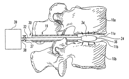
necessary. Referring now to Figs. 2a-2b, a distractor 30 is inserted into the
prepared disc
space 24. Distractor 30 has a shaft 32 extending between a distal end 36 and a
proximal
CA 02455826 2004-O1-29
WO 03/011147 PCT/US02/19921
8
end 38 situated outside the disc space. Adjacent distal end 36 there is an
enlargeable
portion 34 positionable in prepared disc space 24. Enlargeable portion 34 is
inserted into
the disc space in a reduced size configuration, and after proper positioning
in prepared disc
space 12 is confirmed endoscopically, fluoroscopically or via any other
visualization
technique, is thereafter enlarged to contact endplates l la, l 1b and distract
the disc space
to the desired height.
Enlargeable portion 34 is sized with respect to prepared disc space 24 such
that a
void 26 is formed between the enlarged portion 34, inner wall of annulus 14,
and the
endplates 1 la, l 1b generally in the location of the apophyseal ring as shown
in Fig. 3a. In
one form, enlargeable portion 34 is an inflatable balloon or cuff type
structure that is
inserted into the disc space in a deflated condition and thereafter inflated
via an inflation
lumen through shaft 32 to a predetermined pressure with air, gas, or liquid
from an
inflation source 39. A valve 37 can be provided on shaft 32 to block the lumen
therethrough and maintain the inflation pressure in enlargeable portion 34. It
is further
contemplated that enlargeable portion 34 could be made from any material
capable of .
assuming a reduced sized for insertion and withdrawal from the prepared disc
space and
enlargeable for disc space distraction, such as an elastomer, polymer, shape
memory
material or spring steel. Examples of various types of inflatable devices are
described
further below with respect to Figs. 10-17.
In any event, enlargeable portion 34 is sized in the cephalad-caudal
directions
sufficiently to distract the spinal disc space to a desired normal disc space
height and sized
in the lateral and anterior-posterior directions to provide void 26 when
enlarged. A single
centrally placed enlargeable distractor 30 could utilize endplate geometry to
create
lordosis.
In addition to a single distractor having an enlargeable portion inserted into
the
disc space as shown above with respect to Figs. 1-7, other distraction
instruments and
techniques are contemplated. For example, if the enlargeable portion of the
distractor is
inflatable, then the enlargeable portion 34 can be provided with dual chambers
of differing
heights to establish a lordotic effect. In another example, multiple
distractors having
different height enlargeable portions 34 can be inserted and positioned at the
appropriate
locations in the disc space and be enlarged together to provide the desired
endplate
CA 02455826 2004-O1-29
WO 03/011147 PCT/US02/19921
angulation.
As further shown in Figs. 3a and 3b, with distractor 30 enlarged and
maintaining
disc space distraction, a material delivery instrument 40 is inserted into the
disc space in
the access port opposite the distractor access port. Material delivery
instrument 40
includes a working channel 42 through which a first material 50 can be
delivered through
a distal opening 44 and into void 26. First material 50 has a first condition
that allows it to
be selectively placed, injected, flowed, moved or otherwise migrated around
the
enlargeable portion 34 in void 26 such that all or substantially all of void
26 is occupied by
first material 50. First material 50 thereafter changes, cures or transforms
from its first
condition into a second condition in which it forms a solid or semi-solid
interbody device
50' in space 26, as shown in Fig. 4, capable of structurally supporting the
vertebrae at the
desired disc space height. Interbody device SO' thus confornis to the
patient's vertebral
endplate anatomy and also conforms to the shape of void 26 between enlargeable
portion
34 and annulus 14.
It is contemplated that first material 50 can be a cement, poly(methyl
methacrylate), or any other bio-compatible material that has the structural
capabilities to
withstand the spinal column loads applied thereto. It is further contemplated
that first
material 50 can be delivered in a first condition through an instrument
channel or lumen of
instrument 40 and thereafter changed to a second condition via any natural or
chemically
induced or enhanced reaction to form an interbody device 50'. First material
50 can
further be static or include bio-active material to promote bone growth.
While delivery instrument 40 is illustrated as an instrument separate from
distractor 30, it is also contemplated that distractor 30 could be provided
with a working
channel for delivery of first material 50 to void 26 or second material 60 to
central space
52'. For example, as shown in FIG. 3c, distractor 30' has a shaft 32' and an
inflatable
enlargeable portion 34'. Shaft 32' defines an inflation lumen 32a' in
communication with
the interior of enlargeable portion 34'. Shaft 32' further include a material
delivery lumen
32b' extending through enlargeable portion 34' and opening at distal end 36'.
After
distraction with enlargeable portion 34', first material 50 can be delivered
through lumen
32b' into void 26. Such an instrument could be employed for uni-portal
material delivery
and disc space distraction, or used in combination with material delivery
instrument 40 or
CA 02455826 2004-O1-29
WO 03/011147 PCT/US02/19921
another distractor 30' in the opposite access port to provide bi-portal
material delivery. It
is further contemplated that delivery instrument 40 can be a flexible cannula
or catheter
that can be moved or manipulated around void 26 in order to deliver first
material 50 to all
portions thereof. Material delivery instrument 40 can further be provided with
endoscopic
5 capabilities to allow visualization and direct viewing of material delivery.
In another form, one or more flexible material delivery catheters can be
placed
over a guide wire extending through one of the access portals and into the
disc space
around enlargeable portion 34 and at various locations in void 26. The
flexible catheters)
can be placed through only one or both of the access portals 18, 19. With the
desired
10 distraction achieved and the material delivery catheters positioned as
desired, the guide
wires are removed and first material 50 delivered through the flexible
catheter(s). First
material 50 can be delivered sequentially through the catheters or
simultaneously through
the catheters to provide an interbody device 50' that is completely formed
about
enlargeable portion 34 except for an entry port to central cavity 52'.
Interbody device 50'
thus provides balanced spinal load support on the apophyseal ring. Second
material 60
can then be placed centrally into the interbody device in the central cavity
52' previously
occupied by the withdrawn enlargeable portion 34 of distractor 30.
One specific technique for placement of first material 50 via bi-portal,
postero-
lateral access ports was completed as follows. The material delivery
instrument 40
included first and second material delivery catheters each placed in a
respective one of the
first and second access ports 18 and 19. First material 50 was delivered
through one
catheter through the first access port under low pressure until the presence
of first material
50 was detected at the distal end of the first access port or the second
access port. The
catheter was then slowly pulled back through the first access port until first
material 50
was delivered to the distal end of the first access port housing the first
delivery catheter.
Thereafter the first material delivery catheter was withdrawn. First material
50 was then
delivered through the second material delivery catheter positioned in the
second access
port until first material 50 was detected at the distal end of either of the
second access port
or the first access port. The second material delivery catheter was then
pulled back
through the second access port, thereby completely filling the void 26 with
first material
50.
CA 02455826 2004-O1-29
WO 03/011147 PCT/US02/19921
11
Several factors are to be considered in placing first material 50 in the disc
space.
For example, if first material 50 were a cement, factors to consider include
the liquidity of
the cement, the cure temperature of the cement and the insertion pressure of
the cement. If
the cement has a relatively cool temperature, then more time is required for
the cement to
cure which increase operating room time. Curing time can also be affected by
adding
other substances to it, such as growth factors, antibiotics and/or barium
tracer. The
injection pressure of first material 50 can affect whether it will leak out of
small tears in
the annulus or infiltrate interstices and nutrient canals of the vertebral
endplates. It is also
desirable that placement procedures for first material be carried out under
fluoroscopy
with a tracer such as barium in first material 50 to allow monitoring of
material excursion
and its presence in the disc space. Monitoring of the placement of first
material 50 to
confirm its proper positioning in the disc space can be accomplished by AP and
lateral
fluoroscopy or bi-planar fluoroscopy. The presence of material excursion could
signify a
significant annulus or other anatomical or surgically created defect or void.
Such
monitoring provides a safety measure to ensure first material 50 is not placed
into
inappropriate anatomic locations during formation of interbody device 50'.
Referring further to Fig. 4, enlargeable portion 34 is returned to its reduced
size
configuration so it can be removed from interbody device 50' and the disc
space. This
leaves a central cavity 52' surrounded by interbody device 50'. An endoscope
20 can be
used to monitor distractor withdrawal and to check the integrity of interbody
device 50'.
Material delivery instrument 40 can then be repositioned, if necessary, in one
of the access
portals and used to deliver a second material 60 to central cavity 52' as
shown in Fig. 5.
Second material 60 can be artificial disc material, bioactive substance,
rhBMP, autograft,
or bioactive or osteoconductive carrier for bony fusion. In situations where
second
material 60 is fusion material, bony fusion can occur centrally while
interbody device 50'
provides stability of the disc space during fusion. It is further contemplated
that in
situations where fusion is desired, the endplates 1 la, 1 1b could be reduced
to bleeding
bone via scraping, cutting, or reaming prior to placement of second material
60.
Refernng now to Fig. 6, there is shown a partial section view of the spinal
column
segment having interbody device 50' formed in a disc space as described above.
Interbody device 50' conforms with the shape of endplates 1 la, 1 1b and
constrains second
CA 02455826 2004-O1-29
WO 03/011147 PCT/US02/19921
12
material 60 therein. In Fig. 7, there axe shown posterior screws 46a, 46b
secured to
pedicles 16a, 16b and a rod 48 extending between and secured thereto. It is
further
contemplated that posterior stabilization could be provided with screws at the
facet joints,
or via a posterior plate secuxed to the vertebrae. Anterior or latexal
stabilization plates
secured to the vertebrae are also contemplated. Such supplemental fixation and
stabilization devices are known in the art and will not be described further
herein.
Referring now to Fig. 8, there is shown another technique for forming an
interbody
device in a spinal disc space. The instruments used in the technique of Fig. 8
include a left
side lateral distracter 70a and a right side lateral distracter 70b that is
substantially
identical to left side distracter 70a. Lateral distracters 70a, 70b each
include shafts 72a,
72b and an enlargeable portion 74a, 74b, respectively, adjacent a distal end
of the
respective shaft. If enlargeable portions 74a, 74b were inflatable, shafts
72a, 72b would
also define an inflation lumen. After completing procedures to form a prepared
disc space
as discussed above, lateral distracters 70a, 70b are positioned through bi-
portal access
ports 18, 19 and into the disc space 24. Enlargeable portions 74a, 74b each
have a
centavo-convex or banana-shaped configuration so that each can be positioned
along the
inner annulus wall and the apophyseal ring of the upper and lower vertebrae
10a, I Ob
while leaving the central poxtion of the disc space open. Further, the
apophyseal ring in its
most anterior portion between the distal tips of enlargeable portions 74a, 74b
remains open
for placement of material 50 and also remains open along its most posterior
portion
between the distal ends of enlargeable portions 74a, 74b. For example, as
shown in Fig. 8,
first material 50 has been placed in the anterior portion of the disc space by
a material
delivery instrument or catheter inserted through one of the access portals I
8, 19 alongside
the distracter to form a first interbody device segment 50" when cured. First
material 50
could also be placed in the posterior portion to form a second interbody
device segment
(not shown). Additional interbody segments or pillars could be formed in the
disc space,
and second material 60 could then be placed or packed between the interbody
segments.
There are several distraction and material placement techniques afforded by
use of
lateral distracters as shown in Fig. 8. For example, after sequential bi-
lateral distraction of
the disc space, one of the lateral distracters could be reduced in size and
withdrawn and
this same side of the disc space could be provided with first material 50 from
delivery
CA 02455826 2004-O1-29
WO 03/011147 PCT/US02/19921
13
instrument 40 to form a first lateral interbody device segment SOa as shown in
Fig. 9. A
single central distracter 30 can be used to block the central portion of the
prepared disc
space 24 while second lateral distracter 70b blocks the right lateral side of
the disc space.
Second lateral distracter 70b can then be withdrawn and additional first
material 50 is
provided to form a second interbody device segment (not shown) using
enlargeable
portion 34 as a form. After completion of the interbody device segments,
second material
60 can be delivered into the space between the interbody device segments.
Further,
sequential distraction can be done in such a way that two lateral distracters
70a, 70b are
left in prepared disc space 24 and second material 60 can be placed between
the lateral
distracters 70a, 70b. Second material 60 can then be used alone or in
combination with
one of the lateral distracters 70a, 70b as a form for placement of fixst
material 50.
It is further contemplated that the placement location for ftrst material 50
can be
varied at any location about the apophyseal ring by using combinations of
lateral
distracters, anterior and posterior distracters, and central distracters.
Further, it is
contemplated first material 50 could be placed at multiple, discrete locations
about the
apophyseal ring to provide a number of columnar or segmented interbody devices
in the
disc space. These segmented interbody devices could be formed adjacent to and
in centact
with one another or formed with gaps therebetween. It is further contemplated
that the
positioning of the various interbody devices could be varied to accommodate
the approach
desired for material placement, including both uni-lateral injection or a bi-
lateral
placement.
In another embodiment, the banana-shaped lateral distracters 70a, 70b can be
tapered in height to provide angulation between the vertebral endplates. For
example,
lordosis could be established by providing the enlargeable portions 74a, 74b
with a greater
height posteriorly than anteriorly. Further, the lateral distracters 70a, 70b
can be provided
with differing heights in order to distract one side of the disc space more
than the other
side, reducing or eliminating scoliosis. Alternatively, identical inflatable
devices could be
provided in which the inflatable portions have a height that corresponds to
the internal
inflation pressure supplied thereto. One of the lateral distracters could be
inflated to a
greater pressure than the contra-lateral side to provide differential
distraction heights for
each side. The same lateral distracter could be employed bi-laterally to
change the lateral
CA 02455826 2004-O1-29
WO 03/011147 PCT/US02/19921
14
angulation of the disc space by varying the inflation pressure supplied to the
enlargeable
portion thereof.
After repairing scoliosis by providing the appropriate distraction and
interbody
devices, the disc space occupied by the enlargeable portions of the distracter
is available
for placement of bone growth material. For example, if two banana-shaped
inflatable
devices are used, a central cavity encompassed by the enlargeable portions
remains after
the portions are enlarged. Second material can then be placed in this central
cavity.
Additional first material can then be placed in the space previously occupied
by the
enlarged portions to provide struchtral peripheral support. Thus, this
specific example
contemplates initially central placement of a first material, such as bone
growth material,
and then the enlargeable distracters can be sequentially or simultaneously
withdrawn from
the disc space and a second material, such as a cement, placed around the
central core of
first material and against the enlargeable distracter portion, if any,
remaining in the disc
space to provide structural support of the disc space.
As discussed above, enlargeable portion 34 of the distracter 30 can be an
inflatable
device. In Figs. 10-17, there are provided various embodiments of inflatable
devices that
can be used to perform disc space distraction. By providing inflatable devices
of various
shapes and sizes, different vertebral endplate contact areas can be formed
thereby
providing selection of the optimal inflatable device based on vertebral
endplate load
resistance, required distraction force, and the structural integrity of the
pressurized inflated
device. It should be understood, however, that the contact surface areas
provided below
are estimated based on a distraction height of 14 millimeters. The contact
surface area of
each balloon will vary depending on the degree to which the balloon is
inflated. For
distraction heights less than 14 millimeters, the contact are will be greater
than 0.2 square
inches. For distraction heights greater than 14 millimeters, the contact are
will be less than
0.2 square inches. It should be further understood that the contact area for
each balloon
can be varied by changing the lateral and/or anterior-posterior dimensions of
the balloon
while retaining the same balloon shape.
Referring now to Figs. 10a-1 Oc, there is shown a ftrst embodiment an
inflatable
device in the form of a balloon 100 having the shape of a center cylinder with
frusto-
conically tapered ends extending therefrom. Balloon 100 is in communication
with an
CA 02455826 2004-O1-29
WO 03/011147 PCT/US02/19921
inflation lumen 102 and has upper vertebral endplate contacting surface 104
and opposite
lower vertebral endplate contacting surface 106. As shown in Fig. l Ob,
surfaces 104, 106
have an oval shape with the rounded end portions of the oval positioned
laterally of a
longitudinal axis extending through inflation lumen 102 and balloon 100.
Surfaces 104,
5 106 contact endplates 1 la, l 1b of the upper and lower vertebrae 10a, 10b,
respectively, as
shown in Fig. l Oc. Balloon 100 has a central cylindrical portion 108 which
defines
contact surfaces 104, 106, and opposite frusto-conical portions 110, 112
distally and
proximally extending therefrom, respectively, and tapered at an angle that
avoids contact
with the vertebral endplates. In one specific embodiment, it is estimated that
balloon 100
10 has a contact surface area of about 0.2 square inches for each of the upper
and lower
contact surfaces 104, 106 when balloon 100 is expanded to distract the disc
space to a
height of 14 millimeters.
Referring now to Figs. 11 a-11 c, there is shown another embodiment of an
inflatable device in the form of a balloon 120 having a shape of a center
cylinder with a
15 pair of frusto-comically tapered ends extending from each end thereof.
Balloon 120 is in
communication with inflation lumen 122 and has upper vertebral endplate
contacting
surface 124 and opposite lower vertebral endplate contacting surface 126. As
shown in
Fig. l 1b, surfaces 124, 126 have an oval shape with the rounded portions
oriented distally
and proximally along a longitudinal axis extending through inflation lumen 122
and
balloon 120. Surfaces 124, 126 contact endplates 1 la, l 1b of the upper and
lower
vertebrae 10a, l Ob, respectively, as shown in Fig. 11 c. Balloon 120 has a
central
cylindrical portion 128 which defines a portion of contact surfaces 124, 126.
Balloon 120
further includes first frusto-conical portions 130, 132 extending distally and
proximally
therefrom, respectively, which define the remaining portions of contact
surfaces 124, 126.
Frusto-conical portions 130, 132 are only tapered slightly and generally match
the
curvature of the vertebral endplates in order to provide additional contact
area as
compared to balloon 100. In one specific embodiment, balloon 120 has a contact
surface
area of about 0.3 square inches for each of the upper and lower contact
surfaces 124, 126.
Distal frusto-conical portion 134 and proximal frusto-conical portion 136
extend to the
distal end of balloon 120 and to inflation lumen 122, respectively, and
generally do not
contact the vertebral endplates unless the balloon is sufficiently inflated to
create such
CA 02455826 2004-O1-29
WO 03/011147 PCT/US02/19921
16
contact.
Referring to Figs. 12a-12c, there is shown another embodiment an inflatable
device
in the form of a balloon 140 having a vertically oriented cylindrical shape.
Balloon 140 is
in communication with an inflation lumen 142 and has upper vertebral endplate
contacting
surface 144 and opposite lower vertebral endplate contacting surface 146.
Surfaces 144,
146 contact endplates 1 la, 1 1b of the upper and lower vertebrae 10a, l Ob,
respectively, as
shown in Fig. I2c. Balloon 140 has a cylindrical body 148 which has circular
upper and
lower ends 150, 152 that define circular contact surfaces 144, 146 as shown in
Fig. 12b.
In one specific embodiment, balloon I40 has a contact surface area of about
0.5 square
inches for each of the upper and lower contact surfaces 144, 146.
Referring now to Figs. 13a-13c, there is shown another embodiment an
inflatable
device in the form of a balloon 160 having a horizontally oriented cylindrical
shape.
Balloon 160 in communication with an inflation lumen 162 and has a cylindrical
body 168
with distal end 170 and opposite proximal end 172. Balloon 160 further
includes upper
vertebral endplate contacting surface 164 and opposite lower vertebral
endplate contacting
surface I66. As shown in Fig. 13b, contact surfaces 164, 166 have a
substantially
rectangular shape formed by the contact between the cylindrical sidewalls of
cylindrical
body 168 and endplates 1 la, I 1b of the upper and lower vertebrae 10a, 10b,
respectively.
In one specific embodiment, balloon 160 has a contact surface area of about
0.24 square
inches for each of the upper and lower contact surfaces 164, 166.
Referring to Figs. 14a-14c, there is shown another embodiment an inflatable
device
in the form of a balloon 180 having a horizontally oriented cylindrical shape.
Balloon 180
is in communication with inflation lumen 182 and has a cylindrical body 188
with distal
end 190 and opposite proximal end 192. Balloon 180 further includes upper
vertebral
endplate contacting surface I84 and opposite lower vertebral endplate
contacting surface
186. As shown in Fig. 14b, contact surfaces 184, 186 have a rectangular shape
formed by
the contact between the cylindrical sidewalk of cylindrical body 188 and
endplates 11 a,
1 1b of the upper and lower vertebrae IOa, 10b, respectively. In one specific
embodiment,
balloon 180 has a contact surface area of about 0.3 square inches for each of
the upper and
lower contact surfaces I 84, 186. Balloon 180 is similar in shape to balloon
160, but has a
shorter length between its distal and proximal ends to allow balloon 180 to
extend further
CA 02455826 2004-O1-29
WO 03/011147 PCT/US02/19921
17
laterally in the disc space than balloon 160 and thus increasing the vertebral
endplate
contact area.
Referring to Figs. 15a-15c, there is shown another embodiment an inflatable
device
in the form of a balloon 200 having a spherical shape. Balloon 200 is in
communication
with an inflation lumen 202 and has upper vertebral endplate contacting
surface 204 and
opposite lower vertebral endplate contacting surface 206. Surfaces 204, 206
are formed
on spherical body 208 and have a circular shape in contact with endplates l
la, 1 1b of the
upper and Iower vertebrae 10a, l Ob, respectively. Spherical body 208 has
opposite distal
and proximal ends 210, 212 respectively. In one specific embodiment, balloon
200 has a
diameter of 22 millimeters which provides a contact surface area of about 0.35
square
inches for each of the upper and lower contact surfaces 204, 206.
In Figs. 16a-16c there is shown another embodiment spherically shaped balloon
220 having a spherical body 228 in communication with inflation lumen 222.
Spherical
body 228 includes contact surfaces 224, 226 forming a circular contact surface
with
endplates 1 la, l 1b. In this embodiment, balloon 220 has a diameter of 24
millimeters and
the endplate contact surface areas of surfaces 224, 226 are each 0.45 square
inches.
Refernng now to Fig. 17, there is shown an inflatable device having a pear
shaped
balloon 240 in fluid communication with an inflation shaft 242. Balloon 240
includes
upper surface 244 and an opposite lower surface 246. Upper surface 244 has
first
vertebral endplate contacting node 244a, a second vertebral endplate
contacting node 244b
and a concave portion 244c extending therebetween. Similarly, lower surface
246 has first
vertebral endplate contacting node 246a, a second vertebral endplate
contacting node 246b
and a concave portion 246c extending therebetween. Balloon 240 is shaped such
that the
contacting nodes are positionable at the apophyseal ring and the concave
surfaces span
weaker bony material at the central portion of the vertebral endplate. It is
further
contemplated that such a shape could be provided to establish lordosis by, for
example,
providing the anteriorly positioned node with a height less than the
posteriorly oriented
node.
In addition to the above-described shapes, other shapes for the enlargeable
portion
34 of distractor 30 are also contemplated. For example, the enlargeable
portion can have a
shape that corresponds to the shape of the vertebral endplates, such as a
kidney bean
CA 02455826 2004-O1-29
WO 03/011147 PCT/US02/19921
18
shape, or can have a square or rectangular cuboid shape. It is also desirable
that first
material 50 does not adhere to the enlargeable portion 34 while it is curing.
Thus, various
coatings can be applied to the exterior surface of enlargeable portion 34 such
as, for
example, Teflon spray or silicone oil. Other coatings are also contemplated,
so long as
they prevent the adhesion of first material 50 and enlargeable portion 34. For
embodiments in which enlargeable portion 34 is an inflatable device, the
device should
also be made from a tough yet elastic material that can withstand the
inflation pressures
applied thereto while also retaining the capability to return to a reduced
size configuration
for insertion and withdrawal from the disc space and through the access port.
The inflatable devices of the present invention can be designed to accommodate
the patient anatomy. One factor considered in such a design is the force
required to
distract the disc space to the desired disc space height. The ability of the
vertebral
endplates to resist contact pressure has been found to decrease with patient
age. For
example, one study found those persons in the range of 20-30 years have a
vertebral
endplate resistance capability of 1500 pounds per square inch, those persons
in the range
of 40-60 year olds have a vertebral endplate resistance capability of 1050
pounds per
square inch, and those persons over 60 year olds have a vertebral endplate
resistance
capability of 594 pounds per square inch. In order to distract the disc space
with an
inflatable device, sufficient pressure must be exerted to overcome the tension
from the
muscles and ligaments that have become accustomed to the collapsed condition
of the disc
space. However, the pressure on the vertebral endplates must remain within
acceptable
limits.
Based on the contact area of the balloon, the load the balloon will exert on
the
vertebral endplates to distract the disc space can be determined. The pressure
exerted on
the vertebral endplates can also be determined and the balloon sized so that
the contact
pressure does not exceed the vertebral endplate resistance capability of the
patient. The
following table presents the maximum allowable load for various balloon
contact areas
based on the vertebral endplate resistance for the patient ranges provided
above:
CA 02455826 2004-O1-29
WO 03/011147 PCT/US02/19921
19
Maximum Allowable Endplate Load
Contact Area 20-30 yr olds 40-60 yr olds 60+ yr olds
0.5 sq. in. 750 lbs 525 lbs 297 lbs
0.4 sq. in 6001bs 420 lbs 238 lbs
0.3 sq. in. 450 lbs 315 lbs 178 lbs
0.2 sq. in. 300 lbs 210 lbs 119 lbs
0.1 sq. in. 150 lbs 105 lbs 59 lbs
As shown in Fig. 18, a graphical representation is provided to represent the
relationship between the balloon pressure and the load exerted by the balloon
for various
sizes of contact areas for the balloons ranging between 0.1 square inches to
0.5 square
inches. From this information, a balloon contact area size and pressure can
selected that is
within the maximum allowable load for a particular patient. For example, if
100 pounds is
required to distract the vertebrae to the desired height, then a balloon
having contact
surface areas of 0.5 square inches would apply a vertebral endplate load of
about 100
pounds at an inflation pressure of 200 psi. The distraction load of 100 pounds
for the 0.5
square inch contact area is well below the maximum allowable endplate load for
each of
the patient age ranges provided above.
While the invention has been illustrated and described in detail in the
drawings and
foregoing description, the same is to be considered as illustrative and not
restrictive in
character, it being understood that only the preferred embodiment has been
shown and
described and that all changes and modifications that come within the spirit
of the
invention are desired to be protected.