Note: Descriptions are shown in the official language in which they were submitted.
CA 02456025 2007-08-22
1
An Implantable and Sealable System for Unidirectional Delivery of
Therapeutic Agents to Tissues
FIELD OF THE INVENTION
This invention relates to devices and methods for local drug delivery, and in
particular is directed to an implantable system that once is hermetically
sealed to an
organ or tissue protects a therapeutic agent intended for delivery to a target
tissue
from exposure to surrounding tissues and fluids while still achieving
sustained
levels, regionally or systemically, in a mammalian organism, methods for
implanting
same, and methods and devices for treating diseases.
BACKGROUND OF THE INVENTION
The development of drug delivery devices for implantation into a pre-selected
locus
in mammals has been extensively studied. To date, a variety of surgically
implantable drug delivery devices have been developed and patented, and are
discussed below.
U.S. Pat. Nos. 6,217,895; 6,001,386; 5,902,598; and 5,836,935, Ashton et al.
describe a surgically implantable device for local deliver of low solubility
therapeutic agents in an internal portion of the body. The device comprises an
inner
core containing the drug isolated from the surrounding environment by a
permeable
coating polymer which controls the release rate of the drug. The device
delivers the
drug in a multidirectional way from the implantation site, exposing all the
structures
in the site to the delivered agent. Moreover, the drug release occurs and is
through a
complex technology of a coating polymer that is non-bioerodible and permeable
to
the drug.
CA 02456025 2004-01-29
WO 03/020172 PCT/US02/27434
2
U.S. Patent No. 4,378,016, to Loeb, describes a surgically implantable device
for delivering an active factor to a mammalian site. The device comprises a
fluid
permeable membranous sack for implantation within the mammal and an
impermeable hollow tube having one end connected to an opening in the sack and
the
other end designed to remain outside the body of the mammal. The tube provides
an
access passageway to the membranous sack, such that after the sack has been
surgically implanted into the mammal, a cell-containing envelope may be
introduced
into the sack via the tube. Upon insertion of the cell-containing envelope
into the sack
the cells may produce an active factor which subsequently may diffuse into the
surrounding tissue or organ of the recipient.
U. S. Patent 5,182,111, to Aebischer et al., describes a surgically
implantable
device for delivering an active factor to a pre-selected site, for example, a
tissue or
organ in a mammal. The device comprises a semi-permeable membrane enclosing at
least one cell type that produces a specific active-factor and a second cell
type that
produces an augmentory factor. The augmenting factor produced by the second
cell
type subsequently induces the first cell type to produce the active-factor.
U.S. Patent 4,479,796, to Kallok, describes a surgically implantable dispenser
for infusing a pre-selected drug directly into the blood stream. Briefly, the
dispenser
is surgically spliced in line with a blood vessel. The dispenser encloses a
replaceable
cartridge of cells, micro-organisms, which produce and secrete the drug into
blood
flowing past the cartridge.
U.S. Patent No. 4,309,776, to Berguer, describes an intravascular drug
delivery device having a chamber containing transplanted cells for surgical
implantation into the wall of a blood vessel. The device comprises a porous
wall that
permits a hormone produced by the transplanted cells to diffuse out of the
chamber
and into the blood stream
CA 02456025 2004-01-29
WO 03/020172 PCT/US02/27434
3
U.S. Patents 6,251,090; 5,830,173 and 5,725,493, to Avery et al., describe a
drug delivery device, comprising a refillable reservoir connected to the
vitreous cavity
through a tube. This concept requires intraocular invasion, which limits its
application
to situations when the integrity of the targeted tissue is not an issue.
U.S. Patents 6,416,777 and 6,413,540 disclose a device that once positioned
underneath the Tenon's capsule, in contact to the sclera, is supposed to
deliver agents
to the eye. Such system is composed of an outer layer impermeable to the
delivered
therapeutic agent, diminishing its wash out by the periocular fluids. The
device has a
geometry that facilitates its insertion and placement in the sub-Tenon's
space, and
reference is made to a method to place and hold it under the inferior oblique
muscle,
avoiding its dislocation from its original location and proportioning its
positioning
near the macula area. No references are made to methods to hermetically seal
it to the
sclera or to the targeted tissue. Moreover, the design of those devices does
not
accommodate methods to carry more than one agent, as in a bi-compartmental
reservoir neither it refers to refilling ports to allow reposition of the
liquid therapeutic
agents.
The necessity of the use of a hermetically sealed device arises from
characteristics determined by the drugs and tissues. Among the drug-related
factors
are: narrow difference in the efficacious-toxic concentration; high
instability or
susceptibility to inactivation before reaching the aimed tissue; the
requirement of
prolonged and steady release curves, particularly in chronic diseases; and
availability
in liquid or gel state. The tissue factors are mainly related to the level of
topographic
specificity that is required from that agent, and not less importantly to the
harms and
susceptibility of the surrounding tissues to the drug toxic effects.
CA 02456025 2004-01-29
WO 03/020172 PCT/US02/27434
4
The possibility of drug leakage through the device-tissue junction sharply
excludes the use of some important therapeutic agents, not only cytotoxic
drugs, but
other more specific agents. Angiogenic peptides could never be applied and
exposed
to other tissues other than where aimed to act. If aimed to the choroid by
increasing
the blood flow and stimulating capillary growth, its possible exposure to the
vascularized periocular tissue before even crossing the sclera, beyond
dissipating the
angiogenic effect, could increase the flux of blood and plasma around the
implant and
speed up the degradation or neutralization of its active agent. Moreover,
biological
processes occurring in that location may alter significantly the release
pattern of any
agent from the delivery device whether the agent is still active or not.
Inflammatory reactions are the basis of the healing process in mammalians,
involving the release of a wide range of chemical, biological and cellular
factors that
ultimately lead to a reorganization of the tissue. Scar formation and foreign
body
reactions are common responses from an organism to traumatic and surgical
injuries,
particularly if there is exposure to inert or immunogenic materials. These
responses
are created to reconstitute the affected or exposed tissue through a series of
reactions
that frequently culminate with strengthening of the affected tissue and
isolation or
extrusion of the foreign body.
Over the past decades significant experience with periocular implants has been
achieved through the well established practice of encircling elements for
treating
retinal detachment and by the proliferation of filtration devices for the
surgical
therapy of glaucoma. Many polymers were tested for that purpose and the
experience
accumulated over the years showed that the encapsulation of the implant
invariably
happens after periocular implantation. Indeed, even for largely used medical
products
such as silicone, it was shown that the encapsulation process starts as soon
as 3 days
following insertion. Nevertheless, a fibrotic reaction to a prosthesis or to a
structural
CA 02456025 2004-01-29
WO 03/020172 PCT/US02/27434
implant is not so harmful. Instead, it is even desired to provide mechanical
stability to
the implant and enhance its structural function 19, 20, 21, 22
5
The lack of a way to hermetically seal the device to the tissue would not only
affect the way the carried agent would act and react with the surrounding
tissues, but
also the way the surrounding tissues would respond to the agent and the
system. The
encapsulation of such system, and the formation of a layer of scar tissue
between the
drug reservoir and the organ surface would change significantly the pattern of
drug
release altering the main determinants of diffusion through that surface,
which is
primarily composed by a membrane with known characteristics, and diffusion
coefficients for certain molecules.
In Ophthalmology, several studies were carried out to characterize the sclera,
the most external layer of the eyeball, as a membrane. Many experiments
justified the
use of periocular injections to deliver drugs to the eye. Edelhauser et al.
studied
extensively the properties of the sclera as a permeable membrane. His in vitro
studies
were further enhanced by in vivo studies to show how periocular injections can
deliver agents to the internal eye tissues. It was shown that molecules as
large as 70
KDa can diffuse across the sclera and reach the intraocular space, even
against a
pressure gradient. Such properties are partially explained by porous
characteristics of
the scleral collagen, although the whole mechanism is still not totally
understood,
particularly the mechanisms these large molecules can reach the intravitreal
space,
bypassing tight junctions of very selective barriers such as the outer blood-
retinal
barrier. Indeed, the unprotected transcleral route has been used for many
years and
has proved to be effective with the administration of certain drugs. Anti-
inflammatory
steroids are injected through the conjuctiva into the subTenon space and put
directly
CA 02456025 2004-01-29
WO 03/020172 PCT/US02/27434
6
in contact with the sclera, which allows the diffusion of the drug toward the
intraocular space, providing high therapeutic levels of the drug to the
various layers of
the eye. Deposit formulas of steroids are available with demonstrated safety
and
equivalency, or superior effectiveness to the systemic route, but without its
inconvenient side effects. However, because these injections are unprotected
from the
surrounding orbital tissue, much of the injected dose is absorbed systemically
and
carried away from the site. The therapeutic effect is short-lived 7,8,1 .
Some other drugs cannot be administered by this periocular route because of
significant irritation and toxicity to the adjacent tissues at the high levels
necessary to
permeate the layers of the eye. High concentrations of agents are necessary
because of
dissipation of the drug in the periocular tissue. This is mainly attributed to
a washout
mechanism by the periocular soft tissue or inactivation of the agents by
inflammatory
cells, immunoglobulins and plasma components before they reach the targeted
structure.
In certain conditions, such as in endophthamitis, the intraocular use of the
drug
is appropriate by providing high levels of the antibiotic available in a short
period of
time. However, for chronic use, repeated intraocular injections bring an
unnecessary
high risk of complications, either from the injection procedure or from high
drug
concentrations instantaneously provided by the direct injection. Intraocular
procedures are not always possible. Inflammatory conditions such as uveitis,
particularly in the severe disorders, such as in Behcet's disease, even
minimally
invasive intraocular procedures can lead to a severe and prolonged hypotony.
Intraocular cancers also require non-invasive approaches due to the risk of
cancer
cells being disseminated throughout the orbit.
Retinoblastoma, most common primary intraocular tumor in childhood, is an
ideal
disorder for the local delivery of chemotherapeutics. One of its clinical
presentations,
CA 02456025 2004-01-29
WO 03/020172 PCT/US02/27434
7
characterized by seeding of tumor cells in the vitreous gel, is currently
treated by
systemic chemotherapy. The failure of systemic treatment is frequently due to
limited
achievement of therapeutic levels of the drugs in that location, and often
leads to
removal of the eye. Administering the drug directly into the vitreous is
impossible
because of the risk of tumor cell dissemination, directly leading to death.
Regional therapy is an alternative and is currently under clinical trials.
Promising efficacy has been achieved but some toxic side effects were reported
as
well. In this specific situation high levels of cytotoxic drugs, such as
carboplatin in
the orbit, can result in unpredictable side effects during the patient's
lifetime,
particularly in the retinoblastoma population which is more susceptible to
secondary
neoplasias due to gene mutations. Similar therapeutic levels of the drug in
the eye
could be achieved if the periocularly injected drug was isolated or protected
from the
extraocular connective tissue, which offers potential advantages of prolonged
release
time and certainly fewer side effects to the orbital structures and optic
nerve.
Furthermore, a controlled release of those agents could be achieved since the
interface
area with the drug is well defined, a main predictor of drug diffusion rates
across the
sclera. The positioning of the drug in contact with a specific area of the
sclera would
also avoid exposure of more sensitive structures, e.g. optic nerve, to
potentially toxic
drugs at high concentrations.
Regional therapy has been extensively studied and has proved to be
efficacious in several conditions. Although drug delivery systems based on
polymer
technology have improved the bioavailability and pharmacokinetics of
therapeutic
agents in the targeted sites, lack of local specificity is still a major
limitation to its
clinical applicability.
CA 02456025 2004-01-29
WO 03/020172 PCT/US02/27434
8
New classes of therapeutic agents have demonstrated promise, but the inability
to efficiently and specifically deliver such agents to the target limited the
achievement
of successful results to the in vitro studies. A number of those when tested
in vivo,
fail to produce the same results as in vitro.
Moreover, tumor cells as well as infectious agents can spread to other organs
or even systemically, once the natural barriers of the organ are surgically
broken. The
systems aforementioned, when not delivering the agent directly to the
interstice of the
aimed tissue, can still provide therapeutic levels by releasing the agent to
the cavity or
the surrounding space and fluids. This ultimately can lead to uptake from the
organ
and from any of the adjacent structures. Such perfusion systems lack
specificity and
are not suitable for clinical use when the drugs are toxic to the surrounding
strucures.
This problem becomes more prominent when the agent may trigger other
pathologic
processes. This is more frequent when using viral gene vectors, inhibitors of
biological factors and non-specific sensitizers.
Patched delivery systems have been developed for transdermal or
transmucosal release of drugs. Such systems are designed to have one interface
with
the dermal or mucosal epithelium through which the diffusion of drug occurs.
The
other interface is usually external of the target body tissue, e.g. the
external
environment in the case of a transdermal patch, or the intestinal lumen or
oral cavity,
in transmucosal models. The main concern in designing those devices is to
protect the
carried agent from the secretions of the gastrointestinal, oral and nasal
tracts and
consequently to allow more drug to reach the systemic circulation, instead of
directly
acting in a targeted organ or tissue''2'3.
With transmucosal devices any release from the external surface will be
neutralized by luminal enzymes, flora or physical inactivation, or will reach
the
CA 02456025 2004-01-29
WO 03/020172 PCT/US02/27434
9
systemic circulation after distal absorption what is ultimately the goal of
most of these
drug delivery systems. Neither transdermal or transmucosal delivery systems
were
conceptualized to be surgically implanted nor designed to meet the level of
biocompatibility necessary to be exposed to internal body fluids, e.g. blood,
connective tissue, or any internal cellular response. Their application is
under
exposure to body secretions, therefore, they are not usually subject to severe
inflammatory reactions and do not require high levels of biocompatibility,
factors that
make them unsuitable for surgical implantationl 2.
Systems like polymer shields for drug release as the ones available for ocular
use, share some of the characteristics of the transdermal and transmucosal
systems.
They do not aim to deliver the drug directly to the cornea or conjunctiva or
to any
specific ocular structure, but release the agent to a body secretion fluid as
the the
lacrimal film, in a multidirectional way. From the tear film agent diffusion
occurs
throughout the ocular surface and later to the lacrimal drainage system and
nasopharyngeal mucosa, again exposing other tissues to side toxic effects.
These
systems can provide a sustained release of an agent, but in a non-selective
way,
dissipating its effects to all the surrounding structures, e.g. conjunctiva,
lid skin,
cornea, lacrimal system. As with the transdermal and transmucosal systems,
those
systems were designed to offer the advantage of non-invasive sustained
release, not
be implanted through surgical procedures, but just attachment to body or
mucosal
surfaces. as
Experimental and clinical evidences suggest that organ surfaces exposed to
high levels of drugs can lead to internal therapeutic levels even higher than
those
achieved by systemic administration. The potential diffusion properties of
organs and
tissues are discussed, as well as the advantages of its exploration as a
therapeutic
route.
CA 02456025 2004-01-29
WO 03/020172 PCT/US02/27434
Bioactive peptides are agents necessary and naturally present in biological
process, but may also be undesirably present in pathogenic situations, e.g.
tumors,
choroidal neovascular membranes, and absent as well, e.g. ischemic areas of
the
myocardium. The over or down regulation of such factors can lead to the
5 improvement of pathologic conditions, and their efficient use as therapeutic
agents,
require the ability to provide to the target tissue the desired quantity in a
sustained and
prolonged fashion. The same protected regulated delivery is required for gene
vectors,
antisense agents, antibiotics, cytotoxic drugs, enzymes, certain hormones,
etc. Other
agents known as sensitizers also require a specific action, and the drug
uptake by the
10 targeted tissue will later define the efficiency of the definitive
treatment, e.g. chemo,
laser, radio or thermal therapies, in restricting and enhancing its effects,
as well as
side effects, to that site.
Local drug delivery is also under clinical studies for the treatment of
intracranial tumors. Some neural origin tumors, such as malignant glioma have
received most of the attention. These tumors are treated by a standard
combination of
surgical resection and external beam radiation. Due to the ability of this
tumor to
invade the normal adjacent brain it often recurs in the adjacent margins of
resection.
Based on those characteristics and the tumor unresponsiveness to systemic
chemotherapy, the local delivery of drugs, sensitizers and peptide vectors
started to be
considered and studied as a treatment option, with potential effects on the
quality of
life of affected subjects.
Brem et al. have reported prolonged survival using polymers containing
BCNU in controlled trial for recurrent glioblastoma. Such polymers are
prepared to
release 50 % of the drug in the first 24 hours, and 95 % by 120 hours'"'
CA 02456025 2004-01-29
WO 03/020172 PCT/US02/27434
11
Another study reported a high incidence of perioperative complications, such
as wound infection and seizures, without showing advantages over the
conventional
treatment 12, Exposing tissues to higher concentrations of a therapeutic agent
increases
the chance of a greater efficacy without systemic side effects, but also
increases the
risk of local side effects, usually dose-related.
The prior art did not recognize that a selective and protected local delivery
system could substantially improve the effectiveness of the treatment, as well
as make
available other agents never accepted for that use because of potential
toxicity to the
adjacent structures, and prior art systems designed to deliver drugs to the
site where
they are implanted provide no protection for other sensitive normal structures
nearby.
For example, regional therapy to deliver bioactive agent to the myocardium
and epicardial space has been extensively explored. Pericardial effusion
syndrome
and metastatic tumors were shown to respond very well to local delivery of
chemotherapeutics by intrapericardial perfusion of 5-Fluorouracil and
cisplatin
through a catheter. This technique is efficacious in providing the epicardium
space
with high levels of drug, but imposes the risk of secondary infection if used
in a
chronic basis. 13,14
An elegant study by Darsinos et al. showed the pharmacokinetics of digoxin
and lidocain in the various heart tissues, including valves. Their study
showed that
these compounds follow an irregular distribution among cardiac tissues, after
pericardium injection. Again, specificity of an agent to a determined region
of the
same organ is desirable for conditions such arrythmias and dysfunctional
cardiopathies. Absorption of digoxin by the atria and absorption of both drugs
by
intrapericardial aorta were higher than that of other heart tissues, between
20 and 60
minutes. At 30 and 60 minutes, lidocaine was evenly distributed across the LV
wall
CA 02456025 2004-01-29
WO 03/020172 PCT/US02/27434
12
while digoxin 50 micrograms was mainly concentrated subepicardially. This
distribution limits the intrapericardial route for administering those agents
to
situations where higher levels in those areas are desired15. The same author
showed in
another study that the concentration of amiodarone injected into the
pericardium was
higher in the subepicardium compared to deeper layers of the left ventricule,
without
measurable concentration in the blood 16. The preferential distribution of
those agents
is due to an increased uptake of the drug by certain areas. Since this
injection exposes
the whole area of the myocardium surface to the agent, it is susceptible to
different
uptake rates between regions, and consequently to a non-controlled
preferential
delivery.
The effectiveness of bioactive agents as therapeutics depends on their
delivery
routes. For some bioactive reagents, their natural biological occurrence make
them
subject to inactivation or saturation by a variety of factors normally present
in fluids
and tissues before they reach their targets. Some growth factors and other
compounds
were shown to increase the vascularization of infarcted areas of the
myocardium.
Uchida et al. showed in a dog model of myocardial infarction, that the
transcatheter
intrapericardial injection of basic Fibroblast Growth Factor (bFGF) plus
heparin
sulfate is effective in causing angiogenesis and myocardial salvage more in
the
subepicardial infarcted area than in the subendocardial area. Further studies
done in
porcine model of chronic myocardial infaction confirmed the effectiveness of
intrapericardial injection of b-FGF in inducing vascularization of myocardium.
17,18
Although this shows promising results in animal studies, it is still
questionable
whether this route will be feasible in patients with prior instrumentation,
including
bypass surgeries.
CA 02456025 2004-01-29
WO 03/020172 PCT/US02/27434
13
The intravenous route was also considered and clinically studied, but did not
show benefits compared to placebo. The use of this delivery route imposes
concerns
about a potential acceleration of retinal vascular diseases and occult
neoplasias.
Vascular growth factors tend to bind to their receptors or be inactivated, so
they are subject to saturation before reaching deeper layers of the tissues.
Consequently, if vascular growth factors are unequally distributed among
different
layers of the tissue, their effects are expected to be as well. To allow them
to reach
deeper layers of the myocardium, it is necessary to protect them from
unaffected
areas, and limit their action to a defined pathologic area, where they will
have a better
chance to reach deeper after a longer period of exposure. A method for
delivering the
agent in a localized, sustained, protected and very selective manner, would
more
likely perform those tasks, with less side effects, through a minimally
invasive
implantation procedure, potentially benefiting a significant affected
population that is
not eligible for more morbid procedures. This strategy offers the advantages
of the
intrapericardial procedures, with comparable efficacy to intramyocardial
approaches.
The use of bioactive agents locally has been subject to a number of studies.
Inhibitors of vasculogenesis are potential tools for treating
angioproliferative eye
diseases such as retinopathy of prematurity and age-related macular
degeneration, two
leading causes of blindness in premature newborns and the elderly population.
Background Section USPTO Database:
U.S. Patent Nos. 6,217,895; 6,001,386; 5,902,598; and 5,836,935, Ashton et al.
U.S. Patent No. 4,378,016, to Loeb
U. S. Patent No 5,182,111, to Aebischer et al
U.S. Patent No 4,479,796, to Kallok
CA 02456025 2007-08-22
14
U.S. Pat. No. 4,309,776, to Berguer
U.S. Pat. Nos. 6,251,090, and 5,725,493, to Avery
U.S. Pat. Nos. 6,416,777 and 6,413,540, to Yaacobi et al.
Background Section References
1. Torres-Lugo M, Peppas N A: Transmucosal delivery systems for calcitonin: a
review. Biomaterials 2000 June; 21(12):1191-6.
2. Benes L, Claustrat B, Horriere F, Geoffriau M, Konsil J, Parrott K A,
DeGrande G, McQuinn R L, Ayres J W: Transmucosal, oral controlled-release, and
transdermal drug administration in human subjects: a crossover study with
melatonin. J Pharm Sci 1997 October; 86(10):1115-9.
3. Sayani A P, Chien Y W: Systemic delivery of peptides and proteins across
absorptive mucosae. Crit Rev Ther Drug Carrier Syst 1996; 13(1 2):85-184.
4. Gebhardt B M, Kaufman H E: Collagen as a delivery system for hydrophobic
drugs: studies with cyclosporine. J Ocul Pharmacol Ther 1995 Fall; 11(3):319-
27.
5. Kanpolat A, Batioglu F, Yilmaz M, Akbas F: Penetration of cyclosporin A
into the rabbit cornea and aqueous humor after topical drop and collagen
shield
administration. CLAO J 1994 April; 20(2):119-22.
6. Lehr C M: From sticky stuff to sweet receptors--achievements, limits and
novel approaches to bioadhesion. Eur J Drug Metab Pharmacokinet 1996 April
June;
21(2):139-48.
7. Rudnick D E, Noonan J S, Geroski D H, Prausnitz M R, Edelhauser H F: The
effect of intraocular pressure on human and rabbit scleral permeability.
Invest
Ophthalmol Vis Sci 1999 November; 40(12):3054-8.
8. Olsen T W, Aaberg S Y, Geroski D H, Edelhauser H F: Human sclera:
thickness and surface area. Am J Ophthalmol 1998 February; 125(2):237-41.
CA 02456025 2004-01-29
WO 03/020172 PCT/US02/27434
9. Olsen TW, Edelhauser HF, Lim JI, Geroski DH: Human scleral permeability.
Effects of age, cryotherapy, transscleral diode laser, and surgical thinning.
Invest
Ophthalmol Vis Sci 1995 Aug;36(9):1893-903
10. Brem H, Piontadosi S et al.: Placebo controlled trial of eficaccy of
intraoperative
5 controlled delivery of biodegradable polymers of chemotherapy for recurrent
gliomas.
1995, Lance 345: 1008-1012.
11. Trials of local delivery of chemotherapy as first line treatment for
malignant
gliomas were also conducted, at this time using interstitial application of
chemotherapeutics impregnated polymers. The results were promising, showing a
10 prolonged survival in the treated group. (Valtonen S, Timonen U, Toivanen
P, Kalimo
H, Kivipelto L, Heiskanen 0, Unsgaard G, Kuurne T: Interstitial chemotherapy
with
carmustine-loaded polymers for high-grade gliomas: a randomized double-blind
study. Neurosurgery 1997 Jul;41(1):44-8; discussion 48-9.
12. Subach BR, Whitam TF: Morbidity and survival after 1,3-bis(2-chloroethyl)-
15 1 -nitrosurea wafer implantation for recurrent glioblastoma: a
retrospective case-
matched cohort series. 1999 Neurosurgery 45: 17-22.
13. Tabeta H, Watanabe R, Kimura H et al.: Controlling malignant pericardial
effusion by intrapericardial carboplatin administration in patients with
primary non-
small-cell lung cancer.[ Br J Cancer. Oct;83(7):858-62., 2000.
14. Lerner-Tung MB, Chang AY, Ong LS, Kreiser D.: Pharmacokinetics of
intrapericardial administration of 5-fluorouracil. Cancer Chemother Pharmacol
1997;40(4):318-20.
15. Darsinos JT, Samouilidou EC, Krumholz B, Kontoyanni M, Pistevos AK,
Karli JN, Theodorakis MG, Levis GM, Moulopoulos SD: Distribution of lidocaine
and digoxin in heart tissues and aorta following intrapericardial
administration.Int J
Clin Pharmacol Ther Toxicol 1993 Dec;31(12):611-5.
CA 02456025 2004-01-29
WO 03/020172 PCT/US02/27434
16
16. Darsinos JT, Karli JN, Samouilidou EC, Krumbholz B, Pistevos AC, Levis
GM: Distribution of amiodarone in heart tissues following intrapericardial
administration. Int J Clin Pharmacol Ther 1999 Jun;37(6):301-6.
17. Uchida Y, Yanagisawa-Miwa A, Nakamura F, Yamada K, Tomaru T, Kimura
K, Morita T: Angiogenic therapy of acute myocardial infarction by
intrapericardial
injection of basic fibroblast growth factor and heparin sulfate: an
experimental study.
Am Heart J 1995 Dec; 130(6):1182-8.
18. Laham RJ, Rezaee M, Post M, Novicki D, Sellke FW, Pearlman JD, Simons
M, Hung D. Intrapericardial delivery of fibroblast growth factor-2 induces
neovascularization in a porcine model of chronic myocardial ischemia. J
Pharmacol
Exp Ther 2000 Feb;292(2):795-802.
19. D'Hermies F, Korobelnik J-F, Chauvaud D, Pouliquen Y, Parel J-M, Renard
G: Scleral and episcleral histological changes related to encircling explants
in 20
eyes. Acta Ophthalmol Scand. 1999: 77: 279-285.
20. D' Hermies F, Korobelnik J-F, Caputo G et al.: Encapsulation of Scleral
Buckling Materials. A Study of Sixty Specimens. Ophthalmology, 1998: 105(6):
1079-1086.
21. Ricci B, Ricci F: Octyl 2-cyanoacrylate tissue adhesive in experimental
scleral
buckling. Acta Ophthalmologica Scand. 2001: 78: 506-508.
22. Korobelnik JF, D'Hermies F, Chauvaud D et al.: Expand
Polytetrafluoroethylene Episcleral Implants Used as Encircling Scleral
Buckling. An
Experimental and Histopathological Study. Ophthalmic Res 2000; 32: 110-117.
CA 02456025 2004-01-29
WO 03/020172 PCT/US02/27434
17
In view of the foregoing, it is desired to have drug delivery devices that
directly interface with a target tissue, with no or minimal drug being
released to non-
target tissues.
SUMMARY OF THE INVENTION
In an embodiment, an implantable and sealable drug delivery system is
provided, that provides local sustained release of a therapeutic agent or
agents directly
and selectively to a mammalian internal organ, tissue or system. A preferred
embodiment comprises an isolated drug reservoir that solely delivers the agent
through an interface that can be selectively exposed with the targeted
structure. The
control over the interface is achieved by a sealing mechanism provided by a
sealing
base and methods described therefore.
A simple and novel method of providing local or systemic therapeutic levels
through a direct, unidirectional and protected delivery of agents to a
mammalian
organ, tissue or system is also disclosed. Devices of the present invention
can deliver
therapeutic agents to specific tissues surrounded by internal body fluids in a
preferential manner, exposing only the targeted sites to high therapeutic
levels of the
agent for a prolonged period of time, and avoiding undesired toxic effects to
adjacent
structures.
In an embodiment, the drug reservoir is isolated from adjacent structures and
fluids by an outer layer of polymer impermeable to the carried therapeutic
agent. A
delivery port or interface window is provided in the housing of the device for
providing targeted release of a drug contained therein. The interface window
is sealed
to the tissue surface by a surrounding sealing base associated to designed
structures to
assure the hermetical seal necessary for the control of the interface
diffusion
mechanism. The delivery port or interface window may be covered by a
structural
layer that is permeable to the therapeutic agent contained within the device
reservoir,
or by a layer that is biodegradable. In certain instances, the therapeutic
agent is
CA 02456025 2010-10-01
18
contained in a slow release formulation that does not require that the
delivery port
or interface window be covered during implantation, so that a portion of the
agent
bolus in the device reservoir is directly contacted with the target tissue. In
an
embodiment, the device housing includes an attachment mechanism for attaching
the device to a target tissue. This is provided by a series of structures that
in
combination allow a hermetical seal between the system and the targeted
tissue.
This invention can provide therapeutic or prophylactic levels of therapeutic
or physiological agents to mammalian organs, tissues or systems. This
invention to
provide sustained levels of physiological or therapeutic agents to artificial
organs,
cell cultures, cell or tissue scaffolds and transplanted organs or tissues.
This
invention can be used to implant through minimally invasive procedures a
foldable, elastic, flexible or expandable drug delivery device to provide
selective
delivery of therapeutic or physiological agents to mammalian organs, tissues
or
systems, through a sustained and protected release of an agent, assuring an
unidirectional diffusion through the target interface, and avoiding
dissipation of the
agent to adjacent structures. The invention can also provide to a mammalian
organ
or tissue a selective delivery of sensitizers, magnetic or radioactive agents
that will
offer benefits in treating or diagnosing those structures.
In accordance with one aspect of the present invention, there is provided an
implantable delivery device for delivery of at least a first therapeutic agent
into a
target tissue, comprising a housing, said housing comprising a reservoir with
a
drug release port for release of at least the first therapeutic agent into the
target
tissue, said reservoir having at least a first wall that is substantially
impermeable to
the first therapeutic agent to be placed therein; a sealing base for sealing
said
release port to the target tissue, wherein when said release port is sealed to
the
target tissue, the first therapeutic agent in said reservoir is substantially
prohibited
from release by said device other than through said release port into the
target
tissue; and an attachment mechanism to facilitate sealing of said release port
to the
target tissue, said attachment mechanism comprising at least one of a
sufficient
amount of an adhesive for adhering said sealing base to the target tissue
wherein
CA 02456025 2010-10-01
19
said adhesive is held within at least one cavity or channel within said
sealing base,
or a suture holder for engaging at least one suture operatively attached to
the
surrounding tissue, or a band for engaging said device with the target tissue.
In accordance with another aspect of the present invention, there is provided
an implantable delivery device for delivery of at least a first therapeutic
agent into
a target tissue. The implantable delivery device comprises a housing, said
housing
comprising a reservoir with a release port for release of at least the first
therapeutic
agent into the target tissue and said reservoir having at least a first wall
that is
substantially impermeable to the first therapeutic agent to be placed therein;
a sealing base for sealing said release port to the target tissue, wherein
when said
release port is sealed to the target tissue the first therapeutic agent in
said reservoir
is substantially prohibited from release by said device other than through
said
release port into the target tissue; and an attachment mechanism to facilitate
sealing of said release port to the target tissue, said attachment mechanism
comprising at least one member of the group consisting of a sufficient amount
of
an adhesive for adhering said sealing base to the target tissue wherein said
adhesive is held within at least one cavity or channel within said sealing
base, and
at least one stabilizer on said first wall for engaging a buckling band or
suture for
sealably engaging said device with the target tissue.
In accordance with another aspect of the present invention, there is provided
an implantable delivery device for delivery of at least a first therapeutic
agent into
a target tissue, comprising a housing, said housing comprising a reservoir
with a
release part for release of at least the first therapeutic agent into the
target tissue,
said reservoir having at least a first wall that is substantially impermeable
to the
first therapeutic agent to be placed therein; a sealing base for sealing said
release
port to the target tissue, wherein when said release port is sealed to the
target tissue
the first therapeutic agent in said reservoir is substantially prohibited from
release
by said device other than through said release port into the target tissue;
and an
attachment mechanism to facilitate sealing of said release port to the target
tissue,
CA 02456025 2007-08-22
wherein said attachment mechanism comprises a sufficient amount of an adhesive
for adhering said sealing base to the target tissue, wherein said adhesive is
held
within at least one cavity or channel within said sealing base.
5 In accordance with another aspect of the present invention, there is
provided
an implantable delivery device for delivery of at least a first therapeutic
agent into
a target tissue, comprising a housing, said housing comprising a reservoir
with a
release port for release of at least the first therapeutic agent into the
target tissue,
said reservoir having at least a first wall; a sealing base for sealing said
release port
10 to the target tissue, wherein when said release port is sealed to the
target tissue the
first therapeutic agent in said reservoir can be released through said release
port
into the target tissue; and an attachment mechanism to facilitate sealing of
said
release port to the target tissue, wherein said attachment mechanism comprises
a
suture holder for engaging at least one suture operatively attached to tissue
15 surrounding the target tissue, and wherein said first wall is impermeable
to the first
therapeutic agent placed in said reservoir, wherein said release port has a
perimeter, and said suture holder comprises at least one groove in said first
wall,
wherein said at least one groove can be engaged by at least one suture or
buckling
band to fix said device to the target tissue so that said release port is
sealed to the
20 tissue.
In accordance with another aspect of the present invention, there is provided
an implantable delivery device for delivery of at least a first therapeutic
agent into
a target tissue, comprising a housing, said housing comprising a reservoir
with a
release port for release of at least the first therapeutic agent into the
target tissue,
said reservoir having at least a first wall; a sealing base for sealing said
release port
to the target tissue, wherein when said release port is sealed to the target
tissue the
first therapeutic agent in said reservoir can be released through said release
port
into the target tissue; and an attachment mechanism to facilitate sealing of
said
release port to the target tissue, wherein said device further comprises a
refill port,
wherein said refill port comprises a material selected from the group
consisting of
CA 02456025 2007-08-22
21
a dye, a radiosensitive marker, and an echogenic marker so that said refill
port is
indicated by said material.
In accordance with another aspect of the present invention, there is provided
an implantable delivery device for delivery of at least a first therapeutic
agent into
a target tissue, comprising a housing, said housing comprising a reservoir
with a
release port for release of at least the first therapeutic agent into the
target tissue,
said reservoir having at least a first wall that is substantially impermeable
to the
first therapeutic agent to be placed therein; a sealing base for sealing said
release
port to the target tissue, wherein when said release port is sealed to the
target tissue
the first therapeutic agent in said reservoir is substantially prohibited from
release
by said device other than through said release port into the target tissue;
and an
attachment mechanism to facilitate sealing of said release port to the target
tissue,
said attachment mechanism comprising at least one member of the group
consisting of a sufficient amount of an adhesive for adhering said sealing
base to
the target tissue wherein said adhesive is held within at least one cavity or
channel
within said sealing base, and at least one stabilizer on said first wall for
engaging a
buckling band or suture for sealably engaging said device with the target
tissue,
wherein said sealing base has a greater curvature than the targeted tissue
surface.
The present invention may be better understood by reference to the figures and
further detailed description below.
BRIEF DESCRIPTION OF THE DRAWINGS
FIG. 1. Cross-sectional schematic view of the eye
FIG. 2. Cross-sectional histological schematic view of the eye
FIG. 3. Angled superior view of the invented device
FIG. 4. Cross-sectional microscopic and schematic view of the device sealed to
the
eye
CA 02456025 2007-08-22
22
FIG. 5. Cross-sectional schematic view of the device with a bi-compartmental
reservoir
FIG. 6. Inferior view of a bi-compartmental invented device, its sealing base
and
inner coating with a bioadhesive layer.
FIG. 7. Angled inferior view of the invented device, relations between suture
stabilizer, reservoir, sealing base and adhesive coating.
FIG. 8. Lateral view of a human eye and schematic representation of the
invented
system and the method for its use
FIG. 9. Angled superior view of the device and its relation to a tissue or
organ
surface
FIG. 10. Angled inferior view of a single cavity device and its relation with
the
sealing base and coating adhesive
FIG. 11. Cross-sectional schematic representation of the device, its relations
to an
organ surface and methods for accomplishing a hermetical sealing to its target
FIG. 12. Cross-sectional schematic representation of the device applied to the
eye
FIG. 13. Cross-sectional schematic representation of the device applied to the
eye
and methods for refilling its reservoir
FIG. 14. Angled inferior view of the invented system and relations between its
reservoir, refilling port, suture stabilizer and sealing base
FIG. 15. Angled superior view of the invented system and relations between its
refilling port (distinguishable from the outer surface), the reservoir and the
sealing
base
FIG. 16. Angled superior view of the invented device applied to an organ
surface
and the method for hermetical sealing of its base to tissue by way of a
surrounding
suture through designed roles in its base
FIG. 17. Lateral view of a system applied to the scleral surface of an eye and
the
method of providing hermetical sealing of its base to the target tissue
FIG. 18. Encircling band stabilizer designed to hold a buckle and maximize the
sealing of the device's base to the tissue's surface
FIG. 19. Lateral view of the device applied to the eye and the method to
achieve
sealing to the scleral surface through the use of a encircling element
CA 02456025 2007-08-22
23
FIG. 20. Posterior and cross-sectional view of the device applied and sealed
to
sclera of a human eye by using a encircling element
FIG. 21. Inferior view of the device with a coating layer of a structural
biodegradable polymer to provide stability to the reservoir contain,
surrounded by
an adhesive layer
FIG. 22. Angled inferior view of the device and relations between the inner
biodegradable layer and its association to sealing structures such as the
suture
stabilizer and surrounding coating adhesive
FIG. 23. Cross-sectional view of the device, comprising an inner biodegradable
layer, a sealing base and a bioadhesive coating, and its microscopic relations
to the
sclera.
DETAILED DESCRIPTION OF THE DRAWINGS
The present invention relates to the field of local delivery device. The
system
described is intended to be used for treating diseases or conditions in
mammalian
organisms where a local delivery of therapeutic factors or agents are desired.
The
system was designed to be applied to a tissue or organ surface and there
perform its
function of releasing therapeutic agents. The invention consists on a device
that
provides very controlled conditions for therapeutic agents to permeate and
distribute to an organ or tissue layers. Methods for achieving its functions
comprise designed structures that allow a drug reservoir to be hermetically
sealed
to the target surface, keeping the characteristics that dictate the diffusion
pattern of
the drug constant for a prolonged period of time. The embodiment also
incorporates structures to allow prolongation of its effective life by
replacement or
refilling with the therapeutic agent and use of more than one agent.
The invented system was designed to be applied to any surface of any
organ or tissue surface. The drawing is a representation of its application to
the
eye, although the same methods are expected to be use for other tissues.
The FIG. 1 shows a schematic representation of the human eye. The
relation between the diverse structures is important for understanding the
application of the described system.
CA 02456025 2007-08-22
24
The eye is delineated anteriorly by the cornea (9) and posteriorly by the
sclera (1). The cornea (9) is covered by the lacrimal film and exposed to the
environment, while the sclera is surrounded by periocular tissue (6),
including
Tenon's capsule and extraocular muscles. The sclera relates to the posterior
segment of the eye and the cornea to the anterior, each being separated from
the
other by the lens and mainly formed by a cavity. The cavity is filled
anteriorly by
aqueous humour (8), and posteriorly by the vitreous gel (4). In certain
conditions
the vitreous gel is replaced by aqueous humor or synthetic substitutes, such
as
silicone oil or gas. The aqueous humor is produced by the cilliary body (7).
The
impairment of the aqueous outflow leads to ocular hypertension and lately to a
condition called glaucoma. Interfering with the aqueous humor productions is
one
of the ways to decrease of the IOP. The vitreous gel is the remaining of some
fetal
structures, occupying most of the posterior segment volume and composed by
water and a collagen-proteoglicans network that is not replaced during the
life
time. The sclera relates internally to the choroid (2) which is composed
basically
by network of vessels delineated from the retina by the Bruch's membrane,
retinal
pigment epithelium (RPE) basement membrane and the RPE. These last structures
play vital importance in the vision processes for being closely related to the
photoreceptors, the most internal layer of the retina (3). The choroid extends
anteriorly to become the pars plana (10) at the level of the ora serrata.
Choroid,
pars plana, pars plicata and iris are all constituted of uveal tissue and the
site of a
number of inflammatory and infectious processes of the eye. The sclera is
perforated posteriorly by the ganglion cells of the retina at a site called
lamina
cribosa. The retina axons extend to form the optic nerve, which is lately
responsible to conduct the vision signaling to the visual cortex of the brain.
FIG. 2. Schematic microscopic cross-sectional view of the eye layers,
showing the correlation between the sclera (1), choroid (2), RPE complex (13),
retina (3) and vitreous gel (4).
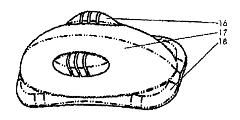
FIG. 3. The invented device in an top-side view of its outer surface (17). The
system comprises a reservoir-like body that is built in a polymer structure,
preferentially but not limited to injection, compression, transfer and
extrusion
CA 02456025 2007-08-22
molding, depending on the polymer, co-polymer or matrix to be used. The choice
of the polymer is driven basically by the characteristics of the organ or
tissue to be
implanted. It is preferentially made of but not limited to to poly-ethylene,
silicone,
hydrogels, poly-orthoester, poly-glycolic acid, poly-lactic acid, poly-
caprolactone,
5 polyvinyl-alcohol or any derivatives. The structure 16 is designed to
stabilize the
buckling suture. Sealing base 18 will maximize the hermetical seal to the
target
surface.
FIG. 4. The cross-sectional view of the embodiment comprised by the
reservoir 20 containing the therapeutic agent. The external surface 17
continues
10 with the sealing base 18, which will provide a hermetically sealed
attachment with
the target surface 24 through the use of an adhesive layer 22.
FIG. 5. The cross-sectional view of a bi-compartmental embodiment. The
reservoir is divided by the internal wall 28, which extends beyond the
curvature of
the target surface at 29, to provide a mild buckle or sealing effect with the
tissue,
15 and to avoid interaction between the agents before they reach the surface
30. The
suture stabilizers 16 are also disclosed.
FIG. 6. The bottom view of the device showing bi-compartmental reservoir
32, the dividing internal wall 28, the sealing base 18 with an adhesive layer
22
applied to it.
20 FIG. 7. The bottom-side view of a single reservoir embodiment 20 and its
relation to a sealing base 18, adhesive layer 22, external surface 17 and
suture
stabilizer 16. Preferentially, the internal curvature of the sealing base 18
follows
the curvature of the target tissue.
FIG. 8. Example of application of the device to an eye. In this case to
25 illustrate in an eye containing an intraocular tumor called retinoblastoma,
most
frequent primary intraocular tumor during the childhood. The sclera surface 41
is
exposed by peritomy and dissection of the adjacent tissue is performed. To
better
control the area of the sclera to be exposed the extraocular muscles 40 can be
isolated by standard techniques. Once the sclera surface is clean of
periocular
tissue in that area, the implant, in this case carrying cytotoxic drugs, is
held in
place by the use of an applicator or just the hands. Sutures 45 are placed
from one
CA 02456025 2007-08-22
26
side of the implant to the other, having it crossing and fitting the
stabilizers 16 to
allow a mild buckle effect of the sealing base 18 and maximize its sealing
effect.
The sutures are passed through the scleral thickness. After the implantation
the
muscles are released and the conjunctiva is brought back to its original
position
covering the eye surface and sutured near the cornea.
FIG. 9. A top-side view of the implant 49 sutured to a tissue 46. The suture
stabilizers 16 allow the sutures to hold the implant in place, but mainly
creating a
hermetical seal between the device and target surface.
FIG. 10. A bottom-side view illustrating the internal surface of the device
that will be in contact to the sclera surface. The interface window 55 is
surrounded
by a sealing base 18. The sealing base 18 is coated in its most peripheral
aspect by
an adhesive layer 22.
FIG. 11. A cross-sectional view of the device applied to the tissue surface,
showing the interface 60 between the reservoir and the tissue. The relations
between the sealing base 18 and the target tissue are also disclosed. In this
case the
hermetical seal was achieved by using an adhesive layer 22 covering the base
18.
FIG. 12. A cross-sectional view of the device and target tissue showing the
interface between the sealing base 18 and the sclera 1 with a hermetical seal
provided by the adhesive layer 22.
FIG. 13. A cross-sectional view of a single compartment 20 device in
apposition to the sclera 1. The method for refilling the reservoir is
disclosed. The
external surface 17 of the device comprises a refilling port 68, made
preferentially
of self-sealing rubber. The drug, solution or suspension is injected through a
cannula or needle device 69. A irrigation-aspiration device may also be used
for
aspirating any remaining solution and refilling the reservoir with the new
solution
or drug. The refilling port 68 is built in an angle to favor its localization
and
insertion of the needle 69.
FIG. 14. A bottom-side view of the internal surface of the device, showing
the relations between the sealing base 18, the adhesive layer 22, external
surface
17, suture stabilizer 16, and a refilling port 68 communicating the external
environment with the interior of the reservoir 20. The refilling port is
preferentially
CA 02456025 2007-08-22
27
made during the molding process for the external surface 17, and lately by a
second process to incorporate the self-sealing rubber, preferentially but not
limited
to silicone, to the port cavity or hole. The location of the port 68 as well
as its
angle to access the reservoir 20 follow the most appropriate way to insert the
refilling needle or cannula.
FIG. 15. The top-side view of the device illustrating the relation between the
refilling port 68 and the outer surface 17. To improve the self-sealing
performance,
depending on the thickness of the external wall 17, it may be built angled in
a way
to increase the length of the tunnel and maximize the sealing properties of
the port
68.
FIG. 16. Illustrates the relations between the device 49 and a tissue surface
46. Note that the sealing base 18 is perforated by multiple holes 83 to allow
a
sewing suture with a sealing effect on the base 18. A variation is to build a
thinned
tunnel along the surface of the sealing base and surrounding it and would
suitable
to continuous suturing by either a manual or automatic technique applied
during
the implantation surgical procedure.
FIG. 17. Illustrates an example of application of the device 49 to the sclera
surface of a human eye, where a sewing suture 88 was used to accomplish a
sealing effect of the base 18.
FIG. 18. Discloses an embodiment where a trail or tunnel 94 was built in the
device external surface 17, crossing its diameter to provide a method for
sealing
the base to a target tissue. The trail 94 is aimed to be fitted by an
encircling
element, preferentially made, but not limited to silicone, although variations
by
using any king of explants are expected.
FIG. 19. Illustrates the application of the device 49 positioned in contact to
the sclera 1 and underneath an encircling element 93. The encircling element
93 is
placed by established techniques, associated or not to a procedure for retinal
detachment treatment. Once the encircling element is tied up, the base 18 is
expected to function as a hermetically sealed interface.
FIG. 20. Illustrates the application of the device 49, in this case bi-
compartmental under a encircling element 93. A hermetical apposition 99 effect
of
CA 02456025 2007-08-22
28
the device against the sclera is achieved by this method. Furthermore, other
device
can be positioned anywhere underneath the encircling element, depending on the
number and dose of the agents necessary for treating that condition.
FIG. 21. Discloses a variation of the internal surface where a layer of
biodegradable polymer 100 is applied to contain the agent inside the
reservoir. The
membrane is preferentially applied by apposition between the sealing base and
the
adhesive layer 22. It is accomplished by creating a series of fenestrations
along the
most peripheral aspect of the biodegradable layer 100. Variations are expected
and
discussed hereinbelow.
FIG. 22. The bottom-side view of the device illustrating the relations
between the body 103, suture stabilizer 16, sealing base 18, biodegradable
layer
100, and the adhesive layer 22.
FIG. 23. Illustrates the cross-sectional view of the relations between the
device and the ocular tissue. Here the device contains a single compartment 20
where the solution with the drug is located and held by a coating
biodegradable
layer 100 to prevent if from leaking or premature exposure before the
hermetical
seal is accomplished. The layer 100 is expected to play a structural function.
Once
it is dissolved the drug or agent will be exposed to the sclera and penetrate
the eye
layers.
DETAILED DESCRIPTION OF THE INVENTION
In an embodiment, the present invention involves a new method of
selectively deliver therapeutic agents to mammalian organs, tissues or systems
through a surgically implantable and hermetically sealable device system that
provides a sustained and protected release of an agent, assuring an
unidirectional
diffusion through the target interface, and avoiding dissipation of the agent
to
adjacent structures.
The invention was based on unexpected findings that agents can be safely
and predictably delivered at therapeutic or prophylactic levels to specific
tissues,
even in a local context, through the control of the organ surface exposed to
an
agent as well as by control of the agent's exposure to the internal body
tissues and
CA 02456025 2007-08-22
29
fluids. This can be achieved by maintaining the organ interface permeable to
the
agent through osmotic agents, physical, chemical or biological treatment, and
by
isolating and localizing the interface area of exchange through a sealing
mechanism.
The control of the agent exposed to the targeted tissue can be obtained by
using drug-associated polymers, osmotic agents and by preferably by coating
the
drug reservoir with a non-drug-permeable polymer, wherein the drug to which
the
polymer is not permeable is the active agent(s)), avoiding dissipation and
toxic
effects of the agents to adjacent structures and fluids, and higher
availability to the
targeted structure. This is proportioned by a series of structures designed to
maintain a hermetically sealed contact between the device and the target
tissue.
The inventors found that this system can offer a tremendous advantage over
the conventional way the drugs are delivered to tissues or organs, allowing
even
agents never considered for clinical use due to non-specificity and toxicity
to be
reconsidered for use. This enables new agents to be developed based on this
alternative of drug delivery technology.
This invention allows new therapeutic modalities, such as organ
transplantation, tissue regeneration techniques, artificial organs or tissue
implantation, to be developed. This will provide a therapeutic and physiologic
support to any new technology that will depend on biological local
incorporation
or maintaining in an internal body portion.
Drug within the reservoir can be associated or mixed with another agent, a
polymer or an osmotic agent. Layers of drug can be provided, wherein a first
drug
is delivered, followed by a second drug. A multi-compartmental reservoir is
also
designed having an inner wall separating the cavities. The body BI comprises
the
construction of a wall dividing the cavities. Preferentially the dividing wall
slightly
extends beyond the corresponding height to the curvature of the sclera or the
surface to isolate the compartments at the interface level. It minimizes the
possibility of mixture and interaction between the agents before they reach
the
target surface.
CA 02456025 2007-08-22
The interface window may have be coated and/or contain an enhancer of
tissue diffusion, such as an enzyme. Collagenases, prostaglandin analogues,
matrix
metalloproteinases, hyluronidases are enzymes that can modify the diffusion
properties of the sclera or tissue surface. The coating process is
preferentially done
5 when compressing the solid drug or during the drug preparation and mixture
with
its polymer or vehicle. If it requires a steady and sustained effect it can be
dispersed throughout the reservoir or restricted to the internal surface to be
place in
contact with the sclera. Depending on the stability and interaction between
the
enhancer and the active therapeutic agent a layer of the enhancer may be
10 incorporated to the internal surface of the reservoir. Preferentially it is
made using
a biodegradable material such as a collagen biomaterial, gelatin, glycolic
acid,
cellulose and lactic acid. Alternatively it can be made of any material that
does not
interfere directly in the release rate of the agent from the reservoir and its
exposure
to the target tissue. In other words, it is not the material carrying the
enhancer
15 expected to play a direct role in the diffusion rate, but the action of
enhancer on the
target surface. We refer to this layer as a functional layer for containing an
agent or
enhancer that will affect the diffusion rate and lately the pharmacokinetics
of the
given therapeutic agent.
An internal layer of a rapidly biodegradable polymer, preferentially a
gelatin,
20 hialuronic acid, methyl-cellulose, poly-glycolic, poly-lactic is envisioned
to be
built for allowing liquid, powder and viscous agents to be held in the
reservoir it
gets stable on the target surface. This process is preferentially accomplished
by
interpositioning the layer between the sealing base and the adhesive layer, in
its
more inner aspect, still allowing a strong adhesion between the adhesive layer
and
25 the sealing base in its most peripheral aspect. A tunnel surrounding the
interface
window is also envisioned to allow the entrapment of the layer in the tunnel
using
silicone or any material of the same class of the sealing base or the device
to build
a ring to be fitted in the tunnel by mechanical apposition or adhesive
attachment.
The interface window, where the reservoir is exposed to the target tissue is
30 surrounded by a sealing base that may be a continuation of the polymer
composing
the external wall or may constitute a different polymer incorporated to
previous
CA 02456025 2007-08-22
31
one by mechanical attachment or use of adhesives. The internal surface of the
sealing base may also be composed by a different part, said sealing part, that
once
is mechanically incorporated to the main part, said the core device, can
entrap a
layer of polymer necessary to hold a liquid or viscous suspension in the
reservoir
avoiding premature exposure or leakage, or to stabilize the above described
layer
of enhancer carrier. The process is envisioned as a sandwich-like apposition
still
respecting the window area that will lately determine the interface between
the
drug reservoir and the target surface. The sealing part will then have the
incorporated characteristics described above for the sealing base in order to
allow
hermetical sealing between the device and the exposed tissue.
Materials useful in constructing the device include but are not limited to
poly-ethylene, silicone, hydrogels, poly-orthoester, poly-glycolic acid, poly-
lactic
acid, poly-caprolactone, polyvinyl-alcohol, polyvinyl-pylirridone, and any
derivatives thereof, and biopolymers, such as hyaluronic acid, fibrin, methyl-
cellulose, collagen, gelatin, or any derivatives might be used in other parts
of the
device.
Preferably, the device allows and protects the preferential flow of the
therapeutic agent across the targeted interface. This is accomplished by using
design structures to allow a hermetical sealing of the device to the target
surface.
Such unidirectional flow will be made possible by means of an external surface
impermeable to the drug. Whether the external will be permeable or not to the
external body fluids, will depend on the characteristics of the drug(s) and
carrier
polymer, as well as to the need for a dissolving agent to regulate the release
of the
drug from the reservoir. Such a mechanism of drug release may be as simple as
the
dissolution of the pure drug/polymer contained in the reservoir by the
incoming
fluids, or using an osmotic agent to regulate the water inflow and dissolution
rate
of the drug, before it permeates the targeted surface.
As mentioned before the embodiment incorporates a series of structures to
allow a hermetical sealing to the target surface. The first is the sealing
base, which
consists on the primary way of achieving sealing. The extended surface beyond
the
interface window is aimed to increase the sealing contact area, whether or not
it is
CA 02456025 2007-08-22
32
coated with an adhesive layer. The sealing base preferentially follows the
same
curvature of the target surface, although a slightly more curved base is
envisioned
to maximize the contact, particularly when flexible materials are used. The
combination of one or more or more of the other characteristics to ameliorate
the
sealing affect, said accessory sealing structures, will provide the
characteristics for
accomplishing the controlled and protected drug delivery.
The first accessory is described as a buckle suture stabilizer or suture
stabilizer. This is a built bump, lane or tunnel on the external surface to
prevent the
suture to slide out of the implant once it buckles the device in apposition to
the
tissue. One or more can be built depending on the size and position of the
device in
relation to the target surface. Preferentially those suture stabilizers are
made of the
same material for the outer surface during the molding process. Alternatively,
it
can be incorporated to the device later on the process using different
materials.
The second accessory is described as a buckling band tunnel or trail. It
consists of a depression on the outer surface, crossing its diameter, to allow
an
encircling element to be place and provide a sealing apposition between the
device
and the target tissue. Preferentially it is built on the device external
surface during
the molding process.
The third accessory is described as a multiple holes base where a sewing
suture should be applied to seal the base of the device. The holes again are
preferentially created during the molding process for the sealing base.
Alternatively a flexible material can be used as sealing base with a linear
surrounding thinning to allow the suture to be performed by an automatic
apparatus.
The above mentioned methods for creating a hermetical seal between the
drug delivery device are essentials in diminishing the interference of
surrounding
fluid and tissues in the diffusion mechanism provided by the drug-tissue
interface.
Moreover, they play a significant role in avoiding unnecessary exposure of
surrounding tissues to toxic effects of the pharmaceutical agents.
Fluid transport before drug dissolution occurs is possible through two
distinct mechanisms. The first is across the organ surface through an osmotic
or
CA 02456025 2007-08-22
33
pressure gradient driven diffusion. The second is across the outer wall
polymer
mainly driven by an osmotic gradient between the reservoir and the outside
tissue
as well as the characteristics of the polymer.
Among the factors related to the permeation of agents through biological
membranes, the surface contact area, concentration of the agent in the donor
side
and the molecular weight of the drug are balanced to provide the tissue with
the
desired levels of the agent in the specific regions. Other factors taken in
account
are the membrane properties and pharmacokinetics of the drug in the tissue.
Those,
despite being biological, can be altered through physical, chemical or
biological
methods, before the exposure to the therapeutic agent and device. In other
words,
the bioavailability and pharmacokinetics of the permeating agents are expected
to
be different through this proposed route, and will be helpful in establishing
the
appropriate combination of the compounds.
It is envisioned that the system of the present invention has numerous
variations. For example, the device can carry an enhancer agent,
preferentially, but
not limited to an enzyme and a protein, such as albumin. The external surface
can
be composed by a polymer non-permeable to the carried agent, preferentially
formed but not restricted to a silicone, poly-glycolic acid, poly-lactic acid,
hyaluronate derivatives, polyvinyl alcohol, acrylate, methacrylate, cellulose,
collagen, metals, any derivatives or associations of the above mentioned
polymers
or others that retain characteristics of non-permeability to the carried
agent.
The external surface of the device may include a refilling port preferentially
made of, but not restricted to a self-sealing material, such as silicone
rubber. It is
envisioned that in using a multiple compartmental device, multiple refilling
ports
are also built in the device. These structures are built on the external
surface
communicating the exterior environment to the interior of the reservoir. To be
recognized after the surgical procedure the port is stained by a
biocompatible,
radiosensitive, echogenic marker or dye. Alternatively, its is also extended
beyond
the outer surface of the device and place in a more accessible part of the
body.
The device may be foldable or flexible to allow insertion through small
incisions, and to conform and tightly fit to irregular organs surfaces.
CA 02456025 2007-08-22
34
The invention includes methods for selective administration to a mammalian
organ, tissue or system desired levels of a therapeutic agent through a
controlled
drug permeation across a target device interface. The interface with the
tissue can
be directly with drug contained within the device reservoir or through a
biodegradable polymer, preferentially composed of but not restricted to
gelatin,
caprolactone, hyaluronic acid, cellulose, poly-glycolic acid, poly-lactic
acid, and
derivatives thereof. These compounds and/or compositions may be pressure,
heat,
photo, or chemically sensitive.
The active agents may be in an encapsulated form, such as liposomes or
microspheres.
Thus, the present invention includes a method of local, protected and
sustained delivery of therapeutic agents directly through a targeted tissue
surface in
a unidirectional way, avoiding dissipation of the agent to surrounding tissues
and
fluid, after surgical implantation into a mammalian organism. The method
involves
placing the drug-loaded device interface window in contact with the targeted
tissue. The method includes sealing the device to the target tissue by way of
adhesives, buckling or suturing or the combination of any of those. To build
the
adhesive layer it preferentially uses but is not limited to a hydrogel,
hyaluronate
and fibrin adhesive. It is incorporated to the sealing base by the use of a
film or
layer containing adhesive in its both sides, or by the pre-application of the
adhesive
to the internal side of the sealing base. For holding the adhesive in place
multiple
cavities, single cavity or a channel system along the internal surface of the
sealing
base are preferentially used. Such sealing structures are made preferentially
during
the molding process of the device. After placement of the adhesive in contact
to the
base, a film may be placed in contact to the adhesive layer. Preferentially
the film
is non-reactive with the adhesive used and can peeled off before the
implantation
procedure. The use of an exposed sealing base, presenting the structures
mentioned
above to hold the adhesive in place allows also its application right before
the
implantation procedure, particularly if a biological adhesive such as fibrin
sealant
is desired to be used.
CA 02456025 2007-08-22
The device may be interfaced with an artificial organ, a synthetic or
biological platform for cells or biological agents, a scaffold for tissue or
cell
regeneration, and/or a transplanted tissue or organ.
The method of the present invention can achieve local or systemic,
5 physiological or pharmacological effects in a mammalian organism, by using a
surgically implantable device that delivers an agent directly and
preferentially
through its interface with the targeted tissue or organ, keeping the rest of
its surface
non-permeable to the carried agent.
The therapeutic agent may be a prophylactic agent. The system or device
10 may carry an osmotic agent.
The effect or diffusion of the agent may be started or enhanced after the
implantation procedure through the use of a secondary agent, whether it is
chemical, physical or biological.
Some non-limiting examples of diseases for which the present inventions
15 may be used include, myocardial ischemic disease, hepatic cancers, hepatic
metastasis of colon cancers, gall bladder tumors, adrenal tumors,
neuroblastomas,
and kidney and pancreatic cancers. The device of the present invention can be
loaded with the desired active agent (i.e., drug(s) and/or prodrug(s)) and can
be
implanted and attached to an anatomical or histological surface. For example,
the
20 device can be glued to the pericardium surface to deliver an agent to the
pericardial
space, allowing the drug in the reservoir to diffuse to the whole myocardium.
It can
also, through an opening of the pericardium, be glued directly to the
myocardium
(note that the pericardium is a sac, mostly acellular, but is delineated from
other
structures by a histopathological and anatomical surface, and the myocardium
25 which is mostly cellular, is also delineated by the pericardium by surface,
which is
ultimately the muscle cell, but there is still a distinguished surface). It is
preferred
that the device not be implanted inside the myocardium to deliver drug to a
deeper
layer of the muscle or a specific group of cells, as it is preferred that such
invasive
techniques be minimized. Hence, it is preferred that the devices, when
implanted,
30 not degrade the histological structure of the tissue that will be treated
(the target).
CA 02456025 2007-08-22
36
In an embodiment, the present invention has numerous applications in
ophthalmology, with the eye providing several locations where loaded devices
may
be applied. Preferentially, in Ophthalmology, the device is used to be in
placed in
contact to the sclera. Alternatively, between the outer layer of the eye,
known as
the sclera, and the vitreous there is suprachoroidal space (accessible through
a
scleral incision) or even the subretinal space. For the subretinal space,
either a
choroidal incision or a retinotomy could be made to allow the insertion of the
implant. Diseases in ophthalmology that may be treated with the present
inventions
and other ophthalmic applications of the present invention include but are not
limited to intraocular tumors, e.g. retinoblastoma, melanoma, macular
degeneration, delivery to the posterior pole (e.g., choroidal and RPE layers)
of
growth factors, antiangiogenic factors, photosensitizers (which may be subject
to
application of laser), gene vectors, etc. The present invention may be applied
to
glaucoma by delivering antiglaucoma drug(s) via the device to the cilliary
body
directly through the sclera. The present invention may also be applied to
retinitis
pigmentosa, to deliver growth factors or to deliver immunosupressive agents to
protect a retina or RPE graft, without an intraocular surgical procedure that
would
jeopardize the graft.
The present invention is designed for implantation, rather than for external
body surface or buccal applications. The present invention makes possible the
targeting of specific tissues within the body or eye, and takes into account
that
many drugs are more specific and toxic to certain groups of cells than others.
In
situations where the surrounding target tissue can be harmed by the applied
drug,
the present invention provides a superior solution by focusing the drug on the
target tissue.
In addition to treating localized diseases, the present invention can be used
to
provide systemic benefits. Using a system of the present invention to deliver
growth factors to the pancreas of a diabetic patient can change the context of
the
systemic disease. Application of appropriate agents using the system of the
present
invention to an inoperable liver affected by a colonic metastatic carcinoma
can
reduce the size of the tumor and make it ressectable. In addition to cures,
the
CA 02456025 2007-08-22
37
present invention can also be used for treatments aimed to improve the quality
of
life of patients, or improve their cost-effectiveness. The local delivery of
cytotoxic
agents by the device to a tumor expanding and compressing the esophagus can
make a difference in the patient's quality of life preventing more complex
interventions, such as a surgical ressection. Thus, even palliative care is
facilitated
by the present invention. The invention is particularly useful in tumor
treatments
when the tumor or effected organ has a distinguishable surface to which can be
sealed the interface window incorporated in a drug delivery device of the
present
invention.
The method of delivering the loaded drug delivery devices of the present
invention may involve a variety of implantation techniques either manually or
through an injector. The devices may be implanted under direct visualization
or
under indirect visualization techniques, such as ultra-sound, MRI, CT-scan
guided,
laparoscopy, etc.
While exemplary embodiments of the present invention have been set forth
above, it is to be understood that the pioneer inventions disclosed herein may
be
constructed or used otherwise than as specifically described.