Note: Descriptions are shown in the official language in which they were submitted.
CA 02456055 2004-01-29
WO 03/014380 PCT/IB02/04767
1
Microorganisms and cells for diagnosis and therapy of tumors
The present invention relates to diagnostic and pharmaceutical
compositions comprising a microorganism or cell containing a
DNA sequence encoding a detectable protein or a protein
capable of inducing a detectable signal, e.g. a luminescent or
fluorescent protein, and, in a particular embodiment,
furthermore (a) DNA sequence(s) encoding (a) protein(s)
suitable for tumor therapy and/or elimination of metastatic
tumors, e.g. a cytotoxic or cytostatic protein.
Presence of bacteria in tumors was reported approximately
fifty years ago. Several publications substantiated the
earlier clinical findings that unexpectedly large numbers of
bacteria were discovered in excised tumors from human
patients. Investigators argue that chronic infections may
predispose cells to malignant growth. Chronic infections of
various strains of Chlamydia have been associated with lung
and cervical cancer as well as malignant lymphoma. Another
well described association between the presence of a specific
bacterial species and cancer development is Helicobacter
pylori in patients with gastric ulcers. Elevated levels of H.
pylori-associated antibodies have been found in patients with
duodenal ulcer and gastric adenocarcinoma. These observations
demonstrate a concomitant presence of bacteria at tumor sites;
however, it was not yet clear whether the microorganisms were
the cause of tumor formation or whether the tumorous tissues
were more susceptible to bacterial colonization. Intravenously
injected strict anaerobic bacteria, Clostridium pasteurianum,
into mice replicated selectively in the tumor suggesting a
hypoxic microenvironment in the necrotic center. Intravenous
injection of attenuated Salmonella typhimurium mutants
resulted in elevated bacterial titers in the tumor tissues in
comparison to the other organs of mice upon histologic and
bacteriologic analyses.
CA 02456055 2004-01-29
WO 03/014380 PCT/IB02/04767
2
Similarly, the presence of virus particles was reported in
excised human breast tumors as early as 1965. More recently,
based on polymerase chain reaction (PCR) data, the human
papillomavirus has been claimed to be associated with
anogenital tumors and esophageal cancers, breast cancers, and
most commonly, cervical cancers. In addition, the presence of
hepatitis C virus in human hepatocellular carcinoma,
Epstein-Barr virus in squamous cell carcinoma in Kirnura's
disease, mouse mammary tumor virus-like paiticles (MMTV) in
human breast cancer, SV40 virus in macaque astrocytoma, and
herpesvirus in turtle fibropapilloma has been reported.
Surprisingly, the concentration of virus particles in the
tumors shows variations among patients. The presence of human
papillomavirus in squamous cell carcinomas of the esophagus
ranges from 0 to 72% (10- 15) . In contrast to tumor tissues,
no virus particles have been found in tumor-free areas of the
esophageal epithelium of the same patient suggesting that the
virus particles are located only in the tumor tissues.
However, so far it could not be shown definitely whether the
above discussed microorganisms are responsible for the
development of disorders like tumors (except for
papillomaviruses) or whether, e.g., tumors can attract and/or
protect viruses or bacteria. Accordingly, there was no basis
for the use of such microorganisms for the diagnosis or
therapy of tumors. Conventional tumor diagnostic methods, such
as MRI (Magnetic Resonance Imaging) lack sensitivity and
specificity and therapeutic methods, e.g. surgery, are
invasive and not very sensitive.
Therefore, it is the object of the present invention to
provide a means for the efficient and reliable diagnosis as
well as the therapy of tumors which overcomes the
disadvantages of the diagnostic and therapeutic approaches
presently used.
CA 02456055 2004-01-29
WO 03/014380 PCT/IB02/04767
3
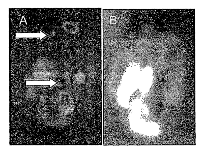
According to the present invention this is achieved by the
subject matters defined in the claims. When Vaccinia virus
(LIVP strain) carrying the light emitting fusion gene
construct rVV-ruc-gfp was injected intravenously into nude
mice, the virus particles were found to be cleared from all
internal organs within 4 days, as determined by extinction of
light emission. In contrast, when the fate of the injected
Vaccinia virus was similarly followed in nude mice bearing
tumors grown from subcutaneously implanted C6 rat glioma
cells, virus particles were found to be retained over time in
the tumor tissues, resulting in lasting light emission. The
presence and amplification of the virus-encoded fusion
proteins in the same tumor were monitored in live animals by
observing GFP fluorescence under a stereomicroscope and by
collecting luciferase-catalyzed light emission under a
low-light video-imaging camera. Tumor-specific light emission
was detected 4 days after viral injection in nude mice
carrying subcutaneous C6 glioma implants ranging in size from
25 to 2500 mm3. The signal became more intense after the 4th
postinjection day and lasted for 30 to 45 days, indicating
continued viral replication. Tumor accumulation of rVV-ruc-gfp
virus particles was also seen in nude mice carrying
subcutaneous tumors developed from implanted PC-3 human
prostate cells, and in mice with orthotopically implanted
MCF-7 human breast tumors. Further, intracranial C6 rat glioma
cell implants in immunocompetent rats and MB-49 mouse bladder
tumor cell implants in C57 mice were also targeted by the
Vaccinia virus. Cross sections of a C6 glioma revealed that
light emission was clustered in õpatchesõ at the periphery of
the tumor where the fast-dividing cells reside. In contrast,
cross sections of breast tumors revealed that fluorescent
,,islands,, were distributed throughout the tumors. In addition
to primary breast tumors, small metastatic tumors were also
detected externally in the contralateral breast region, as
CA 02456055 2004-01-29
WO 03/014380 PCT/IB02/04767
4
well as in nodules on the exposed lung surface, suggesting
metastasis to the contralateral breast and lung. In summary,
light-emitting cells or microorganims, e.g. Vaccinia virus can
be used to detect and treat primary and metastatic tumors.
Similar results were obtained with light-emitting bacteria
(Salmonella, Vibrio, Listeria, E. coli) which were injected
intravenously into mice and which could be visualized in whole
animals under a low light imager immediately. No light
emission was detected thirty-six hours after bacterial
injection in both athymic (nu/nu) mice and immunocompetent C57
mice as a result of clearing by the immune system. In the
cutaneous wound of an intravenously injected animal, the
bacterial light emission increases and remains detectable up
to six days post-injection. In nude mice baring tumors
developed from implanted C6 glioma cells, light emission was
abolished from the animal entirely thirty-six hours after
delivery of bacteria, similar to mice without tumors. However,
forty-eight hours post-injection, unexpectedly, a strong,
rapidly increasing light emission originating only from the
tumor regions was observed. This observation indicates a
continuous bacterial replication in the tumor tissue. The
extent of light emission is dependent on the bacterial strain
used. The homing-in process together with the sustained light
emission was also demonstrated in nude mice carrying prostate,
bladder, and breast tumors. In addition to primary tumors,
metastatic tumors could also be visualized as exemplified in
the breast tumor model. Tumor-specific light emission was also
observed in immunocompetent C57 mice with bladder tumors as
well as in Lewis rats with brain glioma implants. Once in the
tumor, the light-emitting bacteria were not observed to be
released into the circulation and to re-colonize subsequently
implanted tumors in the same animal. Further, mammalian cells
expressing the Ruc-gfp fusion protein, upon injection into the
bloodstream, were also found to home into and propagate in
glioma tumors.
CA 02456055 2004-01-29
WO 03/014380 PCT/IB02/04767
These findings open the way for (a) designing multifunctional
viral vectors useful for the detection of tumors based on
signals like light emission and/or for suppression of tumor
development and/or angiogenesis signaled by, e.g., light
extinction and (b) the development of bacterium- and mammalian
cell-based tumor targeting systems in combination with
therapeutic gene constructs for the treatment of cancer. These
systems have the following advantages: (a) They target the
tumor specifically without affecting normal tissue; (b) the
expression and secretion of the therapeutic gene constructs
are, preferably, under the control of an inducible promoter,
enabling secretion to be switched on or off; and (c) the
location of the delivery system inside the tumor can be
verified by direct visualization before activating gene
expression and protein delivery.
Accordingly, the present invention relates to a diagnostic
and/or pharmaceutical composition comprising a microorganism
or cell containing a DNA sequence encoding a detectable
protein or a protein capable of inducing a detectable signal.
In a preferred embodiment, the microorganism or cell of said
diagnostic and/or pharmaceutical composition furthermore
contains (a) DNA sequence(s) encoding (a) protein(s) suitable
for tumor therapy and/or elimination of metastatic tumors,
such as a cytotoxic protein, a cytostatic protein, a protein
inhibiting angiogenesis or a protein stimulating apoptosis.
Such proteins are well-known to the person skilled in the art
and further examples of suitable proteins are given below.
Any microorganism or cell is useful for the compositions of
the present invention, provided that they replicate in the
organism, are not pathogenic for the organism, e.g.
attenuated, and are recognized by the immune system of the
organism, etc. Examples of microorganisms useful for the
CA 02456055 2004-01-29
WO 03/014380 PCT/IB02/04767
6
present invention are bacteria and viruses. The term
"bacteria" as used herein refers to bacteria which are per se
not tumor-targeted (i.e. they can not differentiate between a
cancerous cell or tissue and the non-cancerous counterpart
cell or tissue) since the results of the experiments leading
to the present invention show that bacteria etc. accumulate in
the tumor due to the fact that in this environment they are
not exposed to attack by the immune system of the host. A list
of candidate bacteria which might be useful for the purposes
of the present invention and which might not be tumor-targeted
are given in Table 1, below. The person skilled in the art can
easily identify such bacteria which are not tumor-targeted by
commonly available methods, e.g. the methods described in
section 6.1 of WO 96/40238. Preferably, said bacteria are
intercellular bacteria such as E. coli, E. faecalis, Vibrio
cholerae, Vibrio fischeri, Vibrio harveyi, Lactobacillus spp.,
Pseudomonas spp. In the method of the present invention,
viruses and cells, particularly mammalian cells, are preferred
which are not tumor-targeted. Particularly preferred are
cytoplasmic viruses.
In a particularly preferred embodiment, the diagnostic and/or
pharmaceutical composition comprises a microorganism or cell
containing a DNA sequence encoding a luminescent and/or
fluorescent protein.
As used herein, the term õDNA sequence encoding a luminescent
and/or fluorescent protein,, also comprises a DNA sequence
encoding a luminescent and fluorescent protein as fusion
protein.
In an alternative preferred embodiment of the diagnostic
and/or pharmaceutical composition, the protein encoded by the
DNA sequence is a cell receptor capable of binding a ligand
which can be a diagnostic or therapeutic ligand. The ligand
CA 02456055 2004-01-29
WO 03/014380 PCT/IB02/04767
7
can be a protein (including large or small peptides,
antibodies, etc.), a synthetic compound (such as a synthetic
steroid analog), etc. Therefore, the in vivo location of
labeled ligands in live animals and human patients can be
visualized, e.g, in real time by SPECT or PET. Almost any
known protein ligand-receptor pair for tumor labeling is
useful in the method of the present invention. Preferably,
to increase specificity, mutant protein ligands (or analogs
if it is a chemical compound) or mutant ligand receptors can
be genetically or chemically engineered so that they will
not bind to any endogenous molecules. In addition to
increasing specificity, these mutants/analogs will also
limit adverse effect to the normal host physiology.
In a more preferred embodiment of the diagnostic and/or
pharmaceutical composition of the present invention, the
ligand is a radionuclide-labelled ligand. Said ligand is,
e.g., useful for tumor visualization by single-photon
emission computed tomography (SPECT) or positron-emission
tomography (PET) resulting from the binding of radionuclide-
labelled ligand to its receptors expressed specifically on
the surface of tumor cells following intravenous delivery
of, e.g., engineered bacteria, viruses, or mammalian cells
carrying the receptor protein gene constructs. Radionuclides
may be used for conventional tumor scintigraphy, PET and
possibly internal radiotherapy of tumors. Examples of
radionuclides useful in the present invention are (a) !3+-
emitters such as 11C, 13N, 150 or 64Cu or (b) y-emitters such
as 1231 Other radionuclides that can, e.g., be used as
tracers for PET include 55Co, 67Ga, 68Ga, 60Cu (I I) , 67Cu (I I) ,
57Ni, Co, 5552Fe, 18F, etc.
SPECT and PET are sensitive techniques that may be used for
tumor imaging according to the present invention. Both SPECT
and PET are capable of detecting trace amounts of 8+ and y
CA 02456055 2004-01-29
WO 03/014380 PCT/IB02/04767
8
emission from radionuclides. PET is even more sensitive than
SPECT. In experiments using small laboratory animals, tumor
imaging is performed using a microPET instrument, which is
commercially available through, e.g., Concorde Microsystems
(Knoxville, TN).
Examples of useful radionuclide-labeled agents are 64Cu-labeled
engineered antibody fragment (Wu et al., PNAS USA 97 (2002),
8495-8500), 64Cu-labeled somatostatin (Lewis et al.,
J.Med.Chem. 42 (1999), 1341-1347, 64Cu-pyruvaldehyde- bis(N4-
methylthiosemicarbazone) (64Cu-PTSM) (Adonai et al., PNAS USA
99 (2002), 3030-3035, 52Fe-citrate (Leenders et al., J.Neural
Transm. Suppl. 43 (1994), 123-132, 52Fe/52mMn-citrate
(Calonder et al., J.Neurochem. 73 (1999), 2047-2055) and
52Fe-labeled iron(III) hydroxide-sucrose complex (Beshara et
al., Br.J.Haematol. 104 (1999), 288-295, 296-302).
In order to apply the radionuclide-labeled ligand in tumor
detection, the genes encoding the receptor proteins that the
ligands may bind are delivered by intravenously injected
bacteria, viruses, or mammalian cells according to the
present invention. Since it could be shown in the examples,
below, that certain intravenously injected bacteria, viruses
and mammalian cells replicate specifically in the tumors,
expression of the receptor proteins in the tumors will mark
the tumors for targeting by the radionuclide-labeled ligands.
For example, in the case of Vaccinia virus, to allow efficient
tumor detection, the virus can be used to carry gene
constructs encoding receptor proteins that can bind
specifically to the ligands. Intravenously injection of
recombinant Vaccinia virus allows the delivery of the receptor
gene and surface expression of the receptor protein in the
tumor tissues. Then, the radionuclide-labeled ligands are
injected intravenously into the host. The specific binding
between radionuclide-labelled ligands and their receptors
CA 02456055 2004-01-29
WO 03/014380 PCT/IB02/04767
9
expressed on the tumor cell surface allows the detection of
tumors based on f+- or y-emissions originating only from the
tumors. Labelling the tumors with radionuclides will allow
easy distinction between tumors and normal tissues.
Therefore, handhold I3+- or y-detectors that are attached to a
surgical blade holder can be designed. Based on the signal
of !3+- or y-emission, the labelled tumors can be cleanly
excised while the removal of normal tissues is kept at a
minimum.
Particularly preferred is gallium-67 for the purposes of the
present invention. Gallium-67 (67Ga) can be used for
diagnostic imaging using PET, SPECT, or scintigraphy. It is
known for its ability to accumulate in inflammatory lesions
and tumors, especially in lymphomas, but also in many other
types of tumors, such as in pancreatic tumors, lung tumors
etc. The mechanism of 67Ga uptake has been proposed to be
through both transferrin-dependent route and transferrin-
independent route. In the transferrin-dependent route, it
has been shown that over-expression of transferrin receptor
significantly increases 67Ga uptake by tumor cells.
Furthermore, anti-transferrin receptor antibody
significantly blocks 67Ga by tumor cells. For small tumor
imaging, very high concentrations of 67Ga are required to
overcome background signal. Thus, in this case, the use of
recombinant Vaccinia virus carrying a transferrin receptor
gene construct for over-expressing transferrin receptors
specifically on the surface of tumor cells in live animals
or human patients following intravenous injection of the
viruses is preferred. 67Ga will be also delivered
intravenously. High level accumulation of 67Ga in tumor cells
with the help from the over-expressed transferrin receptors
helps to significantly improve tumor detection abilities in
live animals and human patients.
CA 02456055 2004-01-29
WO 03/014380 PCT/IB02/04767
In an alternative preferred embodiment, the diagnostic and/or
pharmaceutical composition of the present invention comprises
a microorganism or cell containing a DNA sequence encoding a
protein capable of inducing a signal detectable by magnetic
resonance imaging (MRI), e.g. metal binding proteins.
Furthermore, the protein can bind contrast agents,
chromophores, ligands or compounds required for visualization
of tissues. Preferably, said protein is a cell receptor
capable of binding a paramagnetic- or superparamagnetic metal-
labelled ligand. Tumor visualization by MRI, resulting from
the binding of paramagnetic- or superparamagnetic-metal-
labelled ligands to their receptors expressed specifically
on the surface of tumor cells following, e.g., intravenous
delivery of engineered bacteria, viruses, or mammalian cells
according to the present invention carrying the receptor
protein gene constructs has several advantages. The high
level of accumulation of these metals in the tumors will
facilitate tumor detection. Virtually any paramagnetic or
superparamagnetic metals can be used for this purpose.
Preferably, due to the systemic toxicity of naked heavy
metals, the paramagnetic or superparamagnetic are carefully
selected to keep adverse effects at minimal. In most
currently available contrast agents, the metal particles are
either linked to, chelates or are coated with polymers, which
are much safer to use than naked particles. Therefore, the
use of ligands tagged with chelated or polymer-coated metals
is preferred.
Methods for generating contrast agents linked with proteins
are well known to the person skilled in the art, e.g., the
protein ligand (or other chemical compound ligand) is first
chemically attached to the chelates (e.g. diethylene
triamine pentaacetic acid (DTPA)). Then, the chelate-protein
complex is labeled with metals. A similar labeling method
has been used in generating gallium-labeled somatostatin
CA 02456055 2004-01-29
WO 03/014380 PCT/IB02/04767
11
analogs and used in producing indium-111-labeled low density
lipoprotein using DTPA-bis(stearylamide). Details of protein
modifications with chelates used in contrast agents have
been previously described. For example, modifying proteins
with the bifunctional chelate 2-(4-isothiocyanotobenzyl-6-
methyl) diethylenetriamine-pentaacetic acid (MX-DTPA))
(Mirzadeh et al., Bioconjug. Chem. 1 (1990), 59-65) and
modifying proteins with the cyclic anhydride of
diethylenetriamine-pentaacetic acid (cDTPAA) (Duncan and
Welch, J. Nucl. Med. 34 (1993), 1728-1738) have been
described.
Alternatively, MRI can be carried out by the following
approaches:
(a) Modified gadolinium activation due to enzyme/protein
delivery
This approach is based on the principles of Gene-Directed
Enzyme Prodrug Therapy (GDEPT), in which case the
enzymes/proteins activate a systemically delivered nontoxic
prodrug into active toxic drug. Specific activation of the
MRI contrast agent in weak relaxivity state into strong
relaxivity state by the enzyme/protein in the tumors allows
tumor detection. The enzyme/protein delivery to tumors is
achieved through intravenously injected engineered bacteria,
viruses, and mammalian cells, which carry the gene encoding
13-galactosidase (or any other related enzymes). The 13-
galactosidase can be in either extracellular secreted form
or expressed an the bacteria or mammalian cell surface. An
example of an MRI agent that can be cleaved by 13-galactosidase
and used in MRI imaging for tumor detection is (4,7,10-
tri(acetic acid)-1-(2-13-galactopyranosylethoxy)-1,4,7,10-
CA 02456055 2004-01-29
WO 03/014380 PCT/IB02/04767
12
tetraazacyclododecane)gadolinium (Egad) (Moats et al., 1997,).
In this compound, a galactopyranose residue is positioned at
the 9th coordination site of the Gd3+ ion. Due to this
blockage, water protons are excluded from interacting with Gd3+
ion, and therefore diminished effect on Tl relaxation time. In
the presence of 8-galactosidase, the enzyme cleaves the
galactopyranose from the chelate, freeing the coordination
site, and allows irreversible transition of the contrast agent
from a weak to a strong relaxivity state. Since the bacterium-
, virus-, or mammalian cell-based enzyme expression occurs
only in tumors, the enzyme-mediated relaxivity state
transition will also occur only in the tumors. Therefore, this
enzyme-mediated activation of MRI contrast agents can be used
for tumor detection.
In addition to the above MRI contrast agent, similar new
contrast agents may be developed in which other types of
residues may be attached to the chelates. These residues can
be removed by their corresponding enzymes (similar to the
removal of galactopyranose by galactosidase) (e.g. a-
mannosidases, a- and j3-glucosidases, (3-glucuronidases) to free
the 9th coordination site of the Gd3+ ion and modify the Ti
relaxation time for tumor detections. The genes encoding these
enzymes may all be delivered by the engineered bacteria,
viruses, or mammalian cells according to the present
invention. Moreover, new contrast agents using metals other
than gadolinium may be developed for enzyme-activated MRI
tumor imaging.
This approach can be combined with therapy as shown in the
following sections.
CA 02456055 2004-01-29
WO 03/014380 PCT/IB02/04767
13
(b) Engineering of delivery vector systems for activation of
the contrast agents (e.g. modified gadolinium) first and
subsequently, the activation of an injected prodrug by the
same constitutively expressed enzyme (e.g. 5-galactosidase) -
One-Gene-Product-Based Detection and Therapy System (OGPBDTS)
For example, the gadolinium-based contrast agent can be
activated by an enzyme (e.g. (3-galactosidase), as described
above, which can be used for tumor detection. Subsequently,
the intravenously delivered prodrugs, such as CBI, TMI, PCI
(described in US-patent 5,646,298), may be cleaved by the same
J3-galactosidase in the tumors, to yield active cytotoxic drugs
against tumor cells for cancer treatment.
(c) Engineering of delivery vector systems with the Two-
Gene-Product-Based Detection and Therapeutic Systems
(TGPBDTS)
Gene 1 is linked to a constitutive promoter and produces the
enzymes/proteins for sensing the contrast agents (e.g.
metal-binding proteins, modified gadolinium, or other
agents) and gene 2 is linked to an exogenously activatable
promoter, which is silent without the activator drug, and
only turned on after the detection vector system is found
positive by MRI. The activation of the vector-based promoter
gene construct is achieved by injection or by oral
administration of the activator drug. The repeated injection
of the contrast agent allows the real-time monitoring of
tumor size and location of metastases during the treatment
procedure.
CA 02456055 2004-01-29
WO 03/014380 PCT/IB02/04767
14
Preferably, for transfecting the cells the DNA sequences
encoding the (diagnostic or therapeutic) proteins described
above, e.g., a luminescent and/or fluorescent protein are
present in a vector or an expression vector. A person
skilled in the art is familiar with examples thereof. The
DNA sequences can also be contained in a recombinant virus
containing appropriate expression cassettes. Suitable
viruses that may be used in the diagnostic or pharmaceutical
composition of the present invention include baculovirus,
vaccinia, sindbis virus, Sendai virus, adenovirus, an AAV
virus or a parvovirus, such as MVM or H-1. The vector may
also be a retrovirus, such as MoMULV, MoMuLV, HaMuSV, MuMTV,
RSV or GaLV. For expression in mammals, a suitable promoter
is e.g. human cytomegalovirus õimmediate early promoter,,
(pCMV) . Furthermore, tissue and/or organ specific promoters
are useful. Preferably, the DNA sequences encoding, e.g., a
luminescent and/or fluorescent protein are operatively
linked with a promoter allowing high expression. Such
promoters, e.g. inducible promoters are well-known to the
person skilled in the art. Preferably, the above constructs
are inserted into the bacterial genome by stable
integration. If such constructs are made in mammalian cells
using mammalian vectors, stably transformed cell lines with
a single-copy insertion will be generated for long-term
expression in tumor cells.
For generating the above described DNA sequences and for
constructing expression vectors or viruses which contain said
DNA sequences, it is possible to use general methods known in
the art. These methods include e.g. in vitro recombination
techniques, synthetic methods and in vivo recombination
methods as described in Sambrook et al., Molecular Cloning, A
Laboratory Manual, 2"d edition (1989) Cold Spring Harbor
Laboratory Press, Cold Spring Harbor, NY, for example. Methods
of transfecting cells, of phenotypically selecting
CA 02456055 2004-01-29
WO 03/014380 PCT/IB02/04767
transfectants and of expressing the DNA sequences by using the
above described vectors are known in the art.
The person skilled in the art knows DNA sequences encoding
proteins, e.g., luminescent or fluorescent proteins that can
be used in the diagnostic and/or pharmaceutical composition of
the present invention. During the past decade, the
identification and isolation of structural genes encoding
light-emitting proteins from bacterial luciferase from Vibrio
harveyi (Belas et al., Science 218 (1982), 791-793) and from
Vibrio fischerii (Foran and Brown, Nucleic acids Res. 16
(1988), 177), firefly luciferase (de Wet et al., Mol. Cell.
Biol. 7 (1987), 725-737), aequorin from Aequorea victoria
(Prasher et al., Biochem. 26 (1987), 1326-1332), Renilla
luciferase from Renilla reniformis (Lorenz et al., PNAS USA 88
(1991), 4438-4442) and green fluorescent protein from Aequorea
victoria (Prasher et al., Gene 111 (1987), 229-233) have been
described that allow the tracing of bacteria, viruses or
mammalian cells based on light emission. Transformation and
expression of these genes in bacteria allows detection of
bacterial colonies with the aid of the low light imaging
camera or individual bacteria under the fluorescent microscope
(Engebrecht et al., Science 227 (1985), 1345-1347; Legocki et
al., PNAS 83 (1986), 9080-9084; Chalfie et al., Science 263
(1994), 802-805).
Luciferase genes have been expressed in a variety of
organisms. Promoter activation based on light emission, using
lux AB fused to the nitrogenase promoter, was demonstrated in
Rhizobia residing within the cytoplasm of cells of infected
root nodules by low light imaging (Legocki et al., PNAS 83
(1986), 9080-9084; O'Kane et al., J. Plant Mol. Biol. 10
(1988), 387-399). Fusion of the lux A and lux B genes resulted
CA 02456055 2004-01-29
WO 03/014380 PCT/IB02/04767
16
in a fully functional luciferase protein (Escher et al., PNAS
86 (1989), 6528-6532). This fusion gene (Fab2) was introduced
into Bacillus subtilis and Bacillus megatherium under the
xylose promoter and then fed into insect larvae and was
injected into the hemolymph of worms. Imaging of light
emission was conducted using a low light video camera. The
movement and localization of pathogenic bacteria in transgenic
arabidopsis plants, which carry the pathogen-activated PAL
promoter-bacterial luciferase fusion gene construct, was
demonstrated by localizing Pseudomonas or Ervinia spp.
infection under the low light imager as well as in tomato
plant and stacks of potatoes (Giacomin and Szalay, Plant Sci.
116 (1996), 59-72) .
All of the luciferases expressed in bacteria require
exogenously added substrates such as decanal or coelenterazine
for light emission. In contrast, while visualization of GFP
fluorescence does not require a substrate, an excitation light
source is needed. More recently, the gene cluster encoding the
bacterial luciferase and the proteins for providing decanal
within the cell, which includes 1uxCDABE was isolated from
Xenorhabdus luminescens (Meighen and Szittner, J. Bacteriol.
174 (1992), 5371-5381) and Photobacterium leiognathi (Lee et
al., Eur. J. Biochem. 201 (1991), 161-167) and transferred
into bacteria resulting in continuous light emission
independent of exogenously added substrate (Fernandez-Pinas
and Wolk, Gene 150 (1994), 169-174). Bacteria containing the
complete lux operon sequence, when injected intraperitoneally,
intramuscularly, or intravenously, allowed the visualization
and localization of bacteria in live mice indicating that the
luciferase light emission can penetrate the tissues and can be
detected externally (Contag et al., Mol. Microbiol. 18 (1995),
593-603).
CA 02456055 2004-01-29
WO 03/014380 PCT/IB02/04767
17
Preferably, the microorganism of the present invention is a
bacterium which is not tumor-targeted, e.g. an attenuated
bacterium. A list of candidate bacterial strains that might
be useful for the present invention (i.e. BMPT (bacterium-
mediated protein therapy) and tumor imaging) and which might
not be tumor-targeted are listed in Table 1. The person
skilled in the art can easily identify such bacteria which
are not tumor-targeted by commonly available methods, e.g.
the methods described in section 6.1 of WO 96/40238.
For safety and direct applicability to humans, bacterial
cell lines such as milk and cheese associated microorganisms
are preferred which are naturally consumed by most
individuals and have intrinsic anti-tumor activity when
injected directly to solid tumors. Such bacteria also
include bacteria that were naturally isolated from human
tumors which developed co-existence (symbiosis) with a
variety of types of tumors or with a specific type of tumor.
Extracellular secretion of the therapeutic proteins by
bacteria is mediated through either signal peptides or
endogenous protein secretion pathways. To provide additional
safety to the BMPT, bacterial inducible promoters, such as
IPTG-induced lac promoter may be used to exogenously
regulate therapeutic protein production by bacteria. IPTG-
regulated expression of mammalian proteins in bacteria has
been well documented. IPTG has also been shown to be
functional in promoter activation in animals. In addition to
lac promoter, other examples of promoters that allow
regulation of gene expression in bacteria include the ara
promoter (activated by arabinose) and PLtetO-1 promoter
(activated by anhydrotetracycline (aTc)) (Lutz and Bujard,
Nucleic Acids Res. 25 (1997), 1203-1210).
CA 02456055 2004-01-29
WO 03/014380 PCT/IB02/04767
18
Table 1. Examples of candidate bacterial strains which might be useful in the
present invention:
Bacterial Brief description Reference
strain T
Aerobes, gram
positive
Lactobacillus Yogurt bacteria; nonpathogenic (part of normal flora be- Simonds
et al.
bulgaricus neficial organism needed by human body); blastolysin 1971, J Bacte-
fraction isolated from L. bulgaricus shows antitumor riol 107:382-
effect (Bogdanov et al. 1975; Bogdanov et al. 1977; Ket- 384;
linskii et al. 1987); may reduce the risk of developing
colon tumors in humans (Wollowski et al. 2001); Bogdanov et
ATCC# 11842. al. 1975,
FEBS Lett
57:259-261;
Bogdanov et
al. 1977, Biull
Eksp Biol Med
84:709-712;
Ketlinskii et
al. 1987, Vopr
Onkol 33:51-
56;
Wollowski et
al. 2001, Am J
Clin Nutr
73:451S-455S
Lactobacillus Nonpathogenic (part of normal flora beneficial organism Kato et
al.
casei needed by human body); iv, injected LC9018 strain of L. 1981, Gann
casei shows markedly inhibition of the growth of subcuta- 72:517-523;
neously inoculated sarcoma-180 in mice (Kato et al.
1981); shows antitumor activity from i.p. injected L. casei Kato et al.
(LC9018 strain) (Kato et al. 1988); ATCC# 393. 1988, Cancer
Immunol
Immunother
26:215-221
Lactobacillus Nonpathogenic (part of normal flora beneficial organism Mizutani
and
acidophilus needed by human body); inhibitory effect on liver tumori- Mitsuoka
genesis (Mizutani and Mitsuoka 1980); reduces 1,2-di- 1980, Cancer
methylhydrazine (DMH)-induced intestinal tumors in male Lett 11:89-95;
Sprague-Dawley rats (McIntosh et al 1999); shows strong
antiproliferative effect of milk fermented by L. acidophi- Biffi et al.
lus on the growth of a human breast cancer cell line (Biffi 1997, Nutr
et al. 1997); Cancer 28:93-
99;
McIntosh et
al. 1999, Nutr
CA 02456055 2004-01-29
WO 03/014380 PCT/IB02/04767
19
Cancer
35:153-159
Lactobacillus Nonpathogenic (part of normal flora beneficial organism
brevis needed by human body);
Lactobacillus Nonpathogenic (part of normal flora beneficial organism Biffi et
al.
paracasei needed by human body); shows antiproliferative effect of 1997, Nutr
milk fermented by L. paracasei on the growth of a human Cancer 28:93-
breast cancer cell line (Biffi et al. 1997); 99
Lactobacillus Nonpathogenic (part of normal flora beneficial organism Murosaki
et
plantarum needed by human body); Murosaki et al. (2000) described al. 2000,
Can-
that "that daily administration of L. plantarum L-137 is cer Immunol
required to exert an antitumor effect at the late stages of Inununother
tumor development when IL-12 production is considerably 49:157-164
impaired";
Lactobacillus Nonpathogenic (part of normal flora beneficial organism
rhamnosus needed by human body);
Lactobacillus Nonpathogenic (part of normal flora beneficial organism O'Mahony
et
salivarius needed by human body); modification of enteric flora in al. 2001,
Ali-
IL-10 knockout mice by probiotic lactobacilli (through ment Pharma-
milk feeding) was associated with reduced prevalence of col Ther
colon cancer and mucosal inflammatory activity 15:1219-1225
(O'Mahony et al. 2001);
Lactobacillus Nonpathogenic (part of normal flora beneficial organism
s oro genes needed by human body);
Lactobacillus Nonpathogenic (part of normal flora beneficial organism
lactis needed by human body);
Lactobacillus ATCC#9338
ermentum
Streptococcus Yogurt bacteria; nonpathogenic (part of normal flora be- Kelkar
et al.
thermophilus neficial organism needed by human body); reported tu- 1988,
Cancer
mor-specific transplantation resistance in mice after Lett 42:73-77;
treatment of initial tumors with S. thermophilus (Kaklij
and Kelkar 1996); T-lymphocytes are involved in antitu- Kaklij et al.
mor activity exhibited by S. thermophilus (Kaklij et al. 1991, Cancer
1991); intraperitoneal administration of S. thermophilus Lett 56:37-43;
resulted in complete cure in a very significant proportion
of ascitic form of sarcoma-180 or Ehrlich ascites carci- Kaklij and
noma tumor-bearing mice (Kelkar et al. 1988); ATCC# Kelkar 1996,
BAA-250D. Microbiol
Immunol
40:55-58
Bacillus sub- Nonpathogenic; B. subtilis bacteremia has been signifi- Banerjee
et al.
tilis cantly linked with cancer patients (Banerjee et al. 1988); 1988, Arch
Intern Med
148:1769-
1774
Bacillus Nonpathogenic; peptidoglycan from B. megaterium inhi- Nauciel and
megaterium bits tumor growth (Nauciel and Goguel 1977); Goguel 1977,
J Natl Cancer
Inst 59:1723-
1726
CA 02456055 2004-01-29
WO 03/014380 PCT/IB02/04767
Bacillus Nonpathogenic;
polymyxa
Mycobacte- Nonpathogenic; fast-growing; Saito and Watanabe (1981) Lamensans et
rium smeg- showed that "the bacteriocin fromM smegmatis produced al. 1975,
Proc
matis morphological alterations and inhibition of synthesis of Natl Acad Sci
ribonucleic acid, deoxyribonucleic acid and protein in the USA
transformed but not in the nontransformed cells"; 72:3656-3660;
Saito and
Watanabe
1981, Micro-
biol Immunol
25:13-22
Mycobacte- Nonpathogenic;
rium vaccae
Mycobacte- Nonpathogenic; ATCC# 35782
rium microti
Mycobacte- Nonpathogenic; slow-growing, photochromogen
rium habana originially isolated from monkeys;
Listeria Intracellular pathogen
monocytoge-
nes
Enterococcus Have been isolated from tumors and infection endocardi- Milbrandt
faecalis tis; ATCC# 29212, 51299 1998, Clin
Cardiol
21:123-126
Aerobes, gram
negative
Escherichia Intravenously injected E. coli shows tumor-specific locali-
Shabahang et
coli zation. al.
unpublished
data
CA 02456055 2004-01-29
WO 03/014380 PCT/IB02/04767
21
Salmonella Bermudes and associates have used intravenously injected Pawelek et
al.
typhimurium Salmonella as a protein delivery vector (Pawelek et al. 1997,
Cancer
1997; Bermudes et al. 2000; Clairmont et al. 2000; Zheng Res 57:4537-
et al. 2000;)Bermudes and associates showed a Salmo- 4544;
nella (TK)-dependent [(14)C]FIAU accumulation of at
least 30-fold higher in tumor tissue compared to muscle Bermudes et
tissue (Tjuvajev et al. 2001); Phase I study of the intrave- al. 2000, Adv
nous administration of attenuated Salmonella typhimu- Exp Med Biol
rium to patients with metastatic melanoma showed NO 465:57-63;
ANTITUMOR effect (Toso et al. 2002)
Clairmont et
al. 2000, J
Infect Dis
181:1996-
2002;
Zheng et al.
2000, Oncol
Res 12:127-
135;
Tjuvajev et al.
2001, J
Control Re-
lease 74:313-
315;
Tosoetal.
2002, J Clin
Oncol 20:142-
152
Vibrio cholera Intravenously injected V cholera shows tumor-specific Shabahang
et
localization; nonpathogenic strains of V cholera are al.
available; unpublished
data
Vibrio harveyi Nonpathogenic; luminescent bacteria; ATCC4 700106.
Pseudomo- Nonpathogenic; motile by means of multiple polar Hsueh et al.
nas fluores- flagella; P. fluorescens bacteremia has been re- 1998, J Clin
cens ported in cancer patients (Hsueh et al. 1998); P. Microbiol
fluorescens can bind to nerve cells and behave as 36:2914-2917;
a pathogen (Picot et al. 2001); normally grow at 26-
30 C, but ATCC# 17583 can grow at 37 C. Picot et al.
2001, Micro-
bes Infect
3:985-995
Pseudomonas P. putida bacteremia has been reported in cancer patients Pekhov
et al.
putida (Martino et al. 1996); L-Methioninase from P. putida 1983, Biull
depletes methionine and inhibits tumor growth (Kokkina- Eksp Biol Med
kis et al. 1997); normally grow at 26 C, but ATCC# 95:87-88;
43142, 47054 can grow at 37 C.
Anaissie et al.
CA 02456055 2004-01-29
WO 03/014380 PCT/IB02/04767
22
1987,AmJ
Med 82:1191-
1194;
Martino et al.
1996, Eur J
Clin Microbiol
Infect Dis
15:610-615;
Kokkinakis et
al. 1997, Nutr
Cancer
29:195-204;
Miki et al.
2000, Cancer
Res 60:2696-
2702
Anaerobes,
gram positive
Bifidobacte- Nonpathogenic (part of normal flora beneficial organism Kimura et
al.
rium bifidum needed by human body); showed by Kimura et al. (1980) 1980,
Cancer
that "selectively localized and proliferated in several types Res 40:2061-
of mouse tumors following i _v_ administration" and "None 2068;
of the same bacilli could be detected in the tissues of
healthy organs such as the liver, spleen, kidney, lung, Biffi et al.
blood, bone marrow, and muscle 48 or 96 hr after i.v. 1997, Nutr
administration into tumor-bearing mice"; shows antiproli- Cancer 28:93-
ferative effect of milk fermented by B. bifidum on the 99
growth of a human breast cancer cell line (Biffi et al.
1997); ATCC# 11863, 15696
Bifidobacte- Nonpathogenic (part of normal flora beneficial organism Mizutani
and
rium longum needed by human body); inhibitory effect on liver tumori- Mitsuoka
genesis (Mizutani and Mitsuoka 1980); B. longum was 1980, Cancer
shown to "selectively localized to and proliferated in 7,12- Lett 11:89-95;
dimethylbenz[a]anthracene-induced rat mammary tumors
after systemic application." (Yazawa et al. 2001); was Yazawa et al.
shown to selectively localize and grow in hypoxic tumors 2000, Cancer
(Yazawa et al. 2000); ATCC# 15707 Gene Ther
7:269-274;
Yazawa et al.
2001, Breast
Cancer Res
Treat 66:165-
170
Bifidobacte- Nonpathogenic (part of normal flora beneficial organism Kohwi et
al.
rium infantis needed by human body); shows strong antiproliferative 1978, Gann
effect of milk fermented by B. in antis'on the growth of a 69:613-618;
CA 02456055 2004-01-29
WO 03/014380 PCT/IB02/04767
23
human breast cancer cell line (Biffi et al. 1997) and other
tumor cells or tumors (Kohwi et al. 1978; Sekine et al. Sekine et al.
1985; Sekine et al. 1995); ATCC# 15697 1985, Cancer
Res 45:1300-
1307;
Sekine et al.
1995, Biol
Pharm Bull
18:148-153;
Biffi et al.
1997, Nutr
Cancer 28:93-
99
Bifidobacte- Nonpathogenic (part of normal flora beneficial organism
rium latero- needed by human body);
sporus
Bifidobacte- Shows antiproliferative effect of milk fermented by B. Biffi et
al.
rium animalis animalis on the growth of a human breast cancer cell line 1997,
Nutr
(Biffi et al. 1997); ATCC# 25527 Cancer 28:93-
99
Actinomyces Actinomycetes are fungus-like bacteria that form fila-
israelii mentous branches. They are known to reside in the mouth
and in the intestinal tract. Pathogenic proliferation of the
organisms, which is usually a result of trauma to the re-
ion of infection, can lead to actinomycosis.
Eubacterium Eubacterium species are normal flora of the intestinal
lentum tract. However, they may cause opportunistic infections.
E. lentum, the most often isolated species, has been linked
to endocarditis and some wound infections.
Peptostrepto- One of the most common bacteria found in non-sparing Chatterjee
and
coccus an- anaerobic (NSA) infections in certain surgical group of Chakraborti
aerobius patients; ATCC# 27337 1995, J Indian
Med Assoc
93:333-5, 339
Peptococcus Found in wound infections; Chatterjee and
prevotii Chakraborti
1995, J Indian
Med Assoc
93:333-5, 339
Clostridium Strict anaerobe; wildly in soil; motility is inhibited in the Dang
et al.
novyi presence of oxygen; Vogelstein lab (Dang et al. 2001) 2001, Proc
showed that "intravenously injected C. novyi-NT spores Natl Acad Sci
germinated within the avascular regions of tumors in mice USA
and destroyed surrounding viable tumor cells", "Large 98:15155-
numbers (up to 10$ in a volume of 500 l) of C. novyi and 15160;
C. sordellii spores could be injected intravenously into
normal mice without causing any noticeable side effects."; Nuyts et al.
ATCC# 19402 2002, Anti-
cancer Drugs
CA 02456055 2004-01-29
WO 03/014380 PCT/IB02/04767
24
13:115-125
Clostridium ATCC# 9714
sordellfi
Clostridium ATCC# 27555
absonum
Clostridium Theys et al. (2001) demonstrated "Specific targeting of Theys et
al.
acetobutyli- cytosine deaminase to solid tumors by engineered Clostri- 1999,
Appl
cum diem acetobutylicum"; Other related papers (Theys et al. Environ Mic-
1999); ATCC# 824 robiol
65:4295-4300;
Theys et al.
2001, Cancer
Gene Ther
8:294-297
Clostridium ATCC# 17836
bi ermentans
Clostridium The link between C. difficile and cancer patients has been Anand
and
difficile documented (Anand and Glatt 1993; Simon et al. 2000); Glatt 1993,
ATCC# 700057 Clin Infect Dis
17:109-113;
Schuller et al.
1995, Arch
Dis Child
72:219-222;
Wehl et al.
1999, Med
Pediatr Oncol
32:336-343;
Simon et al.
2000, Infect
Control Hosp
Epidemiol
21:592-596
Clostridium ATCC# 19401
histol ticum.
Clostridium Pathogenic; C. perfringens bacteremia has been often Bodey et al.
perfringens reported in cancer patients; C. perfringens enterotoxin 1991,
Cancer
kills tumor cells and reduce tumor growth (Michl et al. 67:1928-1942;
2001); ATCC# 3624, 13124
Michl et al.
2001,
Gastroentero-
logy 121:678-
684
Clostridium Brown and associates at Stanford described that "To de- Minton et
al.
beijerinckii monstrate the specificity of this approach for tumor targe- 1995,
FEMS
tin g, we intray_enousl _, injected the inactive spore form of Microbiol Rev
CA 02456055 2004-01-29
WO 03/014380 PCT/IB02/04767
C. beijerinckii, which upon transition to a reproductive 17:357-364;
state will express the E. coil nitroreductase gene. Nitrore-
ductase activity was detectable in 10 of 10 tumors during Fox et al.
the first 5 days after intravenous injection of inactive 1996, Gene
clostridial spores, indicating a rapid transition from spore Ther 3:173-
to reproductive state" (Lemmon et al. 1997); other related 178;
papers on C. beijerinckii tumor targeting (Minton et al.
1995, FEMS Microbiol Rev 17:357-364; Fox et al. Lemmon et al.
1996); ATCC# 25752 1997, Gene
Ther 4:791-
796
Clostridium Harmless saprophyte; Brown and associates at Stanford Liu et al.
sporogenes described that "systemic-delivery-of 5-FC into mice previ- 2002,
Gene
ously injected with CD-transformed spores of C. sporo- Ther 9:291-
genes produced greater antitumor effect than maximally 296
tolerated doses of 5-FU"; ATCC# 19404
Staphylococ- Non-motile, non-sporing and facultatively anaerobic; a
cus aureus halotolerant (salt tolerant) organism associated with the
nasal mucosa of mammals which has both benign and
pathogenic strains; S aureus bacteremia is frequently seen
in cancer patients; ATCC# 25923
Staphylococ- Nonpathogenic normal microflora component of the skin;
cus epidermi-
dis
Anaerobes,
gram negative
Acidamino- Found in wound infections; Chatterjee and
coccus fer- Chakraborti
mentans 1995, J Indian
Med Assoc
93:333-5, 339
Plant bacte-
ria, gram
positive
Clavibacter Pathogen of tomato
michiganensis
subsp. michi-
ganensis
Plant bacte-
ria, gram
negative
Agrobacte- A. tumefaciens in plant tumors (Ullrich and Aloni 2000); Aksac
1974,
rium tumefa- Kunik et al. (2001, Proc Natl Acad Sci U S A 98:1871- Turk Hij
Tecr
ciens 1876) described "Agrobacterium attaches to and geneti- Bi of Der
CA 02456055 2004-01-29
WO 03/014380 PCT/IB02/04767
26
cally transforms several types of human cells"; it has been 34:48-51;
reported that antibodies against A. tumefaciens were
found in patients with various cancers (Aksac 1974, Turk Zambryski et
Hij Tecr Biyol Derg 34:48-51); grow at 26-30 C; ATCC# al. 1982, J
15955 Mol Appl
Genet 1:361-
370;
Rezmer et al.
1999, Planta
209:399-405;
Ullrich and
Aloni 2000, J
Exp Bot
51:1951-1960;
Azmi et al.
2001, Planta
2001
May;213(1):2
9-36;
Kunik et al.
2001, Proc
Natl Acad Sci
USA
98:1871-1876
Erwinia her- Non-capsulated, non-spore forming, short rods with a Meadows et
bicola single monotrichous polar flagellum, and are harmless to al. 1976, Can-
humans; purified tyrosine phenol-lyase from E. herbicola cer Res
significantly inhibited growth of established B-16 mela- 36:167-167
noma tumors;
Azorhizobium Symbiotic, colonize plants, fix nitrogen;
caulinodans
Xanthomonas Pathogen of pepper and tomato
campestris pv.
vesicatoria
Xanthomonas Pathogen of beat and cabbage; E. coli lac promoter was Soby and Da-
campestris pv. shown to be functional in this plant pathogen; niels 1996,
campestris Appl Micro-
biol Biotech-
nol 46:559-
561
Note: The content of this table is by no-means to be exhaustive. Any other
similar bacterial
strains, which are not listed in this table, are also considered to be
included.
Particularly preferred is attenuated Vibrio cholerae.
CA 02456055 2004-01-29
WO 03/014380 PCT/IB02/04767
27
A further example of a bacterium useful for the preparation of
a diagnostic composition for tumor-imaging or monitoring a
therapeutic tumor treatment is a magnetic bacterium (Metal-
Binding-Bacteria-Mediated Tumor Detection (MBBMTD)). Such
bacteria allow tumor detection through the accumulation of
iron-based contrast agents. Magnetic bacteria can be isolated
from fresh and marine sediments. They are capable of
producing magnetic particles (Fe304) (Blakemore, Annu. Rev.
Microbiol. 36 (1982), 217-238). To do so, they have
efficient iron uptake systems, which allow them to utilize
both insoluble and soluble forms of iron. Magnetospirillum
magneticum AMB-1 is an example of such magnetic bacteria
that has been isolated and cultured for magnetic particle
production (Yang et al., Enzyme Microb. Technol. 29 (2001),
13-19). Since it can be expected that these magnetic
bacteria (naturally occuring or genetically modified), when
injected intravenously, possess the similar tumor
accumulation ability as that of Vibrio cholera, for example,
these bacteria can be used for accumulating iron-based
contrast agents in the tumors, which in turn allows tumor
detection by MRI. Similarly, other naturally isolated metal-
accumulating strains of bacteria can be used for tumor
targeting, absorption of metals from contrast agents, and
eventually tumor imaging.
Alternatively, viruses such as Vaccinia virus, AAV, a
retrovirus etc. are also useful for the diagnostic and
therapeutic compositions of the present invention; see Table 2
listing examples of viruses useful in the present invention.
CA 02456055 2004-01-29
WO 03/014380 PCT/IB02/04767
28
Table 2. Examples of viruses useful in the present invention:
Viral strains Brief description References
Vaccinia virus We have shown that viral thymidine kinase mutation is not Yu et
al. 2002, un-
necessary for tumor accumulation by intravenously injec- published data
ted Vaccinia virus.
Epstein-Barr
virus
Human zur Hausen 2002, Nat
papillomavi- Rev Cancer 2:342-350
rus
Retrovirus
Adenovirus Lindsey 2002, Lancet
Oncol 3:264
Adenoassoci-
ated virus
SV40 virus
Cytomegalovi-
rus
Newcastle safe, replicates in and kill tumor cells; local injection is
Schirrmacher et al.
Disease Virus more effective than systemic injection 2001, Int J Oncol
18:945-952
Bovine en- Bovine enterovirus has been shown to exhibit wide tissue Smyth et
al. 2002, Int J
terovirus tropism for cell types in vitro. It also shows oncolytic Mol Med
10:49-53
activity towards human cells.
Lymphocytic high stability, low toxicity, and broad host range Beyer et al.
2002, J
choriomenin- Virol 76:1488-1495
gitis virus
(LCMV)
Lentiviruses
Derivatives of safe, live attenuated, has been shown to induce regression
Grote et al. 2001, Blood
the Edmon- of human lymphoma xenografts in immunodeficient mice 97:3746-3754
ston-B strain
of measles
virus (MV Ed)
Herpes sim- Lachmann and
plex virus type Efstathiou 1999, Curr
1 Opin Mol Ther 1:622-
632;
Wu et al. 2001, Cancer
Res 61:3009-3015
Attenuated powerful vaccine, safe Barrett 1997, Biologi-
yellow fever cals 25:17-25;
(YF) virus
McAllister et at 2000, J
Virol 74:9197-205
Note: The content of this table is by no-means to be exhaustive. Any other
similar viral strains,
which are not listed in this table, are also considered to be included.
CA 02456055 2004-01-29
WO 03/014380 PCT/IB02/04767
29
Preferably, the virus is Vaccinia virus.
Preferably, the cell of the diagnostic or therapeutic
composition of the present invention is a mammalian cell such as
stem cells which can be autologous or heterologous to the
organism. Examples of suitable cell types are shown in Table 3.
Table 3. Examples of mammalian cells useful in the present invention:
Mammalian cells Brief description Reference
Stem cells It has been shown that neural stem Aboody et al. 2000, Proc
cells implanted intravenously Natl Acad Sci U S A
outside the central nervous system 97:12846-12851;
target to an intracranial tumor.
Additionally, when implanted Herrlinger et al. 2000, Mol
intracranially at distant sites from Ther 1:347-357
the tumor, such as into the
contralateral hemisphere or into
the cerebral ventricles, the donor
neural stem cells migrate through
normal brain tissues to target the
human glioblastoma cells.
Different types of tumor cells For example, ovarian cancer cells
have been shown to infiltrate the
pleural space of patients to target
malignant pleural mesotheliomas
(Harrison et al. 2000, Ann Thorac
Surg 70:407-411). We have
shown that intravenously injected
fibrosarcoma cells accumulate in
breast tumors and subcutaneous
glioma tumors (Yu et al.
unpublished data).
Note: The content of this table is by no-means to be exhaustive. Any other
similar types of
mammalian cells, which are not listed in this table, are also considered to be
included.
In a further preferred embodiment of the diagnostic and/or
therapeutic composition of the present invention the luminescent
or fluorescent protein is a luciferase, green fluorescent
protein (GFP) or red fluorescent protein (RFP).
CA 02456055 2004-01-29
WO 03/014380 PCT/IB02/04767
In a particularly preferred embodiment, the microorganism or
cell of the diagnostic and/or pharmaceutical composition of the
present invention additionally contains a gene encoding a
substrate for the luciferase. In an even more preferred
embodiment, the microorganism or cell of the diagnostic and/or
pharmaceutical composition of the present invention contains a
ruc-gfp expression cassette which contains the Renilla
luciferase (ruc) and Aequorea gfp cDNA sequences under the
control of a strong synthetic early/late (PE/L) promoter of
Vaccinia or the luxCDABE cassette.
A preferred use of the microorganisms and cells described
above is the preparation of a diagnostic composition for
tumor-imaging. The diagnostic composition of the present
invention can be used e.g. during surgery, to identify tumors
and metastasis. Furthermore, the diagnostic composition of the
present invention is useful for monitoring a therapeutic tumor
treatment. Suitable devices for analysing, e.g., the
localization or distribution of luminescent and/or fluorescent
proteins in an organism, organ or tissue are well known to the
person skilled in the art and, furthermore described in the
literature cited above as well as the examples, below.
Additionally, the microorganisms and cells can be modified in
such a way that they bind metals and consequently are useful
in MRI technology to make tumor localization more specific.
The present invention also relates to the use of an
antimicrobial, e.g. antibacterial or antiviral compound, e.g.,
peptide or protein fused to a detectable protein for the
preparation of a diagnostic composition for tumor-imaging or
monitoring a therapeutic tumor treatment. This diagnostic
composition is useful for tumor detection through the binding
of the antimicrobial compound, e.g., a light-emitting or
radiolabeled antimicrobial peptide/protein with bacteria
localized in tumors (Peptide-Linked-Tumor-Targeting-Vector-
CA 02456055 2004-01-29
WO 03/014380 PCT/IB02/04767
31
Detection (PLTTVD)).
The present invention also relates to the use of an
antimicrobial, e.g. antibacterial or antiviral compound, e.g.,
peptide or protein fused to a therapeutic protein for the
preparation of a pharmaceutical composition for tumor therapy.
Examples of naturally occurring antimicrobial proteins include
ubiquicidin (UBI, 6.7 kDa) and lactoferrin (hLF, 77 kDa). UBI
was originally isolated from murine macrophages and later from
human airway epithelial cells. hLF is found in mucosa and
neutrophils, and is known to bind to surface structures of
both gram-negative and gram-positive bacteria. Small synthetic
peptides containing the bacteria binding domains of UBI or hLF
have been designed for imaging of bacterial infections, which
can be discriminated from sterile inflammations (Nibbering et
al., Nucl. Med. Commun. 19 (1998), 1117-1121; Welling et
al., Nucl. Med. Biol. 29 (2000), 413-422; Welling et al.,
Eur. J. Nucl. Med. 27 (2002), 2929-301) . These peptides are
radiolabeled with 99mTc, which allows real-time visualization
in live animals using planar scintigraphy. For example, a
planar gamma camera (e.g. Toshiba GCA 7100/UI, Tokyo, Japan)
equipped with a low-energy general-purpose parallel-hole
collimator can be used to visualize the distribution of
99mTc-labelled antimicrobial peptides in live animals or
humans. To apply these synthetic peptides for tumor
detection, first the tumorous individuals are infected with
a particular strain of extracellular bacteria. After
bacterial colonization of the tumors, the radiolabelled
compounds, e.g., peptides are delivered intravenously.
Specific binding of the labeled peptides to the bacteria in
the tumors can be visualized in real time by scintigraphy,
which therefore allows the localization of tumors in humans
CA 02456055 2004-01-29
WO 03/014380 PCT/IB02/04767
32
and animals. In addition to labeling with 99mTc, the
compounds, e.g., synthetic peptides can also be labeled with
paramagnetic- or superparamagnetic-metal particles for
visualization using MRI or the compounds can be labeled with
radionuclides, such as "CO, 6$Ga, 60CU (I I) , 1211 etc. for non-
invasive visualization by SPECT or PET. Furthermore, the
compounds, e.g., synthetic peptides can also be tagged with
fluorescent probes, fluorescent proteins (such as green
fluorescent protein (GFP) or DsRed), or luminescent proteins
(such as Renilla luciferase, firefly luciferase) for real-
time visualization in an individual.
In addition to UBI and hLF, there are many other examples of
antimicrobial peptides/proteins produced by bacteria,
plants, invertebrates, and vertebrates including humans
(Schroder, Cell. Mol. Life Sci. 56 (1999), 32-46; Cole and
Ganz, Biotechniques 29 (2000), 822-826, 828, 830-831) which
are useful for the purposes of the present invention, e.g.
pediocin PA-1 from lactic acid bacteria, gramicidin S from
Bacillus brevis), protegrin-1 from porcine leukocytes, SMAP-29
from sheep myeloid cells, buforin I & buforin II from Asian
toad, beta-defensins, LL-37, a fragment of human cathelicidin
hCAP-18, arasin I from catfish, granulysin, PAMP from
Propionibaeterium jensenii etc. Based on these naturally
occurring antimicrobial proteins, small peptides can be
designed and synthesized to retain the bacteria-binding
capability while eliminating the bacteria-killing effect by
the original proteins. These synthetic peptides could then be
used in bacterium-mediated tumor detection.
Similar to detection of bacteria by antibacterial compounds
(e.g. peptides/proteins), antiviral compounds (e.g.
peptides/proteins) may be used for detecting viral particles
in the tumors. Lactoferricin is one of the examples of
CA 02456055 2004-01-29
WO 03/014380 PCT/IB02/04767
33
antiviral peptide/protein that may be used to design peptides
for detecting viral particles. In addition to the
antimicrobial peptides/proteins discussed, radiolabelled- or
light-emitting-protein/probe-labeled antibody fragments can
also be used to target bacteria or viruses localized in tumors
for tumor detection.
Moreover, the antimicrobial compound can be fused to a metal-
binding protein (Fusion-Peptide-Linked Tumor Targeting Vector
Detection (PLTTVD)) for detecting the accumulation of
bacteria, viruses etc. according to the present invention in
tumors. After allowing the binding of the fusion construct to
the engineered bacteria etc. in tumor, metal agents (which can
be used for detection by MRI, PET or SPECT) are injected
intravenously. Binding and absorption of the metal agents by
the bacteria etc. will allow indirect tumor detection.
The present invention also relates to a pharmaceutical
composition comprising a microorganism or cell containing DNA
sequence(s) encoding a cell receptor capable of binding a
ligand and furthermore a ligand which is coupled to a toxin (_
chimeric toxin) . Suitable toxins which can be coupled to the
ligand are well known to the person skilled in the art.
Several chimeric toxins have been described previously.
First, the Tfn-CRM 107 chimeric toxin was prepared by
conjugating transferrin to the mutated diphtheria toxin CRM
107 (Johnson et al., J. Neurosurg. 70 (1989), 240-248),
which targets the transferrin receptor. Second, the 425.3-PE
immunotoxin was prepared by conjugating anti-EGF receptor
antibody to the whole Pseudomonas exotoxin A (Myklebust et
al., Cancer Res. 53 (1993), 3784-3788). Third, the Tfn-PE
was prepared by conjugating transferrin to the Pseudomonas
exotoxin (Hall et al., J. Neurosurg. 76 (1992), 838-844;
Hall et al., Neurosurgery 34 (1994), 649-656). It could be
shown in animal models and human patients that these
CA 02456055 2004-01-29
WO 03/014380 PCT/IB02/04767
34
chimeric toxins have anti-tumor activities against
medulloblastoma, small cell lung cancer, glioma xenografts
and intracranial tumors. Thus, e.g. recombinant Vaccinia
virus can be designed to specifically express transferrin
receptors on the surface of tumor cells allowing the use of
these receptor-targeting chimeric toxins for antitumor
treatment.
The present invention also relates to a pharmaceutical
composition comprising (a) a microorganism or cell containing
a DNA sequence encoding a cell receptor capable of binding a
ligand and (b) said ligand, which is coupled to a therapeutic
protein. Suitable therapeutic proteins as well as methods
for generating therapeutic ligand fusion proteins by linking
therapeutic proteins to the ligand proteins are well known
to the person skilled in the art. Examples of suitable
therapeutic proteins are shown in Tables 4 and S.
CA 02456055 2004-01-29
WO 03/014380 PCT/IB02/04767
Table 4. Examples of enzyme/prodrug pairs in which the delivery of the enzyme
gene is facilitated
b intravenously injected microorganisms and cells:
Therapeutic proteins Description References
(enzymes), drugs,
prodrugs
Herpes simplex virus Most well-known Moolten Cancer Res. 1986;46:5276-
thymidine kinase (HSV- enzyme/prodrug combination. 5281
TK)
+ Ganciclovir (GCV)
Herpes simplex virus thy- A-5021 has highly selective anti- Hasegawa et al.
2000, Cancer Gene
midine kinase (HSV-TK) hepatic activity and was selecti- Ther 7:557-562
vely phosphorylated by viral TK
+ A-5021 (1'S,2'R)- in herpes virus-infected cells.
91[1',2'- The anti-herpetic activity of A-
bis(hydroxymethyl)cyclopr 5021 was most potent in compa-
o -1'- l]meth l} anine rison with ACV and penciclovir.
Horseradish peroxidase When activated by purified Greco et al. 2000, Cancer
Gene
(HRP) HRP, IAA was shown to Thera. 7:1414-1420
inhibit colony formation in
+ Indole-3-acetic acid mammalian cells, whereas,
(IAA) neither enzyme nor prodrug
alone was cytotoxic at the
same concentration or times.
The HRP/IAA-induced cell kill
was effective in normoxic and
anoxic conditions.
Bacterial enzyme carboxy- CPG2 can be expressed both Spooner et al. 2000,
Cancer Gene
peptidase G2 (CPG2) intracellularly or tethered to Ther. 7:1348-1356
the outer surface of
+ 4-([2-chloroethyl][2- mammalian cells, where it is Webley et al. 2001, Br J
Cancer
mesyloxyethyl]amino)ben- able to activate mustard 84:1671-1676
zoyl-L-glutamic acid prodrugs for use in suicide
(CMDA) gene therapy.
or + 4-[N,N-bis(2-
iodoethyl) amino]
phenoxycarbonyl L -glu-
tamic acid (ZD2767P)
Human cytochrome P450 Acetaminophen is cytotoxic Thatcher et al. 2000, Cancer
Gene
CYP1A2 through the cytochrome P450- Ther 7:521-525
mediated generation of a
+ acetaminophen chemically reactive metabolite,
N-acet lbenzo uinoneimine
CA 02456055 2004-01-29
WO 03/014380 PCT/IB02/04767
36
(NABQI).
Rabbit cytochrome P450 CYP4B 1 is able to induce Mohr et al. 2000, Cancer Gene
Ther.
4B1 (CYP4B1) tumor cell death at low micro- 7:1008-1014;
molar concentrations in
+ 4-ipomeanol (4-IM) glioblastoma cells after treat- Heuser et al. 2000,
Cancer Gene
ment with the prodrug 4-IM. Ther 7:806-12
Rat cytochrome P450 4B 1 The CYP2B 1 gene product Kammertoens et al. 2000,
Cancer
(CYP2B1) activates oxaphosphorines to Gene Ther 7:629-636
the hydroxy form, giving rise
+ oxaphosporines, such as to the toxic products phos-
ifosfamide (IFO) phamide mustard and acrolein,
which alkylate DNA and pro-
teins, respectively.
E. coli nitroreductase CB1954 is a weak Djeha et al. 2000, Cancer Gene
(NTR) monofunctional alkylating Ther. 7:721-731;
agent that is converted by NTR
+ CB 1954 into 2- and 4-hydroxylamino
derivatives. Cellular thioesters
such as acetyl coenzyme A
subsequently convert the latter
into a powerful bifunctional
alkylating agent that can kill Djeha et al. 2001, Mol Ther 3:233-
both proliferating and 240
nonproliferating cells.
PTX0147 is the plasmid
expressing NTR from the
human cytomegalovirus
(CMV) early pro- Westphal et al. 2000, Cancer Gene
moter/enhancer and also Ther 7:97-106
carries the b-globin second
intron and poly (A) sequences
and a G418 selectable marker.
Weedon et al. 2000, hit J Cancer
86:848-854
E. coil cytosine deaminase Despite CD expression, a Koyama et al. 2000, Cancer
Gene
(CD), E. coil uracil number of tumor cells were 5- Ther. 7:1015-1022;
phosphoribosyltransferase FC-resistant, which may be
(UPRT) attributable to the lack of an Theys et al. 2001, Cancer Gene Ther
active cytosine transport 8:294-297;
CA 02456055 2004-01-29
WO 03/014380 PCT/IB02/04767
37
+ 5-fluorocytosine (5-FC) system in mammalian cells and
to the degradation of the Kammertoens et al. 2000, Cancer
formed 50FU by Gene Ther 7:629-636;
dihydropyrimidine
dehydrogenase (DPD). In the Block et al. 2000, Cancer Gene Ther
gene transfer strategy, to im- 7:438-445;
prove the effect of the CD/5-
FC system, it might be possible Bentires-Alj et al. 2000 Cancer Gene
to transduce the enzyme gene Ther 7:20-26;
that converts 5-FU to its active
forms. One of the candidates Kawamura et al. 2000, Cancer Gene
is E. coli UPRT. It is a Ther 7:637-643;
pyrimidine salvage enzyme and
is characteristic to bacteria. It Li et al. 1997, Cancer Gene Ther
directly converts 5-FU to 5- 4:113-117
fluorouridine monophosphate
(FUMP) at the first step of 5-
FU activation and has the
potential to enhance the
activating pathway against
DPD.
Cytochrome P450 enzymes Liver tissue has a high content Huang et al. 2000,
Cancer Gene
of P450 enzymes active toward Ther. 7:1034-1042;
+ cyclophosphamide CPA and is the major organ
(CPA) responsible for CPA activation. Kan et al. 2001, Cancer Gene Ther
Activated CPA generated in 8:473-482
the liver circulates through the
blood and gains entry to tumor
tissue to exert its therapeutic
effects.
Intratumoral CPA activation
can result in a high, localized
concentration of active drug
metabolite at its site of action,
which may maximize
therapeutic effects while at the
same time minimizing the host
toxicities associated with he-
patic drug activation.
rabbit carboxylesterase Exposure of neuroblastoma Meck et al. 2001, Cancer Res
cell lines or of mixtures of
+ 7-ethyl-l0-[4-(l-piperi- these cell lines with CD34(+) 61:5083-5089
dino)-1-piperi- cells at a ratio of 10:90 to
CA 02456055 2004-01-29
WO 03/014380 PCT/IB02/04767
38
dino]carbonyloxycamp- replication-deficient
tothecin (CPT-11) AdRSVrCE for 24 h and
subsequent exposure of cells to
1-5 microM CPT-11 for 4 h
increased the toxicity of CPT-
11 to three
Neuroblastoma cell lines
(SJNB-1, NB-1691, and SK-
N-SH) from approximately 20-
50-fold and eradicated their
clonogenic potential.
Mushroom tyrosinase A sterically undemanding Jordan et al 2001, Bioorg Med
Chem
prodrug bis-(2-chloro- 9:1549-1558
+ bis-(2-chloroethyl)amino- ethyl)amino-4-hy-
4-hydroxyphenylamino- droxyphenylaminomethanone
methanone 28 28 was synthesised and found
to be oxidised by mushroom
tyrosinase at a superior rate to
tyrosine methyl ester, the
carboxylic acid of which is the
natural substrate for tyro-
sinase.
E. coil (3-galactosidase Prodrug cleaved by Tietze et al. 2001, Chembiochem
galactosidase shows high 2:758-765
+ 1-chloromethyl-5- cytotoxicity towards human
hydroxy-1,2-dihydro-3H- bronchial carcinoma cells of
benz[e]indole (CC-1065) line A549.
or + 1-(1'-chloroethyl)-5-
hydroxy-1,2-dihydro-3H-
benz[e]indole
A mutant of carboxypepti- Activation of all three of the Friedlos et al. 2002,
Cancer Res
dase G2 (CPG2, glutamate prodrugs not only kills the cells 62:1724-1729
carboxypeptidase expressing the mutant CPG2
on the surface but also the
+ 4-[bis(2- neighboring cells through by-
iodoethyl)amino]- stander effect.
phenyloxycarbonyl-L-glu-
tamic acid
or + 3-fluoro-4-[bis(2-
chlorethyl)amino] b enzoyl-
L- lutamic acid
CA 02456055 2004-01-29
WO 03/014380 PCT/IB02/04767
39
or + 3,5-difluoro-4-[bis(2-
iodoethyl)amino]benzoyl-
L- lutamic acid
Note: The content of this table is by no-means to be exhaustive. Any other
similar enzyme-
prodrug pairs, which are not listed in this table, are also considered to be
included.
CA 02456055 2004-01-29
WO 03/014380 PCT/IB02/04767
Table 5. Further examples of therapeutic proteins that can be used for BMPT
(bacterium-
mediated protein therapy), VMPT (virus-mediated protein therapy) or CMPT (cell-
mediated
rotein therapy), e.g. mCMPT (mammalian-cell-mediated rotein therapy), against
tumors:
Therapeutic proteins, Description References
drugs
rsCD40L Ligand of CD40, sensitizing Eliopoulos et al. 2000, Mole. Cell. Biol.
tumor cells to apoptosis 20:5503-5515
induced by a variety of agents,
including TNF-alpha, anti-Fas,
and cytotoxic drugs.
Fas-ligand Sharma et al. 2000, Pharmacol Ther.
88:333-347
TRAIL Ligand for death receptors such Golstein 1997, Curr. Biol. 7:R750-753.
as DcR2, DcR1, DR5, DR4.
TNF TNF is the ligand for TNFR1, Baker and Reddy 1996, Oncogene 12:1-
which mediates cell-death 9;
signaling.
Theys et al. 1999, Appl Environ
Microbiol 65:4295-4300;
Lammertyn et al. 1997, Appl Environ
Microbiol 63:1808-1813
Recombinant antibo-
dies
Anti-GA733-2 Secreted or membrane-anchored Paul et al. 2000, Cancer Gene Ther
form of monoclonal antibody 7:615-623
(mAb) (C017-1A) specific for
Ag (GA733-2) that is present
on the surface of most
gastrointestinal carcinomas.
Anticancer drugs adria- Some tumor cells express Fas Nakamura et al. 1997,
Anticancer Res.
mycin (ADM), cytosine antigen on their surface, and 17:173-179;
CA 02456055 2004-01-29
WO 03/014380 PCT/IB02/04767
41
arabinoside (Ara-C), apoptosis is induced in those
cisplatin, doxorubicin, cells by IgM-anti-Fas MoAb. Micheau et al. 1997, J.
Natl. Cancer
mitomycin C, Inst. 89:783-789;
fluorouracil, campto-
thecin, cis-diammine- Chang et al. 1998, Osaka City Med. J.
dichloroplatinum (II), Clinically relevant 44:173-180;
CDDP, etc. concentrations of anticancer
drugs enhance Fas receptor Mizutani et al. 1997, Cancer 79:1180-
+ anti-Fas antibody expression on the plasma 1189;
membrane of tumor cells.
Jiang et al. 1999, Hepatology 29:101-
110;
By the combination of ADM or
Ara-C with IgM-anti-Fas Mizutani et al. 1998, J. Urol. 160:561-
MoAb, the induction of 570
apoptosis in HL60 leukemic
cells was enhanced signifi-
cantly. Therefore, we may use
bacteria or viruses to deliver
the gene encoding anti-Fas
antibody. Expression of the
anti-Fas antibody on the
surface of tumor cells may
sensitizes these cells to chemo-
therapy agents, which therefore
may improve the efficiency of
chemotherapy.
Bacterial
toxins
Microcin E492 A low-molecular mass channel- de Lorenzo 1984, Arch Microbiol
forming bacteriocin (7,887 Da) 139:72-75;
produced by Klebsiella
pneumoniae. It has been shown Hetz et al. 2002, Proc Natl Acad Sci U
that microcin E492 induces S A 99:2696-2701
apoptosis in certain human cell
lines. Treatment with zVAD-
fink, a general caspase in-
hibitor, completely blocked the
cytotoxic effect of this bacte-
riocin, which may provide a
safety mechanism when micro-
cin is used in anti-tumor
treatment. Microcin-E492-
insensitive mutants of E. coli
K12 have been isolated, which
may be used as a carrier to
CA 02456055 2004-01-29
WO 03/014380 PCT/IB02/04767
42
deliver microcin (Pugsley et al.
1986, J Gen Microbiol
132:3253-3259).
diphtheria toxin (DT) Toxin-labeled monoclonal
antibodies have been used to
target tumor cell surface
receptors for cell killing (Kreit-
man 2001, Curr Pharm
Biotechnol 2:313-325; Thomas
et al. 2002, J Pediatr Surg
37:539-544). Toxin-label
protein ligands have also been
used to target cell surface
receptors (Olson et al. 1997, Int
J Cancer 73:865-870; Arora et
al. 1999, Cancer Res 59:183-
188; Wild et al. 2000, Br J
Cancer 83:1077-1083).
Pseudomonas exotoxin
Escherichia coli Shiga Shiga toxins, Shiga-like toxin I O'Brien et al. 1992,
Curr Top Micro-
toxins (SLT-I) and Shiga-like toxin II biol Immunol 180:65-94;
(SLT-II) are cell-associated
cytotoxins, which have been Nakao and Takeda 2000, J Nat Toxins
shown to be able to kill tumor 9:299-313
cells. The toxins kill target
cells by inducing apoptosis.
Escherichia coli Vero- VT1 is an E. coli elaborated Farkas-Himsley et al.
1995, Proc Natl
toxin I (VTI) subunit toxin active only Acad Sci U S A 92:6996-7000;
against (tumor) cell lines that
express the VT1 receptor, Arab et al. 1999, Oncol Res 11:33-39
globotriaosyl ceramide-Gb3. In
an example of VT1 action, it
has been shown that VT 1 elimi-
nates human astrocytoma
xenograft in nude mice.
CA 02456055 2004-01-29
WO 03/014380 PCT/IB02/04767
43
Plant toxins
Hyperforin Hyperforin is a plant derived Hostanska et al. 2002, Pharmazie
antibiotic. It was shown that 57:323-331;
hyperforin can inhibit tumor
growth by activating mitochon- Schempp et al. 2002, Oncogene
dria-mediated apoptosis 21:1242-1250
pathway.
Note: The content of this table is by no-means to be exhaustive. Any other
similar therapeutic
proteins, which are not listed in this table, are also considered to be
included.
Since the tumor tissues can be labeled with ligand receptors
using intravenously injected engineered bacteria, viruses or
cells, the binding of therapeutic-ligand fusion proteins to
the ligand receptors will enable the targeting of the
therapeutic proteins to the tumor tissues. Intracellular
bacteria and viruses are particularly useful in labeling the
tumor cell surface with designated receptor proteins. However,
due to "bystander effect" of therapeutic proteins on
neighboring cells, extracellular bacteria and mammalian cells
can also be used to deliver the therapeutic proteins.
In the case of Gene-Directed Enzyme Prodrug Therapy (GDEPT)
(see below), for example, extracellular bacteria may be used
to secrete the enzyme-ligand fusion protein. The tumor cell
surface may be labeled with receptor proteins by viruses.
After the intravenous delivery of prodrug, not only the cells
expressing the receptors will be bathed with and killed by the
active drug, the surrounding tumor cells (which may not be
labeled with receptors) will also be killed.
Viruses (such as Vaccinia virus) may also be used to label the
tumor cell surface with receptor proteins. For example, the
CA 02456055 2004-01-29
WO 03/014380 PCT/IB02/04767
44
tumor tissues can be specifically infected by intravenously
injected engineered Vaccinia virus carrying, e.g., a
transferrin receptor gene (also encoding a single peptide for
cell surface expression) . Expression of the transferrin
receptor on the tumor cell surface will mark these cells for
targeting by therapeutic-ligand fusion proteins. In this case,
the ligand is the transferrin protein, and the therapeutic
protein could be a Pseudomonas exotoxin (or any other
cytotoxic therapeutic proteins). Tumor cell internalization of
the transferrin/transferrin receptor pair allows the
internalization of the therapeutic protein, which in turn
delivers the cytotoxicity specifically to the tumor cells.
Trans ferrin/transferrin receptor pair is only one of many
examples of ligand-receptor pairs that can be used. In
addition, mutant ligands and mutant receptors with highly
specific affinity toward each other may be used to avoid the
binding to endogenous proteins.
Additional examples of suitable proteins are human endostatin
and the chimeric PE37/TGF-alpha fusion protein. Endostatin is
a carboxyterminal peptide of collagen XVIII which has been
characterized (Ding et al., PNAS USA 95 (1998), 10443). It has
been shown that endostatin inhibits endothelial cell
proliferation and migration, induces G1 arrest and apoptosis
of endothelial cells in vitro, and has antitumor effect in a
variety of tumor models. Intravenous or intramuscular
injection of viral DNA and cationic liposome-complexed plasmid
DNA encoding endostatin result in limited expression levels of
endostatin in tumors. However intratumoral injection of
purified endostatin shows remarkable inhibition of tumor
growth. Pseudomonas exotoxin is a bacterial toxin secreted by
Pseudomonas aeruginosa. PE elicits its cytotoxic effect by
inactivating elongation factor 2 (EF-2), which results in
blocking of protein synthesis in mammalian cells. Single chain
PE is functionally divided into three domains: domain Ia is
CA 02456055 2004-01-29
WO 03/014380 PCT/IB02/04767
required for binding to cell surface receptor, domain II is
required for translocating the toxin into the target cell
cytosol, and domain III is responsible for cytotoxicity by
inactivating EF-2. PE40 is derived from wild type Pseudomonas
exotoxin that lacks the binding domain Ia. Other proteins such
as antibody fragments or protein ligands can be inserted in
place of the binding domain. This will render the PE40-ligand
fusion protein specific to its receptor. One of the highly
specific engineered chimeric toxins is the TGF alpha/PE40
fusion protein, where the C-terminus of TGF alpha polypeptide
has been fused in frame with the N-terminus of the PE40
protein. TGF alpha is one of the ligands of epidermal growth
factor receptor (EGFR), which has been shown to be
preferentially expressed on the surface of a variety of tumor
cells. TGF alpha-PE40 fusion protein has been shown to be
highly toxic to tumor cells with elevated EGFRs on the cell
surface and while it is less toxic to nearby cells displaying
fewer numbers of surface EGFR. The toxicity of TGF alpha-PE40
chimeric protein is dependent on a proteolytic processing step
to convert the chimeric protein into its active form, which is
carried out by the target. To overcome the requirement for
proteolysis, a new chimeric toxin protein that does not
require processing has been constructed by Theuer and
coworkers (J. Biol. Chem. 267 (1992), 16872). The novel fusion
protein is termed PE37/TGF alpha, which exhibited higher
toxicity to tumor cells than the TGF alpha-PE40 fusion
protein.
In a particular preferred embodiment of the diagnostic and/or
pharmaceutical composition of the present invention the ligand
is not the naturally occurring ligand but any compound which
is capable of specifically binding to the receptor, e.g. an
antibody. The term "antibody", preferably, relates to antibodies
which consist essentially of pooled monoclonal antibodies with
different epitopic specificities, as well as distinct monclonal
antibody preparations. Monoclonal antibodies are made from an
CA 02456055 2004-01-29
WO 03/014380 PCT/IB02/04767
46
antigen containing fragments of the particular receptor by
methods well known to those skilled in the art (see, e.g.,
Kohler et al., Nature 256 (1975), 495). As used herein, the term
"antibody" (Ab) or "monoclonal antibody" (Mab) is meant to
include intact molecules as well as antibody fragments (such as,
for example, Fab and F(ab')2 fragments) which are capable of
specifically binding to a receptor. Fab and F(ab')2 fragments
lack the Fc fragment of intact antibody, clear more rapidly from
the circulation, and may have less non-specific tissue binding
than an intact antibody (Wahl et al., J. Nucl. Med. 24:316-325
(1983). Thus, these fragments are preferred, as well as the
products of an FAB or other immunoglobulin expression library.
Moreover, antibodies of the present invention include chimeric,
single chain, and humanized antibodies.
Antibody-ligands are, e.g., useful in anti-tumor therapy. using
fusion proteins (i.e. immunotherapeutic proteins) between
antibodies and therapeutic proteins: Firstly, the genes
encoding cell surface receptors are delivered to tumors cells
in live organisms via intravenously injected engineered
bacteria and viruses. Such receptors can be, e.g., transferrin
receptor, EGF receptor, somatostatin receptor, etc. Over-
expression of receptors on the cell surface following
bacterial or viral infections will be used to mark the tumor
cells. Secondly, a fusion protein (an immunotherapeutic
protein) between an antibody, preferably an antibody fragment,
(specific to the overexpressed surface receptors) and a
therapeutic protein (e.g. any type of toxins, see Table 5) is
prepared. The intravenously injected fusion protein may bind
to the marked tumor cells and exhibit cytotoxicity toward
these cells in the tumor tissues.
The present invention also relates to a diagnostic and/or
pharmaceutical composition containing a microorganism or cell
as described above and furthermore (a) an antimicrobial
compound fused to a protein suitable for tumor therapy and/or
CA 02456055 2004-01-29
WO 03/014380 PCT/IB02/04767
47
elimination of metastatic tumors and/or (b) an antimicrobial
compound fused to a detectable protein or a protein capable of
inducing a detectable signal. Anti-tumor therapy using such
fusion proteins between antimicrobial compounds (e.g.
peptides/proteins) and therapeutic proteins can be carried
by firstly preparing fusion gene constructs encoding hybrid
proteins between antimicrobial peptides/proteins and
therapeutic proteins (see Table 5, above) . After protein
expression, the hybrid proteins are purified and ready for
intravenous injection. Secondly, e.g. light-emitting
bacteria, which can be recognized and bound by the
antimicrobial peptides/proteins, are injected intravenously
into the subject for tumor-specific accumulation. The
specific binding of antimicrobial peptides/proteins to
bacteria in the tumors helps to concentrate therapeutic
proteins specifically in the tumors, which therefore may
elicit tumor specific cytotoxicity.
Furthermore, the protein suitable for tumor therapy and/or
elimination of metastatic tumors can be an enzyme converting
an inactive substance (prodrug) administered to the organism
into an active substance, i.e. toxin, which kills the tumor or
metastasis (Gene-Directed Enzyme Prodrug Therapy (GDEPT)). The
principle of GDEPT is that an enzyme/protein activates a
systemically delivered nontoxic prodrug into active toxic
drug, which is cytotoxic to tumors. To be specific, GDEPT is a
two-step treatment. In the first step, the gene encoding a
foreign enzyme is administered and directed to the tumor,
where it may be expressed using specific transcriptional
elements. In the second step, prodrugs are administered and
activated by the foreign enzyme expressed at the tumor. If the
enzyme/proteins are present only in the tumors, the active
drugs will also be produced only in the tumors, and therefore
exhibit cytotoxicity only toward the tumor cells, while at the
same time, systemic toxicity is kept to a minimum. The gene
CA 02456055 2004-01-29
WO 03/014380 PCT/IB02/04767
48
encoding enzyme/protein is delivered specifically to the
tumors using intravenously injected engineered bacteria,
viruses, or (mammalian) cells according to the present
invention. These genes can be under the control of either
constitutive promoters or exogenously regulated inducible
promoters additionally ensuring that the conversion of the
prodrug into the toxin only occurs in the target tissue, i.e.
tumor. Such promoters are e.g. IPTG-, antibiotic-, heat-, pH-,
light-, metall-, aerobic-, host cell-, drug-, cell cycle- or
tissue specific-inducible promoters.
For example, the enzyme can be glucuronidase converting the
less toxic form of the chemoterapeutic agent
glucuronyldoxorubicin into a more toxic form. Preferably, the
gene encoding such an enzyme is directed by a promoter which
is a constitutive or inducible promoter. Further examples of
enzyme/prodrug pairs which are useful in the pharmaceutical
composition of the present invention are listed in Table 4,
above.
Moreover, the delivery system of the present application even
allows the application of compounds which could so far not be
used for tumor therapy due to their high toxicity when
systemicly applied. Such compounds include proteins inhibiting
elongation factors, proteins binding to ribosomal subunits,
proteins modifying nucleotides, nucleases, proteases or
cytokines (e.g. IL-2, IL-12 etc.), since experimental data
suggest that the local release of cytokines might have a
positive effect on the immunosuppressive status of the tumor.
Furthermore, the microorganism or cell can contain a BAC
(Bacterial Artificial Chromosome) or MAC (Mammalian Artificial
Chromosome) encoding several or all proteins of a specific
pathway, e.g. antiangiogenesis, apoptosis, wound-healing or
anti-tumor growth. Additionally the cell can be a computer-
CA 02456055 2004-01-29
WO 03/014380 PCT/IB02/04767
49
designed cyber cell or cyber virus endocing these proteins in
multiple combinations.
For administration, the microorganisms or cells of the present
invention are preferably combined with suitable pharmaceutical
carriers. Examples of suitable pharmaceutical carriers are well
known in the art and include phosphate buffered saline
solutions, water, emulsions, such as oil/water emuslions,
various types of wetting agents, sterile solutions etc.. Such
carriers can be formulated by conventional methods and can be
administered to the subject at a suitable dose. Administration
of the microorganisms or cells may be effected by different
ways, e.g. by intravenous, intraperitoneal, subcutaneous,
intramuscular, topical or intradermal administration. The
preferred route of administration is intravenous injection. The
route of administration, of course, depends on the nature of the
tumor and the kind of microorganisms or cells contained in the
pharmaceutical composition. The dosage regimen will be
determined by the attending physician based on various clinical
factors. As is well known in the medical arts, dosages for any
one patient depends on many factors, including the patient's
size, body surface area, age, sex, the particular compound to be
administered, time and route of administration, the kind, size
and localization of the tumor, general health and other drugs
being adminstered concurrently.
Preferred tumors that can be treated with the microorganisms
or cells of the present invention are bladder tumors, breast
tumors, prostate tumors, brain tumors, colon tumors, lung
tumors, ovarial carcinomas, and pancreatic carcinomas; liver
tumors, skin tumors.
Brief description of the drawings
Figure 1: External imaging of GFP expression in subcutaneous
C6 glioma tumors in nude mice
CA 02456055 2004-01-29
WO 03/014380 PCT/IB02/04767
C6 glioma cells (5 x 10⁵) were implanted subcutaneously into
the right lateral thigh. At designated days after tumor cell
implantation, the animals were infected intravenously with 1 x
108 pfu of rVV-ruc-gfp virus particles. GFP expression was
monitored under a fluorescence stereomicroscope. Bright field
(top), fluorescence (middle), and bright field, fluorescence
overlay (bottom) images of subcutaneous glioma tumor are
shown. GFP signal can be observed in tumors as small as 22 mm3
in size (B-B"), or as old as 18 days (about 2500 mm3 in size)
(A-A" ). In older tumors, GFP expression was seen in
"patch"-like patterns (indicated by arrows in A'). Marker gene
expression in the tumor of the same animal can be monitored
continuously 4 (C-C"), 7 (D-D"), and 14 (E-E") days after
intravenous viral injection. (Bars = 5 mm.)
Figure 2: Visualization of tumor angiogenesis
C6 glioma cells (5 x 10⁵) were implanted subcutaneously into
the right lateral thigh of nude mice. Ten days after tumor
cell implantation, the animals were infected intravenously
with 1 x 10⁸ pfu of rVV-ruc-gfp. GFP expression was monitored 7
days post-viral injection. Vascularization at the surface of
the subcutaneous C6 glioma tumor is shown against the bright
green fluorescent background in the tumor following Vaccinia-
mediated gene expressions. Bright field (A), fluorescence (B),
and bright field, fluorescence overlay (C) images of
subcutaneous glioma tumor are illustrated. (Bars = 5 mm.)
Figure 3: Expression of GFP in subcutaneous glioma tumor of
the same animal
Five days after the subcutaneous implantation of 5 x 10⁵ C6
glioma cells into the right lateral thigh, 108 of rVV-ruc-gfp
virus particles were injected intravenously. Five days after
viral injection, the animal was anesthetized and sacrificed
for analysis of GFP expression under fluorescence microscope.
The tumor was visualized externally (A-K), with the overlying
CA 02456055 2004-01-29
WO 03/014380 PCT/IB02/04767
51
skin reflected (B-B " ), in cross section (C-C " ), and in the
amputated leg (D-D " ). Bright field (A), fluorescence (B), and
bright field, fluorescence overlay (C) images of subcutaneous
glioma tumor are illustrated. The strongest GFP expressions
are seen as patches located along the outer surface of the
tumor on the right (double arrows in C-C " ). Sharp difference
of GFP expression in tumor tissue and in the normal muscle
tissue (arrows in D-D ") is clearly visible. Asterisks mark
the reflected skin (B-B" and D-D"). (Bars = 5 mm.)
Figure 4: Bright field (A) and fluorescence (B) images of
tumor cells expressing GFP
Frozen sections (30 m thick) of the glioma tumor tissues were
prepared from a nude mouse that has been intravenously
injected with 1 x 10⁸ of rVV-ruc-gfp virus particles. (Bars =
50 gm.)
Figure 5: Low light image of the anesthetized nude mouse to
indicate the location of Renilla luciferase-triggered light
emission in the presence of intravenously injected substrate
coelenterazine (5 pg ethanol solution)
Figure 6: Monitoring tumor-specific viral infection based on
GFP gene expression in a variety of tumor models
including subcutaneous PC-3 human prostate tumor (A-A") and
MCF-7 human breast tumor (B-B ") in nude mice, intracranial C6
rat glioma tumor (C-C'', arrows indicate the location of the
tumor) in Lewis rats, and MB-49 mouse bladder tumor (D-D ") in
C57 mice. Animals were monitored 7 days after intravenous
injections of 1 x 10⁸ of rVV-ruc-gfp virus particles. Bright
field (top), fluorescence (middle), and bright field,
fluorescence overlay (bottom) images of the tumor are
illustrated. (Bars = 5 mm.)
CA 02456055 2004-01-29
WO 03/014380 PCT/IB02/04767
52
Figure 7: Monitoring Vaccinia-mediated GFP expression in a
breast tumor model
Nude mouse carrying breast tumor was injected intravenously
with 1 x 10⁸ of rVV-ruc-gfp virus particles. Both the primary
tumor (A-A", B-B " , and C-C ") and the metastasized tumor
(D-DI", E-E" , and F-F") were visualized externally (A-A"
and D-D " ), with overlying skin removed (B-B" and E-E " ), and
when they were split open (C-C" and F-F") in a set of bright
field, fluorescence (`) and bright field, fluorescence overlay
(") images. GFP expression in lung metastases in the same
animal was also visualized (G-G "). (Bars = 5 mm (A-A" to
F-F"), and Bars = 1 mm (G-G").
Figure 8: Visualization of the clearance of light emitting
bacteria from nude mice based on the detection of light
emission under the low light imager
Nude mice were intravenously injected with 107 cells of
attenuated S. typhimurium (A, B) and V. cholera (C, D). Both
strains were transformed with pLITE201 carrying the lux
operon. Photon collection was done 20 min (A, C) and 2 days
(B, D) after bacterial injections.
Figure 9: Homing of glioma tumors by attenuated bacteria
Nude mice with a C6 glioma tumor in the right hind leg were
intravenously injected with 107 attenuated S. typhimurium (A-D)
and with V. cholera (E-H) both transformed with pLITE201
plasmid DNA encoding the lux operon. Photon collection was
carried out for one minute under the low light imager. Mice
injected with S. typhimurium exhibited luminescence
immediately through the whole animal (A). In contrast,
luminescence in the mice injected with V. cholera was visible
in the liver area (E) . Two days after bacterial injection,
both groups of mice demonstrated luminescence only in the
tumor region (B, F). The light emission in the tumors infected
with S. typhimurium slowly diminished four (C) and six (D)
CA 02456055 2004-01-29
WO 03/014380 PCT/IB02/04767
53
days after bacterial injection. Tumors infected with V.
cholera showed enormously increased light emission four (G)
and six (H) days after injection suggesting continued
replication of the bacteria in the tumor tissues.
Figure 10: Homing in of bacteria to breast tumors
Nude mice with breast tumors in the right breast pad were
intravenously injected with 107 attenuated V. cholera (A-D) or
with 107 E. coli (E-F) transformed with pLITE201 plasmid DNA
encoding the lux operon. Photon collection was carried out for
one minute under the low light imager. Twenty minutes after
bacterial delivery, luminescent V. cholera were observed in
the liver (A). Forty-eight hours after injection, light
emission was noted in the primary breast tumor in the right
breast area and a metastatic tumor (arrow) in the left breast
area, and in the incision wound (B) . At five days, the light
emission was visible only in the tumor regions, and not at the
wound (C). Eight days after bacterial injection, the
luminescent activity was abolished from the smaller tumor
region but remained strong in the primary breast tumor (D).
Homing in of E. coli to breast tumors in nude mice was also
observed two days after intravenous bacterial injection (E:
side view, F: ventral view).
Figure 11: Homing in of bacteria to bladder tumors in C57 mice
C57 mice were intravenously injected with 107 attenuated V.
cholera transformed with pLITE201 encoding the lux operon.
Nine days after bacterial delivery, luminescence was noted in
the bladder region of the whole animal (A). The animal was
sacrificed and an abdominal incision was made to expose the
bladder. The light emission was limited to the bladder region
(B). With the removal of the bladder (C) from the mouse, the
entire source of light emission was removed (D) as
demonstrated by the overlay of the low light photon emission
image over the photographic image of the excised bladder.
CA 02456055 2004-01-29
WO 03/014380 PCT/IB02/04767
54
Figure 12: Homing in of bacteria to brain glioma tumors in
Lewis rats
Lewis rats were intravenously injected with 108 cells of
attenuated V. cholera transformed with pLITE201 encoding the
lux operon. Twenty-four hours after bacterial injection, faint
luminescence was noted in the head region of the whole animal
during visualization under the low light imager. The animals
were sacrificed and their brain removed. Photon collection was
carried out for one minute from rats with (A) and without (B)
brain tumors. Strong luminescence was confirmed in regions of
the brain of the rats with the brain tumor (marked with arrows
in A). Luminescence was completely absent in the control brain
tissues (B).
Figure 13: Transformed human fibrosarcoma cells home in on
subcutaneous glioma tumors in nude mice
Nude mice with subcutaneous glioma tumors were injected
intravenously with 5 x 10⁵ human fibrosarcoma cells, which were
permanently transformed with retrovirus derived from pLEIN.
Seven days post-injection, the animals were anesthetized and
monitored under a fluorescent stereomicroscope. Fluorescent
cells were noted only in the tumor region of the whole mice
through the skin (Al-3). Upon exposure of the tumor tissues by
reflection of the overlying skin (B1 -3), and in cross
sections of the tumors (Cl -3), fluorescent patches were
visible in distinct regions. Close examination of the organs
of the mice showed the presence of small clusters of
fluorescent cells in the lungs of the animals, demonstrating
the affinity of the fibrosarcoma cells for the lungs in
addition to the tumorous tissues (Dl-3). (Bars = 5 mm (A1-C3),
= 1 mm (Dl-D3)).
Figure 14: Homing of attenuated Listeria monocytogenes into
subcutaneous prostate tumors
CA 02456055 2004-01-29
WO 03/014380 PCT/IB02/04767
Nude mice with subcutaneous human PC3 prostate tumor in the
right hind leg were intravenously injected with 107 attenuated
L. monocytogenes transformed with psod-gfp plasmid DNA
carrying the gip cDNA, GFP fluorescence was observed under a
fluorescence stereo microscope. Twenty-seven hours after
bacterial injection, GFP signal was detected only in the tumor
region. The tumor is shown in a set of visible light (a),
fluorescent (b), and visible and fluorescent light overlay (C)
images. (Bars = 5 mm.)
The present invention is explained by the examples.
Example 1: Materials and Methods
(A) Bacterium strains. The bacterial strains used were
attenuated Salmonella typhimurium (SL7207 hisG46,
DEL407[aroA544::Tn10]), attenuated Vibrio cholerae
(Bengal 2 Serotyp 0139, M010 DattRSl), and attenuated
Listeria monocytogenes (D2 mpl, actA, plcB).The bacterial
strains were kindly provided by Prof. W. Gobel
(University of Wurzburg, Germany).
(B) Plasmid constructs. The plasmid pLITE201 containing the
luxCDABE cassette was obtained from Dr. Marines, Biotech
24,1998, 56-58). The plasmid pXylA-dual with the operon
sequence of gfp-cDNA, lux AB, lux CD, and lux E under the
control of the Xylose promoter was kindly provided by Dr.
Phil Hill (University of Nottingham, UK).
(C) Transformation of bacteria
The bacteria were transformed by electroporation.
(D) Tumor Cell lines. The rat C6 nitrosourea-induced glioma
cell line (ATCC, Rockille, MID) was cultured in RPMI-1640
CA 02456055 2004-01-29
WO 03/014380 PCT/IB02/04767
56
medium (Cellgro, Mediatech, Inc., Herndon, VA) supplemented
with 10% (v/v) FBS and 1 x penicillin/streptomycin. The human
PC3 prostate carcinoma cell line (ATCC, Rockville, MD) and the
MB-49 mouse bladder tumor cells and rat 9L glioma cells were
maintained in DMEM medium (Cellgro, Mediatech, Inc., Herndon,
VA) supplemented with L-glutamine and 10% (v/v) FBS. HT1080
fibrosarcoma cells (ATCC, Manassas, VA) were cultured in F12
minimal essential media (Cellgro, Mediatech, Inc., Herndon,
VA) supplemented with 10% FBS and 1 x penicillin/ streptomycin.
The MCF-7 human mammary carcinoma cell line (ATCC, Rockville,
MD), permanently transformed with a plasmid carrying pro-IGF-
II cDNA (obtained from Dr. Daisy De Leon, Loma Linda
University, Loma Linda, CA) was cultured in DMEM/F12 medium
supplemented with 5% FBS and 560 pg/ml of G418 (Life
Technologies, Grand Island, NY).
(E) Production and propagation of retrovirus to generate a
light-emitting stably transformed cell line. PT67 packing
cells (Clontech, Palo Alto, CA) were cultured in DMEM medium
supplemented with 10% (v/v) FBS. At 70% confluence, PT67'cells
were transformed with pLEIN (Clontech, Palo Alto, CA) using
calcium phosphate precipitation method (Protection Mammalian
Transfection Systems, Promega, Madison, WI) for 12 hours.
Fresh medium was replenished at this time. Retroviral
supernatant collected from PT67 cells 48 hours post
transformation was filtered through a 0.45 m filter and was
added to target HT1080 cells along with polybrene to a final
concentration of 4 pg/ml. The medium was replaced after 24
hours and the cells were treated with G418 selection at 400 p
g/ml and stepwise increased to 1200 pg/ml.
(F) Propagation of recombinant Vaccinia virus. Vaccinia virus
Lister strain (LIVP) was used as a wild type virus.
Recombinant Vaccinia virus rVV-ruc-gfp was constructed by
inserting, via homologous recombination, the ruc-gfp-cassette
(Wang et al., Proc. Biolumin. Chemilumin. 9, 1996, 419-422)
CA 02456055 2004-01-29
WO 03/014380 PCT/IB02/04767
57
into the Vaccinia virus genome. The virus was amplified in
CV-1 cells by addition of virus particles at a multiplicity of
infection (MOI) of 0. 1 pfu/cell to CV-1 cell monolayers
followed by incubation at 37 C for 1 h with brief agitation
every 10 min. At this time, the supernatant fluid with virus
particles was removed, and the cell monolayers were washed
once with serum free medium. Complete growth medium was then
added and the cells were incubated at 37 C. rVV-ruc-gfp virions
propagated in CV-1 cells were purified through a sucrose
gradient. A plaque assay was used 72 h after infection to
determine the titer of recombinant virus by staining the cells
with 50% crystal violet solution in ethanol.
(G) Generation of mice carrying tumor implants. Five- to
six-week-old male BALB/c athymic nu/nu mice (25-30 g in body
weight) and Lewis rats (250-300 g in body weight) were
purchased from Harlan (Frederick, MD). C57BL/6J Min/-F mice
were obtained from Jackson Laboratories (Bar Harbor, ME), Min
(multiple intestinal neoplasia) is an autosomal dominant trait
involving a nonsense mutation in codon 850 of the murine Apc
gene, which renders these animals susceptible to spontaneous
intestinal adenoma formation.
To obtain tumors in nude mice, C6 glioma cells were grown,
harvested and the cell number was determined by the Trypan
Blue exclusion method. Disinfectant was applied to the skin
surface, then approximately 5 x 10⁵ cells were suspended in 100
l of phosphate buffered saline (PBS) and injected
subcutaneously into the right lateral thigh of each mouse.
Tumor growth was monitored by recording the size of the tumor
with a digital caliper. Tumor volume (mm3) was estimated by the
formula (L x H x W)/2, where L is the length, W is the width,
and H is the height of the tumor in mm.
CA 02456055 2004-01-29
WO 03/014380 PCT/IB02/04767
58
Intracerebral glioma tumors were generated by injecting C6
glioma cells into the head of rats. Prior to injection, rats
were anesthetized with sodium pentobarbital (Nembutal Sodium
solution, Abbot Laboratories, North Chicago, IL; 60 mg/kg body
weight). A midline scalp incision (0.5 -1 cm) was made, the
skin was retracted, and a 1 mm burr hole was made in the skull
at a location 2 mm to the left and 2.5 mm posterior to the
brigma. Tumor cells were pipetted into an insulin syringe,
which was fitted with a 29-gauge needle and mounted in a
stereotactic holder. The needle was inserted vertically
through the burr hole to a depth of 3 mm. After injection into
the brain of 5 x 10⁵ C6 cells in a 10 l volume, the needle was
kept in place for 15 sec and then withdrawn. The skin incision
was closed with surgical clips. Mice bearing subcutaneous
prostate tumors were generated over a period of one month
following subcutaneous implantation of 3 x 10⁶ PC3 human
prostate cells.
MB-49 mouse bladder tumor cells were implanted in the C57
mouse bladder to produce animals with bladder tumors. To
generate animals with breast cancer (Tian and DeLeon,
submitted for publication), female nude mice were first
implanted with 0.72 mg/90 day-release 1713-estradiol pellets
(Innovative Research, Rockville, MD) in the skin to facilitate
breast tumor development and metastasis. One day after
estrogen pellet implantation, 1 x 10⁶ MCF-7 human breast
carcinoma cells transformed with pro-IGF-II (Dull et al.,
Nature 310 (1984), 777-781) were implanted in the mammary fat
pad. For orthotopic transplants, tumors developed from
implanted cells were resected and minced into 1-mm3 cubes for
tissue transplantation into the mammary fat pad.
(H) Assay of Renilla luciferase in live animals. Mice were
anesthetized with Nembutal (60 mg/kg body weight) before every
Renilla luciferase assay. Renilla luciferase activities were
CA 02456055 2004-01-29
WO 03/014380 PCT/IB02/04767
59
determined after intravenous injection of a mixture of 5 l of
coelenterazine (0.5 g/ l diluted ethanol solution) and 95 l
of luciferase assay buffer (0. 5 M NaCl; 1 mM EDTA; and 0. 1 M
potassium phosphate, pH 7.4) . Whole live animals were then
imaged in a dark box using the Hamamatsu ARGUS100 low light
video camera, and the images were recorded using Image Pro
Plus 3.1 software (Media Cybernetics, Silver Spring, MD) . The
pseudocolored photon emission image was superimposed onto the
gray scale image of the animal in order to precisely locate
the site of light emission.
(I) Detection of luminescence and fluorescence. Immediately
before imaging, mice and rats were anesthetized with Nembutal
(60 mg/kg body weight) . The animals were placed inside the
dark box for photon counting and recording superimposed images
(ARGUS 100, Hamamatsu, Hamamatsu, Japan). Photon collection
was for one minute from ventral and dorsal views of the
animals. A light image was then recorded and the low light
image was then superimposed over the light image to record the
location of luminescent activity.
Imaging of GFP expression in tumors of live animals was
performed using a Leica MZ8 stereo fluorescence microscope
equipped with a mercury lamp power supply and a GFP filter
(excitation at 470 nm). Images were captured using a SONY
DKC-5000 3CCD digital photo camera.
(J) Histology of tumor tissues. Under anesthesia, the animals
were euthanized with an overdose of Nembutal . The tissues of
interest were removed, embedded in Tissue-Tek OCT compound
(Miles Scientific, Naperville, IL) and immediately frozen in
liquid nitrogen without fixation. Frozen sections were cut at
-20 C using a Reichert-Jung Cryocut 1800 cryostat. GFP
fluorescence of the tissues was monitored under a Leica
CA 02456055 2004-01-29
WO 03/014380 PCT/IB02/04767
fluorescence microscope and the images were recorded using
Photoshop software.
Example 2: Results obtained by intravenous injection of
recombinant Vaccinia virus rVV-ruc-gfp into mice
(A) Monitoring of virus-mediated marker gene expression in
immunodeficient mice
Vaccinia virus (1 x 10⁸ pfu) carrying the Renilla luciferase -
GFP fusion expression cassette (rVV-ruc-gfp) was introduced
intravenously into nude mice with no tumors. The animals were
observed once every 3 days over a two-week time period under
the low-light imager to monitor luciferase catalyzed light
emission immediately after intravenous injection of
coelenterazine, and under a fluorescence microscope to
visualize GFP expression. Neither apparent luminescence nor
green fluorescence was detected in the animals when imaged
externally, except at certain locations that had small skin
lesions. Such luminescence and fluorescence signals
disappeared after a few days once the lesions had healed.
Animals were sacrificed one week and two weeks after viral
infection, and their organs were removed and examined for the
presence of luminescence and GFP fluorescence signals. One
week after viral injection, no luminescence or green
fluorescence could be detected in brain, liver, lung, spleen,
kidney or testis. These results indicated that the rVV-ruc-gfp
virus did not show organ specificity after injection and that
the virus seemed to be cleared from the animal by the immune
system soon after systemic delivery via the bloodstream.
(B) Visualization of Vaccinia virus-mediated marker gene
expression in glioma tumors of live nude mice
CA 02456055 2004-01-29
WO 03/014380 PCT/IB02/04767
61
The distribution of injected Vaccinia virus in nude mice
bearing subcutaneously implanted C6 glioma tumors was
examined. Nude mice with tumors approximately 500 mm3 in size
were injected intravenously with 1 x 10⁸ pfu of the rVV-ruc-gfp
virus. Seven days after virus injection, the animals were
monitored for GFP expression under a fluorescence microscope
to determine the presence of viral infection and
multiplication in the tumors, which had grown to approximately
2500 mm3 in size. Surprisingly, green fluorescence was detected
only in the tumor regions in live animals. Seven days after
viral injection, the GFP fluorescence was very intensely
localized in a patch-like pattern restricted to the tumor
region (Fig. lA-A'') . These patches, often seen at the end of
blood vessel branches, may have indicated local viral
infection of tumor cells that surround the leaky terminals of
capillary vessels. During real-time observation of the same
tumors, the GFP signal from the center of these patches
started to disappear, and new green fluorescent centers
appeared in the form of rings at the periphery of the fading
patches. The new sites of intense GFP fluorescence may have
resulted from progression of the viral infection to nearby
cells within the tumor during tumor growth and expansion.
After careful examination of the mice, with the exception of
the tumor region, no detectable green fluorescence was seen
elsewhere on the body surface or in the dissected organs. This
experiment clearly showed that a mature solid tumor could be
easily localized by the labeled Vaccinia virus, based on
light-emission, and it also demonstrated the affinity of virus
particles for the tumor tissue.
To determine whether tumor size and vascularization are
decisive factors for viral retention in tumors, nude mice were
intravenously injected with 1 x 10⁸ rVV-ruc-gfp Vaccinia virus
particles one day after subcutaneous C6 cell implantation.
Surprisingly, 4 days after viral injection GFP expression was
CA 02456055 2004-01-29
WO 03/014380 PCT/IB02/04767
62
seen in 5-day-old C6 tumors that had a volume of about 25 mm3
(Fig. 1B-BI ). Examination of labeled Vaccinia virus tumor
targeting by visualization of GFP expression in implanted
tumors younger than 5 days was not feasible in live mice,
since sufficient levels of marker gene expression required
approximately 4 days to allow detection under a fluorescence
microscope.
The finding that injection of the rVV-ruc-gfp Vaccinia virus
into the bloodstream of the host resulted in GFP expression
and accumulation in tumors suitable for non-invasive tumor
detection allowed us to follow the entry and replication
process of this virus in the same animal in real time (Fig. 1
C-C " , D-D ' ' and E-E "). A continuously increasing level of
GFP fluorescence was observed in the same animal throughout 20
days following viral injection, which was the time scheduled
before sacrificing the animals. Such an increase in detectable
fluorescence was indicative of a very strong viral replication
in the tumor tissue, the latter appearing to function as a
protective immunoprivileged environment for viral replication.
The viral replication in the tumors was verified by
determining the viral titer and light emission of the isolated
viral particles in cell cultures. Interestingly, the location
of blood vessels and the neovascularization within the
periphery of the enlarging tumor were readily visible and
confirmed by external visualization against a bright green
fluorescent background (Fig. IA-A", D-D", E-E" and Fig. 2).
To determine the location of viral infection within the
tumors, the animals were sacrificed and the skin over the
tumor was carefully reflected to expose the tumor. In the
exposed tumor, GFP fluorescence was found to be concentrated
exclusively in the tumor tissue (Fig. 3B-B" and D-D" ). The
non-tumorous thigh muscles did not show any fluorescence of
viral infection, as indicated by arrows in Fig. 3D-DI". The
skin overlying the tumor was also non-fluorescent (indicated
CA 02456055 2004-01-29
WO 03/014380 PCT/IB02/04767
63
by asterisks in Fig. 3B-B'' and D-D'' ) . Cross sections of the
tumor, however, revealed that strong green fluorescent regions
were mostly found as patches in the periphery of the tumor
(double arrows in Fig. 3 C-C '' ) where the actively dividing
tumor cells are presumably located.
To further examine the pattern of viral infection in C6 glioma
tumors based on GFP expression, the tumor tissues were
sectioned for microscopic analysis under the fluorescence
microscope. Comparative analysis of various tissue sections
revealed that GFP fluorescence was present in large clusters
of cells within the tumor (Fig. 4), but no fluorescence was
visible in normal tissues such as the heart, lung, liver,
spleen, and kidney.
In addition to GFP, the recombinant rVV-ruc-gfp virus carried
a second marker gene, which encoded the Renilla luciferase in
the form of a fusion protein with GFP. Therefore we were able
to directly superimpose the site of GFP fluorescence with
light emission from Renilla luciferase in the tumors.
Immediately after coelenterazine (substrate for Renilla
luciferase) was delivered by intravenous injection, a very
strong luciferase activity was recorded only in the tumor
region under a low light video camera (Fig. 5). By lowering
the sensitivity of the low light video camera to avoid
saturation of light detection, we were able to identify the
Renilla luciferase gene expression in localized patches in the
periphery of the tumor. These patch-like patterns precisely
correlated with the GFP signals.
(C) Affinity of Vaccinia virus delivered to the bloodstream
for different tumors implanted into animals
To determine whether the attraction of the Vaccinia virus was
limited to glioma tumors or whether this attraction could be
CA 02456055 2004-01-29
WO 03/014380 PCT/IB02/04767
64
observed in other tumors, recombinant Vaccinia virus was
recombinantly introduced into mice that carried different
types of implanted tumors. One of these tumor models was a
nude mouse with implanted subcutaneous PC-3 human prostate
carcinoma. Although the PC3 implants from which tumors
developed grew at a much slower rate than the implanted
subcutaneous glioma tumors, these tumors showed the same
dynamics with regards to Vaccinia virus infection when
identical titers (1 x 10⁸) were injected intravenously (Fig.
6A-A" ) . Similar to our findings with glioma tumors, GFP
expression was initially detected 4 days after virus
injection, and the fluorescence lasted throughout the 3-week
observation period.
Female nude mice with established breast tumors were also used
for labeled Vaccinia injections. These breast tumors were
allowed to grow for 6 months after the animals received
implants of MCF-7 human breast carcinoma cells transformed
with pro-IGF-11 cDNA. At the time of Vaccinia virus injection,
the tumors had reached maximum growth and the tumor volume
(about 400-500 mm3) did not change significantly during the
experimental period. Similar to previous experiments, 6 days
after intravenous delivery of 1 x 10⁸ rVV-ruc-gfp virus
particles, strong GFP expression was observed in the breast
tumor region (Fig. 6B-B", Fig. 7A-A" and B-B ") and nowhere
else in the body.
Examination of cross sections of virus-infected breast tumors
revealed luminescent õislands,, throughout the tumors without
any indication of central or peripheral preference of
infection (Fig. 7C-C" " ). The MCF-7 tumor cells used in these
breast tumor models are known to metastasize and in addition
to, the primary solid tumor, a smaller metastasized tumor found
on the left side of the body showed GFP fluorescence (Fig. 7
D-D" , E-E" , and F-F"). Excised lung tissues were also
CA 02456055 2004-01-29
WO 03/014380 PCT/IB02/04767
examined for detection of metastases. Metastasized tumors as
small as 0.5 mm in diameter on the surface of the lung were
positive for GFP fluorescence (Fig. 7G-G"). The presence of a
strong Renilla luciferase-mediated light emission confirmed
the expression of the luciferase-GFP fusion protein in these
breast tumors but nowhere else in the body when the substrate
coelenterazine was injected intravenously into the live
animals. These experiments showed that intravenously delivered
Vaccinia virus particles were selectively accumulated to and
replicated in primary and metastasized breast tumors in nude
mice, likely as a result of the immunocompromised state of the
tumor microenvironment.
To determine whether virus particles could move out of tumors
and re-enter the circulation, we injected C6 glioma cells into
the thigh of mice to form a second tumor in animals already
carrying a breast tumor infected with labeled Vaccinia virus.
If the virus particles were released from the tumor to
re-enter the circulation in significant numbers, they would be
able to colonize the newly implanted glioma tumor. Monitoring
of these second tumors showed that no GFP signal was visible
in the new glioma tumor 7 and 14 days after implantation of
the glioma cells. To demonstrate that the newly implanted
glioma tumors could be targeted by labeled Vaccinia virus, a
second dose of rVV-ruc-gfp virus (1 x 10⁸ pfu) was
intravenously injected. Five days later, tumor-specific GFP
expression was detected in the newly formed glioma tumor in
addition to GFP expression seen in the original breast tumor.
These findings suggested that the virus particles in infected
tumors were either not released back into the circulation at
all, or were not released in sufficient numbers to infect and
replicate in a second tumor.
Two additional tumor models, including Lewis rats with
intracranial C6 rat glioma tumors and C57 mice with MB-49
CA 02456055 2004-01-29
WO 03/014380 PCT/IB02/04767
66
mouse bladder tumors in the bladder, were used for Vaccinia
injections. To determine whether tumor-affinity of virus
particles is a phenomenon limited to tumors in nude mice with
a diminished T-lymphocyte function or whether it is a general
protective property of tumors that may be demonstrated also in
immunocompetent animals, Lewis rats with intracranial C6 rat
glioma tumors and C57 mice with MB-49 mouse bladder tumors in
the bladder were used. A total of 5 x 10⁵ C6 glioma cells in a
100 gl volume were stereotactically implanted in the brains of
2 of 4 immunocompetent Lewis rats, and the tumors were allowed
to grow for 5 days. The other 2 rats were injected
intracranially with phosphate-buffered saline to serve as
controls. On day six, all 4 rats were intravenously injected
with rVV-ruc-gfp virus particles via the femoral vein. Five
days after virus injection, all 4 animals were sacrificed, and
their brains were carefully excised for analysis by
fluorescence microscopy. GFP expression was detected in the
brains with implanted intracranial tumors (Fig. 6C-C'') while
no GFP expression was seen in the control brains. In parallel
experiments, C57 mice, with or without bladder tumors, were
divided into two groups. One group was injected intravenously
with rVV-ruc-gfp Vaccinia virus (1 x 10⁸ pfu) and the other
with saline solution as control. Five days after virus
injection, the animals were sacrificed and examined under the
fluorescence microscope. GFP expression was observed in the
bladder tumor region in C 57 mice but not in control mice
(Fig. 6D-D").
Taken together, these experiments show that Vaccinia virus
particles were selectively accumulated and retained in a
variety of tumors, probably protected by the tumor
microenvironment, and that they were not able to survive in
the non-tumorous tissues of immunocompromised as well as
immunocompetent animals. The tumor-targeting process by
intravenously injected Vaccinia virus carrying the
CA 02456055 2004-01-29
WO 03/014380 PCT/IB02/04767
67
light-emitting dual marker gene demonstrated the ability of
the Vaccinia virus system to detect primary and metastatic
tumors in live animals.
Example 3: Results of intravenous injection of bacterial and
mammalian light-emitting cells into mice
(A) Visualization of light emitting bacteria present in whole
animals after intravenous injection
To determine the fate of intravenously injected luminescent
bacteria in the animals, 107 bacteria carrying the pLITE201
plasmid in 50 l were injected into the left femoral vein of
mice under anesthesia. Following closure of the incision with
sutures, the mice were monitored under the low light imager
(ARGUS 100 Camera System, Hamamatsu, Hamamatsu, Japan) in real
time and photons were collected for one minute. The imaging
was repeated in two-day time intervals to determine the
presence of light emission from a given animal. It was found
that the distribution pattern of light emission following an
intravenous injection of bacteria into mice was characteristic
of the bacterial strains used. Injection of the attenuated V.
cholera into the bloodstream resulted in light emission
localized in the liver immediately. Injection of S.
typhimurium, however, was widely disseminated throughout the
body of the animal suggesting a difference in the interaction
with host cell system (Figure 8A-8D). Imaging the same animals
24 and 48 hours post-infection showed that all of the
detectable light emission from the earlier time diminished
rapidly and was eliminated completely from the injected
animal. These findings suggest that light emitting bacteria
injected into the bloodstream via the femoral vein are
cleared. This process was confirmed by photon emission
analysis of excised organs, which were found to lack light
CA 02456055 2004-01-29
WO 03/014380 PCT/IB02/04767
68
emission. Similar data were obtained in immunocompetent mice
and rats suggesting that the removal of bacteria from the
blood is efficient in both systems.
(B) Bacteria home in to glioma tumors in nude mice
To determine if bacteria preferentially colonize tumorous
tissues, nude mice with ten-day-old tumors (about 500 mm3) in
the right hind leg were injected intravenously via the femoral
vein with 107 S. typhimurium or 107 V. cholera in a 50 l
volume of bacterial suspension. Following injection, the
incision wounds were sutured and the animals were monitored
for six days under the low light imager. At each observation
time point, photons were collected for exactly one minute. In
mice injected with S. typhimurium, luminescent bacteria were
disseminated throughout the whole body of the animal similar
to the findings in the non-tumorous mice (Figure 9A). Nude
mice injected with V. cholera, demonstrated luminescent
activity only in the liver region during the early observation
period (Figure 9E). Regardless of the bacterial strain
injected, two days after injection, luminescent activity was
observed only in the tumor region (Figures 9B and 9F).
Monitoring of the mice under the low light imager on days four
and six post-injection showed decreased amounts of detectable
luminescence in the tumors of animals injected with S.
typhimurium (Figures 9C and 9D. This finding was in marked
contrast with the findings in the tumors of mice injected with
V. cholera, which demonstrated not only survival but also
propagation of the bacteria in the tumor mass with a dramatic
increase in light emission (Figures 9G and 9H).
Nude mice bearing subcutaneous human PC3 human prostate tumors
in the right hind leg were intravenously injected with 107
attenuated L. monocytogenes transformed with psod-gfp plasmid
DNA carrying the gfp cDNA. GFP fluorescence was observed under
CA 02456055 2004-01-29
WO 03/014380 PCT/IB02/04767
69
a fluorescence stereomicroscope. Twenty-seven hours after
bacterial injection, GFP signal was detected only in the tumor
region (Figure 10) . No GFP signal was observed in the rest of
the animal.
(C) Determination of minimum size and age of glioma tumors
necessary for bacterial infection.
The purpose of this experiment was to determine whether the
size of the tumor has any influence on its ability to be
colonized by bacteria. Tumors were induced in the right hind
leg of nude mice by subcutaneous injection of glioma cells as
described. On days 0, 2, 4, 6, 8, and 10 of tumor induction,
attenuated S. typhimurium and V. cholera with the pLITE201
plasmid were injected intravenously through the femoral vein.
Presence of luminescent bacteria in the tumor was determined
by photon collection for exactly one minute under the low
light imager two and four days post-infection. The tumor
volume was also determined by measuring the dimensions with a
digital caliper. The earliest time-point when luminescent
activity was noted in the tumors was on day eight after tumor
induction.- Corresponding tumor volumes were approximately 200
mm3
(D) Bacteria home in to breast tumors of nude mice
In order to determine whether bacterial colonization of tumors
is limited to glioma cells or whether this is a general
phenomenon observed with all tumors, female nude mice bearing
tumors in the right breast pad were intravenously injected
with 107 V. cholera in a 50 gl volume of bacterial suspension.
The animals were monitored within the first 10 minutes after
inoculation under the low light imager for one minute and
demonstrated the typical luminescent pattern in the liver
region (Figure 11A). Two days later, while the liver had
CA 02456055 2004-01-29
WO 03/014380 PCT/IB02/04767
become clear of luminescent bacteria, the breast tumor was
colonized by the labeled V. cholera. In addition to the main
tumor, a metastatic tumor in the left breast demonstrated
luminescent activity (Figure 11B) . On day five, the animals
had cleared the bacteria that colonized the incision wound,
however, the tumors remained luminescent (Figure 11C). Figure
11D shows the continued colonization and propagation of the
bacteria in the main tumor, while the metastatic, smaller
tumor had become cleared. Luminescent activity continued for
over 45 days in the right breast tumor. Similar experiments
were conducted using E. coli to demonstrate that homing in of
tumors by bacteria is not strain dependent (Figures 11E and
11F).
To determine whether the bacteria from the tumor enter the
blood circulation in significant quantities to colonize other
sites, a second tumor (C6 glioma) was induced in these animals
in the right hind leg. The tumor was allowed to grow for 10
days. No luminescent activity was observed in the glioma tumor
demonstrating the absence of a significant bacteria that would
cause colonization of this tumor. However, when the animal was
rechallenged with 107 attenuated V. cholera intravenously, the
leg tumor showed strong luminescent activity.
The findings of these experiments demonstrate that larger
tumors retain bacteria more effectively over time.
Furthermore, the bacteria within the tumors do not escape into
the blood in sufficient quantities to infect susceptible sites
such as other tumors.
(E) Bacteria home in to bladder tumors in immunocompetent mice
C57 mice were intravenously injected with 107 attenuated V.
cholera transformed with pLITE201 encoding the lux operon. On
day nine after bacterial delivery, luminescent activity was
CA 02456055 2004-01-29
WO 03/014380 PCT/IB02/04767
71
recorded by photon collection for one minute under the low
light imager. Light emission was noted in the bladder region
of the whole animal (Figure 12A). The animals were sacrificed
and an abdominal incision was made to expose the bladder.
Luminescent activity was positively confirmed to be limited to
the bladder (Figure 12B). Upon removal of the bladder from the
mice, luminescent activity was no longer visible anywhere in
the animals, however, the excised bladders continued to
demonstrate light emission (Figure 12C). Based on the results
of this experiment, bacteria can target tumors in
immunocompetent as well as nude mice. Furthermore, the
bacteria can also target smaller tumors.
(F) Bacteria home in to glioma tumors in the brain of rats
Lewis rats with glioma tumors in the brain were intravenously
injected with 108 attenuated V. cholera with the pLITE201
plasmid through the left femoral vein to determine if bacteria
can cross the blood-brain barrier and target tumors in
immunocompetent animals. The whole animals were monitored for
one minute under the low light imager the following day and
low levels of luminescent activity was observed through the
skull. The rats were sacrificed and the brain tissue was
removed in one piece in order to further evaluate the exact
location of the luminescent bacteria. Visualization of the
excised brain under the imager demonstrated strong luminescent
activity in specific regions of the brain (Figure 13A).
Similar imaging of control rats without brains tumors, which
were intravenously injected with the labeled bacteria,
demonstrated absence of any luminescent activity (Figure 13B).
(G) Transformed human fibrosarcoma cells home in to
subcutaneous glioma tumors in nude mice
CA 02456055 2004-01-29
WO 03/014380 PCT/IB02/04767
72
Nude mice with human breast tumors were injected intravenously
with 5 x 10⁵ human fibrosarcoma cells, which were permanently
transformed with retrovirus derived from pLEIN. Seven days
post-injection, the animals were anesthetized with Nembutal,
and monitored under a fluorescent stereomicroscope.
Fluorescent cells were noted only in the tumor region of the
whole mice through the skin (Figure 14A1-3). Upon exposure of
the tumor tissues by reflection of the overlying skin (Figure
14B1-3), and in cross sections of the tumors (Figure 14C1 -3),
fluorescent patches were visible in distinct regions. Close
examination of the organs of the mice showed the presence of
small clusters of fluorescent cells in the lungs of the
animals, demonstrating the affinity of the fibrosarcoma cells
for the lungs in addition to the tumorous tissue.
Example 4: Construction of bacterial plasmid vectors that
carry the light-emitting protein encoding expression cassettes
and the therapeutic gene expression constructs in cis
configuration
(A) Rationale
Using the light-emitting expression systems described above,
tumors could be imaged based on light emission for up to 45
days in animals. These findings suggest a remarkable plasmid
DNA stability in bacteria in the absence of selection.
Therefore, by placing the therapeutic gene cassette in cis
configuration with the light-emitting protein expression
cassette on the same replicon, light emission can be used as
an indicator of therapeutic construct presence and stability.
In contrast to light-emitting proteins, the therapeutic
proteins, endostatin and Pseudomonas exotoxin/TGF alpha fusion
protein, are required to be secreted from the bacteria into
the medium or into the cytosol of tumor cells for inhibition
CA 02456055 2004-01-29
WO 03/014380 PCT/IB02/04767
73
of tumor growth. To achieve protein secretion from the
extracellularly replicating E. coli cells into the tumor, two
constructs with different signal sequences can be designed.
For secretion of endostatin, the ompF signal sequence can be
placed upstream of the coding sequence of endostatin, which
facilitates the secretion into the periplasmic space. To
release the endostatin into the medium, an additional protein,
the PAS protein, needs to be coexpressed with endostatin. PAS
has been shown to cause membrane leakiness and the release of
secreted proteins into the medium (Tokugawa et al.,
J.Biotechnol. 37 (1994), 33; Tokugawa et al., J.Biotechnol. 35
(1994), 69). The second construct for the secretion of
Pseudomonas exotoxin/TGF alpha fusion protein from E. coli has
the OmpA signal sequence upstream of the fusion gene and the
release from the periplasmic space into the medium is
facilitated by sequences present in domain II of the exotoxin
(Chaudhary et al., PNAS 85 (1988), 2939; Kondo et al.,
J.Biol.Chem. 263 (1988), 9470; Kihara and Pastan,
Bioconj.Chem. 5 (1994), 532). To promote secretion of
endostatin and Pseudomonas exotoxin/TGF alpha fusion protein
from L. monocytogenes, the signal sequence of listeriolysin
(LLO) (Mengaud et al., Infect.Immun. 56 (1988), 766) can be
placed upstream of each coding sequence.
For regulation of endostatin and Pseudomonas exotoxin/TGF
alpha fusion protein expression levels in bacteria, vectors
can be generated where the therapeutic protein encoding genes
are under the control of the T7 promoter or the P5pac synthetic
promoter (Freitag and Jacobs, Infect.Immun. 67 (1999), 1844).
Without exogenous induction, the levels of the therapeutic
proteins are low in E. coli and in L. monocytogenes. The
minimal levels of therapeutic proteins in bacteria provide
greater safety following intravenous injection of the
engineered bacteria. In the following, the construction of six
plasmid DNAs for constitutive and regulated expression of
CA 02456055 2004-06-18
74
endostatin and Pseudomonas exotoxin/TGF alpha fusion protein in E.
coli and L. monocytogenes is described. All plasmids to be
transferred into E. coli will carry the constitutively expressed
bacterial lux operon, and all the plasmids to be transferred into L.
monocytogenes will carry the constitutively expressed sod-gfp
cassette. Plasmids BSPT#1-ESi and BSPT#2-Pti are able to replicate
in E. coli only, and plasmids BSPT#3, #4, #5, and #6 replicate in E.
coli and L. monocytogenes.
(B) Construction of plasmid vectors for protein expression and
secretion from E. coli
The construction of the endostatin secretion vector to be used in E.
coli is as follows. The coding sequence of human endostatin (591 bp)
will be amplified by PCR from the plasmid pES3 with the introduction
of the required restriction sites on both ends, followed by ligation
into a pBluescript (Clontech Corp., USA) cloning vector to generate
pBlue-ES. The ompF signal sequence (Nagahari et al., EMBO J. 4
(1985), 3589) is amplified with Taq polymerase and inserted upstream
in frame with the endostatin sequence to generate pBlue-ompF/ES. The
expression cassette driven by the T7 promoter will be excised, and
inserted into the pLITE201 vector described in Example 1(B), above,
carrying the luxCDA B E cassette, to produce the plasmid
pLITE-ompF/ES. The sequence encoding the PAS factor (a 76 amino acid
polypeptide) will be amplified from the chromosomal DNA of Vibrio
alginolyticus (formerly named Achromobacter iophagus) (NCIB 11038)
with Taq polymerase using the primers 5'-GGGAAAGACATGAAACGCTTA3-1
(SEQ ID NO: 1) and 5'-AAACAACGAGTGAATTAGCGCT-3' (SEQ ID NO: 2), and
inserted into the multiple cloning sites of pCR-Blunt (Clontech
Corp., USA) to create the expression cassette under the control of
the lac promoter. The resulting plasmid will be named pCR-PAS. The
lac promoter linked to the pas gene will be excised from pCR-PAS and
CA 02456055 2004-01-29
WO 03/014380 PCT/IB02/04767
inserted into pLITEompF/ES to yield the final plasmid
BSPT#1-ESI.
Plasmid pVC85 (Kondo et al., 1998, J.Biol. Chem. 263: 9470-
9475) contains a T7 promoter, followed by an ompA signal
sequence, and a sequence encoding domain II and III of
Pseudomonas exotoxin (PE40). The DNA sequence encoding PE40
will be excised with restriction enzymes and replaced with a
fragment of PE37/TGF alpha (Pseudomonas exotoxin A 280-613/TGF
alpha) obtained from the plasmid CT4 (Kihara & Pastan, 1994,
Bioconjug. Chem. 5: 532-538) to create the plasmid
pVC85-PE37/TGF alpha. The expression cassette of ompAPE37/TGF
alpha linked to the T7 promoter will be excised and inserted
into pLITE201 to yield the final plasmid BSPT#2-PTI.
(C) Construction of plasmid vectors for protein expression and
secretion from L. monocytogenes
Genes encoding endostatin or PE37/TGF alpha will be inserted
downstream of the listeriolysin (LLO) signal sequence in the
plasmid pCHHI to generate pCHHI-ES and pCCHI-PE37/TGF alpha.
Constitutive expression of the therapeutic proteins will be
obtained by linking the above secretion cassettes to the
listeriolysin promoter obtained from the pCHHI vector. The
sod-gfp expression cassette, excised from the plasmid psod-gfp
(Gotz et al. PNAS in press.) will be inserted into pCHHI-ES to
generate BSPT#3-ESc, and into pCCHI-PE37/TGF alpha to generate
BSPT#4-PTc. For the expression of the therapeutic proteins
under the control of an IPTG inducible promoter, the
listeriolysin promoter in BSPT#3-ESc and BSPT#4-PTc will be
replaced with the Pspac promoter from the plasmid pSPAC (Yansura
and Henner, PNAS USA 81 (1984), 439) to generate BSPT#5-ESi
and BSPT#6-PTi. P,spac is a hybrid promoter consisting of the
Bacillus subtilis bacteriophage SPO-1 promoter and the lac
operator. IPTG-induced GFP expression from the Pspao promoter
CA 02456055 2004-01-29
WO 03/014380 PCT/IB02/04767
76
has been documented in L. monocytogenes in the cytosol of
mammalian cells.
Example 5: Demonstration of the expression of luciferase and
GFP in bacteria and verification of the secretion of
endostatin and recombinant toxin/TGF alpha fusion protein and
their function in cell culture assays
To be able to detect the presence of E. coli and L.
monocytogenes in tumor tissues in live animals, the levels of
the constitutively expressed luciferase and GFP in bacteria
need to be adequate. Therefore, after transformation of
recipient E. coli or L. monocytogenes with the constructs
described in Example 4, the colonies with the highest
luciferase light emission or GFP fluorescence will be
selected. In addition to characterizing light emission from
each selected colony before intravenous injection, the ability
of the selected transformants to secret endostatin and
Pseudomonas exotoxin/TGF alpha fusion protein into the medium
needs to be confirmed. The presence of endostatin and
Pseudomonas exotoxin/TGF alpha fusion protein synthesized
within E. coli and L. monocytogenes will be determined by
extracting these proteins from the cell pellet. The secreted
proteins in the medium will be concentrated and analyzed by
gel separation and the quantity will be determined by Western
blotting. It is imperative to determine the percentage of the
newly synthesized proteins expressed from each plasmid
construct in either E. coli or L. monocytogenes that is
present in the medium. It is also essential to confirm, in
addition to constitutive expression of endostatin and
Pseudomonas exotoxin/TGF alpha fusion protein, that expression
can be induced in E. coli and in L. monocytogenes upon the
addition of IPTG to the bacterial culture medium. For the
design of future tumor therapy protocols, the relative amounts
CA 02456055 2004-01-29
WO 03/014380 PCT/IB02/04767
77
of protein secreted by the constitutive expression system
needs to be compared to the induced expression levels for a
defined time period first in bacterial cultures. It is equally
essential to determine that both proteins when synthesized in
E. coli and L. monocytogenes are biologically active if
generated from the proposed constructs. Both proteins were
synthesized previously in E. coli and were shown to be active.
The results of the experiments described below should confirm
whether endostatin is successfully secreted from E. coli using
the OmpF signal peptide in combination with PAS pore forming
protein expression. These experiments will also show if the
PE40/TGF alpha and PE37/TGF alpha fusion proteins are secreted
from bacteria using the OmpA signal peptide in combination
with domain II of PE. Further, the listeriolysin signal
peptide may also facilitate the secretion of endostatin and
the chimeric toxin/TGF alpha fusion protein into the medium as
well as into the cytosol of infected tumor cells. Using the
endothelial cell migration inhibition assay and the protein
synthesis inhibition assay, it can be expected to determine
that both proteins secreted into the medium are biologically
active. The presence and quantities of these proteins may be
regulated by replacing the constitutive promoters with
promoters that can be induced by IPTG.
In addition to the secretion system described below,
alternative secretion systems such as the E. coli
HlyBD-dependent secretion pathway (Schlor et al.,
Mol.Gen.Genet. 256 (1997), 306), may be useful. Alternative
secretion signals from other gram positive bacteria, such as
the Bacillus sp. endoxylanase signal peptide (Choi et al.,
Appl.Microbiol.Biotechnol. 53 (2000), 640; Jeong and Lee,
Biotechnol.Bioeng. 67 (2000), 398) can be introduced.
(A) Confirmation of endostatin and Pseudomonas exotoxin/TGF
CA 02456055 2004-01-29
WO 03/014380 PCT/IB02/04767
78
alpha fusion protein secretion from bacteria into growth
medium
E. coli strains (DH5a and BL21 (2DE3) will be transformed with
BSPT#1-ESi and BSPT#2-PTi plasmid DNA. L. monocytogenes strain
EGDA2 will be transformed with plasmids BSPT#3-ESc,
BSPT#4-PTc, BSPT#5-ESi, and BSPT#6-PTi individually. After
plating on appropriate antibiotic-containing plates,
individual colonies will be selected from each transformation
mixture. These colonies will be screened under a low light
imager and fluorescence microscope for luciferase and GFP
expression, respectively. Three colonies with the most intense
light emission from each transformation batch will be chosen
for further studies. To verify the secretion of endostatin and
Pseudomas exotoxin/TGF alpha fusion protein from each selected
transformant, the cells will be grown in minimal medium to log
phase. After centrifuging down the bacteria, the supernatants
will be passed through a 0.45- m-pore-size filter, and the
bacterium-free medium will be used for precipitation of the
secreted proteins. The precipitates will be collected by
centrifugation. Pellets will be washed, dried, and
re-suspended in sample buffer for protein gel separation.
Proteins from aliquots corresponding to 10 l of bacterial
culture will be compared to proteins from 200 gl of culture
supernatant after separation in a 10% SDS-polyacrylamide gel.
Western blot analysis will be performed using polyclonal
antibody against endostatin (following the antibody production
protocol described by Timpl, Methods Enzymol. 82 (1982), 472)
and monoclonal antibody against TGF alpha (Oncogene Research
Products, Cambridge, MA, USA). The optimal growth conditions
will be established for secretion by sampling the growth
medium at different times during growth. A similar method has
been used previously to analyze secreted proteins in
Salmonella typhimurium culture supernatant (Kaniga et al.,
J.Bacteriol. 177 (1995), 3965). By use of these methods, the
CA 02456055 2004-01-29
WO 03/014380 PCT/IB02/04767
79
amount of secreted proteins in the bacterial culture medium
generated by each of the constructs without induction will be
established. To estimate the increase in the amount of
secreted proteins in the medium, IPTG-dependent promoter
activation experiments will be carried out by adding IPTG to
the bacterial culture in log phase for 3 to 6 hours, and the
secreted proteins will be assayed as above.
(B) Verification of the biological activity of endostatin
secreted by E. soli and L. monocytogenes using a migration
inhibition assay
It has been shown that endostatin inhibits vascular.
endothelial growth factor (VEGF)-induced human umbilical vein
endothelial cell (HUVEC) migration. Thus, the biological
activity of endostatin secreted by bacteria can be tested
using the HUVEC migration assay provided by Cascade Biologics,
Portland, OR. The inhibition of cell migration will be
assessed in 48-well chemotaxis chambers (Neuro Probe,
Galthersburgs, MD) (Polverine et al., Methods Enzymol. 198
(1991), 440) . Bacterium-free supernatant from each secretion
construct will be added to HUVECs for preincubation for 30
min. After incubation, the HUVECs will be placed in the upper
chamber. The migration of HUVECs into the lower chamber
induced by VEGF16.5 (R&D Systems, Minneapolis, NIN) will be
quantified by microscopic analysis. The concentration of
functional endostatin in the medium will be directly
proportional to the degree of inhibition of HUVEC migration.
(C) Testing the cytotoxic activity of secreted recombinant PE
toxin in tumor cell cultures
The inhibitory activity of the chimeric toxin in mammalian
cells will be measured based on inhibition of de novo protein
synthesis by inactivating EF-2 (Carroll and Collier,
CA 02456055 2004-01-29
WO 03/014380 PCT/IB02/04767
J.Biol.Chem. 262 (1987), 8707). Aliquots of bacterium-free
supernatants obtained from the expression of various
recombinant PE secretion constructs in E. coli and in L.
monocytogenes will be added to the C6 glioma cells or to HCTI
16 colon carcinoma cells. Following treatment with medium, the
mammalian cells will be pulsed with [3H]-leucine, and the
incorporation will be determined in the protein fraction. To
determine the presence of secreted chimeric toxin proteins in"
L. monocytogenes-infected mammalian cells, the bacteria will
be eliminated from the medium by gentamicin treatment. The
mammalian cells containing L. monocytogenes in the cytosol
will be lysed, and the released bacteria removed from the
lysate by filtration. The mammalian cell lysate containing the
secreted chimeric toxins will be assayed in protein synthesis
inhibition experiments. The inhibition of [3H]-leucine
incorporation in tumor cell culture will be directly
proportional to the amount of the biologically active chimeric
toxin protein in the medium and cell lysate.
Example 6: Determination of the entry, localization and
distribution of intravenously injected bacteria in tumors of
live animals
(A) Rationale
Since only a small number of intravenously injected bacteria
escape the immune system by entering the tumor, their
immediate localization is not possible due to limited light
emission in live animals. Their location can only be verified
by sectioning the tumor to identify the early centers of light
emission. Looking at sections at a later time point, bacteria
can be seen throughout the entire tumor due to rapid
replication. To determine whether one or multiple bacteria
enter through the same site, red fluorescent protein can be
CA 02456055 2004-01-29
WO 03/014380 PCT/IB02/04767
81
used to label the extracellularly replicating E. colt and
green fluorescent protein for the intracellularly replicating
L. monocytogenes. By visualizing the distribution of the red
and green fluorescence in tissue sections, the entry sites as
well as replication and localization of E. coli and L.
monocytogenes can be determined individually and
simultaneously in the central or peripheral regions of the
tumor. It can be expected that the patterns of entry and
distribution obtained in implanted tumors mimic those of
spontaneous tumors, accordingly, the bacterium-based diagnosis
and protein therapy will become a valid approach.
With the experiments described in section (B), below, the
entry, replication, and distribution of light-emitting
bacteria in spontaneous tumors can be compared to the
distribution patterns in implanted tumors. Further,
double-labeling experiments will allow the operator to
precisely locate the extracellularly replicating E. coli and
the intracellularly replicating L. monocytogenes in the same
tumor sections. Lastly, it can be determined (subsequent to a
five-day bacterial colonization) whether bacteria are
distributed evenly in the tumors or preferential localization
occurs in the periphery of the tumor or in the necrotic
center. A possible reduction in bacterial entry into
spontaneously occurring tumors due to the immunocompetence of
these animals can be overcome by increasing the number of
intravenously injected bacteria.
(B) Intravenous injection of E. coli expressing red
fluorescent protein and L. monocytogenes expressing green
fluorescent protein into nude mice and into rodents with
implanted and spontaneous tumors
E. coli (DH5(x) carrying the DsRed (Matz et al., Nat.Biotech.
17 (1999), 969) expression cassette under the control of a
CA 02456055 2004-01-29
WO 03/014380 PCT/IB02/04767
82
constitutive promoter are used in this experiment. L.
monocytogenes EGD strain derivatives with in-frame deletion in
each of the virulence genes were individually labeled with the
green fluorescent protein cassette driven by the constitutive
SOD promoter.
The localization and intratumoral distribution of bacteria
will first be studied in nude mice with implanted C6 glioma or
HCT116 colon carcinoma tumors. C6 glioma or HCT116 colon
carcinoma cells (5 x 10⁵ in 100 l) will be subcutaneously
injected into the right hind leg of the animals. Twelve days
after tumor cell injection, the animals will be anesthetized,
and the left femoral vein surgically exposed. Light-emitting
bacteria (1 x 10⁶ cells re-suspended in 50 l of saline) will
be intravenously injected, and the wound incision will be
closed with sutures. Tumors will be measured three times a
week using a caliper. Tumor volume will be calculated as
follows: small diameter x large diameter x height / 2.
The localization of bacteria in the tumor, based on GFP or
RFP, will also be analyzed using cryosectioned tumor tissues.
A reliable morphological and histological preservation, and
reproducible GFP or RFP detection may be obtained using frozen
sections after a slow tissue freezing protocol (Shariatmadari
et al., Biotechniques 30 (2001), 1282). Briefly, tumor tissues
will be removed from the sacrificed animals to a Petri dish
containing PBS and dissected into the desired size. The
samples will be mixed for 2 h in 4% paraformaldehyde (PFA) in
PBS at room temperature. They will be washed once with PBS,
and embedded in Tissue-Tek at room temperature, and then kept
in the dark at 4 C for 24 h and slowly frozen at -70 C. Before
sectioning, the tissue will be kept at -20 C for 30 min. Then,
10- to 50- m-thick sections will be cut with a Reichert-Jung
Cryocut 1800 cryostat and collected on poly-L-lysine
(1%)-treated microscope slides. During sectioning, the
CA 02456055 2004-01-29
WO 03/014380 PCT/IB02/04767
83
material will be kept at room temperature to avoid several
freezing and thawing cycles. Finally, the sections will be
rinsed in PBS and mounted in PBS and kept in the dark at 4 C.
To monitor the entry of light emitting E. coli and L.
monocytogenes from the blood stream into the tumor, 27 nude
mice will be injected with C6 tumor cells, and 27 nude mice
with HCT116 colon carcinoma cells. Twelve days after tumor
development, 9 animals from the C6 group and 9 from the HCT116
group will receive an intravenous injection of E. coli with
the RFP construct. Another 9 animals from each group will
receive an intravenous injection of L. monocytogenes
transformed with the GFP cDNA construct. The third group of 9
animals from each tumor model will receive both E. coli and L.
monocytogenes (1 x 10⁶ cells of each) . Five hours, 25 hours,
and 5 days after injection, three animals of each treatment
group will be sacrificed, their tumors excised, and processed
individually as described in the above cryosectioning
protocol. After freezing, each tumor will be cut into two
halves. One half of the tumor will be used for preparing thick
sections (60-75 m), which will be analyzed under a
fluorescence stereomicroscope to observe the distribution of
bacteria in the sections of tumors obtained from each time
point of the experiment. The regions of interest will be
identified, thin sectioned, prepared, and analyzed with laser
scanning cytometry and under the confocal microscope followed
by image reconstruction.
In parallel experiments, animals with spontaneous tumors, as
listed in Table 6, will be obtained and used in intravenous
injection experiments with E. coli carrying the bacterial lux
operon. Two animals of each tumor model will be used, and the
luciferase light emission monitored daily under the low light
imager. It is expected that the spontaneously occurring tumors
can be imaged similarly to the implanted tumors based on
CA 02456055 2004-01-29
WO 03/014380 PCT/IB02/04767
84
bacterial luciferase expression. Two of the spontaneous tumor
models, mice with adenocarcinoma of the large intestine and
mice with adenocarcinoma of the mammary tissue, will be used
for bacterial localization experiments following intravenous
injection of E. coil expressing RFP and L. monocytogenes
expressing GFP as described above. It can be expected that
these experiments will emphasize the significance of the
bacterium-based diagnosis and protein therapy system.
Table 6
Spontaneous tumor animal models
Animal Strain name Tumor Source Reference
species description
Mouse 129/Sv- Spontaneous Jackson Zhu et al.,
Madh3tmIPar adenocar- Laborato- Cell 94
cinoma of ries (1988), 703
large Bar Harbor,
intestine ME
Mouse FVB/N- Spontaneous Jackson Zhang et
TgN(UPII- carcinoma Laborato- al., Cancer
SV40T)29Xrw of bladder ries Res. 59
with Bar Harbor, (1999),
metastasis ME 3512
to the
liver
Mouse FVB- Spontaneous Jackson Guy et al.,
neuN(N#202) adenocar- Laborato- PNAS USA 89
cinoma of ries (1992),
mammary Bar Harbor, 10578
tissue ME
Rat F344/CrCr1B Spontaneous Charles Hosokawa et
R carcinoma River al.,
of Laborato- Toxicol.Pat
pituitary ries, hol. 21
CA 02456055 2004-01-29
WO 03/014380 PCT/IB02/04767
Wilmington, (1993), 283
MA
Example 7: Verification of bacterium-mediated tumor targeting
and bacterium-secreted protein therapy in rodents with
implanted or spontaneous tumors
(A) Rationale
As shown in the previous examples, intravenous injection of
light-emitting bacteria results in entry, replication, and
accumulation only in the tumor regions in animals. This
process can be monitored by imaging of light emission in
tumors. Placing the endostatin and chimeric toxin expressing
gene cassettes in cis configuration with a light-emitting gene
cassette provides an indirect detection system in vivo for
their temporal and spacial delivery via bacteria.
The endostatin and chimeric toxin gene cassettes are linked to
signal peptide encoding sequences, which facilitate the
secretion of these proteins into the extracellular space in
the tumor or into the cytosol of infected tumor cells. Both
proteins secreted from bacteria into the extracellular space
of the tumor are expected to function similarly to directly
injected purified proteins. Both proteins secreted from L.
monocytogenes into the cytosol of the infected tumor cells
will resemble the viral delivery system reported earlier for
endostatin. The bacterial systems can be used as a
constitutive secretion system or as an exogenously added
IPTG-activatable secretion system in the tumor. By regulating
the expression levels of the therapeutic proteins in bacteria
that colonize the tumor, the secreted amount of proteins
inhibiting tumor growth can be determined. Without the
CA 02456055 2004-01-29
WO 03/014380 PCT/IB02/04767
86
addition of IPTG, the inhibitory protein secretion from the
intravenously injected bacterla will be kept at minimum while
in blood circulation. This will provide added safety to the
recipient tumorous animals during delivery of bacteria. Using
the BSPT system, the onset and duration of the therapy can be
controlled by the addition of IPTG. Upon completion of the
treatment, the bacterial delivery system can be eliminated by
administration of antibiotics, similar to treating a bacterial
infection.
(B) Determination of the effect of endostatin and Pseudomonas
exotoxin/TGF alpha fusion protein secreted by E. coli and L.
monocytogenes on tumor growth in animals with implanted tumors
The inhibitory effect of endostatin and the cytotoxicity of
the chimeric toxin secreted by E. coli and L. monocytogenes in
tumors will be determined as follows. Thirty-five nude mice
bearing 10-day-old C6 tumors will be injected with bacterial
constructs as follows: (a) Five mice with E. colt engineered
to secrete endostatin; (b) Five mice with E. coli engineered
to secrete chimeric toxin; (c) Five mice with L. monocytogenes
engineered to secrete endostatin; (d) Five mice with L.
nionocytogeties engineered to secrete chimeric toxin; (e) Five
mice with E. coli secreting endostatin and chimeric toxin; (f)
control group: five mice injected with E. coli expressing
bacterial luciferase alone, and five mice with L.
monocytogenes expressing GFP. At the time of bacteria
injection, each tumor volume will be determined. Three days
after injection, the replication of bacteria in the tumors
will be monitored under a low light imager or under a
fluorescence stereomicroscope. The light emission and the
tumor volume will be measured daily up to 20 days after
bacterial injection. Ten days after injection, one animal from
each group will be sacrificed and the levels of the secreted
proteins present in the tumor tissue will be analyzed using
CA 02456055 2004-01-29
WO 03/014380 PCT/IB02/04767
87
Western blot analysis. These experiments will result in
inhibition of tumor growth in endostatin treated animals or a
more dramatic tumor regression in animals treated with
chimeric toxin proteins. The tumor growth in control animals
is not expected to be affected by the bacteria alone.
In a follow-up experiment, mice with spontaneous
adenocarcinoma of mammary tissue (strain FVB-neuN(N4202),
Table 6) will be used to study the effect of secreted proteins
on tumor growth. An experimental scheme identical to that
described for the C6 tumor analysis will be used. At the
completion of tumor therapy, the presence of endostatin or
chimeric toxin in the tumor tissue will be determined by
Western blot analysis. An identical experimental design will
be used to assay the effect of IPTG-induction of endostatin
and chimeric toxin production in bacteria in C6 tumors as well
as in the spontaneously occurring breast tumor mouse model. It
is expected that multiple IPTG induction of protein expression
in bacteria might be required for successful tumor therapy.
At any stage of tumor treatment, it may be required to remove
the light emitting and therapeutic gene containing bacteria
from the animal. To carry out this experiment, mice with
12-day-old C6 tumors will be intravenously injected with E.
coli expressing the bacterial luciferase. Three days after
injection, antibiotic therapy will be initiated by
intraperitoneal administration of gentamicin (5 mg/kg body
weight) twice daily, or the newly discovered clinafloxacin
(CL960) (Nichterlein et al., Zentralbl.Bakteriol. 286 (1997),
401). This treatment will be performed for 5 days, and the
effect of antibiotics on the bacteria will be monitored by
imaging light emission from the animals daily.
By completing the above experiments, it is expected that
endostatin and chimeric toxin proteins secreted into the
tumors will cause the inhibition of tumor growth and
CA 02456055 2004-01-29
WO 03/014380 PCT/IB02/04767
88
measurable tumor regression. It is anticipated that tumor
regression will be achieved in both groups of rodents with
implanted tumors and with spontaneously occurring tumors.
Experiments with simultaneous application of secreted
endostatin and chimeric toxin proteins in tumor treatment may
give the most promising results. The removal of the engineered
bacteria from the tumor by administration of antibiotics is an
added safety measure of the bacterium-secreted protein therapy
(BSPT) of the present invention.
CA 02456055 2004-06-18
89
SEQUENCE LISTING
<110> Genelux GmbH
<120> Microorganisms and cells for diagnosis and therapy of tumors
<130> 11279-28
<140> CA 2,456,055
<141> 2002-07-31
<150> EP 01 118 417.3
<151> 2001-07-31
<150> EP 01 125 911.6
<151> 2001-10-30
<160> 2
<170> Patentln version 3.2
<210> 1
<211> 21
<212> DNA
<213> Artificial
<220>
<223> Primer
<400> 1
gggaaagaca tgaaacgctt a 21
<210> 2
<211> 22
<212> DNA
<213> Artificial
<220>
<223> Primer
<400> 2
aaacaacgag tgaattagcg ct 22