Note: Descriptions are shown in the official language in which they were submitted.
CA 02456818 2011-07-19
NON-INVASIVE OCULAR ASSESSMENT METHOD
AND ASSOCIATED APPARATUS
BACKGROUND OF THE INVENTION
1. Field of the Invention
The present invention relates to a method of ocular assessment to determine on
an
essentially real time basis certain physical conditions in the body and to
apparatus for effecting
such monitoring.
2. Description of the Prior Art
It has long been known to examine the eye to determine certain characteristics
of
the eye, such as near and far vision in order to ascertain whether an
individual might need to
wear corrective lenses in the form of eyeglasses or contact lenses. for
example.
It has also been known to monitor the eye to determine other physical
characteristics of the eye, such as the shape of the cornea. See, for example,
U.S. Patent
4.995,716 and 5,159,361.
U. S. Patent 4,995,716 discloses apparatus for measuring the topography of a
cornea. Light projection means projects a grid pattern on the eye which is
coated with a
substance capable of making the eye non-transparent. An electronic camera is
provided in a
second pathway in line with the eye for obtaining and producing an image of
the grid pattern
projected onto the eye. One arm of the apparatus carries the light projection
means and the grid
means on one side of the centerline and the camera means on the other side.
Processing means
are connected to the camera for obtaining data from the image of the grid
pattern projected onto
the eye thereby producing quantitative and qualitative analysis of the contour
of the cornea. See.
also, U.S. Patent 5,159, 361.
In summary, it has been known to provide apparatus to which non- contacting
optical and electronic apparatus can make certain determinations about the eye
itself as well as
other conditions in the body as a result of changes in the eye.
In spite of the foregoing, there remains a real and substantial need for a
method
and associated apparatus for effecting automated determinations regarding
certain specific
conditions in the body based upon examination of the eye.
SUMMARY OF THE INVENTION
The present invention has met the above-described needs.
I
CA 02456818 2011-07-19
The present invention has provided a method of monitoring a medical condition
including a toxin in a subject. The method comprises administering a chemical
substance to the
subject, impinging light on a subject's eye, directing reflected light from
the impinging light
beam to a photosensor, converting the reflected light into corresponding
electrical signals,
delivering the electrical signals to a processor which contains stored
information regarding
desired parameters for the particular medical condition including a biomarker
related to the
toxin, and. effecting a comparison between the photosensor delivered
electrical signals and the
stored information to determine if an undesirable medical condition exists and
if such
undesirable medical condition exists communicating such event.
In accordance with another aspect of the present invention, there is provided
a
method of monitoring a medical condition in a subject. The method comprises
administering a
chemical substance to the subject, impinging light on a subject's eye,
directing reflected light
from the impinging light beam to a photosensor, converting the reflected light
into corresponding
electrical signals, delivering the electrical signals to a processor which
contains stored
information regarding desired parameters for the particular medical condition,
effecting a
comparison between the photosensor delivered electrical signals and the stored
information to
determine if an undesirable medical condition exists and if such undesirable
medical condition
exists communicating such event, employing a toxin sensitive chemical
substance as the
chemical substance, and administering the toxin sensitive chemical substance
by injection.
In accordance with another aspect of the present invention, there is provided
a
method of monitoring a medical condition in a subject. The method comprises
administering a
chemical substance to the subject, impinging light on a subject's eye,
directing reflected light
from the impinging light beam to a photosensor, converting the reflected light
into corresponding
electrical signals, delivering the electrical signals to a processor which
contains stored
information regarding desired parameters for the particular medical condition,
effecting a
comparison between the photosensor delivered electrical signals and the stored
information to
determine if an undesirable medical condition exists and if such undesirable
medical condition
exists communicating such event, employing a toxin sensitive chemical
substance as the
chemical substance, and administering the toxin sensitive chemical substance
topically to the
surface of the eye or its surrounding tissues.
2
CA 02456818 2011-07-19
In accordance with yet another aspect of the present invention, there is
provided a
method of monitoring a medical condition in a subject. The method comprises
administering a
chemical substance to the subject, impinging light on a subject's eye,
directing reflected light
from the impinging light beam to a photosensor, converting the reflected light
into corresponding
electrical signals, delivering the electrical signals to a processor which
contains stored
information regarding desired parameters for the particular medical condition,
effecting a
comparison between the photosensor delivered electrical signals and the stored
information to
determine if an undesirable medical condition exists and if such undesirable
medical condition
exists communicating such event, based upon the monitoring determining a
likely toxic cause of
the undesirable medical condition, employing a toxin-sensitive chemical
substance as the
chemical substance, and the toxin sensitive chemical substance responsive to
the existence of a
predetermined level of the toxin will effect a change in the eye for the
monitoring so as to permit
said determination to be made.
In accordance with yet another aspect of the present invention, there is
provided a
method of monitoring a medical condition in a subject. The method comprises
administering a
chemical substance to a subject, impinging light on a subject's eye, directing
reflected light from
the impinging light beam to a photosensor, converting the reflected light into
corresponding
electrical signals, delivering the electrical signals to a processor which
contains stored
information regarding desired parameters for the particular medical condition,
effecting a
comparison between the photosensor delivered electrical signals and the stored
information to
determine if an undesirable medical condition exists and if such undesirable
medical condition
exists communicating such event, and employing fluorescein as the chemical
substance.
In accordance with yet another aspect of the present invention there is
provided an
apparatus for monitoring a medical condition including a toxin in a subject.
The apparatus
comprises a light source for directing light onto at least one eye of the
subject, sensor means for
receiving reflected light from the eye and converting the light into
corresponding electrical
signals, a processor for receiving the electrical signals, the processor
having stored information
regarding desired parameters of the medical condition including a biomarker
related to the toxin,
and the processor having the capability of effecting a comparison of
information obtained from
the electric signals with the stored information and emitting a determination
of the comparison as
to whether the medical condition exists.
3
CA 02456818 2011-07-19
In accordance with yet another aspect of the present invention, there is
provided
an apparatus for monitoring a medical condition in a subject. The apparatus
comprises a light
source for directing light onto at least one eye of the subject, sensor means
for receiving reflected
light from the eye and converting the light into corresponding electrical
signals, a processor for
receiving the electrical signals, the processor having stored information
regarding desired
parameters of the medical condition, the processor having the capability of
effecting a
comparison of information obtained from the electric signals with the stored
information and
emitting a. determination of the comparison as to whether the medical
condition exists, and the
stored information including information regarding toxic thresholds of at
least one material
selected from the group consisting of heavy metals, neurotoxins,
organophosphates, fertilizers
and pesticides and changes in the eye created by exceeding one or more of the
thresholds.
It is an object of the present invention to provide an efficient and accurate
means
of employing information obtained from the eye to determine whether certain
changes in the
physical condition ol'an individual have occurred.
It is a further object of the present invention to effect such determinations
rapidly
in minimum time.
It is yet another object of the present invention to provide apparatus for
making
such determinations in vehicles, customized head mounted apparel at
workstations and other
ways which facilitate ongoing monitoring of the eye without interfering
meaningfully with
activities of the individual being monitored.
It is yet a further object of the present invention to provide such a method
and
apparatus which employs computerized processing through comparison of the data
obtained
from observations of the eye with either prior data obtained from the salve
individual or
standardized data regarding normal and/or abnormal conditions in the body.
It is another object of the invention to provide an automated system for
continuous
or intermittent monitoring of some optically apparent characteristic that
corresponds to an
undesirable metabolic state or to a toxic exposure.
It is yet another object of the present invention to provide a system which
serves
as an early warning or generalized information leading to subsequent medical
analysis.
4
CA 02456818 2011-07-19
It is yet another object of the invention to provide such a system which
permits
frequent monitoring of certain medical conditions as determined from external
observation of the
eye in order to minimize safety and health risks.
These and other objects of the invention will be more fully understood from
the
following description of the invention with reference to the drawings appended
hereto.
BRIEF DESCRIPTION OF THE DRAWINGS
Figure 1 is a schematic illustration of the human eye viewed from the
exterior.
Figure 2 is a schematic cross-section of the human eyeball.
(0
4A
CA 02456818 2010-06-03
Figure 3 is a flow diagram showing apparatus and a method of monitoring an
individual.
Figure 4 is a schematic flow diagram showing one form of information
processing information. employed in the present invention.
Figure 5 is a schematic of a modified form of method of processing
information of the present invention.
Figure 6 is a schematic view of a form of apparatus employed to examine a
single eye at a time.
Figure 7 is a schematic view similar to Figure 6, except showing apparatus
which is adapted to be employed in inspecting both eyes either sequentially or
simultaneously
and showing modified apparatus support structure.
Figure 8 is a front elevational view of a combination light source and camera
employable in the present invention.
Figure 9 is a top plan view, illustrating conceptionally the use of a
combination
light and lens on a single eye.
Figure 10 is a top plan view illustrating a modified form of apparatus of the
present invention, having separate light sources and sensors.
Figure 1 I is a front elevational view showing a form of head-restraint usable
in
the present invention.
Figure 12 is a schematic prospective view of a form of apparatus showing head
restraint and light source and associated and sensing and processing
apparatus.
DESCRIPTION OF THE PREFERRED EMBODIMENTS
As employed herein, the term "subject" refers to human beings and other
members of the animal kingdom unless in a specific usage an express indication
to the
contrary is provided.
As employed herein, "medical condition" means a condition of the body (other
than direct measurement of vision) which condition can be determined through
(a)
monitoring of the condition of the eye or portions thereof (b) or changes in
the condition of
the eye or portions thereof and shall expressly include, but not be limited
monitoring the
amount of carbon monoxide and other toxic substances including but not limited
to heavy
metals, neurotoxins, organophosphates, fertilizers and pesticides.
The health and function of the eye reflects the general health of the body.
The
neural retina and optic nerve are extensions of the central nervous system. As
a result, agents,
which have general neurotoxic effects, will often affect the retina and optic
nerve as well.
5
CA 02456818 2010-06-03
The eye is supplied with blood by the ocular vasculature, which are visible
through the front
of the eye. Generalized diseases that affect the cardiovascular system are
reflected in changes
in these ocular vessels.
Fluids in the eye interact with the lymphatic system of the body. Many
chemicals and metals which enter the body, are transported by the vascular or
lymphatic
systems and are deposited in the eye. The cornea is an extremely sensitive
ectodermal tissue
that is sensitive to many of the same agents that affect the skin.
The method and apparatus of the present invention may be used in conjunction
with a "dye" or other chemical injected into the blood stream or applied to
the surface of the
eye or its surrounding tissues. This dye or chemical might then react with a
toxin and
fluoresce or change color. The changes in the dye in time through the blood
stream or on the
surface of the eye or tissue surrounding the eye could provide indications of
the presence of
toxins or the health of vasculature and tissue or the accumulation of
chemicals on or in
vessels or tissues. Examples of this include, but are not limited to, current
uses of fluorescein
or other chemicals applied to the surface of the eye or orbit or injected to
reveal vessels of the
eye. retina, pupil and other ocular tissues. The system might monitor these
changes statically
or dynamically on the externally visible portions of the eye and its
surrounding tissues or
within the eye.
Referring to Figure 1. there is shown an exterior view of a human eye which
consists of an eyebrow 2, an upper eyelid 4, and eyelash 6 on the upper lid 4,
a lacrimal duct
8, the white portion or sclera 12, a lower eyelid 14, an iris 16, a pupil 18,
which is an opening
in the iris 16, and the cornea 20.
Figure 2 illustrates a cross-section of the human eyeball with the portion
exposed to the exterior of the person appearing at the left where the
5A
CA 02456818 2004-02-16
WO 03/015660 PCT/US02/26127
conjunctiva 30 is located. An anterior chamber 32 and aqueous humor 34 are
provided in the forefront. The suspensory ligament 36 is operatively
associated with
the lens 38. Other components of the eye are the vitreous body 40 which is
disposed rearwardly of the lens 38 and forwardly of the retina 44 which has
the
fovea 48, optic nerve 52 and papilla 56 and choroid 50 positioned behind the
same.
The eye also contains the posterior chamber 35 and the ciliary process.
Light impinging upon the retina 44 in a particular pattern related to
what the eye has observed is converted by the retina 44 into an electrical
signal
which is transmitted by the optic nerve 52 to the brain.
As will be described in greater detail hereinafter, where monitoring
will occur at infrequent intervals in respect of a particular subject,
apparatus suitable
for receiving and supporting the subject's head so as to resist undesired
movement
may be combined with a structure supporting the light source and detectors
which in
turn are operatively associated with a processor. Where, however, ongoing
monitoring on a continuous or frequent basis is contemplated, the invention
should
preferably be integrated into the subject's environment. Preferably, it would
be
integrated into a heads up display system, which while providing information
to the
subject would also permit monitoring of the subject's eye. Alternatively, the
invention could be incorporated into the dashboard or visor or windshield of a
vehicle with a zoomed view of the pilots/drivers eye.
Ambient illumination is preferred, but the heads up display could also
serve as an illumination source. Alternatively, the system could be based on a
non-
visible wavelength of the electromagnetic spectrum, such as infrared viewing,
either
passively or actively illuminated.
An optics system could be employed for creating an image of the
subject's eye on a sensor. Part of this system could be the light splitting
system of a
heads up display. The system could also employ a direct view of the area of
interest
without the use of an official interface such as in the use of a CCD chip, for
example. The sensor is preferably a digital camera, but could be other types,
such
as video CCD cameras, CMOS devices, simple arrays of photo detectors, film, or
6
CA 02456818 2004-02-16
WO 03/015660 PCT/US02/26127
others. The sensor can be selected to be sensitive to target portions of the
light
spectrum, however, simple gray scale cameras are probably adequate.
Filters can be utilized on the light beam reflected from the eye to
further vary light spectrum selectivity. The miosis application, for example,
does
not require spectral measurements, but spectral filters may be utilized to
minimize
noise from the subject's environment or to filter out unwanted effects from
the
heads up display.
Focusing on a medical condition which involves determining if the
subject is toxic one may look for a related biomarker. It is preferred to
administer a
toxin sensitive chemical substance which may be a dye or other chemical
injected
into the bloodstream or applied topically to the eye or its surrounding
tissues. This
dye or chemical would either enhance the biomarker or react with a specific
toxin or
toxins and would either fluoresce or change color or change in another manner
or
create changes perceived through the bloodstream or on the surface of the eye
or
surrounding tissue. This thereby provides a more tangible indication of the
presence
of an undesired toxin or toxins. These changes can be monitored on the eye and
its
surrounding tissues.
The stored information may consist of results of prior tests of the
specific subject or general average information regarding the normal and
abnormal
toxic conditions. The stored information would also contain data regarding the
nature of specific changes in the eye and how they relate to toxicity. This
information will permit ready determination which correlates observed changes
in
the eye with toxicity and thereby enabling the monitoring process to make the
desired determination.
A processor for analyzing images, preferably a computer, but also
including custom designed circuits may be employed. This also requires the
appropriate means of conveying the image from the sensor into the processor.
In
one form, this could consist of a high-speed digital bus. The processor would
also
in a manner well known to those skilled in the art have appropriate algorithms
7
CA 02456818 2004-02-16
WO 03/015660 PCT/US02/26127
designed for isolation of information as to certain portions of the eye and
measured
characteristics thereof.
The results may then be communicated which could include radioing
distress signals, vibratory output, audible tones, visual indicators or
automatic
changes in the system being operated. Also, CRT displays, hard copy output and
storage in the processor may be provided with or without computer enhancement.
Referring now to Figure 3 a form of apparatus employable in a
medical condition monitoring of the present invention will be considered. This
embodiment may be employed, for example, to monitor toxins. A light source
100,
which may be infrared light, emits light through optics 102 which may be an
appropriate lens or lens system, causes the light to impinge upon a subject's
eye
104. The reflected light 110 passes through optics 114, which may be a lens or
lens
system, through filter 120 to a sensor or sensor array 130 which may, for
example,
be a camera or a self-scanning array of photodiodes which responsive to the
impinging light emits a corresponding electrical signal 132 to a processor 140
which
processor 140 may be any suitable processor programmed to process the data
received. Stored data 138 has been introduced into processor 140. This stored
data
may be subject's specific data previously obtained from the particular subject
or data
regarding normal and abnormal toxic conditions as well as eye changes
resulting
from such toxicity. Processor 140 through lead 141 controls the frequency of
cycle
initiation by controlling light 100. In a preferred embodiment of the
invention, the
microprocessor 140, which may be a computer or an intelligent chip, will have
or
stored therein or have access to a desired normal range of data which may
either be
that obtained from a general population source or in certain instances data
obtained
from the particular individual. When the data acquired departs from the
desired
range by a predetermined amount, the processor 140 may, for example, emit a
signal 142 to an alarm 144 the alarm may take the form of an audible alarm, a
visual alarm or a vibratory output which provides tactile feedback or a
combination
of these and other alarms. They may alert the individual being monitored or
another to an undesired condition such as the onset of sleep or intoxication
or
8
CA 02456818 2004-02-16
WO 03/015660 PCT/US02/26127
reactions to drugs, including pharmaceutical and over-the-counter drugs, as
well as
illegal narcotics. This provides an opportunity for the individual to be
spared from
undesired consequences of one or more of these conditions or others. The
information may then be delivered to a medical professional for appropriate
treatment of the subject and if appropriate modification of the conditions
that
resulted in toxic exposure.
The present invention may also take advantage of the fact that many
toxins have common biomarkers which tend to group together. For example,
neuro-toxins tend to be similar to each other and heavy metals tend to be
similar to
each other. These characteristics serve to assist with the determination of
relationships.
The preferred sensor is a system of one, two or more photosensors.
The primary sensor should have optics gathering light for it from the sclera,
or
white of the eye, when the subject is looking in a prescribed direction
(straight
ahead, at a specific instrument or portion of a heads up display.) A second
sensor
could be utilized for measuring ambient light levels and associated
measurement
corrections. A third sensor could be positioned so that when the subject is
looking in
the prescribed direction (straight ahead), the low light reflectance
corresponding to
the dark pupil area of the eye could serve as a trigger to indicate that
measurements
of the white of the eye were valid.
Alternatively, the sensor could be the array of sensors of a digital
camera where appropriate algorithms would determine where valid eye white
measurements could be obtained. An algorithm for segmenting the sclera from a
whole image could be a classic textbook segmentation algorithm or custom
algorithms designed for pupil isolation. Also relative data between two parts
of the
eye may be compared such as data for the iris compared to data for the pupil.
Also,
measurements of both the external and internal portions of the eye may be
employed.
Filters can be utilized to further vary light spectrum selectivity to the
red portion of the spectrum.
9
CA 02456818 2004-02-16
WO 03/015660 PCT/US02/26127
A means for analyzing sensor data, preferably a custom circuit, but
also including general microprocessors may be employed. This also employs
appropriate means of conveying the sensor data from the sensor into the
processor.
In a preferred embodiment, this would consist of a set of digitized voltages.
The
processor could be placed in close proximity to the sensor or, if desired, be
remotely located with wired or wireless communication between the processor
and
sensors and related components.
A means of correcting the measurement for ambient light variations
may be provided in the processor.
A means of notifying someone of the results, could include radioing
distress, audible tones for the subject, visual output, vibratory output or
putting a
vehicle into a "safe" mode of operation.
As in each of the processes, if desired, in addition to issuing or not
issuing an alarm, a computer stored record of the inspection cycles with or
without
enhancement and with or without hard copy output or visual display on a
monitor
may be employed.
Referring now again to Figure 4, the flow chart of a form of process
of the present invention will be considered. In effecting monitoring for
medical
conditions related to toxicity such as might be encountered for example in
industrial
exposure to elements toxic to the subject, such as for example lead or
mercury,
periodic tests will normally be run with a comparison being made between
current
measurements and stored information related to the particular subject and the
particular toxin or toxins. As shown in Figure 4, the start 170 of the process
involves making of measurements 172 and comparing 174 the measurements with
the baseline measurements contained in the stored information of the same
subject's
eye or eyes. A logic choice 176 is then made. If there are no significant
differences, the test goes to the end 180. If there are significant
differences, the
differences are highlighted for medical professional 182 and may be
characterized
184 according to changes in the eye such as shape, size, color, fluorescence
and any
other bio-markers which will in the processor be compared with existing data
CA 02456818 2004-02-16
WO 03/015660 PCT/US02/26127
correlating such changes with levels of toxicity. This comparison 186
determines
the cause or most likely cause which is presented 190 to the medical
professional or
others involved in the process. In this approach, the testing is normally
performed
at prolonged intervals as defined herein with corresponding changes being made
in
the subject's exposure to the toxic material if toxicity to one or more
elements is
determined to exist.
In an alternate form of the method of the present invention with
reference to Figure 5, the database will contain a large amount of information
regarding normal levels of various toxins for purposes of comparison with the
measurements on the subject. An alternate approach would be to have a smart
system wherein a set of heuristic rules were created on the basis of the
database
information and are employed. Also stored would be information regarding
normal
and abnormal bio-markers such that the determined eye conditions can be
correlated
with a normal and acceptable toxicity level or an unacceptable one.
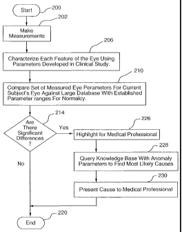
Referring Figure 5, it is seen at the start 200 that measurements are
made 202 with the stored information in the nature of normal and abnormal
parameters determined from clinical studies 206 being employed. The database
information regarding various conditions of the eye being monitored and how
they
correlate to the normal parameters is made 210. A logic decision 214 then
results in
the absence of a departure from normalcy indicating the end 220 of the test.
If there
is a meaningful departure, this information is provided to a medical
professional
226. Departures from desired eye structure or conditions are then compared in
the
processor with various anomaly parameters 228 with the cause 230 being
presented
to the medical professional.
It will be appreciated that depending upon the nature of the testing
done and the equipment available the subject's eyes may be monitored
sequentially
or simultaneously.
Referring to Figure 6, there is shown, schematically a subject 260
which is a human being, wearing a mask 262, which is secured to the individual
by
a strap 264 and defined an opening through which a major portion of the face
and,
11
CA 02456818 2004-02-16
WO 03/015660 PCT/US02/26127
specifically, the eyes 270, 272 are exposed. Integral with the mask 262 is a
lower
chin support portion 280, which in turn is supported by a suitable post 282,
which
may be anchored to any suitable base (not shown). Supported by the upper mask
portion 284 by means of a strut member 286, and a depending arm 290, with a
reinforcing angle brace 292 is a combination light source and sensor 294.
Referring
to Figure 8, there is shown the combination light source and sensor, which has
a
plurality, circumferentially spaced light sources 296, 298, 300, for example,
and a
centrally located camera 302.
In operation of this embodiment of the system, the light source and
lens combination 294 will be employed to examine eye 270 with a plurality of
light
beams 310 fully illuminating eye 270, and reflected light being received in
camera
302, which converts the received light into an output electrical signal
corresponding
thereto over line 320 to processor 324 wherein stored data, relating to
various eye
conditions is provided, and a comparison with the data received is made. The
system otherwise may function in the manner hereinbefore described.
Referring to Figure 7, the individual 260 has a chin support 340
supporting the chin 342 with a pair of systems adapted to examine,
respectively,
each eye 270 and eye 272. A pair of combination light sources and cameras 350,
352 may be supported by an anchor plate 354 and appropriate linkage 356, 358
with
a suitable adjustment linkage assembly 360 connected to arm 362 by
articulating
connector 364, which further may be connected to arm 366 through articulating
connector 368 which, in turn, may be secured to support column 370. A similar
support linkage may be provided for a combination light source and lens 350,
which
has anchor plate 380 and linkage elements 382, 384 supporting the combination
light source and lens 350. It will be appreciated that with this embodiment,
the
output of combination light source and lens 350 will be provided over lead 390
to
processor 324 and the output of combination light source and camera 352 will
be
provided over lead 392 to processor 324. In this embodiment, both eye 270, 272
may be examined sequentially under the control of processor 324 or
simultaneously,
12
CA 02456818 2010-06-03
if desired. In the embodiments of Figure 6 and 7, the operation of the light
source may be
controlled by processor 324 in a manner well-known to those skilled in the
art.
Referring to Figure 9, this figure shows a top plan view of a human subject
260
wherein the combination light source and camera 400, has a plurality of light
sources 402,404
and a camera having a forwardly projecting camera lens 406, with the combined
beam of
light 410 impinging on eye 270 and the reflected light for 414 indicated
generally by an
arrow entering camera 406. The electrical output of the light source camera,
unit 400, is
passed over lead 416 to processor 324.
With reference to Figure 10, there is shown a subject 260 and a modified
embodiment of the invention. In this embodiment of the invention, which is
structured to
provide analysis for both eyes 270,272, and the first light source 420 creates
a light beam 422
which impinges on eye 270 with. the resultant reflected light as represented
by arrow 426
being received in camera 430, which in turn converts the received light into
corresponding
electrical signal which passes over lead 432 to processor 324. Similarly,
second light source
434 creates a light beam 436 which impinges on eye 272 and, as indicated by
arrow 440,
causes reflected light to enter camera 450 which converts the received light
into
corresponding electrical signals, which pass over lead 452 to processor 324.
Figure 11 illustrates a modified form of restraint, which includes a chin
support 480 supporting post 496 and a broken band 482 which has a gap 484 and
positioning
elements 490, 492 secured thereto.
Figure 12 shows a chin support 480 supporting the chin of subject 260 with an
underlying support post 496. Rather than having a discontinuous band
supporting the upper
head portion, in this embodiment, a first support member 500, which is secured
to a support
arm 502 and a second support member 504, which is secured to a second support
arm 506,
cover only a portion of the subject's head and a rear support 510, which is
secured to a
support arm 512, serves to immobilize the subject's head. The light source
520, impinges a
light beam 524 on eye 270 with reflected light as represented by arrow 530
being received
within camera 532 which
13
CA 02456818 2004-02-16
WO 03/015660 PCT/US02/26127
is provided with adjustable supports 534, 536 and provides output over lead
538 to
processor 324.
If desired, additional restraints such as one contacting the rear of the
head and the forehead, for example, (not shown) may be employed if desired. In
this manner, inaccuracies in monitoring due to head movement will be reduced
or
substantially completely eliminated. The processor will generally be
programmed to
confirm that the eye is in the desired position before processing the data
received.
With respect to particular monitoring support and positions for the
apparatus of the present invention numerous modes of energizing and
communicating with the same will be known to those skilled in the art. To the
extent to which that it is to be mounted on head supporting apparatus, the
system
and a source of energizing the same may all be contained within the head
supporting
apparatus with a suitable means for monitoring at least one eye of the user.
Mounting such a system in the user environment has been disclosed, for
example, in
the product offered by Iscan, Inc. of Burlington, Maine under the general
trade
designation "HEADHUNTER." Devices may also be mounted in vehicles or in
regions adjacent to where the individual will be positioned.
It will be appreciated that for short interval monitoring, it will
generally be preferred to have at least one eye of the individual monitored by
the
system at frequent predetermined intervals. The frequency of such monitoring
will
depend to a great extent upon the nature of the activity, the purpose for
which
monitoring is being initiated, the nature of the characteristic being
involved, the
degree of the potential health or safety hazard involved, as well as other
factors.
For miosis and carbon monoxide, it will generally involve a monitoring cycle
occurring about every 1/60 to 30 seconds and preferably about every 1/2 to 10
seconds. This provides not only frequent data, but also facilitates monitoring
trends.
It will be appreciated that the invention may also be employed
advantageously to provide for periodic monitoring of patients at intervals of
days,
weeks, months or years for comparison purposes in order to determine if
14
CA 02456818 2004-02-16
WO 03/015660 PCT/US02/26127
meaningful changes have occurred over time. For convenience of reference
herein,
in order to distinguish these longer periods of time from the shorter repeated
cycles
which may be about 1/60 to 30 seconds between cycles, such longer periods
between monitored cycles will be referred to as "prolonged intervals", and the
shorter intervals of less than one hour, will be referred to as "short
intervals".
It will be appreciated from the foregoing that various monitoring
functions with respect to the eye not including checking of an individual's
vision
directly but rather employing the conditions of the eyes as an indication of
the
presence or absence of toxicity or other conditions within the subject may be
employed. For example, visible light images of the retina including nerves and
vasculature may be obtained. Spectrally filtered images of the retina
including the
typical red-green-blue color, an array of discrete spectral images and special
filters for highlighting known sources of aberrations indicative of specific
afflictions
may be employed. Further, these items may be expanded beyond the visible
spectrum into non-visible regions of the spectrum. The lighting which is
employed
will be so designed so as to facilitate structural measurements of various eye
components such as the lens, for example. Further, dyes or other toxin
sensitive
chemical elements may be employed in order to enhance determination of
anomalies.
It will be appreciated from the foregoing that the present invention
provides an effective means for monitoring of medical conditions so as to
provide
indications of potentially hazardous conditions with primary emphasis on
toxicity
from a number of sources.
While for convenience of disclosure herein reference has been made
to the human eye, in certain instances advantageous use of the invention may
be
made on animals, such as guard dogs, livestock, fish, working animals, or
wildlife,
for example. All of this has been accomplished in an economical, simple and
efficient automated manner.
Whereas particular embodiments of the invention have been described
herein for purposes of illustration, it will be evident to those skilled in
the art that
CA 02456818 2004-02-16
WO 03/015660 PCT/US02/26127
numerous variations of the details may be made without departing from the
invention as defined in the appended claims.
16