Note: Descriptions are shown in the official language in which they were submitted.
CA 02457797 2004-02-17
WO 03/016843 PCT/GB02/03811
SYSTEM AND METHOD FOR SCANNING IR MICROSCOPY
This invention relates to a microscope which can be used in FT-IR
spectroscopy. FT-IR microscopes generally operate in conjunction with an FT-
IR spectrometer. The IR beam used by the microscope is produced by the
spectrometer which also controls the scanning.
Background
Currently available FT-IR microscopes capable of generating an infrared image
fall broadly into two categories: focal plane array systems, where a
relatively
large array detector is used to generate an image of a portion of a stationary
sample; and single detector systems, where an image of arbitrary size is built
up from individual pixels by translating the sample in small steps on a
motorised stage in some form of raster scan. The large array based systems are
generally obliged, by the low frame rates available from typical detectors, to
use a very slow spectrometer scan, often a stepped scan, where an
interferometric image of the sample is acquired at each spectrometer scan
position before moving on to the next scan position; meanwhile, the sample
does not move. The single detectors in contrast have much higher bandwidth,
permitting a much more rapid spectrometer scan, typically not stepped, with
interferogram data collected continuously during the scan. The present
invention is concerned with this latter type of system where the sample is
moved stepwise in its own plane after each spectrum acquisition, and concerns
CA 02457797 2006-10-25
WO 03/016843 PCT/GB02/03811
2
the speed and efficiency of data collection. The detector does not have to be
a
single detector but could be a small array.
One known FT-IR microscope currently generates images (or maps), one pixel
at a time by a step and repeat sequence of operations. The cyclic sequence
consists of;
step the motorised sample stage to the next sample location;
start the spectrometer scan;
wait for the spectrometer scan to complete and stop;
transfer the data;
step the stage to the next sample location......
The sequential nature of the operations produces substantial delays as each
operation waits for the previous to complete, with the result that it takes a
very
long time to collect a reasonably-sized image.
The proposal of the present invention is to synchronise the stage movement
with the scan, minimising the lost time and preferably effecting the stage
movement within dead time encountered in the natural course of repeated
spectrometer scans. Coupled with an extension of the operating principle to
the
collection of several pixels in parallel by utilising a small array in place
of the
CA 02457797 2004-02-17
WO 03/016843 PCT/GB02/03811
3
single detector, the result is a dramatic reduction of process time so that
image
collection can occur in times comparable to those achieved with the much more
expensive large array based systems.
Embodiments of the invention can be implemented as follows:
The first step is to keep the spectrometer scanning continuously in order to
avoid the delays inherent in starting the scan from a halted condition. Then
it
becomes possible to step the microscope stage at any desired moment and
simply wait for data to be collected from the next complete scan. This
requires
abandoning any scan actually in progress during the stage movement, which
spoils the data being collected during that scan.
The next step is to synchronise the stage movement with the scan so that the
loss of a scan is predictable rather than random. For example, suppose the
stage movement, including settling time, took a substantial fraction of an
individual scan time. Then depending upon exactly where the stage movement
occurred, either one or two scans might be lost. Synchronising the stage
movement to the end of the scan would ensure that only one scan was lost.
This degree of synchronism can be achieved simply by waiting for data transfer
to complete but since stage movement can begin as soon as the previous scan
has finished, without waiting for data transfer, it is preferable to arrange
for a
stage movement trigger signal to be passed just as soon as the previous scan
has completed. Such a trigger signal might be propagated through the system
CA 02457797 2004-02-17
WO 03/016843 PCT/GB02/03811
4
using existing software channels.
The loss of one scan still has a substantial effect on the overall mapping
rate.
For example if we collect only one scan per pixel, the rate will be halved by
the loss of every alternate scan. Obviously it would be preferable to lose no
scans at all and this can be achieved by having a stage stepping time,
including
settling, that is shorter than the natural dead time between scans that occurs
during the reversal of the mechanical scan direction. Even if the stage
stepping
time is a little longer than the scan dead time, it may be advantageous to
artificially increase the scan dead time rather than to revert to losing a
whole
scan. Such tight synchronism requires rather rapid communication of a trigger
signal from the scan controller to the stage controller just as soon as the
previous scan has completed and this may be better achieved in direct hardware
communication rather than through software with the delays typically
encountered in non-real-time operating systems.
The final possibility is to move the stage continuously and at the same time,
to
scan continuously. This presents difficulties. Firstly, the sample spectrum is
potentially changing while the scan is in progress and careful analysis is
needed to determine what undesirable side-effects this may have. A second
problem is to establish the correlation between scan and effective stage
position. This requires a recording means to note the co-ordinates of the
stage
at, say, the start of each scan. Finally, there is the problem of error
recovery. If
CA 02457797 2004-02-17
WO 03/016843 PCT/GB02/03811
either the stage or the scan does not operate at a constant rate, the data
will
appear at varying increments of stage movement. While small problems of this
nature could be dealt with by interpolation, if either of the two mechanisms
malfunctions even momentarily, recovery from the error will be fairly complex.
5
The preferred embodiment uses synchronous stepping of the stage in the dead
time between scans as being the most appropriate solution. The scan controller
notifies the stage controller that the previous scan has finished and that the
stage may now be moved, by means of a dedicated hardware control line to
obviate any unnecessary delays. The cyclic sequence of events then becomes:
wait for data collection from the scan currently in progress to complete;
send an immediate trigger signal to the stage controller....
to step the motorised sample stage to the next sample location...
while at the same time transferring the data;
allow the scan to start its next data collection immediately after
turnaround.. ...
It is believed to be a novel concept to provide a FT-IR microscope based on a
rapid scan spectrometer in which the incremental sample stage movement is
synchronised with the end of the spectrometer scan in such a way that
effectively no lost data collection time is incurred. It is believed to be
particularly advantageous to do this in a microscope which uses a small array
CA 02457797 2004-02-17
WO 03/016843 PCT/GB02/03811
6
detector in place of the single element detectors used previously. Such a
microscope is described in EP-A-1184703.
The invention will be described now by way of example only with particular
reference to the accompanying drawing which is a block schematic
diagrammatic view of an embodiment in accordance with the present invention.
The invention is applicable to a wide range of FT-IR microscopes which
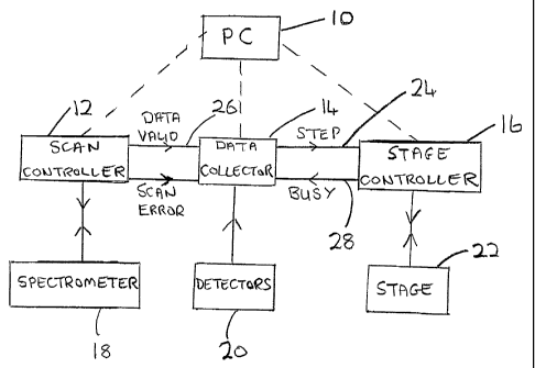
operate in conjunction with scanning spectrometers. The drawing shows a
microscope imaging system which has four functional control blocks:- a
personal computer PC (10), a scan controller (12) ( part of the spectrometer),
a
microscope data collection (14) and a microscope stage controller (16). Real-
time (guaranteed fast) links between the four blocks are shown as solid
connecting lines and non-real-time links are shown as dashed.
The PC (10) acts as a master controller and has the overall task of
coordinating
the actions of the remaining three control blocks (12, 14, 16). As a result of
the at-times-unpredictably slow response of its operating system, the PC is
not
relied upon to make real-time control decisions during the operating of the
system but rather sets up the control functions in the other controllers ahead
of
time and then simply monitors the operation and processes incoming data when
it can.
CA 02457797 2004-02-17
WO 03/016843 PCT/GB02/03811
7
The scan controller (12) has the function of supervising the interferometer
scan
in the spectrometer (18). As will be known to the those skilled in the art it
scans continually backward and forward, monitoring the position reached, by
counting the periods of a sinusoidal signal generated as a function of
distance
travelled by means of laser metrology within the interferometer. Thus the
controller (12) controls and monitors the length of active scan during which
data should be collected and determines the characteristics and timing of the
scan turnaround.
The microscope data collector (14) has the function of generating a stream of
digitised data, converting the analog signals from the infrared detectors (20)
of
the microscope to numeric form at regular intervals determined by the same
sinusoidal signal used by the scan controller (12). Digitised data is
collected
during the active scans and is ignored during the scan turnaround, i.e. the
period between the end of one scan and the start of the next. The data
collector receives a "scan active" signal issued by the scan controller (12)
when
a scan is currently active.
The microscope stage controller (16) has the function of controlling advances
of the microscope stage (22) after data collection at a particular location
has
been completed and before data collection at the next location can begin. In a
sweep across the sample, the step size is constant and scan be set up
beforehand. As a result, it is only necessary for the stage controller (10) to
be
CA 02457797 2004-02-17
WO 03/016843 PCT/GB02/03811
8
told when to step to the next location and this is communicated from the
microscope data collector (14) by a single control line (24).
The system operates as follows: The PC (10) instructs the other control
systems (12, 14, 16) in the basic parameters of the measurements: length of
scan for the spectrometer and any details of scan turnaround; then number of
data points per scan and number of scans to collect at each location for the
microscope data collector; and finally, sample step size for the microscope
stage controller. After allowing the systems to initialise themselves, the PC
then issued a command to start the measurement.
The scan controller (12) issues the "data valid" signal on a line (26) to the
microscope data collector as soon as the start of scan is reached and the data
collector gathers digitised signals until the end of scan is signalled by the
scan
controller through the "data valid" signal. Data gathering recommences with
the start of scan and the cycle continues until the requisite number of scans
has
been collected at the current sample. Note that if any errors are detected,
the
data collector can discard the current scan and simply wait for the next scan
instead. Once the data collector has collected enough scans at the current
sample location, it can signal the stage controller to advance the sample
location by way of the line (24). This moment occurs at the end of the last
scan, once the "data valid" has signalled the end of the scan and the data
collector has verified that the scan is satisfactory. Provided any unnecessary
CA 02457797 2004-02-17
WO 03/016843 PCT/GB02/03811
9
delays are avoided, the signal to step the stage is sent by the data collector
(14)
to the stage controller immediately after the end of scan, just as the
turnaround
is starting. Typically, there will be sufficient time to advance the sample
prior
to the next scan, is which can data collection at the step sample location can
resume with the following scan with consequently no loss of collection
efficiency.
In some combination of circumstances, it may be that the sample step cannot
be completed prior to the next scan. To guard against collecting data while
the
sample is still moving, the stage controller can flag to the data collector
(14)
via line (28) that it is currently moving the stage. If the data collector
detects
that the next scan has started before the move is completed, it can then
discard
the next scan and wait for the following one by which time the stage
controller
should certainly have finished the task of advancing the sample.