Note: Descriptions are shown in the official language in which they were submitted.
CA 02457930 2010-05-27
Diagnostic testing process and apparatus
Field of the Invention
This invention relates to a diagnostic testing process and in particular
to an apparatus for use in carrying out an assay process and to a method of
carrying out an assay process using that apparatus.
Background of the Invention
Lateral flow and flow-through technology have been used for
diagnostic assays for almost twenty years. Lateral flow technology is
currently dominant because lateral flow devices are easy to produce and the
assay can be performed in a simple 2-step process that can be adapted for
whole blood separation. This results in a simple device that can be used in
the field as a rapid point-of-care diagnostic (Cole et al 1996 Tuberc. Lung.
Dis.
77:363-368). However, multiple disease diagnosis using lateral flow
technology is very difficult because of differences in lateral diffusion
between
samples and variation in flow rates between batches of the partitioning
membrane. This means that antigen or antibody signal strengths may vary
both within tests and between batches of tests, resulting in inconsistent
results.
Existing flow-through diagnostic tests can be completed in less than
two minutes compared with typical times of five to fifteen minutes for lateral
flow tests. This advantage in speed however, is often at the expense of
sensitivity. A further disadvantage is that higher volumes of sample are
required to achieve the same sensitivity as lateral flow. This maybe
problematic in some situations. For example, the diagnosis of analytes
(reagents) in whole blood requires the separation of plasma from whole blood
cells. The higher volumes of whole blood required for this would quickly
block the membranes in the flow-through format.
The basic principal of flow-through assays is well established. The
tests are designed to determine the existence of, and in some cases, the
quantity of, a predetermined analyte/reagent in a sample. Often the reagent
will be a protein but other reagents can be tested for. If the assay is to
test for
the existence of a particular disease in a patient, the patient's body fluids
may
be tested for an antibody or other protein produced by the patient in response
to the infection, or for a protein which is expressed by the bacterium or
viral
CA 02457930 2004-02-18
WO 03/016902 PCT/AU02/01119
2
agent or the like causing the disease. In a typical flow through assay a
liquid
sample which is believed to contain the reagent is sucked into an absorbent
pad via a membrane to which is bound a capture analyte which is known to
bind to the reagent. The membrane is then typically washed with a buffer
and a liquid containing a detection analyte which also binds to the reagent
and which includes a tracer or marker which is detectable, is applied to the
membrane. The detection analyte binds to the immobilised reagent bound to
the membrane and can be seen or otherwise detected to indicate the presence
of the reagent.
US 4246339 discloses a test device for assaying liquid samples for the
presence of a predetermined reagent. The device includes telescoping top
and bottom members defining a liquid reservoir therebetween and resilient
means for biasing the members in the open position. The top member defines
a series of test wells each of which has a base defined by a microporous
membrane with a capture analyte immobilised on the membrane surface.
Absorbent means are located in the bottom member, spaced from the
membrane in the open position but in contact therewith in the closed
position. US 4246339 discloses the adding the test serum diluted with a
buffer to a test well, and incubating the device at room temperature for ten
minutes prior to depressing the cassette to the closed position to pass the
sample through the membranes into the absorbent material. When the
membranes are dry, the membrane is washed and then covered with a
solution containing a detection analyte which binds to the immobilised
reagent followed by a subsequent step in which a stain is applied.
It will be appreciated that the process described in US 4246339, is a
somewhat long drawn out, time consuming and tedious process and also
lacks sensitivity.
A more recent flow through device is described in US 5185127 which
discloses an assay device including a filter stack and an enclosure having a
base portion and a lid. The filter stack has a hydrophilic membrane having a
capture analyte thereon, referred to in US 5185127 as a binder. A
hydrophobic membrane is located under the hydrophilic membrane and a
pad of absorbent material is located under the hydrophobic membrane. The
lid includes an upwardly extending rib which defines a recess having an
insert therein. In use, a sample containing the reagent (referred to in US
5185127 as the analyte) is placed in the well of the assay device at which
CA 02457930 2004-02-18
WO 03/016902 PCT/AU02/01119
3
time the reagent/analyte binds to the capture analyte/binder. Flow of the
assay solution however, does not take place because the aqueous solution
does not wet the hydrophobic membrane placed under the hydrophilic
membrane in the filter stack. Thus as much time is necessary to complete the
binding of the detection analyte to the reagent is allowed. When binding is
judged to be complete, flow may be initiated by adding a wetting agent which
wets the hydrophobic membrane. After which time the aqueous liquid flows
into pad of absorbent material. The membrane may then be washed and
treated with a detection analyte/tracer which may be an antibody which
specifically binds to the analyte, the antibody having a label covertly
conjugated thereto. Again the sensitivity of US 5185127 is lacking and is not
equivalent to that obtainable in lateral flow or ELISA formats.
It is one object of the present invention to provide an improved method
and apparatus for use in an assay process such as an immunoassay,
diagnostic assay or the like in which the process and apparatus are capable of
screening a wide range of samples such as whole blood, plasma/serum, or
samples with a high particulate load such as crushed grain (eg wheat heads)
and which is simple and rapid to perform whilst still maintaining
sensitivities at least equivalent to that obtainable in lateral flow or ELISA
formats.
A related object of the present invention is to provide a method and
apparatus which can be successfully used for multiple disease diagnosis from
a single whole blood or other sample in a single test. An extension would be
successful screening of a sample in a single test for the presence of multiple
analytes not necessarily related to disease (e.g drugs, agriculture,
veterinary
testing).
Any discussion of documents, acts, materials, devices, articles or the
like which has been included in the present specification is solely for the
purpose of providing a context for the present invention. It is not to be
taken
as an admission that any or all of these matters form part of the prior art
base
or were common general knowledge in the field relevant to the present
invention as it existed in Australia or elsewhere before the priority date of
each claim of this application.
Because the prior art is not consistent in its terminology, for the
avoidance of doubt and for the purpose of clarity, the following terms used in
the specification below, are defined as follows. The term "reagent' is used to
CA 02457930 2004-02-18 PCT/AU02/01119
Received 02 May 2003
4
refer to the compound protein or the like which is to be detected by the
assay.
The term "capture analyte" is used to refer to a compound which is bound to a
membrane and to which the reagent will bind. The term "detection analyte"
is used to refer to a compound which will also bind to the reagent and which
carries a tracer or some other element whose presence may be detected,
typically visually detected whether under visible light, or fluorescence.
Summary of the Invention
In a first broad aspect, the present invention provides an apparatus and
method for use in an assay process which is characterised by providing a
"pre-incubation step" in which the sample and detection analyte (which may
typically be an antibody bound to colloidal gold or a fluorescent tag) may
bind together, which has the effect of both improving the sensitivity of the
assay and reducing the volume of sample required for an assay prior to
reaction of the sample/analyte complex with a reaction membrane to which
one or more ligands are bound.
Thus, in one aspect of the present invention there is provided an
apparatus for use in an assay process comprising:
a first member comprising a first, porous, membrane to which is bound
a capture analyte for binding to a reagent to be detected, the member having
an upper surface and a lower surface;
a second member being a body of absorbent material disposed below
and touching the lower surface of the first member;
a vessel for containing a liquid sample spaced above the first member
said vessel having side walls and a base, the base being defined by a second
membrane, the vessel being capable of retaining a liquid sample for a
predetermined incubation period; and
means for supporting the vessel above the first member in two
positions, a first position in which the membrane is spaced a sufficient
distance from the first member so as to not permit fluid transfer from the
vessel to the body of absorbent material, and a second position in which the
second membrane is in contact with the first member, such contact
permitting fluid transfer from the vessel through the first and second
membranes to the body of absorbent material.
In a related aspect the present invention provides a method for
assaying for the presence of a pre-determined reagent in a liquid sample
comprising the steps of:
a) providing a first porous membrane to which capture analytes for
binding to the reagent have been bound;
AMENDED SHE
IP lAU
CA 02457930 2004-02-18 PCT/AU02/01119
Received 02 May 2003
b) placing a sample to be assayed and a detection analyte in a vessel
having a base defined by a second porous membrane, the vessel being
capable of retaining the liquid sample for a predetermined incubation period;
c) allowing a sufficient period of time to pass for the detection
5 analyte to bind to the reagent, if present in the liquid sample;
d) contacting the base of the vessel with the first porous membrane;
and
e) causing the liquid sample to flow through the membranes to allow
the reagent to bind to the capture analyte carried on the first membrane.
Thus, the present invention provides a chamber which may serve as a
pre-incubation chamber in which a pre-incubation step can occur where the
sample and detection analyte combine, which improves the sensitivity of the
test and reduces the volume of sample required for the assay. It has been
found that the pre-incubation step increases the test sensitivity for a
typical
existing flow-through apparatus by approximately ten times to equivalent
levels of sensitivity compared with lateral flow technology, while still
allowing the assay to be completed in around two minutes compared to 10
minutes for lateral flow formats.
For example a ground wheat head suspension may be solubilised, and
mixed and pre-incubated in the chamber with a detection analyte in the form
of an antibody against alpha-amylase linked to a colloidal gold particle. The
contents of the chamber may then be allowed to flow through to the first
membrane containing a capture analyte in the form of an immobilised anti-
amylase antibody, and anti-amylase antibody/gold complexes will bind to the
immobilised antibody forming a detectable signal. The signal can be detected
by the removal of the pre-incubation unit and washing of the reaction
membrane with buffer.
This format can also be used for detecting reagents in whole blood
since whole red blood cells can be removed in the pre-incubation chamber
and the plasma allowed to flow-through to the reaction membrane containing
a bound capture analyte. In this format, the base membrane defined at the
base of the pre-incubation chamber will typically be a membrane which has
the correct pore size to retain the red blood cells and allow the plasma to
pass
through on contact with the first membrane. Similarly particulate samples
containing grain extracts, cell or microbial extracts can be analysed with
this
flow-through format since particulate matter can be removed in the pre-
u MEP,1DED HE 2: If
PE-'N/AU
CA 02457930 2004-02-18
WO 03/016902 PCT/AU02/01119
6
incubation chamber and therefore cannot block the reaction area on the
upper surface of the reaction membrane.
The apparatus can also be used for detecting analytes in body fluids
other than blood, such as plasma, sera, urine, saliva and sputum. In this
case, the sample can be retained in the pre-incubation chamber by use of a
hydrophobic membrane. To obtain efficient flow through capillary action to
the second member when the pre-incubation chamber is lowered, the
reaction membrane is pre-wet with a wetting agent containing a detergent or
the reaction membrane is blocked with a hygroscopic solution such as
sucrose, trehalose, fructose, or alternatively, glycerol.
This changes the characteristics of the reaction membrane from a non-
hygroscopic to a hygroscopic membrane allowing the sample to flow through
to the second member upon contact of the membrane at the base of the pre-
incubation chamber with the reaction membrane.
In a yet further embodiment, if a hydrophobic membrane is used as the
base of the pre-incubation chamber, the apparatus may be used with the
hydrophobic membrane and reaction membrane in contact, with the operator
adding a wetting agent to the sample to cause flowthrough, when desired.
Thus, in a related aspect, there is provided an apparatus for use in an
assay process comprising a housing including:
a first member comprising a first, porous, membrane to which is bound
a capture analyte for binding to a reagent to be detected, the member having
an upper surface and a lower surface;
a second member being a body of absorbent material such as tissue
paper or the like disposed below and touching the lower surface of the first
member;
a chamber located above the first member said chamber having side
walls, and a base including a second, hydrophobic, membrane, having an
upper and a lower surface, the pre-incubation chamber being supported
above the first member with the lower surface of the hydrophobic membrane
in contact with the upper surface of the first member.
The pre-incubation chamber can also be used to remove analytes that
may interfere with the assay, such as human anti-mouse antibodies
(HAMAS), in solution or by binding anti-analyte antibodies to the surface of
the chamber. The chamber can also be used to extract the analyte of interest
from an absorbent surface such as a swab, which has been taken from the
CA 02457930 2004-02-18
WO 03/016902 PCT/AU02/01119
7
throat of a patient, by swirling the swab in an extraction solution in the
chamber. The pre-incubation chamber may be part of a pre-filter unit which
acts also to pre-filter the sample prior to contact with the upper surface of
the
first member.
Examples of assays that can be preformed by this method where two
reaction steps are involved (the incubation of the analyte with the labeled
anti-analyte followed by the binding of this complex to a solid-phase anti-
analyte), are:
Direct antigen assay
1. Ag* (analyte) + Ab*1(anti-Ag)-label
2. Solid phase-Ab2 (anti-Ag) + Ag/Ab1(anti-Ag)-label complex
Direct antibody assay (i)
1. Ab1(analyte = anti-Ag) + Abe (anti-Abj -label
2. Solid phase-Ag + Ab1 (anti-Ag)/Ab2 (anti-Ab1)-label complex
Direct antibody assay (ii)
1. Ab1 (analyte = anti-Ag) + Ab2 (anti-Abj -label
2. Solid-phase-Ab3 (anti-Ag)/Ag + Ab1(anti-Ag)/Ab2 (anti-Abl) -label
complex
Indirect antigen assay
1. Ag (analyte) + Ab1(anti-Ag) + Ab2 (anti-Abj -label
2. Solid-phase-Ab3 (anti-Ag) + Ag/Ab1 (anti-Ag)/Ab2 (anti-Abl) -label
complex
Indirect antibody assay (i)
1. Ab1 (analyte = anti-Ag) + Ab2 (anti-Ab1) + Ab3 (anti-Ab2)-label
2. Solid phase Ag + Ab1 (anti-Ag)/Ab2 (anti-Ab1)/Ab3 (anti-Abj -label
complex
Indirect antibody assay (ii)
1. Ab1 (analyte = anti-Ag) + Ab2 (anti-Ab1) + Ab3 (anti-Ab2)-label
2. Solid phase Ab4 (anti-Ag)/Ag + Ab1 (anti-Ag)/Ab2 (anti-Ab1)/Ab3 (anti-
Ab2)-label complex
CA 02457930 2004-02-18
WO 03/016902 PCT/AU02/01119
8
*Ag indicates antigen
*Ab indicates antibody
A piezoelectric driven printer may be used to dispense precise amounts
of multiple disease ligands such as antigens or antibodies or an analyte as a
micro array onto a reaction membrane for use in the apparatus of the first
aspect of the present invention. The ligands or analytes may be dispensed in
particular patterns, e.g. letters for ease of recognition of results.
Typically,
100 pl of fluid reagent (1 drop), or multiples thereof, is dispensed, but this
will vary depending on the application. The resultant size of the spot on the
membrane is about 55 microns or more in diameter subject to fluid diffusion
on the membrane, but again this will vary depending on the application. It is
possible to dispense droplets with diameters of 5-10 microns, and hence
lower volumes of fluid reagent (for example, 1-10 pl) can be applied. Using
precise quantitative printing of micro arrays of antibodies, antigens, or
other
analytes means that tests using precise quantities of these reagents can be
produced for multi disease diagnosis of a single sample. This array
technology can be applied to tests for drugs or other markers across all
diagnostic fields.
Alternatively, an adult/neonatal syringe pump 1235 from ATOM
Medical Corporation, Japan, typically used to administrator small quantities
of intravenous liquids through a catheter to hospital patients can be adapted
to apply single or multiple lines of a capture analyte to the first membrane
eg
nitrocellulose.
In one preferred embodiment, ligands for detecting tuberculosis, HIV,
hepatitis, syphilis and malaria antibodies may be deposited onto a reaction
membrane. This would allow the simultaneous diagnosis of tuberculosis,
HIV, hepatitis, syphilis and malaria from a single blood sample without the
need for intermediate sample treatment steps.
Utilising the present invention allows the assaying of small volumes of
whole blood and thus the present invention provides a very rapid diagnostic
assay device that is simple to use and can be used in both laboratory and
point-of-care field diagnostic locations. For example, a finger prick of blood
would be sufficient to perform an assay. Similarly large volumes of sample
can be used in this device by increasing the amount of absorbent material
CA 02457930 2010-05-27
9
(second member). For instance, 10 mis of dilute fluids like urine can be can
be assayed to detect low abundance molecules.
Analytes and/or ligands (e.g. antigens or antibodies) can be printed
down in titrating amounts and/or concentrations. Thus, in an individual
screen, this would provide a means of quantitating analyte-ligand levels
within the sample solution.
The pre-incubation step may also be carried out with a multi-analyte
detector where any number of detection analytes can be attached to a gold
particle or a similar detectable tag e.g. a fluorescent marker.
Brief Description of the Drawings
A specific embodiment of the present invention will now be described
by way of example only and with reference to the accompanying drawings in
which:
Figure 1 is a schematic drawing of an apparatus embodying aspects of
the present invention in a first configuration;
Figure 2 is a schematic drawing of the apparatus of Figure 1 in a second
configuration;,
Figure 3 is a perspective view of an assay apparatus or cassette
embodying aspects of the present invention;
Figure 4 is an exploded view of the components of the cassette shown
in Figure 3;
Figures 5a to 5d show various stages in the use of the apparatus of
Figure 3 in carrying out an assay; and
Figure 6 is a graph comparing test results from samples spiked with
alpha amalyse undergoing no-pre-incubation with samples undergoing a one
minute pre-incubation.
Brief Description of a Preferred Embodiment
Capture analytes in the form of ligands such as antigens or antibodies
(e.g. TB, HIV-1) are printed onto a protein-capture membrane matrix (e.g. a
nitrocellulose membrane) in an appropriately sized array using piezoelectric
chemical printing technology. A suitable chemical printing system for use in
the present invention involves the use of piezoelectric drop-on-demand ink
jet printing technology for micro-dispensing fluids in DNA diagnostics or the
Combion Inc. synthesis process called "CHEM-JET". To explore drop on
*Trade-mark
CA 02457930 2010-05-27
demand fluid dispensing for DNA diagnostics, an eight fluid printer has been
developed as part of the Genosensor Technology Development (GTD) project
funded by the Institute of Standards and Technology (USA). Research to
date, is focused on printing oligonucleotide micro-spots onto solid supports.
5 In the CHEM-JET technique, which was developed at the California Institute
of Technology, tiny volumes of reagent bearing liquid are squirted onto
specific spots or addresses of a solid substrate much as an ink-jet printer
squirts ink onto a page. By repeatedly returning to each address with one or
another of a small set of building blocks, in this case, nucleotides modified
10 for the process, huge two-dimensional libraries of short DNA chains
(oligonucleotides) can be assembled. Such a device including an imaging
means is described in the applicant's co-pending International patent
application No PCT/AU98/00265.
In the described embodiment, antigen is
printed onto a reaction membrane in 100 pl droplets, or multiples thereof (eg.
10 ni), with each aliquot being 1 mm apart. However, these volumes and
distances can be increased/decreased accordingly depending on the chosen
antigen titre and array size. For example, it is possible to dispense droplets
with volumes as low as 1-10 pl.
In a particularly preferred embodiment, antigens or antibodies can be
printed down in a matrix of dots or lines or in the shape of letters so that
quantitative multiple analyte analysis of a single sample is possible.
After the dispensed antigen has dried, non-specific protein-binding
sites on the (nitrocellulose) membrane are blocked using 0.5% (v/v) casein in
phosphate buffered saline (PBS) + 0.05% (w/v) sodium azide + 0.1% (v/v)
Tween-20 (PBSA wash buffer). It is however an option to leave the
membrane unblocked following the printing of the antigen (or antibody) or
other ligand.
In another preferred embodiment syringe pump technology used for
the administration of liquids intravenously to patients can be adapted to lay
down single or multiple lines on nitrocellulose membranes.
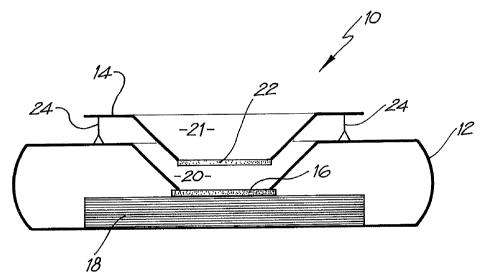
Turning to the drawings, Figure 1 shows a flow-through assay device
10, which utilises the nitrocellulose membrane described above. The device
is in the form of a cassette 12 and an associated removable filter frame 14.
Inside the cassette there is the membrane (typically nitrocellulose) 16 on
which capture analytes in the form of ligands are printed, as described above,
CA 02457930 2010-05-27
11
which is located on top of an absorbent matrix 18. The absorbent matrix
preferably comprises multiple layers of absorbent tissue or an absorbent pad
such as blotting paper, in the specific embodiment twenty-four layers (double
ply), which have been found to possess an ideal porosity that permits the
most rapid flow-through of various solutions. This rapid flow-through is
important as it results in lower backgrounds with higher reaction specificity
and higher signal resolution.
As shown in Figure 1, the top of the cassette defines an opening in its
upper face and a depending generally frusto-conical well whose sides depend
down as far as the membrane 16, to define a chamber having sloping sides
and a base defined by the membrane 16.
The filter unit frame 14 is spaced above the upper surface of the
cassette 12. It also defines a depending conical well in the form of a chamber
21 also referred to as a "pre-incubation chamber" having sloping sides and a
base 22 formed from a 5 m Whatman grade 1 membrane or a 0.22 gm
hydrophilic Durapore membrane filter (Millipore, North Ryde, Australia).
However, other types of filter/membrane and pore size would be suitable
depending on the application. The function of the membrane is to retain a
sample to be assayed in the well or pre-incubation chamber 21 long enough
for a "pre-incubation step" to take place. When membrane 22 is lowered to
contact the membrane 16, capillary attraction draws the sample from the
chamber 20 through membranes 22 and 16 and into the tissue 18.
For ease of use, two pins 24 are provided which support the filter frame
14 at an appropriate distance above the cassette 12 during the pre-incubation
step but which allow the filter frame to be pushed down so that the
membranes 22 and 16 are in contact for the second stage of the process
shown in Figure 2. The frame 14 is also removable so that the membrane 16
can be viewed to determine the results of the assay.
Figures 3 to 5d illustrate one commercial assay device design
embodying the aspects of Figures 1 and 2.
In those Figures, the components which are equivalent to components
shown in Figures 1 and 2 carry the same reference numerals. The cassette 12
comprises an upper moulding 12a and a lower moulding 12b. The porous
membrane 22 is defined by the base of a pressed filter paper frustro cone 22a
held in place by a filter retainer 23. The filter unit frame 14 defines two
*Trade-mark
CA 02457930 2004-02-18
WO 03/016902 PCT/AU02/01119
12
dimples 14a on which an operator's thumbs may press when depressing the
filter frame to contact the membranes 22 and 16.
Figures 5a to 5d illustrate the stages of operation of the apparatus.
Figure 5a illustrates the filter frame separate from the cassette 12. Figure
5b
illustrates the pre-incubation positioned with the base of the chamber/well 21
spaced from membrane 16. Figures 5c and 5d illustrate the device after the
filter unit has been pressed down to bring the membranes 22 and 16 into
contact to allow the sample to flow through to the blotting paper 18.
If the membrane 22 is replaced with a hydrophobic membrane, it is
possible to operate the device with a pre-incubation step solely in the
position shown in Figures 3 and 4 with the membranes 22 and 16 always in
contact. The hydrophobic membrane 22 will prevent flow of the sample in
the incubation chamber 21 to the reaction membrane 16. After a sufficient
period of time has past for detection analyte in the chamber 21 to bind to the
reagent, a suitable wetting agent is added to the sample in the chamber which
allows the sample to flow through the hydrophobic membrane past the
reaction membrane 16 and into an absorbent matrix 20.
Example 1
Application of the pre-filter chamber
Whatman membrane (paper) or Reemay filters (polyester; 1cm2) are
inserted into the chamber 21 in the filter frame to form a conical retaining
vessel (pre-filter unit).
The sample is pipetted into the plastic pre-filter chamber (50-100 ul)
along with a detection analyte in the form of a detecting antibody (50-100 ul)
bound to colloidal gold (particle size 20-50 nm). The sample is pre-incubated
with the gold-conjugate (O.D.4) within the pre-incubation chamber for thirty
seconds after gentle pippetting to ensure adequate mixing. After thirty
seconds the chamber is pressed into the well 20 of the test cassette 12. Upon
contact with the membrane 16 containing the detection zone, the solution
filters through to the absorbent layer 18 beneath. The pre-filter 14 is
discarded when the solution has filtered through and two drops of PBSA
wash buffer are then added to the reaction membrane to wash away excess
gold-conjugate revealing the results of the assay on membrane 16.
The use of the pre-incubation of the sample with the detection analyte
increases sensitivity by approximately ten fold. Further, any particulate
CA 02457930 2004-02-18
WO 03/016902 PCT/AU02/01119
13
matter is retained in the pre-incubation chamber all of which can be removed
to provide a clear signal. The use of the preincubation chamber with the dual
roles of permitting a pre-incubation step and a pre-filtering step, also
allows
multi-analyte detection on the reaction membrane by pre-incubating with a
multi-analyte probe, e.g. colloidal gold bound to different detecting
analytes.
In addition, interfering analytes or substances that could cause false
positives
or negatives in the assay can be removed or absorbed out in the pre-
incubation step, e.g. human antibodies to mouse antigens can be absorbed out
by anti-HAMA antibodies.
Although the above described example relates to the antigens relating
to disease, the immunoassay apparatus could be used, for example, as an
allergy test kit, as a test kit for drugs of abuse or for analysing non-human
derived samples e.g. bovine, porcine, veterinary tests, and tests in
agriculture
such as grain quality evaluation, etc.
The method and apparatus of the present invention is particularly
suited to use with swabs which can be simply placed into the chamber 21,
swirled around in liquid containing a detecting antibody (50-100 ul) bound to
colloidal gold for 30 seconds before the pre-filter unit is depressed to
contact
the membranes 22 and 16 together.
Any combination of ligands and analytes can be applied to the system
of the present invention. The choice of ligands could be tailored to detect
prevalent diseases in a particular country or population. For example,
analytes from the following combination of diseases could be used for
diagnosis using this array.
1. TB and HIV
2. Hepatitis-B & C, HIV
3. Chagas, HIV, TB, Syphilis and Hepatitis-B & C
4. Malaria, Dengue, TB, Chagas.
Alternatively antigens representing different varieties of wheat or other
agricultural products could be printed on the reaction membrane enabling
detection of multiple strains with a single test.
CA 02457930 2010-05-27
14
Example 2
The assay device can also be used for detecting analytes in body fluids
other than blood such as plasma, sera, urine, saliva and sputum. In this
system, the sample can be retained in the pre-incubation chamber 22 by use
of a hydrophobic membrane such as Reemay or Hollingsworth*and Voss' 7303
instead of the Whatman grade 1 membrane or a 0.22 .tm hydrophilic
Durapore membrane filter described above. The sample is mixed with the
detection analyte for the required pre-incubation period. To obtain efficient
flow through capillary action to the absorbent layer 18 when the pre-
incubation chamber 22 is lowered onto the cassette 12, one of two procedures
can be followed:
1. The membrane 16 containing the capture analyte is pre-wet with
at least one drop of wash buffer containing 0.01 M phosphate, 0.15 M NaC1,
0.0% Azide, 0.5% Tween 20 or any wetting agent containing a detergent;
2. The membrane 16 containing the capture analyte is blocked with
a hygroscopic solution such as sucrose, trehalose, fructose, or alternatively,
glycerol. This changes the characteristics of the membrane 16 from a non-
hygroscopic to a hygroscopic membrane allowing the sample to flow through
to the absorbent layer 18 upon contact of the membrane at the base of the pre-
incubation chamber 22 with membrane 16.
Example 3 (Comparative Example)
Comparison of no pre-incubation and 1 minute pre-incubation of a
sample spiked with alpha amylase in the above described format.
Procedure
A 6% solution of bovine sera albumin was spiked with 0.1ng/ml,
0.5ng/ml, 1ng/ml, 10ng/ml, 50 ng/ml, 100ng/ml, 500ng/ml and 1000ng/ml and
applied to the above format according to the following procedure:
No-preincubation
I. The pre-incubation chamber was pressed down so that the base of
the chamber comes into contact with the first member containing the
capture antibody against alpha amylase.
*Trade-mark
CA 02457930 2004-02-18
WO 03/016902 PCT/AU02/01119
II. Sixty microlitres of 0.5% tween in saline was added to the pre-
incubation chamber and allowed to filter through to the absorbent
material beneath the first membrane.
III. One hundred microliters of spiked alpha amylase sample was
5 added to the chamber and allowed to filter through to the absorbent
material beneath the first membrane.
IV. Sixty microlitres of 0.5% tween in saline was added to the pre-
incubation chamber and allowed to filter through to the absorbent
material beneath the first membrane.
10 V. Sixty microlitres of anti-alpha amylase antibody linked to colloidal
gold (particle size 20-50nm) was added to the pre-incubation chamber
and allowed to filter through to the absorbent material beneath the first
membrane.
VI. Sixty microlitres of 0.5% tween in saline was added to the pre-
15 incubation chamber and allowed to filter through to the absorbent
material beneath the first membrane.
VII. The pre-incubation chamber was removed and the result on the
reaction membrane scanned with a densitometer. Signal strength was
measured in pixel intensity.
One minute pre-incubation
I. Sixty microliters of 0.5% tween in saline was added to first
membrane and allowed to filter through to the absorbent material
underneath.
II. The pre-incubation chamber was suspended over the first membrane
so that there was a space between the chamber and the membrane.
III. One hundred microliters of spiked alpha amylase sample and 60
microliters of anti-alpha amylase antibody linked to colloidal gold
(particle size 20-50nm) were incubated in the pre-incubation
chamber for 1 minute.
IV. The chamber was lowered until it came in contact with the first
membrane and the mixture of sample and antibody-gold conjugate
allowed to filter through to the absorbent material.
V. Sixty microliters of 0.5% tween in saline was added to the pre-
incubation chamber and allowed to filter through to the absorbent
material.
CA 02457930 2004-02-18
WO 03/016902 PCT/AU02/01119
16
VI. The pre-incubation chamber was removed and the result on the
reaction membrane was scanned with a densitometer. Signal strength
was measured in pixel intensity.
Each data point on the graph is the average of two experiments using
the apparatus described above. The results show that pre-incubation of the
sample with the detection analyte has a minimal detection limit defined in
pixel density of around 500 pg/ml of alpha amylase. This is compared to a
minimum detection limit without the pre-incubation of about 50ng/ml and
indicates the pre-cubation increases the sensitive by around 10 fold.
Example 4 (Comparative Examples)
Demonstration of in creased sensitivity with increased pre-incubation of
the sample with the detection analyte.
Samples of amylase diluted in 0.5% saline to 400 ng/mL were treated
with immunogold conjugate against amylase and aliquotted onto the flow-
through format in different protocols as shown below.
A. The sample was added to the format (without a filter present) and
allowed to filter through prior to adding conjugate, followed by an
aliquot of conjugate immediately the sample had passed through the
membrane.
B. The sample was mixed in the correct proportions with gold conjugate
and aliquotted immediately onto the flow-through format.
C. The sample was mixed as with protocol B but added to the flow
through format after a 60 second interval.
The results presented in pixel intensity are shown in the tables below
(for 2 experiments):
Protocol Sample Control Sample Control S/PC ratio
peak peak area area
A 82 286 657 2120 317
B 288 758 2062 5509 383
C 823 949 5843 6765 884
CA 02457930 2004-02-18
WO 03/016902 PCT/AU02/01119
17
Protocol Sample Control Sample Control S/PC ratio
peak peak area area
A 89 516 588 3890 588
B 482 830 3736 6345 602
C 708 829 4506 5822 792
Clearly there is a significant increase in the sample signal when the
analyte is preincubated with the conjugate probe, as distinct to sequential
detection on the flow-through format. The difference in detection levels (for
the 400 ng/mL sample) equated to between a 7.5-fold to 10-fold increase in
detectable amylase in the flow through format when the sample is
preincubated separately to the detecting capture antibody.
It will be appreciated by persons skilled in the art that numerous
variations and/or modifications may be made to the invention as shown in the
specific embodiments without departing from the spirit or scope of the
invention as broadly described. The present embodiments are, therefore, to
be considered in all respects as illustrative and not restrictive.