Note: Descriptions are shown in the official language in which they were submitted.
CA 02458176 2004-02-19
WO 03/026346 PCT/US02/29920
NONLINEAR NOISE REDUCTION FOR
MAGNETOCARDIOGRAMS USING WAVELET
TRANSFORMS
BACKGROUND OF THE INVENTION
Field of Invention
The present invention relates generally to the field of magnetocardiography
and
electrocardiography. More specifically, the present invention is related to
nonlinear noise
l0 reduction for magnetocardiograms using wavelet transforms.
Discussion of Prior Art
Magnetocardiography (MCG) is the measurement of magnetic fields emitted by the
heart from small currents by electrically active cells of the heart muscle. It
is a noninvasive
15 diagnostic method still not introduced into routine clinical practice.
Magnetocardiography consists of measurements of time-varying magnetic fields
generated above the torso (or maternal abdomen in fetal magnetocardiography)
by the
electrophysiological processes in the heart. The measurement of these fields
over the torso
provides information which is complementary to that obtained by
electrocardiography, and is
20 used especially in diagnosing abnormalities of heart function.
Due to the extremely weak strength of these signals (one millionth or less of
the
earth's magnetic field), currently only Superconducting Quantum Interference
Devices
(SQUII7s) are capable of such a task. Such sensors operate only at very low
(cryogenic)
temperatures and must be placed inside a special enclosure (cryostat). Its
external walls are at
25 room temperature, while inside the low temperature is attained, usually by
filling by a low-
temperature liquid (cryogen), most typically liquid helium. The cryostat with
SQUID sensors
is positioned close to, but without any contact with the human body. It is
possible, in
principle, to replace liquid helium by liquid nitrogen, by utilizing high
temperature
superconductor SQUIDS (HTSQULDs) and gradiometers. Although this would greatly
CA 02458176 2004-02-19
WO 03/026346 PCT/US02/29920
simplify the handling, to date demonstrated HTS-based MCG systems have not
been entirely
practical.
Although magnetocardiography has several advantages compared with
electrocardiography, a breakthrough for a practical clinical use is still
missing. Therefore, it
is necessary to develop convincing and attractive results for medical doctors,
and to reduce
the costs of SQUID systems. Both can be achieved on the basis of an improved
noise
cancellation method.
The existence of much stronger, natural and human-generated external signals
results
in extremely low (less than one in a million) signal-to-noise-ratio values.
These signals are
l0 unusable unless methods of external signal (noise) suppression are
employed.
The most effective, but also the most expensive and inflexible method of noise
suppression is the operation of the method in high-quality magnetically
shielded rooms.
However, such rooms have been proven to be unacceptable in cardiological
practice.
Operating outside magnetic shielding and without highly balanced SQUID
gradiometer
15 systems is essential for a clinical acceptance. Therefore, the emphasis of
recent efforts has
been on the development of MCG systems that can be operated in the absence of
magnetically
shielded rooms. The main technique of noise suppression utilized has been
higher order
gradiometry.
In diagnostic applications, the magnetic field of the heart may be analyzed
spatially
20 and/or over time in order to identify complex changes in cardiac electrical
activity due to
pathological functional or structural changes in the myocardium. These may
result from
ischemia, myocardial infarction, volume or pressure changes in the cardiac
chambers, or
arrhythmia.
Magnetocardiographic imaging by arrays of SQUID sensors is increasingly being
25 investigated for use in the diagnosis of ischemia, heart muscle vitality
(differentiation
between hibernating and necrotic tissue) and in arrhythmia risk analysis.
Biomagnetic
localization can be used in cardiology in order to identify focal activity in
the cardiac
conduction system. Specifically, accessory pathways as in the Wolf Parkinson-
White
syndrome, the origin of ventricular extra systoles or ventricular tachycardias
may be localized
30 non-invasively with a precision of millimeters.
2
CA 02458176 2004-02-19
WO 03/026346 PCT/US02/29920
The potential significance of MCG is that it is a totally noninvasive, non-
contact
diagnostic and functional imaging method, for which very high sensitivities
and specificities
have been demonstrated in some clinical studies involving several hundreds of
cardiac arterial
disease patients.
Magnetocardiograms measured outside magnetic shielding suffer from
environmental
noise superimposed onto the signal of the heart. One can distinguish three
types of noise:
homogenous noise (e.g. the magnetic field of the earth), stochastic noise
(white noise, colored
noise, 1 /f noise), and deterministic noise (e.g., power line disturbances
with peaks at 50/60
Hz in power spectrum). The homogenous and deterministic noise components often
exceed
to the signal by orders of magnitude. Additionally, stochastic and
deterministic noise varies in
time so that an adaptive noise cancellation is required.
Deterministic noise components may be either low, medium or high frequency.
Low
frequency deterministic noise (0.1 to 1 Hz) is typically due to moving
elevators, metal doors,
metal chairs or other moving metallic (magnetic) objects. Magnetic implants
such as
15 defibrillators, pacemakers, sternal wires or dental work may oscillate with
the breathing
frequency of the patient. Breathing causes a movement of the magnetic parts,
which results in
an offset in the cardiac time series of usually high amplitude. Moreover,
magnetic parts
within the body may vibrate due to the mechanical pumping of the heart. The
vibration
frequency is then strongly correlated to the heartbeat, leading to what is
commonly referred to
2o as "correlated noise".
Middle frequency deterministic noise (1 Hz to 20 Hz) is typically caused by
spinning
fans, air conditioners, or other clinical apparatus. Vibrations of the
building and the system
itself as well as flux jumps may also cause disturbances in this middle
frequency range.
High frequency noise (> 20Hz) is mostly due to power supplies, monitor
frequencies,
25 or other electronic devices.
These various deterministic noise sources make it difficult to extract the
useful,
undistorted magnetocardiograph [MCG] data that is required for
magnetocardiograph
analysis.
Many techniques have been exploited in attempts to diminish or remove such
3o unwanted noise from a signal. The most common noise reduction methods
utilized have been
CA 02458176 2004-02-19
WO 03/026346 PCT/US02/29920
hardware and software gradiometry techniques combined with classical filtering
using low-
pass, high-pass or notch filters.
Classical filters of various types have not performed well in this area.
Filters are non-
adaptive, and their use results in insufficient signal preservation,
especially in the case of
notch filters.
First or higher order hardware gradiometers have been utilized to provide a
suppression of homogenous or gradient fields of lower orders. This method
efficiently
reduces the influence of the homogeneous magnetic field of the earth, e.g.,
and has only a
small effect on the hearts' signal. However, deterministic and stochastic
noise components
l0 originating from nearby sources, and having significant spatial gradients
are not suppressed
sufficiently even by high-precision higher-order gradiometers, which, in
addition, are difficult
to fabricate and thus expensive.
The most successful prior art method to eliminate deterministic noise is the
use of
multiple reference sensors. By adaptively applying cross-correlation
techniques in various
15 ways (Robinson, 'Environmental Noise Cancellation For Biomagnetic
Measurements' (1989),
and Rueders et al., 'Frequency Dependent Gradiometry: A New Non-Invasive
Method Of
Improved Noise Cancellation Applied To Magnetocardiography' (1989)), it is
possible to
subtract noise peaks from the signal sensor, provided the noise peaks are
correlated. In this
context, correlated means that the (deterministic) noise is self correlated,
whereas it is not
20 correlated with the signal.
The problem with the mufti-sensor technique is that, for a sufficient noise
gradient
suppression, at least seven, and up to twenty-five reference sensors are
needed.
Furthermore, multiple reference sensors, even when coupled with cross-
correlation
signal processing, fail to solve a significant problem in signal
identification and analysis, that
25 of stochastic noise. Stochastic noise survives the multiple reference
sensor procedure since it
doesn't correlate at all.
Many attempts have been made to minimize or remove stochastic noise from
signals.
A method using local projections in state space and the covariance matrix (as
in the paper by
Schreiber et al. entitled, 'Nonlinear Noise Reduction For Electrocardiograms',
Chaos 6:87,
30 (1995)) has been shown to be useful in reducing stochastic noise. In this
procedure, the
4
CA 02458176 2004-02-19
WO 03/026346 PCT/US02/29920
signals' signature is localized in state space and is projected onto a noise-
free subspace
indicated by the largest eigenvalues of the covariance matrix. This method
works well, but
only if the dimension of the signals' subspace in state space is known.
Generally, in case of high noise levels, the dimension of the signals'
subspace in state
space is not known and the spectrum of the eigenvalues is flat.
In magnetoencephalography (MEG), mathematical approaches to spatial filtering
such
as nonlinear beamformers, and specifically synthetic aperture magnetometry,
have been used
to localize electric and magnetic activity sources in the brain as described
in S.E. Robinson
and J. Vrba, Comparison of SAM and MUSIC Performance for Unaveraged MEG, and
J.
Vrba and S.E. Robinson, Differences between Synthetic Aperture Magnetometry
and Linear
Beamformers, Proceedings of Biomag 2000, 12th International Conference on
Biomagnetism,
HUT, Espoo, Finland.
Such methods are also helpful for noise separation. However, according to the
authors, it is unlikely that synthetic aperture magnetometry or analogous
methods could be
easily applied to MCG. The main reason is that the human heart represents, at
least in the
QRS and ST intervals of the cardiac cycle, a spatially extended electric and
magnetic source,
as opposed to the very local activity sources in the brain.
Hence, until now, no satisfactory technique has been available to
substantially reduce
all types of noise in magnetocardiograph data.
2o Some prior art patents and literature in this field are described below.
Several patents
utilize wavelet transforms to remove noise from a signal.
Abdel-Malek et al., USP 5,497,777, entitled Speckle Noise Filtering In
Ultrasound
Imaging and assigned to General Electric Company, discloses a method of
filtering noise
from a signal of interest using wavelet transforms. Some of wavelet transform
coefficients
contributed by the noise components are eliminated, and only the coefficients
belonging to
the true signal are inverse transformed. The inverse transform recovers an
approximation of
the true signal without the noise component.
However, this approach is based on the assumption that noise and signal are
represented by different coefficients and especially are not overlapping in
some coefficients.
Additionally, knowledge of which coefficients contain signal information and
which contain
CA 02458176 2004-02-19
WO 03/026346 PCT/US02/29920
noise is required in order to reject only those belonging to the noise. Both
assumptions are not
fulfilled in magnetocardiographic time series.
Kumar et al., USP 6,208,951, entitled Method And An Apparatus For The
Identification And/Or Separation Of Complex Composite Signals Into Its
Deterministic And
Noisy Components and assigned to the Council of Scientific & Industrial
Research, also
discloses a method for separating noise components from a signal of interest
using a wavelet
transform. A composite signal is wavelet transformed before the noise
components are
eliminated utilizing the properties of the wavelet transform and its different
dimensions to
separate the true and noise signals and recover the desired signal.
The problem with this approach is that it requires that signal and noise be
separated
prior to performing the wavelet transform. This is not the case in measured
MCG time series.
Therefore, a technique is needed which does not require the prior separation
of noise and
signal in order to perform the wavelet transform. What is needed is a
technique which
reorganizes the time series in a way that applying the wavelet transform leads
to the desired
separation (a steep eigenspectrum).
Tran et al. USP 6,249,749, entitled Method And Apparatus For Separation Of
Impulsive And Non-Impulsive Components In A Signal and assigned to Ford Global
Technologies, Inc., discloses a method for separating two signals within a
composite signal
by performing a statistical analysis on the wavelet transform coefficients,
and detecting their
2o contributions to the different signals. The coefficients contributing to
either signal are
separately inverse transformed in order to individually recover each signal.
As with Kumar et al., Tran requires the prior separation of signal and noise
components. These patents reflect the easiest way to use the wavelet transform
for the
separation of signals) from noise.
Noise reduction techniques are also disclosed in the non-patent literature.
L.Rebollo-
Neira, A.Costantinides, T.Stathaki, "Signal Representation For Compression And
Noise
Reduction Through Frame-Based Wavelets", IEEE Trans. Signal Processing 46(3):
587-597
(1998) discusses a method for noise reduction that uses wavelet transform.
This paper
mentions suppression of some wavelet subspaces where it is assumed the signal
may be noise
contaminated.
6
CA 02458176 2004-02-19
WO 03/026346 PCT/US02/29920
Leder et al., 'Reproducibility of HTS-SQUID Magnetocardiography in an
Unshielded
Clinical Environment', (International Journal of Cardiology 79 (2-3), July
2001 ), discloses a
technique that measures the magnetic field of the human heart using high
temperature
superconducting (HTS) sensors. These sensors are operated at the temperature
of liquid
nitrogen and without electromagnetic shielding. This article highlights the
need for a still
missing universal noise cancellation technique.
HTS SQUID technology is not yet suitable to measure magnetocardiograms outside
shielding. Although there are some promising results, high temperature
superconductors are
less sensitive compared to low temperature conductors (4-5 times). This will
always decrease
the system performance such that details in the magnetic signature of the
heartbeat won't
show up in HTS systems. It is even worse for fetal MCG because the field
strength is at least
one order of magnitude lower than in adults.
Koch, SQUID magnetocardiography: Status and perspectives, IEEE Transactions On
Applied Superconductivity 11: (1) 49-59, Part 1 (March 2001), details recent
advances in
SQUID-system technology such as improved noise suppression techniques, better
field
sensitivity (in particular for HTSQUIDs), real time options, vector
magnetometers and novel
signal analysis approaches have appreciably reduced the technical constraints
that hindered
until recently the implementation of magnetocardiography techniques into
practical clinical
use. This article summarizes the state of the art in SQUID
magnetocardiography.
2o Zhang et al., Second-order, High-Temperature Superconducting Gradiometer
For
Magnetocardiography In Unshielded Environment, Applied Physics Letters 76: (7)
906-908
Feb 14, 2000, discloses a second-order gradiometer for magnetocardiography in
unshielded
environment. This high-temperature SQUID system is demonstrated to be
diagnostically
relevant for magnetocardiograph in terms of signal-to-noise ratio, spatial
resolution,
frequency bandwidth, rejection of environmental disturbances, and long-
term.stability
considerations. Zhang discloses an unshielded single channel system in a
transportable
Dewar, which can be used directly at the patient's bed. Compared to low
temperature
superconductor SQUID performance, it is very weak. However, its performance
may be
sufficient for its narrow intended use for monitoring ST-segments in
infarction patients.
Robinson, Environmental Noise Cancellation For Biomagnetic Measurements,
7
CA 02458176 2004-02-19
WO 03/026346 PCT/US02/29920
Advances in Biomagnetism, Plenum Press, New York 1989, provides a general
description of
the state of the art in biomagnetic denoising. This article describes the use
of reference
sensors and noise cancellation based on cross-correlation techniques.
This article is the principal authority for cross correlation denoising. The
approach
described in the article is presently being utilized commercially for the
denoising. In this
article, the minimum number of needed reference sensors was found to be 7.
Denoising By Soft-Thresholding, IEEE Trans. Inform. Theory 41:613, (1995)
discloses that the hard or soft threshold of wavelet coefficients is well
suited for signal
recovery even in state space as described by Effern et al. "Nonlinear
Denoising Of Transient
Signals With Application To Event Related Potentials", Physica D 140(3-4), Jun
15, (2000).
This article proposes removing transients from an EEG time series. Event
related
potentials (ERPs) are evoked by applying a stimulus to a patient. A
corresponding region in
the brain shows a particular responding waveform that is, according to its
polarity and time-
after-event classified. Effern analyzed the P300 which is a very weak wave
with a signal-to-
noise-ratio (SNR) of much below 1. Since the P300 usually occurs only for some
milliseconds a denoising is very difficult.
Effern's key concept is so called circular embedding. He used Takens' theorem
to
embed an artificial time series that he created by continuously adding all
single P300 time
series leading to one "big" time series. Wavelet transforming of embedded
vectors helped
him to identify transients, which he then removed.
Whatever the precise merits, features and advantages of the above mentioned
prior art,
none of them disclose a common technique to substantially reduce all types of
noise in
magnetocardiograph data.
It addition to its other uses, it would be highly desirable to develop such a
procedure
for use in fetal magnetocardiography. Fetal magnetocardiography has potential
as an
alternative method of fetal surveillance. Since fetal heart signals are 10
times weaker than
those of adults, a better magnetic field resolution is required (<10 fT/Hz'~z
versus < 50
ff/Hz'~2 for adults). Fortunately, a rather limited signal bandwidth of 25 Hz
is usually
sufficient.
CA 02458176 2004-02-19
WO 03/026346 PCT/US02/29920
Thus far, only fetal magnetocardiography inside magnetically shielded rooms
(MSR)
has been convincingly demonstrated and reported in the literature. Attempts to
use
gradiometers without shielding, especially HTS gradiometers, have been, thus
far, relatively
unsuccessful. In the third trimester of pregnancy, it is not reliably possible
to measure
electrical activity using abdominal leads. This is due to the presence of an
electrically
insulating layer, vernix caseosa, on the fetus during this period. As magnetic
fields propagate
relatively undisturbed through body tissue, it is possible to record the fetal
magnetocardiography more precisely than the fetal ECG.
Fetal magnetocardiography may be used to examine signal morphology, cardiac
time
l0 intervals and heart rate variability. This will allow the assessment of the
fetal cardiac
conduction system, arrhythmias, cardiac congenital defects, growth,
development of the
autonomic nervous system, acidosis and fetal stress.
An overview of the current status of fetal heart diagnostics based on fetal
magnetocardiography is given in Fetal Biomagnetism in Frontiers in Fetal
Health 1:(S)
15 November 1999, Satellite Symposium of the 4th Hans Berger Conference, Jena,
Germany,
September 26, 1999, Ed., A.L. Pastuszak.
The significance of fetal magnetocardiography resides in its unique monitoring
and
diagnostic capabilities. The various reported and possible diagnostic uses of
fetal
magnetocardiography can be broken down in two periods of application: during
gestation and
20 at the time of delivery.
During gestation fetal magnetocardiography may be used in 1 ] the analysis of
cardiac
rhythm, especially when a cardiac arrhythmia or a conduction disturbance (AV
block) is
suspected; 2] the analysis of the PR interval in the fetus and diagnosis of 1
st degree AV block
in the fetal population at risk (Lupus Erythematosus, autoimmune disease,
etc.); 3] the
25 analysis of the amplitude of the QRS complex and diagnosis and follow up of
the fetus with
ventricular hypertrophy (fetus of diabetic mother, mother receiving steroids,
etc.); 4] the
analysis of repolarization phase (e.g., ST segment changes related to fetal
ischemia); 5]
assessment of the fetus well being (heart rate variability); and 6] the
detection of fetus at risk
from long QT syndrome for which fetal magnetocardiography may be the only
method
30 available.
9
CA 02458176 2004-02-19
WO 03/026346 PCT/US02/29920
During the intrapartum period fetal magnetocardiography may be used in 1 ]
assessment of the fetal well being during the different phases of delivery
(HRV study); 2]
direct analysis of the AV conduction (PR interval) to provide useful
information on the fetal
well being/ distress; and 3] ST segment analysis to provide useful information
on cardiac
ischemia during fetal distress.
It is therefore an object of this invention to provide a procedure for
detecting and
analyzing fetal health during gestation and intrapartum periods.
It is also an object of this invention to provide an effective system and
method to
substantially eliminate deterministic and stochastic noise from measured
magnetocardiograph
or electrocardiograph time series.
It is an object of this invention to provide adaptive noise cancellation
methods,
particularly with reference to de-noising signals obtained from
magnetocardiography or
electrocardiography.
It is another object of this invention to provide adaptive noise cancellation
methods
utilizing only one reference sensor to remove stochastic noise.
It is an object of this invention to provide adaptive noise cancellation
methods where
the signals subspace in state space is not known.
It is another object of this invention to provide adaptive noise cancellation
methods
where a simple wavelet transform of the time series has a flat eigenspectrum,
which would
ordinarily preclude separation of the signal from noise components.
SUMMARY OF THE INVENTION
The present invention provides for a system and method to substantially
eliminate
deterministic and stochastic noise from measured magnetocardiograph or
electrocardiograph
time series more effectively than known prior art methods. It requires only
that the signal be
approximately deterministic. This is the case when magnetocardiograph or
electrocardiograph
time segments of four seconds or longer duration are used.
CA 02458176 2004-02-19
WO 03/026346 PCT/US02/29920
DESCRIPTION OF THE DRAWINGS
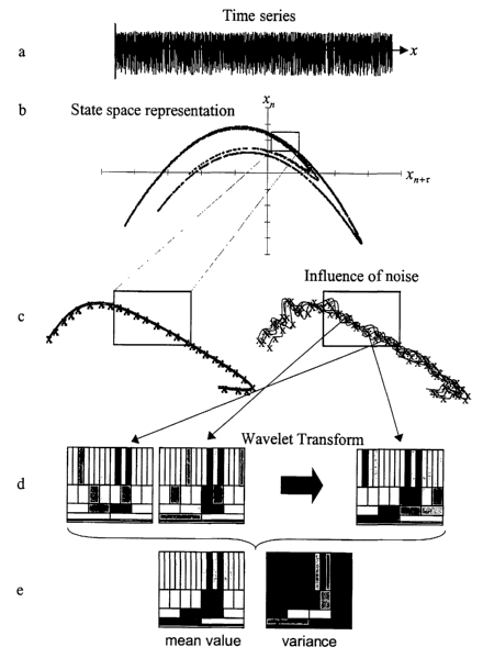
Figure 1 a represents an observed system viewed in terms of time.
Figure 1b represents an observed system viewed in terms of reconstructed state
space
and shows the densely lying trajectories of an at least approximately
deterministic system.
Figure lc depicts a portion of the state space of Figure 1b before and after
the
introduction of noise to the signal.
Figure 1 d is a mufti-resolution representation of the state space vectors in
the wavelet
domain.
Figure 1 a illustrates the high entries in wavelet coefficients representing
signal related
to directions and low entries for those of stochastic noise related
directions.
Figure 2a represents S seconds of electrocardiograph data recorded at 200 Hz
as the
pure signal recorded by the main sensor.
Figure 2b shows the frequency spectrum of the electrocardiograph after pre-
filtering
by a SOHz notch filter and a second-order low pass filter at 100Hz.
15 Figure 2c shows the signal of Figure 2b with added white noise
superimposed.
Figure 2d shows the resulting noise spectrum of Figure 2c.
Figure 2e shows the cleaned time series, after wavelet transformations and
subtraction
in state space of the signal of Figure 2c.
Figure 2f shows the frequency spectrum of the electrocardiograph after wavelet
2o transformations and subtraction in state space.
Figure 3a represents 5 seconds of magnetocardiograph signal recorded outside a
shielding room where only the main component of the heart signal (R wave) is
visible.
Figure 3b represents the frequency spectrum of the signal of Figure 3a.
Figure 3c represents 5 seconds of the simultaneously recorded noise signal of
Figure
25 3a.
Figure 3d represents the Fourier spectrum of the signal shown in Figure 3c.
Figure 3e shows the time series resulting from the present de-noising
procedure.
11
CA 02458176 2004-02-19
WO 03/026346 PCT/US02/29920
Figure 3f shows the Fourier spectrum corresponding to the Figure 3e time
series.
Figure 4a shows the original time series using the data of Example 1.
Figure 4b represents the frequency spectrum of the signal of Figure 4a.
Figure 4c shows the time series after noise reduction with ghkss.
Figure 4d represents the power spectrum of the signal of Figure 4c.
Figure 4e depicts the residuum of noise in the signal of Fig. 4a using the
present de-
noising method.
Figure 4f depicts the residuum of noise in the signal of Fig. 4a after noise
reduction
with 'ghkss'.
l0 Figure Sa depicts an excerpt of three seconds of a time series recorded
from a
pregnant woman with a low temperature SQUID within shielding.
Figure Sb shows some of the typical noise peaks at 50 Hz are missing, which
indicates
the use of a shielding chamber.
Figure Sc depicts the result after applying NLD showing the MCG of the mother
15 visible but contaminated with low frequent (respiratory) artefacts, which
may be removed by
increasing the observation time.
Figure Sd depicts the power spectrum, free from noise peaks and showing a
decreased
white noise level.
Figure Se depicts the spectrum of the QRS complexes of the foetal MCG after
20 removal of the mother's MCG from the time series and applying NLD again,
demonstrating
that previously overlapping heartbeats have been separated.
Figure Sa depicts the spectral energy of the mother's MCG.
Figure Sf depicts the spectral energy of the foetal MCG, which is much lower
but lies
within the same bandwidth as that of the mother (d) and demonstrates the
importance of
25 highly adaptive denoising procedures.
12
CA 02458176 2004-02-19
WO 03/026346 PCT/US02/29920
DESCRIPTION OF THE PREFERRED EMBODIMENTS
I have discovered the hard or soft threshold of wavelet coefficients is well
suited for
signal recovery in state space and have applied this technique to the de-
noising of
magnetocardiograph or electrocardiograph time series signals.
The present invention provides a method and system for nonlinear de-noising
(NLD)
of magnetocardiograph or electrocardiograph time series signals by performing
local
projections in the reconstructed state space using the wavelet transform to
identify and
describe deterministic structures. Thus, the goal is to locate and separate
subspaces generated
by any deterministic process independent of its source (be it the noise or the
signal of the
heart). The method consists of first separating a subspace from stochastic
noise followed by
separating different subspaces.
To represent the dynamical properties of an observed system it is useful to
operate in
the reconstructed state space (see F. Takens, 'Detecting Strange Attractors in
Turbulence',
Lecture notes in math., Springer, New York, 1981 ) instead of the time domain
(Figure 1 a).
An at least approximately deterministic system leads to densely lying
trajectories and is
constrained to a subspace (Figure 1 b) whereas a stochastic process causes a
random
distribution within the entire state space. Superimposing white noise onto a
deterministic
signal causes distortions of the primary densely lying trajectories (Figure
lc).
To identify and to describe a deterministic structure in state space, it is
useful to
transform the state space vectors into a suitable basis system. "Suitable"
means that one
attempts to find a basis function, which adapts best to the specific
deterministic structure
present.
It is possible to describe the determinism by only a few coefficients in the
domain of
the new basis system, due to the fact that directional information is
compressible. In contrast,
stochastic noise is incompressible and, therefore, needs a complete set of
basis coefficients to
be reproduced.
The wavelet transform provides many highly adaptive basis functions called
wavelets.
It is defined by translations and dilations of a basis function (a wavelet)
convolved with a
signal x (t). An additional scaling factor (mostly a power of 2) lets the
wavelet transform act
13
CA 02458176 2004-02-19
WO 03/026346 PCT/US02/29920
like a mathematical microscope; it lets one observe signal information at
different scales
dependent on its location. Exactly this property is useful, because one
obtains a multi-
resolution representation of the state space vectors in the wavelet domain
(Figure 1 d).
The general mathematical background of wavelet transforms is well known and an
introduction can be found in the paper by Mallat et al. entitled A Theory For
Multiresolution
Signal Decomposition: The Wavelet Representation, IEEE Trans. Pat. Rec. Mach.
Intel.
11:674, (1989). A comprehensive database containing the available literature
and wavelet
applications is presented in Amara Graps website:
http://www.amara.com/current/wavelet.html.
l0 It is important to choose an optimum wavelet. An optimally chosen wavelet
(analyzing function) is one which best represents the signal. For example, in
fast Fourier
transform, the analyzing functions are sine and cosine waves. Applied to a
pure sine wave, the
fast Fourier transform yields a single peak in the spectrum. However, applying
a fast Fourier
transform to a rectangular pattern requires huge amount coefficients to
properly describe this
15 pattern. The same is true with the wavelets: the better the wavelet matches
the function-of
interest (here: heartbeat) the better. It is possible to design a problem-
oriented wavelet, one of
the big advantages of the wavelet transform. For the purposes of this
invention, the best
choice in this case is the well-known Coiflet using filterorder 6. Other
Coiflet wavelet
transforms may be used, as well as Haar, Morlet, Mexican Hat, biorthogonal
spline,
20 Daubechies, Malvar, Lemarie, Meyer, and Symlet wavelet types.
The optimally chosen wavelet provides high entries in wavelet coefficients
representing signal related directions and low entries for those of stochastic
noise related
directions (Figure 1e). This allows the definition of a shrinking condition
for the projection
towards the direction of the maximal variance effectuated by the determinism
of the signal.
25 Finally, the inverse wavelet transform recovers the state space vectors
from which the cleaned
time series can be reconstructed.
The deterministic noise fills additional subspaces, which have to be separated
from
the manifold of the signal. The noise related subspaces are localized and
described by
recording the noise in an additional reference sensor and transforming the
state space vectors
30 into the wavelet basis system. Then, their signature in the time series of
the source sensor is
14
CA 02458176 2004-02-19
WO 03/026346 PCT/US02/29920
identified and a simple subtraction in state space is performed. This
procedure is superior to
common cross-correlation techniques because the dynamical properties of the
deterministic
noise are considered. It is believed that the wavelet transform has never been
used for this
purpose, especially not in conjunction with reference sensors.
The noise reduction methods described are particularly useful in obtaining
useful data
from magnetocardiographs. One particularly beneficial use of the cleaned
signal is in
determining the well being of a fetus carned by a pregnant mammal, especially
a human
being. During certain phases of pregnancy the fetal ECG is very difficult to
record because of
the insulating fat layer in the fetus. Since the magnetic permeability of
tissue is that of free
to space, MCG's of the fetus do not suffer from this failing. However, until
now, it has been
impossible to diagnose the presence of cardiac abnormalities in the fetus
using SQU)D
systems outside shielding due to the very weak signal of the fetus, and an
unusable low
signal-to-noise-ratio. Using the techniques described herein it is now
possible to separate the
signals received from the mother from those of the fetus and to determine
abnormalities in the
fetal heartbeat.
The disclosed NLD technique also provides significant advantages in
conjunction
with SQUID technology. A shielded room is not necessary in SQUID
magnetocardiography;
however the absence of shielding results in increased noise and requires more
powerful noise
cancellation techniques such as that described herein.
One of the key aspects of the inventive method is the use of adaptive
thresholding. As
used herein, thresholding means dividing the eigenspectrum of the wavelet
coefficients.
After embedding the time series into the state space, nearest neighbor search
is
performed for each single state space vector "x" and the wavelet transform is
applied. Then, a
center-of mass wavelet is created by building the mean from all transformed
vectors that are
the nearest neighbors to "x".
If it were possible to perfectly separate subspaces occupied by noise and
signal a hard
thresholding could be performed. In that case all coefficients belonging to
noise are set to
zero and the rest are kept as it is. However, since, in general, subspaces
overlap, an adaptive
thresholding is required, which accommodates the fact that some coefficients
contain both
signal and noise information.
CA 02458176 2004-02-19
WO 03/026346 PCT/US02/29920
In soft thresholding, noise coefficients are set not to zero (hard) but to a
certain value,
e.g. the mean value (soft). This keeps some information of these particular
coefficients but
decreases their importance. The more noise that overlaps with the subspace of
the signal the
more difficult it is to separate them and the more important adaptive
thresholding becomes
The concept underlying the mathematical methodology of NLD is the performance
of
local projections in the reconstructed state space using the wavelet transform
to identify and
describe deterministic signal structures. The goal is to locate and separate
subspaces
generated by any deterministic process independent of its source (be it the
noise or the signal
of the heart). The procedure consists of two parts: ( 1 ) the separation of a
subspace from
t 0 stochastic noise and (2) the separation of different subspaces, which are
described below.
To represent the dynamical properties of an observed system it is useful to
operate in
the reconstnzcted state space instead of the time domain. Fig. 1 a shows the
time domain plot
of the x-component of a sample time series, which is known as Henon map and
defined as
follows:
xn+~ =1.4 - x~ + 0.3yn
yn+I xn
Obviously, it is impossible to recognize any dynamical property of the
underlying
(deterministic) system. Time delay embedding of the Henon map leads to the
following state
space vectors:
"(n) - (xn ~'xn-s ~'x'n-2r s..., xn_~m_~~r )
where i denotes the time delay and m the embedding dimension. The state space
representation of the Henon map is given in Fig. 1b). Here, using T = 1 and m
= 2 the
components of the state space vectors are depicted in a two dimensional graph
by plotting
component x"+r against x". An at least approximately deterministic system
leads to densely
lying trajectories and is constrained to a subspace whereas a stochastic
process causes a
random distribution within the entire state space.
Superimposing white noise to a deterministic signal causes distortions of the
primary
densely lying trajectories. The left graph of Fig lc shows an excerpt of some
(bunched)
16
CA 02458176 2004-02-19
WO 03/026346 PCT/US02/29920
trajectories of Fig 1b. The effect of superimposing noise to this excerpt is
demonstrated in the
right part of Fig. 1 c.
The next step is to identify and to describe a deterministic structure in
state space. For
this purpose it is useful to transform the state space vectors into a suitable
basis system.
"Suitable" means that one attempts to find a basis function that adapts best
to the
deterministic structure. In this case it is possible to describe the
determinism by only a few
coefficients in the domain of the new basis system. This is due to the fact
that directional
information is compressible. In contrast, stochastic noise is incompressible
and, therefore,
would need a complete set of basis coefficients to be reproduced.
The wavelet transform provides many highly adaptive basis functions called
wavelets.
It is defined by translations and dilations of a basis function (a wavelet)
convolved with a
signal x(t). An additional scaling factor (mostly a power of 2) lets the
wavelet transform act
like a mathematical microscope, which means that it lets one observe signal
information at
different scales dependent on its location. Exactly this property is useful,
because one obtains
a mufti-resolution representation of the state space vectors in the wavelet
domain (see Fig 1 d).
With an optimally chosen wavelet one can expect high entries in wavelet
coefficients
representing signal related directions and low entries for those of stochastic
noise related
directions (Fig. le). This enables one to define a shrinking condition for the
projection
towards the direction of the maximal variance effectuated by the determinism
of the signal.
Finally, the inverse wavelet transform recovers the state space vectors from
which the cleaned
time series can be reconstructed.
Adaptive (hard or soft) thresholding of wavelet coefficients is well suited
for signal
recovery even in state space and is important in de-noising of MCG or ECG time
series
signals.
The deterministic noise fills additional subspaces, which have to be separated
from
the manifold of the signal. In application to MCG, the noise related subspaces
are localized
and described by recording the noise in an additional reference sensor and
transforming the
state space vectors into the wavelet basis system. Then, their signature in
the time series of
the source sensor is identified and a simple subtraction in state space is
performed. This
17
CA 02458176 2004-02-19
WO 03/026346 PCT/US02/29920
procedure is superior to common cross-correlation techniques because the
dynamical
properties of the deterministic noise are considered.
The significance of NLD resides in its potential ability to separate weak
useful
bioelectric or biomagnetic signals from many orders of magnitude stronger
noise, without
recurnng to intensive signal averaging and filtering (both of which distort
the signal to be
measured.)
To demonstrate the efficiency of the novel de-noising scheme, it was applied
to
simulated signals using electrocardiographic data of a healthy patient wherein
the data is
recorded at 200Hz as the pure signal recorded by a main sensor
l0 Example 1
NLD was applied to simulated noisy signals, starting from a 5 second ECG
recording
of a healthy heart, recorded at 200Hz bandwidth, and taken as the pure signal
from the main
sensor. This ECG was pre-filtered by a SOHz notch filter and a second-order
low pass filter at
100Hz (Figs. 2a and 2b).
15 Subsequently, white noise is added with an amplitude variance of 30%
referred to the
electrocardiograph's variance, and the deterministic noise. The deterministic
noise had
frequency peaks at 16 2/3Hz, SOHz (rail power supply in Europe and
subharmonics), and
60Hz (signal analysis systems) with an amplitude variance of 100%.
The deterministic noise had frequency peaks at 16 2/3Hz, SOHz (power supply in
20 Europe and subharmonics), and 60Hz (signal analysis systems) with an
amplitude variance of
100% (see Figs. 2c and 2d). A reference noise time series was created using
the same
parameters as mentioned above, but additionally, with variations in amplitude
and a constant
phase shift for the deterministic noise components.
Figure 2c shows the signal with added white noise superimposed, and Figure 2d
the
25 resulting noise spectrum. The reference time series is generated by
creating noise using the
same parameters as mentioned above, but additionally, with variations in
amplitude and a
constant phase shift for the deterministic noise components.
After wavelet transformations and subtraction in state space, Figure 2e shows
the
cleaned time series. A reference time series is generated by creating noise
using the same
18
CA 02458176 2004-02-19
WO 03/026346 PCT/US02/29920
parameters as mentioned above, but additionally, with variations in amplitude
and a constant
phase shift for the deterministic noise components. Figure 2f shows the
frequency spectrum
of the electrocardiograph after wavelet transformations and subtraction in
state space.
One can infer from Figure 2e that the baseline between the heartbeats (a good
indicator of the de-noising quality) is almost noise free. Hence, the present
invention's
method performed both signal preservation and considerable noise reduction.
Example 2
As an example of measured signal data, data obtained from the
magnetocardiograph
l0 of a healthy patient recorded outside a shielding room using a laboratory
HTSQUID system is
depicted in Figure 3. Five [5] seconds of magnetocardiograph signal was
obtained as depicted
in Figure 3a. The patient's heartbeat is only barely visible in Figure 3a. A
simultaneously
recorded noise time series was recorded as depicted in Figure 3c.
The frequency spectrum of the signal depicted in Figure 3a is shown in Figure
3b; that
15 depicted in Figure 3c is shown in Figure 3d. Due to the width of the 50 Hz
peak in the
spectrum no notch filter was used.
For this measurement, two axial gradiometers of first order with 7 cm baseline
were
mounted at a distance of 7 cm one above the other. In this example, the top
gradiometer
recorded the reference signal (Figures 3c and 3d).
20 Figures 3e and 3f show the time series along with its corresponding Fourier
spectrum
resulting from the present de-noising procedure. In the reconstructed
magnetocardiograph
[MCG] of Figure 3e, even small details of the heartbeat are revealed. Again,
the baseline
between the heartbeats is almost noise free.
Example 3
25 Figure 4a-b illustrates the superiority of the present invention's system
and method
over one of the prior art de-noising techniques.
The analysis of the Example 2 data set based upon this method is shown in
Figures 4a
and 4b. The tool 'ghkss' described in the paper by Hegger et al. entitled,
'Nonlinear Time
Series Analysis (TISEAN)', incorporated herein by reference, is used to
analyze the data set.
19
CA 02458176 2004-02-19
WO 03/026346 PCT/US02/29920
This is the algorithmic form of "Nonlinear Noise Reduction For
Electrocardiograms" (Chaos
6:87,1995).
The tool 'ghkss' was applied to the data set and obtained the results shown in
Figs. 4c
and 4d. Obviously, NLD reaches a better noise reduction quality in this case,
clarified by the
respective residuums (see Figs. 4e and 4f). This is due to the fact that
'ghkss' is not able to
separate overlapping subspaces in state space, which is one of the most
important features of
NLD.
To illustrate this, an analysis of the same data is performed based upon the
technique
described in the paper by Schreiber et al. entitled, 'Nonlinear Noise
Reduction For
Electrocardiograms' (Chaos 6:87, 1995), the disclosure of which is
incorporated herein by
reference. In summary form, the procedure reduces stochastic noise by
performing local
projections in state space using the covariance matrix. The signals' signature
is localized in
state space and is projected onto a noise-free subspace indicated by the
largest eigenvalues of
the covariance matrix. This method works well, but only if the dimension of
the signals'
subspace in state space is known.
The results of the analysis are shown in Figures 4c and 4d. It should be noted
that
NLD reaches a better noise reduction quality in this case. This is also
demonstrated by the
respective residuums of noise (Figures 4e and 4f). The NLD residuum is much
lower than
that of 'ghkss'. This is due to the fact that, in contrast to NLD, 'ghkss' is
not able to separate
overlapping subspaces in state space, while NDL does. That separation ability
is one of the
most important features of NLD.
NLD was also compared with another existing technique, Frequency Dependent
Gradiometry (FDG) and NLD were applied to the same MCG sample, and it turned
out that
NLD performed a much superior noise reduction.
Example 4
The example shows the applicability of the inventive method to measurement of
a
foetal heartbeat using MCG. Figure Sa shows an excerpt of three seconds of a
time series
recorded from a pregnant woman with an LTSQUID within shielding. In Figure Sb
some of
CA 02458176 2004-02-19
WO 03/026346 PCT/US02/29920
the typical noise peaks at SO Hz are missing, which indicates the use of a
shielding chamber.
In the first NLD step the deterministic noise components are removed.
Figure 5c shows the result after applying the second NLD step. The MCG of the
mother is visible being still contaminated with low frequent (respiratory)
artefacts, which may
be removed by increasing the observation time. Its power spectrum in Figure Sd
is free from
noise peaks and shows a decreased white noise level.
Removal of the mother's MCG from the time series and applying NLD again, the
QRS complexes of the foetal MCG are obtained as shown in Figure 5e. Note that
even
previously overlapping heartbeats are separated. The spectral energy of the
foetal MCG
shown in Figure Sf is much lower but lies within the same bandwidth as that of
the mother's
shown in Figure Sd. This further demonstrates the importance of highly
adaptive denoising
procedures.
The programming of the present invention may be implemented by one of skill in
the
art of digital signal processing.
The above examples demonstrate the effective implementation of a nonlinear
noise
reduction method for magnetocardiograms using wavelet transforms. While
various preferred
embodiments have been shown and described, it will be understood that there is
no intent to
limit the invention by such disclosure, but rather, it is intended to cover
all modifications and
alternate constructions falling within the spirit and scope of the invention,
as defined in the
2o appended claims.
21