Note: Descriptions are shown in the official language in which they were submitted.
CA 02458880 2004-02-26
WO 03/017867 PCT/US02/23908
-1-
INTRAOCULAR LENS IMPLANT HAVING
EYE ACCOMMODATING CAPABILITIES
BACKGROUND OF THE INVENTION
Field of the Invention
The present invention is broadly concerned with improved intraocular lenses
which can be surgically implanted as a replacement for the natural crystalline
lenses in
the eyes of cataract patients. More particularly, the invention is concerned
with such
intraocular lenses which have a specially configured, resilient optic
positioning element
serving to maintain the equatorial segment of the positioning element in
substantial
contact with the corresponding equatorial portion of the capsule of the eye.
Description of the Prior Art
Cataracts occur when the crystalline lens of the eye becomes opaque. The
cataracts may be in both eyes and, being a progressive condition, may cause
fading
vision and eventual blindness. Cataracts were once surgically removed along
with the
anterior wall of the capsule of the eye. The patient then wore eyeglasses or
contact
lenses which restored vision but did not permit accommodation and gave only
limited
depth perception.
The first implant of a replacement lens within the eye occurred in 1949 and
attempted to locate the replacement lens in the posterior chamber of the eye
behind the
iris. Problems such as dislocation after implantation forced abandonment of
this
approach, and for some period thereafter intraocular lenses were implanted in
the
anterior chamber of the eye.
Others returned to the practice of inserting the lens in the area of the eye
posterior to the iris, known as the posterior chamber. This is the area where
the
patient's natural crystalline lens is located. When the intraocular lens is
located in this
natural location, substantially normal vision may be restored to the patient
and the
problems of forward displacement of vitreous humor and retina detachment
encountered
in anterior chamber intraocular lenses are less likely to occur. Lenses
implanted in the
CA 02458880 2004-02-26
WO 03/017867 PCT/US02/23908
-2-
posterior chamber are disclosed in U.S. Patent Nos. 3,718,870, 3,866,249,
3,913,148,
3,925,825,4,014,049,4,041,552,4,053,953, and 4,285,072. None of these lenses
have
focusing capability.
Lenses capable of focusing offered the wearer the closest possible substitute
to
the crystalline lens. U.S. Patent No. 4,254,509 to Tennant discloses a lens
which moves
in an anterior direction upon contraction of the ciliary body located anterior
to the iris.
Though providing focusing capabilities, it presents the same disadvantages as
other
anterior chamber lenses. U.S. Patent No. 4,253,199 to Banko approaches the
problem
ofproviding a focusable lens differently, byproviding a replacement lens of
deformable
material sutured to the ciliary body. This lens functions much as the original
crystalline
lens but risks bleeding from the sutures.
U.S. Patent No. 5,674,282 to Cumming is directed towards an accommodating
intraocular lens for implanting within the capsule of an eye. The Cumming lens
comprises a central optic and two plate haptics which extend radially outward
from
diametrically opposite sides of the optic and are movable anteriorly and
posteriorly
relative to the optic. However, the Cumming lens suffers from the same
shortcomings
as the Levy lens in that the haptics are biased anteriorly by pressure from
the ciliary
bodies. This will eventually lead to pressure necrosis of the ciliary body.
Finally, U.S. Patent No. 4,842,601 to Smith discloses an accommodating
intraocular lens having anterior and posterior members which urge against the
anterior
and posterior walls of the natural lens capsule. The muscular action exerted
on the
natural capsule will thus cause the lens to flatten, thereby changing the
focus thereof.
The Smith lens is formed of first and second plastic lens members connected to
one
another adjacent their peripheral edges so as to provide a cavity
therebetween. The
connection between the lens members is accomplished by way of a U-shaped
flange on
the first member which forms an inwardly facing groove for receiving an
outwardly
extended flange on the second member. The Smith lens is lacking in that the
first and
second members must be separately inserted into the capsule and assembled
within the
capsule which is extremely difficult for even highly skilled surgeons to
accomplish.
CA 02458880 2004-02-26
WO 03/017867 PCT/US02/23908
-3-
SUMMARY OF THE INVENTION
The present invention represents a significant advance in the art and provides
an accommodating intraocular lens for implantation substantially within the
confines
of the capsule of the human eye between the anterior and posterior capsule
walls. The
lens comprises a single optic presenting opposed anterior and posterior
surfaces,
together with a resilient optic positioning element coupled to the optic to
cooperatively
present a shape that generally conforms to the shape of the capsule. The optic
positioning element has a posterior face configured for yieldable engagement
with the
posterior capsule wall, and an anterior face configured for yieldable
engagement with
the anterior wall of the capsule. The positioning element also defines an
equatorial
segment of maximum diameter between the, anterior and posterior faces. The
positioning element is operable to substantially maintain the equatorial
segment thereof
in contact with at least a part of the capsule equatorial portion in
essentially all
orientations and conditions of accommodation of the lens within the capsule.
The positioning element is preferably formed of a yieldable synthetic resin
material to present a unitarily formed, seamless body having an elastic
memory. In
practice, the lens of the invention is surgically implanted within a capsule,
so as to take
full advantage of the "rubber band effect." This in turn assures accurate lens
accommodation in response to contraction and relaxation of the ciliary body,
acting
through the zonules and the elastin tissue of the eye.
BRIEF DESCRIPTION OF THE DRAWINGS
Figure 1 is a generally schematic vertical sectional view of an intraocular
lens
in accordance with the invention, shown mounted in the capsule of an eye;
Fig. 2 is a view similar to that of Fig. 1, but illustrating the intraocular
lens in
an accommodated position owing to relaxation of the ciliary muscle;
Fig. 3 is a plan view of a preferred lens of the invention;
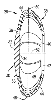
Fig. 4 is a vertical sectional view taken along line 4-4 of Fig. 3 and further
illustrating the construction of the intraocular lens;
Fig. 5 is a top perspective view of the lens of Fig. 3;
Fig. 6 is a bottom perspective view of the lens of Fig. 3;
CA 02458880 2004-02-26
WO 03/017867 PCT/US02/23908
-4-
Fig. 7 is an enlarged, fragmentary view of another embodiment in accordance
with the invention, including a thin membrane in covering relationship to
openings
present in the optic positioning element to impede migration of cells
therethrough; and
Fig. 8 is a vertical sectional view taken along line 8-8 of Fig. 7 and further
depicting the construction of the Fig. 7 embodiment.
DETAILED DESCRIPTION OF THE PREFERRED EMBODIMENT
Turning now to the drawings, the present invention provides an intraocular
lens
for surgical replacement of the human lens in the treatment of cataracts in
the human
eye. Figs. 1 and 2 illustrate various components of the human eye. Briefly,
the eye 10
includes a frontal portion 12 and a rearward portion including the retina (not
shown).
The frontal portion 12 of the eye 10 is covered by a cornea 14 which encloses
and forms
an anterior chamber 16. The anterior chamber 16 contains aqueous fluid and is
bounded at the rear by an iris 18. The iris 18 opens and closes to admit
appropriate
quantities of light into the inner portions of the eye 10. The eye 10 also
includes a
capsule 20 which ordinarily contains the natural crystalline lens. When the
eye 10
focuses, the capsule 20 changes shape to appropriately distribute the light
admitted
through the cornea 14 and the iris 18 to the retina at the rearward portion of
the eye 10.
The capsule 20 is supported within eye 10 by means of the ciliary muscle 22
which supports zonules 24, the latter including elastin tissue substantially
about the
equatorial portion 27 of the capsule 20.
The retina is composed of rods and cones which act as light receptors. The
retina includes a fovea which is a rodless portion that provides for acute
vision. The
outside of the rearward or posterior portion 14 of the eye 10 is known as the
sclera
which joins into and forms a portion of the covering for the optic nerve.
Images
received by the retina are transmitted through the optic nerve to the brain.
The area
between the retina and the capsule 20 is occupied by vitreous fluid.
Ocular adjustments for sharp focusing of objects viewed at different distances
is accomplished by the action of the ciliary body 22 on the capsule 20 and the
naturally
occurring crystalline lens. This is accomplished through the zonules 24 and
the elastin
tissue 26 forms a part thereof creating a so-called "rubber band effect." For
example,
CA 02458880 2004-02-26
WO 03/017867 PCT/US02/23908
-5-
when the ciliary body 22 contracts, the zonules 24 and elastin tissue 26
exerts forces
on the capsule 20 to achieve a more spherical shape as shown in Fig. 2 for
viewing
objects that are nearer the viewer. When the ciliary body 22 retracts and
pulls on the
zonules 24, a force is exerted in an opposite direction to make the capsule 20
more
discoid; objects at a distance can then be viewed in proper focus.
Referring now to Figs. 3-6, a preferred intraocular lens (IOL) 28 is
illustrated.
The IOL 28 includes a central optic 30 which may be formed of an acrylic or
similar
synthetic resin material and presents an anterior surface 32 and an opposed
posterior
surface 34. The surfaces 32, 34 are normally convex, although the shape of
these
surfaces and the overall size of the optic 30 an be varied depending upon the
user's
eyesight. The optic 30 is also provided with four circumferentially spaced
through-
openings 36.
The IOL 28 further includes a resilient positioning element 38 which serves to
locate the optic 30 within a human capsule 20 and to effect accommodation of
the lens.
The element 38 may be integral with optic 30 or may be structurally distinct;
in either
case the element 38 is preferably unitarily formed as a seamless component. As
illustrated, the element 38 includes an annular posterior segment 40 with a
central
opening 42. A plurality of circumferentially spaced, arcuate in cross-section
positioning
legs 44 extend from the segment 42 and are joined to the margin of optic 30,
with
openings 46 defined between adjacent pairs of the legs 44. As perhaps best
seen in Fig.
4, the legs 44 cooperatively present, with the optic 30, a substantially
discoid shape with
a central chamber 45. However, the legs 44 also define an annular equatorial
segment
48 disposed on opposite sides of equatorial axis 50 (Fig. 4). The overall IOL
further
presents a central polar axis 52 as shown. Preferably, the outside dimension
of the IOL
28 at the equatorial segment 48 is from about 9-11mm, usually about 10mm. On
the
other hand, the outside dimension along polar axis 52 is typically from about
2-4mm,
usually about 3mm.
The positioning element 38 is preferably formed of any appropriate
biologically
inert material conventionally used in IOL construction (e.g., elastic,
synthetic resin
materials). Examples of suitable lens materials include acrylates (such as
polymethylmethacrylates), silicones, and mixtures of acrylates and silicons.
It is
CA 02458880 2009-07-03
WO 03/017867 PCT/US02/23908
-6-
particularly preferred that lenses according to the invention be constructed
of a material
having an elastic memory (i.e., the material should be capable of
substantially
recovering its original size and shape after a deforming force has been
removed). An
example of a preferred material having elastic memory is MEMORYLENS (available
from Mentor Ophthalmics in California).
A particular feature of IOL 28 is that the positioning element 38 thereof is
configured so as to substantially conform with the capsule 20, particularly to
the
equatorial portion 27 of the capsule. This is shown in Figs. 1 and 2, where it
will be
observed that the equatorial segment 48 of the IOL 28 is in substantially
conforming
contact with the inner surface of the equatorial portion 27 of capsule 20.
Note also that
this close conforming relationship is maintained notwithstanding the extent of
accommodation of the lens 28. In this fashion, the lens 28 makes full use of
the "rubber
band effect" of the natural eye 10, and thus more closely mimics accommodation
of the
eye's natural lens.
Intraocular lens 28 substitutes both locationally and functionally for the
original,
natural, crystalline lens. In order to insert the lens 28 into the capsule 20,
an ophthalmic
surgeon would remove the natural lens (and thus the cataracts) by conventional
methods, leaving an opening 54 in the anterior wall of the capsule 20. Lens 28
is then
folded into a compact size for insertion into the capsule 20 through the
opening 54.
Once inserted, the capsule 20 is filled with fluids (e.g., saline solution)
which enter the
lens 28, causing the lens 38 to return to its original, non-deformed state as
shown in Fig.
1. There is no need to suture the lens to the capsule 20 because, due to the
size and
shape of the lens 28 as described above, the lens 28 will not rotate or shift
within the
capsule 20.
Implantation of the IOL 28 restores normal vision because, not only does the
lens 28 replace the patients occluded natural lens, but the normal responses
of the ciliary
body 22 cooperate with the zonules 24 and elastin tissue 26 during focusing of
the lens
28. In Fig. 2, the focal length between the posterior surface 34 of optic 30
and the fovea
is greater to permit viewing of nearby objects. The focal length is greater
because the
ciliary muscle or body 22 has contracted, making the capsule 20 more spheroid;
this
causes the lens 28 to be maintained in its tensioned state, positioning the
optic 30
* Trade-mark
CA 02458880 2009-07-03
WO 03/017867 PCT/US02/23908
-7-
anteriorly. The lens 28 thus follows the eye's natural physiology for focusing
to provide
a substitute means of optical accommodation. When the object of observation
becomes
more distant, the sensory cells within the retina signal the ciliary body 22
to relax, thus
pulling on the zonular fibers 24 to make the capsule more discoid as shown in
Fig. 1.
In so doing, the polar dimension of the capsule 20 is narrowed, which in turn
causes the
polar dimension of the lens 28 to narrow in a similar manner. This narrowing
causes
the optic 30 to move posteriorly as the capsule 20 and the lens 28 become more
discoid.
The focal length between the posterior surface 34 of optic 30 and the fovea is
thus
shortened, and the object remains in focus.
Figs. 7 and 8 illustrate a modified IOL28awhich is identical in all respects
with
IOL 28, save for the provision of a very thin membrane 56 in covering
relationship to
the openings 46 between positioning legs 44. It is contemplated that the
membrane 56
would be formed of the same synthetic resin as the positioning element 38, but
would
be much thinner (on the order of a few thousandths of an inch) than the
remainder of
the element 38. The purpose of membrane 56 is to prevent or at least impede
the
passage of migratory cells through the openings 46 and into the chamber 45 of
the IOL.
The subject matter ofU.S. Patent No. 6,217,612 issued April 17, 2001, and the
subject matter ofU.S. Patent No. 6,299,641 issued October 9, 2001 are relevant
subject matters for this patent application.
