Note: Descriptions are shown in the official language in which they were submitted.
~ f Y"T y:,e;~
CA 02459002 2004-02-27
iu vad._~~~y,.,.
''~ IE:50 Fram-GILL JENNINGS & EVERY +,~4 20 t3tt 1310 T-A09 P.~D6/Q13
A CATHETER P~StTIONtNG DEVICE
Field of the lnventivn
The present invention relates to medical devices for controlling the
positioning
of an infra vascular catheter device. In particular, the present invention is
concerned
with a positioning device forrelative positioning of lumen in a multi-lumen
intravascular
catheter.
Background to the Invention
1D The human vascular system may suffer from a number of problems. These
may broadly be characterised as cardiovascular and peripheral vascular
disease.
Among the types of disease, atherosclerosis is a particular problem.
Atheroscierotic
plaque can develop in a patient's cardiovascular system. The plaque can be
quite
extensive and vcGude.a substantial length ofthe vessel. Additionally, the
plaque may
be inflamed and unstable, such plaque being subject to rupture, erosion or
ulce!-ation
which can cause the patient to experience a myocardial infarction, thrombosis
yr other
traumatic and unwanted effects.
Our cv-pending International Application No. PCTIEP01~04401 discloses a
vascular catheter apparatus for temperature measurement of vascular tissue.
' z o Imporkantly, it has been reported that unstable and inflamed plaque can
cause the
temperature of the artery waif to elevate up to 2.5°C proximate the
inflamed plaque.
With the vascular catheter apparatus described in PCTIEP01I04401, detection of
the
temperature at the vascular wall is enabled. 'The temperature information ~ is
subsequently transferred via the carrierto a remote device where the wall
temperature
can be detected arid recarded_ The device is able to locate inflamed plaque by
monitoring the vascular wall tar elevated temperatures. This may be achieved
by
measuring temperature relative to normal segments of a vessel or absolute
temperature values.
The tharmography catheter is inserted into the target tissue using usual
catheterisation techniques. This usually involves the use of a guide catheter.
Problems associated with systems inGVrporating a guide catheter include its
relative positioning with respect to the catheter. For example, where it is
desired tp
deliver a catheter to the coronary arteries, the guide catheter is maneuvered
.to the
opening of the coronary arteries by a physician, in order that the catheter
may be
AMENDED SHEET°
Empf .~e i t :18/ 1 ~l~flt73 11: 49 ~, m . ~~.... . ~.m~~ -nr -: 717 P .006
CA 02459002 2004-02-27
WO 03/022345 PCT/EP02/09430
2
introduced to the coronary arteries. A problem may arise when the catheter is
moved
in the coronary arteries. As the physician tries to maneuver the catheter in
the cardiac
tissue, this may cause the guide catheter to be pushed away from the entrance
to the
coronary artery, consequently causing the catheter to be dragged out of the
coronary
artery. This problem is exaggerated where mufti-lumen catheters are employed,
such
as the thermography catheter described in PCT/EP01104401.
In orderto accurately identify regions of unstable plaque, the vascular
catheter
apparatus described in PCT/EP01104401 is controlled by a positioning device,
referred
to as a pull-back device. The pull-back device includes a number of lumen
mounts
that can be selectively connectable to a drive mechanism that can be used to
manipulate and maneuver a catheter within a guide catheter and cardiac tissue.
A
guide catheter mount is fixed relative to the patient so that the guide
catheter, once
in position, does not move relative to the patient.
A problem associated with such a system is that the length of guide catheter
remaining outside the patient is dependent on the size of the patient. For
example,
interventional cardiovascular treatment of a large patient will leave a
shorter length of
guide catheterand catheteroutside the patient than treatment of the equivalent
region
in a smaller patient. Furthermore, the relative length of catheter to guide
catheter left
outside the patient for treatment of different regions, eg. peripheral heart
tissue rather
2 o than the main coronary arteries, will differ. This poses a problem as, in
use, the guide
catheter and catheter must both be fixed to the positioning device in what is
a fixed
relative separation dictated by the physical dimensions of the positioning
device and
the lumen mounts.
Summary of the Invention
According to a first aspect of the invention, a catheter positioning system
comprises a guide catheter extension adapted to co-operate with a guide
catheter, a
catheter positioning device adapted to engage a catheter and guide the
catheter
within the guide catheter extension, wherein the guide catheter extension
further
3 0 comprises a plurality of engagement means for engaging the positioning
device and
thereby fixing the relative positions of the guide catheter extension and the
positioning
device at any one of a number of positions over its length.
The present invention allows the distance between a guide catheter and a
positioning device to be manipulated by the user. Thus the guide catheter may
be
CA 02459002 2004-02-27
WO 03/022345 PCT/EP02/09430
3
fixed in position relative to both patient and positioning device, while
providing the
optimum distance between the effective length of the guide catheter (guide
catheter
and guide catheter extension) and the points at which the catheter is fixed to
the
positioning device.
The guide catheter extension of the present invention is adapted to receive a
catheter used in interventional cardiology. Preferably, the body of the guide
catheter
extension_is substantially cylindrical in cross section and has a diameter in
the range
of 1-15 mm. Preferably the diameter is in the range of 2-10 mm, more
preferably 3-7
mm. Preferably, the length of the guide catheter extension is in the range of
0.1 m to
1 m. More preferably, the length of the guide catheter extension is 0.15-0.5
m.
The body of the guide catheter extension may be formed from standard guide
catheter materials. For example nylon, PTFE, polyurethane, polyethylene and
nitinol
and mixtures thereof may be used. It may also be made from metals such as
aluminium, steel and alloys thereof.
The guide catheter extension preferably has a number of points adapted for
engagement with the catheter positioning device. Notches, annular
indentations, and
any other suitable means may be used. Preferably there are 2-200 fixation
points,
more preferably 5-100, most preferably 10-50 fixation points. These engagement
means enable the guide catheter extension to be fixed in place, at selected
positions
2 0 over its length, on the catheter positioning device.
The guide catheter extension comprises a distal and a proximal end.
Preferably, the distal end is adapted for engagementwith the guide catheter,
while the
proximal end is adapted for engagement with the catheter positioning device.
According to a second aspect of the present invention, there is provided a
guide catheter extension for a guide catheter having a proximal end and a
distal
end, the guide catheter extension being capable of receiving a catheter and
comprising a substantially rigid tubular section capable of sealing engagement
within
a compression fitting of the guide catheter provided at the proximal end of
the guide
catheter.
3 o Where positioning of the catheter, therefore translationai movementwithin
the
vascular tissue (therefore also within the guide catheter and guide catheter
extension)
is required, the arrangement allows the junction between the guide catheter
extension
and guide catheter to be sealed by tightening the compression fitting, but
does not
allow the junction to impinge on the catheter within. The seal is preferably
achieved
CA 02459002 2004-02-27
WO 03/022345 PCT/EP02/09430
4
by providing a sealing element in the guide catheter extension which forms a
low
friction, slidable seal with the sheath of the catheter. Thus the catheter is
able to be
moved and positioned within the apparatus without undue friction being applied
to the
catheter. This is particularly important as a Y-piece, in addition to being
used as the
injection point for contrast medium into the patient, is also used as a
pressure
measurement point during the interventional procedure. In order for the
pressure of
the patient to_be_celiab(y_measured,~the system-_must._be substantially
_closed,
otherwise the pressure will vent at a non closed section. ' This will lead to
loss of
pressure, loss of blood, and unreliable pressure readings. However, the
present
system maintains the pressure of the system as the guide catheter and guide
catheter
extension junction is sealed and the diameter of the catheter is generally
slightly less
than the diameter of the internal lumen of the guide catheter extension.
Alternatively,
the pressure is maintained by providing the above mentioned sealing element in
the
guide catheter which forms a low friction, slidable seal with the sheath of
the catheter.
Most preferably, the distal end of the guide catheter extension is adapted for
engagement with a standard Y-piece used in interventional cardiology, having a
compression fitting. This substantially prevents loss of blood or fluid at the
junction
between the guide catheter and the guide catheter extension.
In a most preferred embodiment, the distal end of the guide catheter extension
2 0 comprises a substantially rigid tubular section which is fixed to a
flexible section, and
which is co-axial therewith. The rigid tubular section may be integrally
moulded with
the flexible section. Alternatively, it is fixed to the flexible section by
any suitable
means, for example, glue, soldering, welding and the like.
The catheter positioning device is preferably a type for positioning a
catheter
and comprises a first lumen mount for holding a first lumen of the catheter, a
second
lumen mount for holding the guide catheter extension, and a drive mechanism,
wherein the first lumen mount is selectively connectable to the drive
mechanism for
relative movement with respect to the second lumen mount.
The second lumen mount preferably includes a bracket, preferably adapted for
3 o engagement with the guide catheter extension. The bracket is usually
located at one
end of an extension arm, while the other end is connected to the body of the
positioning device.
In a preferred embodiment, the positioning device is a pull-back device which
is used for positioning and/or controlled withdrawal of a catheter.
CA 02459002 2004-02-27
WO 03/022345 PCT/EP02/09430
In a particularly preferred embodiment, the present invention is used in
concert
with a vascular catheter apparatus, especially multi-lumen and/or sheathed
catheters;
which require precise positioning and or maneuvering within vascular tissue.
For
example, the present invention finds particular utility with catheters used
for
5 measurement of a physical parameter and/or treatment of vascular tissue, and
which
comprise a flexible body, and at least one sensor and/or treatment means.
-Treatment means include those-usuall-y used in inter_venfiional cardiology,
such
as ablation apparatus (for example electrodes), drug delivery ports, tissue
removal
apparatus, scents and scent positioning means (for example, balloons or
sleeves) and
1 o the like.
In a particularly preferred embodiment, the present invention is used in
concert
with a vascular catheter apparatus, preferably a catheter apparatus comprising
a
body, at least one resiliently biased projection depended from the body, a
sensor
carried by the projection, and an electrical carrier connected to the sensor
for
l5 . transmitting data from the sensor to a remote device, wherein the
electrical carrier is
coiled. Such a device is described in our earlier filed European patent
application no.
01306599.0 In this embodiment, the electrical connection is coiled to reduce
the strain
at critical points where it is necessary to maintain a seal, and hence
electrical
isolation. The design is also especially suitable for use with a vascular
thermography .
2 o catheter apparatus of the type described in our earlier filed
International patent
application no. PCT/EP01/04401.
Preferably, the electrical carrier is coiled around the body of the
projection.
Preferably, the pitch of the coil is arranged such that there are 5 to 10
turns per
cm.
2 5 Preferably, a heat shrink wrapping is applied over at least a portion of
the
length of the projection. A heat shrink material is generally a polymeric
material
capable of being reduced in size upon application of heat. These are generally
used
in the form of a tube. Suitable materials include polyesters, PVC,
polyolefins, PTFE
and the like. The preferred material is a polyester.
3 o Generally, the thermography catheter comprises a plurality of co-axial
lumen.
Preferably, the thermography catheter comprises a central lumen adapted to be
mounted on a standard angioplasty guide wire suitable for vascular
intervention. The
apparatus is preferably based on the rapid-exchange orthe monorail system,
although
over-the-wire techniques are also envisaged. Preferably, outside the central
lumen
2 r~0or~ ' ' CA 02459002 2004-02-27 ~~O.ZO~~3~
~'~" '1"u~-VOb'LV~.. «'10:50 From-GILL JENNINGT & EVERY +44 20 73f'~ 1310 T-
80B P.OOTl01~3 ..__
6
is located an intermediate lumen. Preferably, outside the intermediate lumen
is
mounted an external lumen, hereinafter ro(erred to as a sheath. Preferably, at
the
distal tip of the apparatus is a guide member. Other lumen may be present and
all the
lumen may house components within themselves or between adjacent lumen. .
The projection is preferably mounted on the central or intermediate lumen but
ma_ y be attached to any lumen inside the sheath.
The central lumen may be formed from the standard catheter Lumen materials,
for example, nylon, FEP, polyurethane, polyethylene and nitinol and mixtures
thereof.
The intermediate lumen and the sheath are generally construcked from, but
s o individually selected from, the standard catheter lumen materials
discussed above.
The sheath is adapted to fit ever the adjacent lumen housed inside the sheath
and should be able to move relative to the adjacent lumen under the control of
a
remote device.
Preferably, the central and intermediate lumen are bound to one another and
1S are not moveable relative to one another
Preferably, the flexible body of the catheter has a longitudinal axis and at
least
part of the projections are extensible radialty from the longitudinal ~aacis
of the body.
Generally, the projection$ have an elongate shape, preferably having
dimensions in
the range of 2 mm to 15 mm, mare preferably 3 to 7 mm in length. The
projections
2 o preferably have a caliper of 0.3 mm to 5 mrn, more preferably 0,5 mm to 3
mn7.
A first end of the projection is preferably attached to the body, preferably
the
intermediate andlor the central lumen, while a second end comprises one yr
more
sensors. The second end is preferably free, ie, not attached to any of the
lumen, and
is adapted to be radially movable away from the central lumen.
~25 Twv or more sensors, preferably 2 to 10 sensors, more preferably 2 to 6
sensors may be utilised in the catheter apparatus. Preferably, each sensor is
mounted on a separate projectivn_ In a particularly preferred example, four
projections, each having a single sensor mounted thereon, are provided.
The sensors are preferably located on an outer face of the projection,
n:lative
3 o the central lumen, ie., facing the vascular tissue in use. Each sensor
should
preferably be located toward, ar at the distal tip of the projection.
The projections need not be mounted in substantially the same circumferential
plane of the catheter body, but this cvnfigurafiivn is preferred.
......F ..... ... .....~...... . .;..........,
AMENDED~SHEET
Fmpf_aPit:lR~l~/'~flfl~ ll:dq ~w~ 'tmpT.rii~'::717 P.007
CA 02459002 2004-02-27
WO 03/022345 PCT/EP02/09430
7
The projection preferably comprises super-elastic material. Super-elasticity
refers to the ability of certain metals to undergo large elastic deformation.
Such
compounds favorably exhibit features such as biocompatibility, kink
resistance,
constancy of stress, physiological compatibility, shape-memory deployment,
dynamic
interference, and fatigue resistance.
A large number of super-elastic materials may be utilised, particularly binary
Ni-Ti with-betweev-50-and-60 atomic--percent nickel: While-many-metals.
exhibit
superelastic effects, Ni-Ti-based alloys appear to be best suited for
deployment in the
human body due to them being chemically and biologically compatible.
1 o Preferably, the projection, when not restrained will adopt a deployed
configuration in which a free end of the projection is extended away from the
central
lumen. In this deployed configuration, the projection is resiliently biased
against the
vascular wall in use, thus Initiating contact between the sensor and said
wall. This
achieves an adequate thermal contact with the vascular wall, without
substantially
compromising blood flow.
In an alternative example, the projection may be mounted to achieve a similar
resiliently biased effect. For example, one method of achieving this would be
to mount
the projection on a spring, preferably a micro-spring, such that when
unrestrained, the
projection is extended against the vascular wall as discussed above.
2 o The sensors may be any form of temperature sensor and are preferably
selected from thermistors, thermocouples, infra red sensors and the like.
Preferably,
the sensors are thermistors. These are preferably semi-conductor materials
having
an electrical impedance in the range of 1-50 Kf~. Such thermistors prove
extremely
reliable regarding the relation between the temperature changes and resistance
2 5 changes.
Preferably, the catheter comprises a radiopaque marker which aids in the
location of the device by fluoroscopy during interventional surgery. More
preferably,
at least one sensor includes a marker so that it is discernible via
fluoroscopy. Most
preferably, individual sensors include different markertypes, so thatusing
fluoroscopy,
3 o the individual sensors can be identified and their spatial orientation and
relative
location to a desired part of the vessel wall thus clearly defined.
The distal tip may additionally comprise an ultrasound probe system that can
give images of the arterial wall. This may be achieved by the incorporation to
the
distal catheter tip of a phased array of high-frequency ultrasonic crystals or
a
CA 02459002 2004-02-27
WO 03/022345 PCT/EP02/09430
8
mechanical sector ultrasound element. In this way, intravascular ultrasound
(IVUS)
images may be captured simultaneously with the temperature data. This is
extremely
useful for morphological data acquisition, correctly recognizing the area of
interest and
for accurate catheter positioning.
The proximal section of the catheter incorporates a connector for coupling the
temperature data signals to a remote device such as a personal computer. These
signals-ar-e_transmitted-along_the-wires.fr-om-the--sensors. The_wires-are-pr-
eferably
housed within the sheath and are preferably electrically isolated from the
patient.
Preferably, the wires are housed between the central lumen and the
intermediate
lumen, within the outer sheath.
When the pull-back device is used in concert with the types of vascular
catheter described above, the pull-back device comprises a first lumen mount
for
holding a first lumen of the catheter, and a second lumen mount for holding a
second
lumen of the catheter, a third lumen mount for holding a guide catheter
extension, and
a drive mechanism, wherein each of the first and second lumen mounts is
selectively
connectable to the drive mechanism for both independent and relative movement
with
respect to the third lumen mount and to one another to control the
configuration of the
catheter.
The pull-back device enables a guide catheter and the catheter to be stabily
2 0 mounted. In particular, the pull-back device enables relative movement
between the
guide catheter and the thermography catheter but, in use, allows the catheter
to move
relative to the patient and restrains movement of the guide catheter relative
to the
patient. The pull-back device additionally allows a controlled retraction and
positions)
retention of the associated sheath, thus ensuring atraumatic . expansion of
the
2 5 projections on the catheter.
Preferably, the pull-back device comprises a fixed mount for the guide
catheter
extension, a mount for the sheath and a mount for the combined inner and
intermediate lumen. Hereinafter, the guiding catheter extension mount is
referred to
as mount A, the sheath mount as mount B, and the inner and intermediate lumen
3 0 mount as mount C.
MountA preferably has a fixed position during pull-back but may be adjustable.
Mount B and C are preferably moveable relative to one another and to mount A.
Mount B and C may be motor driven, in particular stepper motor driven. While
mount
B and G are moveable, they are preferably adapted to enable selective locking
in
1 ~ .~ r~ r~oo3 ' CA 02459002 2004-02-27 e'~PQ~dg,~,~~ j ,,,
,.
'wro°~vo~-t~~~ '1D:50 FroA!-GILL JENNINGS $ EVERY +44 20 T377 1310 T-
808 P.008/Di3 ; ._~f~ ~ ....,. ~.~~
9
place relative to one another and/or to mount A. Mount C is preferably mounted
on
the drive mechanism although mount B and C may bath be mounted on the drive
mechanism. The drive mechanism enables the catheter to be driven towards or
away
fmm the patient via movement of mounts B andlvr C.
The interlocking of mount B and C prevents the sheath from moving rela4ve
to the lumens housed inside the sheath, thereby ensuring the projections
remain in
the deployed configuration and engaged with the vascular tissue in the area of
interest.
The locking mechanism on the pull-back device includes a restraining
s o mechanism, preferably a stopper rvd_ This is provided with means far
engaging
projections within mounts B andlor C_ A similar set of pmjectivns within the.
same
mounts are used to selectively cannectthe mounts to the drive~rod. These
projections
may be actuated by a user who can selecEi~ely control which of the mounts is
Ivc(ced
and which a~ d 'rnren, and the interaction between the mounts.
The drive mechanism is preferably driven by a motor, and preferably gearing
is provided along with control and monitoring means.
It is particularly important that substantial occlusion of the vascular tissue
is
prevented. This is achieved by the present invention as the apparatus in a
deployed
configuration does not substantially increase its radial cress sectional area
beyond the
2 0 radial cross sectional area of the apparatus in a retracted configuration.
Preferably, the ratio of the area ofthe cross-sections! profiles of the
apparatus
in the deployed to retracted configurations is in the range 4:1-1:1,
preferably 3:1-
1 _25:1, mere preferably 2.5:1-2:1. most preferably 1 _75:1-1 _25:1.
The vascular catheter apparatus, subsequent to the identification and
measurement of vasculartissue, in particular, atherascleratic plaque, may be
used to
treat an area identified as being at risk of rupture of said plaque. Treatment
rnay be
effected by reinserting the catheter to a predetermined area of the vascular
tissue.
This reinsertion may be achieved in a controlled manner as the prior
temperature
measurement scan with the device may be used to produce a temperature map of
the
3 0 vascular tissue. This information may be stared in the remote device and
can be used
to relocate the area of risk. This procedure requires less contrast media to
be infused
into the patient than would normally be required in similar vascular
interventivnal
procedures as the position of the catheter is known due to the data stored in
the
remote device_ The pull-back device may then, under the control
AMENDED.-SHEET:-
Fm~f _aPi t:18/1~1~003 11:49 ~. .... . ~ ~mp,~ ~hry:71? P.008
CA 02459002 2004-02-27
WO 03/022345 PCT/EP02/09430
of a user, be used to drive the catheter back to, for example, the starting
point of the
temperature measurement or any point along the path of the temperature data
acquisition, for further temperature measurements or alternative treatments of
the
vascular tissue.
5 For example, the catheter apparatus can then be used to treat the area by
any
of the usual therapeutic procedures, including localised delivery of a
therapeutic
agent; delive-ry-of-a scent; br-ashy therapy; ablation of selected-tissue_etc.-
.Thus~the
thermography catheter may additionally comprise angioplasty balloons or
sleeves.
10 Brief Description~of the Drawings
Examples of the present invention will now be described in detail with
reference to the accompanying drawings, in which:
Figure 1 shows a schematic diagram of a system for conducting vascular
catheterisation of a patient;
Figure 2 shows a side view of the distal tip of a thermography catheter device
in a temperature sensing (deployed) configuration;
Figure 3 shows a side view of the distal tip of the device in a non-
temperature
sensing (retracted) configuration;
Figure 4 shows the pull-back device in side view;
2 o Figure 5 shows the pull-back device in plan view;
Figure 6 shows a section of the guide catheter extension distal end; and,
Figure 7 is a flow diagram illustrating the steps involved with conducting
intravascular catheterisation of a patient and the associated data capture and
image
processing.
Detailed Description
Figure 1 is a schematic diagram of a system for conducting vascular
catheterisation of a patient.
The system includes a personal computer (PC) 1 that presents a graphical user
3 0 intertace (GUI) via a number of monitors 2. The user interface system is
based on a
Microsoft Windows T"' platform. Multiple windows may be used to
acquire/project data
from/to the user. Although not shown, the PC can accept user inputs via a
keyboard
and mouse, or other pointing device, in the usual manner. The PC includes a
number
of data stores 7, which may be external, and a CD ROM reader/writer device 3.
CA 02459002 2004-02-27
WO 03/022345 PCT/EP02/09430
11
The PC is coupled via a data interface 4 to a thermography catheter 5, details
of which will be described below. In this example, the thermography catheter 5
transmits four channels (one for each sensor) which are received by the data
intertace
4. An analogue temperature data signal on each channel is converted to a
digital
signal using an A/D converterwithin the data interface 4 at a user configured
sampling
rate of up to 2.5 KHz. Typically, the sampling rate would be set at around ~5
to 50 Hz
~o_re~luce~he quantity of_data_acq.uir_ed._ ..~_ ~ ,-_. __ _ .__ __
The data interface 4 includes a multiplexer (not shown) that combines the four
digital channels into a single time division multiplexed (TDM) signal. This
TDM signal
1 o is coupled to the PC over a PCi bus. The data from each channel are
written into an
area of memory within the data store 7 reserved for that channel where they
can
subsequently be retrieved for data processing along with the corresponding
time
sequenced data from other channels and image data from other sources.
The temperature data from the thermography catheter 5 are introduced to the
system software running on the PC using function calls. Temperature data are
input
to the software as the actual voltage at the A/D hardware inputs, and
therefore they
have to be converted to temperature. A sensor data convert function handles
this
process.
The system is designed to be used in conjunction with a fluoroscopy x-ray
2 o apparatus and therefore includes a video frame capture interface 6 that
couples
fluoroscopy video data inputs to the PC via a PCI bus. Similarly, it can be
used in
conjunction with intravascular ultra-sound (IVUS) image data fed from the
thermography catheter 5 (when provided with the appropriate hardware). The
system
software allocates sufficient memory area to the systems memory for this data,
taking
into account the current system configuration, for example sampling rate,
recording
time, and video frame size. A memory handle hDib is used to map video data
directly
through the PCI bus from the video frame capture interface 6 to this allocated
area in
memory. hDib memory is divided into i equal chunks, each of a size equal to
the
frame capture intertace frame-buffer. Optionally, hDib [i] data can also be
mapped to
3 0 a memory area of a screen-video buffer, giving capability of live preview
during
recording. Each time the software records an x group of four (or more)
temperature
measurements, it prompts for a frame capture at hDib [x]. A user configuration
file
determines the ratio between temperature data:fluoroscopy video frame capture.
CA 02459002 2004-02-27
WO 03/022345 PCT/EP02/09430
12
Whilst in normal circumstances the thermography catheter 5 is inserted
manually, it is intended that when performing vascular measurements the
thermography catheter 5 is pulled back relative to a predetermined start
position using
an electro-mechanical pull-back drive 8 coupled to the body of the catheter.
The pull-
s back drive 8 is controlled by the PC via a pull-back drive interface 9. The
system
software accesses user-defined configuration files to get the necessary
information
-about controlling-the-systems-automati~pull-back irate-dace-9. -Data ampling
rate,
recording duration and pre-selected retraction rate are taken into
consideration for
adjusting the pull-back speed. The software routines control a D/A converter
(not
1 o shown) that feeds the input of the pull-back interface 9 with an
appropriate control
voltage. The controlled pull-back process will be described in more detail
below.
Temperature data plotting may be both on-line and/or off-line. In an on-(ine
mode, the monitor presents a temperatureltime-distance graph, where
temperature
is continuously plotted as connected dots. In an off-line mode, temperature
data can
15 be loaded from the data store 7 (or other media) and plotted on the screen
graph.
The user can scroll to different time/temperature locations, white several
automated
functions may be provided, for example auto min-max marking, colour coding of
temperature on a bullseye graph, colour thermal maps, and 3D temperature
coding
on a cylinder model. In the latter case, an artificial colour 3D cylinder that
represents
2 0 the vessel is divided into splines equal to the temperature channels. The
channel
temperature is coded on each spline with colours varying from dark-blue
(minimum
temperature) to flashing-red (maximum temperature). The user can rotate the
cylinder
as he wishes in a virtual 3D world. The focus is set to the specific
time/distance that
corresponds to the mouse position on the screen temperature/time graph. 3D
position
2 5 control is performed using multi cubic-bezier lines, where the curvation
control points
change in relation to the cylinders position in the virtual world. A separate
window
shows numeric details for the particular time/distance position. Video frame
data from
simultaneous fluoroscopy/IVUS are plotted as image frames in a separate
window.
By moving to a specific time/temperature position, the corresponding video
frame is
3 0 automatically projected. In this way, temperature and video frames are
accurately
synchronised.
The system software is designed to provide basic and advanced image
processing functions for the captured fluoroscopy/IVUS video frames, such as
filtering
and on-screen measurement functions. The user can filter the captured frame to
CA 02459002 2004-02-27
WO 03/022345 PCT/EP02/09430
13
discard unwanted information while focusing on the desired one. There are
several
auto-filter options as well as manual adjustment of the image curve. In
addition, the
user can calibrate the system and proceed in performing on-screen measurements
of
both distances and/or areas. Automatic routines pertorm quantification of the
measurements giving significant information on lesion characteristics. The
temperature can also be colour coded on the fluoroscopy frame, providing
unique
--information-about the-correlation~ between temperature and morphology.
By using temperature data and video frame data, the system software uses
advanced algorithms based on interpolation and fractal theory to plot a 3D
reconstruction of the vessel under measurement with colour coding of
temperature.
The user can freely move the virtual camera inside the reconstructed vessel in
360°,
and/or fly-through the vessel. 2D reconstructions are also provided.
Temperature
data can be processed on the basis of mean temperature, or on a channel-by-
channel
basis.
Figures 2 and 3 show an example of the distal tip of a thermography catheter
incorporating sensors 10 mounted circumferentially about a central lumen 14.
In this
example, four sensors 10 are mounted on resiliently biased projections 11
circumferentially about the central lumen at 90° intervals, although
only one sensor
is shown here for the sake of clarity. The projections 11 are made of NiTinol.
The sensors 10 are NTC thermistors. Such thermistors prove extremely
reliable regarding the relation between the temperature changes and resistance
changes. An NTC thermistor having a 30 Kf2 impedance at 25°C typically
maintains
linearity between 35°C and 45°C, at a resolution of 0.01
°C - 0.1 °C.
The construction of the thermistors .10 are that of two rectangular plates
with
a semi-conductor material in the centre. The thermistor has dimensions in the
range
of 0.25mm - 5mm, and a caliper less than 1 mm.
Each thermistor 10 is attached to the end of each projection 11 by bonding
with an thermally conducting epoxy glue 12. Each thermistor 10 is connected to
an
insulated bifilar wire 13. The wire 13 has a low impedence and is constructed
from
3 0 nickel and/or copper. This wire provides an electrical connection with the
proximal end
of the device (not shown).
As shown in the Figures, the wire 13 is coiled around the length of the
projection 11. This feature has the effect of substantially eliminating strain
when the
projection 11 flexes. The pitch of the coil is typically arranged to be such
that there
CA 02459002 2004-02-27
WO 03/022345 PCT/EP02/09430
14
are 5 to 10 turns over a length of 10 mm. As will be described below, a heat
shrink
wrapping 15 is applied over the projection 11 to prevent damage to the wire 13
during
retraction and replacement of an outer sheath 16. The heat shrink wrapping
also
provides an additional degree of electrical isolation.
To assemble a projection, a NiTinol arm is first pretreated by placing it in a
bending tool and heating to around 700°C to impart a bend in the arm.
The NiTinol
ar_m~s then head_straighf i~a chick-ar~d_a t~termisto~l_bifil_ar.wire
assernbl_y_is attached
to a free end of the arm using a UV cure adhesive. The wire 13 is then spun
around
fihe length of the NiTinol arm. Finally, the heat shrink wrapping 15 is placed
over the
1 o length of the NiTinol arm to a point just beyond that of the thermistor.
In this example
the heat shrink wrapping is supplied as a polyester tube that is cut to
length. An
epoxy resin is Then injected into the end of the tube. The assembly is
subsequently
heat treated to shrink the tube and set the epoxy resin. The heat shrink
wrapping is
then trimmed back to expose at least part of the epoxy resin coated
thermistor, while
maintaining electrical isolation of the bifilar wires. After heat treatment,
the heat shrink
has a wall thickness of around l0pm.
As shown in the Figures , the thermography catheter is mounted on an
angioplasty guide wire (not shown) which runs through the central lumen 14 and
a
guide member 17 which defines the tip of the thermography catheter.
2 o In use, the apparatus may be actuated between a non-wall-temperature
sensing configuration and a temperature sensing configuration. The non-
temperature
sensing configuration is hereinafter referred to as the retracted
configuration. The
temperature sensing configuration is hereinafter referred to as the deployed
configuration. An example of the deployed configuration is shown in Figure 2.
An
2 5 example of the retracted configuration is shown in Figure 3.
In the retracted configuration, the sheath 16 encompasses the projections 11
so that they are constrained to lie parallel to the longitudinal axis of the
catheter and
therefore cannot take up a deployed position. The sheath 16 extends as far as
the
rear end of the guide member 17 and may or may not overlap the guide member.
3 0 Where there is overlap, a smooth profile of the catheter is maintained.
Any
protrusions from the thermography catheter which could lead to damage of the
vascular wall, are minimised in the retracted configuration. This is
particularly
important where a vessel is angulated or there is bifurcation of the vessel.
Such
features lead to bending of the thermography catheter and would emphasise any
CA 02459002 2004-02-27
WO 03/022345 PCT/EP02/09430
protrusions. Hence, in this example the sheath 16 and the guide member 17
present
a smooth profile when adjacent to one another in the retracted configuration.
To adopt the deployed configuration, the sheath 16 is withdrawn away from the
extreme distal tip i.e., away from the guide member 17, towards the proximal
section,
5 to expose the projections 11: When the sheath 16 is withdrawn to the extent
shown
in Figure 2, the resiliently biased projections 11 take up the deployed
configuration.
-It-should-be-noted-that the-sheath-is-controlled from-the. proximal end of
the apparatus
and is not shown in its entirety in the Figures.
The projections 11 individually extend a certain distance (r) away from the
1 o longitudinal axis of the catheter. In the deployed configuration, r has a
value in the
range of 2-4 mm. However, r is not fixed and varies with the diameter of the
vascular
tissue being measured due to the flexibility of the projections 11.
Different diameter catheters may be used for different diameters of vascular
tissue. However, as it is desirable to minimize the diameter of catheters in
all
15 interventional vascular treatments, it is desirable to adapt the length of
the projections
and/or the angle to which the projections may extend away from the central
lumen
depending on the dimensions of the vascular tissue being measured rather than
increasing catheter body dimensions. Thus, the projections for a large blood
vessel,
for example 8 mm diameter, will generally require a length of projection in
the range
2 0 of 5 mm to 10 mm. Smaller diameter vascular tissue, for example 2.5 mm
diameter,
will generally require a length of projection in the range of 2 mm to 6 mm.
Typically,
the ratio of the area of the cross-sectional profiles of the apparatus in the
deployed
to retracted configurations is up to 4:1.
The thermography catheter includes a valve system (not shown) allowing the
annular gap between the sheath and the intermediate lumen to be flushed in an
adequate way, thus minimising the possibility of air bubbles or debris within
the
sheath. Such a valve is constructed to enable engagement by a 2 mm, 5 mm, or
10
mm, 6° luer syringe. The thermography catheter maybe flushed with a
suitable fluid
such as saline. When flushing the catheter, fluid should exit via the distal
tip of the
3 o catheter, indicating proper flushing of the sheath. In addition, the
catheter includes
a female luer fitting (not shown) attached to the proximal end of the central
lumen, to
enable the central lumen to be flushed in a similar way to the sheath.
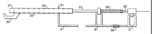
The body of a pull-back device is illustrated in Figures 4 and 5. The proximal
section of the thermography catheter described above is constructed to enable
r s c E
CA 02459002 2004-02-27 E~~2fl9~~'3(? E
F 1h6~='Gee=Zfl53 '10:55- From-GILL JENNINGS &~EVERY +44 25 T3T7 1316 T-B5fi
P.GG91~13 r aa'a'' ' '~" '= '°
16
remote deployment and retraction ofthe projections. This is effected via
rnanipulativn
of the sheath. A two-lumen telescopic construction 2D is used to manipulate
the
sheath 21 between the retracted and the deployed configuration. One lumen is
connected to, or integral with, the outer sheath and can slide aver an
adjacent lumen
which comprises or is connected to one of the lumen housed within the sheath.
Rotation of one tube inside the other is prevented by slotting of the lumen or
other
features on the lumen. Additionally, seating markings {not shown), may be
provided
to avoid over-retraction of the tubes.
The pull-back device includes a drive module 23 which includes a molar,
1o gearing system, typically a speed reducer, control and monitoring means,
and
engagement gear far a driving rod 22. The drive module may be formed
separately
from the body of the pull-back device so that it may be reused. The body of
the pull-
back device must be kept sterile and may be formed from a material such as
polyurethane. This allows the body to be cheaply and easily produced and may
be
7.5 disposable. Alternatively, or additionally, the pull-back device may be
enclosed in a
sterile, flexible plastic sheath when in use, so as to maintain stelllify.
The pull-back device comprises a driving rod 22, adapted for engagementwith
an engagement geaC of the drive module 23 and mount G. Mounts G and B are
adapted to engage the centralfintermediate lumen 25 and the sheath lumen 27
2 o respectively. A Mount A is provided which is adapted to engage the guide
catheter
extension 24. Mount A includes a~bracket 31 for connection of mount A to the
guide
catheter extension trxation paints 32. When engaged, mount B may be moved
.~;._....~, ,:. .' towards C to place fine thermograph catheter in the open
configuration: ~ C may be
selectively driven reversibly over a range of travel (usually about 60 mm)
suitable far
2 5 withdrawal of the catheter apparatus over the measured region. The driving
rod Z2
is a worm-screw type which interacts with the engagement gear of the drive
module
23, fihus providing a smoothly driven apparatus.
The mounts 8 and C may individually be locked in position relative to one
another yr may be selectively unlocked in order to allow movement of the lumen
25,
3 o sheath 21 and guide catheter extension 24 relative to one another.
With reference to figures 6 and 7, in use, the sequence of events begins with
the insertion of a guiding catheter into the area of general interest (step
100), tar
example the cardiac region. Where, for example, the coronary arteries are to
be
examined, the guiding catheter is inserted so that it is adjacent the opening
of the
AMEN~DED~.SHEET
EmPf.zPit:1811~/~003 11:49 .~ w. ~ .., .~~mp~. yr.~':717 P.009
1~ /~~ i CA 02459002 2004-02-27
~~' ~~~v~t0~"c~>'
_. °ta:5D From-GILL JENNINCS & EVERY +44 Zo 737 i3lD T-eos P.olnlal3
17
coronary arteries (step 110). An angioplasty guide wire is then inserted into
the
coronary artery, past the paint of speafic interest. The guide wire is usually
inserted
with the aid of standard fluoroscopic techniques, as is the guide catheter.
The guide catheter, when in place vvetthe entrance to the coronary (or other
target) artery wilt protrude a distance from the patient once in place. This
is then fixed
to,the guide catheter extension 24, The guide catheter extension will be fixed
to the
guide catheter by inserting the non-compressible tube 27 intv.the Y piece 28.
The
gland nut29 and o-ring seal (compression fitting) is tightened to seal the
joint between
the guide cathetetand guide catheter extension and a securing means 30 is
provided
Z o which holds the Y piece in place relative to the guide catheter extension.
Alternatively,
the outside surface of the non-compressible tube rnay be profiled with shallow
circumferential grooves, to ensure that the tube will not pull out when held
in the
compression fitting of the Y piece (not shown).
A seat element 31 is provided within the guide catheter extension. This is
sandwiched between the non-compressible tube and the guide catheter extension
body. This provides 3 sealing engagement between the non-compressible tube and
the catheter.
Once the guide catheter, guide catheter extension and guide wire are in
position, the catheter is maneuvered over the guide wire to a position beyond
the
z 0 specific area of interest in the coronary artery (step 120) with the aid
of tluorascvpy.
The catheter is then fixed in position on the pull-back device by clipping
into mounts
B and C_ The guide catheter extension is then fixed in position on the mount
A, at a
~fucation paint along its length which optimises the distance between mount A
and B ...,.,,..w~~.~.....,..,..t~4;-~.,:
and C_ Thus, the guide catheter extension should be fixed to mount A so that
the .
catheter may be mounted on mounts B and C in a closed configuration.
An angivgram is taken (step 13D? to assess the position of the catheter in the
vasculartissue. This image is saved and the position of the catheter is
rnarised an the
image so as to define a starting point for the controlled pull-back step.
The sheath 21 is then retracted to al(vw the projections to adopt the deployed
3 0 configuration. This is achieved by moving mount B towards mount C (usually
manually). Mount C at this time is locked relative to mount A. Once the sheath
21 is
retracted sufficieptly to allow expansion of the nailiently biased
projections, mount B
is locked in position and mount G is pulled back by the drive
AMENDEp SHEET'
EmP f .ze i t :18l 1 ~1~003 11: 50 ~ ~' ' ' " ' ' tmE~ r W : : 717 P .010
CA 02459002 2004-02-27
WO 03/022345 PCT/EP02/09430
18
mechanism until the projections are housed in the sheath. This is feasible if
the
sheath 21 is retracted sufficiently (equal or greater than the length of the
pull-back
distance during which measurement takes place) to allow the
intermediate/central
lumen 25 to be retracted in the sheath 21 without the sheath impacting on the
projections along the length of measurement.
Alternatively, the mount B and C are locked in position once the thermography
-catheter-is-in-the-deployed-configuratior-~--and-both-mounts-ar-e-pulled-back-
by the-drive
mechanism.
The locking mechanism includes a stopper rod 26. This is provided with
0 graduations capable of engaging electrically actuated locking pins (not
shown) within
mounts B and/or C. A similar set of electrically actuated locking pins (not
shown)
within the same mounts are used to selectively connect the mounts to the drive
rod
22. A set of locking pins on any particular mount may not be connected to both
the
drive rod 22 and the stopper rod 26 simultaneously. Thus, each mount is either
in
Z 5 drive or stop mode. Alternatively a ratchet mechanism may be provided as
the locking
mechanism.
When the mount C is in drive mode, it moves relative to mount A and B. Mount
C cannot be moved towards mount B when attached to the pull-back device.
The catheter may be marked to indicate when the sensors are in a deployed
2 0 or in a retracted position. This may be achieved by provision of a
telescopic tubing 20
with appropriate indicators or by simply marking the extreme deployed or
retracted
position on the apparatus.
Controlled pull-back of the catheter then takes place (step 140). The pull-
back
takes place at a constant speed and is controllable by the user. Pull-back
typically
2 5 takes place at speeds of 0.1 to 2 mm in divisions of 0.1 mm or so.
The pull-back takes place over a distance of the vascular tissue being
measured. Temperature readings may be taken intermittently or substantially
continuously. The data transmitted by the sensors from the vascular wall is
captured
for data and image processing (step 150) together with a fluoroscopylIVUS
image
3 0 frame.
As the catheter is withdrawn inside the artery, the projections automatically
adjust their angle following the wall's morphology without losing the desired
thermal
contact. The result is that the thermal contact between the sensors and the
wall is
CA 02459002 2004-02-27
WO 03/022345 PCT/EP02/09430
19
continuously maintained, even when the catheter is crossing very irregular
plaque
formations.
Once the pu((-back has been completed, the central/intermediate lumens are
retracted such that the projections are withdrawn into the sheath 21 in order
to place
the sensors in the retracted configuration. This restores the original smooth
profile of
the catheter. The catheter may then be detached from the pull-back device and
wifihdrawn-from the-patient-or-may-be-reinse -rted-into the same or-another-
.blood vessel
in order to take another reading. Alternatively, the catheter may be
reinserted in order
to enable a therapeutic or surgical intervention.