Note: Descriptions are shown in the official language in which they were submitted.
CA 02459462 2004-03-03
WO 03/023363 PCT/US02/28920
MICROCANTILEVER APPARATUS AND METHODS
FOR DETECTION OF ENZYMES
Technical field
The general field of the invention relates to an apparatus and a method for
detecting
the presence of an enzyme in a sample by measuring a deflection of a
microcantilever, the
surface of the microcantilever having a substrate for the enzyme. In various
embodiments,
the invention is of use in proteomics, drug discovery, medical research,
medical, veterinary,
dental diagnostics, forensics, and military applications.
l0 Background
A large variety of enzymes are important in medicine, industry, and other
applications. Discovery of novel enzymes has gone hand-in-hand with
development of
certain industries, for example the discovery of bacterial restriction enzymes
and the
development of genetic engineering. Enzymes are important in various medical
pathologies
(Fang J., et al., Proc. Natl. Acad. Sci. U.S. 97: 3884-3889, 2000), as novel
therapeutics
(LT.S. patent number 6,210,667 issued April 3, 2001), as targets for
development of novel
therapeutic agents, for example, HIV protease (U.S. patent number 6,271,235
issued
August 7, 2001), in industrial processes such as antibiotic biosynthesis
(CT.S. patent number
6,258,555 issued July 10, 2001), degradation of unwanted materials such as
polyurethane
(U.S. patent number 6,180,381, issued Jan.30, 2001) and in the food industry
(U.S. patent
number 5,827,712, issued Oct. 27,1998). The need to obtain novel enzyme
activities is so
great that protein engineering research has been directed toward development
of catalytic
antibodies (U.S. patent number 5,807,688, issued Sep. 15, 1998).
Thin bimorph microcantilevers can undergo bending (deflection) due to
differential
stresses following exposure to and binding of a compound from their
environment, for
example in a fluid sample. Soft microcantilevers having spring constants less
than 0.1 N/m
are sensitive to stress differentials that arise as a result of interactions
between extremely
small amounts of a substrate material on a surface of the microcantilever and
one or more
materials in a sample. For a given microcantilever with a specially designed
coating layer,
3o the deflection yields information about components of the environment to
which the
microcantilever is exposed. Microcantilevers are capable of detecting
calorimetric enzyme-
mediated catalytic biological reactions with femtoJoule resolution. (Thundat
et al.,
"Microcantilever Sensors", Microscale Thermophysical Engr. 1, pgs. 185-199,
1997.)
Oligonucleotide interactions within a sample can be detected using a
monolithic array of
CA 02459462 2004-03-03
WO 03/023363 PCT/US02/28920
test sites formed on a surface to which the sample is applied (U.S. patent
number
5,653,939).
There is a need for methods and an apparatus for detecting an interaction
between an
enzyme and its enzymatic substrate, or detecting a protein having an enzymatic
activity or a
related molecule, such as a catalytic antibody, or a binding protein, as
measured by a
response of a microcantilever to a stress caused by changes in free surface
energy and
bonding energy. There is a need in medical and veterinary diagnostics, and in
research, for
detection and analysis of binding and activities of enzymes and enzyme-like
proteins.
Summary
1 o The invention in one embodiment provides a method for detecting an enzyme,
the
method comprising: depositing a coating material on a first surface of at
least one
microcantilever; adding at least one substrate to the coating material, the
substrate capable
of interacting with the enzyme; exposing the microcantilever with the
substrate to a sample;
and measuring a deflection of the microcantilever, wherein the deflection
indicates the
15 presence of the enzyme in the sample. In a related embodiment, adding the
substrate
comprises adding at least one biomaterial, a biomaterial selected from the
group consisting
of a nucleic acid, a protein, a lipid, a hydrocarbon, and a polysaccharide,
for example. In
another related embodiment, the substrate is a drug.
In a related embodiment of this method, the deflection is caused by a change
in
20 stress on the surface of the microcantilever. In a preferred embodiment,
the deflection is
measured by observing the change by an optical means, which preferably
includes a laser.
Alternatively, an electron tunneling means, a capacitive means, a
piezoelectric means or a
piezoresistive means may be used to observe the change in deflection.
In a related embodiment, the method further comprises analyzing the deflection
of
25 the microcantilever as a function of a time parameter determined from the
time of exposing
the microcantilever to the sample. Analyzing the deflection comprises using a
microprocessor adapted for comparing, calculating, and storing the deflection
of the
microcantilever as a function of a time parameter. Analyzing the deflection
further
comprises analyzing a parameter selected from the group of concentration of
enzyme,
3o concentration of substrate, presence of a cofactor and presence of an
inhibitor.
In a related embodiment, the method comprises the microcantilever having a
length
that is at least about 20~m, at least about 20~,m to about 150~m, the length
is for example
about SO~.m to about 250~,m, about 100~m to about 400p.m, about 200~,m to
about SOO~,m,
or about 250~m to about 7500,m. Further, the width can be at least about S~m
to about
2
CA 02459462 2004-03-03
WO 03/023363 PCT/US02/28920
20~.m, about 10~m to about 30~,m, about 20~,m to about SO~,m, about 25~,m to
about
100~m, or to about 300pm. The height can be at least about 0.1 Vim, for
example, at least
about 0.4~,m , about 4p,m to about 10~m. Depositing the coating material
further comprises
depositing a metal. The metal is selected from at least one of the group
consisting of
aluminum, copper, gold, chromium, titanium, and silver. Fox example, the metal
is gold.
In a related embodiment, the method further comprises depositing a plurality
of
metals. Depositing a plurality of metals further comprises depositing a first
layer of
chromium and a second layer of gold. In a related embodiment, the method
further
comprises depositing a first layer of titanium and a second layer of gold. The
metal in other
1 o embodiments is an amalgam or an alloy.
In a related embodiment, the microcantilever has a second surface selected
from the
group consisting of aluminum oxide, iridium oxide, silicon, silicon oxide,
silicon nitride,
tantalum pentoxide, and a plastic polymer.
In a related embodiment of the invention provides at least one microcantilever
which
15 is a block array having a plurality of microcantilevers.
In a related embodiment, the method further comprises, prior to adding the
substrate
to the first surface, reacting the microcantilever with a bifunctional cross-
linker, the
bifunctional cross-linker capable of further reacting with the substrate. The
bifunctional
cross-linker is selected from the group consisting of: dithiobis(succinimido
undecanoate
20 (DSU); long chain succinimido-6-[3-(2-pyridyldithio)-propionamido]
hexanoate
(LCSPDP); succinimidyl-6-[3-(2-pyridyldithio)-propionamido] hexanoate (SPDP);
and m-
maleimidobenzoyl-N-hydroxysuccinimide ester. For example, the bifunctional
cross-linker
is DSU.
In a related embodiment of the method, the microcantilever detects an enzyme
25 selected from the group consisting of a hydrolase, an oxidoreductase, a
transferase, a lyase,
and a ligase. For example, the enzyme is a hydrolase. The hydrolase is a
protease. For
example, the protease is a metalloprotease or a serine protease. Further, the
enzyme is
selected from the group of consisting of a kinase, a phosphatase, an
endopeptidase, an
exopeptidase, a restriction endonuclease, an exonuclease, and a polymerase.
30 The transferase is selected from the group~consisting of a glycosyl
transferase, a
glutathione S-transferase, an acetyl transferase, and a DNA methyl
transferase. For
example, the lyase is selected from the group consisting of: a polysaccharide
lyase, a 3-
hydroxy-3-methylglutaryl CoA lyase, an argininosuccinate lyase and an
isocitrate lyase.
For example, the oxidoreductase is selected from the group consisting of a
hydroxylamine
CA 02459462 2004-03-03
WO 03/023363 PCT/US02/28920
oxidoreductase, a glyphosphate oxidoreductase, a quinine oxidoreductase, a
ubiquinone
oxidoreductase, and a protein disulfide oxidoreductase. In a related
embodiment of the
invention, the sample comprises an enzyme that is substantially purified.
According to a
further embodiment of the method, the sample comprises a biological fluid. The
biological
fluid is selected from the group consisting of: a cell lysate, a culture
medium, a spent
medium, an animal extract, and a plant extract. For example, the biological
fluid comprises
a bodily fluid from a vertebrate animal, such as a human or other mammal.
According to an
embodiment provided by this method, the bodily fluid is selected from the
group consisting
of: blood, lymph, tissue fluid, urine, bile, sweat, synovial fluid, amniotic
fluid, abdominal
to fluid, pericardial fluid, pleural fluid, cerebrospinal fluid, gastric
juice, intestinal juice, joint
cavity fluid, tears, and nasal discharge.
In a related embodiment of the invention, the enzyme is associated with a
medical
condition in a vertebrate animal. The medical condition is a genetic defect
for example, the
medical condition is selected from the group consisting of Fabry disease,
Gaucher disease,
15 Lesch-Nyhan disease, Tay-Sachs disease, mannosidosis disease, ~-linked
glomerular
disease, and mucopolysaccharidosis. In another embodiment, the medical
condition is a
cancer, for example, the cancer is selected from a cancer of the bxain, liver,
pancreas, lung,
prostate, or breast. In a related embodiment, the cancer is prostate, and the
enzyme is
prostate specific antigen. In a related embodiment, the cancer is breast
cancer, and the
2o enzyme is a collagenase. The medical condition in another embodiment is the
presence of
an infectious agent. For example, the infectious agent is selected from the
group consisting
of: a virus, a bacterium, a fungus, a protozoan, and a helminth.
An embodiment of the invention provides a method for detecting in a sample an
associating substance that binds to a substrate, wherein detecting the
substance involves at
25 least one microcantilever configured to be responsive to a micro-force, the
method
comprising: depositing a coating material on a first surface of the
microcantilever; adding at
least one substrate to the coating material, the substrate capable of
interaction with the
substance; exposing the microcantilever with the substrate to the sample; and
measuring a
resulting free surface energy change on the surface of the microcantilever,
wherein the
30 surface energy change indicates binding to the substrate by the associating
substance in the
sample.
In a related embodiment of the invention, the associating substance is
selected from
the group consisting of: a binding protein, an enzyme, a cofactor, a receptor
Iigand, an
antibody, a polysaccharide, a lipid, a nucleic acid, and a steroid. For
example, the
4
CA 02459462 2004-03-03
WO 03/023363 PCT/US02/28920
associating substance is an enzyme wherein the enzyme binds the substrate and
fails to
dissociate. In another example, the enzyme has no activity on the substrate.
In yet another
example, the substrate is a non-cleavable pseudosubstrate.
The substrate in a related embodiment is a plurality of biomaterials. The
substrate in
another related embodiment comprises an inhibitor of enzymatic activity.
In one embodiment, the invention provides a method of screening for an
inhibitor of
an enzyme, wherein detecting the inhibitor involves having a substrate for the
enzyme on a
microcantilever, the method comprising: adding the substrate to a first side
of a first
microcantilever having a coating, the substrate capable of interacting with
the enzyme and
with the coating; exposing the first microcantilever with the substrate to a
sample, the
sample containing a candidate inhibitor and the enzyme; and measuring a
deflection of the
first microcantilever in comparison to a deflection of a second
microcantilever exposed to
the enzyme in the absence of the candidate inhibitor. In a related embodiment
of the
method, the first microcantilever and the second microcantilever are located
in a first
15 interaction cell and a second interaction cell of a microfluidics device.
In a related
embodiment, a third microcantilever and a fourth microcantilever are located
in a third
interaction cell and a fourth interaction cell, the third cell and fourth cell
having different
concentrations of enzyme than the first cell and the second cell. In a related
embodiment, a
third microcantilever and a fourth microcantilever are located in a third and
fourth
2o interaction cell, the third cell and the fourth cell having different
samples of candidate
inhibitors than the f rst cell and the second cell.
An embodiment of the invention provides an apparatus to measure a microforce
generated by an interaction between an enzyme and a biomaterial, comprising:
at least one
microcantilever, wherein the microcantilever has a length, a width, and a
thickness; a
25 coating material deposited on a first surface of the microcantilever; a
biomaterial capable of
attachment to the coating material; and at least one interaction cell, wherein
the
microcantilever with the coating material and the biomaterial is exposed to a
sample, the
sample comprising the enzyme. The biomaterial comprises an enzymatic
substrate.
Alternatively, the biomaterial comprises an enzymatic pseudosubstrate. The
3o microcantilever in certain embodiments comprises a block array having a
plurality of
microcantilevers. The microcantilever has dimensions that are microscopic,
having a length
that is at least about 20~m, for example, about SO~,m to about 150~m, about
SO~m to about
250~m, about 100~m to about 400~m, about 200~,m to about SOO~,m, or about
250~.m to
about 750~m. Further, the width is at least about S~,m, for example, the width
is about S~m
CA 02459462 2004-03-03
WO 03/023363 PCT/US02/28920
to about 20~,m, about 10~m to about 30~,m, about 20~m to about SO~m, about
25~,m to
about 100~m, or up to about 300~m. The height can be at least about 0.1 Vim,
for example,
at least about 0.4~m , about 4~m to about 10~.m. The microcantilever coating
is selected
from at least one of the group consisting of copper, gold, aluminum, chromium,
titanium,
and silver. For example, the coating is gold coating. According to a further
embodiment of
this apparatus, a second surface of the microcantilever is selected from the
group consisting
of silicon, silicon nitride, other silicon compounds, metal compounds, gallium
axsenide,
germanium, germanium dioxide, glass, zinc oxide, diamond, quartz, palladium
and a plastic
polymer. The apparatus in one embodiment is disposable. In another embodiment,
the
1 o apparatus is reusable.
Brief description of the drawings
Figure 1 is a schematic representation of a partial top view of a
microcantilever
showing three dimensions, first and second surfaces, and substrate molecules
deposited on
the first surface.
is Figure 2 is a schematic diagram of a side view of a microcantilever having
molecules of a bifunctional cross-linking agent attached to the surface of the
microcantilever and to a biomaterial, and the biomaterial bound directly to
the surface of the
microcantilever. Various types of enzymes and modes of binding to and
digesting substrate
molecules are shown.
2o Figure 3 is a schematic view of a microcantilever showing various potential
positions of deflection and return to an original position.
Figure 4 is a time course (in seconds, on the abscissa) of microcantilever
deflection
(in nm, on the ordinate) as a result of papain digestion of IgG (upper
function), compared to
a prior control using the same microcantilever exposed to buffer only (lower
function).
25 Description of specific embodiments
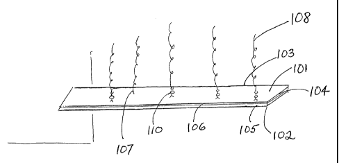
Figure 1 shows a microcantilever having a first surface 101, a second surface
105, a
height I02, a width 104, and a length 109. The first surface can have at least
one coating
106. An enzymatic substrate I08 is affixed to the first surface directly 107,
or by covalent
reaction with a bifunctional cross-linking agent 110. Non-covalently bound
substrate
3o molecules can be washed from the first surface following a reaction with
the cross-linking
agent, for example by use of a buffer having a low pH, or a mild detergent.
Covalent
linking of substrate molecules, rather than direct binding, is a preferred
embodiment, as the
former process produces a more geometrically homogeneous array of substrate
molecules.
CA 02459462 2004-03-03
WO 03/023363 PCT/US02/28920
Figure 2 illustrates the interaction of classes of enzymes with substrate
molecules on
a first surface of a microcantilever, following addition of an enzyme sample
to the
microcantilever. Prior to addition of sample, all substrate molecules are full-
length, as
shown in Panel A, second substrate molecule from right. Panel A shows an
enzyme as a
cross-hatched circle, binding to a recognition site on the interior of the
substrate molecule
(substrate molecule at left), and cleaving the substrate molecule, leaving a
shorter product
covalently attached to the surface (second, third, fifth, seventh, etc.,
substrate molecules
from left). This result would be obtained from digestion of a DNA substrate
molecule by an
endonuclease such as a restriction enzyme, e.g., BamHl or EcoRl, or from
digestion of a
l0 protein substrate molecule by a protease such as trypsin. Panel B shows an
enzyme as a
cross-hatched circle, binding to a free end of a substrate molecule distal
from the attached
end, and cleaving the substrate processively. This result would be obtained
from digestion
of a nucleic acid or a protein substrate molecule by, for example, an
exonuclease or an
exopeptidase, respectively. Panel C shows interaction of binding proteins
(open or stippled
circles), or inactive enzymes, with substrate molecules. Following binding, no
digestion of
substrate molecules is obtained.
Figure 3 shows deflection of a microcantilever from an initial position, A.
Addition
of substrate molecules to a first surface can alter the position of the
microcantilever to a new
position, e.g., position B or position C. Subsequent enzyme digestion as in
Fig. 2, panel A,
2o or Fig. 2, panel B, can further alter the deflection, e.g., from position B
to position C, or
from position C to position A. Binding of inactive enzyme or of a binding
protein to the
substrate can alter the position of the microcantilever, causing deflection,
for example, from
position C to position B.
Figure 4 shows a time course of deflection of a microcantilever, the first
surface of
which has been covalently attached to a protein substrate, a solution of IgG
antibody
molecules. The microcantilever having covalently attached antibody is first
exposed to a
control buffer (lower function), as a result of which exposure no change is
observed in the
deflection. The same microcantilever is then exposed to an appropriate
concentration of the
protease papain, as described in Example 1. The data show a significant change
in
3o deflection, of about 60nm, occurring over a time course of several minutes
following
exposure to the papain.
Definitions
Unless the context otherwise requires, as used in this description and in the
following claims, the terms below shall have the meanings as set forth below.
7
CA 02459462 2004-03-03
WO 03/023363 PCT/US02/28920
The term "microcantilever" is a structural term that refers to a flexible beam
that
may be bar-shaped, V-shaped, or have other shapes, depending on its
application. One end
of the microcantilever is fixed on a supporting base, another end standing
freely.
Microcantilevers are usually of microscopic dimensions, for example, the
microcantilever
has dimensions that are microscopic, having a length that is at least about
20~m, about
SO~.m to about 150~,m, fox example, about SO~.m to about 250~.m, about I OO~m
to about
400pm, about 200pm to about SOOpm, or about 250p,m to about 750~m. Further,
the width
is at least about S~,m, for example, the width is about Spm to about 20p,m,
about 10~,m to
about 30pm, about 20~.m to about SO~.m, about 25~,m to about 100~m, or up to
about,
300pm. The height can be at least about O.l ~,m, for example, at least about
0.4~,m , about
4~,m to about l Op.m.. Silicon and silicon nitride are the most common
molecules used to
fabricate microcantilevers. Other molecules have also been reported for making
microcantilevers, including piezoelectric molecules, plastic molecules and
various metals.
Specifically, microcantilevers can be manufactured from a variety of
materials,
including for example, ceramics, silicon, silicon nitride, other silicon
compounds, metal
compounds, gallium arsenide, germanium, gerrrianium dioxide, zinc oxide,
diamond, quartz,
palladium, tantalum pentoxide, and plastic polymers. Plastics can include:
polystyrene,
polyimide, epoxy, polynorbornene, polycyclobutene, polymethyl methacxylate,
polycarbonate, polyvinylidene fluoride, polytetrafluoroethylene, polyphenylene
ether,
polyethylene terephthalate, polyethylene naphthalate, polypyrrole, and
polythiophene.
Microcantilevers that are custom fabricated can be obtained for example from a
manufacturer such as Diffraction Ltd., Waitsfield, VT.
Microcantilevers with a compound immobilized on the surface on the free end
have
been used to detect and screen receptorlligand interactions, antibody/antigen
interactions
and nucleic acid interactions (U.S. patent number 5,992,226, issued on
November 30,
I999). Deflection is measured using optical and piezoelectric methods.
Microcantilevers
can measure concentrations using electrical methods to detect phase difference
signals that
can be matched with natural resonant frequencies (U.S, patent number
6,041,642, issued
March 28, 2000.) Determining a concentration of a target species using a
change in
3o resonant properties of a microcantilever on which a known molecule is
disposed, for
example, a biomolecule such as DNA, RNA, and protein, is described in U.S.
patent
number 5,763,768.
A method and apparatus for detecting and measuring physical and chemical
parameters in a sample media uses micromechanical potentiometric sensors (U.S.
patent
CA 02459462 2004-03-03
WO 03/023363 PCT/US02/28920
number 6,016,686, issued January 25, 2000). Detection of a chemical analyte is
described
in U.S. patent number 5,923,421, issued July 13, 1999. Magnetic and electrical
monitoring
of radioimmune assays, using antibodies specific for target species which
cause
microcantilever deflection, e.g., magnetic beads binding the taxget to the
microcantilever,
are described in U.S. patent number 5,807,758, issued September 15, 1998.
The term "first surface" as used herein refers to that geometric surface of a
microcantilever designed to receive and bind to molecules of a substrate for
an enzyme.
One or more coatings can be deposited upon this first surface. Thus the term
"second
surface" refers to the area of the opposite side of the microcantilever which
is designed not
to to contain coating or enzyme substrates. As the second surface is generally
not coated, it is
generally comprised of the material from which the microcantilever or
microcantilever array
is fabricated, prior to any coating procedure applied to the first surface.
Alternatively, it
may be coated with a material different from the first surface's coating.
A first surface of a microcantilever can be fabricated to have an intermediate
layer,
15 for example, sandwiched between the first surface comprising for example,
gold, and the
second surface, comprising for example silicon nitride. The intermediate layer
may be an
alloy comprising a plurality of metals, for example, the intermediate layer
may be an
amalgam comprising mercury with at least one of chromium, silver, and
titanium. While
mercury is not generally compatible with an environment having proteins such
as enzymes,
2o in some embodiments the amalgam or alloy of a middle layer may comprise
mercury.
U.5. patent numbers 6,096,559 issued August l, 2000, and 6,050,722 issued
April
18, 2000, describe fabrication of a microcantilever, including use of material
such as
ceramics, plastic polymers, quartz, silicon nitride, silicon, silicon oxide,
aluminum oxide,
tantalum pentoxide, germanium, germanium dioxide, gallium arsenide, zinc
oxide, and
25 silicon compounds. Coating of micromechanical sensors with various
interactive molecules
is described in U.S. patent number 6,118,124, issued September 12, 2000.
Deflection or bending of a microcantilever from a first position to at least a
second
position may be due to differential stress on a first surface of the
microcantilever in
comparison to a second surface, the change in surface stress resulting from
exposure of the
3o microcantilever to a component of a particular environment. A
microcantilever can be
deflect following a change from a first environment to a second environment.
For example,
the environment can be altered in many possible ways including: an enzyme can
be added
or deleted or the enzyme concentration can be lowered or raised; a specific co-
factor of an
enzyme can be added or deleted or the concentration of the co-factor can be
lowered or
CA 02459462 2004-03-03
WO 03/023363 PCT/US02/28920
raised; a specific inhibitor of an enzyme can be added or deleted or the
concentration of the
inhibitor can be lowered or raised; a sample can be diluted or concentrated
prior to, during
or after exposure to a microcantilever; a sample can experience a temperature
change prior
to, during or after exposure to a microcantilever; a sample can experience a
change in pH
prior to, during or after exposure to a microcantilever; a sample can
experience a change in
conductivity prior to, during or after exposure to a microcantilever; and a
sample can
experience a change in viscosity prior to, during or after exposure to a
microcantilever.
Measuring a deflection is measuring the distance moved or change in position
of a
microcantilever that alters from a first occupied position, at which first
position the
to microcantilever with the biomaterial on the first surface of the
microcantilever has not yet
bound or reacted with the enzyme, to a second position occupied by the
microcantilever
after it has altered its position because of binding to or reaction of the
biomaterial on the
microcantilever with the enzyme in the environment, and consequent alteration
of the
biomaterial.
15 A deflection characteristic is a pattern of deflection of a microcantilever
which is
reproducible in extent of distance traveled, for example as measured in nm,
and frequency
' per unit time. The deflection characteristic can distinguish specific
conditions of enzyme
and substrate, and further reaction conditions such as temperature,
concentration, ionic
strength, presence of an ion or other co-factor, presence of a preservative
such as a protease
2o inhibitor, and other conditions cell-known to one of skill in the
enzymological arts. The
extent of a deflection under a particular set of these conditions can become a
signature for a
specific reaction. A deflection characteristic is calculated from a
measurement of extent of
movement of the microcantilever, as a function of the time of addition of a
sample, or as an
extent of the movement as a function of concentration of an enzyme, of
concentration of a
25 substrate, of concentration of an inhibitor, of concentration of a co-
factor, of pH, or of
temperature, and the like.
A microprocessor can be included in an apparatus or a method, such as an
integrated
circuit containing the arithmetic, Logic, and control circuitry required to
intezpret and
execute instructions from a computer program. The microprocessor components of
the
3o measuring devices reside in an apparatus for detection of microcantilever
deflection.
Detection of an enzyme in an environment
The term "environment" means the entire complex of factors to which the
microcantilever is exposed. For example, the complex of factors may include a
sample
having a substance such as: a substantially purified enzyme; a bodily fluid
containing at
CA 02459462 2004-03-03
WO 03/023363 PCT/US02/28920
least one enzyme; a substantially pure inhibitor; a bodily fluid containing an
inhibitor, and
combinations of such components, and the like. The term "environment" also
includes the
concentration of each of any of components of the sample that can affect
enzyme activity.
Factors such as temperature of the environment, while contributing to stress,
are controlled
by standard means, cell known to one of ordinary skill in the art, such as use
of an insulated
and thermally controllable housing, and by monitoring of deflection of a
reference
microcantilever in an environment designed to omit either the substrate, the
enzyme, or an
essential co-factor. The reference microcantilever may be exposed to
inactivated enzyme,
or it may contain a control enzyme compared to that found in the sample. The
difference
lo between the environments of the reference microcantilever and the
experimental
microcantilever results in a measure of the amount of deflection experienced
by the
experimental microcantilevers compared to the deflection seen in the reference
environment
as the background against which all other microcantilever deflections are
measured.
As used herein, deflection of a microcantilever from a first position to at
least a
15 second position can occur by a physical or chemical alteration of an enzyme
substrate
molecule linked to a surface of a microcantilever, due to enzyme activity. For
example, a
physical alteration which is a change in surface tension stress of the sensor
material, e.g., of
a substrate molecule, can occur when a DNA substrate reacts with either a DNA
nuclease,
such as an endonuclease or an exonuclease, or with a DNA Iigase. In the first
case, the
2o alteration is a reduction in the amount of material on the microcantilever.
In the second
case, the alteration is increase in the amount of material on the
microcantilever. Surface
stress on the surface of the microcantilever will change as a result of the
enzyme activity
acting upon the substrate. Deflection of the microcantilever changes also when
a nuclease
enzyme molecule binds to the DNA molecule. Following digestion of the
substrate and
25 release or removal of the enzyme, deflection of the microcantilever to
another position can
be observed. Similarly, deflection of the microcantilever can change from a
first position to
a second position due to a change in mechanical stress from an additional
amount of
material on the surface when a substrate interacts with and binds the enzyme.
Deflection
can change from a second position to at least a third position, following, for
example,
3o activity of a ligase molecule results in addition of a length of DNA to the
DNA substrate
molecule. Deflection can change from a third position to at least a fourth
position when the
Iigase dissociates from the DNA substrate.
Another embodiment of a deflection of a microcantilever is observed when, for
example, a physical alteration of a substrate molecule occurs when a DNA
substrate reacts
11
CA 02459462 2004-03-03
WO 03/023363 PCT/US02/28920
with a DNA endonuclease or exonuclease. Deflection of the microcantilever can
change
from a first position to at least a second position due to the increased
amount of material on
the surface when the substrate interacts with the enzyme. Deflection can
change from a
second position to at least a third position when the nuclease removes DNA
from the DNA
substrate molecule. Deflection can change from a third position to at least a
fourth position
when the nuclease disassociates from the DNA substrate. However, as these
interactions
occur at nsec to sec speeds, real time monitoring of deflection is a
measurement of an
overall change in all of the material on the surface of the microcantilever,
including amount
of substrate due to the enzymatic activity of the enzyme, and and amount of
enzyme on the
l0 surface.
The deflection of a microcantilever can be measured by a means that is
capacitive,
piezoelectric, piezoresistive, or optical. The term "capacitive" means storage
of energy in a
non-conducting material resulting from a force or stress on the surface of the
material. This
force or stress can result in a deflection of the microcantilever. The term
"piezoelectric"
is means a voltage andlor current produced between surfaces of a solid non-
conducting
material when a mechanical stress is applied to it. The term "piezoresistive"
means a
change in electrical resistance of a substance when a pressure or force is
exerted on the
surface of the substance. Optical means include use of ambient light and other
sources of
light, including lasers. Detection of microcantilever deflection by optical,
electrical and
20 mechanical means is shown in U.S. patent number 5,653,939 issued Aug. 5,
1997. Use of
laser light sources is shown in 6,016,686 issued Jan. 25, 2000, and 6,123,819,
issued Sept.
26, 2000. Majumdar et al. (WO 01/14823 A1 international publication date March
1, 2001)
uses measurement of defraction of incident light to measure microforces with a
set of
microcantilever finger array blocks that can deflect relative to a set of
fixed frame fingers.
25 Magnetic and electrical means for detection of deflection are shown in U.S.
patents
5,807,758 issued Sept. 15, 2998, 5,156,810 issued Oct. 20, 1992, and in
5,981,297, issued
Nov. 9, 1999, and 6,107,000 issued Aug. 22, 2000, respectively. Piezoelectric
means for
measuring deflection are shown in U.S. patent numbers 5,814,525 issued Sept.
29, 1998;
5,445,008 issued Aug. 29, 1995; and 5,719,324, issued Feb. 17, 1998,
respectively.
3o A time parameter is a time interval fax measuring an event or an occurrence
from a
first point of time to at least a second point of time, and also a third, a
fourth, etc., points in
time. In general, the first point in time is noted as the time of exposing the
microcantilever
to the sample.
12
CA 02459462 2004-03-03
WO 03/023363 PCT/US02/28920
A stress is a force exerted on a surface of a microcantilever which can be
associated
with intermolecular interactions on that surface, such as: enzymatic
alteration of a substrate
on a first surface of a microcantilever, followed by enzyme release; or,
irreversible binding
of a protein in a sample to the substrate. Stress includes any type of force
exerted on a
surface of a microcantilever resulting from the interaction of a specific
enzyme substrate, or
a specific enzyme inhibitor, or a potential substrate, with an enzyme.
Microcantilevers are
sensitive to stress differentials due to different types of interaction of a
component of the
sample with one or more materials that have been added to a coating layer on
top of a first
material.
to The term "responsive" means that the microcantilever, including all
coatings and
sensor materials such as a substrate for an enzyme, is can deflect as a result
of the stress
generated by an interaction force that arises when an enzyme specifically
interacts with the
substrate. The resulting force may comprise chemical-mechanical forces,
thermomechanical forces, electrostatic forces, magnetic forces, and other
types of forces,
1 S alone or in combination.
Enz~rnes
The term "enzyme" encompasses a large number of protein biological catalysts,
which are known to or are predicted to catalyze a reaction. Most commonly, an
enzyme can
catalyze at least one of many different possible biochemical reactions that
comprise
20 biological pathways. Further, an enzyme can catalyze an organic chemical
reaction, such as
conversion of ethanol to acetic acid, or an inorganic reaction, such as
reduction of molecular
nitrogen.
The molecules that are the results of an enzymatically catalyzed reaction are
referred
to as "products." The terms enzyme, substrate, and product are standard terms
in the arts of
25 enzymology and biochemistry. The term enzyme can include, for example, an
active
enzyme in a sample capable of modifying its enzymatic substrate to yield an
enzymatic
product on a microcantilever; a genetically altered enzyme having a catalytic
defect; an
enzyme lacking a cofactor essential for catalytic activity; and an enzyme in a
sample
binding irreversibly to a pseudosubstrate. The interaction forces generated by
enzyme
3o activity on a substrate molecule may comprise chemical-mechanical forces,
thermal-
mechanical forces, electrostatic forces, magnetic forces, and other types of
forces.
Enzymes encompass six general classes based on the reaction being catalyzed,
including: isomerases, oxidoreductases, transferases, hydrolases, lyases, and
ligases.
Isomerases catalyze the conversion of a substrate which is a chemical
compound, to a
13
CA 02459462 2004-03-03
WO 03/023363 PCT/US02/28920
different chemical compound product that contains the same number and type of
atoms, but
in a different structural configuration. Oxidoreductases are involved in
oxidation, reduction,
and electron or proton transfer reactions of the substrate. Transferases
catalyze reactions in
which groups of atoms are transferred to or from substrate molecules.
Hydrolases cleave
s one or more of a variety of covalent bonds of the substrate by hydrolysis.
Ligases join two
or more substrate components to form a covalent bond, each component being
part of a
substrate complex. Enzymes that are known in the art can be purified from
cells that have
been collected and concentrated as the enzymes are thus purified. Cells are
ruptured by
methods commonly employed by artisans in microbiology and cell biology, for
example,
l0 sonication, French press, freeze thawing, and detergent lysis.
Secreted microbial enzymes can be obtained from spent culture medium, i.e.,
growth
medium from which cells have been removed following culture and growth of
cells.
Enzymes can be purified by pxocedures including column chromatography,
particularly
affinity column chromatography, and also ion-exchange column chromatography,
size
is exclusion column chromatography, and, as fusion proteins, can be purified
using highly
specific affinity ligands (see New England Biolabs Catalog, 2000-2001, pp. 142-
143).
Enzymes are purified and stored in suitable buffers containing anti-oxidant
agents,
such as dithiothreitol or mercaptoethanol, to maintain native cysteine
disulfide bonds in a
reduced condition, and with chelators such as EDTA to protect the enzyme from
heavy
20 metal inactivation. Enzymes can be stored at -20° C or -70°
C, with an agent such as
glycerol or DMSO to prevent water crystal formation, or in a suitable buffer.
Many
enzymes of interest are commercially available (Sigma Aldrich, Inc., St.
Louis, MO;
Calbiochem, San Diego, CA; New England Biolabs, Inc. Beverly, MA), as are
suitable
buffers for storage and concentrated reaction mixes that are formulated for
optimal enzyme
25 activity and include appropriate ions. Alternatively, enzymes are available
as purified
crystals, which can be dissolved in a suitable buffer at a specific
appropriate concentration
prior to use.
Enzymes herein include in scope any genetically engineered or semi-synthetic
peptide-containing molecule capable of reacting with another molecule to
promote a
30 chemical change, for example, a catalytic antibody. The term enzyme is
further envisioned
to include an activity that has not yet been characterized, but for which a
substrate and assay
system can be devised, for example, a DNA restriction endonuclease that
recognizes and
binds to a palindromic or non-palindromic sequence consisting of 10 or more
nucleotides.
Further, the term enzyme includes naturally-occuring or genetically engineered
derivatives
14
CA 02459462 2004-03-03
WO 03/023363 PCT/US02/28920
of an enzyme with known activity, including a derivative having reduced or
essentially no
activity.
Enzymes having a known activity are characterized using the methods and
apparatuses herein by parameters of that activity associated with a particular
enzymatic
substrate, including affinity for the substrate, and rate of turnover of the
substrate to yield
product. The parameters are known as Km (Michaelis constant) as a measure of
affinity for
a substrate and V",~, which is a maximum velocity. These parameters are
determined by
analyses of enzyme activity as a function of concentrations of enzyme and
substrate, and by
observing the reaction as a function of time. Mutated enzymes, and active
enzymes in the
to presence of an enzyme inhibitor, can exhibit a lower affinity for a
particular substrate
(increased Km) or a lower turnover number (decreased Vm~). The methods and
apparatus of
the present invention can be optimized to determine changes in Km and Vm~ of
enzyme
derivatives, and for identification and analysis of enzyme inhibitors.
Substrates for enzymatic activity
The term "substrate" means a molecule specifically chosen by one of ordinary
skill
in the biochemistry of enzymes, because it is known to be a substance that
reacts with an
enzyme of interest. The molecule of substrate, or mixture of different
molecules of different
substrates, can be chosen because at least one of the types of molecules is
known to bind
specifically to the active site of the enzyme, such that the enzyme acts to
catalyze a
2o chemical reaction that alters the substrate. For example, the substrate can
be a particular
protein for an enzyme which is protease; or, the substrate can be a DNA
molecule having a
particular nucleotide sequence that can be recognized by a molecule of a
restriction
endonuclease.
A substrate can be designed to detect a novel enzymatic activity, i.e., an
enzymatic
activity that might be present but is not currently known to be present in one
of a plurality
of natural product samples, or from a library of mutated enzymes. The term
"substrate" is
commonly used in the engineering arts to indicate a surface which acts as a
support for
another material, for example, in I1. S. Patent No. 6,123,819, issued Sept.
26, 2000. In the
present application the term "substrate" is used to refer only to a member of
that particular
3o class of molecule which specifically can interact with an enzyme of choice,
and which can
be bound by the enzyme and be further chemically altered by a reaction
catalyzed by the
enzyme, to yield a product that is chemically different from the initial
substrate material.
Substrates need not be the natural substrate of an enzyme, and can be designed
according to the particular purpose of the user, including diagnostics,
inhibitor search,
CA 02459462 2004-03-03
WO 03/023363 PCT/US02/28920
purity monitoring, or novel enzyme discovery. Substrates can be nature-
identifical, e.g., a
protein in a native configuration, or can be denatured and further chemically
modified. The
substrate can also be further modified for use with other means of detection,
for example, a
substrate can be colorigenic, fluorogenic, or radioactive, although these
modifications need
not affect an aspect of microcantilever deflection.
Under some circumstances it is desirable to have a dense array of substrate
molecules, for example, short substrate molecules, as opposed to a less dense
array of
longer substrate molecules. The kinetics of enzyme digestion of a substrate on
a surface of
a microcantilever can depend on the size of the particular enzyme, fox
example, the Stokes
1 o radius of the enzyme, so that an optimal extent of density and size of
substrate molecules
should be determined by the user experimentally. The density of the substrate
on the first
surface of the microcantilever can be adjusted by varying one or more of the
factors,
including the concentration of the enzyme, the temperature of the reaction of
enzyme with
cross-linking agent, or the duration of time of this reaction. Further, the
substrate can be a
15 mixture of suitable molecules, as can be determined by one of skill in the
art of
enzymology.
A sensor material can be deposited on the surface of a microcantilever, and
can
interact with a component of a sample, for example, the sensor material is a
biomaterial. In
another embodiment, the sensor material can be any substance with which a
protein,
20 particularly an enzyme can interact, and which can be immobilized on
microcantilever.
The term "biomaterial" means any organic material isolated from a natural
source,
or produced synthetically, or produced semi-synthetically by chemical
synthesis with an
organic starting material. For example, a biomaterial can be isolated from a
natural source
such as an animal tissue, a plant, or from bacterial cells, using technology
cell known to one
25 skilled in the art. A biomaterial such as a protein can be synthesized semi-
synthetically
using recombinant DNA technology, or in a eukaryotic cell-free system, by
methods which
are cell known to one skilled in the art. A protein can also be synthesized de
novo using
solid state or solution peptide synthesis chemistry, with commercially
available devices and
substrates cell known to one skilled in the art of peptide synthesis. A
biomaterial can be all
30 or a portion of a cell. A sensor for the detection of bound E. coli cells
immobilized using
antibodies on microfabricated structures is disclosed in Ilic et al.
"Mechanical resonant
immunospecific biological detector", Appl. Phys. Lett. Vol. 77, No. 3, pgs.
4S0-452,
17 July 2000. Biomaterials and other sensor materials can be obtained
commercially, or can
be produced by the artisan in the laboratory.
16
CA 02459462 2004-03-03
WO 03/023363 PCT/US02/28920
The phrase "non-cleavable pseudosubstrate" means a molecule that is chemically
similar to a natural substrate of the enzyme, which can bind the enzyme, but
which
pseudosubstrate is not altered chemically. A pseudosubstrate can bind
covalently or non-
covalently to the enzyme active site, but cannot be converted to the end
pxoduct of the
chemical reaction. For example, a proteinaceous protease inhibitor can act as
a
pseudosubstxate for a protease, for example, a synthetic inhibitor can act as
a
pseudosubstrate for a cAMP-dependent protein kinase.
The phrase "substantially pure" means that the enzyme of interest has been
physically manipulated to increase the final concentration in comparison to
the initial
1 o concentration, with respect to other non-enzyme materials, for example, so
that the enzyme
solution is at least 80% pure, is at least 90% pure, is at least 95% pure, or
is at least 99%
pure with respect to non-enzyme components of the solution.
Samples
The term "sample" means the components dissolved or dispersed in a fluid
state. A
15 sample of interest can be assayed for the presence of a diagnostically
important enzyme in a
sample from a subject; alternatively, a sample can be assayed for presence of
a novel
enzyme activity.
The term "bodily fluid" means any fluid produced or secreted within or by a
body of
an animal, blood, lymph, tissue fluid, urine, bile, sweat, synovial fluid,
amniotic fluid,
2o abdominal fluid, pericardial fluid, pleural fluid, cerebrospinal fluid,
gastric juice, intestinal
juice, joint cavity fluid, tears, and nasal discharge.
The phrase "medical condition" means any condition in which the health of a
subject
is impaired. The medical condition can include for example a genetic defect,
an infection, a
cancer which can be a leukemia or a tumor, and the like.
25 The term "infection" is meant to include disorders of a human or animal
subject
caused by one or more species of bacteria, viruses, fungi, or protozoans,
which are
disease-producing organisms collectively referred to as "pathogens." The term
"fungi" is
meant to include the yeasts. In this invention, pathogens are exemplified by,
but not limited
to: Gram-positive bacteria such as Enterococcus fecalis, Hemophilus
pneumoniae, Listeria
30 monocytogenes, Mycobacterium tuberculosis, M. leprae, Pr~opr~ionibacterium
aches,
Staphylococcus aureus, S. epidermis, S. intermedias, Streptococcus
hemolyticus, S
pneumoniae; Gram-negative bacteria such as Flavobacterium meningosepticum,
Helicobacter pylori, Hemophilus pneumoniae, H. influenzae, Iflebsiella
pneumonia,
Neisseria gonorrlzoeae, Pseudomonas aeruginosa, Slzigella dysentef~ia,
Salmonella typlzi,
17
CA 02459462 2004-03-03
WO 03/023363 PCT/US02/28920
S paratyphi, Escherichia coli serotype 0157:H7, Chlanzydia species; viruses
such as
HIV-1, -2, and -3, HSV-I and -II, non-A non-B non-C hepatitis virus, pox
viruses, rabies
viruses, and Newcastle disease virus; fungi such as Candida albicans, C.
tropicalis, C.
krusei, C. pseudotf°opicalis, C. parapsilosis, C. quillermondii, C.
stellatoidea, Aspergillus
fumigates, A. Niger, A. nidulahs, A. flavus, A. terreus, Absidia corymbifera,
A. ramosa,
Cryptococcus neoforms, Histoplasma capsulatum, Coccidioides immitis,
Pheumocystis
carixzii, Rhizopus arrhizus, R. oryzae, Mucor pusillus and other fmgi; and
protozoa such as
Erztamoeba histolytica, Entarnoeba coli, Giardia lamblia, G. irztestizzalis,
Eimeria sp.,
Toxoplasma sp., Cryptosporidium parvum, C. muris, C. baileyi, G meleagridis,
C. wrairi,
to and C. hosarz~m. Obtaining unique epitopes from these organisms by
screening proteins
and by assaying peptides in vitro are commonly known to those skilled in the
art.
The phrase "genetic defect" means any inheritable pathological condition which
is
caused by the presence of a mutant allele or disease gene. Examples include
but are not
limited to: Fabry disease, Gaucher disease, Tay-Sachs disease, Lesch-Nyhan
disease,
mannosidosis disease, X-linked glomerular disease, and mucopolysaccharidosis.
Cross-linkin a ents
The term "attachment" with respect to an enzymatic substrate and a first
surface of a
microcantilever, means a covalently bonded or other physically connected
molecule of
substrate that is connected to the coating material on the first surface of
the microcantilever.
2o In a preferred embodiment, an attachment is a covalent bond from the
substrate to an atom
of a chemical linker, e.g., a bifunctional cross-linking reagent or "cross-
linker", which is
also covalently bonded through a different atom to the first surface.
Attachment can also be
by direct non-covalent connection of the biomaterial to the coating material
on the f rst
surface without modification of either the first surface or the biomolecule.
Such connection
can be due to complementarity of shape, charge, andlor to exclusion of waters
of hydration,
hydrophobicity, or other characteristics of the particular combination of the
first surface and
the particular substrate (U.S. patent number 6,123,819, issued Sept. 26,
2000).
The phrase "bifunctional cross-linkex" means a substance which can connect a
first
component to a second component, wherein the cross-linker consists of a carbon
chain and
3o has a first chemically reactive group at a first end of the substance and a
second bioreactive
group at a second end of the substance. A chemical reaction between the first
end of the
substance with a first component, and a chemical reaction between the second
end of the
substance with a second component, results in the linkage of the first and
second
components of the invention herein. A bifunctional cross-linker is used to
bind a substrate
18
CA 02459462 2004-03-03
WO 03/023363 PCT/US02/28920
molecule to a first surface of a microcantilever, for example, to bind a
protein substrate such
as a collagen to a first surface having a gold coating.
For example, bifunctional cross-linkers can include the following compounds:
dithiobis(succinimidyl-undecanoate) (DSU), and can be purchased from Pierce
Endogen,
Inc. (Rockford, IL); long chain succinimido-6-[3-(2-pyridyldithio)-
propionamido]
hexanoate (LCSPDP), contains pyridyldithio and NHS ester reactive groups which
react
with sulfliydryl and amino groups, can be purchased from Pierce; succinimidyl-
6-[3-(2-
pyridyldithio)-propionamido] hexanoate (SPDP) contains pyiidyldithio and NHS
ester
reactive groups which react with sulfhydryl and amino groups, can be purchased
from
to Pierce (Rockford, IL); and m-maleimidobenzoyl-N-hydroxysuccinimide ester
(MBS)
contains NHS ester and maleimide reactive groups which react with amino and
sulfhydryl
groups, and can be purchased from Pierce (Rockford, IL).
The terms "protein", "polypeptide", and "peptide", as used herein, shall have
the
same meaning.
15 The above embodiments of the invention, having been fully described, are
illustrated
by the following Examples and claims, which are not intended to be further
limiting. The
contents of all cited references are hereby incorporated by reference herein.
EXAMPLES
2o Example 1. Papain digestion of an immunoglobin IgG antibody substrate.
A surface having a gold-coated microcantilever was cleaned by exposure to an
ozone-enriched atmosphere for 10 min. The cross-linking agent was attached by
immersing
the microcantilever in a solution of 0.1% (w/v) DSU in dioxane for 60 min. The
microcantilever was washed three times with dioxane, followed by a wash with
phosphate
z5 buffered saline (PBS), pH 7.6.
The microcantilever was further incubated with a solution of Immunoglobulin G
(1 mg/mL; CalBiochem, San Diego, CA) in PBS solution for 60 min, to covalently
attach a
protein substrate for the enzyme papain to the coated f rst surface of the
microcantilever.
The microcantilever was removed from the antibody solution and immersed in a
carbonate
3o buffer solution, pH 8.5, for 30 min to hydrolyze any unreacted DSU.
The microcantilever was mounted in a cell of an atomic force microscope (AFM),
and measurement of microcantilever deflection was initiated. After attainment
of a stable
baseline, a 100 microliter sample of a PBS solution containing a 0.1 % (w/v)
solution of the
detergent Tween was injected into the cell. Microcantilever deflection was
monitored as a
19
CA 02459462 2004-03-03
WO 03/023363 PCT/US02/28920
function of time, as is depicted in Fig. 4 as "control." Next, a 100
microliter sample of
papain (100 micrograms per mL; CalBiochem, San Diego, CA) was injected into
the cell.
Microcantilever deflection was monitored as a function of time, and the
results are depicted
in Fig. 4, labeled as "papain."
The steady upward bending of the microcantilever shown in Fig. 4 denotes a
change
in the surface tension on the microcantilever from a change in the protein
substrate from the
gold-coated first surface of the microcantilever. The data shown are one
example of several
observations, having the same result. The data show monitoring of enzymatic
activity of
papain as a function of time. Further, these data show the capability of the
microcantilever
1 o to measure enzymatic activity.
Example 2. Neisseria secreted protease digestion of IgG substrate.
A surface having a gold-coated microcantilever is cleaned by exposure to an
ozone-enriched atmosphere for 10 min. The cross-linking agent is attached by
immersing
the microcantilever in a solution of 0.1% (wlv) DSU in dioxane for 60 min. The
microcantilever is washed with dioxane, followed by a wash with phosphate
buffered saline
(PBS), pH 7.6.
The microcantilever is further incubated with a solution of Immunoglobulin G
(1 mg/mL; CalBiochem, San Diego, CA) in PBS solution for 60 min to covalently
attach
the IgG protein substrate to the surface. The microcantilever is removed from
the antibody
2o solution and immersed in a carbonate buffer solution, pH 8.5, for 30 min to
hydrolyze any
unreacted DSU.
The microcantilever is mounted in a cell of an AFM and measurement of
deflection
is initiated. After attainment of a stable baseline, a 100 microliter aliquot
of a PBS solution
containing 0.1 % (w/v) solution of the detergent Tween is injected into the
cell.
Microcantilever deflection is monitored as a function of time. Next, a 100
microliter aliquot
of a sample containing a Neisseria secreted protease is injected into the
cell.
Microcantilever deflection is further monitored as a function of time.
The steady upward bending of the microcantilever indicates change of surface
tension of the protein substrate from the gold-coated side of the
microcantilever. Many
other bacterial pathogens secrete a similar antibody-specific proteolytic
enzyme during a
course of pathogenesis, which enzyme can be detected by use of a
microcantilever.