Note: Descriptions are shown in the official language in which they were submitted.
CA 02459541 2004-02-16
WO 03/015640 PCT/US02/24590
BIOACTIVE OCCLUSION COIL
-1-
BACKGROUND OF THE INVENTION
The present invention deals with implantable
medical devices. While conceivably the devices could
be utilized in the context of a variety of body
spaces, the present description, for the sake of
brevity, will often be described in the context of
the treatment of vascular aneurysms. Accordingly,
one aspect of the present invention deals with an
implantable medical device for at least partially
obstructing the neck portion of a vascular aneurysm.
Another aspect of the present invention pertains
to a medical device for forming an embolism within
the vasculature of a patient. More particularly, it
is a vaso-occlusion device at least partially coated
with a bioactive agent, an absorbable material or
biopolymer or an absorbable or biopolymer coating
optionally containing or coated with other bioactive
agents. A highly flexible vaso-occlusive device
coated with such materials also forms a variation of
the invention.
Vascular aneurysms are typically formed due to a
weakening in the walls of an artery. Often aneurysms
are the site of internal bleeding and,
catastrophically, the site of strokes. Different
implantable medical devices have been developed for
treating vascular aneurysms. Treatments commonly
known as "artificial vaso-occlusion" treatments are
known to be useful in treating aneurysms by filling
CA 02459541 2004-02-16
WO 03/015640 PCT/US02/24590
-2-
associated undesirable vascular spaces. A variety of
different vaso-occlusive devices are known to be at
least arguably effective for the treatment of
aneurysms.
Vaso-occlusive devices are surgical implants
that are placed within open sites in the vasculature
of the human body. The devices are introdiiced
typically via a catheter to: the site. within the
vasculature that is to be clos,ed-. That site may be
within the lumen of a blood vessel or perhaps within
an aneurysm stemming from a blood vessel.
There are a variety of materials and devices
that have been used t, o create emboli in the
vasculature of the human body. For instance,
injectable fluids such as microfibrillar collagen,
various polymeric foams and beads have been used.
Certain injectable fluid devices can be introduced
through a catheter and are capable of forming a solid
space-filling mass in a target location. Polymeric
resins, particularly cyanoacrylate resins, have been
used as injectable vaso-occlusive materials. Both
the injectable gel and resin materials are typically
mixed with a radio-opaque material to allow accurate
setting of the resulted materials. Although some of
these agents provide for excellent short-term
occlusion, many are thought to allow. vessel
recanalization due to absorption of the agents into
the blood. In addition, there are significant risks
involved in use of cyanocrylates, and similar
CA 02459541 2004-02-16
WO 03/015640 PCT/US02/24590
-3-
materi.als, due to the potential for misplacement.
Such misplacement can create emboli in undesired
areas. Generally, injectable fluid occlusion devices
are somewhat difficult, if not impossible, to
retrieve once they are improperly placed.
In some instances, materials such as hog hair
and suspensions of metal particles have been
introduced into an aneurysm by'those wishing to form
occlusions. It is believed that these materials
encourage natural cell growth within the sac portion
of an aneurysm.
Several patents describe different deployable
vaso-occlusive devices that have added materials
designed to increase their thrombogenicity. For
example, fibered vaso-occlusive devices have been
described in a variety of patents assigned to Target
Therapeutics, Inc., of Fremont, California. Vaso-
occlusive coils having attached fibers are shown in
U.S. Patent Nos. 5,226,911 and 5,304,194, both to
20. Chee et al. Another vaso-occlusive coil having
attached fiberous materials is found in U.S. Patent
No. 5,382,259, to Phelps et al. The Phelps et al.
patent describes a vaso-occlusive coil which is
covered with a polymeric fiberous braid on its
exterior surface. U.S. Patent No. 5,658,308, to
Snyder, is directed to a vaso-occlusive coil having a
bioactive core.
To further increase occlusive properties and
thrombogenicity, a variety of vaso-occlusive devices
CA 02459541 2004-02-16
WO 03/015640 PCT/US02/24590
-4-
have been treated with a variety of substances. For
instance, U.S. Patent No. 4,994,069, to Ritchart et
al., describes a vaso-occlusive coil that assumes a
linear helical configuration when stretched and a
folded, convoluted configuration when relaxed. The
stretched condition is used in placing the coil at
the desired site (via passage through the catheter)
and the coil assumes a relaxed configuration -- which
is better suited to occlude the vessel -- once the
device is so-placed. Ritchart et al. describes a
variety of shapes. The secondary shapes of the
disclosed coils include "flower" shapes- and double
vortices. The coils may be coated with agarose,
collagen, or sugar.
U.S. Patent No. 5,669,931, to Kupiecki,
discloses coils that may be filled or coated with
thrombotic or medicinal material. U.S. Patent, No.
5,749,894, to Engelson, discloses polymer-coated
vaso-occlusion devices. U.S. Patent No. 5-, 690, 671 to
McGurk discloses an embolic element which may include
a coating, such as collagen, on the filament surface.
U.S. Patent No. 5,536,274 to Neuss shows a
spiral implant which may assume a variety of
secondary shapes. Some complex shapes can be formed
by interconnecting two or more of the spiral-shaped
implants. To promote blood coagulation, the implants
may be coated with metal particles, silicone, PTFE,
rubber lattices, or polymers.
CA 02459541 2004-02-16
WO 03/015640 PCT/US02/24590
-5-
As has been alluded to above, advancements in
the artificial occlusion of aneurysms have occurred
due to the delivery and implantation of metal coils
as vaso-occlusive devices.
Vaso-occlusion coils are generally constructed
of a wire, usually made of a metal or metal alloy,
which is wound into a helix. Most commonly, these
coils are introduced in a stretched linear form
through a catheter to the selected target site, such
as a particular aneurysm. The vaso-occlusi.on coils
typically assume an irregular shape upon discharge of
the device from the distal end of the catheter. The
coils may undertake any of a number of random
configurations used to fill an aneurysm. In some
instances, vaso-occlusion coils are adapted to assume
a predetermined secondary shape designed to enhance
the ability to fill undesirable vascular spaces.
A variety of vaso-occlusion coils and braids are
known. Tungsten, platinum, and gold threads or wires
are said to be preferred. Vaso-occlusion coils have
a variety of benefits including that they are
relatively permanent, they may be easily imaged
radiographically, they may be located at a well
defined vessel site, and they can be retrieved.
In some instances, particularized features of
coil designs, such as specialized mechanisms for
delivering vaso-occlusion coils through delivery
catheters and implanting them in a desired occlusion
site, have been described. Examples of categories of
CA 02459541 2004-02-16
WO 03/015640 PCT/US02/24590
-6-
vaso-occlusion coils having specialized delivery
mechanisms include pushable coils, mechanically
detachable coils, and electrolytically detachable
coils. =
Pushable coils are commonly provided in a
cartridge and are pushed or plunged from an engaged
delivery catheter into an aneurysm. A pusher wire
advances the pushable coils through and out of the
delivery catheter into the site for occlusion.
Mechanically detachable vaso-occlusive devices
are typically integrated with a pusher wire and are
mechanically detached from the distal end of that
pusher wire after exiting a delivery catheter.
A variety of mechanically detachable devices are
also known. For instance, U.S. Patent No. 5,234,437,
to Sepetka, shows a method of unscrewing a helically
wound coil from a pusher having' an interlocking
surface. U.S. Patent No. 5,250,071, to Palermo,
shows an embolic coil assembly using interlocking
clasps - that are mounted both on the pusher and on the
embolic coil. U.S. Patent No. 5,261,195, to Twyford
et al., shows a pusher-vaso-occlusive coil assembly
having an affixed, proximately extending wire
carrying a ball on its proximal end and a pusher
having a similar end. The two ends are interlocked
and disengaged when expelled from the distal tip of
the catheter. U.S. Patent No. 5,312,415, to Palermo,
also shows a method for discharging numerous coils
from a single pusher by use of a guidewire which ha's
CA 02459541 2004-02-16
WO 03/015640 PCT/US02/24590
-7-
a section capable of interconnecting with the
interior of the helically wound coil. U.S. Patent
No. 5,350,297, to Palermo et al., shows a pusher
having a throat at its distal end and a pusher
through its axis. The pusher sheath will hold onto
the end of an embolic coil and will then be released
upon pushing the axially placed pusher wire against
the member found on the proximal end of the vaso-
occlusive coil.
Within electrolytically detachable vaso-
occlusive devices, the vaso-occlusive portion of the
assembly is attached to a pusher wire via a small
electrolytically severable joint. The
electrolytically severable joint is severed by the
placement of an appropriate voltage on the core wire.
The joint erodes In preference either to the vaso-
occlusive device itself or to the pusher wire. In
accordance with principles of competitive erosion,
parts of the wire that are not intended to erode are
often simply insulated' to prevent such an
electrolytic response caused by the imposition of the
electrical current.
U.S. Patent No. 5,354,295 and its parent
5,122,136, both to Guglielmi et al., describe an
electrolytically detachable embolic device. That is
to say that a joint between the pusher wire and the
vaso-occlusive portion dissolves or erodes when an
electrical current is applied to the pusher wire.
CA 02459541 2004-02-16
WO 03/015640 PCT/US02/24590
-8-
Some vaso-occlusive devices include specialized
mechanical features and/or shapes. Various shaped
coils have been described. For example, U.S. Patent
No. 5,624,461, to Mariant, describes a three-
dimensional in-filling vaso-occlusive coil. U.S.
Patent No. 5,639,277, to Mariant et al., describes
embolic coils having twisted helical shapes and U.S.
Patent No. 5,649,949, to Wallace et al., describes
variable cross-section conical vaso-occlusive coils.
A random shape is described, as well. U.S. Patent
No. 5,648,082, to Sung et al., describes methods for
treating arrhythmia using coils which assume random
configurations upon deployment from a catheter. U.S.
Patent No. 5,537,338 describes a multi-element
intravascular occlusion device in which shaped coils
may be employed. Spherical shaped occlusive devices
are described in U.S. Patent No. 5,645,558 to Horton.
Horton describes how one or more strands can be wound
to form a substantially hollow spherical or ovoid
shape when deployed in a vessel. U.S. Patent Nos.
5,690,666 and 5,718,711, by Berenstein et al., show a
very flexible vaso-occlusive coil having little or no
shape.after'introduction into the vascular space.
One type of aneurysm commonly known as a "wide-
neck aneurysm" is known to present particular
difficulty in the placement and retention of vaso-
occlusive devices. Furthermore, vaso-occlusive
devices, in particular, vaso-occl.usion coils, lacking
substantial secondary shape strength may be difficult
CA 02459541 2004-02-16
WO 03/015640 PCT/US02/24590
-9-
to maintain in position within an aneurysm no matter
how skillfully they are placed.
Vaso-occlusive devices are typically placed in
an aneurysm in the following fashion. A micro-
catheter is initially steered into or adjacent the
entrance of an aneurysm, typically aided by the use
of a steerable guide wire. The guide wire is then
withdrawn from the micro-catheter and replaced by the
vaso-occlusive device. The vaso-occlusive device is
advanced through and out of the micro-catheter,
desirably being completely delivered into the
aneurysm. After, or perhaps, during, delivery of the
device into the aneurysm, there is a specific risk
that the device or a portion of the device might
migrate out of the aneurysm entrance zone and into
the feeding vessel. The presence of the device in
the feeding vessel may cause the undesirable response
of an occlusion in the feeding vessel. Also, there
is a quantifiable risk that blood flow in the feeding
vessel and the aneurysm may induce movement of the
device further out of the aneurysm, resulting in a
more developed embolus in the patent vessel.
As noted above, aneurysms present particularly
acute medical risk due to the dangers associated with
an inherently thin vascular wall. The utilization of
vaso-occlusive devices to occlude an aneurysm without
occluding the adjacent vasculature poses a special
challenge. Methods that meet this challenge and
still avoid undue risk of an aneurysm rupture are
CA 02459541 2004-02-16
WO 03/015640 PCT/US02/24590
-10-
desirable. None of the above documents discuss vaso-
occlusive devices such as those found below.
SUMMARY OF THE INVENTION
One aspect of the present invention pertains to
an implantable medical device for at least partially
obstructing a neck portion of a vascular aneurysm.
The implantable medical device includes an occlusion
subassembly having a central tubular member and at
least one lateral protrusion fixedly attached to the
central tubular member. The lateral protrusion(s)
and the central tubular member are of a size and
overall flexibility to lodge at the neck portion of
the vascular aneurysm. A cylindrical helical coil is
attached to the lateral protrusion.
Another aspect of the present invention pertains
to another implantable ' medical device. The
implantable medical device includes a loop of wire
having first and second ends connected to a base
member. A cylindrical helical coil is radially
disposed about a portion of the loop of wire. A
material for encouraging a cellular response is
disposed on at least one portion of the coil. The
material for encouraging the cellular response is
also biodegradable.
Still another aspect of the present invention
pertains to another implantable medical device. The
medical device includes a loop of wire having first
and second ends connected to a base member. A
CA 02459541 2009-01-13
-11-
cylindrical helical coil is radially disposed about a
portion of the loop of wire. A fiberous woven
tubular member coaxially engages at least one portion
of the cylindrical helical coil. -
According to one aspect of the invention there is
provided an implantable medical device for at least
partially obstructing a neck portion of a vascular
aneurysm, comprising:
an occlusion subassembly comprising a central member
and a loop of wire having first and second ends
connected to the central member, said loop of wire and
the central member being of a size and overall
flexibility to lodge at the neck portion of the
vascular aneurysm, and a coil attached to and radially
disposed about a portion of the loop of wire, wherein
the subassembly further comprises a fibrous woven
tubular member extending coaxially over at least a
portion of the coil, the tubular member comprising a
material for encouraging a cellular response.
BRIEF DESCRIPTION OF THE DRAWINGS
FIG. 1 is a partial sectioned view of a catheter
extending toward an aneurysm emanating from the wall
of a blood vessel.
FIG. 2 is a side view of an implantable bridge
assembly.
FIG. 3 is a partial sectioned view of the
implantable bridge assembly inserted within the
catheter.
FIG. 4 is a partial sectioned view of the
implantable bridge assembly.
FIG. 5 is an end view, taken along line 2A in
FIG. 2, of the implantable bridge assembly.
CA 02459541 2009-01-13
- lla-
FIG. 6 is a detailed end view of a lateral
protrusion portion of the impZantable bridge
assembly.
FIGS. 7A to 7F are partial sectioned views of
the aneurysm and illustrate procedural elements
associated with using the implantable bridge
assembly.
FIG_ 8 is a perspective view of one embodiment
of the inverition.
FIG. 9 is a perspective view of another
embodiment of the invention showing a coil having a
CA 02459541 2004-02-16
WO 03/015640 PCT/US02/24590
-12-
permanently bonded inner coating of a thrombotic
agent and a water-soluble, dissolvable outer coating
of an anti-thrombotic agent.
FIG. 10 is a detailed end view of a lateral
protrusion portion of the implantable bridge assembly
in accordance with another embodiment of the present
invention.
FIG. 11 is a detailed end view of a lateral
protrusion portion of the implantable bridge assembly
in accordance with another embodiment of the present
invention.
DETAILED DESCRIPTION OF THE ILLUSTRATIVE EMBODIMENTS
FIG. 1 illustrates a partial sectioned view of
an aneurysm 100 emanating from the wall of a feeding
vessel 105. A catheter 110 is shown having a radio-
opaque band 115 at its distal end. As is known in
the art, radio-opaque band 115 assists in the
guidance of catheter 110 through a vascular system
utilizing principles of radiography or fluoroscopy.
As illustrated, the distal end of catheter 110 has
been guided so as to extend through a neck portion
120 of aneurysm 100.
FIG. 2 illustrates a side view of an implantable
retainer bridge assembly 200 in accordance with one
aspect of the present invention. Assembly 200
includes a plurality of lateral protrusions 205,
which are fixedly connected to a base section 210.
In accordance with one embodiment, base section 210
CA 02459541 2004-02-16
WO 03/015640 PCT/US02/24590
-13-
is a central tubular member. Lateral protrusions 205
in combination with base section 210 make up a bridge
subassembly 202. While lateral protrusions 205 are
illustratively wire loops, other types of lateral
protrusions should be considered within the scope of
the present invention. For example, lateral
protrusions 205 could be formed as a plurality of
non-looping arms extending from base section 210. In
addition, while FIG. 2 illustratively includes three
lateral protrusions 205, more or fewer lateral
protrusions could be utilized.
Retainer assembly 200 further includes a core
wire 215 (also know as a pusher wire) having a distal
end 220 which includes a severable joint 225. Bridge
subassembly 202, more particularly, base section 210,
is fixedly connected to distal end 220 of core wire
215 and is positioned just distally of severable
joint 225. In accordance with one embodiment, as
will be described below, the bridge subassembly is
directly connected to a portion of severable joint
225.
Retainer assembly 200 is deliverable through a
tul:5ular member such as-catheter 110 in FIG. 1. The
shape of retainer assembly 200 shown in FIG. 2 is the
secondary shape or deployed shape found after the
assembly has been pushed from a distal end of
catheter 110. As retainer assembly 200 is pushed
through catheter 110, it generally has a relatively
retracted or low profile shape, which can be referred
CA 02459541 2004-02-16
WO 03/015640 PCT/US02/24590
-14-
to as the delivery shape or primary shape. The
delivery shape is essentially the shape of the
interior of catheter 110.
FIG. 3 is an illustration of retainer assembly
200 in the delivery shape, as it is being delivered
through catheter 110. The same reference numbers are
used in FIG. 3 for elements that are the same or
similar to those elements illustrated in FIGS. 1 and
- 2. After deployment from catheter 110, retainer
assembly 200 assumes its secondary shape as is seen
in FIG. 2. To undergo such massive changes in shape,
lateral protrusions 205 are typically produced of
material such as a super-elastic alloy. Super-
elastic and pseudo-elastic shape recovery alloys and
shape memory polymers (i.e., *urethanes) are well
known in this art. These alloys are especially
suitable for lateral protrusions 205 because of their
capacity to recover --almost completely-- to an
initial configuration once stress is removed. In
addition to super-elastic and pseudo-elastic alloys,
other materials having shape memory characteristics
are within the scope of the present invention.
Severable joint 225 (FIGS. 2 and 3) may also be
called a sacrificial link. Severable joint 225
includes- means for severing bridge subassembly 202
from most, if not all, of core wire 215. In one
embodiment of the present invention, bridge
subassembly 202 is directly and fixedly connected to
a distal pottion of severable joint 225, enabling a
CA 02459541 2004-02-16
WO 03/015640 PCT/US02/24590
-15-
complete severance of subassembly 202 from core wire
215. In another embodiment, subassembly 202 is
fixedly connected to a small portion of core wire 215
(distally located from joint 225) that remains with
subassembly 202 following severance of joint 225.
For example, the small portion of core wire 215
might, following severance, be substantially
contained within base portion 210 of subassembly 202.
The severing action of joint 225, as will be
described in greater detail below', enables
subassembly 202 to remain in a portion of aneurysm
100 (FIG. 1) after most or all of core wire 215 and
catheter 110 have been removed from feeding vessel
105. In accordance with one illustrative embodiment,
severable joint 225 causes severance via mechanical
means. Other means, however, should be considered
within the scope of the present invention.
For the purpose of simplifying description, it
will be assumed that severable joint 225 is an
20. electrolytic severable joint. It should be noted
that the Figures reflect this embodiment of the
present invention. In accordance with the
embodiment, as will be described in greater detail in
relation to FIG. 4, core'wire 215 is coated with an
electrical insulator that is not susceptible to
dissolution via electrolysis in blood or other ionic
media. Severable joint 225 is not coated with such
insulator and is constructed of a material that is
susceptible to electrolytic dissolution in blood.
CA 02459541 2004-02-16
WO 03/015640 PCT/US02/24590
-16-
Severable joint 225 is also significantly more
susceptible to electrolytic dissolution than base
section 210 and lateral protrusions 205 (bridge
subassembly 202). In accordance with one embodiment,
lateral protrusions 205 are attached to base section
210 but are not in an electrically conductive
relationship therewith, and further, are coated with
an electrical insulator that is not susceptible to
dissolution via electrolysis in blood or other ionic
media. In accordance with one aspect of the present
invention, in response to an electrolytic control
signal, only severable joint 225 dissolves, such that
bridge subassembly 202 is severed from core wire 215.
As was described above, subassembly 202 could be
directly connected to a portion of severable joint
225 or, alternatively, base section 210 of
subassembly 202 could be fixedly connected to a small
portion of core wire 215 (distally located from joint
225) that remains with subassembly 202 following
severance of joint 225.
FIG. 4 is a partial sectional view of an
embodiment of an implantable bridge assembly similar
to the one illustrated in FIG. 2. The same reference
numbers are used in FIG. 4 for elements that are the
same or similar to those illustrated in previously
described embodiments. It should be noted that the
severable joint 225 within the FIG. 4 embodiment is
illustratively consistent with the electrolytic,
severance embodiment described above. As was
CA 02459541 2004-02-16
WO 03/015640 PCT/US02/24590
-17-
previously mentioned, other severance methods could
be utilized.
In FIG. 4, implantable bridge assembly 200
includes lateral protrusions 205 that each
illustratively include an attached marker coil 400.
Marker coils 400 are illustratively constructed of
radio-opaque material (i.e., platinum) that assists
in the guidance of bridge subassembly 202 through a
tubular delivery device (such as catheter 110 in FIG.
1) and through a vascular system, utilizing
principles of radiography or fluoroscopy. In
particular, marker. coils 400 assist in the
positioning of bridge subassembly 202 within an
aneurysm, such as aneurysm 100 (FIG. 1). Bridge
assembly 200 also includes base section 210 that
comprises an outer marker coil 405 and an inner
marker coil 410. In accordance with illustrative
embodiments of the present invention, either, neither
or both of outer marker coil 405 and inner marker
coil 410 could be constructed of a radio-opaque
material. As was previously described, such material
assists in the guidance of subassembly 202 through a
vascular system and into a target aneurysm.
Continuing ,with the description of FIG. 4,
lateral protrusions 205 each illustratively include a
plurality of ends 415 that are fixedly secured
between outer marker coil 405 and inner marker coil
410. Other means for securing.lateral protrusions
205 to base section 210 should be considered within
CA 02459541 2004-02-16
WO 03/015640 PCT/US02/24590
-18-
the scope of the present invention. Inner marker
coil 410 is adapted to radially surround and fixedly
secure the most distal point of core wire 215. In
another embodiment (not illustrated), as was
described above, inner marker coil 410 could be
adapted to fixedly connect to a distal portion of
severable joint 225. In accordance with the
electrolytic severance embodiment of severable joint
225, core wire 215 is covered with an insulation
material 425 such that severable joint 225 is the
only completely exposed portion of core wire 215. As
was discussed above, this encourages the electrolytic
severabilty of severable joint 225 when an
electrolytic control signal is applied to assembly.
200. Finally, retainer assembly 200 includes an
optional marker coil 420 enclosed within insulation
material 425. Optional marker coil 420 is
constructed of a radio-opaque material (i.e.,
platinum) to provide further assistance in the
location and precise placement of bridge subassembly
202 within a vascular system, and to locate a
relative position of subassembly 202 with respect to
a delivery catheter.
FIG. 5 is an end view of an embodiment of a
bridge subassembly 202 portion of an implantable
bridge assembly 200 similar to those illustrated in
FIGS. 2 and 4. The FIG. 5 end view represents a view
taken along line, 2A in FIG. 2.. The same reference
numbers are used in FIG. 5 for elements that are the
CA 02459541 2004-02-16
WO 03/015640 PCT/US02/24590
-19-
same or similar to those elements illustrated in
previously described embodiments.
As is illustrated, retainer sub-assembly 202
includes lateral protrusions 205, a base section 210
and distal end 220 of core wire 215. In accordance
with another embodiment, as was described above, base
section 210 could alternatively be fixedly secured to
a distal portion of a severable joint 225. Base
section 210 further comprises inner marker coil 410
and outer marker coil 405. The plurality of ends 415
associated with lateral protrusions 405 are
illustratively fixedly secured between outer marker
coil 410 and inner marker coil 405.
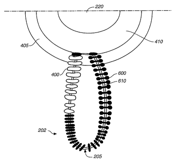
FIG. 6 is an end view illustration of one
particular lateral protrusion 205, in accordance with
an illustrative embodiment of the present invention.
Any of the lateral protrusions 205 described in
relation to other embodiments of the present
invention could be configured similar to the FIG. 6
embodiment described below. The same reference
numbers are used in FIG. 6 for elements that are the
same or_:similar to those illustrated in previously
described embodiments.
Lateral protrusion 205 illustrated in FIG. 6
includes an interior wire 610 having an attached
marker coil 400. Details pertaining to marker coil
400 were described above in relation to FIG. 4.
Lateral protrusion 205 further includes a suture
material 600 wrapped or braided around a portion of
CA 02459541 2004-02-16
WO 03/015640 PCT/US02/24590
-20-
interior wire 610 that is not covered by marker coil
400. While FIG. 6 illustratively shows all of
interior wire 610 covered either by marker coil 400
or suture material 600, some portions of wire 610
could, in accordance with one embodiment of the
present invention, be exposed. In addition,
additional suture material 600 could, in accordance
with another embodiment, be attached to any portion
of bridge subassembly 202 (i.e., attached to inner
coil 410 or outer coil 405) Suture material 600
could, in a,ccordance with yet other embodiments, also
be attached to the distal end 210 or to marker coils
400.
Suture material 600 is illustratively a
therapeutic agent. In accordance with one
embodiment, suture material 600 is or contains a
bioactive material, such as a drug, protein, 'or
genetic material, useful for the medical treatment of
an aneurysm or other medical disorder. In accordance
with another embodiment, suture material 600 is a
bioactive material of a different type, such as a
material selected or designed to encourage cell
growth within a vascular aneurysm. In accordance
with this embodiment, the material could
illustratively be a natural bio-material, such as
collagen, gelatin, fibrin, fibronectin, fibrinogen,
hyaluronic acid, polysaccharides, or proteoglycans,
or any combination thereof; or a combination of
natural bio-materials and synthetic absorbable
CA 02459541 2004-02-16
WO 03/015640 PCT/US02/24590
-21-
materials. In accordance with another embodiment,
suture material 600 is constructed of a material that
encourages cell growth within a targeted portion of
an aneurysm, and is biologically absorbed by the
human body. While there are many materials within
the scope of the present invention that could be
utilized as suture material 600, two that are
biologically absorbable and designed to encourage
cell growth are polylactic acid (PLA) and
polyglycolic acid (PGA). In accordance with one
embodiment, a mixture or composite composition
comprising PLA and PGA could be utilized. Other
potential suture materials that may encourage cell
growth include polymers containing c-caprolactone,
trimethylene carbonate, and p-dioxanone. The suture
materials presently listed are only examples of the
many potential materials that should be considered
within the scope of the present invention.
Suture material 600 could be applied to any or
all portions of bridge subassembly 202 in accordance
with a variety of methods, all of which are
embodiments of the present invention.
Illustratively, suture material 600 is replaced by a
material having a substantially liquid form which is
sprayed on subassembly 202 or applied using a dip
coating procedure. In that embodiment-, the entire
subassembly 202 can be coated with the therapeutic
agent. Of course, suture material 600 or other forms
CA 02459541 2004-02-16
WO 03/015640 PCT/US02/24590
-22-
of the therapeutic agent can be applied to
substantially any portion of subassembly 202.
In addition, some materials suitable for use as
suture material 600 (such as polylactic acid,
polyglycolic acid or a mixture thereof) are available
in extruded or molded forms. Extruded or molded
materials such as these can be formed into desired
shapes and applied to any portion of bridge
subassembly 202. In accordance with one embodiment
of the present invention, the material is formed into
a tubular form and slipped over a portion of
subassembly 202, such as over a portion of the wire
forming a lateral protrusion 205. In accordance with
another embodiment, as is illustrated in FIG. 6, the
material is formed into a solid or strand form and is
wrapped or braided around portions of bridge
subassembly 202. In accordance with yet another
embodiment, the material is heated and wrapped or
braided around a mandrel having a desired shape
(i.e., having a curvature consistent with a portion
of subassembly 202). After the wrapped or braided
material has cooled, it is removed from the mandrel
and then has a permanent relaxed shape convenient for
application to a bridge subassembly 202.
FIGS. 7A-7F are a series of partial sectioned
views of an aneurysm 100 emanating from the wall of a
feeding vessel 105. The same reference numbers are
used in FIGS. 7A-7F for elements that are the same or
similar to those illustrated in previously described
CA 02459541 2004-02-16
WO 03/015640 PCT/US02/24590
-23-
embodiments. FIGS. 7A-7F illustrate procedural
elements associated with using an implantable bridge
assembly consistent with the present invention, as
has been described in relation to the above described
illustrative embodiments.
In accordance with the present invention, as is
represented by FIG. 1, catheter 110 is initially
steered into or adjacent to the entrance of an
aneurysm, typically aided by the use of a steerable
guide wire (not illustrated). As was discussed above
in relation to FIG. 1, radio-opaque band 115 may be
used to assist in the steering of catheter 110
through a vascular system.
When catheter 110 has been positioned relative
to an aneurysm, the guide wire is removed. As was
discussed in relation to FIG. 3, implantable bridge
assembly 200 is then pushed through catheter 110 so
that bridge subassembly 202 exits a distal end of
catheter 110 and' takes on a deployed shape (similar
to FIG. 2) within aneurysm 100. FIG. 7A illustrates
subassembly 202 in the deployed shape within aneurysm
100. In accordance with the embodiment of FIG. 7A,
subassembly 202 is positioned such that lateral
protrusions 205 extend into a sac portion 700 of
aneurysm 100.
FIG. 7B illustrates an alternate placement of a
deployed subassembly 202 within an aneurysm 100. In
accordance with the FIG. 7B embodiment of the present
invention, subassembly 202 is positioned such that
CA 02459541 2004-02-16
WO 03/015640 PCT/US02/24590
-24-
lateral protrusions 205 engage neck portion 120 of
aneurysm 100. Depending on characteristics of the
aneurysm being treated, particularly depending on the
size of neck portion 120, either of the embodiments
illustrated in FIGS. 7A and 7B may be most
appropriate.
It should be noted that marker coil devices,
such as marker coils 400, inner coil 410-, outer coil
405 and optional coil 420, described above in
relation to FIG. 4 could be utilized to steer and
position subassembly 202 with an aneurysm. In
accordance with an embodiment of the present
invention, any or all of these radio-opaque markers
could be utilized by an operator of the present
implantable medical device to provide steering
capability utilizing principles of radiography or
fluoroscopy.
After bridge subassembly 202 is placed within a
portion of aneurysm 100, the next step is to sever
the subassembly from pusher wire 215. This severance
occurs as described above in relation to the
description of severable joint 225. In accordance
with one embodiment, severable joint 225 dissolves in
response to an electrolytic signal being applied
thereto, thereby disengaging subassembly 202 from all
or most of core wire 215. FIG. 7C is an illustration
of bridge subassembly 202 engaged within aneurysm 100
after joint 225 has been severed.
CA 02459541 2004-02-16
WO 03/015640 PCT/US02/24590
-25-
After joint 225 has been severed, core wire 215
is removed from catheter 110. In accordance with one
embodiment of the present invention, catheter 110 is
then withdrawn, leaving subassembly 202 bridging neck
120 of aneurysm 100. As was described in relation to
FIG. 6, in accordance with one embodiment of the
present invention, subassembly 202 includes an
attached suture material or other form that serves as
a therapeutic agent for the treatment of aneurysm
100. In accordance with one embodiment, as was
described above, the therapeutic agent is a
biologically absorbable material that encourages cell
growth in the neck 120 portion of aneurysm 100 and is
biologically absorbed. Accordingly, subassembly 202
is capable of serving as a device for at least
partially obstructing the neck 120 portion of an
aneurysm. In accordance with another embodiment, as
was also described above, the suture material on
subassembly 202 simply serves as a drug delivery
agent.
In accordance with one aspect of the present
invention, bridge subassembly 202 can be utilized to
retain vaso-occlusive devices, such as vaso-occlusion
coils, within an aneurysm. Accordingly, as is
illustrated in FIG. 7D, after core wire 215 has been
removed from catheter 110, the distal end of catheter
110 is then engaged with an opening in bridge
subassembly 202. Next, vaso-occlusive devices,
illustratively vaso-occlusion coils 705, are pushed
CA 02459541 2004-02-16
WO 03/015640 PCT/US02/24590
-26-
through catheter 110 into aneurysm 100. Then, as is
illustrated by FIG. 7E, catheter 110 is removed from-
.feeding vessel 105 and subsequently from the vascular
system. Of course, in accordance with another
embodiment of the present invention, coils 705 can be
placed in the aneurysm 100 through a separate
delivery catheter after placing subassembly 202 but
prior to detaching it. FIG. 7F is an illustration of
this latter embodiment wherein coils 705 are
transported through a catheter 710 that is
independent of catheter 110.
Regardless of the method of implantation, the
implanted subassembly 202 illustratively includes an
attached suture material that encourages cell growth
in the neck 120 portion of aneurysm 100.
Accordingly, subassembly 202, in combination with the
attached suture material, serves as a retaining
device for retaining vaso-occlusion coils 705 within
aneurysm 100. In accordance with one embodiment, as
described above, the suture material is biologically
absorbable. In accordance with another embodiment of
the present invention, vaso-occlusive devices are
delivered before severance of severable joint 225
through a catheter 710 or 110 and through an opening
within base section 210 of bridge subassembly 202.
Another aspect of the present invention pertains
to a vaso-occlusive device having an outer coating of
a collagen-based material or other bioactive
material. It may have other functional drugs,
CA 02459541 2004-02-16
WO 03/015640 PCT/US02/24590
-27-
genetic material, or proteins associated (chemically
linked or physically mixed) with the collagen. The
collagen-based material is for the purpose of
enhancing the rate and density of the occlusion
produced by the vaso-occlusive device at the selected
body site and specifically to promote permanent
cellular in-growth at that site. The therapeutics,
drugs, genetic material, or proteinaceous material
associated with the collagenous material are placed
in the collagen to provide specific effects outlined
below.
As used, the outer, collagen-based or other
bioactive-based coating is preferably placed over an
inner tie layer coating or treatment. The binding
layer preferably provides a layer contiguous to the
vaso-occlusive device and the outer coating. The
inner coating is generally bonded to the vaso-
occlusive member. The inner coating may be of known
silane coupling agents or primer polymer agents
(e.g., low molecular weight polymer adhesives) or the
like. The inner coating may also be deposited on the
member by plasma treatment or may simply be a plasma
treatment of the type intended to etch the substrate.
The inner coating may also include vapor-deposited
polymers, e.g., polyxyxylene and the like. Other
methods for applying the thin polymeric inner
coating, e.g., by dripping or spraying dilute
polymeric solution, may also be employed.
CA 02459541 2009-01-13
-28_
Preferably, the inner coating is permanently
bonded to the coil and either chemically or
physically bonded to the outer coating so that
shortly after coil deployment, the outer material can
safely perform its intended purposes, i.e. beginning
the healing cascade within the vessel.
Another suitable tie layer coating involves
"plasma treatment" of coils. These plasma-treated
coils exhibit an amino-functionality which may be
measured using known chemical methods. When the devices
treated by this process are placed in the bloodstream,
the amino-functionality results in a slight positive
ionic charge on the surface of the fibers. This
amino-functionality attracts platelets and
thrombogenic proteins from the bloodstream. Plasma
treatment may be carried out using e.g., a plasma
generator such as that found in U.S. Patent No.
3,847,652. The plasma may comprise a nitrogen-
containing gas, preferably those containing diatomic
nitrogen or ammonia. Gas pressures are
advantageously maintained at a very low level, e.g.,
no greater than about 5 millimeters of mercury,
preferably from 0.1 to 2 millimeters of mercury.
The period of time in which the vaso-occlusive
device is subjected to the plasma need not be great.
That is to say that for most applied power settinas
below about 200 watts and in the radio frequency
reg_on between 1 and 50 megaHertz, the time of
CA 02459541 2004-02-16
WO 03/015640 PCT/US02/24590
-29-
reaction need not be greater than 10 minutes to
achieve the results described herein.
Other plasma treating steps which are intended
to etch the substrate are also suitable for this
invention.
FIGS. 8 and 9 show typical vaso-occlusive
devices suitable for use with this procedure. FIG. 8
shows a typical vaso-occlusive device 1100. Vaso-
occlusive device 1100 is shown in FIG. 8 to include a
helically wound coil 1102 having tips 1104 to ease
the potential of the component wire to cause trauma
in a blood vessel. The device may include tufts or
fiber bundles attached to it, so as to increase the
amount and volume of fiber held by the coil and
thereby to promote overall thrombogenicity of the
device. Typical of a vaso-occlusive device
comprising a helical coil having attached fiberous
elements such as shown in FIG. 8 is found in U.S.
Patent No. 5,226,911, to Chee et al.
FIG. 9 shows a vaso-occlusive device 1200
comprising a helically wound coil 1202, an inner tie
coating 1204 and an outer collagenous coating 1206.
The inner coating is generally a substance,
preferably proteinaceous, which is bound to the coil
1202 and which is also bound, physically or
chemically, to the outer collagenous covering 1206.
The occlusion devices of the invention may be
made using conventional equipment and procedures.
CA 02459541 2009-01-13
-30-
For example, helical coils may be prepared by
wrapping a suitable wire about a cylindrical or
cop_ical mandrel. The strand(s) are then placed
axially through the core of the helix and, if a
multiplicity of strands are employed, their ends may
be bound by heat, adhesives, or mechanical means.
Radial filaments may be attached to the windings of
the helix by tying or with adhesives.
The polymeric materials used in the vaso-
occlusive devices in FIG. 8 and FIG. 9 are known
materials. They are those materials which are
generally approved for use as implants in the body or
could be so approved. They may be of polymers such
as polyethylene, polypropylene, polyvinylchloride,
polyamides such as Nylon, polyurethanes,
polyvinylpyrrolidone, polyvinyl alchohols,
polyvinylacetate, cellulose acetate, polystyrene,
polytetrafluoroethylene, polyesters such as
polyethylene terephthalate (DacronT11), silk, cotton,
and the like. When the polymers are fiberous, they
are often looped or tufted as shown in the drawings.
Although it is not =critical to this invention, they
are usually assembled in bundles of 5 to 100 fibers
per bundle. Preferred materials for the polymer
component of vaso-occlusive devices comprise
polyesters, polyethers, polyamides, and
polyfluorocarbons_ Especially pre=erred is
polyethyleneterephthalate, sold as DacronT'4. Placing a
CA 02459541 2004-02-16
WO 03/015640 PCT/US02/24590
-31-
protein-based covering on the fibers is a variation
of the invention.
Another variation of the invention includes the
specific use of polymers which evince an angiogenic
response, preferably, biodegradable polymers, that
are associated with the vaso-occlusive support base.
By "associated" is meant that the material is tied to
or is made to adhere to the' vaso-occlusive support
base. The composition may be a fabric or gauze-like
structure. It may also be a non-woven or loose
agglomeration of individual fibers. In general, they
need to stay in place during the placement of the
device in the body.
Preferably, the associated covering is a
polymeric material such as a biodegradable polymer,
e.g., polyglycolic acid, polylactic acid,
reconstituted collagen, poly-p-dioxanone, and their
copolymers such as poly(glycolide-lactide) copolymer,
poly(glycolide-trimethylene carbonate) coploymer,
poly(glycolide-E-caprolactone) copolymer, glycolide-
trimethylene carbonate triblock copolymer, and the
like. Mixtures of the noted polymers, e.g., of
polylactide and polyglycolide may also be used. The
associated coverings may also be used in conjunction
with the bioactive coatings discussed elsewhere.
The coils (1102 in FIG. 8 and 1202 in FIG. 9)
may be made of any of a wide variety of biocompatible
metals or polymers or carbon. In particular, the
metals may be selected from gold, rhenium, platinum,
CA 02459541 2004-02-16
WO 03/015640 PCT/US02/24590
-32-
palladium, rhodium, ruthenium, various stainless
steels, tungsten, and their alloys, titanium/nickel
alloys particularly nitinoltype alloys. The
preferred alloy is one comprising upwards of 90
percent platinum and at least a portion of the
remainder, tungsten. This alloy exhibits excellent
biocompatibility and yet has sufficient strength and
ductility to be wound into coils of primary and
secondary shape and will retain those shapes upon
placement of the vaso-occlusive device in the human
body. The diameter of the wire typically making up
the coils is often in a range of 0.005 and 0.050
inches. The resulting primary coil diameter
typically is in the range of 0.008 and 0.085 inches.
Smaller coil diameters are used for finer problems
and larger coil diameters and wire diameters are used
in larger openings in the human body. A typical coil
primary diameter is 0.015 and 0.018 inches. The axial
length, of a vaso-occlusive device may be between 0.5
and 100 centimeters. The coils are typically wound
to have between 10 and 75 turns per centimeter.
In addition to the coils shown in the Figures,
the vaso-occlusive device may comprise a substrate
comprising a woven braid rather than the helical coil
shown in those Figures. The vaso-occlusive device
may comprise a mixture of the coil and braid.
Indeed, it is within the scope of this invention that
a portion of the coil be polymeric or a combination
of metal and polymer.
CA 02459541 2004-02-16
WO 03/015640 PCT/US02/24590
-33-
It is further within the scope of this invention
that the vaso-occlusive device comprise shapes or
structures other than coils or braids, for example,
spherical structures and the like.
In one aspect of the present invention, the
vaso-occlusive devices described above and those
similar to those specifically described above, are
first optionally treated with a tie layer coating and
then subjected to treatment to provide the outer
collagenous, proteinaceous, or bioactive material
layer. Preferably, neither the inner nor outer
coating interfere with the shape of the coil after
deployment. In one variation of the invention, the
outer layer is applied to the vaso-occlusive base
without the inner tie layer, but is applied in such
an amount that theresulting assembly is not
significantly more stiff than is the vaso-occlusive
device without the covering. That is to say, the
coated device is not more than 35%, preferably not
more than 15%, and most preferably not more than 5%,
stiffer than is the untreated device base.
Preferably, the covering is less than about 1.0 mil,
more preferably less than about 0.5 mil in thickness.
When a collagen layer, the outer collagenous
layer may be of a wide variety of types, natural or
synthetic, but preferably comprises a phot-
polymerizable collagen which will bind both with the
inner tie layer and with the added bioactive agents.
The preferred collagenous materials have the same
CA 02459541 2004-02-16
WO 03/015640 PCT/US02/24590
-34-
surface functional groups as to Type I and Type IV
natural collagens. Those functional groups are
typically of the type which bind to acrylate-type
linkages.
The outer collagenous or proteinaceous coating
may further contain additional materials which have
one or more functions, including, but not limited to,
reducing friction, providing a therapeutic for local
or blood borne delivery, or enhancing thrombosis,
coagulation, or platelet activity. The additional
materials may be applied either as a substantially
pure layer over the collagenous layer or chemically
bonded to (and interspersed with) the collagenous
layer or physically bonded to the outer collagenous
layer. The added bioactive materials may be, e.g.,
genes, growth factors, biomolecules, peptides,
oligonucleodites, members of the integrin family,
RGD-containing sequences, oligopeptides, e.g.,
fibronectin, laminin, vitronectin, hyaluronic acid,'
silk-elastin, fibrogenin, and other basement membrane
proteins with bioactive agents.
Non-limiting examples of bioactive coating or
materials suitable in this invention include both
natural and synthetic compounds, e.g., fibrinogen,
other plasma proteins, growth factors (e.g., vascular
endothelial growth factor, "VEGF"), synthetic
peptides of these and other proteins having attached
RGD (arginine-glycine-aspartic acid) residues
generally at one or both termini, or other cell
CA 02459541 2004-02-16
WO 03/015640 PCT/US02/24590
-35-'
adhesion peptides, i.e., GRGDY, oligonucleodides,
full or partial DNA constructs, natural or synthetic
phospholipids, or polymers with phosphorylcholine
functionality.
Other bioactive materials which may be used in
the present invention include, for example,
pharmaceutically active compounds, proteins,
oligonucleotides, ribozymes, anti-sense genes, DNA
compacting agents, gene/vector systems (i.e.,
anything that allows for the uptake and expression of
nucleic acids), nucleic acids (including, for
example, naked DNA, cDNA, RNA, DNA, cDNA, or RNA in a
non-infectious vector or in a viral vector which may
have attached peptide targeting sequences; antisense
nucleic acid (RNA or DNA); and DNA chimeras which
include gene sequences and encoding for ferry
proteins such as membrane translocating sequences
("MTS") and herpes simplex virus-1 ("VP22")), and
viral, liposomes and cationic polymers that are
selected from a number of types depending on the
desired application, including retrovirus,
adenovirus, adeno-associated virus, herpes simplex
virus, and the like. For example, biologically
active solUtes include anti-thrombogenic agents such
as heparin, heparin derivatives, urokinase, PPACK
(dextrophenylalanine proline arginine
chloromethylketone), rapamycine, probucol, and
verapimil; angiogenic .and anti-angiogenic agents;
anti-proliferative agents such as enoxaprin,
CA 02459541 2004-02-16
WO 03/015640 PCT/US02/24590
-36-
angiopeptin, or monoclonal antibodies capable of
blocking smooth muscle cell proliferation, hirudin,
and acetylsalicylic acid; anti-inflammatory agents
such as dexamethasone, prednisolone, corticosterone,
budesonide, estrogen, sulfasalazine, and mesalamine;
antineoplastic/antiproliferative/anti-mitotic agents
such as paclitaxel, 5-fluorouracil, cisplatin,
vinblastine, vincristine, epothilones, endostatin,
angiostatin and thymidine kinase inhibitors;
anesthetic agents such as lidocaine, bupivacaine, and
ropivacaine; anti-coagulants such as D-Phe-Arg
chloromethyl keton, and RGD peptide-containing
compound, heparin, antithrombin compounds, platelet
receptor antagonists, anti-thrombin antibodies, anti-
platelet receptor antibodies, aspirin, prostaglandin
inhibitors, platelet inhibitors and tick antiplatelet
factors; vascular cell growth promotors such as
growth factors, growth factor receptor antagonists,
transcriptional activators, and translational
promotors; vascular -ce1l growth inhibitors such as
growth factor inhibitors, growth factor receptor
antagonists, transcriptional repressors,
translational repressors, replication inhibitors,
inhibitory antibodies, antibodies directly against
growth factors, bifunctional molecules consisting of
a growth factor and a cytotoxin, bifunctional
molecules consisting of an antibody and a cytotoxin;
cholesterol-lowering agents; vasodilating agents;
agents which interfere with endogenous vasoactive
CA 02459541 2004-02-16
WO 03/015640 PCT/US02/24590
-37-
mechanisms, and combinations thereof. These and
other compounds are applied to the device.
Polynucleotide sequences useful in practice of
the invention include DNA or RNA sequences having a
therapeutic effect after being taken up by a cell.
Examples of therapeutic polynucleotides include anti -
sense DNA and RNA; DNA coding for endogenous
molecules. The polynucleotides of the invention can
also code for therapeutic polypeptides. A
polypeptide is understood to be any translation
production of a polynucleotide regardless of size,
and whether glycosylated or not. Therapeutic
polypeptides include as a primary example, those
polypeptides that can compensate for defective or
deficient species in an animal, or those that act
through toxic effects to limit or remove harmful
cells from the body. In addition, the polypeptides
or proteins that can be incorporated into the polymer
coating 130, or whose DNA can be incorporated,
include without limitation, proteins competent to
induce angiogenesis, including factors such as,
without limitation, acidic and basic fibroblast
growth factors, vascular endothelial growth factor
(including VEGF-2, VEGF-3, VEGF-A, VEGF-B, VEGF-C)
hif-1 and other molecules competent to induce an
upstream or downstream effect of an angiogenic
factor; epidermal growth factor, transforming growth
factor alpha and beta, platelet-derived endothelial
growth factor, platelet-derived growth factor, tumor
CA 02459541 2004-02-16
WO 03/015640 PCT/US02/24590
-38-
necrosis factor alpha, hepatocyte growth factor and
insulin like growth factor; growth factors; cell
cycle inhibitors including CDK inhibitors; thymidine
kinase ("TK") and other agents useful for interfering
with cell proliferation, including agents for
treating malignacies; and combinations thereof.
Still other useful factors, which can be provided as
polypeptides or as DNA encoding these polypeptides,
including monocyte chemoattractant protein ("MCP-l"),
and the family of bone morphogenic. proteins
("BMP's"). The known proteins include BMP-2, BMP-3,
BMP-4, BMP-5, BMP-6 (Vgr-1), BMP-7 (OP-1), BMP-8,
BMP-9, BMP-10, BMP-11, BMP-12, BMP-13, BMP-14, BMP-
15, and BMP-16. Currently preferred BMP's are any of
BMP-2, BMP-3, BMP-4, BMP-5, BMP-6, and BMP-7. These
dimeric proteins can be provided as homodimers,
heterodimers, or combinations thereof, alone or
together with other molecules. Alternatively or, in
addition, molecules capable of inducing an upstream
or downstream effect of a BMP can be provided. Such
molecules include any of the "hedgehog" proteins, or
the DNA's encoding them.
In one exemplary embodiment of the present
invention, the medical device has recombinant nucleic
acid incorporated therein, wherein the recombinant
nucleic acid comprises a viral vector having linked
thereto an exogenous nucleic acid sequence.
"Exogenous nucleic acid sequence" is used herein to
mean a sequence of nucleic acids that is exogenous to
CA 02459541 2004-02-16
WO 03/015640 PCT/US02/24590
-39-
the virus from which the vector is derived. The
concentration of the viral vector, preferably an
adenoviral vector, is at least about 1010 plaque
forming units ("p.f.u."), preferably at least about
1011 p.f.u. Alternatively, the concentration of the
viral vector is limited by the concentration that
results in an undesirable immune response from a
patient.
Treatment of vaso-occlusive coils with the
described materials may be carried out using known
methods, for example dip coating, spray coating,
wiping, vapor deposition or the like.
The devices that are treated according to the
procedure of this invention are often introduced to a
selected site using the procedure outlined below.
This procedure may be used in treating a variety of
maladies. For instance, in treatment of an aneurysm,
the aneurysm itself may be filled with the devices
made according to the procedure specified here.
Shortly after the devices are placed within the
aneurysm, a thrombus begins to form and, at some
later time, is at least partially replaced by
cellular material formed around the vaso-occlusive
devices.
In general, a selected site is reached through
the vascular system using a collection of
specifically chosen catheters and guide wires. It is
clear that should the aneurysm be in a remote site,
e.g., in the brain, methods of reaching this site are
CA 02459541 2004-02-16
WO 03/015640 PCT/US02/24590
-40-
somewhat limited. One widely accepted procedure is
found in U.S. Patent No. 4,994,069 to Ritchart, et
al. It utilizes a fine endovascular catheter such as
found in U.S. Patent No. 4,739,768, to Engelson.
First of all, a large catheter is introduced through
an entry site in the vasculature. Typically, this
would be through a femoral artery in the groin.
Other entry sites sometimes chosen are found in the
neck and are in general well known by physicians who
practice this type of medicine. Once the introducer
is in place, a guiding catheter is then used to
provide a safe passageway from the entry site to a
region near the site to be treated. For instance, in
treating a site in the human brain, a guiding
catheter would be chosen which would extend from the
entry site at the femoral artery, up through the
large arteries extending to the heart, around the
heart through the aortic arch, and downstream through
one of. the arteries extending from the upper side of
the aorta. A guidewire and neurovascular catheter
such as that described in the Engelson patent are
then placed through the guiding catheter as a unit.
Once the tip of the guidewire reaches the end of the
guiding catheter, it is then extended using
fluoroscopy by the physician to the site to be
treated using the vaso-occlusive devices of this
invention. During the trip between. the treatment
site and the guide catheter tip, the guidewire is
advanced for a distance and the neurovascular
CA 02459541 2004-02-16
WO 03/015640 PCT/US02/24590
-41-
catheter follows. Once both the distal tip of the
neurovascular catheter and the guidewire have reached
the treatment site, and the distal tip of that
catheter is appropriately situated, e.g., within the
mouth of an aneurysm to be treated, the guidewire is
then withdrawn. The neurovascular catheter then has
an open lumen to the outside of the body. The
devices of this invention are then pushed through the
lumen to the treatment site. They are held in place
variously because of their shape, size, or volume.
These concepts are described in the Ritchart et al.
patent as well as others. Once the vaso-occlusive
devices are situated in the vascular site, the
embolism forms.
FIGS. 10 and 11 are end view illustrations of
particular lateral protrusions 205, in accordance
with illustrative embodiments of the present
invention. Any of the lateral protrusions 205
described in relation to other embodiments of the
present invention could illustratively be configured
similar to the FIG. 10 or FIG.' 11 embodiments
described below. The same reference numerals are
used in FIGS. 10 and 1.1 for elements that are the
saine' or similar to those elements illustrated and
described in relation to previous embodiments and
previous Figures.
Lateral protrusion 205, in both FIG. 10 and FIG.
11, includes an interior wire 610 having an attached
marker coil 400. Details pertaining to marker coil
CA 02459541 2004-02-16
WO 03/015640 PCT/US02/24590
-42-
400 were described in relation to FIG. 4. It should
be noted that marker coil 400 is an optional element.
In FIG. 10, a cylindrical helical coil 1300 is
disposed about a portion of wire 610 that is not
covered by marker coil 400. In FIG. 11, a
cylindrical helical coil 1305 is similarly
configured. It should be noted that either coil
could take a non-helical configuration without
departing from the scope of the present invention.
While FIGS. 10 and 11 illustratively depict all of
wire 610 covered either by marker coil 400 or coils
1300 or 1305, some portions of wire 610 could, in
accordance with one embodiment of the present
invention, be exposed. In accordance with another
embodiment, marker coil 400 could be eliminated and
coil 1300 or coil 1305 could extend around all or any
portion of wire 610. Illustratively, multiple coils
1300 or multiple coils 1305 could be attached to a
single wire 610 in place of a single continuous coil
1300 or a single continuous coil 1305.
As is depicted in both FIG. 10 and FIG. 11, wire
610 includes first and second ends that are fixedly
secured between inner coil 410 and outer coil 405 of
bridge subassembly 202. With this arrangement, coils
1300 and 1305 can be secured and maintained on their
respective wires 610.
Illustratively, coils 1300 and 1305 may be made
of any of a wide variety of biocompatible metals or
polymers or carbon. In particular, the metals may be
CA 02459541 2004-02-16
WO 03/015640 PCT/US02/24590
-43-
selected from gold, rhenium, platinum, palladium,
rhodium, ruthenium, various stainless steels,
tungsten, and their alloys, titanium/nickel alloys
particularly nitinol type alloys. In accordance with
one embodiment, coils 1300 and 1305 are flexibly
constructed so as to accommodate delivery of
subassembly 202 through a tubular delivery device.
In accordance with one embodiment, subassembly
202 may be equipped with a broad range of bioactive
and/or therapeutic capabilities simply by attaching a
coil, having attached bioactive and/or therapeutic
material, to wire 610.
With reference to FIG. 10, in accordance with
one embodiment, a therapeutic agent may be attached
to coil 1300, and then coil 1300 can be placed over
wire 610. The first and second ends of wire 610 can
then be secured between coils 405 and 410.
In accordance with one embodiment, coil 1300 in
FIG. 10 is similar to any of the coil-like vaso-
occlusive device embodiments described above in
relation to FIGS. 8 and 9. It should be noted,
however, that for the FIG. 8 and FIG. 9 vaso-
occlusive device embodiments to be incorporated as a
coil 1300 in FIG. 10, tips 1104 (FIG. 8) require
modification to include a hollow opening that enables
wire 610 (FIG. 10) to extend there through.
In accordance with one embodiment, at least one
element in the form of a fiber is attached to coil
1300. A single, a multiplicity, or even tufts of
CA 02459541 2004-02-16
WO 03/015640 PCT/US02/24590
-44-
fibers may illustratively be attached to coil 1300.
The fibers could be attached using a variety of
methods, including tying the fibers to the coil,
securing tufts or bundles of fibers between openings
in the coil, etc.
Illustratively, the fibers attached to, or
otherwise disposed on, coil 1300 may comprise
polymeric occlusion-causing material, thrombogenic
material, and/or fibrogenic material. In accordance
with another embodiment, the fibers comprise
biodegradable material, such as (but not limited to)
polyglycolic acid, polylactic acid, reconstituted
collagen, poly-p-dioxanone, and their copolymers.
Mixtures of these sorts of material may also be used.
In addition, the fibers may comprise any of the
materials discussed above in relation to materials
for incorporation into outer coating 1206 (FIG. 8) of
vaso-occlusive device 1100.
Referring to FIG. 11, in accordance with an
embodiment of the present invention, fibers, having
any of the above-mentioned compositions, could be
woven or braided into a fiberous woven or braided
tubular member 1310. Member 1310 may be suitably
woven to enable a coaxial extension over at least one
portion of coil 1305. Member 1310 is intended to
illustrate another way in which fibers or a fiberous
material having a broad range of therapeutic
properties could be attached to a coil that is
attachable to a wire 610 portion of a subassembly
CA 02459541 2004-02-16
WO 03/015640 PCT/US02/24590
-45-
202. In accordance with another embodiment, rather
than a woven or braided member, tubular member 1310
could take other tubular member configurations. For
example, it could be a substantially continuous
(substantially without gaps or openings) tube of
material that coaxially extends over at least one
portion of coil 1305. Alternatively, it could be a
somewhat continuous tubular member but with holes or
slits. These and other tubular member configurations
could incorporate material having characteristics
similar to those described above in relation to other
embodiments. For instance, a given tube of material
could incorporate a therapeutic agent, be
biodegradable, and/or be constructed of a material
for encouraging a cellular response. In one
embodiment, material could be sprayed or dip coated
on a portion of coil 1305.
Although the present invention has been
described with reference to preferred embodiments,
workers skilled in the art will recognize that
changes may be made in form and detail without
departing from the spirit and scope of the invention.