Note: Descriptions are shown in the official language in which they were submitted.
CA 02459603 2004-03-15
WO 03/039657 PCT/US02/06457
1
ELECTRICAL TISSUE STIMULATION APPARATUS AND METHOD
BACKGROUND OF THE INVENTION
For more than 30 years, electrical stimulation of nervous tissue has been used
to
control chronic pain. Therapy originates from an implanted source device,
called an electric
signal generator. The electrical signals, usually a series of brief duration
electrical pulses,
are delivered through one or more implanted leads that communicate with the
source
device, and contain several conductive metal electrodes to act as low
impedance pathways
for current to pass to tissues of interest. For example, in spinal cord
stimulation (SCS)
techniques, electrical stimulation is provided to precise parts of the human
spinal cord
through a lead that is usually deployed in the epidural space dorsal to the
spinal cord. Such
techniques have proven effective in treating or managing disease and chronic
pain
conditions.
Percutaneous leads are small diameter leads that may be inserted into the
human
body through a Tuohy (non-coring) needle, which includes a central lumen
through which
the lead is guided. Percutaneous leads are advantageous because they may be
inserted into
~0 the body with a minimum of trauma to surrounding tissue. On the other hand,
the designs
of lead structure that may be incorporated into percutaneous leads are limited
because the
lead diameter or cross-section must be small enough to permit the lead to pass
through the
Tuohy needle, generally less than 2.0 mm diameter. Typically, the electrodes,
also called
contacts, on percutaneous leads are cylindrical metal structures, with a
diameter of
ZS approximately 1.0 rnm and a length of 4.0 to 10.0 mm. Of course, half of
each of these
electrodes, facing away from the tissue of interest, is not very useful in
delivering
therapeutic current. Thus the surface area of electrodes that face the tissue
to be excited is
small, typically 3.0 to 10.0 square mm. Electrodes must be approximately this
size for
many human applications, especially SCS, to allow sufficient charge to be
delivered with
30 each electrical pulse to excite cells, but without a high charge density
(charge / pulse /
square mm) that might damage tissue or the electrode itself.
CA 02459603 2004-03-15
WO 03/039657 PCT/US02/06457
2
Paddle leads, like Model 3596 Resume~ Lead, Model 3982 SymMix~ Lead or
Model 3991 Transverse Tripole~ Lead of Medtronic, Inc., have been developed to
offer
improved therapy control over some aspects of percutaneous leads. Paddle leads
include a
generally two-dimensional array of electrodes on one side of an insulative
body, for
providing electrical stimulation to excitable tissue of the body. A paddle
design allows
electrodes to be considerably wider than percutaneous leads, up to 4.0 mm or
more. Two-
dimensional arrays of electrodes allow programming of active sites and better
control of the
electric field distribution.
One disadvantage recognized in known paddle leads is that their installation,
repositioning and removal necessitates laminectomy, which is a major back
surgery done by
neurosurgeons and orthopedic surgeons, involving removal of part of the
vertebral bone.
Laminectomy is required because paddle leads have a relatively large width (up
to 1.0 cm or
more) compared to percutaneous leads. Thus, implantation, repositioning or
removal of a
paddle lead requires a rather large opening between the vertebral bones.
Electrodes on paddle leads can easily have larger surface areas than
percutaneous
leads, typically 8.0 to 20.0 square mm or more. Such electrodes are mainly
circular or
rectangular, and require welds to fine, flexible wires passing through the
length of the lead
~0 body. Such welds are prone to breakage in high flex situations unless a
relatively thick
paddle is used to shield the welds and support the electrodes. One advantage
of preferred
embodiments of the invention is that welds are not required in places where
they might
encounter flexing.
ZS Because of the size and relative stiffness of paddle leads compared to the
tissues
they lie near to, more scar tissue or fibrosis tends to form around them over
time than
around percutaneous leads. 'This can reduce electrical efficiency, and lead to
the need for
larger currents over time. Such scar tissue also necessitates greater surgical
efforts for
removal of paddle leads, if required. On some occasions, physicians have even
clipped off
30 the lead body and left a paddle permanently in a patient rather that
surgically remove it, if
the system should cease giving therapeutic benefit.
CA 02459603 2004-03-15
WO 03/039657 PCT/US02/06457
3
For these above listed benefits and liabilities, there is a need for a lead
that can be
percutaneously inserted through a Tuohy-type needle, but which can create
electrodes, each
with substantial 2-dimensional surface area, at positions that are more
lateral than the
current percutaneous lead bodies. Furthermore, if such a lead could be safely
removed by
simple traction on the lead body, the increased surgical efforts that are
required of paddle-
type leads could be avoided.
The prior art has shown some examples of leads that can be expanded in situ,
but
they cannot perform all of the above listed features.
Mullett in U.S. Patent No. 5,121,754 described a percutaneously-inserted
epidural
stimulation lead that can be straightened by a stylet and inserted into the
epidural space
through a Tuohy needle, and then will assume a sigmoidal shape that had been
preset in it
once the stylet is removed. This allows a plurality of electrodes to be
positioned at a variety
of longitudinal and lateral positions over the dorsal surface of the spinal
cord. Because each
electrode is a cylindrical metal electrode of fixed size and shape, the device
cannot reliably
place several electrodes at each longitudinal position. With a diameter less
than 2.0 mm,
each electrode must have a length of several millimeters to pass adequate
currents for SCS
(typically 10 - 20 milliamperes). Hence on such simple percutaneous leads the
electrodes
2,0 are manufactured as metal cylinders whose diameter matches the lead
diameter. In addition,
there is a problem with getting the various electrodes into lateral positions:
once the stylet
is removed, the preset sigmoidal shape returns, but only until the lateral
forces generated by
the preset curves equal the strength of various unpredictable adhesions
between the dura and
the vertebral bones or ligamentum flavum to resist the forces. In practice,
since such leads
ZS are near the delicate spinal cord and flexible dura, they must have a high
degree of
flexibility once the straightening stylet is removed, and this may prevent
achievement of the
degree of lateral electrode positioning that is desired.
Conducting coils have been used in at least parts of leads to assist
defibrillation of
30 the heart (Smits ~ Camps, U.S. Pat. No. 5,105,826; Holleman, Sandstrom,
Rugland &
Williams, U.S. Patent No. 4,971,070). While these have a degree of flexibility
and even
sigmoidal or spiraling shapes, they were designed to not change their shape,
nor will they
CA 02459603 2004-03-15
WO 03/039657 PCT/US02/06457
4
pass through a Tuohy needle lumen of 2.0 mm or less. Another conducting coil
was built to
have two or more alternating, generally coplanar curves to act as a
defibrillation device
inside the heart (Stein, U.S. Patent No. 5,405,374). However, this has a very
large curving
electrode, spanning an area of approximately 40 mm x 40 mm, designed to touch
the heart
tissue at two or three places, and does not curl back upon itself in a spiral
manner. Cardiac
leads often have preset curves to enable the electrodes to contact specific
tissue inside the
heart (Kruse, Lokhoff and van Venrooij, U.S. Patent No. 5,628,778; Hughes,
U.S. Patent
No. 4,394,866). One design had a "resiliently coiled configuration", with two
360-degree
turns (Ayers, U.S. Patent No. 5,476,498), but the curving parts are insulated,
several
centimeters in diameter, and used for fixation of the lead inside the heart
chambers.
A shape-memory neurological lead for use in the epidural space was described
in
WPI Acc No: 93-342955/199343. 'The lead as finger-shaped wings made from shape-
programmable, thermal sensitive metal and /or polymer, e.g., a bimetal or
nitinol alloy. At
room temperature, the wings will lay along side the lead body, which can be
inserted
through a needle to be positioned in the epidural space. Once implanted, at
body
temperature the wings will move outward into their pre-programmed shape,
expanding each
on in one direction, to fixate the lead optimally with respect to the
boundaries of the
epidural space. However, there are no conducting electrodes on the tips of the
stabilization
2,0 wings, and the motion is more like a person raising their arnis out from
the body, and not,
like a person with outstretched arms curling up their fingers to form fists.
Siekmeyer and van Erp (U.S. Patent No. 5,846,196) describe a temporary
multielectrode cardiac mapping probe. The probe is believed to likely have a
larger
~5 diameter than will fit through the lumen of an epidural Tuohy needle (about
2.0 mm
maximum). In one embodiment, two member wires are advanced out of a confining
sheath
inside a heart chamber, and due to their preset elastic curves, expand to
stretch out a sheet
array of many recording electrodes that was folded or rolled into a compact
shape inside the
sheath. The electrodes are each of a fixed 2-dimensional size and rectangular
shape. Since
30 the device was not intended to be permanently implanted in the human body,
the advanced
members are withdrawn back into the sheath after the mapping or ablation
procedure is
done, collapsing by rolling or folding the sheet of electrodes again to fit in
the narrow width
CA 02459603 2004-03-15
WO 03/039657 PCT/US02/06457
sheath. The device has the ability to carry electrodes to more lateral
positions than the
width of the sheath. However, the sheath must be wide enough to accommodate
the widths
of numerous hard, metal electrodes when the sheet of them is made compact
again. If those
electrodes were of the size required for tissues stimulation, and not
recording electrodes, the
5 sheath would be 10 mm in diameter or larger. The planar sheet of electrodes
may have a
backing of shape memory material, perhaps made of nitinol, which also can
change its
conformational shape due to change in temperature inside versus outside the
body, or by
means of heating elements.
Chilson and Smith (U.S. Patent No. 4,699,147) also described a cardiac mapping
device that had four wires each with multiple recording electrodes, that will
move apart in
their middle region once they are deployed out of a sheath, forming a 3-
dimensional
surface, but it is similar to the device in the '196 patent, and will not
perform any better for
chronic tissue stimulation.
However, an optimal permanently implantable lead for tissue stimulation must
have
several additional features for use in the human body. It must allow the
placement and use
of substantially large conducting electrodes that are needed to safely and
reliably pass
stimulation electrical pulses of adequate amplitudes to excite tissue cells
over indefinitely
long periods of time, typically each about 2.0 x 4.0 mm or larger. To greatly
minimize
surgical trauma during implantation, the lead should be able to have the
electrodes assume a
1-dimensional shape that is very narrow (less than 0.5 mm) inside the lead
body (or sheath)
for passage through a small catheter or Tuohy needle, and to assume a 2-
dimensional shape
when outside the lead body. Since there may be considerable deposits of
fibrosis or scar
tissue around each electrode within a few months of permanent implantation, if
necessary,
the lead should be able to be removed by gentle traction on the lead body, and
have all parts
easily disengage from the tissue, again without major surgical trauma.
King, Rise, Schendel and Schallhorn (LT.S. Patent No. 6,161,047) described
seven
30 lead designs that are compact and can be inserted through a sheath or Tuohy
needle, and can
be expanded in situ or even collapsed and removed through the lead body or
sheath. Some
of these use preset elastic materials to help the lead expand once it is in a
position where
CA 02459603 2004-03-15
WO 03/039657 PCT/US02/06457
6
expansion is safe, i.e., in a tissue space in the body. However, in each
instance the
conducting electrodes are metallic with a permanent, sizeable 2-dimensional
surface at all
times.
Furthermore, many of the current designs of implanted epidural stimulation
leads do
not have sufficient flexibility to function well in areas of great mechanical
movement. For
example, epidural stimulation leads in the cervical spinal cord are under
great movement
due to flexing of the neck. Percutaneous leads, and even paddle leads, can
deliver
paresthesia (the tingling feeling of stimulation that is necessary for pain
relief). However,
with currently available models, after implant the paresthesia rnay vary from
nonexistent to
very painful (too intense) during modest movements of the head. This is most
frustrating to
the patient, and prevents use of stimulation during sleep, when it may be most
needed.
Practitioners have gone to great lengths, and extensive surgery, fo suture
small paddles to
the dura mater for cervical applications. An implantable lead with an array of
electrodes
that is very flexible and that even can urge each electrode toward the dura
mater
independently would be a very useful for epidural stimulation in the cervical
region.
Finally, the dura mater is curved. Paddle leads generally are flat, so it is
possible
that several of their electrodes might not be touching the dura mater at all
times. If some of
ZO them should be several millimeters away from the dura mater, scar tissue or
even the fat
cells that are found in the epidural space might become lodged between the
dura mater and
the electrodes, greatly diminishing the efficiency of the stimulation due to
higher impedance
for current that might otherwise pass into the spinal cord.
SUMMARY OF THE INVENTION
This invention relates to implantable leads for delivering electrical
stimulation to
tissue in the human body. Specifically, this invention relates to implantable
leads that have
thin, wire-like, moveable elongate members that may be elastically deformed,
but with a
30 distal tip that can curl up in a space inside the body to form a 2- or 3-
dimensional electrode
for delivery of electrical pulses. Members can be positioned axially or at
variable non-axial
CA 02459603 2004-03-15
WO 03/039657 PCT/US02/06457
7
distances from the lead body. This invention also relates to mechanisms for
accomplishing
the insertion of multiple electrodes in a manner that is minimally invasive,
even through a
narrow lumen like a vertebral foramen. An array of such electrodes can also be
easily
removed without major surgical intervention.
Preferred embodiments of the invention combine the advantages of percutaneous
leads with those of paddle leads, both of which are permanently implanted in
the human
body for electrical stimulation of excitable tissue. In a preferred
embodiment, a lead body is
provided that can be passed through a Tuohy needle and which can spread over
several
dimensions an array of 2-dimensional electrodes. These electrodes are located
on the tips of
moveable, extendable members, which, once deployed beyond the confines of the
lead
body, will curl up into 2-dimensional electrodes. If the lead should need to
be removed, the
lead body or its extendable members can be retracted, and the electrodes will
uncurl and
become straight as they are drawn back into the lead body. This can be done
without major
surgical intervention.
The part of an extendable member that curls into a 2-dimensional conductive
pad or
3-dimensional electrode is composed of a robust and safe material, such as
platinum or
platinuxn/iridium. Those metals, or a composite of similar metals over a
substrate, are
~0 treated by heat, pressure or chemicals so that they have a preset tendency
to curl up,
especially when it is no longer confined in a channel of the lead body. The
tip that curls
may be a coiled conductor, much like a spring.
In an embodiment, the curling part of an extendable member may have a
bimetallic
2,5 nature so it will curl at a given temperature, or it may be made of
nitinol or other
hyperelastic materials, that may require heating to certain temperatures to
effect shape
changes.
Regarding the positioning of electrodes, in a preferred embodiment, each
extendable
30 member can be positioned independently, or groups of them can be moved in
unison. Each
member has a portion that may have a preset curve to allow the tip of that
member, with its
curled electrode, to be positioned more laterally or more ventrally (toward
the dura matter
CA 02459603 2004-03-15
WO 03/039657 PCT/US02/06457
8
for epidural stimulation) than the tip of the lead body itself. Each member
may have an
asymmetry to match an asymmetry in its channel, so that its deployment is in a
fixed
direction from the lead body. Alternatively, the implanting physician may be
able to use
fluoroscopy to send each member's tip in any preferred direction.
By having a curve to allow deployment of the extendable member's tip non-
axially,
various degrees of non-axial placement of a electrode can be controlled by the
length of
deployment of the extendable member outside of the lead body. The member in
this case
needs an elastic ability to be straightened (when so confined) or to curve
(when no longer
confined).
For epidural SCS, if there is a curve in the extendable member to allow
deployment
of the member's tip ventrally (toward to the spinal cord), each member may be
positioned to
allow it's curled top to lie against the curved surface of the dura matter.
Thus, an array of
such electrodes can match the curvature of the dura mater, and keep a more
constant
distance from the spinal cord.
In an embodiment, the extendable member may be composed of a coiled conductor
to have great flexibility. This design would use an internal wire spanning at
least some
2,0 portions to give the member sufficient curvature to allow its deployment
from the lead body
ZS
in specific directions. There would be insulation on the outside except at the
proximal end,
which is electrically connected to the pulse source, and at the distal end,
which curls into a
conducting electrode. There might also be two or more coiled conductors,
dissimilar in
properties, which are bonded, hooked or welded to the tip of the member.
In another embodiment, a coiled conductor may be found only at the distal end
of
the extendable member, with most of the length of the member being a simple
metal wire,
insulated to prevent current loss except at the conducting tip. This coiled
conductor may
screw on to the end of the wire.
In order to prevent curling of the electrode before the end of the extendable
member
is in its final position, the tip of the member may be coated with a material
that keeps it
CA 02459603 2004-03-15
WO 03/039657 PCT/US02/06457
9
rigidly straight. This material would dissolve over time in the environment of
the body,
allowing curling of the tip into an electrode. The material may also have a
sharp point, to
make the deployment of the member through adhesions or other tissue easier.
In a further embodiment, the lead may be designed to allow placement of
sizeable 2-
or 3-dimensional electrodes through a very small lumen in the body, such as a
vertebral or
sacral foramen, for peripheral nerve stimulation. This can be done with a
smaller diameter
lead body than other currently available lead designs, which have rigid
electrodes.
Other advantages, novel features, and further scope of applicability of the
present
invention will be set forth in the detailed description to follow, taken in
conjunction with the
accompanying drawings, and in part will become apparent to those skilled in
the art upon
examination of the following, or may be learned by practice of the invention.
For example,
although the examples herein depict electrical electrodes that are essentially
2-dimensional,
a 3-dimensional ball electrode may also be assembled by curling of the tip of
an extendable
member that has been appropriately preset by treatments.
BRIEF DESCRIPTION OF THE DRAWINGS
The accompanying drawings, which are incorporated into and form a part of the
specification, illustrate several embodiments of the present invention and,
together with the
description, serve to explain the principles of the invention. The drawings
are only for the
purpose of illustrating a preferred embodiment of the invention and are not to
be construed
as limiting the invention. In the drawings, in which like numbers refer to
like parts
2,5 throughout:
FIG. 1 is a schematic view of a patient with a chronically implanted
neurological
stimulation system employing a preferred embodiment of the invention.
FIG. 2 is a cross sectional view of the spinal cord showing implantation of a
preferred lead.
CA 02459603 2004-03-15
WO 03/039657 PCT/US02/06457
FIG. 3 is a coronal view of the dorsal surface of the spinal cord showing the
distal
end of an implanted lead.
5 FIG. 4 is a cross-sectional view showing both ends of an extendable member
used
with the lead.
FIG. 5 is a cut-away view of the distal end of an implanted lead, with a cross-
sectional view of the epidural portion of the lead.
FIG. 6 is a view of the distal end of an implanted lead, with a cross-
sectional view of
the epidural portion of the lead showing another embodiment.
FIG. 7 is a side view of the surface of the implanted lead, showing the distal
tip and
a middle portion when the electrodes are not yet deployed, and a. cross-
sectional view of a
middle portion of the lead.
FIG. 8 is a cross-sectional view of an electrical receptacle of an extension
or power
source into which the proximal ends of each of the lead's six deployable
members may be
2,0 electrically grounded.
FIGS. 9A and 9B show two views of the distal end of a another embodiment of
the
lead, with FIG 9A illustrating the distal end a dissolvable covering material
that keeps it
straight, and FIG 9B illustrating the distal end after dissolution of the
covering material,
ZS with the tip curled into a two-dimensional electrode.
FIG. 10 is a cross-sectional view of the distal end of another embodiment of
an
extendable member, prior to deployment, showing how two dissimilar springs can
be
attached to allow different mechanical characteristics.
CA 02459603 2004-03-15
WO 03/039657 PCT/US02/06457
11
FIG. 11 is a cross-sectional view of the distal end of another embodiment of
an
extendable member after deployment, showing how a spring-like ending, which is
not
insulated, can be attached to an insulated proximal wire portion.
FIG. 12 is a cross-sectional view of a portion of an extendable member near
the
distal end prior to deployment, showing how a spring-like coiled conductor
ending, which is
not insulated, can be screwed onto an insulated proximal wire portion.
FIG. 13 is a cross-sectional view of a lead portion near the distal end
showing how
five electrodes can be positioned at various lateral and ventral positions to
match the shape
of the dura.
FIG. 14 is a cut-away view of a the distal tip of a lead body, with one
extendable
member passing through a sacral foramen and another following a sacral nerve,
allowing
placement of electrodes near a peripheral nerve.
DESCRIPTION OF PREFERRED EMBODIMENTS
FIG. 1 is a schematic view of patient 10 having an implant of a neurological
ZO stimulation system employing an embodiment of the invention. The preferred
system uses a
programmer 18 that is coupled via conductor 22 to radio-frequency antenna 24.
This
permits attending medical personnel to change various stimulation parameters
after implant
using the radio-frequency communication.
This communication is directed to an implantable pulse generator 20. The
stimulation pulses are produced by implantable pulse generator 20, which is
preferably an
Itrel IIOO or Synergy~ implantable neurological pulse generator available from
Medtronic,
Inc.
30 The stimulation pulses produced by implantable pulse generator 20 are
coupled to
spinal cord 12 using insulated lead 16, sometimes using also a connecting
segment called an
CA 02459603 2004-03-15
WO 03/039657 PCT/US02/06457
12
extension (not shown). The electrodes of insulated lead 16 are located at its
distal end 14
located near the spinal cord 12.
Though the preferred mode employs fully implanted elements, systems employing
partially implanted generators and radio-frequency coupling from an external
battery may
also be used with leads of alternative embodiments of the invention.
FIG. 2 is a cross-sectional view of spinal cord 12 showing implantation of the
distal
end 14 of the lead 16 within the epidural space 50. Also shown for purposes of
orientation
are the dorsal columns 55 of the spinal cord, the dura mater 60, the vertebral
bone 62, the
arachnoid membrane 61 generally adherent to the dura mater, and the
intrathecal space 54
containing CSF. The distal tip 14 has two extendable members 32 deployed
laterally, and
each has one electrode 33 on its distal tip. As an extendable member 32 is
passed distally,
out the tip 14 of the lead, its most distal part, no longer constrained by the
confines of the
lead body, will curl up due to preset elastic properties and form a 2-or 3-
dimensional
electrode 33. If the extendable member 32 is pulled back into the lead tip 14,
each electrode
33 will uncurl and straighten out again.
FIG. 3 is a coronal view (from the top, if patient lies on stomach) of the
dorsal
surface of the spinal cord 12 showing the distal end 14 of an implanted lead.
The lead's
distal ending 14 is placed near the midline of the spinal cord 34, parallel to
the cord. Left
dorsal roots 36 and right dorsal roots 37 are shown as if the dura mater was
transparent,
passing further laterally off the dorsal surface of the spinal cord. ~ The
distal tip of the lead
14 has a narrow width, capable of passing through the lumen of a Tuohy needle,
typically
2,5 14 to 15 gauge. There are four electrodes 33 depicted. Each one is formed
from a curling
of the tip of one extendable member 32 after it has passed out of the lead's
distal tip 14.
Notice that due to other preset elastic properties of the extendable member
32, as it is passed
beyond the narrow confines of the distal tip 14, it will curve laterally,
allowing the
electrodes 33 to be located much more laterally than the diameter of the lead
body. The
3Q degree of exposed curvature, hence the lateral position of the electrodes
33 can be
controlled in two ways: 1) by only extending the members a small distance out
of the lead
tip 14 depicted for the two members on the left, or, 2) by extending them
beyond the
CA 02459603 2004-03-15
WO 03/039657 PCT/US02/06457
13
portions of each extendable member that are preset as curved, as depicted for
the two
members on the right, in which case the maximum possible lateral position is
achieved for
that member and its electrode. Also, by passing the two members on each side a
variable
distance past the tip of the lead body 14, the electrodes on each side can be
placed at
different spinal levels.
FIG. 4 depicts a cross-section of the proximal and distal ends of one of the
extendable members that can be slid in or out of the end of the lead 14. The
member has an
electrically conducting metallic coil 43 running its entire length. This
allows it to be very
flexible, which is advantageous for the member to stay close to the dura mater
in spite of
great flexibility of the spine, especially in the cervical area. However, as
the extendable
member is pushed out of the distal tip of the lead 14, it needs sufficient
stiffness and
direction so that its motion is in a desirable direction, such as lateral. An
internal wire 44
inside the spring 43 in one or more portions of the extendable member is able
to give this
direction and stiffness.
Near the distal end of the member, the internal wire 44 has a distal tip 45,
and
extending beyond that is only the coiled conductor electrode 33. This part of
the conducting
coil has been prestressed, by heat, pressure or chemical treatment or by use
of bimetallic
ZO metals or nitinol material, so that once the end of the member is beyond
the tip of the lead
14, it will curl back upon itself at least one time, creating a two-
dimensional circular, oval
or rectangular pad or electrode 33. If the extendable member were retracted
back into the
lead tip 14, the curled portion would straighten again inside the lead tip 14.
Hence, this
member can be easily retracted from the body merely by pulling it back into
the lead body.
ZS The 'entire epidural lead, in spite of having sizeable 2-dimensional
electrodes, can be
removed from the epidural space by pulling it out. This is an advantage that
conventional
paddle leads do not possess.
There is insulation 42 on all outer surfaces of the extendable member except
in two
30 sites. One is the proximal end 40. This part is a conductive metal, such as
stainless steel, to
which a proximal end of the conductor coil 41 can be welded. This proximal end
can fit
into an electrical receptacle such as an extension or the pulse generator
itself, so that electric
CA 02459603 2004-03-15
WO 03/039657 PCT/US02/06457
14
currents can be passed into the member. The insulation 42 prevents leakage of
current
except at the other end of the member, beyond the tip of the internal wire 45,
where the
conductor coil 43 is also not insulated, and becomes curled into the pad-like
electrode 33
once the member's tip is deployed in the epidural space. Due to the
prestressing, the
conductor coils at the tip electrode 33 might not have the same size, shape or
consistency of
the conductor coils more proximally.
The electrically conductive area of this electrode 33 should be large enough
to allow
therapeutic electric currents (typically up to 20 ma) to pass at voltages
available from the
implanted pulse generator (typically up to 15 Volts). Hence, enough of the
distal end of the
extendable member must be uninsulated so that the impedance of the member from
proximal to distal end is less than 500 Ohms, and potentially less than 100
Ohms, since
other parts of the system like the extension and pulse generator and the
tissue itself may also
have impedance that restricts the amount of current to flow.
The exposed, uninsulated electrode 33 is typically made of robust and
nonreactive
materials, like the metallic electrodes of commercially available implanted
stimulation
leads, which often use platinum or platinum/iridium blends. If the entire
impedance of the
extension and conductive members and tissue paths is 500 Ohms, and the
implanted power
ZO source delivers 10 Volts, the current that flows is 20 milliamperes. If the
electrical signal is
composed of substantially square wave pulses, with a 200 microsecond duration,
for
example, then each pulse delivers 4 microCoulombs of charge (Current x pulse
width). The
charge density at an electrode of exposed surface area 8.0 square millimeters
is thus 50
microCoulombs/ square centimeter/ pulse. This is below the charge density at
which pure
ZS platinum or platinum/iridium electrodes cause production of oxygen or
hydrogen gas at
their surface, which would soon cause damage to the electrodes or tissue
(Table 2.4, page
57, in Neural Prostlr.eses: Furadarraeratal Studies, ed. W. F. Agnew and D. B.
McCreery,
Prentice Hall, Englewood Cliffs NJ, 1990). Considerations like these must be
used to
design and build the coiling electrodes that have enough surface area to be
safe and reliable.
30 In addition, each electrode should have a tight-enough curl and orientation
so that the area
of that electrode presented toward the surface of the nervous tissue being
activated, is
CA 02459603 2004-03-15
WO 03/039657 PCT/US02/06457
reasonably compact. This area is typically at least 6.0 and at most 24.0
square millimeters,
at least for epidural SCS.
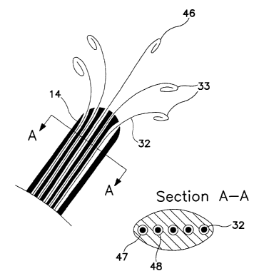
FIG. 5 is a cut-away view of the distal end 14 of an implanted lead with
extendable
5 members 32 deployed, and also a cross-sectional view of the epidural portion
of the lead.
Each member 32 can be extended beyond the lead tip 14 and it will gently curve
t o allow
lateral positioning of electrodes 33 or not, as shown by a midline electrode
46, depending
upon whether that particular member as a preset curve. The curve is due to
preset elastic
properties of the internal wire 44 of FIG. 4. Section A-A is an axial cross-
section of a
10 portion of the distal end of the lead 14. As depicted, there are five
channels, with two of
them labeled 47 and 48, each one with an extendable member 32 that may be slid
back and
forth, except when there is an anchor placed to prevent movement of the member
relative to
the lead body. Such an anchor may be simply a suture tied by the implanting
physician
tight enough to compress the lead body around the members, or may be more
elaborate with
15 set screws, collars, etc. In another embodiment, there may be designed an
asymmetric
feature in both the member and its channel so that each member has a fixed
aligmnent
relative the lead body. This would make the curving of the extended parts of
the member
predictable, as shown in FIG. 5, where the electrodes lie in a nearly planar
array that may be
next to the dura. Note that the cross-section A-A is not circular. While Tuohy
needles are
ZO widely available, more recently other shapes of hollow needles are being
considered for
epidural placement of leads, and these may allow slightly larger cross-
sectional areas, or
shapes that are not only circular.
An alternative design would not require an individual channel for each
extendable
~5 member, but rather a single lumen in the middle, or one lumen for several
members that will
curve toward a given side. The implanting physician could selectively pass
each member
out the tip of the lead 14, and would use fluoroscopy to determine the
direction of curving
and lateral motion, using rotational torque on each member to optimize its
position.
However, either such a multi-member channel would be narrow enough to prevent
curling
30 of the tips inside the lead body, or, should the tips curve and bend
backwards inside the
channel, the tips must be sufficiently flexible and the stiffening wires
sufficiently strong to
CA 02459603 2004-03-15
WO 03/039657 PCT/US02/06457
16
still pass the member distally, whereupon the tip 33 will form complete and
adequate 2- or
3-dimensional pads or electrodes.
FIG. 6 is a view of the distal end of an implanted lead 14 with members 32
deployed
outwardly. There is an axial cross-sectional view depicting another
embodiment, Section
B-B. This view shows six channels 47 for passage of extendable members 47, and
a central
open lumen 49, which can be used for a stylet to help in initial positioning
of the lead body
in the epidural space. Such a stylet is typically removed prior to closing all
incisions in the
patient because it is too stiff to leave there permanently. A symmetric,
hexagon shape for
the location of these channels is one way to best use the available space in
the lead body,
but other positions are possible, especially if the axial cross-section shape
is not circular.
FIG. 7 is a side view of the surface of the implanted lead, showing the distal
tip 14
and a middle portion 15 when the electrodes are not yet deployed, and another
portion 17
that is more proximal. When the middle portion is in the depicted position,
with separation
"L" between portion 15 and portion 17, the extendable members 32 are still
inside the distal
tip 14. By design, this gap should be located along the lead body so that its
position will lie
in the skin incision where the implanting physician can access it. Members 32
are attached
~0 permanently to lead body portion 17. When ready to deploy the members 32,
the physician
will hold the portion 15 steady with an instrument, and will push the portion
17 forward to
close the gap "L". This will simultaneously slide all six extendable members
32 distally,
with their tips extending out of the distal tip 14 of the lead body. In
another embodiment,
lead body portion 17 may have several independent parts, each one to be able
to deploy one
2,5 or more members independently. Section C-C is an axial cross-section of
portion 17 near to
the gap "L". It shows six members 32, each in their own channel, although the
channels
may be open to a central lumen 19. There is a stylet 18 in the center. It may
be later
removed to allow greater flexibility of the lead, or it may remain. In this
embodiment, if the
channels open up to a central lumen 19, the stylet 18 may serve the function
of keeping
30 each member 32 in its channel, due to its adequate diameter.
CA 02459603 2004-03-15
WO 03/039657 PCT/US02/06457
17
FIG. 8 is a cross-sectional view at the level of the most proximal ends of the
extendable members 40, where electrical connections are made to a receptacle
56 of either
an extension or implanted pulse generator. The view depicts six of the
proximal endings of
members 40, as shown in FIG. 4. Each one can be placed or pressed into an
electrical
connector, which has a conducting electrode 57, and each of these in turn has
a wire 58 that
is the source of electrical signal from the next component of the system. The
member's
proximal ending 40 can have a secure electrical communication with the
electrode 57 either
by use of set screws or Ball-seals~, like current commercially-available
electrical systems,
or can be held into position by the depicted elastic band 59, as shown. This
connection
must also have the flanges of the electrical receptacle 56 seal against the
surrounding elastic
band 59, which is an insulator like silicone rubber, so that current will not
leak from one
conducting electrode 57 to the next one. Either the elastic band 59 or another
insulated boot
must go over these connections to pernlanently seal out ionic solutions, which
might short
out the signals.
In the design of FIG. 7, all six proximal endings of the extendable members 32
will
have the same longitudinal position along the lead body, and hence the
electrical receptacle
56 in FIG. 8 may have a short axial length to will accommodate all of the
member's
proximal endings 40. However, if each extendable member is advanced by itself
or in
groups to varying degrees, there will be infra-lead redundancy in the lengths
of the members
that must be handled at the site of the electrical receptacle 56. In another
embodiment of
the connection described in FIG. 8, the axial length of the receptacle 56 and
its electrodes
57 are long enough to handle this redundancy. Alternatively, redundant infra-
lead lengths
of extendable members are looped or bunched up under an insulated elastic band
59 or
insulated boot, which may also be filled with silicone rubber for added
insulation. All of
these connections may be made in a subcutaneous pocket, where there is some
leeway for
size. If the lead must be removed or replaced, or the extendable members
repositioned,
surgical access to this pocket is necessary, regardless of the type of lead
used.
30 FIG. 9 shows two views of the distal end of a lead of another embodiment to
help
deployment of the extendable members and placement of the electrodes. In FIG.
9A, the
conducting tip of the member, which will eventually have a curl, is straight
35 while the
CA 02459603 2004-03-15
WO 03/039657 PCT/US02/06457
18
member's tip is still inside the lead body, or while it is being deployed in
directions
determined by the preset curvature of the member. This degree of straightness
is caused by
a thin but strong coating 62. This coating may be made from a wide variety of
materials
that are nontoxic, and which will dissolve in a matter of minutes to hours.
The coating will
enable the straight tip of the member 35 to poke through adhesions or
fibrosis, with minimal
deflection. After this coating has dissolved, FIG. 9B shows that the
conductive tip of the
member can curl up to form the conducting electrode 33. The coating may be
pointed in
shape, to make deployment easier. Since the member's tip 35 is metallic, it
will be easily
visible on fluoroscopy. In another embodiment, if there is a bimetallic metal
component or
nitinol is used to curl and uncurl the member's tip, controlled with the use
of electric
currents, then the transition from curling to uncurling can be done
repeatedly, or electively
only when there are obstructions that make positioning of the member's tip
difficult.
FIG. 10 is a cross-sectional view of the distal end of an extendable member 35
of
another embodiment of the invention, prior to deployment. Figure 10 shows how
two
dissimilar conductor coils can be attached to allow different mechanical
characteristics. The
proximal coil 43 goes from the proximal end out to a position near the end of
the internal
wire 44. It carries the electric signals, and is insulated 42 except for the
proximal end where
it has electrical connection to the extension or implanted pulse generator. A
dissimilar coil
ZO 70 begins at the end of the first spring 43 or is intertwined partially in
the coils of the first
spring as shows, and constitutes most of the member tip that will curl to form
an electrode.
The tip coil 70 will have the preset properties that allow it to curl once the
member is
deployed out of the lead body, and may be considerably more flexible than the
proximal
coil 43. Use of two dissimilar conductor coils may be useful, especially since
one, coil 42,
ZS needs a low impedance, and the other, coil 35 may need the ability to
accept a preset coiling
tendency.
FIG. 11 is a cross-sectional view of the distal end of another embodiment of
an
extendable member of the invention after deployment, showing how a conductor
coil ending
30 that is not insulated can be attached to an insulated proximal wire
portion. Here any
proximal conductor coil from the most proximal tip of the member is not
needed. The wire
44 comprises the member itself proximally, and has an insulative coating 42.
Attached to
CA 02459603 2004-03-15
WO 03/039657 PCT/US02/06457
19
the distal end of this wire 44, both mechanically and electrically is a member
tip 35 that will
curl to form a 2-dimensional electrode after deployment. Most of this member
tip 35 is not
insulated.
FIG. 12 is a cross-sectional view of a portion of an extendable member near
its distal
end, prior to deployment, showing how a conductor coil ending 35 that is not
insulated can
be screwed onto an insulated proximal wire portion 44. This is a most
convenient way to
assemble the tip of the extendable member. As the ending 35 curls to form a 2-
dimensional
electrode, one edge of this electrode will be the conductor coil that is
screwed onto the
threaded wire tip.
FIG. 13 is a cross-sectional view of a lead portion 16 near the distal lead
end 14
showing how five electrodes 33 can be positioned at various lateral and
ventral positions
and match the shape of the dura mater 60. In this example, one electrode is
fixed to the tip
of an extendable member that passes straight out of a central channel and has
a slight
ventral curve 72. This curve can be modest, if the lead end 14 is close to the
dura, or it can
be much sharper, if the lead end 14 is nearer to the vertebral bone 50. The
other four
electrodes 33, are located on members that curve both laterally upon
deployment and also
ventrally. The degree of ventral curve can be matched to the shape of the dura
mater 60 at
ZO that particular lateral position. In this way, the electrodes can be
positioned up against the
dura mater 60, allowing electrical efficiency due to less impedance, since the
impedance of
the epidural space 50, filled with fat or blood vessels, is substantial more
than the
impedance of the CSF in the subdural space.
~5 FIG. 14 is a cut-away view of a the distal tip of a lead body 14, showing
two
embodiments that allow the safe introduction of electrodes near to delicate
and small
peripheral nerves. The view shows a dorsal sacral foramen 73 and a ventral
sacral foramen
74, which are holes in the sacral bone that allow nerves to pass into the
body. Two nerves
are shown, a dorsal sacral nerve 75, for example the S3 nerve, and a ventral
sacral nerve 76,
30 going inside the pelvis to visceral organs and muscles. Often, screening
leads are placed in
or through the ventral sacral foramen 74, to see if urinary incontinence can
be improved or
visceral pain lessened. If there is success, then percutaneous-type permanent
leads with
CA 02459603 2004-03-15
WO 03/039657 PCT/US02/06457
electrodes are placed, but they often no longer give as much therapeutic
benefit. This could
be improved if the physician could implant a larger electrode near the ventral
sacral root 76.
Preferred embodiments of the invention make it possible to place a larger
electrode near the
root than the spaces through which that electrode must be passed. In one
method, a lead tip
5 14 is placed over the dorsal ventral foramen 73. One or more extendable
members 32 is
passed through both sacral foramena, and its tip 33 can curl into an electrode
whose
dimensions can be larger than the diameter of the accessible lumens. Some
physicians
today pass standard percutaneous SCS leads caudally, following a ventral
sacral nerve 77
from inside the sacral bone through the ventral sacral foramen 74. This may be
dangerous
10 since each sacral root nearly fills up the lumen of its passage near and
through the ventral
sacral foramen 74. An alternative method using the invention would have the
physician
pass an extendable member 77 from above, also the side of the ventral sacral
nerve 76, and
out of the ventral sacral foramen 74. In that foramen, or beyond, the
electrode 78 can curl.
The extendable member 77 can be of a very small diameter so there is room next
to the
15 ventral sacral nerve 76, and the electrode can form in a space where there
is adequate room.
Thus, preferred embodiments of the invention allows introduction of an
electrode
through a smaller lumen in the body. On the other side of the lumen, the tip
can curl into its
preset shape and become a 2- or 3- dimensional electrode. If necessary in the
future, simple
~0 traction upon the lead body or each extendable member will allow the tips
to uncurl and
retract through the narrow lumen. A good example of this application is
placement of an
electrical electrode through a vertebral or sacral foramen for peripheral
nerve stimulation of
a particular nerve outside the vertebral bones. A larger, 2-dimensional
electrode has a better
chance to have a stable excitation of a nerve than a percutaneous-type
electrode with a 1.0
ZS mm width. Placing two such electrodes along one nerve may also be
desirable, both to be
sure to excite the axons in the nerve by one or both of the electrodes, and to
stimulate across
the nerve with one electrode on either side, if one is a cathode and the other
an anode.
Another useful application is to treat trigeminal neuralgia, wherein the lead
body or member
is passed through the foramen rotundum of the cheekbone into the space of
Gasserian
ganglion.
CA 02459603 2004-03-15
WO 03/039657 PCT/US02/06457
21
While the above examples show use of preferred embodiments of the invention
for
stimulation of spinal cord or peripheral nerves, the same techniques can be
used for
stimulation of any excitable tissue where there is sufficient space for the
tips of the
extendable members to curl into electrodes, e.g., inside the ventricles of the
brain or on any
surface of the brain. Such methods may be very advantageous when very flexible
but
removable electrodes are needed, for example, in intrathecal or subdural
stimulation. On
occasion, sufficient space can be created through prior use of dilators,
especially for
stimulation on the surface of muscles or subcutaneously.
While the preferred aspects of the invention have primarily been described
with
respect to use of medical or implantable medical leads used for electrically
stimulating
tissue, such as nervous tissue, it will be understand that such medical leads
may also be
employed for sensing or monitoring physiological parameters, such as for
example
electrical activity within the spine or brain.
Those skilled in the art will recognize that the preferred embodiments may be
altered
or amended without departing from the true spirit and scope of the invention,
as defined in
the accompanying claims.