Note: Descriptions are shown in the official language in which they were submitted.
CA 02460227 2004-03-10
WO 03/037428 PCT/US02/32810
IMPLANTABLE MEDICAL DEVICE FOR MONITORING
CARDIAC BLOOD PRESSURE AND CHAMBER DIMENSION
FIELD OF THE INVENTION
The present invention relates generally to implantable medical devices (IMDs)
for
monitoring signs of acute or chronic cardiac heart failure and providing blood
pressure and
heart chamber dimension data to a physician to diagnose the condition of the
heart and
prescribe appropriate therapies including multi-chamber pacing optimized as a
function of
the measured blood pressure and heart chamber dimensions.
BACKGROUND OF THE INVENTION
Patients suffering from chronic heart failure including congestive heart
failure
(CHF) manifest an elevation of left ventricular end-diastolic pressure,
according to the
well-known heterometric autoregulation principles espoused by Frank and
Starling. This
may occur while left ventricular end-diastolic volume remains normal due to a
decrease in
left ventricular compliance concomitant with increased ventricular wall
stiffness. CHF
due to chronic hypertension, ischemia, infarct or idiopathic cardiomyopathy is
associated
with compromised systolic and diastolic function involving decreased atrial
and
ventricular muscle compliance. These may be conditions associated with chronic
disease
processes or complications from cardiac surgery with or without specific
disease
processes. Most heart failure patients do not normally suffer from a defect in
the
conduction system leading to ventricular bradycardia, but rather suffer from
symptoms
which may include a general weakening of the contractile function of the
cardiac muscle,
attendant enlargement thereof, impaired myocardial relaxation and depressed
ventricular
filling characteristics in the diastolic phase following contraction.
Pulmonary edema,
shortness of breath, and disruption in systemic blood pressure are associated
with acute
exacerbations of heart failure. '
All these disease processes lead to insufficient cardiac output to sustain
mild or
moderate levels of exercise and proper function of other body organs, and
progressive
worsening eventually results in cardiogenic shock, arrhythmias,
electromechanical
CA 02460227 2004-03-10
WO 03/037428 PCT/US02/32810
2
dissociation, and death. In order to monitor the progression of the disease
and to assess
efficacy of prescribed treatment, it is necessary to obtain accurate measures
of the heart
geometry, the degree of heart enlargement, and the mechanical pumping
capability of the
heart, e.g., ejection fraction, under a variety of metabolic conditions the
patient is likely to
encounter on a daily basis. These parameters are typically measured through
the use of
external echocardiogram equipment in the clinical setting. However, the
measurement
procedure is time consuming to perform for even a resting patient and cannot
be
practically performed replicating a range of metabolic conditions. Typically,
the
echocardiography procedure is performed infrequently and months or years may
lapse
between successive tests, resulting in a poor understanding of the progress of
the disease
or whether or not intervening drug therapies have been efficacious. Quite
often, only
anecdotal evidence from the patient is available to gauge the efficacy of the
prescribed
treatment.
Moreover, in many cases, diseased hearts exhibiting left ventricular
dysfunction
(LVD) and CHF also have conduction defects wherein cardiac depolarizations
that
naturally occur in one upper or lower heart chamber are not always conducted
in a timely
fashion either within the heart chamber or to the other upper or lower heart
chamber. In
such cases, the right and left heart chambers do not contract in optimum
synchrony with
each other, and cardiac output suffers due to the conduction defects. In
addition,
spontaneous depolarizations of the left atrium or left ventricle occur at
ectopic foci in
these left heart chambers, and the natural activation sequence is grossly
disturbed. The
natural electrical activation system through the heart involves sequential
events starting
with the sino-atrial (SA) node, and continuing through the atrial conduction
pathways of
Bachmann's bundle and internodal tracts at the atrial level, followed by the
atrio-
ventricular (AV) node, Common Bundle of His, right and left bundle branches,
and final
distribution to the distal myocardial terminals via the Purkinje fiber
network. A common
type of infra-atrial conduction defect is known as infra-atrial block (IAB), a
condition
where the atrial activation is delayed in getting from the right atrium to the
left atrium. In
left bundle branch block (LBBB) and right bundle branch block (RBBB), the
activation
signals are not conducted in a normal fashion along the right or left bundle
branches
respectively. Thus, in a patient with LBBB or RBBB, the activation of the
ventricles is
CA 02460227 2004-03-10
WO 03/037428 PCT/US02/32810
slowed, and the QRS is seen to widen due to the increased time for the
activation to
traverse the conduction path. For example, in a patient with LBBB, the delay
in the
excitation from the RV to the LV can be as high as 120 to 150 ms. Cardiac
output
deteriorates because the contractions of the right and left heart chambers are
not
synchronized sufficiently to eject the maximal blood volume. Furthermore,
significant
conduction disturbances between the right and left atria can result in left
atrial flutter or
fibrillation.
More particularly, as described in commonly assigned LT.S. Patent No.
6,129,744,
patients suffering from LVD are also known to have elevated levels of
catecholamines at
rest because the body is attempting to increase cardiac output that induce a
higher resting
heart rate. In addition, the QT interval for such a patient is affected by the
catecholamine
level and thus has a changed pattern during exercise as well. These patients
have a
decreased QT response, or smaller change in QT, during exercise, such that the
QT
interval shortening during exercise is smaller than that found normally.
Although QT
interval is influenced independently by heart rate alone, as well as by
exercise and
catecholemines, it is not known to what extent each of these factors or both
are responsible
for the changed QT response to exercise in LVD patients. However, it is known
that
patients suffering LVD clearly have a different pattern of QT interval
shortening during
exercise. Moreover, the changed conductive patterns or a heart in heart
failure are
manifested by other changes in the PQRST waveforms, particularly an abnormally
wide or
long duration of the ventricular depolarization signal, or QRS.
These observed conduction defects have caused physicians to prescribe
implantation of conventional, atrioventricular (AV) synchronous pacing
systems,
including DDD and DDDR pacing systems, marketed by Medtronic, Inc. and other
companies, in certain patients for treatment of heart failure symptoms.
Certain patient
groups suffering heart failure symptoms with or without bradycardia tend to do
much
better hemodynamically with AV synchronous pacing due to the added
contribution of
atrial contraction to ventricular filling and subsequent contraction. However,
fixed or
physiologic sensor driven rate responsive pacing in such patients does not
always lead to
improvement in cardiac output and alleviation of the symptoms attendant to
such disease
processes because it is difficult to assess the degree of compromise of
cardiac output
CA 02460227 2004-03-10
WO 03/037428 PCT/US02/32810
4
caused by CHF and to determine the pacing parameters that are optimal for
maximizing
cardiac output, particularly the AV delay.
Determining an optimal AV delay requires performing echocardiography studies
or
obtaining pressure data involving an extensive patient work-up as set forth in
commonly
assigned U.S. Patent No. 5,626,623. Moreover, conventional DDD and DDDR
pacemakers pace and sense only in the right atrium and right ventricle and
cannot alleviate
or alter IAB, LBBB, RBBB and QT interval widening.
Consequently, while some improvement has been reported in certain patients
receiving two-chamber DDD or DDDR AV sequential pacemakers, the efficacy of
the
treatment is not established for larger patient populations. Therefore,
efforts have been
undertaken to develop more appropriate therapies, to identify patients who
would benefit
from such therapies, and to provide tools to assess the efficacy of the
applied therapies.
A great deal of testing and data collection is necessary to obtain a thorough
understanding of the heart failure condition and disease etiology of a
symptomatic heart
failure patient in order to prescribe any therapy, including drug therapies
and IMD
delivered stimulation therapies. Therefore, a number of other approaches have
been
proposed and advanced involving implantation of physiologic cardiac monitors
for
deriving and storing electrical EGM signals and mechanical performance
indicating
parameters over a prolonged time period and development of three and four-
chamber
pacing systems having the same capabilities.
An implantable EGM monitor for recording the cardiac electrogram from
electrodes remote from the heart is disclosed in commonly assigned U.S. Patent
No.
5,331,966 and PCT publication WO 98/02209 and is embodied in the MedtronicOO
REVEALO Insertable Loop Recorder having spaced housing EGM electrodes. More
elaborate implantable hemodynamic monitors (IHMs) for recording the EGM from
electrodes placed in or about the heart and other physiologic sensor derived
signals, e.g.,
one or more of blood pressure, blood gases, temperature, electrical impedance
of the heart
and/or chest, and patient activity have also been proposed. The Medtronic~
CHRONICLED IHM is an example of such a monitor that is coupled through a lead
of the
type described in commonly assigned U.S. Pat. No. 5,564,434 having capacitive
blood
pressure and temperature sensors as well as EGM sense electrodes. Such
implantable
CA 02460227 2004-03-10
WO 03/037428 PCT/US02/32810
monitors when implanted in patients suffering from cardiac arrhythmias or
heart failure
accumulate date and time stamped data that can be of use in determining the
condition of
the heart over an extended period of time and while the patient is engaged in
daily
activities. A wide variety of other IMDs have been proposed to monitor many
other
physiologic conditions as set forth in U.S. Patent No. 6,221,011
With respect to stimulation therapies other than DDD or DDDR pacing therapies,
it
was observed in the early days of implantable cardiac pacing that paired
pacing (two or
more closely spaced pacing pulses delivered at the time-out of an escape
interval) and
triggered or coupled pacing (one or more pacing pulses delivered following the
detection
of a P-wave or R-wave terminating an escape interval) with relatively short
interpulse
intervals (150 to 250 milliseconds in dogs and about 300 milliseconds in human
subjects)
beneficially slowed heart rate and increased cardiac output. The result of the
second
pulse, applied within the relative refractory period of the first paced or
spontaneous
depolarization, is to prolong the refractory period and effect a slowing of
the heart rate
from its spontaneous rhythm without an attendant mechanical myocardial
contraction.
This slowing effect has been employed since that time in many applications,
including the
treatment of atrial and ventricular tachycardias, where a single pulse or a
burst of pulses
are coupled to a spontaneous tachycardia event with a coupling interval that
is shorter than
and can be set as a fraction of the tachycardia interval as taught, for
example, in U.S.
Patent Nos. 3,857,399 and 3,939,844. The slowing of the heart rate by coupled
pacing is
accompanied by the ability to increase or decrease the rate with subsequent
coupled pacing
within wide limits.
Paired and coupled stimulation of a heart chamber also cause a potentiation of
contractile force effect through a phenomenon known as post-extrasystolic
potentiation
(PESP) described in detail in commonly assigned U.S. Patent No. 5,213,098. The
force of
contraction of the heart is increased during the heart cycle that the paired
or coupled
stimulation is applied, and the increase persists but gradually diminishes
over a number of
succeeding heart cycles. Other measurable PESP effects that also persist but
gradually
decline over a number of heart cycles include changes in the peak systolic
blood pressure,
the rate of contraction of the ventricular muscle with a resulting increase of
the rate of rise
CA 02460227 2004-03-10
WO 03/037428 PCT/US02/32810
6
of intraventricular pressure (dP/dt), an increase in coronary blood flow, and
an increase in
the oxygen uptake of the heart per beat.
Various burst pulse stimulation regimens have been proposed for the treatment
of
heart failure including CHF that involve application of supra-threshold and/or
sub-
threshold stimulation paired or coupled pacing pulses or pulse trains.
Moreover, various
electrodes have been proposed for single site and multi-site delivery of the
stimulation
pulses to one or more heart chamber in the above-referenced patents and
publications.
However, it remains difficult to economically determine appropriate candidates
that would
benefit from such stimulation and to measure the efficacy of a given
stimulation regimen
and/or electrode array. Extensive catheterization procedures must be conducted
of a heart
failure patient to determine if he or she is a candidate for implantation of
such a system.
Then, the efficacy of any given treatment must be assessed at implantation and
in periodic
post-implant follow-up clinical tests. The patient work-up and follow-up
testing must take
into account or simulate known patient activities, patient posture, and
whether the patient
is awake or asleep in order to be representative of the heart failure
condition over a daily
time span
Consequently, determining the most efficacious burst stimulation parameters
can
be difficult and the results vary over time and due to a number of factors.
Thus,
widespread adoption of burst stimulation therapies for treating heart failure
has not
occurred.
A number of proposals have been advanced for providing pacing therapies to
alleviate heart failure conditions and restore synchronous depolarization and
contraction of
a single heart chamber or right and left, upper and lower, heart chambers as
described in
detail in the above referenced '744 patent and in commonly assigned U.S.
Patent Nos.
5,403,356, 5,797,970 and 5,902,324, 6,219,579 and in U.S. Patent Nos.
5,720,768 and
5,792,203. The proposals appearing in U.S. Patent Nos. 3,937,226, 4,088,140,
4,548,203,
4,458,677, 4,332,259 are summarized in U.S. Patent Nos. 4,928,688 and
5,674,259. The
advantages of providing sensing at pace/sense electrodes located in both the
right and left
heart chambers is addressed in the '688 and '259 patents, as well as in U.S.
Patent Nos.
4,354,497, 5,174,289, 5,267,560, 5,514,161, and 5,584,867.
CA 02460227 2004-03-10
WO 03/037428 PCT/US02/32810
7
The medical literature also discloses a number of approaches of providing bi-
atrial
and/or bi-ventricular pacing as set forth in: Daubert et al., "Permanent Dual
Atrium Pacing
in Major Intra-atrial Conduction Blocks: A Four Years Experience", PACE (Vol.
16, Part
II, NASPE Abstract 141, p.885, April 1993); Daubert et al., "Permanent Left
Ventricular
Pacing With Transvenous Leads Inserted Into The Coronary Veins", PACE (Vol.
21, Part
II, pp. 239-245, Jan. 1998); Cazeau et al., "Four Chamber Pacing in Dilated
Cardiomyopathy", PACE (Vol. 17, Part II, pp. 1974-1979, November 1994); and
Daubert
et al., "Renewal of Permanent Left Atrial Pacing via the Coronary Sinus", PACE
(Vol. 15,
Part II, NASPE Abstract 255, p. 572, April 1992).
In most cases, it has been proposed that bi-ventricular pacing pulses be
applied
simultaneously to the right and left ventricles. An observation is made in
commonly
assigned U.S. Patent No. 6,219,579 that the exact timing of mechanical events
are
important for properly controlling right and left heart chamber pacing so as
to optimize
left ventricular output. Specifically, it is known that actual contraction of
one ventricular
chamber before the other has the effect of moving the septum so as to impair
full
contraction in the later activated chamber. Thus, while concurrent or
simultaneous pacing
of the left and right ventricle may achieve a significant improvement for CHF
patients, it
is better to provide for pacing of the two ventricles in such a manner that
the actual
mechanical contraction of the left ventricle, with the consequent closing of
the valve,
occurs in a desired time relationship with respect to the mechanical
contraction of the right
ventricle and closing of the right value. For example, if conduction paths in
the left
ventricle are impaired, delivering a pacing stimulus to the left ventricle at
precisely the
same time as to the right ventricle may nonetheless result in left ventricular
contraction
being slightly delayed with respect to the right ventricular contraction.
In the above-referenced ' 324 patent, an AV synchronous pacing system is
disclosed providing three or four heart chamber pacing through pace/sense
electrodes
located in or adjacent one or both of the right and left atrial heart chambers
and in or
adjacent to the right and left ventricular heart chambers. During an AV delay
and during a
V-A escape interval,'a non-refractory ventricular sense event detected at
either the right or
left ventricular pace/sense electrodes starts a programmable conduction delay
window
(CDW) timer. A ventricular pace pulse is delivered to the other of the left or
right
CA 02460227 2004-03-10
WO 03/037428 PCT/US02/32810
ventricular pace/sense electrodes at the time-out of the CDW if a ventricular
sense event is
not detected at that site while the CDW times out. However, it is not always
easy to
determine just how to program the CDW duration to optimize the hemodynamics of
the
heart. As a consequence, it is important to provide a technique for
measurement of
mechanical events, such as a mechanical closure point of each of the
ventricles, so as to be
able to accurately program the sequence of pacing to achieve the desired dual
ventricular
pacing which optimizes ejection fraction, or cardiac output, for the
individual patient.
Moreover, while such AV sequential, three or four-chamber pacing systems can
be
programmed to at least initially restore right and left and upper and lower
heart synchrony
in the clinical setting, they are not always able to maintain that synchrony
over a range of
heart rates and as the patient is exposed to other conditions of daily life
including stress
and exercise.
It is understood that the amount of blood being pumped by the heart is
governed
not only by the intrinsic or multi-chamber paced heart rate, but also by the
stroke volume
of the heart which is adversely lessened by heart failure. It has been
recognized that it
would be desirable to measure the contractility or displacement of the heart
wall to
determine the hemodynamic efficiency of the heart alone in an implanted
monitor or in the
context of controlling the operations of therapy delivery IMDs.
For example, the use an accelerometer positioned within a lead that is located
within one of the chambers of the heart is disclosed in U.S. Patent No.
5,549,650. The
lead is attached to one of the walls of the heart so that movement of the wall
of the heart
causes the accelerometer that to develop an accelerometer signal that is
processed to
provide a first signal indicative of the contractility of the heart and a
second signal
indicative of the physical displacement of the wall of the heart. It is
proposed in U.S.
Patent No. 4,730,619 to derive a measure of the ejection time of the
ventricles, which is
derived from the duration of contraction of the right ventricle which is
deterniined from
changes in right ventricular pressure. The right ventricular blood pressure is
measured by
a hermetically sealed absolute strain gauge transducer or a piezoresistive
transducer
mounted within a transvenous lead. The signals derived in the '650 and '619
patent are
employed by the pacing system to adjust the pacing parameters to improve the
CA 02460227 2004-03-10
WO 03/037428 PCT/US02/32810
hemodynamic efficiency of the heart as this information is directly related to
the volume
of blood being pumped by the heart during each ventricular contraction.
In an approach related to monitoring rejection of heart transplants, a
magnetic field
responsive Hall effect device and a permanent magnet are implanted directly
across the
septum or a heart wall as taught in U.S. Patent No. 5,161,540, and the Hall
effect device is
powered by an implantable generator and telemetry transceiver. The compliance
of the
heart wall is monitored to detect any loss of compliance characteristic of
rejection of the
heart transplant is transmitted from the implanted system.
A discussion of a wide number of mechanical and electrical parameter sensors
employed in the art to assess cardiac functions and hemodynarnic efficiency is
set forth in
U.S. Patent No. 5,243,976. In the '976 patent, continuous wave (CW) and pulsed
wave
(PW) Doppler emitters are incorporated into pacing leads to measure blood
flow, and the
flow measurements are employed to regulate atrial and ventricular pacing
parameters and
for other purposes.
In the above-referenced ' 579 patent, impedance measurements are made in or
across the heart chambers from which accurate timing signals are obtained
reflecting
mechanical actions, e.g., valve closures, so that accurate timing information
is available
for controlling electrical activation and resultant mechanical responses for
the respective
different heart chambers. The impedance or mechanical sensing determinations
are
preferably made by multiplexing through fast switching networks to obtain the
desired
impedance measurements in different heart chambers. In a preferred embodiment,
control
of left heart pacing, is based primarily upon initial detection of a
spontaneous signal in the
right atrium, and upon sensing of mechanical contraction of the right and left
ventricles.
In a heart with normal right heart function, the right mechanical AV delay is
monitored to
provide the timing between the initial sensing of right atrial activation (P-
wave) and right
ventricular mechanical contraction. The left heart is controlled to provide
pacing which
results in left ventricular mechanical contraction in a desired time relation
to the right
mechanical contraction; e.g., either simultaneous or just preceding the right
mechanical
contraction; cardiac output is monitored through impedance measurements, and
left
ventricular pacing is timed to maximize cardiac output. In patients with IAB,
the left
atrium is paced in advance of spontaneous depolarization, and the left AV
delay is
CA 02460227 2004-03-10
WO 03/037428 PCT/US02/32810
adjusted so that the mechanical contractions of the left ventricle are timed
for optimized
cardiac output from the left ventricle.
The ' 579 patent also sets forth algorithms using the impedance measurements
to
obtaining and storing data reflecting heart failure state and for optimizing
bi-ventricular
5 pacing to provide maximum cardiac output.
A CHF monitor/stimulator is disclosed in commonly assigned U.S. Patent No.
6,104,949 that senses the trans-thoracic impedance as well as patient posture
and provides
a record of same to diagnose and assess the degree and progression of CHF. The
sensed
10 trans-thoracic impedance is dependent on the blood or fluid content of the
lungs and
assists in the detection and quantification of pulmonary edema symptomatic of
CHF.
Trans-thoracic impedance is affected by posture, i.e. whether the subject is
lying down or
standing up, and the sensed trans-thoracic impedance is correlated to the
output of the
patient posture detector to make a determination of presence of and the degree
of
pulmonary edema for therapy delivery and/or physiologic data storage
decisions.
A monitor/stimulator is disclosed in U.S. Patent No. 5,417,717 that monitors
and
assesses level of cardiac function then permits a physician to arbitrate the
therapy mode, if
therapy is indicated. The monitor stimulator assesses impedance, EGM, and/or
pressure
measurements, and then calculates various cardiac parameters. The results of
these
calculations determine the mode of therapy to be chosen. Therapy may be
administered
by the device itself or a control signal may be telemetered to various
peripheral devices
aimed at enhancing the heart's function. Alternatively, the device may be
programmed to
monitor and either store or telemeter information without delivering therapy.
One
suggested therapy comprises delivery or AV synchronous, bi-ventricular pacing
pulses to
the heart.
Particularly, the implantable monitor/stimulator of the '717 patent monitors
conventional parameters of cardiac function and contractile state, including
all phases of
the cardiac cycle. Thus, assessments of contractile state measured include
indices of both
cardiac relaxation and contraction. Utilizing the dual source ventricular
impedance
plethysmography technique described in U.S. Patent No. 4,674,518, the
monitor/stimulator
monitors cardiac function by assessing hemodynamic changes in ventricular
filling and
CA 02460227 2004-03-10
WO 03/037428 PCT/US02/32810
11
ejection or by calculating isovolumic phase indices by known algorithms. The
primary
calculations involve: ( 1 ) the time rate of change in pressure (dP/dt) or
volume (dV/dt) as
isovolumic indicators of contractility; (2) ejection fraction as an ejection
phase index of
cardiac function according to the known quotient of stroke volume divided by
end
diastolic volume; (3) Maximal elastance, EM; (4) regression slope through
maximal
pressure-volume points as a further ejection phase index of contractility
using the method
of Sagawa; (5) stroke work according to the known pressure-volume integration;
(6) the
time course of minimum (end) diastolic pressure-volume measurements according
to the
method of Glantz as a measure of diastolic function; and (7) cardiac output
calculation
according to the known product of heart rate and stroke volume as an index of
level of
global function.
While measurement and storage of this group of parameters of cardiac function
and contractile state can provide valuable information about the state of
heart failure, the
sensors are not always easy to implant so that they perform reliably
chronically and under
the range of conditions encountered by the patient and resulting from
progression of the
heart failure. The proposed systems employing locally disposed accelerometers
at one or
more location in the heart or distributed impedance measuring electrodes to
detect and
measure heart motion and to derive the above-described parameters are
difficult to
implement and subject to outside influences that distort the signals.
Chronically collected data from patients with heart failure is needed so that
the
treating cardiologist can properly and accurately chart the progression,
determine the
nature of the heart failure, and be able to implement the optimal treatment in
a timely
fashion. There is a substantial need in the art for a pacemaker or other IMD
having the
capacity to identify the progression or remission of heart failure and to
provide such
indication to the patient's physician so that options can be assessed from
time to time to
treat the changing patient condition.
Given the demonstrated feasibility of PESP and four-chamber cardiac pacing,
and
the availability of techniques for sensing natural cardiac signals and
mechanical events,
there nonetheless remains a need for developing a system which is adapted to
obtain
valuable data and to make changes in the pacing parameters to optimize
mechanical
performance of the heart. There is a need for such an IMD providing bi-
ventricular and/or
CA 02460227 2004-03-10
WO 03/037428 PCT/US02/32810
12
bi-atrial pacing wherein the pacing rate and A-A delay or V-V delay as well as
the AV
delay are periodically optimized by the IMD operating system to provide
appropriate
hemodynamic status during various ambulatory conditions and activities of
daily living
using cardiac pressures, dimensions and wall displacement.
SUMMARY OF THE INVENTION
In view of the above need, the present invention provides a system and method
for
monitoring patient cardiac signals and processing such signals within an IMD
to provide
data from which the onset or progression of heart failure can be determined.
It is to be
understood that the invention is applicable to various forms of heart failure,
including left
heart conduction disorders such as IAB, LBBB and RBBB, and other forms of
heart
dysfunction including LVD.
In accordance with the present invention, an implantable stimulator and
monitor
measures a group of parameters indicative of the state of heart failure
employing EGM
signals, measures of blood pressure including absolute pressure P, developed
pressure DP
(DP = systolic P - diastolic P), and/or dP/dt, and measures of heart chamber
dimension (D)
over one or more cardiac cycles to derive trend data indicative of the state
of heart failure.
The measures of pressure and dimension developed over heart cycles can also be
employed in pressure-dimension relationship analysis to provide other useful
information
about the status of the cardiac function.
The dimension sensor or sensors comprise at least a first sonomicrometer
piezoelectric crystal mounted to a first lead body implanted into or in
relation to one heart
chamber, e.g., the RV, that operates as an ultrasound transmitter when a drive
signal is
applied to it and at least one second sonomicrometer crystal mounted to a
second lead
body implanted into or in relation to a second heart chamber, e.g.,'the LV,
the LA or the
RA, that operates as an ultrasound receiver. The ultrasound receiver converts
impinging
ultrasound energy transmitted from the ultrasound transmitter through blood
and heart
tissue into an electrical signal. The time delay between the generation of the
transmitted
ultrasound signal and the reception of the ultrasound wave varies as a
function of the
distance between the ultrasound transmitter and receiver which in turn varies
with
contraction and expansion of a heart chamber between the first and second
CA 02460227 2004-03-10
WO 03/037428 PCT/US02/32810
13
sonomicrometer crystals. One or more additional sonomicrometer piezoelectric
crystal
can be mounted to additional lead bodies such that the distances between the
three or more
sonomicrometer crystals can be determined. In each case, the sonomicrometer
crystals are
distributed about a heart chamber such that the distance between the separated
ultrasound
transmitter and receiver crystal pairs changes with contraction and relaxation
of the heart
chamber walls whereby the instantaneous measured distance is characterized as,
or is
proportional to, the instantaneous heart chamber dimension D.
The instantaneous heart chamber dimension (D) is an indicator of the
instantaneous
heart chamber volume (V) and can be employed in pressure dimension
relationship
analyses akin to pressure-volume relationship analyses. More than one receiver
crystal
can be positioned about a given heart chamber, e.g., the LV, and paired with a
transmitter
crystal to derive sets of dimension data from which heart chamber volume V may
be more
closely extrapolated.
A heart failure parameter of interest comprises end systolic elastance (EES),
i.e., the
ratio of end systolic blood pressure P to an end systolic volume V or
dimension D of a
heart chamber and the end-diastolic elastance (EED). The EES and EED heart
failure state
parameter is determined and stored periodically when patient posture, activity
level,
intrinsic heart rate, and regularity are within programmable ranges. The EES
and EED
parameter data is associated with a date and time stamp and with other patient
data, e.g.,
patient activity level, and the associated parameter data is stored in IMD
memory for
retrieval at a later date employing conventional telemetry systems.
Incremental changes in
the parameter data over time, taking any associated time of day and patient
data into
account, provide a measure of the degree of change in the CHF condition of the
heart.
The sonomicrometer distance and pressure sensing system and method of the
present invention has particular application to the derivation of LV pressure
and
dimension data and the development of the EES and EED data that provide a
global metric
of heart failure status and remodeling that occurs due to the pathophysiology.
In general
terms, as the heart chamber dimension D and volume V increase and pressure P
decreases
or remains the same, the EES decreases and the EED increases. This is the
common
observation as the heart failure worsens. The data also provides a global
metric of heart
failure status and severe remodeling that occurs during delivery of drug
and/or stimulation
CA 02460227 2004-03-10
WO 03/037428 PCT/US02/32810
14
therapies. In general terms, an effective therapy leading to an improvement in
the heart
failure state is indicated by a reduction in the heart chamber dimension D and
volume V,
pressure P increases or remains the same and EES increases while EED
decreases.
The percent systolic shortening provides additional information which can be
used
to evaluate the AV and VV pacing intervals. Percent systolic shortening is
measured by
dividing the difference of the dimensions at end-systole and end-diastole by
the end-
diastolic value. The amount of shortening occurnng each beat is stable and
decreases as
the amount of ventricular dysfunction increases.
The implantable stimulator and monitor that is capable of performing these
functions comprises an implantable pulse generator (IPG) or monitor and lead
system
extending into operative relation with at least one and preferably multiple
heart chambers
for electrical sensing and stimulation, blood pressure measurement and chamber
volumetric measurement during contraction and relaxation. The IPG/monitor has
a sense
amplifier for each heart chamber of interest that is coupled through a lead
conductor with
electrical stimulation/sense electrodes for sensing cardiac electrical heart
signals
originating in or traversing that heart chamber so that the sense amplifier
can detect a P-
wave in an atrial chamber or R-wave in a ventricular chamber.
Preferably an IPG is provided having timing circuitry for timing out atrial
and/or
ventricular escape intervals and the ESI of coupled or paired PESP stimulating
pulses)
and a pulse generator coupled with at least one stimulation/sense electrode
for delivering
pacing pulses and PESP stimulation pulses to each heart chamber of interest.
The IPG has
blood pressure signal processing circuitry coupled through lead conductors
with a blood
pressure sensor located in a distal lead section in or in operative relation
to each heart
chamber of interest for deriving blood pressure P and dP/dt samples. The IPG
also has
dimension D and volume V determining circuitry coupled with one or more of the
sonomicrometer dimension sensors located in or in relation with each heart
chamber of
interest for deriving a signal representative of heart chamber dimension D and
volume V.
In order to overcome the disadvantages and limitations of previously known
approaches for optimizing pacing therapy, the processing system of the present
invention
processes the derived pressure and dimension to produce signals representative
of stroke
volume, percent systolic shortening, stroke work, cardiac contractility, pre-
ejection period,
CA 02460227 2004-03-10
WO 03/037428 PCT/US02/32810
filling time and ejection time. These signals are used to provide
hemodynamically optimal
pacing therapy while the patient is at rest and to provide hemodynamically
optimal rate-
responsive pacing therapy. Stroke volume, percent systolic shortening, stroke
work,
cardiac contractility, pre-ejection period, filling time and ejection time may
be used,
individually or together in combination, to adjust the parameters of the
implantable cardiac
stimulating device so that hemodynamically optimal pacing therapy may be
provided.
The pressure and dimension signals as provided by the processing system of the
present invention have been found to be related to stroke work. To illustrate,
pressure and
dimension signals from a patient suffering from dilated cardiomyopathy
demonstrate a
10 reduced pulse pressure change and a reduced dimensional change (volume
change) during
a cardiac cycle. Note that both absolute pressure and overall dimension may be
increased
over long time periods, yet the change is attenuated. This indicates that the
total volume
of blood being pumped by the heart during each heartbeat is abnormal.
The present invention is directed to a processing system which processes the
15 pressure and dimension signals to determine cardiac stroke volume, percent
systolic
shortening, stroke work, cardiac contractility, pre-ejection period, filling
time and ejection
time, and then use these calculated values to optimize the timing of the
stimulation
provided to the patient by the rate-responsive pacemaker. In this manner,
operational
parameters of the rate-responsive pacemaker may be adjusted, in a closed loop
manner, as
the circumstances for optimal hemodynamic performance change. For example, the
rate-
responsive pacemaker may continually adjust the heart rate of the patient to
provide
hemodynamically optimal pacing therapy, thereby substantially maximizing
cardiac output
during periods of metabolic need.
The present invention initially establishes optimal values for heart rate, A-
A, V-V
and AV delays. Then, for each optimization cycle, cardiac performance is
measured using
pressure and dimension signals for selected combinations of heart rate, A-A, V-
V and AV
delays. The interval values resulting in the greatest measured cardiac
performance
become the new optimal values for the next cycle.
In another aspect of the present invention, methods for providing
hemodynamically
optimal rate-responsive pacing therapy and hemodynamically optimal pacing
therapy at
rest are described. The methods of providing hemodynamically optimal pacing
therapy
CA 02460227 2004-03-10
WO 03/037428 PCT/US02/32810
16
(for rate-response or at rest) may utilize, individually or in combination,
stroke volume,
percent systolic shortening, stroke work, cardiac contractility, pre-ejection
period, filling
time and ejection time to optimize cardiac performance.
This summary of the invention and the objects, advantages and features thereof
have been presented here simply to point out some of the ways that the
invention
overcomes difficulties presented in the prior art and to distinguish the
invention from the
prior art and is not intended to operate in any manner as a limitation on the
interpretation
of claims that are presented initially in the patent application and that are
ultimately
granted.
BRIEF DESCRIPTION OF THE DRAWINGS
These and other advantages and features of the present invention will be more
readily understood from the following detailed description of the preferred
embodiments
thereof, when considered in conjunction with the drawings, in which like
reference
numerals indicate identical structures throughout the several views, and
wherein:
FIG. 1 is a schematic diagram depicting a mufti-channel, atrial and bi-
ventricular,
monitoring/pacing IMD in which the present invention is preferably implemented
employing distributed sonomicrometer piezoelectric crystals to derive
dimension signals
during systolic and diastolic heart contraction phases;
FIG. 2 is a simplified block diagram of one embodiment of IMD circuitry and
associated leads employed in the system of FIG. 1 enabling selective therapy
delivery
and/or monitoring in one or more heart chamber;
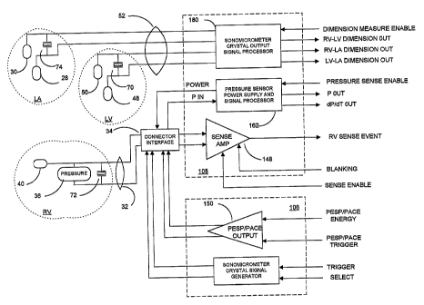
FIG. 3 is a simplified block diagram of a mufti-chamber measurement system for
deriving RV pressure signals, dimension measurements and cardiac EGM signals
employed in monitoring CHF and optionally pacing the heart and delivering
pacing
therapy in accordance with the present invention;
FIG. 4 is a comprehensive flow-chart illustrating the operating modes of the
IMD
circuitry of FIG. 3 in a variety of AV synchronous, bi-ventricular pacing
modes in
accordance with one embodiment of the invention;
FIG. 5 is a flow chart illustrating the steps of delivering ventricular pace
pulses
following time-out of an AV delay in FIG. 4;
CA 02460227 2004-03-10
WO 03/037428 PCT/US02/32810
17
FIG. 6A-6B is a flow chart illustrating the steps of delivering ventricular
pace
pulses following a ventricular sense event during the time-out of an AV delay
or the V-A
escape interval in FIG. 4;
FIG. 7 is a flow chart illustrating the steps of periodically operating the
system of
FIG. 3 to derive RV pressure signals, dimension measurements and cardiac EGM
signals,
storing the signals, optionally processing the signals to update pacing timing
parameters,
and telemetering the stored data and updated parameters to an external
programmer;
FIG. 8 is a flow chart illustrating the steps of operating the system of FIG.
3 to
derive RV pressure signals and dimension measurements and processing the
signals to
provide elastance data in step 5416 of FIG. 7;
FIG. 9 is a graphical depiction of measured left ventricular PV loops during a
modification of preload with end systolic PV points shown at the upper left;
FIG. 10 is a graphical depiction of a linear regression of the end systolic PV
points
of FIG. 18 to derive the slope of the LV EES;
FIG. 11 is a graphical depiction of measured left ventricular PV loops during
normal heart function with end systolic PV points shown at the upper left;
FIG. 12 is a graphical depiction of a linear regression of the end systolic PV
points
of FIG. 20 wherein the determination of slope of the LV EES is not reliable;
FIG. 13 is a flow chart illustrating the steps of employing elastance
parameter data
derived in FIGS. 7 and 8 at differing temporary settings of pacing parameters
to derive the
set of pacing parameters providing optimal right and left mechanical heart
function;
FIG. 14 depicts the relationship of heart chamber EGM, pressure, flow, and
volume during a heart cycle; and
FIG. 15 is a flow chart illustrating an alternative manner of deriving pacing
parameter values from diagnostic values derived from measured pressure and
distance
signals that optimize right and left heart mechanical heart function
DETAILED DESCRIPTION OF THE PREFERRED EMBODIMENTS
In the following detailed description, references are made to illustrative
embodiments for carrying out the invention. It is understood that other
embodiments may
be utilized without departing from the scope of the invention. For example,
the invention
CA 02460227 2004-03-10
WO 03/037428 PCT/US02/32810
18
is disclosed in detail herein in the context of an AV sequential, three
chamber or four
chamber, pacing system operating in demand, atrial tracking, and triggered
pacing modes
for restoring synchrony in depolarizations and contraction of left and right
ventricles in
synchronization with atrial sensed and paced events for treating heart failure
and/or
bradycardia in those chambers. This embodiment of the invention is
programmable to
operate as a three or four chamber pacing system having an AV synchronous
operating
mode for restoring upper and lower heart chamber synchronization and right and
left atrial
and/or ventricular chamber depolarization synchrony.
It should be appreciated that the present invention may be utilized in an
implantable monitor to gather data in patients suffering various forms of
heart failure. The
system of the present invention may also may be incorporated into an anti
tachyarrhythmia system including specific high rate pacing and cardioversion
shock
therapies for providing staged therapies to treat a diagnosed tachyarrhythmia.
In FIG. 1, heart 10 includes the upper heart chambers, the right atrium (RA)
and
left atrium (LA), and the lower heart chambers, the right ventricle (RV) and
left ventricle
(LV) and the coronary sinus (CS) extending from the opening in the right
atrium laterally
around the atria to form the great vein (GV) that extends further inferiorly
into branches of
the GV. FIG. 1 is an illustration of transmission of the cardiac
depolarization waves
through the RA, LA, RV and LV in a normal electrical activation sequence at a
normal
heart rate with the conduction times exhibited thereon in seconds. The cardiac
cycle
commences normally with the generation of the depolarization impulse at the SA
Node in
the right atrial wall and its transmission through the atrial conduction
pathways of
Bachmann's Bundle and the Internodal Tracts at the atrial level into the left
atrial septum.
The RA depolarization wave reaches the atrio-ventricular (AV) node and the
atrial septum
within about 40 msec and reaches the furthest walls of the RA and LA within
about 70
msec, and the atria complete their contraction as a result. The aggregate RA
and LA
depolarization wave appears as the P-wave of the PQRST complex when sensed
across
external ECG electrodes and displayed. The component of the atrial
depolarization wave
passing between a pair of unipolar or bipolar pace/sense electrodes,
respectively, located
on or adjacent the RA or LA is also referred to as a sensed P-wave. Although
the location
and spacing of the external ECG electrodes or implanted unipolar atrial
pace/sense
CA 02460227 2004-03-10
WO 03/037428 PCT/US02/32810
19
electrodes has some influence, the normal P-wave width does not exceed 80 msec
in width
as measured by a high impedance sense amplifier coupled with such electrodes.
A normal
near field P-wave sensed between closely spaced bipolar pace/sense electrodes
and located
in or adjacent the RA or the LA has a width of no more than 60 msec as
measured by a
high impedance sense amplifier.
The depolarization impulse that reaches the AV Node is distributed inferiorly
down the bundle of His in the intraventricular septum after a delay of about
120 msec.
The depolarization wave reaches the apical region of the heart about 20 msec
later and is
then travels superiorly though the Purkinje Fiber network over the remaining
40 msec.
The aggregate RV and LV depolarization wave and the subsequent T-wave
accompanying
re-polarization of the depolarized myocardium are referred to as the QRST
portion of the
PQRST cardiac cycle complex when sensed across external ECG electrodes and
displayed. When the amplitude of the QRS ventricular depolarization wave
passing
between a bipolar or unipolar pace/sense electrode pair located on or adjacent
the RV or
LV exceeds a threshold amplitude, it is detected as a sensed R-wave. Although
the
location and spacing of the external ECG electrodes or implanted unipolar
ventricular
pace/sense electrodes has some influence, the normal R-wave width does not
exceed 80
msec in width as measured by a high impedance sense amplifier. A normal near
field R-
wave sensed between closely spaced bipolar pace/sense electrodes and located
in or
adjacent the RV or the LV has a width of no more than 60 msec as measured by a
high
impedance sense amplifier.
The typical normal conduction ranges of sequential activation are also
described in
the article by Durrer et al., entitled "Total Excitation of the Isolated Human
Heart", in
CIRCULATION (Vol. XLI, pp. 899-912, June 1970). This normal electrical
activation
sequence becomes highly disrupted in patients suffering from advanced CHF and
exhibiting IACD, LBBB, RBBB, and/or IVCD. These conduction defects exhibit
great
asynchrony between the RV and the LV due to conduction disorders along the
Bundle of
His, the Right and Left Bundle Branches or at the more distal Purkinje
Terminals. Typical
infra-ventricular peak - peak asynchrony can range from 80 to 200 msec or
longer. In
RBBB and LBBB patients, the QRS complex is widened far beyond the normal range
to
from >120 msec to 250 msec as measured on surface ECG. This increased width
CA 02460227 2004-03-10
WO 03/037428 PCT/US02/32810
demonstrates the lack of synchrony of the right and left ventricular
depolarizations and
contractions.
FIG. 14 depicts the relationship of heart chamber EGM, pressure, flow, and
volume during a heart cycle reproduced from the above-referenced '464 patent
which
depicts the electrical depolarization waves attendant a normal sinus rhythm
cardiac cycle
in relation to the fluctuations in absolute blood pressure, aortic blood flow
and ventricular
volume in the left heart. The right atria and ventricles exhibit roughly
similar pressure,
flow and volume fluctuations, in relation to the PQRST complex, as the left
atria and
ventricles. It is understood that the monitoring and stimulation therapy
aspects of this
10 invention may reside and act on either or both sides of the heart. The
cardiac cycle is
completed in the interval between successive PQRST complexes and following
relaxation
of the atria and ventricles as the right and left atria re-fill with venous
blood and
oxygenated blood. In sinus rhytlun, the interval between depolarizations may
be on the
order of 500.0 ms to 1,000.0 ms for a corresponding sinus heart rate of 120
bpm to 60
15 bpm, respectively. In this time interval, the atria and ventricles are
relaxed, and overall
atrial size or volume may vary as a function of pleural pressure and
respiration. In the
blood pressure diagrams of FIG. 14, it may be observed that the atrial and
ventricular
blood pressure changes track and lag the P-waves and R-waves of the cardiac
cycle. The
time period To -Tl encompasses the AV delay.
20 In patients suffering from cardiac insufficiency arising from bradycardia
due to an
incompetent SA node or AV-block, atrial and/or ventricular conventional pacing
may be
prescribed to restore a sufficient heart rate and AV synchrony. In FIG. 14,
for example,
atrial and/or ventricular pacing pulses would precede the P-wave and the
deflection of the
QRS complex commonly referred to as the R-wave. Cardiac output may be reduced
by
the inability of the atrial or ventricular myocardial cells to relax following
atrial (To-Tl)
and ventricular (TZ-T4) systolic periods. Prolonged systolic time periods
reduce passive
filling time T4 -T7 as shown in FIG. 14. Thus, the amount of blood expelled
from the atria
and/or ventricles in the next cardiac cycle may be less than optimum. This is
particularly
the case with GHF patients or other patients in whom the stiffness of the
heart is increased,
cardiac filling during the passive filling phase (T4 -T7) and during atrial
systole (To -Tl) is
significantly limited.
CA 02460227 2004-03-10
WO 03/037428 PCT/US02/32810
21
The relationship between pressure and dimension (or volume) provide a closed
curve graph when plotted together (as in Figures 9 and 11). The dimension
measurement
during a cardiac cycle has a similar relationship as volume. The width of the
closed-loop
represents percent of systolic shortening (for dimension) and/or stroke volume
(for
volume) and the height of the loop represents the developed pressure. The area
encircled
by the loop is the stroke work. The different phases of the cardiac cycle are
also
represented in the pressure-dimension/volume relationship loop. The increase
in
dimension at the bottom of the curve represents filling of the ventricles. The
upstroke
(and increase in pressure) represents the isovolumetric contraction and the
decrease in
dimension/volume at the top of the curve represents systole. The downstroke
(and
decrease in pressure) represents the isovolumetric relaxation of the
ventricles and the cycle
repeats.
The method and apparatus of the present invention can be provided within a
three
or four chamber pacing system that can be programmed to restore the
depolarization
sequence and the synchrony between the right and left heart chambers that
contributes to
adequate cardiac output. This restoration is effected through providing
optimally timed
cardiac pace pulses to the RA and/or LA and, after the AV delay, to the RV and
LV as
necessary and to account for the particular implantation sites of the
pace/sense electrodes
in relation to each heart chamber while maintaining AV synchrony. The present
invention
can be employed to obtain data related to the mechanical fttnction of the
heart to aid in the
assessment of the efficacy of the programmed pacing mode and parameter values
and the
progression or regression of heart failure.
In accordance with an aspect of the present invention, a method and apparatus
is
provided to restore the depolarization sequence and the synchrony between the
right and
left ventricular heart chambers that contributes to adequate cardiac output.
This
restoration is effected through providing optimally timed cardiac pace pulses
to the RA
and/or LA and, after the AV delay, to the RV and LV as necessary and to
account for the
particular implantation sites of the pace/sense electrodes in relation to each
heart chamber
while maintaining AV synchrony.
Therefore, FIG. 1 also shows a schematic representation of an implanted, four
chamber cardiac pacemaker of the above noted types for restoring AV
synchronous
CA 02460227 2004-03-10
WO 03/037428 PCT/US02/32810
22
contractions of the atrial and ventricular chambers and simultaneous or
sequential pacing
of the right and left ventricles. The pacemaker IPG 14 is implanted
subcutaneously in a
patient's body between the skin and the ribs. Three endocardial leads 16, 32
and 52
connect the IPG 14 with the RA, the RV and both the LA and the LV,
respectively. Each
lead has two electrical conductors and at least one pace/sense electrode, and
a remote
indifferent can electrode 20 is formed as part of the outer surface of the
housing of the IPG
14. As described further below, the pace/sense electrodes and the remote
indifferent can
electrode 20 (IND CAN electrode) can be selectively employed to provide a
number of
unipolar pace/sense electrode combinations for pacing and sensing functions,
particularly
sensing far field signals, e.g. a far field R-wave (FFRS), or bipolar
pace/sense electrodes.
The depicted positions in or about the right and left heart chambers are also
merely
exemplary. Moreover other leads and pace/sense electrodes may be used instead
of the
depicted leads and pace/sense electrodes that are adapted to be placed at
electrode sites on
or in or relative to the RA, LA, RV and LV.
The depicted bipolar endocardial RA lead 16 is passed through a vein into the
RA
chamber of the heart 10, and the distal end of the RA lead 16 is attached to
the RA wall by
an attachment mechanism 17. The bipolar endocardial RA lead 16 is formed with
an in-
line connector 13 fitting into a bipolar bore of IPG connector block 12. The
in-line
connector 13 is coupled to an RA lead conductor pair within lead body 15 and
connected
with distal tip RA pace/sense electrode 19 and proximal ring RA pace/sense
electrode 21.
Delivery of atrial pace pulses and sensing of atrial sense events is effected
between the
distal tip RA pace/sense electrode 19 and proximal ring RA pace/sense
electrode 21,
wherein the proximal ring RA pace/sense electrode 21 functions as an
indifferent electrode
(IND RA). Alternatively, a unipolar endocardial RA lead could be substituted
for the
depicted bipolar endocardial RA lead 16 and be employed with the IND CAN
electrode
20. Or, one of the distal tip RA pace/sense electrode 19 and proximal ring RA
pace/sense
electrode 21 can be employed with the 1ND CAN electrode 20 for unipolar pacing
and/or
sensing.
Endocardial RV lead 32 is transvenously advanced through the SVC and the RA
and into the RV where its distal tip RV pace/sense electrode 40 is fixed in
place in the
apex by a conventional distal attachment mechanism 41. In accordance with one
aspect of
CA 02460227 2004-03-10
WO 03/037428 PCT/US02/32810
23
the present invention, a blood pressure sensor 38 and a sonomicrometer crystal
72 are
incorporated within a distal segment of the lead body 36 of RV lead 32 to be
located
within the RV when the distal attachment mechanism 41 attaches to the
ventricular apex.
The pressure sensor 38 can be of the type disclosed in the above-referenced
'434
patent and employed with the Medtronic~ CHRONICLE~ IHM monitor. Such
implantable monitors when implanted in patients suffering from cardiac
arrhythmias or
heart failure accumulate date and time stamped data that can be of use in
determining the
condition of the heart over an extended period of time and while the patient
is engaged in
daily activities. The conductive surface of the pressure sensor 38 can be
employed as an
indifferent pace/sense electrode to provide bipolar pacing and sensing with
the distal
pace/sense electrode 40.
The sonomicrometer crystal 72 can be a cylindrical piezoelectric crystal tube
sandwiched between an inner tubular electrode and an outer tubular electrode
and fitted
around the lead body 36 of the type described in U.S. Patent No. 5,795,298.
Various
sonomicrometer systems for measuring distance between an driven piezoelectric
crystal
acting as a transmitter of ultrasonic energy and a receiving piezoelectric
crystal that
vibrates when exposed to the ultrasonic energy and provides an output signal
are disclosed
in U.S. Patent Nos. 5,779,638, 5,795,298, 5,817,022 and 5,830,144. Cylindrical
receiving
crystals are mounted to an ECG mapping lead body and coupled to the lead
conductors in
the '298 patent, and the receiving crystals are employed with externally
located
transmitting crystals to provide a way to locate the mapping electrodes in the
body without
use of fluoroscopy.
The outer tubular electrode of the piezoelectric crystal 72 can also be
employed as
an indifferent pace/sense electrode to provide bipolar pacing and sensing with
the distal
pace/sense electrode 40.
The RV lead 32 is formed with an RV lead conductor pair within lead body 36
extending from an in-line connector 34 fitting into a bipolar bore of IPG
connector block
12. A first conductor or the RV lead conductor pair is connected with distal
tip RV
pace/sense electrode 40, to the inner tubular conductor of the sonomicrometer
crystal 72,
and to a first terminal of the pressure transducer 38. A second conductor of
the RV lead
CA 02460227 2004-03-10
WO 03/037428 PCT/US02/32810
24
conductor pair is connected with the outer tubular conductor of the
sonomicrometer crystal
72 and to a second terminal of the pressure transducer 38.
In this illustrated embodiment, a multi-polar, endocardial CS lead 52 is
advanced
through the superior vena cava (SVC), the RA, the ostium of the CS, the CS
itself, and
into a coronary vein descending from the CS, such as the great vein (GV). The
distal
pace/sense electrodes 48 and 50 are thus located deep in the GV alongside the
LV to allow
the depolarization of the LV to be detected and to allow pacing pulses to be
delivered to
the LV simultaneously with, or in timed relation to the delivery of pacing
pulses of the
RV. In the illustrated four chamber or channel embodiment, LV CS lead 52 bears
proximal LA CS pace/sense electrodes 28 and 30 positioned along the CS lead
body 56 to
lie in the larger diameter CS adjacent the LA. Typically, LV CS leads and LA
CS leads
do not employ any fixation mechanism and instead rely on the close confinement
within
these vessels to maintain the pace/sense electrode or electrodes at a desired
site. The LV
CS lead 52 is fornied with a multiple conductor lead body 56 coupled at the
proximal end
connector 54 fitting into a bore of IPG connector block 12. A small diameter
lead body 56
is selected in order to lodge the distal LV CS pace/sense electrode 50 deeply
in a vein
branching inferiorly from the great vein GV. It will be understood that LV CS
lead 52
could bear a single LA CS pace/sense electrode 28 and/or a single LV CS
pace/sense
electrode 50 that are paired with the IND_CAN electrode 20 or the ring
electrode 21 for
pacing and sensing in the LA and LV, respectively.
In accordance with one aspect of the present invention, a sonomicrometer
crystal
70 is incorporated within a distal segment of the lead body 56 of LV CS lead
52 to be
located alongside the LV at a distance from the sonomicrometer crystal 72. In
addition, a
sonomicrometer crystal 74 is incorporated within a more proximal segment of
the lead
body 56 of LV CS lead 52 to be located alongside the LA at a distance from the
sonomicrometer crystal 72. The sonomicrometer crystal 74 could alternatively
be located
more proximally on lead body 56 to locate it in the RA or SVC. Or, an
additional
sonomicrometer crystal 74 could be located more proximally on lead body 56 to
locate it
in the RA or SVC or on the RA lead body 15 to locate it in the RA or SVC. The
sonomicrometer crystals 70 and 74 can be a cylindrical piezoelectric crystal
tube
sandwiched between an inner tubular electrode and an outer tubular electrode
and fitted
CA 02460227 2004-03-10
WO 03/037428 PCT/US02/32810
around the lead body 36 of the type described in the above-referenced '298
patent. The
outer tubular electrodes of the piezoelectric crystals 70 and 74 can also be
employed as an
indifferent pace/sense electrode to provide bipolar pacing and sensing
replacing the
indifferent pace/sense electrodes 48 and 28, respectively.
5 In this case, the CS lead body 56 would encase electrically insulated LV and
LA
lead conductor pairs extending distally from connector elements of a dual
bipolar
connector 54. The LA lead conductor pair extends proximally from the more
proximal LA
CS pace/sense electrodes 28 and 30 and the inner and outer tubular electrodes
of the
sonomicrometer crystal 74. The LV lead conductor pair extends proximally from
the
10 more distal LV CS pace/sense electrodes 48 and 50 and the inner and outer
tubular
electrodes of the sonomicrometer crystal 70.
The sonomicrometer crystals 70, 72 and 74 are thereby disposed apart and in
relation to the LV, RV, and LA. It will be understood that additional or
alternative
sonomicrometer crystals could be disposed in the RA or SVC. The dimensions D1,
D2
15 and D3 vary during the heart cycle, depending upon the instantaneous state
of contraction
or relaxation of the heart chambers.
It will also be understood that the IPG 14 can comprise an ICD IPG, and that
the
one or more or the leadsl6, 32 and 52 can also incorporate
cardioversion/defibrillation
electrodes and lead conductors extending thereto through the lead bodies for
delivering
20 atrial and/or ventricular cardioversion/de~brillation shocks in any of the
configurations
and operating modes known in the art.
FIG. 2 depicts a system architecture of an exemplary multi-chamber
monitor/therapy delivery system IMD 100 implanted into a patient's body 10
that provides
delivery of a therapy and/or physiologic input signal processing through the
RA, LA, RV
25 and LV lead conductor pairs. The IMD 100 has a system architecture that is
constructed
about a microcomputer-based control and timing system 102 that varies in
sophistication
and complexity depending upon the type and functional features incorporated
therein. The
functions of microcomputer-based multi-chamber monitor/therapy delivery system
control
and timing system 102 are controlled by firmware and programmed software
algorithms
stored in RAM and ROM including PROM and EEPROM and are carried out using a
CPU, ALU, etc., of a typical microprocessor core architecture. The
microcomputer-based
CA 02460227 2004-03-10
WO 03/037428 PCT/US02/32810
26
mufti-chamber monitor/therapy delivery system control and timing system 102
may also
include a watchdog circuit, a DMA controller, a block mover/reader, a CRC
calculator,
and other specific logic circuitry coupled together by on-chip data bus,
address bus,
power, clock, and control signal lines in paths or trees in a manner well
known in the art.
It will also be understood that control and timing of mufti-chamber IMD 100
can be
accomplished with dedicated circuit hardware or state machine logic rather
than a
programmed micro-computer.
The mufti-chamber IMD 100 also typically includes patient interface circuitry
104
for receiving signals from the above-described sensors and pace/sense
electrode pairs
located at specific sites of the patient's heart chambers to derive heart
failure parameters
and to time delivery of mufti-chamber pacing therapies, particularly AV
synchronous, bi-
ventricular pacing therapy to the heart chambers. The patient interface
circuitry 104
therefore comprises a sonomicrometer/pacing stimulation delivery system 106
and a
physiologic input signal processing circuit 108 that are both coupled with the
above-
described RA. RV, LA and LV lead conductor pairs and described in further
detail in
reference to FIG. 3. The patient interface circuitry 104 can be configured to
include
circuitry for delivering cardioversion/defibrillation shocks and/or cardiac
pacing pulses
delivered to the heart or cardiomyostimulation to a skeletal muscle wrapped
about the
heart. A drug pump for delivering drugs into the heart to alleviate heart
failure or to
operate an implantable heart assist device or pump implanted in patients
awaiting a heart
transplant operation can also be incorporated into the mufti-chamber IMD 100.
A battery provides a source of electrical energy to power the mufti-chamber
IMD 100
and to power any electromechanical devices, e.g., valves, pumps, etc. of a
substance delivery
mufti-chamber monitor/therapy delivery system, or to provide electrical
stimulation energy of
an ICD shock generator, cardiac pacing pulse generator, or other electrical
stimulation
generator associated therewith. The typical energy source is a high energy
density, low
voltage battery 136 coupled with a power supply/POR circuit 126 having power-
on-reset
(POR) capability. The power supply/POR circuit 126 provides one or more low
voltage
power sources Vlo, the POR signal, one or more VREF sources, current sources,
an elective
replacement indicator (ERI] signal, and, in the case of an ICD, high voltage
power Vhi to the
CA 02460227 2004-03-10
WO 03/037428 PCT/US02/32810
27
therapy delivery system 106. Not all of the conventional interconnections of
these voltages
and signals are shown in FIG. 2.
Virtually all current electronic mufti-chamber monitor/therapy delivery system
circuitry employs clocked CMOS digital logic ICs that require a clock signal
CLK provided
by a piezoelectric crystal 132 and system clock 122 coupled thereto as well as
discrete
components, e.g., inductors, capacitors, transformers, high voltage protection
diodes, and the
like that are mounted with the ICs to one or more substrate or printed circuit
board. In FIG.
2, each CLK signal generated by system clock 122 is routed to all applicable
clocked logic
via a clock tree. The system clock 122 provides one or more fixed frequency
CLK signals
that are independent of the battery voltage over an operating battery voltage
range for
system timing and control functions and in formatting uplink telemetry signal
transmissions
in the telemetry I/O circuit 124.
RAM memory registers in microcomputer-based control and timing system 102 may
be used for storing data compiled from sensed cardiac activity and/or relating
to device
operating history or sensed physiologic parameters for uplink telemetry
transmission on
receipt of a retrieval or interrogation instruction via a downlink telemetry
transmission. The
criteria for triggering data storage can also be programmed in via downlink
telemetry
transmitted instructions and parameter values The data storage is either
triggered on a
periodic basis or by detection logic within the physiologic input signal
processing circuit
108 upon satisfaction of certain programmed-in event detection criteria. In
some cases,
the mufti-chamber IMD 100 includes a magnetic field sensitive switch 130 that
closes in
response to a magnetic field, and the closure causes a magnetic switch circuit
to issue a
switch closed (SC) signal to control and timing system 102 which responds in a
magnet
mode. For example, the patient may be provided with a magnet 116 that can be
applied
over the subcutaneously implanted mufti-chamber IMD 100 to close switch 130
and
prompt the control and timing system to deliver a therapy and/or store
physiologic episode
data when the patient experiences certain symptoms. In either case, event
related data,
e.g., the date and time, may be stored along with the stored periodically
collected or
patient initiated physiologic data for uplink telemetry in a later
interrogation session.
Uplink and downlink telemetry capabilities are provided in the mufti-chamber
IMD 100 to enable communication with either a remotely located external
medical device
CA 02460227 2004-03-10
WO 03/037428 PCT/US02/32810
28
or a more proximal medical device on the patient's body or another multi-
chamber
monitor/therapy delivery system in the patient's body. The stored physiologic
data of the
types described above as well as real-time generated physiologic data and non-
physiologic
data can be transmitted by uplink RF telemetry from the multi-chamber IMD 100
to the
external programmer or other remote medical device 26 in response to a
downlink
telemetered interrogation command. The real-time physiologic data typically
includes real
time sampled signal levels, e.g., intracardiac electrocardiogram amplitude
values, and sensor
output signals including pressure and dimension signals. The non-physiologic
patient data
includes currently programmed device operating modes and parameter values,
battery
condition, device 117, patient ID, implantation dates, device programming
history, real time
event markers, and the like. In the context of implantable pacemakers and
ICDs, such patient
data includes programmed sense amplifier sensitivity, pacing or cardioversion
pulse
amplitude, energy, and pulse width, pacing or cardioversion lead impedance,
and
accumulated statistics related to device performance, e.g., data related to
detected
arrhythmia episodes and applied therapies. The mufti-chamber monitor/therapy
delivery
system thus develops a variety of such real-time or stored, physiologic or non-
physiologic,
data, and such developed data is collectively referred to herein as "patient
data".
The physiologic input signal processing circuit 108 includes at least one
electrical
sense amplifier circuit for amplifying, processing and in some cases detecting
sense events
from characteristics of the electrical sense signal or pressure sensor output
signal. The
physiologic input signal processing circuit 108 in mufti-chamber
montor/therapy delivery
systems providing dual chamber or mufti-site or mufti-chamber monitoring
and/or pacing
functions includes a plurality of cardiac signal sense channels for sensing
and processing
cardiac signals from sense electrodes located in relation to a heart chamber.
Each such
channel typically includes a sense amplifier circuit for detecting specific
cardiac events and
an EGM amplifier circuit for providing an EGM signal to the control and timing
system 102
for sampling, digitizing and storing or transmitting in an uplink
transmission. Atrial and
ventricular sense amplifiers include signal processing stages for detecting
the occurrence of a
P-wave or R-wave, respectively and providing an RA-SENSE. RV-SENSE, LA-SENSE
and/or LV-SENSE event signal to the control and timing system 102. Such an RV
sense
amplifier circuit 48 is depicted in FIG. 3, for example. Timing and control
system 102
CA 02460227 2004-03-10
WO 03/037428 PCT/US02/32810
29
responds in accordance with its particular operating system to deliver or
modify a pacing
therapy, if appropriate, or to accumulate data for uplink telemetry
transmission or to provide
a Marker Channel~ signal in a variety of ways known in the art.
FIG. 3 schematically depicts certain of the components of
sonomicrometer/pacing
stimulation delivery system 106 and input signal processing circuit 108 in
relation to the
pace/sense electrodes, the pressure sensor 38, and the sonornicrometer
crystals 70, 72 and 74
of the LV and RV leads 32 and 52. Not all of the components of the
sonomicrometer/pacing
stimulation delivery system 106 and input signal processing circuit 108 are
depicted in FIG.
3 in order to make its depiction of the components of interest clearer.
The input signal processing circuit 108 includes at least one pressure signal
processing channel for sensing and processing pressure sensor derived signals
from the RV
pressure sensor 38 coupled to the RV lead conductor pair. Such a pressure
sensor power
supply and signal processor circuit 162 is shown in FIG. 3 coupled to the
pressure sensor 38
through connector 34 and the RV lead conductor pair within RV lead body 32.
The sonomicrometer/pacing stimulation delivery system 106 preferably comprises
an RA pacing output pulse generator, an RV pacing pulse generator, an LV
pacing pulse
generator and optionally an LA pacing pulse generator selectively coupled in
each case to
an RA, RV, LV and LA pace electrode pair which can be programmably selected as
described above. For example, the RA pacing output pulse generator can be
coupled to
the RA lead conductors, the RV pacing pulse generator can be coupled to the RV
lead
conductors, the LV pacing pulse generator can be coupled to the LV lead
conductors, and
the LA pacing pulse generator can be coupled to the LA lead conductor pair for
bipolar
pacing in relation to each chamber. Two, three or four chamber synchronized
pacing is
effected employing combinations of these pacing pulse generators and following
a pacing
timing algorithm carried out by microcomputer-based timing and control system
102 in a
manner disclosed in commonly assigned, U.S. Patent No. 5,902,324. PESP pacing
pulse
trains can also be applied to the selected heart chamber through the selected
pace electrode
pair in order to increase the force of contraction of the heart during the
heart cycle that the
paired or coupled stimulation is applied, and the increase persists but
gradually diminishes
over a number of succeeding heart cycles. The present invention seeks to
optimize the
timing of delivery of RV and LV pacing pulses to alleviate symptoms of heart
failure and
CA 02460227 2004-03-10
WO 03/037428 PCT/US02/32810
optimize cardiac output as a function of measured changes in at least the
dimension D2 of
FIG. 1.
To this end, FIG. 3 shows that the sonomicrometer/pacing stimulation delivery
system 106 comprises a crystal generator 152 for supplying an oscillating
drive signal to a
5 programmably selected one of the sonomicrometer crystals 70, 72 and 74 (the
driven or
ultrasound transmitter crystal). A low energy drive signal at about
1.0 MHz can be applied by crystal generator 152 to the selected one of the
sonomicrometer crystals 70, 72 and 74 to transmit the ultrasonic signal
through the heart
tissue and to induce oscillations at the same frequency in the other selected
one or more of
10 the sonomicrometer crystals 70, 72 and 74. In this case, the driven crystal
is
sonomicrometer crystal 72 coupled through the RV lead conductor pair and lead
connector
34 with the crystal generator 152. The transmitted ultrasonic wave energy
cause the other
sonomicrometer crystals 70 and 74 (in this illustrated case) to vibrate at
their resonant
frequencies in the manner of a microphone after an RV-LV and RV-LA time delay
15 dependent upon the dimensions D1 and D2, respectively, thereby acting as
receiver
crystals. The ultrasound vibrations develop induced signals that are conducted
through the
LV and LA lead conductors to and detected by a sonomicrometer signal processor
circuit
180 within the input signal processing circuit 108. The RV-LV and RV-LA time
delays
depend upon the fixed speed of sound through heart tissue, which typically is
a constant
20 1540 meters/second, and the instantaneous distance between the ultrasound
transmitter
crystal and ultrasound receiver crystal. That distance or dimension varies as
a function of
how much the LV and LA contracts in the systolic phase and relaxes in the
diastolic phase.
Sets of instantaneous dimensions D1 and D2 can be determined during programmed
sample windows of the paced or intrinsic heart cycle from the measured RV-LV
and RV-
25 LA time delays collected as the driven sonomicrometer crystal is
periodically energized at
a defined sample frequency during the defined sample window. The instantaneous
LV-LA
time delays can also be calculated from the measured RV-LV and RV-LA time
delays and
employed to determine the instantaneous dimension D3.
Alternatively, the dimensions D1, D2 and D3 can be derived by cycling through
a
30 routine of selecting and applying ultrasound energy to RV sonomicrometer
crystal 72 and
measuring the dimensions D1 and D2 as described above and then applying
ultrasound
CA 02460227 2004-03-10
WO 03/037428 PCT/US02/32810
31
energy to LV sonomicrometer crystal 70 or LA sonomicrometer crystal 74 and
measuring
dimension D3 from the signal received at the other of the LV sonomicrometer
crystal 70
or LA sonomicrometer crystal 74. A similar routine may be established if the
LA
sonomicrometer crystal 74 is located in the RA or SVC.
This determination of the dimensions D1, D2, and D3 compiles accurate data of
the excursions of the LV and LA walls due to the locations of the
sonomicrometer crystals
70 and 74 without requiring perforation of the LV and LA walls and possible
compromise
of the functions of the LV and LA.
The RV, LV and LA lead conductors can be employed to power the driven
sonomicrometer crystal 72 and to detect the induced ultrasonic frequency
signals on
sonomicrometer crystals 70 and 74, for example, without compromising the
delivery of
pacing pulses or the sensing of the atrial and ventricular EGM. The
sonomicrometer
crystals 70, 72, and 74 exhibit high impedance except at their resonance
frequencies of
about 1.0 MHz, which is orders of magnitude above pacing pulse and EGM
frequency
bandwidths. Therefore, the sonomicrometer crystals 70, 72, and 74 act as open
circuits
and do not conduct or draw current during normal pacing operations but can be
periodically energized during sample windows to gather data for storage or
adjustment of
the AV delay and V-V delay as described further below. The high frequency
ultrasound
energy is blocked by a filter at the sense amplifier input and protection
circuitry at the
output of the pacing pulse generators.
Normal Pacing Modes:
The possible mufti-chamber pacing modes of IMD 100 are depicted in the flow
chart of FIG. 4 and described as follows. The particular operating modes of
the present
invention are implemented as a programmed or hard-wired sub-set of the
possible
operating modes. The AV delay is started in step S 100 when a
P-wave outside of refractory is sensed across the selected atrial sense
electrode pair during
the V-A escape interval (an A-EVENT) as deterniined in step S 134 or an A-PACE
pulse is
delivered to the selected atrial pace electrode pair in step S 118. The AV
delay can be a
PAV or SAV delay, depending upon whether it is started on an A-PACE or an A-
EVENT,
respectively, and is timed out by the an SAV/PAV delay timer. The SAV or PAV
delay is
CA 02460227 2004-03-10
WO 03/037428 PCT/US02/32810
32
terminated upon a non-refractory RV-EVENT or LV-EVENT output by a ventricular
sense amplifier prior to its time-out.
Post-event timers within microcomputer-based control and timing system 102 are
started to time out the post-ventricular time periods and the TRIG PACE
window, and a
V-A escape interval timer within microcomputer-based control and timing system
102 is
started to time out the V-A escape interval in step 5104 if the SAV or PAV
delay times
out in step S 102 without the detection of a non-refractory RV-EVENT or LV-
EVENT.
The TRIG PACE window inhibits triggered pacing modes in response to a sense
event
occurring too early in the escape interval.
Either a programmed one or both of the RV-PACE and LV-PACE pulses are
delivered in step S 106 (as shown in the flow chart of FIG. 5) to selected RV
and LV pace
electrode pairs, and the V-A escape interval timer is timed out in step S 116.
When both of
the RV-PACE and LV-PACE pulses are delivered, the first is referred to as V-
PACE1, the
second is referred to as V-PACE2, and they are separated by a VP-VP delay. As
described in greater detail below in reference to FIGs. 6A-6B, if a bi-
ventricular pacing
mode is programmed in step S 106, it can be selectively programmed in a left-
to-right or
right-to-left ventricle pacing sequence wherein the first and second delivered
ventricular
pace pulses are separated by separately programmed VP-VP delays. The VP-VP
delays
are preferably programmable between about 4 msec and about 80 msec.
The baseline or lower rate SAV, PAV and VP-VP delays are initially selected to
optimize LA function and LV cardiac output in a patient work-up, typically
while the
patient is at rest, as described further below. However, these time delays and
the V-A
escape interval can be programmed to be adjusted within programmed upper and
lower
limits to accommodate the patient's requirements for cardiac output due to
exercise as
reflected by the ACTIVITY signal output by the activity signal processor
circuit. The
pressure (P and dP/dT) and dimension (D1, D2, D3) data associated with the
optimum LA
function and LV cardiac output are also collected and stored in IMD memory
within
microcomputer-based control and timing system 102 during the work-up. That
data is
periodically collected and stored in IMD memory pursuant to the present
invention.
CA 02460227 2004-03-10
WO 03/037428 PCT/US02/32810
33
Moreover, the pressure (P and dP/dT) and dimension (D1, D2, D3) data can be
periodically determined to assess the efficacy of the SAV, PAV and VP-VP
delays that are
initially selected to optimize LA function and LV cardiac output and to cause
the SAV,
PAV and VP-VP delays to be adjusted to optimize LA function and LV cardiac
output.
Additionally, the pressure (P and dP/dT) and dimension (D1, D2, D3) data can
be
used to adjust and augment the parameters for delivery of PESP for improving
cardiac
performance. If necessary, periodic determination of the efficacy of the PESP
parameters
for improving cardiac function can be performed to maximize performance using
the
pressure and dimension feedback information for changing PESP parameters.
Returning to step S 102, the AV delay is terminated if an RV-EVENT or LV-
EVENT (collectively, a V-EVENT) is generated by the RV sense amplifier or the
LV
sense amplifier in step S 108. The time-out of the V-A escape interval and the
post-
ventricular time periods are started in step S 110 in response to the V-EVENT.
In step
S 112, it is determined whether a ventricular triggered pacing mode is
programmed to be
operative during the AV delay. A ventricular triggered pacing mode is
programmed on,
and it is undertaken and completed in step 5114 (FIGS. 6A-6B). Any VSP mode
that may
otherwise be available is programmed off. The time-out of the TRIG PACE window
is
commenced in step S 113 simultaneously with the time-out of the V-A escape
interval and
post-event time periods in step S 110.
The A-PACE pulse is delivered across the selected RA pace electrode pair in
step
S 118, the AV delay is set to PAV in step S 120, and the AV delay is commenced
by the
AV delay timer if the V-A atrial escape interval is timed out in step S 116
without a non-
refractory A-EVENT being sensed across the selected pair of atrial sense
electrodes. But,
the V-A escape interval is terminated if a non-refractory A-EVENT is generated
as
determined in steps S 122 and S 134. The ABP and ARP are commenced upon an A-
EVENT by post-event timers within microcomputer-based control and timing
system 102
in step S 134, the AV delay is set to the SAV in step S 13 8, and the SAV
delay is started in
step S 100 and timed out by the SAV/PAV delay timer.
Assuming that the normal activation sequence is sought to be restored, a
programmed SAV and PAV delay corresponding to a normal AV conduction time from
the AV node to the bundle of His are used or a calculated SAV and PAV delay is
CA 02460227 2004-03-10
WO 03/037428 PCT/US02/32810
34
calculated in relation to the prevailing sensor rate or sensed intrinsic heart
rate and are
used by SAV/PAV delay timer 372.
If an RV-EVENT or LV-EVENT (for simplicity, referred to as a V-EVENT) is
detected in step S 123 during the time-out of the V-A escape interval, then,
it is determined
if it is a non-refractory V-EVENT or a refractory V-EVENT in step S 124. If
the V-
EVENT is determined to be a non-refractory V-EVENT in step S 124, then the
TRIG PACE window is started or restarted, the V-A escape interval is
restarted, and the
post-ventricular time periods are restarted in step 5126.
A determination of whether a ventricular triggered pacing mode is programmed
to
be operative during the V-A escape interval is made in step 5128. Ventricular
triggered
pacing during the V-A escape interval is not progranvned on or not provided in
the pacing
system when triggered ventricular pacing is inappropriate for the patient. If
ventricular
triggered pacing during the V-A escape interval is programmed on, then it is
undertaken
and completed in step S 132 (FIGS. 6A-6B). If ventricular triggered pacing is
not
programmed on as determined in step 5130, then no ventricular pacing is
triggered by the
sensed non-refractory V-EVENT during the V-A escape interval. Steps 5130 and
5132
are merely included herein to complete the disclosure of one form of an AV
synchronous
pacing system in which the present invention may be incorporated. It will be
understood
that the present invention can be incorporated into an AV synchronous pacing
system that
does not include steps 5130 and 5132.
FIG. 5 depicts the step 5106 in greater detail, and FIGS. 6A-6B depict the
steps
S 114 and S 132 in greater detail. If a VP-VP pacing mode is programmed on in
step S 106,
it can be selectively programmed in a left-to-right or right-to-left ventricle
sequence,
wherein the first and second delivered ventricular pace pulses (V-PACE1 and V-
PACE2)
are separated by separately programmed VP-VP delays. If a bi-ventricular
triggered
pacing mode is programmed on in either or both of steps S 114 and S 132, it
can be
selectively programmed to immediately pace the ventricle from which the V-
EVENT is
sensed or a fixed or programmed ventricle regardless of where the V-EVENT is
sensed
with a V-PACE1. Then, the V-PACE2 is generated to synchronously pace the other
ventricle after a programmed VS/VP-VP delay. Or, the triggered pacing mode can
be
selectively programmed in either or both of steps S 114 and 132 to only
synchronously
CA 02460227 2004-03-10
WO 03/037428 PCT/US02/32810
pace the other ventricle than the ventricle from which the V-EVENT is sensed
with V-
PACE2 after separately programmable VS-VP delays, depending on the right-to-
left or
left-to-right sequence. All of these VP-VP, VS/VP-VP, and VS-VP delays are
preferably
programmable between nearly 0 msec and about 80 msec.
As a practical matter, the minimum VSNP-VP, and VP-VP delays may be set to
one half the system clock cycle in order to avoid simultaneous delivery of RV-
PACE and
LV-PACE pulses. The pace pulse width is typically programmable between about
0.5
msec and 2.0 msec, and the pace pulse amplitude is typically programmable
between 0.5
and 7.5 volts. In one embodiment, the system clock provides a full clock cycle
of about
10 8.0 msec. Therefore, the minimum VP-VP delay is set at a half clock cycle
or about 4.0
msec.
As shown in FIG. 5, the IMD 100 of FIG. 3 can be programmed to either only
deliver a single RV-PACE or LV-PACE (V-PACEl) or the pair of RV-PACE and LV-
PACE pulses (V-PACE1 and V-PACE2) separated by the VP-VP delay timed out by a
V-
15 V delay timer within microcomputer-based control and timing system 102. If
delivery of
only a single RV-PACE or LV-PACE is programmed as determined in step 5200,
then it
is delivered in step 5202.
If VP-VP pacing is programmed on in step 5200, then V-PACE1 is delivered in
step 5204 in the programmed RV-LV or LV-RV sequence. Again, the RV-PACE pulse
is
20 typically delivered across the active RV tip electrode 40 and one of the
available
indifferent electrodes that is programmed and selected depending upon which
are present
in the pacing system and the RV pacing vector that is desired as set forth
above. And, the
LV-PACE pulse is delivered across the active LV pace electrode 50 and a
selected
indifferent electrode, e.g. pace/sense electrode 48. The V-PACEl pace pulse is
delivered
25 at a programmed pulse energy dictated by the programmed voltage and pulse
width.
The V-V delay timer is loaded with the programmed VP-VP delay and starts to
time out in step 5206. If the RV-PACE pulse is V-PACE1, then a programmed VP-
VP
delay is timed in V-V delay timer. The LV-PACE pulse is delivered as V-PACE2
in the
LV pacing path between the active LV pace/sense electrode 50 and the selected
indifferent
30 pace/sense electrode 48 in step 5210 after time-out of the programmed VP-VP
delay in
step 5208. Conversely, if the LV-PACE pulse is the first to be delivered (V-
PACED, then
CA 02460227 2004-03-10
WO 03/037428 PCT/US02/32810
36
a programmed VP-VP delay is timed out in the V-V delay timer. The RV-PACE
pulse is
then delivered as V-PACE2 typically across the active RV pace/sense electrode
40 and the
programmed indifferent pace/sense electrode in step 5210 after time-out of the
programmed VP-VP delay in step 5208.
FIGS. 6A and 6B comprise a flow chart illustrating the steps S 114 and S 132
(when
provided or programmed on) of FIG. 4 for delivering ventricular pace pulses
triggered by
a ventricular sense event in step S 108 during the time-out of an AV delay or
in step S 124
during time-out of the V-A escape interval. The sensing of
R-waves in the RV and LV can be accomplished employing several RV-SENSE and LV-
SENSE sensing axes or vectors including a traps-ventricular sensing vector. A
bipolar
RV-SENSE vector (RV pace/sense electrodes 38 and 40), a unipolar RV-SENSE
vector
(RV tip pacelsense electrode 40 and IND CAN electrode 20), and a unipolar LV-
SENSE
vector (LV pace/sense electrode 50 and IND-CAN electrode 20), and a traps-
ventricular,
combined RV-SENSE and LV-SENSE vector (RV pace/sense electrode 40 and LV
pace/sense electrode 50) can be programmed. The selection of the sensing
vectors would
depend upon heart condition and the selection of the pace pulse pathways.
The IMD 100 can be separately programmed in one of three triggered pacing
modes designated VS/VP, VS/VP-VP or VS-VP triggered modes for step 5114. In
the
VS/VP triggered pacing mode, a V-PACE1 is delivered without delay upon a RV-
EVENT
or LV-EVENT to the RV or LV pacing pathway, respectively. In the VS/VP-VP
triggered
pacing mode, the V-PACE1 is delivered without delay upon a RV-EVENT or LV-
EVENT
to the selected RV or LV pacing electrode pair, respectively, and a V-PACE2 is
delivered
to the other of the selected LV or RV pacing electrode pair after the VS/VP-VP
delay
times out. In the VS-VP pacing mode, a RV-EVENT or the LV-EVENT starts time-
out of
a VS-VP delay, and a single pace pulsel(designated V-PACE2) is delivered to
the selected
LV or the RV pace electrode pair, respectively, when the VS-VP delay times
out.
The TRIG PACE time window started by a prior V-EVENT or V-PACE must
have timed out in step 5300 prior to delivery of any triggered ventricular
pace pulses. If it
has not timed out, then triggered pacing cannot be delivered in response to a
sensed V-
EVENT. If the TRIG PACE window has timed out, it is then restarted in step
5302, and
the programmed triggered pacing modes are checked in steps 5304 and 5316.
CA 02460227 2004-03-10
WO 03/037428 PCT/US02/32810
37
When IMD 100 is programmed in the VS/VP-VP triggered mode as determined in
step 5304, the non-refractory RV-EVENT or LV-EVENT or collective V-EVENT of
indeterminable origin is treated as a single V-EVENT. If the TRIG PACE window
has
timed out as determined in step 5300, then the single V-EVENT triggers the
immediate
delivery of a programmed one of the RV-PACE or a LV-PACE as V-PACE1 across the
programmed bipolar or unipolar RV and LV pace electrode pair, respectively, in
step
5306. Thus, V-PACE1 is delivered to a predetermined RV or LV pace electrode
pair,
regardless of whether a RV-EVENT and LV-EVENT is sensed.
Then, a VS/VP-VP delay is started in step 5308 and timed out in step 5310. The
VS/VP-VP delay is specified as a VP-VP delay when the RV-PACE is
V-PACE1 and the LV-PACE is V-PACE2. The VS/VP-VP delay is specified as a
VP-VP delay when the LV-PACE is V-PACE1 and the RV-PACE is V-PACE2. The LV-
PACE or RV-PACE pulse is delivered at the programmed amplitude and pulse width
across the programmed LV or RV pace electrode pair in step 5210.
In the simplest embodiment of the present invention, the VS/VP-VP mode would
be the only triggered ventricular pacing mode provided. The remaining steps of
FIGS. 6A
and 6B are described in the event that the VS/VP and/or the VS-VP triggered
ventricular
pacing mode is included in the pacing system.
In step 5314, it is determined whether the VS-VP triggered pacing mode or the
VS/VP triggered pacing mode is programmed. When the IMD 100 is programmed to a
single heart chamber VS/VP triggered pacing mode, the RV-EVENT or LV-EVENT
triggers the immediate delivery of an RV-PACE or an LV-PACE across a
programmed
bipolar or unipolar RV or LV pace electrode pair, respectively, in step 5316,
regardless of
whether an RV-EVENT or LV-EVENT was sensed.
When the IMD 100 is programmed to the VS-VP triggered pacing mode, an LV-
EVENT as determined in step 5318 loads the appropriate VS-VP delay in V-V
delay timer
in step 5320 and starts the VS-VP delay time-out in step S322. The RV-PACE is
delivered at its time-out in step 5322 (also designated V-PACE2). If an RV-
EVENT is
determined in step 5318, then the appropriate VS-VP delay in V-V delay timer
in step
5326 and the VS-VP delay is timed out in step 5328. The LV-PACE (also
designated V-
PACE2) is delivered at time-out of the VS-VP delay in step 5330.
CA 02460227 2004-03-10
WO 03/037428 PCT/US02/32810
38
In all of steps 5306, 5312, 5316, 5324 and 5330, the LV-PACE pulse is
preferably
delivered as V-PACE2 in the LV pacing path between the active LV pace/sense
electrode
50 and pace/sense electrode 48.
Returning to FIG. 4, the V-A escape interval is timed out in step S 116
following
the completion of the ventricular pacing mode of FIGS 6A-6B. If the V-A escape
interval
times out, then an RA-PACE pulse is typically first delivered across the RA
pace
electrodes 17 and 19 in step S 118, and the AV delay timer is restarted in
step S 100.
Thus, it will be observed that the multi-site, AV sequential, bi-ventricular
cardiac
pacing system described above is selectively programmable to provide
ventricular pacing
pulses delivered to one or both of the RV and LV sites synchronously within a
V-V pace
delay following time-out of an AV delay from a preceding delivered A-PACE
pulse or an
A-EVENT (typically, the RA-PACE pulse or the RA-EVENT) and operating in
accordance with the steps of (a) timing an AV delay from a preceding delivered
A-PACE
pulse or A-EVENT; (b) detecting a V-SENSE at one of a first and second
ventricular site
within the AV delay and, in response, terminating the AV delay and providing a
V-
EVENT; (c) delivering a V-PACE1 pulse to a selected one of the first and
second
ventricular sites upon the time-out of the AV delay or, in a triggered mode,
upon the V-
SENSE; (d) timing a V-V pace delay comprising one of a VS-VP pace delay from a
V-
EVENT occurring prior to the time-out of the AV delay or a VP-VP pace delay
from the
V-PACE1 delivered at the end of the AV delay or a VS/VP-VP pace delay from a
triggered V-PACEl; and (e) delivering a V-PACE2 pulse to the other of the
first and
second ventricular sites upon the time-out of the V-V pace delay.
Mechanical Heart Function Measurement and Optimization'
FIG. 7 illustrates the overall IMD function from the time of implantation
(step
5400) and initial programming (steps 402) and baseline parameter measurements
(step
5404) through successive cycles of gathering parameter data in the IMD (steps
5406 -
5418), optionally adjusting pacing parameters in step 5420 (further described
in reference
to FIG. 13), uplink telemetry transmission of the accumulated data to an
external
programmer (step 5424) for display and analysis (step 5426), leading to
possible
reprogramming (step 5402) and baseline parameter measurement (step 5404) to
better
CA 02460227 2004-03-10
WO 03/037428 PCT/US02/32810
39
assess the heart failure state. The present invention may be implemented into
a versatile
mufti-chamber pacing system as described above or into a less comprehensive
pacing
system offering fewer programmable pacing parameters and operating modes.
Each measured parameter may be programmed ON or OFF, and a particular event
trigger for starting measurement of the programmed ON parameter as well as any
specific
measurement criteria can be programmed in step 5402 using conventional
dovmlinlc
telemetry transmitted commands that are received in the telemetry transceiver
124 and
forwarded to the microcomputer-based control and timing system 102. The
physician may
initially program the pacing system to deliver a pacing therapy in accordance
with options
provided in the flow charts of FIGS. 4, 5 and 6A-6B as described above. At a
minimum,
the pacing system of IMD 100 would be programmed to operate as a bi-
ventricular pacing
system or as an AV synchronous bi-ventricular pacing system.
In step 5404, baseline parameter measurements are optionally performed for
each
programmed ON parameter to collect baseline or reference parameter data, to
both store
such data in IMD memory and to uplink telemeter the parameter data for
observation by
the physician and for use in programming the operating modes and parameter
values. The
initial and updated baseline parameter measurements can be stored in the IMD
RAM
memory and/or stored externally in a patient file maintained by the physician
with a date
and time stamp and other pertinent data, e.g. patient activity level measured
by activity
signal processor circuit 118 and patient heart rate, if measurable.
In accordance with the present invention, the RV and/or LV pressure P and
dP/dt
signals and the dimension data (Dl or D1, D2 and optionally D3) are derived by
activating
the system depicted in FIG. 3 for each of a plurality of programmed AV delays
and V-V
delays. Parameter values are derived by following the processes illustrated in
FIGS. 7 and
8 and described further below.
In addition, particular selected ones of V-V conduction times (including the
VP-
VS and/or VP/VS-VS and/or VS-VS conduction times) can be collected from a
paced or
sensed ventricular event, (typically the RV-PACE or RV-EVENT to the LV-EVENT).
If
AV sequential pacing is operative, then the PAV and SAV delays from a paced or
sensed
atrial event (typically the RA-PACE or RA-EVENT) to a V-EVENT (typically the
first to
occur of the RV-EVENT and the LV-EVENT) are also collected. Other data, e.g.
the RV
CA 02460227 2004-03-10
WO 03/037428 PCT/US02/32810
and LV QRS duration signals can also be collected and employed in at least
initially
optimizing the cardiac output.
After implant, the programmed ON parameters are measured in step 5416 when an
event trigger for the specific parameter occurs and when heart rate and/or
rhythm criteria
5 and patient activity level criteria are met as set forth in steps 5408 -
5414. The event
criteria of step 5406 may be a programmed time or multiple times of every day
or
specified days of the week or month as tracked by a date/time clock within the
microcomputer-based timing and control system 102 or the detection of the
patient
initiated parameter measurement or some other programmed event, e.g., a
combination of
10 the time or times of day and a level of patient exercise indicated by the
activity signal
processor circuit 118.
Typically, the collection of the data in step 5404 and step 5416 should take
place
when the heart rate is in a normal range and is stable within a certain
stability tolerance
which can both be programmed by the physician and are deternzined over a
series of heart
15 cycles in steps 5408 - 5412 in a manner well known in the art. The
measurement of the
data also only takes place in step 5416 when the patient activity level is
appropriate, e.g.,
reflecting rest or steady activity, as determined in step 5414. Typically, in
step 5408,
incidences of spontaneous RA-EVENTs and RV-EVENTS would be monitored while the
escape interval establishing the pacing rate is set to the lower rate limit
(LRL) to determine
20 the intrinsic heart rate.
The heart rate would be established at the pacing LRL or another programmed
rate
in step 5412 if the intrinsic heart rate cannot be determined in this way or
is unstable as
determined in step 5410. The atrial and ventricular pacing pulses will be
delivered during
the test if the patient's intrinsic heart rate is lower than the LRI
established pacing rate,
25 and consequently the heart rate will be inherently low and stable under
these
circumstances.
The measurement and storage of the particular pressure and dimension data is
then
conducted in step 5416 over a programmed number of heart cycles or a time
period if the
activity level criteria are met in step 5414. The heart rate and/or stability
continues to be
30 monitored through steps 5416 - 5420,_and the pressure and dimension
measurement that is
commenced in step 5416 may also be aborted if the heart rate and/or stability
changes
CA 02460227 2004-03-10
WO 03/037428 PCT/US02/32810
41
such that the heart rate/stability criteria become no longer satisfied in step
5410 before the
parameter measurement steps are completed.
The physician may program the IMD 100 to perform one or more of the pressure
and dimension measurements in a single session initiated in step 5406. In each
case, a
single pressure and dimension value can be obtained and stored in steps 5416
and 5418 or
the maximum, minimum and average pressure and dimension values can be obtained
in
step 5416 and stored in IMD memory with a date and time stamp and any other
pertinent
information, e.g., patient activity level, in step 5418. The history of the
number, times and
dates of successive parameter measurements can also be stored in IMD memory,
but the
stored parameter data and related data may be discarded on a FIFO basis if the
memory
capacity assigned to such data storage is exceeded.
Steps 5408 through 5418 are repeated each time that the event trigger criteria
for
the V-V conduction time measurement are satisfied in step 5406. The data
collection
continues until the accumulated data is uplink telemetered to the physician in
steps 5422
and 5424. The physician then reviews the accumulated data in step 5426 to
determine if
the pressure and dimension data reveals a trend. Pressure and dimension trend
data
evidencing any change in the intrinsic or triggered V-V conduction time
between RV and
LV sites gathered over a period of days, weeks and months provides a valuable
indication
as to whether the heart failure state is improving, worsening or staying about
the same.
The physician can then reprogram pacing operating modes and parameter values
in steps
5402 and 5404 to provide a more efficacious therapy.
In addition, the IMD can be programmed to perform step 5420 as depicted in
FIG.
13 to optimize pacing parameter values when the criteria of steps 5406 - 5414
are
satisfied.
The preceding specific embodiments are directed AV sequential pacing wherein
typically the atrial pacing and sensing takes place in one of the RA and LA
and ventricular
pacing takes place in a predetermined one of the RV-LV or LV-RV sequence at
ventricular sites in the RV and LV. However, it will be understood that the
present
invention also embraces locating first and second ventricular pace/sense
electrodes
separated apart from one another but within either the RV or LV.
CA 02460227 2004-03-10
WO 03/037428 PCT/US02/32810
42
Collection of End Systolic Elastance Parameter Data:
The raw collected pressure and dimension trend data may be of use in
monitoring
the state or progression of heart failure. Moreover, the end systolic
elastance EEs
parameter is believed to be a useful indicator of the state of heart failure
and can provide
an indication of the state of progression or regression of the heart failure
through the
comparison of EES parameter data collected over time. The end systolic
elastance EEs
parameter comprises a slope determined from a collection or "cloud" of "n"
data points of
end systolic PES measurements plotted against the simultaneously determined
end systolic
heart chamber volume DES measurements.
FIG. 8 depicts the steps of determining the EES parameter in step 5416 of FIG.
7.
When the EES parameter measurement is started, it can be conducted during "n"
successive paced heart cycles as illustrated in steps 5504 - 5506 or during
intrinsic heart
cycles as illustrated by the broken lines. In the latter case, it may be
advisable to make a
determination that the heart rate and rhythm remain within prescribed ranges
between
steps 5502 and 5512. In the former case, the pacing Escape Interval (EI) is
calculated that
is sufficiently shorter than the intrinsic EI to overdrive pace the heart
chamber in step
5504, and fixed rate pacing is carried out in steps 5504 - 5508 at least for
"n" programmed
pacing cycles.
In either case, the pressure sensor power supply and signal processor 162 is
enabled in step 5512 to measure the heart chamber blood pressure and provide
"N"
sampled P and dP/dt signals over the heart cycle. At the same time, the
sonomicrometer
crystal signal generator 152 is enabled in step 5514 to develop "N" dimension
[D1, D2,
D3] signals over the heart cycle. The "N" sampled P and dP/dt and dimension
[D1, D2,
D3] signals are digitized in step 5516 and applied to control and timing
system 102.
The end systolic point PES and DES is determined in step 5518 and stored in
IMD
memory in step 5520. The determination of the end systolic PES and DES samples
at the
end systolic point in the heart cycle is made by first determining dP/dt MIN
sample and
selecting a P sample and D1 sample at a short time, e.g., 20 ms, prior to the
dP/dt MIN
sample. In this way, "n" sets of [PES, DES] data points are accumulated for
determination
of EES and derivation of a correlation coefficient R and squared correlation
coefficient R2
in step 5526.
CA 02460227 2004-03-10
WO 03/037428 PCT/US02/32810
43
The EES data set count is then incremented in step 5522, and the incremented
count
is compared to a programmed data set count "n" in step S524. The process of
determining
the n end systolic point PES and DES values is commenced again for the next
intrinsic EI at
step 5502 or the next paced EI at step 5504, and the process is repeated until
the
programmed data set count "n" is reached.
It should also be noted that the event trigger criteria of step 5406 can be
programmed in step 5402 to be "all times" that step 5412 is met or axed rate
pacing is
provided in steps 5504 - 5508. In this case, "n" sets of [PES , DES] data
points are
continuously accumulated on a FIFO basis for determination of EES and
derivation of a
correlation coefficient R and squared correlation coefficient RZ in step 5526.
In this
variation, steps 5522 and 5524 are always satisfied when the first "n" sets of
[PES, DES]
data points are accumulated.
Then, in either case, in step 5526, a linear regression of the "n" sets of
[PES, DES] data points is conducted using standard linear regression
techniques to derive
the slope of the sampled data set, EES, a correlation coefficient, R, and the
squared
correlation coefficient RZ as depicted in FIGS. 9 -11 as described further
below.
In step 5528, the squared correlation coefficient R2 of the "n" sets of
[PES, DES] data points data set (the sample squared correlation coefficient
RZ) is compared
to a threshold squared correlation coefficient R2 (e.g. 08 - 0.9) that is
initially programmed
in step 5402.
The slope of the sampled data set of "n" end systolic [PES : VES] data points
determined in step 5526 is saved as the EES in step 5530 if the sample squared
correlation
coefficient RZ exceeds the threshold squared correlation coefficient RZ value
as determined
in step 5528. If the threshold condition is not met, then a slope of the
sampled set of "n"
end systolic [PES , DES] values cannot be meaningfully determined. The
accumulated data
set is either discarded and the EES parameter measurement aborted as shown in
FIG. 7 or
the data set is updated on a FIFO basis by starting again at either step 5502
or step 5506.
The accumulated data set and/or slope EES is then saved with other associated
data in IMD
memory in step. 5530 if the slope can be determined from the clustered plotted
intersecting
data points of "n" end systolic [PES, DES] values.
CA 02460227 2004-03-10
WO 03/037428 PCT/US02/32810
44
Dimension and volume follow the same relationship with respect to pressure for
the pressure-volume relationship during the cardiac cycle. Dimension is
reduced during
systole similar to a reduction in ventricular volume during systole and
likewise an increase
in dimension during ventricular filling similar to an increase in ventricular
volume during
filling. Multiple dimensions can be used to estimate volume similar to the
volumetric
measures used in echocardiography for estimates of ventricular volume from two-
dimensional measurements.
FIG. 9 is a plot of ten consecutive PD loops during a modification of preload
(vena
caval partial occlusion) with end systolic PD points shown at the upper left
of FIG. 9.
When a linear regression is performed using these ten end systolic PD points
of FIG. 9, a
straight line is formed as shown in FIG. 10. The fit of the line shown in FIG.
10 to the
systolic PD points is very good with correlation RZ= 0.998. An end systolic
elastance EEs
of 9.69 is evidenced by the slope of the line. It is expected that the slope
will change in a
manner that signifies the progression or remission of heart failure in a
patient's heart.
By contrast, FIG. 11 is a plot of ten consecutive PD loops at a baseline
condition of
a relatively normal heart evidencing little physiologic change in the measured
P and D.
As a result, the ten end systolic PD points are on top of each other in the
upper left corner
of FIG. 11. When a linear regression is performed using these ten end systolic
PD points
in FIG. 12, these points do not reliably form a good straight line and thus do
not permit an
estimation of EES. The correlation of RZ=0.322 is sufficient to recognize that
the EES slope
of 3.31 is not an accurate reflection of the physiology and would be discarded
following
the comparison step 5526.
The end systolic elastance EES is computed periodically or continuously in
this
manner to store a set of such slopes. The stored slopes are retrieved by
uplink telemetry to
an external programmer and are subjected to linear regression analysis to
determine if a
more recent slope has changed from an earlier slope in a manner that signifies
a
deterioration or improvement in CHF. A decrease in EES implies a decrease in
systolic
function and loss in contractile strength.
It will be appreciated from the above description that the implanted
monitor/stimulator of the present invention may be utilized to obtain the
aforementioned
parameters as stored patient data over a period of time. The treating
physician is able to
CA 02460227 2004-03-10
WO 03/037428 PCT/US02/32810
,45
initiate uplink telemetry of the patient data in order to review it to make an
assessment of
the heart failure state of the patient's heart. The physician can then
determine whether a
particular therapy is appropriate, prescribe the therapy for a period of time
while again
accumulating the stored patient data for a later review and assessment to
determine
whether the applied therapy is beneficial or not, thereby enabling periodic
changes in
therapy, if appropriate. Such therapies include drug therapies and electrical
stimulation
therapies, including PESP or other bust stimulation therapies, and pacing
therapies
including single chamber, dual chamber and mufti-chamber (bi-atrial and/or bi-
ventricular) pacing. Moreover, in patients prone to malignant
tachyarrhythmias, the
assessment of heart failure state can be taken into account in setting
parameters of
detection or classification of tachyarrhythmias and the therapies that are
delivered.
It would be desirable to employ the pressure and dimension data and EES
elastance
to derive the AV delay and V-V pace delay or other parameters. e.g., the
parameters of
burst stimulation therapies, that optimizes cardiac output as measured by the
elastance EES.
FIG. 13 is a flow chart illustrating step 5420 in deriving a set of pacing
parameters
providing optimal right and left mechanical heart function that are employed
until step
5416 is repeated.
In step 5420, incremental changes are automatically made to the SAV delay, PAV
delay and/or V-V delay, and the effects of the changes as evidenced by changes
in the
slope of the EES derived in a series of P and D measurements made after each
change are
determined as would be done in the external programmer as described above.
Step 5420
can be programmed on or off and thereby bypassed in FIG. 7. No parameter
changes are
made if step 5420 is programmed off, but the physician still obtains valuable
data
illustrating the trend in elastance EES in the course of following the steps
of FIG. 7 that can
be analyzed to determine whether the patient's heart failure state is
improving or
deteriorating. If it appears that the elastance EES is remaining stable or
increases over
time, then it may be presumed that the applied pacing therapy and drug therapy
is of
benefit. If the elastance EES is decreased, then adjustments in therapy,
including repeating
steps 5402 and 5404 need to be undertaken.
_ In one variation._of this aspect of the invention, the delay parameters
comprising
one or more of the LRL, the SAV delay, the PAV delay, the A-A delay, and/or V-
V delay,
CA 02460227 2004-03-10
WO 03/037428 PCT/US02/32810
46
providing the optimal elastance EES is derived. The selected delay parameter
is
successively incremented or decremented, an elastance EES value at each
adjusted delay is
derived and compared to the preceding derived elastance EES value to determine
if the
elastance EES value is increased or decreased. The delay parameter is setting
to the newly
derived delay parameter value that provides the optimal elastance EES value.
One manner of determining the values of the LRL, SAV delay, PAV delay, A-A
delay and/or V-V delay that provide the optimal elastance EES value is
illustrated in FIG.
13. An alternative to this dithering approach is to have a preset threshold or
boundary of
the value. If the observed value exceeds the threshold or extends beyond the
boundary
limits, then the algorithm is engaged.
In step 5418, the first measured elastance EES value at the prevailing LRL, A-
A
delay, V-V delay, SAV delay and PAV delay has been stored in step 5418. A
point-in-
time measurement of elastance assumes that the unstressed volume of the
ventricle
remains stable over the test/measurement period.
Each of a series of elastance EES sa~LE values that are measured after a
change in
one or more of the LRL, A-A delay, V-V delay, SAV delay, and PAV delay are
compared
with the preceding or prior measured elastance EES saMrLE value to determine
if the
change has increased the slope. An additional change in the same direction
(increasing or
decreasing the parameter duration) is made if the prior change increases the
slope. But, if
the change results in a decreased slope, then the change direction is reversed
to repeat the
measurement of the elastance EES using the prior parameter value. Only one
reversal in
direction is allowed to inhibit "hunting" that could otherwise occur and cause
the
algorithm to repeat the dithering indefinitely. A rest period of a number of
heart cycles or
a time period is provided between each change in a LRL, A-A delay, V-V delay,
SAV
delay, and PAV delay parameter value to allow the heart to acclimate to the
change.
Thus, in step 5502 one or more of the LRL and/or SAV delay and/or PAV delay
and/or A-A delay and/or V-V delay are either incremented or decremented, the
corresponding increment or decrement flag is set so that the direction of
change (increase
. . _ or decrease) is recorded, and a "NO" count is set to "0". Then, the
resting period is timed
or counted out in steps 5504 and 5506. It will be understood that a physician
may
CA 02460227 2004-03-10
WO 03/037428 PCT/US02/32810
47
establish an incrementing and decrementing routine from the patient work-up in
steps
5402 and 5404 to determine which of the parameters and combinations of
parameters
effect a change in the elastance EES in the particular patient. The physician
can also
program the increment and decrement amounts and the length of the resting
period of
steps 5504 and 5506. The physician can also program the system to abort or
continue the
process after a delay if steps 5410 or 5414 are not satisfied.
At this point, steps 5416 - 5418 are repeated per step 5508 to derive a
succeeding
measured EES saMrLE value at the decremented or incremented one or more of the
LRL, A-
A delay, V -V delay and/or SAV delay and/or PAV delay that is can be stored in
memory
in step 5418 to retain a record of the operation of the algorithm for
retrieval and review by
the physician in a subsequently initiated telemetry session. The succeeding
measured
EES _saNrnLE value is compared to the prior measured EES-sA~LE value in step
5510. If the
succeeding measured EES_ -saMrLE value is greater than the prior measured EES
snMrLE
value, then the flag status is checked in step 5512. If the increment flag was
set in step
5502, and the increment has effected the favorable increase in the elastance
EES, then the
one or more of the SAV delay and/or PAV delay and/or V-V delay that was
incremented
in step 5502 is again incremented in step 5514. Similarly, if the decrement
flag was set in
step 5502, and the decrement has effected the favorable increase in the
elastance EES, then
the one or more of the LRL, SAV delay and/or PAV delay, A-A delay and/or V-V
delay
that was decremented in step 5502 is again decremented in step 5516. The
process of
steps 5504 - 5516 is then repeated to determine if the increase in the
elastance EES can be
furtherincreased.
Returning to step S 510, if the succeeding measured elastance EES_sa~LE value
is
greater than the prior measured EES-s.awrnLE value, which can occur in the
first pass through
steps 5502 through 5508 or in subsequent passes through 5504 - 5516, then a
change in
direction is initiated. The "NO" count (set to "0" in step 5502) is checked in
step 5518
and incremented to "1" in step 5520. The flag status is checked in step 5522
to determine
the prevailing direction of change, and the change in direction is effected in
step 5516 or
5524. Thus, if the one or more of the LRL, A-A delay, SAV delay and/or PAV
delay
and/or V-V delay was decremented previously, then the direction is changed in
step 5524
CA 02460227 2004-03-10
WO 03/037428 PCT/US02/32810
48
to increment the one or more of the LRL, A-A delay, SAV delay and/or PAV delay
and/or
V-V delay and to repeat steps 5504 - 5510.
At some point, the succeeding measured EES-saMrLa value is greater than the
prior
measured EES saMrLE value, and the condition of step 5518 is satisfied. Then,
the prior
measured EES saNrnr.E value is declared the optimal elastance EES, and it and
the
corresponding one or more of the LRL, A-A delay, SAV delay and/or PAV delay
and/or
V-V delay are stored in RAM and employed in the operating system as described
above
with respect to FIGS. 4 through 6B until step 5420 is repeated upon a trigger
event
satisfying step 5406 and satisfaction of the criteria or steps 5408 - S414.
Alternatively, the incremented or decremented preceding value of the one or
more
of the LRL, A-A delay, SAV delay and/or PAV delay and/or V-V delay are stored
in
RAM and employed in the operating system as described above with respect to
FIGS. 4
through 6B the first time the condition of step 5510 is not satisfied.
The physician can also enter programming commands that enable successive
changes in each of the pacing parameter values including the LRL, A-A delay
SAV delay,
PAV delay and V-V delay to be tested pursuant to steps 5502 - 5526 and the
above-
described variants. Therefore, the next one of the synchronous pacing delays
can be tested
after a previous synchronous pacing delay has been derived by repeating steps
5502 -
5526 pursuant to step S28 until all of the delay values have been derived. In
many clinical
cases, only the optimal V-V delay in the RV-LV or LV-RV sequence would be
obtained.
In other clinical cases, the optimal SAV delay would be first obtained, and
then the
optimal V-V delay in the RV-LV or LV-RV sequence would be obtained. In certain
clinical cases, the PAV delay would be automatically set to be the same as the
optimal
SAV delay derived through steps 5502 - 5526. The order of the process and the
tests
included in the process can be left to the clinicians to develop for the
particular patient.
The resulting pacing parameter values of the LRL, SAV delay, PAV delay, A-A
delay and/or the V-V delay are stored with the corresponding elastance EES
data and the
other related data in step 5526 and employed in the operating system depicted
in FIGS. 4
through 6B until the event criteria are next satisfied. Therefore, in this
aspect, the present
invention can be employed to selectively derive the LRL and/or SAV delay
and/or PAV
delay andlor A-A delay and/or the V-V delay that optimizes the elastance EES
over a
CA 02460227 2004-03-10
WO 03/037428 PCT/US02/32810
49
period of weeks or months until the physician is able to analyze the stored
data in step
5428 and perform steps 5402 and 5404 if deemed desirable.
A similar algorithm to that depicted in FIG. 13 can be employed to derive the
optimal parameters of PESP or other burst stimulation therapies for delivery
to the patient.
In this variation, the burst stimulation therapy parameters can be altered
instead of the
LRL, SAV, PAV, A-A delays and V-V delays in steps 5502, 5514, 5516, and 5524 -
5528.
An alternative algorithm for steps 5416 - 5420 of FIG. 7 is provided in FIG.
15.
Measures of pressure P and dimension D are made periodically (even for each
cardiac
cycle) and are stored in device memory in step 5600. The direct ventricular
developed
pressure P and dimensions D1, D2, and/or D3 values may be used for comparison.
In
addition, one or more calculated "diagnostic value" (DV) using pressure and
dimension
data may include, but are not limited to, stroke work (SW), end diastolic
dimension
(EDD), percent systolic shortening (%SS), elastance (EES) and certain
"synchronicity
value(s)" described further below in step 5602. The algorithm illustrated in
FIG. 15
compares a current DV (which may comprise one or more of the above-listed DVs)
to a
defined range comprising a threshold or an upper and lower bound of the
particular
measured or calculated DV in step 5604. The defined range threshold or
boundary may be
directly programmed by the physician or comprise a percentage change or other
mathematical derivation (e.g. standard deviation of multiple recent measures
of the test
value).
The pacing parameters that are adjusted include, but are not limited to, the
lower
rate limit (LRL), the sensed AV delay (SAV) or paced AV delay (PAV) depending
on the
pacing mode, the A-A delay between delivered RA and LA pacing pulses (if
operable in
the system) and/or the V-V delay between delivered RV and LV pacing pulses (if
operable
in the system).
When any of the pacing parameter values (PPVs) are changed, a favorable
therapy
benefit would be expected to be provided when either or both of a change in
pressure (OP)
and a change in dimension (OD) exhibits an increase or no change. In regards
to the other
derived DVs, _a favorable therapy benefit would be expected to be provided
when: SW
exhibits an increase or no change; EDD (two of three dimension measures)
exhibits a
CA 02460227 2004-03-10
WO 03/037428 PCT/US02/32810
decrease or no change; %SS (two of three dimension measures) exhibits an
increase or no
change; and EES exhibits an increase or no change. Thus, the threshold or
range bounds
for each measured DV would be programmed or set-up to fall out of these
desired range.
For example, SW should increase, and if SW instead falls below a threshold or
lower
range bound, then the PPV(s) should be adjusted to increase and bring the
measured SW
back into the defined range or above the threshold.
If the current observed DV(s) are found to be within the defined range in step
5604, then the algorithm returns to collect another updated, current values)
in steps 5600
and 5602. The current DV can be stored or used in the trend diagnostic data
for later
10 retrieval. If the current DV exceeds the threshold or lies outside of the
bounds of the
defined range in step 5604, then the algorithm adjusts the specific PPV in
step 5606, and
the PPV is updated and stored in memory. The PPV is checked to make sure that
it is
within appropriate bounds in step 5608. If the PPV remains in bounds, then a
programmable timer or pacing cycle counter is started in step 5612. The
algorithm restarts
15 upon time-out of the programmed delay or achievement of the accumulated
count of the
programmed number of pacing cycles.
But, if the PPV is found in step 5608 to meet or exceed the defined bound or
threshold for that pacing parameter, then the next pacing parameter in the
defined or
programmed sequence of pacing parameters is selected for adjustment in step
5610, and
20 it's PPV is then adjusted used in steps 5606 - 5614. The algorithm of FIG.
15 thus adjust
the defined PPVs individually or collectively in some combination, perhaps pre-
specified
by a programmed regimen, or in some fixed order. If the new DV(s) that are
derived
while pacing at the new PPVs satisfy step 5604, then the IMD IPG would retain
the new
PPVs derived in step 5606.
25 The time delay between a measured pressure P signal or EGM signal, e.g., a
P-
wave or an R-wave, or a delivered pacing pulse (Vp) and a subsequent dimension
signal D
during the same cardiac cycle can also provide diagnostic data that may be
used to
determine status and synchronicity of the ventricles of the patient as well as
assist in
adjustment of the pacing parameters, including the delivery of PESP
stimulation. For
30 example, the timing of the ventricular pacing Vp spike to the beginning of
the movement
of the individual sonomicrometer crystals (to indicate mechanical movement of
the
CA 02460227 2004-03-10
WO 03/037428 PCT/US02/32810
51
ventricle) may be measured (e.g. Vp to D1 initial movement, Vp to D2 initial
movement
and Vp to D3 initial movement; referenced to FIG. 1). If these time values are
nearly
simultaneous, then the synchronicity of the ventricle is improved (or more
normal). This
parameter can be measured beat-to-beat or over some time period and used as a
clinical
diagnostic in regards to the status of the heart failure of the patient. An
increase in the
standard deviation of these times or a greater difference in these times
indicates a poorer
synchronicity of ventricular contraction and a poorer status of the patient.
For the adjustment of pacing parameters, the timing can be measured with
respect
to the ventricular pacing pulse Vp to the detected movement at the different
crystals. For
example, in biventricular pacing with the RV pacing delivered first
(adjustable AV and V-
V delays), then using the D2 and D3 measures in regards to the RV pace
delivery time
provides time periods T2 (Vp to D2 movement, RV wall movement) and T3 (Vp to
D3
movement, LV wall movement). If the difference of T2 and T3 is greater than
some
threshold or limit (T3 - T2 > threshold), then the V-V delay could be adjusted
such that
the site with the greater time (e.g. T3 > T2) is pre-excited earlier in
relation to the other
site. For example, if T3 = 60 ms and T2 = 10 ms and the threshold is 20 rns,
then T3 - T2
= 50 ms and T3 > T2. Thus pre-excitation of the LV site would decrease the
difference.
Thus, the V-V delay timing would need to be adjusted to pre-excite the LV site
in relation
to the RV site; e.g. if the original V-V delay timing was simultaneous (V-V
delay = 0 ms),
then the new setting could be LV pace followed by RV pace 50 ms later. The
result would
be a more simultaneous contraction of the ventricles and the timing values
would meet the
threshold criteria. ~ If the criteria are met, then the new value would be
stored, and the
algorithm reset to continue to monitor the time periods. As long as the
threshold is met,
then the current parameters would be maintained. If the time periods again
exceeded the
threshold or limit, then the intervallparameters would be adjusted again. This
adjustment
would also be performed within the limits and bounds of a desired window of
pressures
measured using the pressure information simultaneously with the
wall movement information for the adjustment of the pacing parameters. Thus,
the
parameter of synchronicity would operate in a similar algorithm to that
depicted in FIG. 15
and described above. _ . __
CA 02460227 2004-03-10
WO 03/037428 PCT/US02/32810
52
Conclusion:
All patents and publications referenced herein are hereby incorporated by
reference
in there entireties.
It will be understood that certain of the above-described structures,
functions and
operations of the pacing systems of the preferred embodiments are not
necessary to
practice the present invention and are included in the description simply for
completeness
of an exemplary embodiment or embodiments. It will also be understood that
there may
be other structures, functions and operations ancillary to the typical
operation of an AV
synchronous, three or four chamber pacemaker that are not disclosed and are
not necessary
to the practice of the present invention. In addition, it will be understood
that specifically
described structures, functions and operations set forth in the above-
incorporated patents
and publications can be practiced in conjunction with the present invention,
but they are
not essential to its practice. It is therefore to be understood, that within
the scope of the
appended claims, the invention may be practiced otherwise than as specifically
described
without actually departing from the spirit and scope of the present invention.